Note: Descriptions are shown in the official language in which they were submitted.
CA 02546453 2006-O1-10
WO 2005/009499 PCT/US2004/022840
col~LlArIT osT>rossis FIxATIOrI i~.rE
SPECIFICATION
TECIiNICAL FIELD
[0001] 'Ihe iavent3on generally relates to prosthetic implants, speciRcally
relating to
tesorbable prosthetic implants. The invention more particularly coneozns a
resorbable
osteosynthesis fixation plate. Specifically, an implant for joining tissue or
bone fiagments of
the crattium, face and other plasticlreconstxu~ctive ptocedutes.
BACKGROUND ART
[0002] Rigid internal fixation has been indicated for treatment of defects of
the mammalian
skeletal system for decades. Although extcmal fixation such as plaster and
splints have basin
ustd to stabilize the slraleton since ancient times, it was not until the
emezgeaco of steel wire
in the ninetexnth crntvry that a practical method for treating nori-isolatable
boac fragments
such as those found in craniomaxillofacial repair situations was developed.
[0008] A great advancement in skeletal fixation occun~ed in the late 1950s
with the
ilttroduction of metallic plate systems. By securing the plate to the
individual bone
eonsponents wills screws, tb~is relatively simple device prevented ftagtnent
motility commonly
encountered with wire-stabilized repair. These plates are generally sheets of
metal that arc
fenestrated at various points along their lengths for fasteain~g by screw.
Over the years,
metallic plate systems have become minieturizsd and more baocompatible.
Initially made
from stainless steel, subsequent alloys, wlxich include vitgllinna~ and
titaaiura, were .
developed allowiusg for improved sbrtagth and rigidity. A panoply of geometric
oon~gurat~ons is available to meet nearly every conceivable bone fncation
need. The
application of ~naetallie materials has greatly improved aesthetic outcomes
and bas enabled
earlier and more complete suzgical reconstzuctions.
[0004] The search for improved fixation implants has lead to the development
of a pietb~ora
of prostheses for utilization in surgical procedures, for example, fiber
reinforced sheets to
prevent hernias, bone plates to allow healing of bones after flracture and
skull plates for use
after cranial surgery. In particular, bone fixation plate end skull plate
implants ate utilized in
a msrlraer such that their placement may prevent bone fragment movemtnt
relative to the
remainder of the bone.
SUBSTITUTE SHEET (RULE 26)
CA 02546453 2006-O1-10
WO 2005/009499 PCT/US2004/022840
See, e.g.9 LT.S. Patent 3,741,0'. The c~nstruction of these pr~sthe, e~ has
hilt~ric~.lly been oaf
some metal or metal alloy, e.g., surgical stainless steel, titanium, or
~itallium« . See, e.g., IJ.S.
Patent 6,344,042. These metal prostheses have the desired strength and
rigidity to properly
stabilize the area and allow the healing process to occur unimpeded by
fragment and/or bone
shifting. The stabilization may be external to the body, by use of a scaffold
of rods and braces
(see, e.g., LT.S. Patent 3,577,424) or alternatively, implanted internally and
fastened to the bone
via a securing means, such as cementing, medical staples, pins, nails, tacks,
screws or clamps.
See, e.g., TJ.S. Patents 5,01,733; 6,454,770; 6,336,930.
[0005] Metal plates to be utilized as prostheses to immobilize bone fragments
have the ability to
be customized to fit the unique contours of each patient. Customization of the
prosthesis is
accomplished by twisting and bending the plates to fit the surgical site.
Despite the utility of
metallic plate systems, their use is not without problems. Multiple bending
attempts may be
required to achieve a desired fit, potentially fatiguing the metal.
Furthermore, an extended
customization and shaping process may lead to higher risk for the patient, due
to a protracted
period while under anesthesia, as well as increased opportunity for
infection.'
[0006] The consequences of long-term metal implants over a fifty to seventy
year period are not
known. Particles from these devices have been isolated in very distant organs
such as the liver
and the lung. Trace amounts of aluminum and nickel have been found in tissues
surrounding
implants thought to be composed of pure titanium. Metal plates have the
drawback of remaining
in place long after the healing process is complete, unless removed through a
second invasive
procedure. This intransience may be harmful where there is a need for
continued bone growth
and that growth is restrained by the implant, e.g., a child's skull must be
capable of continued
growth through development, and a metal skull plate, if left in place after
cranial surgery, would
interfere with developmental growth. Other postoperative complications from
metallic plating
systems include: visibility or palpability, hardware loosening with resulting
extrusion (e.g.
"screw backout"), temperature sensitivity to cold, screw migration and
maxillary sinusitis, bone
atrophy or osteopenia caused by stress shielding and corrosion, interference
with radiographic
imaging and radiation therapy, allergic reactions, intracranial migration in
cranio-orbital surgery,
and the possibility of causing growth restriction of the craniofacial skeleton
on pediatric patients.
Additionally, a metal prosthesis, if not removed after healing, may over time
corrode or allow
2
CA 02546453 2006-O1-10
WO 2005/009499 PCT/US2004/022840
the leaching of metals to other locations of the body. For these reasons, the
pursuit of other
fi;~at~ion technology has continued.
[0007] In order to overcome some or all of the drawbacks of metal implants,
considerable
attention has been given to the field of biodegradable (absorbable,
resorbable) prostheses. These
prostheses are capable of protecting the injury site, while still allowing the
healing process to
occur; however the resorbable nature of the prostheses allows the prostheses
to remain in place
only as long as would be needed to complete the healing process. The
resorption of the implant
obviates the need for a second surgical procedure to remove the implant, as
might be required for
a non-absorbable prosthesis, thereby reducing opportunity for infection or
other complications.
Additionally, any problems commonly associated with metal implants that may
also be
associated with resorbable implants, such as bone atrophy, would be transient,
as the problem
would not persist beyond the absorption of the implant.
[0008] Tlie use of resorbable materials to form an implantable prosthesis His
not new. See, e.g.,
U.S. Patent 3,739,773. Bioresorbable internal fixation devices have been
available for years
principally as pins, plugs, screws, tacks and suture anchors. In 1996, the
United States Food and
Drug Administration approved the first bioresorbable internal fixation system
for
craniomaxillofacial indications (LactoSorb~, Walter Lorenz Surgical, Inc.,
Jacksonville, FL).
Available in a variety of screws, panels and plate designs, the material is a
non-porous
amorphous copolymer of L-lactic and glycolic acid in a ratio of 82:18 and
engineered to
completely resorb in 9 to 15 months following placement. The material has
manufactured
fenestration points located throughout the device to allow for screw fixation
to bone. Other
resorbable materials now available commercially include Synthes~ and
MacroPQreF~~
fixation plates.
[0009] Prior art discloses that bioresorbable internal fixation plates can be
manufactured by
injection molding or compression molding techniques. For injection molding, a
mold of the .
desired plate is first fabricated. The desired polymer is then heated
significantly above its glass
transition temperature until its viscosity is low enough to allow the polymer
to flow. As this is
occurring, a screw carnes the molten polymer into the mold where it is allowed
to cool below its
glass transition temperature. The polymer is now solidified to the shape of
the mold. The
CA 02546453 2006-O1-10
WO 2005/009499 PCT/US2004/022840
advantage to injecti~~a~ molding is that extremely intricate mold de ,i~~ c~a~
be produced. ~~ne
disadvantage of injection molding, however, is that the polymer may undergo a
relatively long
heat cycle, vrlxich breal~s down the molecular weight of the polyaner,
thereby, affecting material
strength as well as degradation characteristics. Another major disadvantage to
injection molding
is that there will usually be a certain amount of inherent stress within the
plate due to freezing
the polymer in place prior to it obtaining the orientation of lowest energy.
~ver time, or if the
plate is heated prior to use, it will deform in order to relieve the stress in
the part. In the case of
heat application, the screw holes of the plate have been shown to become
distorted causing some
screw points to become unusable or preventing proper thread contact and
alignment. Annealing
may be used to help prevent this deformation from occurring. Annealing
requires holding the
plate in place while heating it above its glass transition temperature and
waiting for the stress to
relieve. This is an additional heating step and can lengthen a manufacturing
process or further
break down the molecular weight of the polymer.
[0010] Another method of thermally forming a polymer into a resorbable
fixation plate is
compression molding. Under this method, a mold is first produced and placed
between hot
platens. The two mold halves are separated and polymer is placed into the
mold. Compressive
pressure is applied to the mold and it is then heated above the glass
transition temperature of the
polymer. The polymer will eventually start to flow as the mold heats up and
the material will
take the shape of the mold. The advantage to this method is that the fixation
plates incur very
little molded-in stress because the polymer only has a relatively short
distance to move.
Disadvantages to this method include an even longer heating cycle than
injection molding, a
slow process that is difficult to use on a large scale, and a process that may
require machining
the holes into the fixation plate as a second operation.
[0011] The materials derived from ordinary therrrial molding techniques
(injection and
compression molding) are not flexible at room temperature. Generally, the
resorbable
craniomaxillofacial products currently on the market are not deformable at
room temperature
and must be heated prior to implantation to adapt the device to the contours
of the wound site.
As the patients often vary in size, and because the bone surfaces are not
flat, during implantation
there exists a need to fit the prosthesis to the particular contours of each
patient. Various
techniques may be utilized to heat the prosthesis (e.g. exposure to hot air,
immersion in hot
CA 02546453 2006-O1-10
WO 2005/009499 PCT/US2004/022840
liquid, e~~po~ure to radiation cr es~posure tc some other heating medium) to a
temperature above
the glass transition point, but below the melting point; thereby making the
prosthesis temporarily
flexible, and allowing it to be ~xtted to an individual by bending, either by
hand or with special
bending tools. See, e.g., TJ.S. Patent No. 5,2q0,281. After heating, the
physician has only a
limited amount of time, often just seconds, in which to accomplish the
bending. Depending on
the thickness of the plate, this period ofmalleability can be as short as two
to three seconds
causing the practitioner in many instances to expend considerable time to
reheat and reshape the
material several times while bending to achieve proper conformity. This
additional time
increases anesthesia requirements and~operating room time and increases the
potential of
infection. See U.S. Patent 6,332,884 for a prosthesis that turns to a clear
solid while heated
above its glass transition temperature, and reverts back to an opaque solid
when cooled below
the glass transition temperature, giving a visual indication to the physician
about the status of the
prosthesis' flexibility. The limited period of time available for bending of
the prosthesis requires
dexterity and care by the physician to create a shape that is adequate for use
with each
individual. The repeated heating, if necessary, to allow careful molding of
the prosthesis adds to
the time and complexity and cost of the procedure, further increasing the risk
to the patient.
Furthermore, if the prosthesis is not properly heated above the glass
transition temperature as
required for flexibility, and is bent while below the glass transition
temperature, then the
prosthesis will remain inflexible or be rather brittle, and likely develop
cracks and/or micro-
cracks upon bending.
[0012] In U.S. Patent No. 6,221,075, there is disclosed a polymer tissue
fixation device that may
be deformed at room temperature. The ability of the polymer to be deformed
without heating is
made possible by an additional manufacturing step incorporated into the
thermal molding
techniques described above, where the polymer material is oriented with an uni-
and/or biaxial
solid state deformation process. The solid state deformation process orients
the molecules of the
polymer, so that room temperature bending is possible without substantial
damage or breaking.
The deformation process adds to the cost of manufacturing the tissue fixation
device, adding to
required labor and time of manufacture. In order to avoid unwanted bending or
warping of the
device while exposed to temperatures above the glass transition point but
below the melting
point, the device must be maintained at elevated temperature after deformation
to allow stress
relief of the polymer molecules. After the deformation step or stress relief
step, any further
CA 02546453 2006-O1-10
WO 2005/009499 PCT/US2004/022840
modifications or machining, such as holes for fastening device, must be
created before use, such
~.s by drilling.
[001] falter et al. in LT.S. Patent hTo. 6,203,573, disclose molded,
biodegradable porous
polymeric implant materials having a uniform pore size distribution. The
materials can be
molded into implants of any desired size and shape without loss of uniformity
of pore size
distribution. The material may be hand-shaped when warmed to body
temperatures, and more
preferably when warmed to at least about 45 degree C. and more preferably to
at least about 50
degree C. Once at an elevated temperature, the implant material can be further
hand-shaped to
fit the defect into which it is placed and fit the desired shape for the
regrown tissue.
[0014] A need, therefore exists for an internal fixation device that can be
resorbed by the body
over time, yet provide sufficient strength to prevent bone fragment motility
over the healing
period necessary for natural repair. Furthermore, the device should be capable
of manual
deformation at room temperature to fit the unique shape of each individual
patient without the
use of heat or chemical manipulation, wherein the deformation may occur by
bending or
application of a compressive force. Also the device should be capable of
resisting the formation
of micro-cracks caused by shaping, or distortions caused by the introduction
of fastening
techniques known in the art.
[0015] A prosthesis as described above would have the benefits of ease of use
in surgery, along
with the associated benefit for the patient of reducing the total time under
anesthesia, and
minimizing the risk of infection for the patient.
[0016] In LT.S. Patents 4,966,599 and 5,413,577, Pollock discloses a set of
pre-formed bone
plates, to be manufactured as a kit. The use of an individual bone plate,
comprising one of many
in the kit, will still require final shaping by bending or crimping of the
plates while the patient is
undergoing surgery. Pollock has taken an approach to minimize the amount of
time required to
customize the implant by manufacturing many plates, encompassing a plurality
of generic shapes
and sizes, such that only minor customization by bending of the appropriate
prosthesis would be
required. A mufti-piece kit, such as described by Pollock, would necessarily
result in waste as
CA 02546453 2006-O1-10
WO 2005/009499 PCT/US2004/022840
tlae unused sizes and shapes of the Zit v.~ould be discarded as neat being
appropriate fear the
specific patient's needs.
[0017] In U.S. Patent No. 4,186,448, Brekke describes the use of a porous
body, made of
biodegradable material, to fill or cover a bone void. This material includes
interconnected,
randomly positioned, randomly shaped and randomly sized voids extending
throughout the mass
of the body member. The voids promote the penetration of blood into the
prosthesis and aid
healing through the facilitation of tissue and/or bone growth into the
prosthesis. The prosthesis
as described promotes tissue ingrowth and is replaced by new bone upon
resorption.
[0018] The use of a resorbable prosthesis that serves as a barrier to cell
permeability, while
allowing bone wound or void healing is disclosed by Hayes et al. in U.S.
Patent No. 6,031,148,
and also by Brekke et al. in U.S. Patent No. 5,855,608. Hayes' prosthetic
material serves as a
pliable barrier to cells, acting to prevent soft tissue growth in areas where
bone growth is
desired. The Hayes patent discloses the pliable prosthesis having a matrix
that is sufficiently
open to allow infiltration of blood and subsequent interconnection of
ingrowing tissue through
the open spaces. Brekke discloses a resorbable implant that is capable of
serving as a barner to
isolate one form of tissue (i.e. bone) from another form of tissue (i.e. soft
tissue)'. Once
implanted, the barrier prosthesis would serve to protect a void or wound in
one tissue (the bone)
from encroachment by the adjoining (soft) tissue, which would otherwise grow
unobstructed into
the void, precluding the void from proper repair with the original type of
(bone) tissue.
[0019] In EPA 0 274 898, Hinsch discloses a foam-like, resorbable, plastic
material,
incorporating textile reinforcing elements made from resorbable plastic
embedded in an open-
cell plastic matrix, the open-cell matrix formed by a vacuum, freeze drying
process. The
application disclosure includes tables demonstrating how the tensile strength
of the implant
increases upon addition of the textile reinforcing elements. This increased
tensile strength
results in an implant that is more resistant to pulling and tearing forces.
One of the stated
objectives of the invention is to have an open cell structure to permit the
growing in of cells and
blood vessels, yet still retain adequate tensile strength to serve as an
implant. According to the
disclosure, the pores must be of sufficient average size to allow the ingrowth
of cells and blood
vessels.
CA 02546453 2006-O1-10
WO 2005/009499 PCT/US2004/022840
[0020] The aforementioned application EPA 0 274 898 (I~insch), as well as U.S.
Patents IVo.
4,186,448 (Brel~l~e), U.S. 6,031,148 (I~ayes) and U.S. 5,8559608 (Brelelge)
disclose resorbable
prostheses used to Pxl1 or cover tissue voids, relying on the formation of new
bone and tissue
within the implant. None of these disclosures anticipate the need of a
prosthesis that conforms
to a surgical site via collapse of pores and is utilised to anchor tissue
fragments together.
[0021] The use of composites in prostheses has been used to improve both
mechanical and
biological properties. In PCT application WO 86/00533, for example, Leenslag
discloses a
composite of fiber material, which may or may not be biodegradable,
incorporated in a porous
matrix of a biodegradable organic polymer material. The material as described
by Leenslag is
suitable for repair or replacement of torn bony material, the term bony
material as used therein
referring to a damaged meniscus, not to a wound in a bone as contemplated by
the subject
invention. The design of the prosthesis is such that it requires rapid
ingrowth of tissue and
vessels as part of its function.
[0022] Bowman et al., in U.S. Patent Application Publication No: US
2002/0127265 A1,
describes a biocompatible tissue repair stimulating implant or "scaffold"
device. The application
discloses an implant that facilitates cellular ingrowth, by the open cell foam
structure of the
polymer, as well as by the delivery of tissue growth stimulating compounds as
biological agents
within the device. The implant as described may incorporate at least one layer
of a mesh or
weave of fibers to lend mechanical support to the device, in order to enable
the device to be
handled in the operating room prior to and during implantation, to enable the
implant to resist
suture pull through, and to enable the foam device to withstand stresses
placed upon it while
implanted. This compound implant of foam and fiber reinforcement is implanted
with the aim
of encouraging tissue ingrowth into the implant, such that as the device is
reabsorbed, tissue
growth penetrates into the device.
[0023] Both the Leenslag patent and the Bowman application are for devices
operating in a
manner similar to the aforementioned devices disclosed by EPA 0 274 898, U.S.
4,186,448, U.S.
6,031,148 and U.S. 5,855,608, in that they function as a void filler or tissue
replacement.
CA 02546453 2006-O1-10
WO 2005/009499 PCT/US2004/022840
[~02~] ~n implantable, bioresorbable membrane used to allow healing of a
tissue defect site is
disclosed by boon et al. in U. S. Patent l~To. 5,94,020. ~s described therein,
the membrane
serves to isolate a tissue defect site from encroachment by adjoining tissue
while allowing the
wound to heal. The implant may also incorporate woven or knitted fabric made
of bioresorbable
fibers as a support embedded in a bioresorbable porous polymer matrix. To
achieve sufficient
malleability and dimensional stability, as well as to avoid prior art, the
patent discloses an
implant whose surfaces have been heated above the glass transition temperature
to 150 C and
forcing a plate with 20 protrusions/cm2 into the already porous device (the
embossing step).
[0025] Vyakarnum in U.S. Patent No. 6,306,424 discloses an implant useful as a
tissue scaffold,
for repair or regeneration of tissue having architectural gradients (e.g.
bone, cartilage, and skin),
wherein the implant relies on gradients that mimic the histologic pattern of
the tissues into which
it is implanted.
[0026] °There exists a need for an implantable, bioabsorbable
prosthesis that is capable of being
customized quickly, effectively and easily for the particular needs of each
patient. The
prosthesis should be capable of being fastened quickly and easily by a variety
of fastening
methods known in the art, including the use of staples, sutures, adhesives,
nails, tacks, pins or
clamps. The prosthesis must allow customization by responding to bending and
compressive
forces by smoothly bending and holding the desired shape, rather than cracking
or breaking
suddenly. The customization process should be simple, without requiring
specialized tools or
heating, thereby saving time and cost in the operation, as well as minimizing
risk to the patient
from prolonged exposure to infection and anesthesia. The prosthesis should
allow the physician
to make the customization in situ, while in the surgery suite, even while
partially implanted.
Furthermore, the absorbable prosthesis should be rigid enough to serve to
isolate and protect the
tissue from shifting. The prosthesis should be capable of being fully absorbed
after the healing
process has completed.
[0027] It is the intent of this invention to overcome these and other
shortcomings of the prior
art.
CA 02546453 2006-O1-10
WO 2005/009499 PCT/US2004/022840
I~IS~L~SLTI~E ~F THE 1~T5~EIUTI~I'~T
[002] The present invention takes advantage of the porous structure of the
prosthesis that
allows deformation of the device without cracking or breaking of the
structure. When bending
pressure, for example, is applied, individual pores will collapse, allowing
the smooth bending of
the prosthesis in a radius, rather than a sudden collapse of the material as
the prosthesis breaks.
Similarly, if compressive force is applied, a portion of the pores may
collapse, allowing the
prosthesis to conform to the shape of the tissue it is pressed against, while
maintaining the
structural rigidity of the device.
[0029] While allowing the flexibility to be custom fit to the patient's tissue
contours at room
temperature in the operating field without special tools, the prosthesis
retains enough structural
rigidity and strength to lend structural support and protect a healing wound
of a patient, e.g., to
serve as a tissue or bone fixation device, a skull plate, or other prosthesis
requiring structural
rigidity. By altering the construction, such as varying the pore size and
number or incorporating
reinforcement materials, the physical characteristics of the prosthesis can be
altered. In this
fashion, a tissue fixation device, e.g., a bone plate, that is more compliant
and having controlled
structural stiffness due to incorporated pores may be manufactured. A tissue
fixation device as
described may be useful as a skull plate for example, where less flexing or
stress is to be
expected. Alternatively, by reducing the size and number of pores, a more
rigid, and less
compliant plate can be manufactured. Such a prosthesis would be more suitable
for higher stress
uses, including immobilizing bone fragments for broken ribs, pelvis, arms or
legs as non-
limiting examples.
[0030] The present invention is distinguishable from the prior art of porous
tissue replacement
'devices, as the tissue replacement devices of the prior art serve to replace
tissue temporarily,
even so much as mimicking the tissue architecture replaced, and encourage the
ingrowth of
blood and tissue to allow new tissue growth to replace the bioeroding
prosthesis. In
contradistinction, the present invention serves to immobilize or hold two
tissue areas together,
functioning as a tissue joining device, and does not require the ingrowth of
new tissue as the
device bioerodes. The device of the present invention is capable of being bent
or altered without
CA 02546453 2006-O1-10
WO 2005/009499 PCT/US2004/022840
requiring any e~~traneous stefas, such as heating or embosJinb c~f the device.
[0031] 'These and other objects of this invention are achieved by providing a
biodegradable
prosthesis, the prosthesis being made of a porous polymer foam material or
alternatively a
composite of a porous polymer foam material and a reinforcing material. The
prosthesis being
capable of room temperature bending, yet retaining sufficient rigidity and
strength to lend
structural support and allow healing while in use. The unique use of pores
within the current
invention provides advantages over previously existing solid polymer and metal
bone fixation
plates, such as:
1) Flexibility over a wide range of temperatures, including room temperature.
2) Ability to be penetrated with fastening devices (e.g., pins, needles,
tacks, screws,
etc.) without preformed holes.
3) Ability to be sutured through its thickness and resist suture pull-through.
4) Pores allow for superior anchorage when using glues/adhesives.
5) Ability to be cut and shaped using surgical scissors.
6) Ability to punch shapes out of large sheets of material.
7) Ability to deliver biologically active agents impregnated within the
polymer of
the prosthesis.
8) Ability to deliver biologically active agents impregnated within the pores
of the
prosthesis.
9) Ability to be impregnated with structural components.
10) Ability to be formed as a mufti-phasic device. '
11) Ability to be formed as a gradient device.
12) Mass of device can be modified by changes in porosity and/or addition of
structural components.
13) Rigidity of device can be modified by changes in porosity and/or
structural
components.
14) Device can be bent or shaped without deformation of preexisting anchorage
holes.
11
CA 02546453 2006-O1-10
WO 2005/009499 PCT/US2004/022840
Bh~F DE~KI~IPTIC1~1 C~F~FIE 1~1~.~~J~a~
[003] Figure 1 (A) Perspective view of prosthetic material shaped as a
rectangular bone plate
having regular, ordered pores; (B) perspective view of prosthetic material
shaped as a
rectangular bone plate having irregular, random pores.
[0033] Figure 2 (A) Cross-section view of the prosthesis showing regular,
ordered pores; (B)
cross-section view of the prosthesis having regular, ordered pores showing the
prosthesis being
bent, along with the resulting compression and subsequent collapse of pores on
inside portion of
curve; (C) cross-section of prosthesis having a porous layer and a solid or
nearly solid layer; (D)
cross-section view of the prosthesis having a porous layer and a solid or
nearly solid layer,
showing the prosthesis as it is being bent, along with the resulting
compression and subsequent
collapse of pores on inside portion of curve, and smooth bend of solid or
nearly solid layer; (E)
cross-section view of the prosthesis having a porous layer, a solid or nearly
solid layer, and a
second porous layer; (F) cross-section of prosthesis having a porous layer, a
solid or nearly solid
layer, and a second porous layer, showing the prosthesis experiencing multiple
bending forces,
along with the resulting compression of pores on inside portions of the
curves, and smooth
bending of solid or nearly solid layer.
[0034] Figure 3 (A) Cross-section view of the prosthesis showing porous zones,
and (B) cross-
section of prosthesis under compressive force, showing collapse of a portion
of a zone of pores
to conform to shape pressed against, and (C) cross-section of prosthesis being
bent, showing
compression and subsequent collapse of pores on'inside portion of curve.
[0035] Figure 4 Perspective and partial cutaway view of suture and.other
fastening devices
penetrating through the prosthesis.
[0036] Figure 5 Cross-section view of the prosthesis having a random,
irregular, porous
structure and also having particulate material or biologically active agents.
[0037] Figure 6 (A) Cutaway perspective view of the prosthesis bone plate
having at least one
reinforcing fiber arranged randomly throughout the prosthesis; (B) cutaway
perspective view of
12
CA 02546453 2006-O1-10
WO 2005/009499 PCT/US2004/022840
the prosthesis bona plate having s, reinforcing vreave or mesh. For
simplicitrgr of drawing, the
porous nature of the material is not depicted.
[00~~] Figure 7 (Pz"z~a' ~1Y~) Cross-sectional views of (A) a solid polymer
bone plate as known in
the prior art, and (B) a solid polymer bone plate, known in the prior art, as
a bending force is
applied at a temperature less than that of the glass transition temperature
for the polymer.
[0039] Figure ~ Instructional depictions of a beam in (A) an unbent condition,
and (B) a bent
condition.
MODES FOR CARRYIhTG OUT THE INVENTION
[0040] The object of the invention is an implantable prosthesis, constructed
of a porous,
resorbable, polymer material. The material comprising the prosthesis or device
(as used herein,
the terms prosthesis and device are used interchangeably) is a resorbable
polymer that is
biocompatible with the systems of a living being. The construction of the
prosthesis is such that
it is to be capable of easily being shaped. The shaping may occur in a variety
of manners, such
as by bending or compressing to conform to a desired shape without requiring
any source of
heating or special tools, and upon removal of the bending or shaping force,
the prosthesis will
remain in the exact shape, or nearly so; thus enabling the prosthesis to ~t
the unique contours of
each patient. Despite being easily manipulated by hand, the prosthesis remains
rigid and strong
enough to lend structural support, and allows protection to the wound of a
living being while the
healing process occurs. The device has several advantages over metal
prostheses, including the
resorbable nature of the prosthesis, which obviates the need for a second
invasive surgery to
remove the device, also the ability to be shaped without the need for special
tools and/or
equipment.
[0041] The prosthesis may be sterilized by any low temperature methods known
in the art (e.g.
exposure to ethylene oxide, hydrogen peroxide gas plasma, e-beam irradiation
or gamma
irradiation). The sterilization minimizes the opportunity of infection to
occur as a result of the
implant.
13
CA 02546453 2006-O1-10
WO 2005/009499 PCT/US2004/022840
[l~0a2] ~ the preferred embodiment of the inerention, the porous prosthesis i~
manufactured
from a resorbable material. The resorption rates of resorbable polymers can be
controlled by
varying the polymer material, molecular weight, additives, processing, and
sterilisation.
Resoaption rates can be adjusted to be shorter for applications that require
mechanical strength
for only a short period of time or longer for applications that require
mechanical strength to be
present for a longer duration. Examples of resorbable polymers that can be
used to form the
prosthesis are shown in following Table 1. These materials are only
representative of the
materials and combinations of materials, which can be used as prosthetic
material.
[0043] Table 1. Examples Bioresorbable Polymers for Construction of the Device
of the
Current Invention:
Alginate
Aliphatic polyesters
Cellulose
Chitin
Chitosan
Collagen
Types 1 to 20
Native fibrous
Soluble
Reconstituted fibrous
Recombinant derived
Copolymers of glycolide
Copolymers of lactide
Elastin
Fibrin
Glycolide/1-lactide copolymers (PGA/PLLA)
Glycolide/trimethylene carbonate copolymers (PGA/TMC)
Glycosaminoglycans
Lactide/tetramethylglycolide copolymers
Lactide/trimethylene carbonate copolymers
Lactide/E-caprolactone copolymers
14
CA 02546453 2006-O1-10
WO 2005/009499 PCT/US2004/022840
I,actide/~-v~lerol~,ctone copolymers
L-lactide/dl-lactide copolymers
Methyl methacrylate-1ZT-vinyl pyrrolidone copolymers
Modified proteins
l~Iylon-2
PHBA/y-hydroxyvalerate copolymers (PHBA/HVA)
PLA/polyethylene oxide copolymers
PLA-polyethylene oxide (PELA)
Poly (amino acids)
Poly (trimethylene carbonates)
Poly hydroxyallcanoate polymers (PHA)
Poly(allclyene oxalates)
Poly(butylene diglycolate)
Poly(hydroxy butyrate) (PHB)
Poly(n-vinyl pyrrolidone)
Poly(ortho esters)
Polyalkyl-2-cyanoacrylates
Polyanhydrides
Polycyanoacrylates
Polydepsipeptides
Polydihydropyrans
Poly-dl-lactide (PDLLA)
Polyesteramides
Polyesters of oxalic acid
Polyglycolide (PGA)
Polyiminocarbonates
Polylactides (PLA)
Poly-1-lactide (PLLA)
Polyorthoesters
Poly-p-dioxanone (PDO)
Polypeptides
Polyphosphazenes
CA 02546453 2006-O1-10
WO 2005/009499 PCT/US2004/022840
Polysaccharides
Polyurethanes (PLT)
Polyvinyl alcohol (PEA)
Poly-j3- hydroxypropionate (PHPA)
Poly-(3-hydroxybutyrate (PEA)
Poly-cr-valerolactone
Poly-(3-alkanoic acids
Poly-~i-malic acid (PMLA)
Poly-s-caprolactone (PCL)
Pseudo-Poly(Amino Acids)
Starch
Trimethylene carbonate (TMC)
Tyrosine based polymers
[0044] Two exemplary processes which may be used for making the present
resorbable porous
polymeric fixation plate are the "plasticized melt flow" or PMF, and "phase
separation polymer
concentration" or PSPC.
[0045] In an embodiment created through the PMF process, the nucleating agent,
if any, can be
mixed into a gas-permeated plasticized polymer. The gas (e.g. air, oxygen,
carbon dioxide,
nitrogen, argon, or any inert gas, including combinations thereof) trapped
within the polymer
begins to expand as the pressure external to the polymer is reduced. As the
gas expands it
attempts to create uniformly dispersed homogeneous spherical pores. When a
nucleating agent is
present, the growth of the pores may be disrupted as the walls defining the
pores thin to the point
that the nucleating agent begins to protrude. In this case, the nucleating
agent may act as a
"modeling agent". As the gas continues to expand the modeling agent particles
begin to
interfere with each other and/or the expanding pore walls, and force the pore
to take on an
irregular shape.
[0046] In an embodiment created through the PSPC process, a modeling agent may
optionally
be dispersed within a polymer solvent solution. In the practice of the PSPC
process, the
temperature of the mixture is lowered until crystals form within the solution.
As the crystals
16
CA 02546453 2006-O1-10
WO 2005/009499 PCT/US2004/022840
gro~,R,r they farce the poly~~er iaat~ a mraaller and smaller area similar to
the expanding gas in the
PI~IF process. The bro~vth of the crystals may be disrupted if they come in
contact with the
optionally added modeling agent. 'The modeling then occurs as the crystals
continue to grow and
press the modeling agent particles in contact with each other and the crystals
are thus forced to
grow around the particles in an irregular fashion. After solidification vacuum
or leaching, a
chilled non-solvent removes the solvent crystals.
[0047] The pore characteristics of devices created through the PMF and PSPC
process may be
controlled, with respect to pore size, shape, and pattern. It is recognized
that the pores of the
device may be manufactured having pores created in a particular shape, whether
regular (e.g.,
columnar, tubular, cuboidal, spheroidal, etc.) or irregular. Furthermore, the
pores may be
arranged in an ordered manner or random manner. That is, an ordered
arrangement of the pores
exists where there is a predictable repeating pattern to the pore arrangement.
Similarly, pores
arranged in a random manner do not have a predictable pore arrangement. As an
example, a
regular ordered pore may be a columnar shape, where the columnar pores are
substantially
parallel in orientation. Alternatively, a regular, random construction of the
pores may be created
where there are columnar pores, but not aligned substantially parallel in
orientation, rather are
randomly oriented.
[0048] In those embodiments incorporating a modeling agent, the porosity, pore
surface texture
and geometry of the matrix may be controlled by varying the ratio of polymer
to modeling agent
in the PMF and PSPC processes; wherein the matrix is polymer, molding agent
and porosity
combined. Low polymer constituent concentrations combined with longer
processing times
allows the growth of large pores, thereby affecting mechanical and physical
properties. The rate
at which the pores grow (via gas expansion or crystal growth, as appropriate)
can determine
where in the polymer mass the modeling agent is located. Slow growth of pores
allows the
modeling agent to migrate within the thinning polymer walls and remain covered
or
encapsulated. Rapid expansion of the pores does not allow sufficient time for
the modeling
agent to migrate within the walls resulting in partial exposures of the
modeling agent. The
modeling agent may also control physical and biologic properties. For example,
the
incorporation of high modulus strengthening components (e.g., polymers,
ceramics or metallics)
in various forms (e.g., particulate, fiber, whisker, etc.) as the modeling
agent will affect the
17
CA 02546453 2006-O1-10
WO 2005/009499 PCT/US2004/022840
strength and tougha~es;~ c~f the xesulting structure.
[~~a.~] The modeling agent does not just affect mechanical properties, but
rather can serve
multiple purposes, which may include but are not limited to:
1. creating a textured surface on the internal surfaces defining the pores;
2. creating a microporous conduit system between pores;
3. reaction-extraction of endogenous growth factors;
4. carrying and/or delivering drugs, biologically active or therapeutic
agents;
5. function as a drug, biologically active or therapeutic agent;
6. modifying mechanical properties (e.g. strength, flexibility, etc);
7. function as an in-vivo leachate to increase the overall porosity.
[0050] The irregular pore surfaces formed by the modeling agent serves
multiple purposes,
which may include but are not limited to:
1. increased surface area provides greater numbers of anchorage points for
cell
attachment;
2. increased surface area permits modification to the leaching rate of drugs
or other
therapeutics;
3. textured surfaces increase quantity of material that can be coated on the
interior
pore surfaces;
4. irregular surfaces increase the resistance to flow through the implant.
5. engineered surfaces can affect how cells attach, thereby modifying the
resulting
tissue that is generated.
6. engineered or roughened surfaces can alter the overall pore geometry, which
can
affect stresses on differentiating cells, thereby dictating cell
differentiation modalities.
[0051] Refernng now to the drawings, wherein like reference characters refer
to like parts, one
contemplated embodiment of the invention is shown in Figure lA: In this
embodiment of a
polymer tissue fixation device, the regular ordered pores 12 comprising the
porous material of
the prosthesis 10 is produced in the approximate shape required for use as a
bone plate implant
18
CA 02546453 2006-O1-10
WO 2005/009499 PCT/US2004/022840
for a patient. Simil~rl~d~ a~ shovrrx in figure l~, the porous matea~~l oaf
the device 10 is composed
of random, irregular pores 14~. In either of these embodiments of the
invention, where the device
is to be utilized to stabilize a fissure created in the parietal bone during
the course of cranial
surgery, a flat disk or rectangle shape might suffice, as shown. The height of
the disk would be
such that while fastened to the skull by any suitable means known in the art
(e.g., adhesives,
clamps, medical staples, nails, pins, tacks, screws, sutures, or wires), the
implant would not
protrude markedly from the skull and would allow replacement of the scalp to
cover the implant.
Similarly, the same implantable device might be used to cover a hole drilled
or a void cut in the
parietal bone. Such an implant would not be subjected to high physical
stresses, and can be
manufactured in an appropriate manner to create a more compliant plate, such
that it is capable
of complying with the curves of the skull surfaces.
[0052] The surgeon, prior to implantation, may customize the length, width,
and shape of the
implantable device. These alterations may include bending a specific portion
of the device,
cutting to the desired size or shape, or punching holes. Due to the porous
nature of the implant,
the alterations may be performed quickly by hand; or alternatively facilitated
through the use of
simple hand tools, for example a scalpel, scissors, a needle or an awl. In
contrast, prior art metal
bone plates are relatively difficult to bend to a desired shape and are not
capable of being cut or
have holes punched through directly before implanting in the patient.
Similarly for the prior art
non-porous polymer bone plates that require heating to the glass transition
temperature in order
to be bent without cracking or breaking, and are also not capable of being cut
or having holes
punched through just before use, as sharp edges, cracks or distortion of the
implant would occur.
The porous structure of the invention allows it to remain flexible over a wide
range of
temperatures, ranging from below freezing and up to the melting point of the
specific polymer or
combination of polymers. This low temperature flexibility allows the device to
be simply and
quickly bent or cut, contemporaneously with the ongoing procedure, therefore
allowing the
procedure to be completed in less time, and reducing the risk of harm to the
patient. The
physical characteristics of the subject invention are such that the surgeon
has great flexibility in
the customization process, the placement of holes, modifications of shape and
various bends can
easily be accomplished in manners not previously possible with prior art solid
bone plates,
whether metal or polymer. Additionally, should the surgeon choose, the porous
polymer device
could be heated above the glass transition temperature and preformed with
greater ease than
19
CA 02546453 2006-O1-10
WO 2005/009499 PCT/US2004/022840
solid polymer sle~~i~;es due to the lov,~er mass of the porous prosthesis, as
compared to a solid
prosthesis cuxxently Down in the art.
[005] Referring again to Figure lA, a larger plate of the prosthetic material
can be made, from
which one or many different prosthetic devices 10 can easily be cut, and in
this manner, a single
piece may be capable of being shaped into several copies of the device 10, all
which may then be
altered further, if necessary, and implanted into a living being. This allows
the surgeon great
flexibility in deciding what size bone plate would be required, and the
general shape required.
Furthermore, due to the ease with which the plate material may be bent, and
formed, the surgeon
is able to achieve a custom fit for each patient very quickly, and with little
time spent forming
the prosthesis to fit.
[0054] Referring to the prior art depicted by Figures 7A and 7B, the effects
of bending forces 80
(shown in orientation here by arrows) upon a solid, polymer bone fixation
plate 75, as known in
the prior art, is depicted in cross section. While at room temperature, and
below the glass
transition temperature of the polymer, bending forces 80 may be applied to the
solid, polymer
bone plate 75, however, due to the physical characteristic of the solid
implant, the solid, polymer
implant may flex, but will not readily conform to a different shape. Rather,
as shown in Figures
7A and 7B, the plate 75 will either crack 77 or break 79. Contrast this now,
with the porous
material of the present invention, as depicted in Figures 2A and 2B, wherein
the polymer device
is capable of being bent without requiring heating. A cross-sectional
depiction of one
possible embodiment of the invention is depicted in Figure 2A, wherein device
10 is shown
having regular ordered pores 12. Figure 2B depicts the device 10 as it is
being bent, wherein the
bending force 80 is applied (shown in orientation here by arrows), causing the
deformation and
collapse of pores in the region along the inside of the bend 22. As discussed
in further detail to
follow, pores in the region on the outside of the bend 24 may also undergo a
shape or size
change, although it may be a more subtle change compared to those on the
inside of the bend.
The collapsing of the pores along the inside of the bend 22 serves to
distribute the bending force
80 applied over an area comprising a radius, thereby ensuring that the
prosthetic device 10 bends
smoothly, forming a radius curve 26, rather than cracking or breaking, as
would a solid piece of
material (the prior art depicted in Figure 7A and 7B) with similar rigidity
and without the
porous construction 12 of the invention. Furthermore, after the bending forces
80 are released,
CA 02546453 2006-O1-10
WO 2005/009499 PCT/US2004/022840
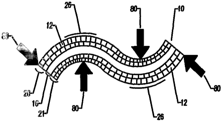
the collapsed pores along the inside region of the bend .22 gill not spring
bath: to their original
shape 12, rather they will remain substantially in the deformed and collapsed
state, thereby
ensuring that the smooth bend forming a radius cursye 26 in the prosthesis 10
remains after the
bending forces 80 are removed. Though not shown, a similar device constructed
of random,
irregular pores (as shown in Figure 1B) would behave similarly under the
application of bending
forces, forming a smooth radius curve by the collapse of pores along the
inside region of the
curve.
[0055] 'The collapsing of a porous layer along the inside of a bend permitting
the bending of a
solid or nearly solid layer into radii previously unachievable without
cracking or breaking can be
seen by refernng to Figures 8A and 8B. 'These figures depict a beam being bent
in an arc. In the
unbent condition (Figure 8A), the upper and lower surfaces have the same
length, but in the bent
condition (Figure 8B), one can see that both surfaces are strained. In
particular, the upper
surface is distended or stretched, and the lower surface is shortened or
compressed. This arises
because the upper and lower surfaces are connected to one another, and they
try to maintain their
continuity after bending. Thus, the upper surface is in a state of tensile
stress, and the lower
surface is in a state of compressive stress. Moreover, there is a region
between the upper and
lower surfaces that does not change in length, and this zone is in a neutral
stress state, being
neither compressed nor in tension. One can also see from the bent beam of
Figure 8B that the
sharper the bend, the smaller are the radii at the inner and outer surfaces,
and the greater the
amount of strain (i.e., fractional change of length) there is of these
surfaces.
[0056] Consider now that the beam is porous, or at least porous at its upper
and lower surfaces. The
collapsing of pores on and near the lower surface is a response to the
compressive stress, and an
accommodation of this stress since the material reacts to an externally
applied stress in such a way
as to minimize internal stress. In other words, the collapsing of pores at the
lower surface reduces
the amount of compressive stress. Similarly, the pores on and near the upper
surface are under a
tensile stress due to the bending, and can respond to this stress by becoming
more elongated in the
tensile direction. This elongation is often accompanied by a shortening of the
pore in a direction
perpendicular to the tensile direction, i.e., a flattening of the pore, so
this, too, can be thought of as a
collapse of the pore. Because the compressive and tensile stresses must be in
balance, the relaxation
of stress at one surface, e.g., due to collapsing or pores, also causes a
relaxation of stress at the
21
CA 02546453 2006-O1-10
WO 2005/009499 PCT/US2004/022840
opposite surface. ~cc~rdinglyr~ the coll~p~ing o~f pores permits amounts of
bending that would
other~,~~ise cause cracking c~r breaking of the solid polymer in beams not
having such a porous layer.
[0057] Another way of looking at the stress and strain states in the bent beam
is as follows. The
bent beam would like to keep the upper and lower surfaces the same length, so
the left edge of
the bent beam would be represented by line c-d. But this it cannot do, since
the upper surface
is firmly connected to the bottom surface, so the left edge ends up being
represented by line e-f.
Thus, the bottom surface is shortened as compressed pores collapse, and the
upper surface is
lengthened as pores in tension also collapse in a direction normal to the
tension. The collapsing
pores serve to relieve the compressive and tensile stresses, and thus relaxing
the overall stress
state of the material. This relaxation allows the material to deform with
lower stress, when
compared with non-porous materials. v
[0058] Similarly, supplying a porous layer or section at or near an outer
surface of an otherwise
non-porous structure will allow stress relaxation through the collapsing (or
elongation) of the
pores. This relaxation will allow deformation at lower stresses, thereby
allowing more strain to
be experienced before the critical breaking stress is reached.
[0059] Figure 2C and 2D depict a cross-sectional view of another possible
embodiment of the
device 10, comprising a porous layer 19 of ordered pores 12 and a layer of
solid or nearly solid
material 16. The transition from porous material 12 to a solid material 16 may
be an abrupt
transition 28 as depicted, or alternatively the transition may be a gradual
transition that occurs as
the size and population density of the pores decreases gradually in
construction (not shown).
Behaving similarly to the depictions of Figs. 2A and 2B, the dual layer
prosthesis of Figs. 2C
and 2D is capable of being smoothly bent, by forming a radius curve 26 when
bending forces 80
are applied. In the depiction of Fig. 2D, the pores 12 comprising the porous
layer 19 along the
inside of the bend 22 will collapse, and allow the formation of a radius in
the solid or nearly
solid layer 16 of material along the outside of the bend 24; resulting in a
smooth bend rather than
cracking or breaking as would the prior art of Fig. 7A and 7B. Though not
shown, a device
constructed from a dual layer device composed of a layer of random, irregular
pores and a solid
or nearly solid layer would exhibit bending behavior similar to that shown by
Figs. 2C and 2D.
22
CA 02546453 2006-O1-10
WO 2005/009499 PCT/US2004/022840
[006~] A~ a non-limiting example, the dual layer device 10 of Fig. 2C may
comprise a solid or
nearly solid layer 16 of synthetic pol,~mer (e.g., PGA, PLA, etc.), and the
regular, ordered porous
layer 19 may be fornled of a porous matrix of non-synthetic material (e.g.,
collagen, alginate,
chitosan, etc.) Due to the nature of the materials utilized for this
particular e~~ample, the porous
layer 19 composed of non-synthetic material, will, while dry, afford
structural support to
facilitate the bending of the solid layer 16, by selectively collapsing a
portion of the pores 12.
However, when wetted (e.g., after implantation, or exposure to a liquid or
solvent), the layer of
regular, ordered porous material 19 comprised of non-synthetic material loses
its rigidity and
becomes soft and compliant, or alternatively may quickly be dissolved
entirely, leaving only the
synthetic solid or nearly solid layer 16 of the device 10, as the non-
synthetic polymers are
soluble in tissue fluids, or lose structural rigidity when wetted. The porous
material layer may be
composed of irregular random pores, and exhibit a similar behavior, though
this is not shown.
[0061] Due to the construction of the dual layer device of Fig. 2C and 2D, the
smooth bending
may occur causing the regular, ordered porous layer 19 to form the inside of
the curve 22. As
the bending forces 80 are applied, they cause the collapse of the regular,
ordered pores 12, with
the collapsed pores distributing the bending forces 80 over a radius, thus
permitting the smooth
bending of the solid or nearly solid layer 16.
[0062] As shown in Figures 2E and 2F, the multi-layer construction herein has
more than two
layers. In this embodiment, there is shown a solid or nearly solid layer 16,
sandwiched between
regular ordered porous layers above 20 and below 21. Though it is recognized,
but not shown,
that the irregular, random pores would behave similarly. In this embodiment,
bending forces 80
may be applied in either orientation, and as the pores 12 are able to collapse
and distribute the
bending forces in either direction, more elaborate multi-directional bends are
possible, without
breaking the solid or nearly solid layer 16 as the bending forces 80 would
form radius curves 26,
and with multiple curves may allow 's' bends or other types of bends.
[0063] A cross-sectional depiction of an alternate, embodiment of the
invention is depicted in
Figure 3A. The prosthesis 10 of Figure 3A features a laminar construction,
which comprises a
series of layers 31,32,33 of varying pore sizes and pore densities. Though the
depiction here is
of regular ordered pores 12, this is for ease of illustration, and may
suitably be irregular, random
23
CA 02546453 2006-O1-10
WO 2005/009499 PCT/US2004/022840
pores. It is conte-rnplated that the prosthesis 10 be made ent~arely of on a
resorbable material, or
alternati-~ely, vrith each of the layers of the laminar construction 31, 32,
33 comprising the same
or a different resorbable material. Furthermore, the prosthesis 10 of this
embodiment or any
other of these embodiments may be fabricated by adding at least one
reinforcing material to the
prosthesis (to be discussed later).
[0064] As shown in Figure 3C, the varying pore sizes of the layers 31, 32, 33
would offer
varying resistance to collapse, with the larger pores of layer 3 l, being more
easily collapsed than
the smaller pores of the intermediate layer 32, which in turn would be more
easily collapsed than
the even smaller pores of the smallest pore layer 33. It is recognized that in
order for the
existence of multi-layer construction, at least two layers are needed. For
ease of illustration,
three distinct layers are depicted by Figures 3A, B, and C, but it is
recognized that there may be
more or less layers. It is also recognized that the interface or transition
between the layers may
be gradual or abrupt; for ease of illustration, abrupt interfaces between the
distinct layers are
depicted. It is also recognized that the distinction of layers may be based
upon some other
characteristic than pore size, such as pore density, material of construction,
or some other
identifiable quality, for ease of illustration, the distinction is made by
pore size and pore density.
[0065] This mufti-layer construction depicted by Fig. 3C would allow bending
forces 80 to be
applied (shown in orientation here by the large black arrows) upon the device
10, with the
resulting smooth bend in a radius curve 26 demonstrated by Figure 3C. As a
result of the
laminar construction, incorporating differing layers having varying resistance
to collapsing of the
pores, a more suitable bone plate may be constructed, relative to a non-porous
bone plate; yet the
laminar porous construction will retain the ability to be smoothly bent to
facilitate customization
of the implant. This may be accomplished, for example, by reducing the pore
sizes in the layer
33 along the outside of the bend 24, resulting in greater strength in that
layer 33, and at the same
time, increasing the pore sizes in the layer 31 along the inside of the bend
22, where additional
flexibility is gained by the more easily collapsed pores of the larger pore
layer 31. In this
manner, the prosthesis 10 is able to bend smoothly, forming a radius curve 26,
rather than
breaking as it would if the same bending force 80 (refernng to the prioY art
of Figures 7A and
7B) was applied and absorbed by only a small area, such as along a narrow
crease in the fold or
bend of the solid plate 75.
24
CA 02546453 2006-O1-10
WO 2005/009499 PCT/US2004/022840
[0066] Furthermore, when bending forces 80 are removed, the smooth bend of the
radius curve
26 in the mufti-layer device shovrn by Fig. 3C will remain, as the
irreversibly collapsed pores
along the inside region of the curare 22 will not return to their original
shape.
[0067] It is recognized that a great number of layers may be constructed into
the device,
comprising various combinations distinguishable by their number, structure, or
other
distinguishable characteristics; such as the structural properties of the
device may be altered by
creating different combinations of layers, pore sizes, construction materials
or other structural
qualities.
[0068] A prosthesis constructed as shown in Figure 3A would be able to conform
to the shape
of an uneven surface, as depicted in Figure 3B. This may be accomplished by
applying
compressive force 81 (shown in orientation here by arrows) upon the device 10,
compressing the
device evenly against an exposed uneven surface 36, with resistance 82 (shown
in orientation
here by arrow) offered by the uneven surface 36 against the device 10. As a
result of the
compressive force 81 and resistance 82, the initial resistance from the
protruding areas 38 would
selectively deform and collapse the larger and more easily collapsed layer of
pores 31, and to a
lesser extent the intermediate pores of layer 32, the affected pores located
proximally to the
protrusion area 38, leaving the layers with smaller pores 33 intact.
Furthermore, the recessed
areas 39 of the uneven surface 36 would not deform or collapse any of the
pores in the layers 31,
32, 33. As a result of the compressive force 81 and the resistance 82,
affecting the pore structure
of the device 10, the prosthesis surface may be altered to take on the inverse
shape of the
exposed surface 36, and therefore complies with the uneven surface 36. Upon
removal of the
compressive force 81, the device 10 will not spring back elastically to the
original shape due to
the fact that a portion of the pores 12 had irreversibly collapsed.
[0069] Referring to Figure 4, wherein a prosthesis 10 having random, irregular
pores 14 is
depicted, the prosthesis 10 with random pores 14 is capable of being used with
fastening systems
known in the art. Though not shown, an alternate embodiment of the device
having regular
ordered pores would similarly be capable of being used with fastening systems
known in the art.
These fastening systems may include adhesives (not shown), medical staples 42,
pins (not
CA 02546453 2006-O1-10
WO 2005/009499 PCT/US2004/022840
shrav,~n)~ mils 44, tsclo~ (not ~h~awga), serevrs 469 or clamps (not shown),
among other suitable
fastening devices.
[0070] In one embodiment, the prosthetic device 10 may be manufactured
incoaporating a hole
40, or alternatively a plurality of holes (not shown), extending at least
partially through the
prosthesis 10, which could accommodate the use of a suitable fastening method,
such as e.g.;
screws 46 or nails 44, to fasten the implantable device 10 through a pre-
existing hole 40.
Depending on the intended use of the prosthesis 10, the hole 40 may be created
during the
manufacturing process.
[0071] Preferably, the surgeon would be able to further customize the
implantable device 10 by
creating any number of needed holes 40 for the procedure. This may be
accomplished by use of
a hole-punch device, alternatively by a scalpel, scissors, the use of a
cutting blade, or by any
means suitable to ,penetrate into and through the prosthesis. The tool will
displace and deform
the porous structure it comes in contact with, such as in separating the
material in making a hole
40, leaving the pores more distant from the tool intact. In this manner, a
hole 40 may be made in
the prosthesis 10, without large-scale tearing or splitting of the prosthesis,
as only the pores
closest to the tool would be disturbed, and thereby limit the effect upon
pores away from the
tool. When used in this manner, the physician retains the flexibility to
locate the fastening points
where, in the physician's judgment, they are most appropriate, without
requiring pre-
manufactured fastening points in the prosthesis 10.
[0072] Most preferably, the prosthetic device 10 may be put in place, and
fastened without the
use of pre-manufactured holes in the prosthesis. This allows the physician to
simply fasten the
prosthesis 10 by any suitable means known in the art, such as by forcing a
screw 46, nail 44 or
staple 42 through the implant, wherein the porous structure 14 of the device
10 limits the amount
of large scale tearing that may occur, as only the pores closest to the tool
will be affected, ' ' '
leaving the rest of the device intact. When used in this manner, the physician
has flexibility to
locate the fastening devices in situ, without needing to approximate where the
location needs to
be created. As a result, there would not be a need to fit the prosthesis 10 to
the appropriate
shape, and then make the holes away from the patient; rather the prosthesis 10
could be fitted to
shape, and while the prosthesis 10 is in place, simply fastened into location
by any suitable
26
CA 02546453 2006-O1-10
WO 2005/009499 PCT/US2004/022840
fastening means lmoc;r~a in the art, v~ithout requiring the existence of a pre-
exacting hole 40. ~s
an alternative to the ezse of rigid fastening systems, such as screws 4d or
nails 44~ to fasten the
prosthesis 10, the porous structure of the device 10 with irregular pores 14,
and also having
regular ordered pores (not shown) is capable of being sutured 4~8 as shown in
Figure 4. The
suture 48 may be non-resorbable, or preferably be of a resorbable nature, so
that it may dissolve
over time, along with the prosthesis 10. The porous structure is compatible
with the use of a
suture 48 as the porous structure of the device 10 will accommodate a needle
by the pores
separating as the needle penetrates, whether through the entire thickness of
the prosthesis, or
merely through a portion of the thickness of the prosthesis. The porous
structure of the device
is able to resist suture pull-through, as the pulling force exerted by the
suture may be
distributed over a large number of pores. For this reason, the thread of the
suture 48 will not
easily rip through the structure and pull out. By use of a suture 48, the
prosthetic device 10 may
be attached to soft tissue, without requiring a pre-manufactured hole 40 for
suturing, or
attachment of sutures before use.
[0073] Still another alternative fastening method 'relies on the use of
adhesives to attach the
prosthesis in place (not shown). While using adhesives, a portion of the pores
along the surface
will be in contact with, and may absorb a portion of a liquid adhesive. As the
adhesive sets, the
prosthesis will be attached to the tissue. Suitable adhesives include fibrin,
polymer, or
cyanoacrylate glue, as well as others known to those skilled in the art. Such
adhesives may be
capable of penetrating into the porous material of the implantable device 10,
with the effect of
multiplying the bonded surface area, thereby resulting in a bond stronger than
would be available
if merely the exposed outer surface of the implant was coated by the adhesive.
[0074] As shown in Figure 5, a cross-sectional depiction of the device as a
composite 51,
comprising the device as previously described, as well as featuring further
additional materials
50. The depiction of Fig. 5 is of a composite device 51 having random,
irregular pore structure
14; however, it is recognized that the composite device 51 may have regular
ordered pore
structure as well, and further containing additional material 50. In one
embodiment, the
additional materials 50 may be high modulus strengthening components (e.g.,
polymers,
ceramics or metallics), where the high modulus material will affect the
physical characteristics of
the composite prosthesis 51, such as increasing the rigidity, strength, and
toughness of the
27
CA 02546453 2006-O1-10
WO 2005/009499 PCT/US2004/022840
resulting ~t~nacti,~re. 'Tlze strengthening agent may be in various foam
~e.g.9 parkiculate, f~ber9
whi~,l~er, mesh, =,veave, l~nit, yam, etc.). The additional material may be
uniformly distributed
throughout the entire composite prosthesis ~ 1, or alternatively selectively
incorporated to
achieve a desired effect.
[0075] The same additional material 50 incorporated to achieve a desired
effect upon the
physical properties of the composite implantable device 51 may also affect its
biologic
properties. As an example, hydroxyapatite would not only improve the strength
of the implant,
but also be capable of, for example, extracting endogenous growth factors from
the host tissue
bed while functioning as a microporous conduit facilitating movement of
interstitial fluid
throughout the isolated porosities of the device. In another embodiment, the
additional materials
50 may alter the resorption qualities of the resorbable porous material. Other
non-limiting
examples of suitable materials that may be added to the prosthesis are listed
in Table 2.
[0076] Table 2: Examples of Materials Incorporated into the Composite Device
in
Accordance with the Present Invention
Alginate
Bone allograft or autograft
Bone Chips
Calcium
Calcium Phosphate
Calcium Sulfate
Ceramics
Chitosan
Cyanoacrylate
Collagen
Dacron
Demineralized bone
Elastin
Fibrin
Gelatin
28
CA 02546453 2006-O1-10
WO 2005/009499 PCT/US2004/022840
O'la~s (e.g.- l~i~a-r~l~~~)
Caold
C'alycosaminoglycans
Hydrogels
Hydroxyapatite
Hydroxyethyl methacrylate
Hyaluronic Acid
Liposomes
Microspheres
Natural Polymers
Nitinol
Oxidized regenerated cellulose
Phosphate glasses
Polyethylene glycol
Polyester
Polysaccharides
Polyvinyl alcohol
Radiopacifiers
Salts
Silicone
Silk
Steel (e.g. Stainless Steel)
Synthetic polymers
Thrombin
Titanium
Tricalcium phosphate
[0077] The additional material 50 can serve multiple purposes, which may
include, but are not
limited to:
1. creating a textured surface on the internal surfaces defining the pores;
2. creating a microporous conduit system between pores;
29
CA 02546453 2006-O1-10
WO 2005/009499 PCT/US2004/022840
reacting e~~tracting of endogenous grovrth factors;
carrgring and/or delivering drugs, biologically active or therapeutic agents;
functioning ae a drug, biologically active or therapeutic agent;
6. modifying mechanical properties (e.g. strength, flexibility, suture
retention, etc.);
7. functioning as an in-vivo leachate to increase the overall porosity.
[0078] The textured surface created by the additional material 50 additionally
serves multiple
purposes that may include but are not limited to:
increased surface area permits modification to the leaching rate of drugs or
other
therapeutics;
2. textured surfaces increase quantity of rriaterial that can be coated on the
interior
pore surfaces;
3. irregular surfaces increase the resistance to flow through the implant.
[0079] Additional materials 50 may also be used at the time of manufacture to
control the
process output (e.g. plasticizers, surfactants, dyes, etc.) For example,
processing the polymer
with stearic agents will cause the thinning of matrix between the pores, which
is most easily
penetrable, or rapidly resorbing, following implantation. This will result in
a composite device
51 with high strength, and interconnected pores.
[0080] The additional materials 50 may lend some other desired property to the
composite
prosthesis 51, such as the capability of delivering biologically active
agents, or of being radio-
opaque, in order to allow imaging by x-ray or MRI techniques while the
prosthesis is implanted
or being implanted in the living being. The additional material 50 would be
capable of being
resorbed in the body, either at the same rate of absorption as the polymer or
at a faster or slower
rate of resorption. Should the prosthesis further contain biologically active
agents, they may be
delivered slowly as the surrounding porous material is resorbed. The period of
delivery of the
biologically active agents from the device may be delayed and/or further
extended by
incorporating drug depots into the composite prosthesis, such that the
biblogically active agents
are slowly released into the body. Alternatively, if the biologically active
agents comprising the
additional material 50 are easily dissolved, tissue fluids may be capable of
leaching out the
CA 02546453 2006-O1-10
WO 2005/009499 PCT/US2004/022840
agents as the tissue fluids permeate the pcr~u~ structure of the cramp~~ite
pr~sthesis 51 a
E~Lamples of biologically active agents that may sere as the additional
material 50 of the
c~mposite pr~sthesis 51 are listed in Table 3.
[0081] The additional material 50 may be in the form of microspheres.
Microspheres can be
made of a variety of materials such as polymers, silicone and metals.
Biodegradable polymers
are ideal for use in creating microspheres f~r use in these embodiments (e.g.,
see those listed in
Table 1). The release of agents from bioresorbable microparticles is dependent
upon diffusion
through the microsphere polymer, polymer degradation and the microsphere
structure. Although
most any biocompatible polymer could be adapted for this invention, the
preferred material
would exhibit in vivo degradation. It is well known that there can be
different mechanisms
involved in implant degradation like hydrolysis, enzyme-mediated degradation
and bulk or
surface erosion. These mechanisms can alone or combined influence the host
response by
determining the amount and character of the degradation product that is
released from the
implant. In the extracellular fluids of the living tissue, the accessibility
of water to the
hydrolysable chemical bonds makes hydrophilic polymers (i.e. polymers that
take up significant
amounts of water) susceptible to hydrolytic cleavage or bulk erosion.
[0082] Several variables can influence the mechanism and kinetics of polymer
degradation.
Material properties like crystallinity, molecular weight, additives, polymer
surface morphology,
and environmental conditions. As such, to the extent that each of these
characteristics can be
adjusted or modified, the performance of this invention can be altered. These
microspheres,
serving as the additional material 50 in the composite device 51, may further
contain and/or
deliver biologically active agents from Table 3.
[0083] Table 3: Examples with Some Types of Biological, Pharmaceutical, and
other
Therapies that can be Delivered via the Composite pevice in Accordance with
the Present
Invention
Adenovirus with or without genetic material
Angiogenic agents
31
CA 02546453 2006-O1-10
WO 2005/009499 PCT/US2004/022840
f~ngioten~ia~ converting En~,e Inhibitors ~t~~E inhabitors~
Angiotensin II antagonists
anti-angiogenic agents
Antiarrhythmics
Anti-bacterial agents
Antibiotics
Erythromycin
Penicillin
Anti-coagulants
Heparin
Anti-growth factors
Anti-inflammatory agents
Dexamethasone
Aspirin
Hydrocortisone
Antioxidants
Anti-platelet agents
Forskolin
Anti-proliferation agents
Anti-rejection agents
Rapamycin
Anti-restenosis agents
Antisense
Anti-thrombogenic agents
Argatroban
Hirudin
GP Ilblllla inhibitors
Anti-virus drugs
Arteriogenesis agents
acidic fibroblast growth factor (aFGF)
angiogerain
aragiotropin
32
CA 02546453 2006-O1-10
WO 2005/009499 PCT/US2004/022840
baa~ac f ba-oblast ~~owa'la factor (bFGF)
~rafae naorpJaogenic proteins (PMP)
epidea-naal gi-owtla factor (EGF)
ftbrin
granulocyte-naacropJaage colony stimulating factor
(GM-CS'F)
hepatocyte growth factor (HGF)
FIIF 1
Indian hedgehog (Inlay
insulin growth factor-1 (IGF 1)
ifaterleukin-~ (IL-~)
MAC 1
nicotinanaide
platelet-derived endothelial cell growth factor (PD-ECGF)
platelet-derived growth factor (PDGF)
transforming growth factors alpha & beta
(TGP .alpha., TGF beta.)
tumor necrosis factor alpha (TNF . alpha.)
vascular endothelial growtJz factor (VEGF)
vascular permeability factor (YPF)
Bacteria
Beta blocker
Blood clotting factor
Bone morphogenic proteins (BMP)
Calcium channel Mockers
Carcinogens
Cells
Stem cells
Bone Marrow
Blood cells
Fat Cells
33
CA 02546453 2006-O1-10
WO 2005/009499 PCT/US2004/022840
~~'~~.~cle Cell
~lattbilical coa-d :ells
Chemotherapeutic agents
Ceratnide
T°axol
Cisplatin
Paclitaxel
Cholesterol reducers
Chondroitin
Clopidegrel (e.g., plavix)
Collagen Inhibitors
Colony stimulating factors
Coumadin
Cytokines prostaglandins
Dentin
Etretinate
Genetic material
Glucosamine
Glycosaminoglycans
GP IIb/IIIa 'inhibitors
L-703,081
Granulocyte-macrophage colony stimulating factor (GM-CSF)
Growth factor antagonists or inhibitors
Growth factors
Autologous Growth Factors
B-cell Activating Factor (BAFF)
Bovine derived cytokines
Cartilage Derived Growth Factor (CDGF)
Endotl2elial Cell Growth Factor (ECGF)
Epidermal growth factor (EGF)
Fibroblast Growth Factors (FGF)
Hepatocyte growtlt factor (HGF)
34
CA 02546453 2006-O1-10
WO 2005/009499 PCT/US2004/022840
Instali~a-life Garo~jt~a Factoa~ (~.g. Ir,~l"-1)
Nerve ga-owtla factor (NGF)
Platelet Derived Growth Factor (PDGF)
l~ecoanbiraarat NGF (rIaNGF)
Tissue necrosis factor (TNF)
Tissue derived cytokiaaes
Transforming growth factors alpha (TGF alpha)
Transforming growth factors beta (TGF beta)
Tlascular Eradothelial Growth Factor (VEGF)
Tlascular permeability factor (ZJPF)
Acidic fibroblast growtla factor (aFGF)
Basic fibroblast growth factoa~ (bFGF)
Epideranal growth factor (EGF)
Hepatocyte growth factor (HGF)
Insulin growth factor-1 (IGF 1)
Platelet derived endotlaelial cell growth factor (PD-ECGF)
Tumor necrosis factor alpha (TNF .alpha.)
Growth hormones
Heparin sulfate proteoglycan
HMC-CoA reductase inhibitors (statins)
Hormones
Eaythropoietin
Immoxidal
Immunosuppressant agents
inflammatory mediator
Insulin
Interleukins
Interlukins
Iaaterlukin-8 (IL-8)
Lipid lowering agents
Lipo-proteins
Low-molecular weight heparin
CA 02546453 2006-O1-10
WO 2005/009499 PCT/US2004/022840
I~~rrralahocites
Lysine
~~C-1
Morphogens
Poatc trtorphogenic pt~oteiats (PIUIPs)
Nitric oxide (NO)
Nucleotides
Peptides
PR39
Proteins
Prostaglandins
Proteoglycans
Perlecan
Radioactive materials
Iodine - 125
Iodine -131
Ia~idium -192
Palladium 103
Radio-pharmaceuticals
Secondary Messengers
Cerarnide
Signal Transduction Factors
Signaling Proteins
Somatomedins
Statins
Stem Cells
Steroids
Thrombin
Sulfonyl
Thrombin inhibitor
Thrombolytics
Ticlid
36
CA 02546453 2006-O1-10
WO 2005/009499 PCT/US2004/022840
T~rrosine l~nase h~hibitors
~'Z'6~~
AC-17
Vasodilator
FlistaTTZiTZe
FOT skOlin
NdtYOg'1J1C2YZTt
Vitamins
E
C
Yeast
[0084] Therapy delivery may come from the pores 14 of the composite prosthesis
51, as
released from physical entrapment of the therapy impregnated within the walls
of the pores 14; it
may come from material adsorbed or loosely adhering to the surface of enclosed
pores 14 or
interconnected pores (not shown); or it may stay suspended within the pores 14
of the composite
device 51 as implanted, awaiting contact with tissue fluid entering the pores
14. Figure 5 depicts
the composite prosthesis 51 having irregular, random pores, it is recognized
that a composite
prosthesis having regulai, ordered pores and additional material would behave
similarly.
[0085] It is recognized that each of the delivery modes could result in
different delivery rates.
That is, therapy may evolve more rapidly from interconnected pores (not
shown), than from
isolated pores 14, which may in-turn release therapy faster than any therapy
delivered by the
polymer constituent (e.g., as it degrades).
[0086] In one embodiment, the therapy delivered via the additional material 50
is co-mingled
with the various other constituents and components prior to the processing.
This allows for
some concentration of the therapy to remain in the polymer constituent, while
some of the same
therapy migrates or precipitates into the porous region of the matrix. An
equilibrium phase
diagram for the components and constituents would allow the tailoring of the
concentration of
therapy in each region (i.e., pore or polymer constituent), additionally,
therapies with low
solubility in either component will aid preferential placement of therapy.
37
CA 02546453 2006-O1-10
WO 2005/009499 PCT/US2004/022840
[00~'~] As shown in Figs. 6A, an alternative embodiment of the composite
prosthesis Sl may
also incorporate random fibers or whiskers 61 as the additional material to
add strength or
another property to the composite prosthesis 51. Alternatively, and as shown
in Figure 613, the
fibers may be arranged in a non-random pattern, such as a weave, knit or mesh,
or scaffold 62.
The patterned fibers 62 may be arranged in a single layer forming a two-
dimensional sheet, or
they may form a reinforcing scaffold extending in three dimensions through the
composite
prosthesis 51. The patterned fibers 62 or the random fibers 61, may extend
throughout the
entirety of the composite prosthesis 51, or alternatively may be limited to a
particular portion or
layer of the composite prosthesis 51, where greater strength or altered
physical characteristic is
desired.
[0088] The incorporation of random fibers 61 or a non-random pattern of fibers
62 into the
porous material of the composite prosthesis 51 may impart additional shear
strength to the
implant, enabling it to further resist mechanical stresses imposed while
implanted in the living
being. The fibers 61, '62 may also serve as an additional safety measure; upon
the formation of a
break or fault in the porous structure of the implant, the incorporated fibers
would ensure that the
entire prosthesis is able'to remain in place, preventing a loose piece of the
implant from being
able to migrate within the being.
[0089] The random fibers or whiskers 61, or the non-random fibers 62,
comprising the
additional material may be biocompatible and non-resorbable, or more
preferably biocompatible
and resorbable, such that as the porous material of the composite device 51 is
absorbed by the
living being, the 'fibers 61, 62 are absorbed as well. Resorbable fibers
comprising the additional
material may be constructed from materials selected from table 1 above, a non-
exhaustive list of
some of the materials from which the resorbable prosthesis may be constructed.
The fibers 61,
62 may be the same material or a different material from the porous material
comprising the
prosthesis. Depending on the materials selected, the composite prosthesis 51
may be resorbed at
the same rate or a different rate from the incorporated fibers 61, 62.
[0090] The structure of the prosthesis may be manufactured in such a way that
there is a layered
appearance to the bone plate when viewed in cross-section. This may be
accomplished by
38
CA 02546453 2006-O1-10
WO 2005/009499 PCT/US2004/022840
n ~etho6s com~nonl-sF lmovrr~ an the art, such as: por~sogen particulate
leaching; blovrn gas
methods; gas forming polymerizations; lyophilization; phase separation, as
described in TJ.S.
6,355,699 Bl (~yalbarnum), and IT.S. patent application Ser. hTo. 10/02212
(Eovrznan). Any of
these methodologies may be utilized to create a prosthesis entirely uniform in
material, however,
having layers with a variety of pore sizes and densities (not shown). For
example, there may be
a first layer of material that is relatively less porous, transitioning
through a first interface to a
layer that is relatively more porous, and transitioning through a second
interface to a layer that is
relatively less porous. By tailoring the manufacturing process, a great many
variety of
combinations may be constructed into a prosthesis.
[0091] In an embodiment of the practice of the current invention, the bendable
porous fixation
device can be machined, punched, or molded into any configuration, such as an
internal fixation
device for use in surgical repair, replacement, or reconstruction of damaged
bone in any area of
the body (e.g. pelvis, orbital floor, palate, jaw, long bone, etc.). Internal
fixation devices may be
successfully employed for many conditions and applications (e.g., orthopedic,
spinal,
maxillofacial, craniofacial, etc.). The devices may take on various forms,
including, but not
limited to, common forms such as sheets, disks, cups, tubes, rolls, blocks,
cylinders or pads
suitable for attachment to tissues. In addition to fixation of tissues, the
bendable porous fixation
device has the ability to retain graft material (e.g., ceramics, demineralized
bone matrix, allograft
bone chips, autograft bone chips, etc.) within a location. For example, a
fixation plate that is
bent into a sleeve can be used to prevent migration of graft material placed
into a segmental
defect of a long bone, and further provide protection and support for
maintaining the proper gap
in the segmental defect.
[0092] In another embodiment, the bendable porous fixation device contains
reinforcing
materials such as long threads, screens, meshes or other fibers. The polymer
making up the
pores supports, confines, and locks the reinforcing material within a spatial
conformation. This
retards the reinforcing material from migrating within or dissection from the
fixation device.
This can be used to alter mechanical properties (e.g., compressive strength)
of the construct.
Additionally, the polymer may improve the biocompatibility of the reinforcing
material (e.g.,
improved cellular attachment or adhesion to a mesh). The reinforcing material
may be centered
within the construct, located on or just below one or more surfaces or
interspersed throughout
39
CA 02546453 2006-O1-10
WO 2005/009499 PCT/US2004/022840
the entire con~tra~rt.
[009] In another embodiment, the pores of the bendsble porous fixation device
are used to
control the location and delivery of biologically active agents (e.g., growth
factors, hormones,
bone morphogenic proteins, drugs, cells, viruses, etc.) (see table 3). The
biologically active
agents could be located within the polymer walls or supported within the pores
making up the
device. Additionally, the biologically active agents could be mechanically or
chemically
attached or bonded to the polymer or suspended within a hydration fluid
located in the pores of
the bendable fixation device. This hydration fluid may contain a soluble
polymer that suspends
or binds the biologically active agent. Additionally, the hydration fluid
containing the soluble
polymer may be removed leaving the soluble polymer as a coating on the pore
walls or
microstructure suspended within the pores.
[0094] In another embodiment, the polymer of the bendable porous fixation
device is used to
control the location and orientation of particulate components compounded into
the porous
material (e.g., tricalcium phosphate, Hydroxylapatite, calcium sulfate,
autologous bone graft
material, allograft bone matrix, polymers, microspheres, etc.). The polymer
supports, confines,
and locks the particulate components within a spatial conformation. This
retards the particulate
from migrating within or disassociating from the fixation device. The
particulate can be used to
alter mechanical properties (e.g., compressive strength) of the construct
[0095] In another embodiment, the'materials made by these various processes
may be cross-
linked to impart improved characteristics such as: mechanical strength (e.g.,
suturablity,
compression, tension, etc.), and biodurability (e.g., resistance to enzymatic
and hydrolytic
degradation, etc.). This enhancement of characteristics may be accomplished
using one or more
of several different cross-linking agents, or techniques (e.g., thermal
dehydration, EDC,
aldehydes (e.g., formaldehyde, gluteraldehyde, etc.), natural cross-linking
agents (e.g., genipin,
proanthocyariidin, etc.). Each type of cross-linking agent/technique or
combinations thereof
imparts diverse mechanical and biological properties on the material. These
properties are
created through the formation of unique chemical bonds that stabilize the
construct. This
stabilization greatly increases the ability of the construct to hold a shape
and conformation;
thereby, preserving the interlaced relationship between the fibers.
CA 02546453 2006-O1-10
WO 2005/009499 PCT/US2004/022840
[009~a] several of these bendable fixation embodiments may als~ be
manufactured in composite
laminate form. That is, flat sheet or shaped embodiments may be affixed to
additional sheets or
other materials (e.g. solid plates, screws, screens, meshes, etc.), by
pressing, gluing, stitching or
other fastening means known t~ those skilled in the art. These macro-
composites may be created
having the respective characteristics of each of the component materials,
thereby creating a
single device having beneficial characteristics from each of the component
materials. For
example, a resorbable, osteoconductive, porous and flexible first layer of the
device, which is
enhanced by being constructed as a laminate with a second layer in the form of
a high strength
mesh affixed to one surface of the first layer; thereby creating a flexible,
yet high strength
device.
[0097] Thus since the invention disclosed herein may be embodied in other
specific forms
without departing from the spirit or general characteristics thereof, some of
which forms have
been indicated, the embodiments described herein are to be considered in all
respects illustrative
and not restrictive, by applying current or future knowledge. The scope of the
invention is to be
indicated by the appended claims, rather than by the foregoing description,
and all changes
which come within the meaning and range of equivalency of the claims are
intended to be
embraced therein.
41