Note: Descriptions are shown in the official language in which they were submitted.
CA 02546460 2006-O1-12
WO 2005/007225 PCT/US2004/022796
DEVICE TO ASSIST HYPERHYDROSIS THERAPY
by
BACKGROUND
The present invention relates to a device for assisting hyperhydrosis
to therapy. In particular, the present invention relates to a dermal overlay
device for assisting hyperhydrosis therapy.
Human sweat as part of a normal thermoregulation process.
Additionally, sweating can be is a normal physiological response to a
Is psychological stress or emotional stimuli. For most people, sweating is
only a minor cosmetic annoyance. For others, however, sweating may
be excessive and become a socially or medically crippling handicap.
Hyperhidrosis is a disorder in which there is an exaggerated sweat
secretion involving both the eccrine and the apocrine sweat glands. The
2o excessive sweating usually occurs in the palms, soles, and axillae.
Palmar hyperhidrosis is a condition of excessive sweating in the hand.
Such condition may be socially embarrassing. Plantar hyperhidrosis is a
condition of excessive sweating in the foot. This condition may cause
blisters, infections, and bromohidrosis. Axillary hyperhidrosis is a
2s condition of excessive sweating in the armpit. In axillary hyperhidrosis,
as much as 26 mUh of sweat can be excreted from each armpit. Such
excessive sweating is not only socially embarrassing but may even
cause staining and rotting of clothes.
so Presently, the cause of hyperhidrosis is unknown. However, what is
known is that the 3 to 4 million sweat glands of the body are under the
control of the hypothalamus and the sympathetic system. Afferent
CA 02546460 2006-O1-12
WO 2005/007225 PCT/US2004/022796
impulses from sensors on the skin and other parts of the body travel to
the hypothalamus, which integrates the information for chemoregulation
of the body. The preoptic area of the anterior hypothalamus then sends
efferent impulses via sympathetic fibers back out to the body. Segment
T2 to T4 of the spinal chord innervate the head and neck area; fibers in
segment T2 to T8 innervate the upper limbs; fibers in segment T6 to
T10 innervate the trunk; and finally fibers in T11 to T12 innervate the
lower extremities.
io Although sympathetic innervations typically rely on adrenergic
neurotransmitters, acetylcholine is the neurotransmitter released by the
sympathetic nerve terminals involved in innervating the sweat glands.
However, that is not to say that only acetylcholine can innervate the
sweat glands. Some reports have shown that eccrine and apocrine
is glands respond to .alpha.- and .beta.-adrenergic agonists as well.
Although the hypothalamus has a significant role in controlling the
rate of sweating, other physical variables may affect the rate of sweat
secretion. For example, sweating rate may also be affected by variables
2o such as wetness and blood flow. Additionally, the rate of sweating varies
greatly among people and is related to acclimatization, sex, age, and
maybe even diet. a
With respect to treating hyperhidrosis, various treatments are being
2s used. For example, topical administration.aluminum chloride is a
common practice. It is thought that aluminum chloride mechanically
obstruct eccrine sweat glands to reduce sweating, although some
evidence shows that the reduction in sweat may result from atrophy of
the secretory cells. A downside of using aluminum chloride is that the
3o aluminum chloride may react with the water content of the sweat to form
hydrochloric acid. The formation of hydrochloric acid may cause severe
skin irritation.
CA 02546460 2006-O1-12
WO 2005/007225 PCT/US2004/022796
Other topical preparations are also being used. For example,
treatment of plantar and palmar hyperhidrosis includes use'of
glutaraldehyde and tannic acid (strong tea). However, this treatment
s may cause a browning of the skin.
Anticholinergics, both systemic and topical, are also being used.
However, most patients cannot tolerate the side effects.
io In addition to the described adverse effect of the above methods, the
above treatment methods are effective to alleviate excessive sweating
for only a brief duration of time, thus requiring frequent treatments, i.e.
daily or weekly. ,
is Surgical treatment involving sweat gland excision and
sympathectomy may provide for a longer duration of alleviation from
hyperhidrosis. However, these invasive treatments are rarely indicated
due to the adverse consequences and cost. For example, surgery may
cause contractures. Sympathectomy may result in complications
2o including infection, pneumothorax, Horner's syndrome, resumption of
sweating, and compensatory hyperhidrosis. Additionally, hyperhidrosis
may resume after surgery or sympathectomy.
Subdermal injections of a botulinum toxin at the site of an excessive
2s sweat secretion have been successfully used to treat hyperhydrosis.
See e.g. Naumann M., Botulinum toxin type A in the treatment of,focal
hyperhidrosis, J. Cutaneous Laser Ther 2001;3(1 ):42-43, and; U.S. .
patent 5,766,605 (Sanders). Treatment typically entails, at each
treatment session, making a number of injections into the hyperhydrotic
so skin, so as to achieve the desired distribution of the botulinum toxin into
the target area, as opposed to making only one or a few injections.
Typically, after determining the dimensions of a dermal area exhibiting
3
CA 02546460 2006-O1-12
WO 2005/007225 PCT/US2004/022796
of excessive sweat secretion, as by use of an iodine starch test, the
attending physician attempts to indicate the locations of botulinum toxin
injection by marking a pattern (a grid) of multiple spaced dots on the
target skin area. Often the injection location dots are neither properly
s spaced nor of an appropriate number when such a freehand method is
used. It is known to use a multiple injection plate for the treatment of
hyperhydrosis wherein five or seven needles puncture the skin at the
same time. Grimalt R., et al., Multi-injection plate for botulinum toxin
application in the treatment of axillary hyperhidrosis, Dermatol Surg
'v
l0 2001 Jun;27(6):543-544.
Botulinum Toxin
The genus Clostridium has more than one hundred and twenty seven
species, grouped according to their morphology and functions. The
is anaerobic, gram positive bacterium Clostridium botulinum produces a
potent polypeptide neurotoxin, botulinum toxin, which causes a
neuroparalytic illness in humans and animals referred to as botulism.
The spores of Clostridium botulinum are found in soil and can grow in
improperly sterilized and sealed food containers of home based
2o canneries, which are the cause of many of the cases of botulism. The
effects of botulism typically appear 18 to 36 hours after eating the
foodstuffs infected with a Clostridium botulinum culture or spores. The
botulinum toxin can apparently pass unattenuated through the lining of
the gut and attack peripheral motor neurons. Symptoms of botulinum
25 toxin intoxication can progress from difficulty walking, swallowing, and
speaking to paralysis of the respiratory muscles and death.
Botulinum toxin type A is the most lethal natural biological agent
known to man. About 50 picograms of a commercially available
so botulinum toxin type A (purified neurotoxin complex)' is a LDSO in mice
1 Available from Allergan, Inc., of Irvine, California under the tradename
BOTOX~ in 100 unit vials)
4
CA 02546460 2006-O1-12
WO 2005/007225 PCT/US2004/022796
(i.e. 1 unit). One unit of BOTOX~ contains about 50 picograms (about
56 attomoles) of botulinum toxin type A complex. Interestingly, on a
molar basis, botulinum toxin type A is about 1.8 billion times more lethal
than diphtheria, about 600 million times more lethal than sodium
s cyanide, about 30 million times more lethal than cobra toxin.and about
12 million times more lethal than cholera. Singh, Critical Aspects of
Bacterial Protein Toxins, pages 63-84 (chapter 4) of Natural Toxins II,
edited by B.R. Singh et al., Plenum Press, New York (1976) (where the
stated LDSO of botulinum toxin type A of 0.3 ng equals 1 U is corrected
to for the fact that about 0.05 ng of BOTOX~ equals 1 unit). One unit (U)
of botulinum toxin is defined as the LDSO upon intraperitoneal injection
into female Swiss Webster mice weighing 18 to 20 grams each.
Seven generally immunologically distinct botulinum neurotoxins have
is been characterized, these being respectively botulinum neurotoxin
serotypes A, B, Ci, D, E, F and G each of which is distinguished by
neutralization with type-specific antibodies. The different serotypes of
botulinum toxin vary in the animal species that they affect and in the
severity and duration of the paralysis they evoke. For example, it has
2o been determined that botulinum toxin type A is 500 times.more potent,
as measured by the rate of paralysis produced in the rat, than is
botulinum toxin type B. Additionally, botulinum toxin type B has been
determined to be non-toxic in primates at a dose of 480 U/kg which is
about 12 times the primate LDSO for botulinum toxin type A. Moyer E et
2s al., Botulinum Toxin Type B: Experimental and Clinical Experience,
being chapter 6, pages 71-85 of "Therapy With Botulinum Toxin",edited
by Jankovic, J. et al. (1994), Marcel Dekker, Inc. Botulinum toxin
apparently binds with high affinity to cholinergic motor neurons, is
translocated into the neuron and blocks the release of acetylcholine.
3o Additional uptake can take place through low affinity receptors, as well
as by phagocytosis and pinocytosis.
s
CA 02546460 2006-O1-12
WO 2005/007225 PCT/US2004/022796
Regardless of serotype, the molecular mechanism of toxin
intoxication appears to be similar and to involve at least three steps or
stages. In the first step of the process, the toxin binds to the presynaptic
membrane of the target neuron through a specific interaction between
s the heavy chain, H chain, and a cell surface receptor; the receptor is
thought to be different for each type of botulinum toxin and for tetanus
toxin. The carboxyl end segment of the H chain, Hc, appears to be
important for targeting of the toxin to the cell surface.
to In the second step, the toxin crosses the plasma membrane of the
poisoned cell. The toxin is first engulfed by the cell through receptor-
mediated endocytosis, and an endosome containing the toxin is formed.
The toxin then escapes the endosome into the cytoplasm of the cell.
This step is thought to be mediated by the amino end segment of the H
Is chain, HN, which triggers a conformational change of the toxin in
response to a pH of about 5.5 or lower. Endosomes are known to
possess a proton pump which decreases intra-endosomal pH. The
conformational shift exposes hydrophobic residues in the toxin, which
permits the toxin to embed itself in the endosomal membrane. The toxin
zo (or at a minimum the light chain) then translocates through the
endosomal membrane into the cytoplasm.
The last step of the mechanism of botulinum toxin activity appears to
involve reduction of the disulfide bond joining the heavy chain, H chain,
2s and the light chain, L chain. The entire toxic activity of botulinum and
tetanus toxins is contained in the L chain of the holotoxin; the L chain is
a zinc (Zn++) endopeptidase which selectively cleaves proteins
essential for recognition and docking of neurotransmitter-containing
vesicles with the cytoplasmic surface of the plasma membrane, and
so fusion of the vesicles with the plasma membrane. Tetanus neurotoxin,
botulinum toxin types B, D, F, and G cause degradation of
synaptobrevin (also called vesicle-associated membrane protein
6
CA 02546460 2006-O1-12
WO 2005/007225 PCT/US2004/022796
(VAMP)), a synaptosomal membrane protein. Most of the VAMP
present at the cytoplasmic surface of the synaptic vesicle is removed as
a result of any one of these cleavage events. Botulinum toxin serotype
A and E cleave SNAP-25. Botulinum toxin serotype C1 was originally
thought to cleave syntaxin, but was found to.cleave syntaxin and SNAP-
25. Each of the botulinum toxins specifically cleaves a different bond,
except botulinum toxin type B (and tetanus toxin) which cleave the same
bond. Each of these cleavages block the process of vesicle-membrane
docking, thereby preventing exocytosis of vesicle content.
to
Botulinum toxins have been used in clinical settirigs for the treatment
of neuromuscular disorders characterized by hyperactive skeletal
muscles (i.e. motor disorders). In 1989 a botulinum toxin type A
complex has been approved by the U.S. Food and Drug Administration
is for the treatment of blepharospasm, strabismus and hemifacial spasm.
Subsequently, a botulinum toxin type A was also approved by the FDA
for the treatment of cervical dystonia and for the treatment of glabellar
lines, and a botulinum toxin type B was approved for the treatment of
cervical dystonia. Non-type A botulinum toxin serotypes apparently
2o have a lower potency and/or a shorter duration of activity as compared
to botulinum toxin type A. Clinical effects of peripheral intramuscular
botulinum toxin type A are usually seen within one week of injection.
The typical duration of symptomatic relief from a single intramuscular
injection of botulinum toxin type A averages about three months,
2s although significantly longer periods of therapeutic activity have been
reported.
Although all the botulinum toxins serotypes apparently inhibit release
of the neurotransmitter acetylcholine at the neuromuscular junction, they
so do so by affecting different neurosecretory proteins and/or cleaving
these proteins at different sites. For example, botulinum types A and E
both cleave the 25 kiloDalton (kD) synaptosomal associated protein
CA 02546460 2006-O1-12
WO 2005/007225 PCT/US2004/022796
(SNAP-25), but they target different amino acid sequences within this
protein. Botulinum toxin types B, D, F and G act on vesicle-associated
protein (VAMP, also called synaptobrevin), with each serotype cleaving
the protein at a different site. Finally, botulirium toxin type Ci has been
s shown to cleave both syntaxin and SNAP-25. These differences in
mechanism of action may affect the relative potency and/or duration of
action of the various botulinum toxin serotypes. Apparently, a substrate
for a botulinum toxin can be found in a variety of different cell types.
See e.g. Biochem J 1;339 (pt 1):159-65:1999, and Mov Disord,
,;;
l0 10(3):376:1995 (pancreatic islet B cells contains at least SNAP-25 and
synaptobrevin).
The molecular weight of the botulinum toxin protein molecule, for all
seven of the known botulinum toxin serotypes, is about 150 kD.
Is Interestingly, the botulinum toxins are released by Clostridia) bacterium
as complexes comprising the 150 kD botulinum toxin protein molecule
along with associated non-toxin proteins: Thus, the botulinum toxin type
A complex can be produced by Clostridia) bacterium as 900 kD, 500 kD
and 300 kD forms. Botulinum toxin types B and Ci is 'apparently
2o produced as only a 700 kD or 500 kD complex. Botulinum toxin type D
is produced as both 300 kD and 500 kD complexes. Finally, botulinum
toxin types E and F are produced as only approximately 300 kD
complexes. The complexes (i.e. molecular weight greater than about
150 kD) are believed to contain a non-toxin hemaglutinin protein and a
2s non-toxin and non-toxic nonhemaglutinin protein. These two non-toxin
proteins (which along with the botulinum toxin molecule comprise the
relevant neurotoxin complex) may act to provide stability against
denaturation to the botulinum toxin molecule and protection against
digestive acids when toxin is ingested. Additionally, it is possible that
so the larger (greater than about 150 kD molecular weight) botulinum toxin
complexes may result in a slower rate of diffusion of the botulinum toxin
away from a site of intramuscular injection of a botulinum toxin complex.
s
CA 02546460 2006-O1-12
WO 2005/007225 PCT/US2004/022796
In vitro studies have indicated that botulinum toxin inhibits potassium
cation induced release of both acetylcholine and norepinephrine from
primary cell cultures of brainstem tissue. Additionally, it has been
s reported that botulinum toxin inhibits the evoked release of both glycine
and glutamate in primary cultures of spinal cord neurons and that in
brain synaptosome preparations botulinum toxin inhibits the release of
each of the neurotransmitters acetylcholine, dopamine, norepinephrine
(Habermann E., et al., Tetanus Toxin and Botulinum A and C
to Neurotoxins Inhibit Noradrenaline Release From Cultured Mouse Brain,
J Neurochem 51 (2);522-527:1988) CGRP, substance P and glutamate
(Sanchez-Prieto, J., et al., Botulinum Toxin A Blocks Glutamate
Exoeytosis From Guinea Pig Cerebral Cortical Synaptosomes, Eur J.
Biochem 165;675-681:1897.. Thus, when adequate concentrations are
is used, stimulus-evoked release of most neurotransmitters is blocked by
botulinum toxin. See e.g. Pearce, L.B., Pharmacologic Characterisation
of Botulinum Toxin For Basic Science and Medicine, Toxicon
35(9);1373-1412 at 1393; Bigalke H., et al., Botulinum A Neurotoxin
Inhibits Non-Cholinergic Synaptic Transmission in Mouse Spinal Cord
2o Neurons in Culture, Brain Research 360;318-324:1985; Habermann E.,
Inhibition by Tetanus and Botulinum A Toxin of the release of
('3HJNoradrenaline and ~HjGABA From Rat Brain Homogenate,
Experientia 44;224-226:1988, Bigalke H., et al., Tetanus Toxin and
Botulinum A Toxin inhibit Release and Uptake of Various Transmitters,
2s as Studied with Particulate Preparations From Rat Brain and Spinal
Cord, Naunyn-Schmiedeberg's Arch Pharmacol 316;244-251:1981, and;
Jankovic J. et al., Therapy t~Vith Botulinum Toxin, Marcel Dekker, Inc:,
(1994), page 5.
so Botulinum toxin type A can be obtained by establishing and growing
cultures of Clostridium botulinum in a fermenter and. then harvesting and
purifying the fermented mixture in accordance with known procedures.
9
CA 02546460 2006-O1-12
WO 2005/007225 PCT/US2004/022796
All the botulinum toxin serotypes are initially synthesized as inactive
single chain proteins which must be cleaved or nicked by proteases to
become neuroactive. The bacterial strains that make botulinum toxin
serotypes A and G possess endogenous pr'oteases and serotypes A
s and G can 'therefore be recovered from bacterial cultures in
predominantly their active form. In contrast, botulinum toxin serotypes
C1, D and E are synthesized by nonproteolytic strains and are therefore
typically unactivated when recovered from culture. Serotypes B and F
are produced by both proteolytic and nonproteolytic strains and
io therefore can be recovered in either the active or inactive form.
However, even the proteolytic strains that produce, for example, the
botulinum toxin type B serotype only cleave a portion of the toxin
produced. The exact proportion of nicked to unnicked molecules
depends on the length of incubation and the temperature of the culture.
Is Therefore, a certain percentage of any preparation of, for example, the
botulinum toxin type B toxin is likely to be inactive, possibly accounting
for the known significantly lower potency of~ botulinum toxin type B as
compared to botulinum toxin type A. The presence of inactive botulinum
toxin molecules in a clinical preparation will contribute to the overall
2o protein load of the preparation, which has been linked to increased
antigenicity, without contributing to its clinical efficacy. Additionally, it
is
known that botulinum toxin type B has, upon intramuscular injection, a
shorter duration of activity and is also less potent than botulinum toxin
type A at the same dose level.
2s
High quality crystalline botulinum toxin type A can be produced from
the Hall A strain of Clostridium botulinum with characteristics of >_3 X 10'
U/mg, an A2sdA2~s of less than 0.60 and a distinct pattern of banding on
gel electrophoresis. The known Shantz process can be used to obtain
3o crystalline botulinum toxin type A, as set forth in Shantz, E.J., et al,
Properties and use of Botulinum toxin and Other Microbial Neurotoxins
in Medicine, Microbiol Rev. 56;80-99:1992. Generally, the botulinum
io
CA 02546460 2006-O1-12
WO 2005/007225 PCT/US2004/022796
toxin type A complex can be isolated and purified from an anaerobic
fermentation by cultivating Clostridium botulinum type A in a suitable
medium. The known process can also be used, upon separation out of
the non-toxin proteins, to obtain pure botulinum toxins, such as for
s example: purified botulinum toxin type A with an approximately 150 kD
molecular weight with a specific potency of 1-2 X 10$ LD5o Ulmg or
greater; purified botulinum toxin type B with an approximately 156 kD
molecular weight with a specific potency of 1-2 X 1 O$ LDSO U/mg or
greater, and; purified botulinum toxin type F with an approximately 155
io kD molecular weight with a specific potency of 1-2 X 10' LDSO U/mg or
greater.
Botulinum toxins and/or botulinum toxin complexes can be obtained
from List Biological Laboratories, Inc., Campbell, California; the Centre
is for Applied Microbiology and Research, Porton Down , U.K.; Wako
(Osaka, Japan), Metabiologics (Madison, Wisconsin) as well as from
Sigma Chemicals of St Louis, Missouri. Pure botulinum toxin can also
be used to prepare a pharmaceutical composition.
2o As with enzymes generally, the biological activities of the botulinum
toxins (which are intracellular peptidases) is dependant, at least in part,
upon their three dimensional conformation. Thus, botulinum toxin type
A is detoxified by heat, various chemicals surface stretching and surface
drying. Additionally, it is known that dilution of the toxin complex
2s obtained by the known culturing, fermentation and purification to the
much, much lower toxin concentrations used for pharmaceutical ;
composition formulation results in rapid detoxification of the toxin unless
a suitable stabilizing agent is present. Dilution of the toxin from
milligram quantities to a solution containing nanograms per milliliter
so presents significant difficulties because of the rapid loss of specific
toxicity upon such great dilution. Since the toxin may be used months or
years after the toxin containing pharmaceutical composition is
n
CA 02546460 2006-O1-12
WO 2005/007225 PCT/US2004/022796
formulated, the toxin can stabilized with a stabilizing agent such as
albumin and gelatin.
A commercially available botulinum toxin containing pharmaceutical
composition is sold under the trademark BOTOX~ (available from
Allergan, Inc., of Irvine, California). BOTOX~ consists of a purified
botulinum toxin type A complex, albumin and sodium chloride packaged
in sterile, vacuum-dried form. The botulinum toxin type A is made from
a culture of the Hall strain of Clostridium botulinum grown in a medium
io containing N-Z amine and yeast extract. The botulinum toxin type A
complex is purified from the culture solution by a series of acid
precipitations to a crystalline complex consisting of the active high
molecular weight toxin protein and an associated hemagglutinin protein.
The crystalline complex is re-dissolved in a solution containing saline
is and albumin and sterile filtered (0.2 microns) prior to vacuum-drying.
The vacuum-dried product is stored in a freezer at or below -5°C.
BOTOX~ can be reconstituted with sterile, "ton-preserved saline prior to
intramuscular injection. Each vial of BOTOX~ contains about 100 units
(U) of Clostridium botulinum toxin type A purified neurotoxin complex,
20 0.5 milligrams of human serum albumin and 0.9 milligrams of sodium
chloride in a sterile, vacuum-dried form without a preservative.
To reconstitute vacuum-dried BOTOX~, sterile normal saline without
a preservative; (0.9% Sodium Chloride Injection) is used by drawing up
2s the proper amount of diluent in the appropriate size syringe. Since
BOTOX~ may be denatured by bubbling or similar violent agitation, the
diluent is gently injected into the vial. For sterility reasons BOTOX'~ is
preferably administered within four hours after the vial is removed from
the freezer and reconstituted. During these four hours, reconstituted
so BOTOX~ can be stored in a refrigerator at about 2° C. to about
3°C.
Reconstituted, refrigerated BOTOX~ has been reported to retain its
potency for at least about two weeks. Neurology, 43:249-53:1997.
12
CA 02546460 2006-O1-12
WO 2005/007225 PCT/US2004/022796
It has been reported that botulinum toxin type A has been used in
clinical settings as follows:
(1) about 75-125 units of BOTOX~ per iritramuscular injection (multiple
s muscles) to treat cervical dystonia;
(2) 5-10 units of BOTOX~ per intramuscular injection to treat glabellar
lines (brow furrows) (5 units injected intramuscularly into the procerus
muscle and 10 units injected intramuscularly into each corrugator
supercilii muscle);
to (3) about 30-30 units of BOTOX~ to treat constipation by intrasphincter
injection of the puborectalis muscle;
(4) about 1-5 units per muscle of intramuscularly injected BOTOX~ to
treat blepharospasm by injecting the lateral pre-tarsal orbicularis oculi
muscle of the upper lid and the lateral pre-tarsal orbicularis oculi of the
is lower lid.
(5) to treat strabismus, extraocular muscles have been injected
intramuscularly with between about 1-5 units of BOTOX~, the amount
injected varying based upon both the size of the muscle to be injected
and the extent of muscle paralysis desired (i.e. amount of diopter
2o correction desired).
(6) to treat upper limb spasticity following stroke by intramuscular
injections of BOTOX~ into five different upper limb flexor muscles; as
follows:
(a) flexor digitorum profundus: 7.5 U to 30 U
2s (b) flexor digitorum sublimus: 7.5 U to 30 U
(c) flexor carpi ulnaris: 10 U to 40 U
(d) flexor carpi radialis: 15 U to 60 U
(e) biceps brachii: 50 U to 200 U. Each of the five indicated muscles
has been injected at the same treatment session, so that the patient
so receives from 90 U to 360 U of upper limb flexor muscle BOTOX~ by
intramuscular injection at each treatment session.
13
CA 02546460 2006-O1-12
WO 2005/007225 PCT/US2004/022796
(7) to treat migraine, pericranial injected (injected symmetrically into
glabellar, frontalis and temporalis muscles) injection of 25 U of BOTOX~
has showed significant benefit as a prophylactic treatment of migraine
compared to vehicle as measured by decreased measures of migraine
s frequency, maximal severity, associated vomiting and acute medication
use over the three month period following the 25 U injection.
Additionally, intramuscular botulinum toxin has been used in the
treatment of tremor in patients with Parkinson's disease, although it has
io been reported that results have not been impressive. Marjama-Jyons,
J., et al., Tremor-Predominant Parkinson's Disease, Drugs & Aging
16(4);273-278:2000.
It is known that botulinum toxin type A can have an efficacy for up to
15 12 months (European J. Neurology 6 (Supp 4): S111-S1150:1999), and
in some circumstances for as long as 27 months, when used to treat
glands, such as in the treatment of hyperhydrosis. See e.g. Bushara K.,
Botulinum toxin and rhinorrhea, Otolaryngol Head Neck Surg
1996;114(3):507, and The Laryngoscope 109:1344-1346:1999.
2o However, the usual duration of an intramuscular injection of Botox~ is
typically about 3 to 4 months.
The success of botulinum toxin type A to treat a variety of clinical
conditions has led to interest in other botulinum toxin serotypes. Two
2s commercially available botulinum type A preparations for use in humans
are BOTOX~ available from Allergan, Inc., of Irvine, California, and
Dysport~ available from Beaufour Ipsen, Porton Down, England. A
Botulinum toxin type B preparation (MyoBloc~) is available from Elan
Pharmaceuticals of San Francisco, California.
In addition to having pharmacologic actions at the peripheral location,
botulinum toxins may also have inhibitory effects in the central nervous
14
CA 02546460 2006-O1-12
WO 2005/007225 PCT/US2004/022796
system. Work by Weigand et al, Nauny Schmiedeberg's Arch.
Pharmacol. 1976; 292, 161-165 and Habermann, Nauny
Schmiedeberg's Arch. Pharmacol. 1974;. 281, 47-56 showed that
botulinum toxin is able to ascend to the spinal area by retrograde
s transport. As such, a botulinum toxin injected at a peripheral location,
for example intramuscularly, may be retrograde transported to the spinal
cord.
A botulinum toxin has also been proposed for the treatment of
to rhinorrhea (chronic discharge from the nasal mucous membranes, i.e.
runny nose), rhinitis (inflammation of the nasal mucous membranes), .
hyperhydrosis and other disorders mediated by the autonomic nervous
system (U.S. patent 5,766,605), tension headache, (U.S. patent
6,458,365), migraine headache (U.S. patent 5,714,468), post-operative
Is pain and visceral pain (U.S. patent 6,464,986), pain treatment by
intraspinal toxin administration (U.S. patent 6,113,915), Parkinson's
disease and other diseases with a motor disorder component, by
intracranial toxin administration (U.S. patent 6,306,403), hair growth and,
hair retention (U.S. patent 6,299,893), psoriasis and dermatitis (U.S.
ao patent 5,670,484), injured muscles (U.S: patent 6,423,319, various
cancers (U.S. patents 6,139,845), pancreatic disorders (U.S. patent
6,143,306), smooth muscle disorders (U.S. patent 5,437,291, including
injection of a botulinum toxin into the upper and lower esophageal,
pyloric and anal sphincters) ), prostate disorders (U.S. patent
2s 6,365,164), inflammation, arthritis and gout (U.S. patent 6,063,768),
juvenile cerebral palsy (U.S. patent 6,395,277), inner ear disorders (U.S.
patent 6,265,379), thyroid disorders (U.S. patent 6,358,513), parathyroid
disorders (U.S. patent 6,328,977) and neurogenic inflammation (U.S.
patent 6,063,768). Additionally, controlled release toxin implants are
so known (see e.g. U.S. patents 6,306,423 and 6,312,708).
is
CA 02546460 2006-O1-12
WO 2005/007225 PCT/US2004/022796
Tetanus toxin, as wells as derivatives (i.e. with a non-native targeting
moiety), fragments, hybrids and chimeras thereof can also have
therapeutic utility. The tetanus toxin bears many similarities to the
botulinum toxins. Thus, both the tetanus to~cin and the botulinum toxins
are polypeptides made by closely related species of Clostridium
(Clostridium tetani and Clostridium botulinum, respectively).
Additionally, both the tetanus toxin and the botulinum toxins are dichain
proteins composed of a light chain (molecular weight about 50 kD)
covalently bound by a single disulfide bond to a heavy chain (molecular
to weight about 100 kD). Hence, the molecular weight of tetanus toxin and
of each of the seven botulinum toxins (non-complexed) is about 150 kD.
Furthermore, for both the tetanus toxin and the botulinum toxins, the
light chain bears the domain which exhibits intracellular biological
(protease) activity, while the heavy chain comprises the receptor binding
Is (immunogenic) and cell membrane trahslocational domains.
Further, both the tetanus toxin and the botulinum toxins exhibit a
high, specific affinity for gangliocide receptors on the surface of
presynaptic cholinergic neurons. Receptor mediated endocytosis of
ao tetanus toxin by peripheral cholinergic neurons results in retrograde
axonal transport, blocking of the release of inhibitory neurotransmitters
from central synapses and a spastic paralysis. Contrarily, receptor
mediated endocytosis of botulinum toxin by peripheral cholinergic
neurons results in little if any retrograde transport, inhibition of
2s acetylcholine exocytosis from the intoxicated peripheral motor neurons
and a flaccid paralysis.
Finally, the tetanus toxin and the botulinum toxins resemble each
other in both biosynthesis and molecular architecture. Thus, there is an
30 overall 34% identity between the protein sequences of tetanus toxin and
botulinum toxin type A, and a sequence identity as high as 62% for
some functional domains. Binz T, et al., The Complete Sequence of
16
CA 02546460 2006-O1-12
WO 2005/007225 PCT/US2004/022796
Botulinum Neurotoxin Type A and Comparison with Other Clostridial
Neurotoxins, J Biological Chemistry 265(16);9153-9158:1990.
Acet Icholine
s Typically only a single type of small molecule neurotransmitter is
released by each type of neuron in the mammalian nervous system,
although there is evidence which suggests that several neuromodulators
can be released by the same neuron. The neurotransmitter
acetylcholine is secreted by neurons in many areas of the brain, but
io specifically by the large pyramidal cells of the motor cortex, by several
different neurons in the basal ganglia, by the motor neurons that
innervate the skeletal muscles, by the preganglionic neurons of the
autonomic nervous system (both sympathetic and parasympathetic), by
the bag 1 fibers of the muscle spindle fiber, by the postganglionic
is neurons of the parasympathetic nervous system, and by some of the
postganglionic neurons of the sympathetic nervous system. Essentially,
only the postganglionic sympathetic nerve fibers to the sweat glands, the
piloerector muscles and a few blood vessels are cholinergic as most of
the postganglionic neurons of the sympathetic nervous system secret
2o the neurotransmitter norepinephine. In most instances acetylcholine
has an excitatory effect. However, acetylcholine is known to have
inhibitory effects at some of the peripheral parasympathetic nerve.
endings, such as inhibition of heart rate by the vagal nerve.
2s The efferent signals of the autonomic nervous system are
transmitted to the body through either the sympathetic nervous system
or the parasympathetic nervous system. The preganglionic neurons of
the sympathetic nervous system extend from preganglionic sympathetic
neuron cell bodies located in the intermediolateral horn of the spinal
so cord. The preganglionic sympathetic nerve fibers, extending from the
cell body, synapse with postganglionic neurons located in either a
paravertebral sympathetic ganglion or in a prevertebral ganglion. Since,
17
CA 02546460 2006-O1-12
WO 2005/007225 PCT/US2004/022796
the preganglionic neurons of both the sympathetic and parasympathetic
nervous system are cholinergic, application of acetylcholine to the
ganglia will excite both sympathetic and parasympathetic postganglionic
neurons.
Acetylcholine activates two types of receptors, muscarinic and
nicotinic receptors. The muscarinic receptors are found in all .effector
cells stimulated by the postganglionic, neurons of the parasympathetic
nervous system as well as in those stimulated by the postganglionic
to cholinergic neurons of the sympathetic nervous system. The nicotinic
receptors are found in the adrenal medulla, as well as within the
autonomic ganglia, that is on the cell surface of the postganglionic
neuron at the synapse between the preganglionic and postganglionic
neurons of both the sympathetic and parasympathetic systems.
is Nicotinic receptors are also found in many nonautonomic nerve endings,
for example in the membranes of skeletal muscle fibers at the
neuromuscular junction.
Acetylcholine is released from cholinergic neurons when small, clear,
2o intracellular vesicles fuse with the presynaptic neuronal cell membrane.
A wide variety of non-neuronal secretory cells, such as, adrenal medulla
(as well as the PC12 cell line) and pancreatic islet cells release
catecholamines and parathyroid hormone, respectively, from large
dense-core vesicles. The PC12 cell line is a clone of rat
2s pheochromocytoma cells extensively used as a tissue culture model for
studies of sympathoadrenal development. Botulinum toxin inhibits the
release of both types of compounds from both types of cells in vitro,
permeabilized (as by electroporation) or by direct injection of the toxin
into the denervated cell. Botulinum toxin is also known to block release
30 of the neurotransmitter glutamate from cortical synaptosomes cell
cultures.
is
CA 02546460 2006-O1-12
WO 2005/007225 PCT/US2004/022796
A neuromuscular junction is formed in skeletal muscle by the
proximity of axons to muscle cells. A signal transmitted through the
nervous system results in an action potential at the terminal' axon, with
activation of ion channels and resulting release of the neurotransmitter
s acetylcholine from intraneuronal synaptic vesicles, for example at the
motor endplate of the neuromuscular junction. The acetylcholine
crosses the extracellular space to bind with acetylcholine receptor
proteins on the surface of the muscle end plate. Once sufficient binding
has occurred, an action potential of the muscle cell causes specific
to membrane ion channel changes, resulting in muscle cell contraction.
The acetylcholine is then released from the muscle cells and
metabolized by cholinesterases in the extracellular space. The
metabolites are recycled back into the terminal axon for reprocessing
into further acetylcholine.
is
What is needed therefore is a method for facilitating hyperhydrosis
therapy by assisting the marking of a target skin area with a grid or
pattern of injection location marks or dots', at which locations (i.e. at the
dots) an antihyperhydrotic pharmaceutical, such as a botulirium toxin
2o can be injected.
SUMMARY
The present invention meets this need and provides needed a device
2s for facilitating hyperhydrosis therapy. The device can be used to assist
marking of a target skin area with a grid or pattern of injection location
marks or dots, at which locations (i.e. at the dots) an antihyperhydrotic
pharmaceutical, such as a botulinum toxiri can be injected.
so The botulinum toxin (as either a complex (i.e. about 300 to about 900
kDa] or as a pure [i.e. about 150 kDa molecule] used can be a
botulinum toxin A, B, C, D, E, F or G.
19
CA 02546460 2006-O1-12
WO 2005/007225 PCT/US2004/022796
As used,herein "about" means approximately or nearly and in the
context of a numerical value or range set forth herein means ~10% of
the numerical value or range recited or clairiied.
s
A devicevfor assisting hyperhydrosis therapy can comprise a material
with an upper face and a lower face. The lower face of the material is
suitable for placement in contact with an area of the dermis of a patient
with hyperhydrosis. The dermal area is an area which exhibits
s r, ,
io excessive sweat secretion. The material can have a plurality of
perforations which extend completely through the material from the
upper face to the lower face.
Additionally, the material can have an exterior border which
is circumscribes the material. The exteri~i- border is not perforated
because a user presses down on the border to hold the device in place
when it is in use.
Preferably, the material is flexible, so that when the material is
2o pressed again the dermal area, substantially all of the exterior border is
in contact with the dermal area. The perforations in the material can be
spaced apart by a first uniform distance. The device can also comprise
1
a second plurality of perforations spaced apart by a second uniform
distance. The first uniform distance is not equal to the second uniform
25 distance.
At least one (and as many as all) of the perforations can have a bore
with a first end opening at the upper face and a second end opening at
the lower face, wherein the diameter of the first end of the bore is
so greater than the diameter of the second end of the bore.
CA 02546460 2006-O1-12
WO 2005/007225 PCT/US2004/022796
A method for assisting a hyperhydrosis therapy through .use of our
device can have the steps of: determining a dermal area of a patient
which exhibits hyperhydrosis; placing in contact with the dermal area
the lower face of the device comprising; extending a marker through a
perforation so as to mark a dermal surface under the lower face of the
material, and; removing the device from contact with the dermal area.
The determining step can be.by use of an iodine starch test. This
method can further comprise after the removing step, the step of
to injecting a botulinum toxin at the location of the mark on the dermal
area.
DRAWINGS
The following drawings are provided to assist understanding of
aspects and features of the present invention.
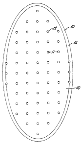
Figure 1 is top view of an embodiment of a device for assisting
2o hyperhydrosis therapy within the scope of the present invention,
showing a plurality of perforations in the device.
Figure 2 is a is top view of a second embodiment of a device for
assisting hyperhydrosis therapy within the scope of the present
2s invention, showing a plurality of more closely set perforations.
Figure 3 is a is top view of a second embodiment of a device fpr
assisting hyperhydrosis therapy within the scope of the present
invention, showing a plurality of two different sef of perforations,
3o Figure 4 is an enlarged side cross sectional view through one of a
perforations in the device of Figures 1, 2 or 3.
21
CA 02546460 2006-O1-12
WO 2005/007225 PCT/US2004/022796
DESCRIPTION
The present invention is based on the discovery that hyperhydrosis
therapy can be assisted by use of a dermal'overlay device. As shown
by Figure 1, an embodiment of our invention can be a device 10
comprised of a material, such as a flexible plastic, suitable (i.e. no sharp
protrusions, non-irritating) for firm, though temporary, placement against
a patch or area of hyperhydrotic skin of a hyperhydrosis patient. The .
device 10 can be made of a transparent material and has a plurality of
~ 'r
io through holes or perforations 12. A border 14 circumscribes the device
10. Preferably, the perforations 12 are separated by a uniform distance
A, so as to facilitate an even distribution of an injected antihyperhydrotic
pharmaceutical. The distance A can be about 2 cm.
is An alternate embodiment of our invention, as shown by Figure 2, can
comprise a device 20 comprised of a material, such as a bendable
plastic, suitable (i.e. smooth, not irritating upon transient skin contact)
for
firm, though temporary, placement against a patch or area of
hyperhydrotic skin of a hyperhydrosis patient. The dbvice 20 can be
2o made of a transparent material and has a plurality of through holes or
perforations 22. A border 24 circumscribes the device 20. Preferably,
the perforations 22 are separated by a uniform distance B, so as to
facilitate an even distribution of an injected antihyperhydrotic
pharmaceutical. The distance B can be about 1.5 cm.
2s
A third alternate embodiment of our invention, as shown by Figure 3,
can comprise a device 30 comprised of a material, such as a flexible
plastic, suitable (i.e. no sharp protrusions, non-irritating) for firm, though
temporary, placement against a patch or area of hyperhydrotic skin of a
so hyperhydrosis patient. The device 30 can be made of a transparent
material and has a plurality of a first set of holes or perforations 32 and
a second set of holes or perforations 34. A border 36 can circumscribe
22
CA 02546460 2006-O1-12
WO 2005/007225 PCT/US2004/022796
the device 30. Preferably, the perforations 32 are separated by a
uniform distance C, so as to facilitate an even distribution of an injected
antihyperhydrotic pharmaceutical. The perforations 34 can be
separated by a uniform distance D, so as to facilitate an even
s distribution of an injected antihyperhydrotic pharmaceutical with a
different injection density (i.e. C is not equal to D). The distance
between the perforations 12, 22, 32 or 34 can be between about 0.1 cm
to about 4 cm.
io As shown by Figure 4 which is an enlarged, side cross sectional view
through one of the perforations of Figures 1, 2 or 3, the device has a top
face 40 and a bottom face 42. A perforation can have a first end which
opens onto the top face 40, which first end has a diameter X. The
perforation can also have a second end which opens onto the bottom
is 42, which first end has a diameter Y. Preferably, and as illustrated by
Figure 4, diameter Y is less than diameter X, so that the bore of the
perforation can have a conical shape. Such a conical shape is a
preferred configuration for a bore of a perforation of the device because
upon insertion of a marker such as a ball point pen into the 'first end of
2o the perforation and through to the second end of the perforation (while
the lower face of the device is in contact with and being pressed against
the skin of a patient), the marker will be held firmly in the perforation and
will make a point mark or dot on the skin of the patient. Since the device
has multiple such perforations, rapid and accurate use of the marker to
2s mark a series of dots onto the skin of the patient is thereby assisted.
In practice the devise can be used by placing the lower face of the
device against an area of target skin (which can be, for example,
hyperhydrotic axial (i.e. armpit), plantar or plamar skin) which has
3o previously been determined to be an area of hyperhydrotic skin, as by
observation or by use of a diagnosed test such as the iodine starch test.
Thus, before treating focal hyperhidrosis, it can be necessary to find out
23
CA 02546460 2006-O1-12
WO 2005/007225 PCT/US2004/022796
what specific area of the body is producing excess sweat. This can be
done using.a diagnostic procedure known as the Minor or iodine starch
test. For this test, a weak solution of iodine is applied to the skin. Then,
powdered starch is dusted over the dried iodine. As the patient sweats,
s the areas where excessive sweating occurs are stained a bluish color by
the iodine starch mixture, thereby showing where the sweat glands are
overactive. Gravimetry is another test which measures exactly how
much a patient sweats. In gravimetry blotting paper is pressed against
the skin to soak up the sweat. Then, the blotting paper is weighed with a
~s~
io delicate scale to determine how much sweat has been absorbed.
The device is pressed against the skin (by pressing down on the
border) and a marker is inserted into each of the perforations in turn.
The device in then removed form contact with the skin leaving a grid
is pattern of dots on the skin showing where, to inject the botulinum toxin.
The material which comprises the device can be a plastic, silicone or
other suitable material. The material can be flexible and can be shaped
and sized so as to follow the contours of an armpit, foot or hand where it
2o can be applied.
Examples of botulinum toxins within the scope of the present
invention include the botulinum toxin types A, B, C, D, E, F, and G.
2s Botulinum toxins for use according to the present invention can be
stored in lyophilized, vacuum dried form in containers under vacuum
pressure or as stable liquids. Prior to lyophilization the botulinum toxin
can be combined with pharmaceutically acceptable excipients,
stabilizers and/or carriers, such as albumin. The lyophilized material
3o can be reconstituted with saline or water to create a solution or
composition containing the botulinum toxin to be administered to the
patient.
aa.
CA 02546460 2006-O1-12
WO 2005/007225 PCT/US2004/022796
EXAMPLE
The following non-limiting example sets forth a specific preferred
s method to use a device within the scope of the present invention and is
not intended to limit the scope of the our invention.
Example 1
Use of Device for Assistina HYperhydrosis Therap,Y
io A female patient, 32 years old, is diagnosed through observation and
use of the iodine starch with axial hyperhydrosis, in both armpits. The
lower side of the device shown in Figure 1 is pressed firmly against her
left armpit (while her left arm is raised above her head) and a ball point
pen is inserted into each of the perforations of the device in turn. The
Is device is removed, leaving a clear grid pattern of dots on her arm pit.
The same procedure is followed for the right armpit. A botulinum toxin
in then injected at the site of each dot, thereby treating her
hyperhydrosis.
2o Although the present invention has been described in detail with
regard to certain preferred methods, other embodiments, versions, and
modifications within the scope of the present invention are possible. For
example, the disclosed device can be made from various materials and
in various shapes, with different perforations spacings and different
2s perforation bore diameters.
All references, articles, patents, applications and publications set
forth above are incorporated herein by reference in their entireties.
3o Accordingly, the spirit and scope of the following claims should not
be limited to the descriptions of the preferred embodiments set forth
2s
Image