Note: Descriptions are shown in the official language in which they were submitted.
CA 02546592 2006-05-18
WO 2005/052562 PCT/SE2004/001725
1
EXAMINATION METHOD AND APPARATUS
FIELD OF THE INVENTION
The invention relates to a method and an apparatus for detection
of ionizing radiation.
BACKGROUND OF THE INVENTION AND RELATED ART
Radiographic imaging detectors comprising an array of small
sensors to capture a radiation-generated image are well known in
the art. A collimated radiation beam is intensity modulated as
it passes through a radiation-absorbing subject and the
transmitted beam as detected thus represents an inverted image
of the absorption by the subject, which in turn is related to
the elemental composition, density, and thickness of the
subject.
To improve contrast the broadband radiation from an X-ray tube
is heavily filtered before being used for radiographic purposes.
It is well known that at X-ray photon energies typically used,
the photoelectric absorption is decreased as a power law as the
X-ray photon energy increases, while unwanted scattering is
increased.
For soft tissue the photoelectric absorption is decreasing
rapidly at energies above about 20 keV and this higher energy X-
ray radiation does not contribute to the image recorded, but
reduces the contrast in the image. Thus, higher energies are
filtered out from the radiation.
SUMMARY OF THE INVENTION
A problem with the known kind of approach is that most X-ray
tubes have low efficiency at such low photon energy as 20 keV,
CA 02546592 2006-05-18
WO 2005/052562 PCT/SE2004/001725
2
i.e. the number of X-rays per unit power supplied to the tube is
low.
Further, all X-ray tubes emit radiation within a wide energy
spectrum. Typically, metallic foils filter the radiation from
the X-ray tube, but simultaneously the flux of X-rays is
reduced. Thus, large load has to be put on the X-ray tube to
obtain a reasonable radiation flux downstream the metallic
foils. Also, the relatively low flux affects the exposure time
in an adverse manner, i.e. makes it long, which obviously limits
the applicability of the technique.
Another issue of high importance is the radiation dose to the
subject in case it is a living organism or part thereof. While
the development of efficient collimators, appropriate filters,
and sensitive detector arrays during the last decades have
effectively reduced the radiation dose; still there is much to
do. Further reduction of the radiation dose is a driving
mechanism in detector design of today.
A main object of the invention is therefore to provide a method
and an apparatus for examination of a subject, which overcome
the above-identified problems as being related with the prior
art.
In this respect there is a particular object to provide such a
method and such an apparatus, which provide for the deposition
of only small amounts of energy in a subject to be examined.
A further object of the invention is to provide such a method
and such an apparatus, which provide for the possibility of
using broadband radiation for the measurement.
A still further object of the invention is to provide such a
method and such an apparatus, wherein radiation in a spectral
CA 02546592 2006-05-18
WO 2005/052562 PCT/SE2004/001725
3
range is used, in which the risk of under- or over exposing some
areas of the image is reduced.
Yet a further object of the invention is to provide such a
method and such an apparatus, wherein radiation over a wide
energy range, and especially at high photon energies, can be
detected with high efficiency.
These objects, among others, are attained by methods and
apparatuses as claimed in the appended claims.
The inventors have found that by preventing Compton scattered
radiation from being detected, and by providing ionizing
radiation within a spectral range such that more, preferably
much more photons, of the ionizing radiation are Compton
scattered than absorbed through the photoelectric effect in the
subject to be examined, an entirely new field of radiology opens
up. Since the probability of scattering is essentially the same
for a broad spectrum of photon energies, broadband radiation
including higher energies can be used for the detection.
Variations in an image, captured at photon energies high
enough to mainly obtain Compton scattering in the subject, are
substantially due to the density only of the examined subject,
provided that its thickness is constant, or known and
corrected for. This is true since the attenuation coefficient
for Compton scattering at photon energies of 10-300 keV is
only weakly dependent on atomic number and photon energy. This
is in sharp contrast to photoelectric absorption, which is
heavily dependent on energy, and even more dependent on atomic
number. Thus, the radiation image obtained is essentially a
shadow image of the density variations in the subject to be
examined.
CA 02546592 2006-05-18
WO 2005/052562 PCT/SE2004/001725
4
In some radiographic applications, however, such as soft
tissue applications including e.g. mammography, the density
variations may be very small, and therefore the contrast in
the recorded images is very low. According to the present
invention, a suitable contrast-enhancing agent is therefore
introduced into the subject to be examined. The suitable
should modify the density of the subject to be examined and
introduce density gradients into there. The density of the
contrast-enhancing agent may be higher or lower than the
density of the subject, but is preferably lower than the
density of the subject. For instance, an ultrasound contrast
agent may be employed. Contrast agents comprising or capable
of generating dispersions of gas microbubbles are preferred,
since such dispersions are particularly efficient due to the
low density and ease of compressibility of the microbubbles.
Thus, ordinary contrast enhancing agents for X-ray
diagnostics, such as iodine, which introduce atomic number
gradients into the subject rather than density gradients, are
less suitable. Further, the ultrasound contrast agent
administered to the subject should be sufficiently stable in
vivo to be recirculated in the blood stream following
administration, so that it may become equilibrated in the
blood pool prior to imaging.
Preferably, Compton scattered radiation is prevented from being
detected by means of a one-dimensional gas ionization detector
including two electrodes, between which an ionizable gas is
located, and a radiation entrance arranged such that said
ionizing radiation enters said detector sideways between the
electrodes, and electrons liberated by interaction between the
ionizing radiation and the gas are accelerated in a direction
essentially perpendicular thereto, wherein the distance
between the electrodes is kept short to essentially only allow
CA 02546592 2006-05-18
WO 2005/052562 PCT/SE2004/001725
radiation collimated in a plane between the electrodes to
ionize the gas. Further, the detector preferably employs
electron avalanche amplification; wherein only radiation
collimated in a very thin plane closest to the cathode
5 electrode will be amplified sufficiently to essentially
contribute to the signal as detected.
An advantage of the present invention is that if broadband
radiation is used for the detection, there is less need of
thick filters, the efficiency of the radiation source is
increased, the load on the radiation source can be lowered,
and the exposure time can be reduced due to the higher photon
f lux .
Further, since a scattered photon deposits only a fraction of
its energy in a subject, whereas a photoelectrically absorbed
photon deposits all its energy, the dose to the subject is
reduced.
In a particular preferred embodiment of the present invention
the above-mentioned novel examination method based on
scattering rather than absorption, is combined with an
ultrasound examination method. Here, the contrast-enhancing
agent can be administered to the subject, after which the
above-mentioned novel examination method based on scattering
and the ultrasound examination method are performed,
preferably simultaneously, using the same contrast-enhancing
agent. This is particularly advantageous for mammography
examinations, wherein the above-mentioned novel examination
method based on scattering provides for the detection of a
high-quality image of a breast to be examined causing an
extremely low dose to the subject. For instance, the dose may
be 20-100 times lower than in prior art X-ray mammography
examinations. The ultrasound examination provides an
CA 02546592 2006-05-18
WO 2005/052562 PCT/SE2004/001725
6
ultrasound image, which serves as a complement for diagnosis.
Some tumors will be better visualized in the ultrasound image.
Further characteristics of the invention and advantages
thereof, will be evident from the detailed description of
preferred embodiments of the present invention given
hereinafter and the accompanying Figs. 1-4, which are given by
way of illustration only and thus, are not limitative of the
present invention.
BRIEF DESCRIPTION OF THE DRAWINGS
Fig. 1 is a schematic diagram illustrating photoelectric
absorption, Compton scattering, pair production and total
attenuation coefficients for human tissue as a function of X-ray
photon energy, and a continuous X-ray spectrum of a typical X-
ray source for use in the present invention.
Fig. 2 illustrates schematically an apparatus for radiography
used in the present invention.
Fig. 3 is a flow diagram of a method according to a preferred
embodiment of the present invention.
Fig. 4 illustrates schematically an apparatus for radiography
according to another preferred embodiment of the present
invention.
DESCRIPTION OF PREFERRED EMBODIMENTS
As can be seen in Fig. l, which is a schematic diagram
illustrating photoelectric absorption, Compton scattering, pair
production and total attenuation coefficient ~.l.pE, l..l,~g, ~PR~ I~oT for
human soft tissue as a function of X-ray photon energy E, the
photoelectric attenuation coefficient ~u.PE decreases as a power
law with photon energy, and at about 25 keV the Compton
CA 02546592 2006-05-18
WO 2005/052562 PCT/SE2004/001725
7
scattering attenuation coefficient ~,~5 is comparable to the
photoelectric absorption attenuation coefficient ~u,PE. Between
about 30 and several hundred keV the Compton scattering
attenuation coefficient ~,~5 is completely dominating, whereas at
higher photon energies (in the order of 1 MeV) the probability
for pair production is increasing rapidly, and becomes the
dominating interaction process . While Fig. 1 is illustrating an
example only for human soft tissue, the relative overall
structure of the diagram holds for a large variety of matter.
The Compton scattering attenuation coefficient ~,~5 is fairly
constant over a large range of photon energies . It can be seen
in Fig. 1 the Compton scattering attenuation coefficient ~u~s for
soft tissue is fairly constant between photon energies of about
30 and several hundred keV.
Further, the photoelectric absorption attenuation coefficient
is heavily dependent on the atomic number of the elements, of
which the matter is comprised, whereas the Compton scattering
attenuation coefficient ~,~5 is only very weakly dependent on the
atomic number.
Still further, the transmission through matter is dependent
exponentially on the total attenuation coefficient ~,~,, on the
density p of the matter, and on the thickness t of the matter
according to:
Transmission ~ exp[-(~"I,ol, * p * t)
Thus, provided that ionizing radiation with photon energies high
enough so that Compton scattering dominates over photoelectric
absorption is passed through matter, the transmission through
there is only very weakly dependent on atomic number of the
CA 02546592 2006-05-18
WO 2005/052562 PCT/SE2004/001725
8
matter, and the actual photon energy, but strongly dependent on
the density of the matter. This is in sharp contrast to the case
where photoelectric absorption is the dominating interaction
mechanism. Here, the transmission through the matter is not only
strongly dependent on the density of the matter, but also on the
atomic number of the matter as well as on the actual photon
energy employed. Thus, if ionizing radiation with photon
energies high enough so that Compton scattering dominates over
photoelectric absorption was used, it can be broadband radiation
without having to perform complex calculations to compensate for
any strong photon energy dependence.
A typical continuous X-ray spectrum from an 30 kV wolfram-based
X-ray tube as filtered by a rhodium filter for use in e.g.
mammography examinations according to prior art is schematically
indicated in Fig. 1 by a dash-dotted line. Here, photoelectric
absorption dominates over Compton scattering. A broadband X-ray
spectrum from an 80 kV tungsten-based X-ray tube as filtered
with a copper filter is indicated by a dashed line. The
broadband radiation spectrum is displaced towards higher photon
energies, at which Compton scattering dominates over
photoelectric absorption.
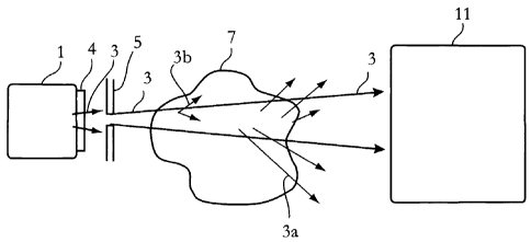
Fig. 2 illustrates schematically, in a side elevation view, an
apparatus for radiography for use in the present invention. The
apparatus comprises, as seen from left to right, an X-ray source
2 5 1, a f i lter arrangement 4 , an optional source aperture 5 and a
detector device 11.
The X-ray source may be a tungsten-based X-ray tube emitting an
X-ray radiation beam within a wide energy spectrum. The beam is
filtered by means of the filter arrangement 4 at the output of
the X-ray source 1. The filter arrangement 4 differs from a
conventional filter in the sense that it transmits higher
CA 02546592 2006-05-18
WO 2005/052562 PCT/SE2004/001725
9
energies, and preferably a much wider spectrum, such as e.g. the
broadband X-ray spectrum illustrated in Fig. 1. The radiation
beam as filtered is subsequently passed through the optional
source aperture 5 to collimate the beam. Preferably, the shape
and size of the source aperture 5 is adapted to the particular
size and kind of detector device 11. Thus, given a one-
dimensional detector device, the aperture 5 is designed with a
slit-shaped radiation transparent window, and given a
rectangular two-dimensional detector device; the aperture 5 is
preferably designed with a rectangular radiation transparent
window.
The source collimator is optional and is used to reduce the dose
to the subject to be examined in case the subject is a living
organism or part thereof, by producing a beam of X-rays, which
only illuminates the sensitive areas of the detector device 11.
The radiation beam 3 as filtered and optionally collimated
enters a region, where a subject, subject-matter, matter, object
or patient 7 to be imaged is located. In the subject 7 some
photons may be photoelectrically absorbed, some may be Raleigh
and Compton scattered (indicated by rays 3a in Fig. 1), and some
photons may be converted into electrons and positrons in a pair
production process, where these electrons and positrons may give
rise to emission of X-ray photons (indicated by rays 3b in Fig.
1). The various processes depend on elemental composition of the
subject 7 and on the photon energies of the incident radiation
beam 3.
The radiation beam transmitted through the subject 7 without
being deflected is detected by the detector device 11, while the
scattered radiation is prevented from being detected. Typically,
however, small amounts scattered radiation might enter into the
detector device 11 and blur the image recorded.
CA 02546592 2006-05-18
WO 2005/052562 PCT/SE2004/001725
According to the present invention the filter arrangement 4 is
adapted to the elemental composition of the subject 7 to be
imaged in a manner such the radiation beam as filtered is
within a spectral range so that more photons of the radiation
5 beam as filtered are Compton scattered than absorbed through
the photoelectric effect in the subject 7, i.e. so that
Compton scattering dominates over photoelectric absorption.
In the case of human soft tissue, such as breast tissue, the
filtered radiation may be broadband X-ray radiation between 10
10 and 300 keV (i.e. similar to the broadband radiation spectrum
of Fig. 1), preferably between 20 and 100 keV, and more
preferably above 30 keV. In other applications the filtered
radiation may be radiation above 30 keV.
Alternatively, the filtered radiation is in a spectral range
such that at least 2 times, more preferably at least 5 times,
and most preferably at least 10 times more photons of the
filtered radiation are Compton scattered than absorbed through
the photoelectric effect in the subject 7. If possible the
filtered radiation should be in a spectral range, at which
photoelectric absorption does not essentially occur in the
subject 7.
The detector 11 has preferably an elongated opening for entry
of the ionizing radiation; and a row of individual detector
elements arranged essentially parallel with the elongated
opening; and is of the kind wherein charges or photons
generated by interactions between the ionizing radiation and a
detection medium within the detector and travelling in a
direction essentially perpendicular to the ionizing radiation,
are detected by the row of individual detector elements.
CA 02546592 2006-05-18
WO 2005/052562 PCT/SE2004/001725
11
The detector is preferably a gaseous-based parallel plate
detector operating in avalanche amplification mode, wherein the
signals in the individual detector elements originate
essentially only from ionization within a thin layer, which may
be at least 2-5 times thinner than the inter-plate distance.
This advantageous behavior is obtained as the amplification is
exponential and electrons liberated closer to the individual
detector elements will not be able to produce signals strong
enough.
For further details regarding different kind of detectors for
use in the present invention, reference is made to the following
U.S. Patents by Tom Francke et al. and assigned to XCounter AB
of Sweden, which patents are hereby incorporated by reference:
Nos. 6,118,125; 6,373,065; 6,337,482; 6,385,282; 6,414,317;
6,476,397; 6,477,223; 6,518,578; 6,522,722; 6,546,070;
6,556,650; 6,600,804; and 6,627,897.
Alternatively, the detector device 11 may more generally be any
one- or two-dimensional detector, which is capable of
discriminating scattered photons to a large extent. The detector
may preferably any of a TFT-based detector; a scintillator-
based detector; a solid state detector such as a CMOS- CCD-,
CdZn- or CdZnTe-based detector; a gaseous-based detector; or a
combination thereof, and is advantageously provided with an
anti-scatter device, particularly an array of radiation
transparent channels arranged in front of the detector.
In order for the invention to operate properly, the scattered
radiation has to be discriminated from being detected to an
especially large extent. Preferably at least 900, more
preferably at least 99~, and most preferably at least 99.9 of
the Compton scattered radiation in the subject 7 is prevented
CA 02546592 2006-05-18
WO 2005/052562 PCT/SE2004/001725
12
from being detected. The parallel plate detector described
above has been shown to easily fulfill such a requirement.
By means of primarily using ionizing radiation at photon
energies where Compton scattering dominates over photoelectric
absorption, and by detecting the transmitted radiation
separate from the radiation scattered in the subject, a number
of advantages arise:
~ Since the radiation is primarily scattered off the
subject 7 and not absorbed in it, the radiation dose to
the subject is reduced. At photon energies of 50 keV a
Compton scattered photon deposits only about l00 of the
energy compared to a photoelectrically absorbed photon.
~ The filters may be made thinner since the radiation has
not to be that heavily filtered (due to the Compton
scattering attenuation coefficient compared to the
photoelectric absorption attenuation coefficient). Less
radiation is scattered in a thin filter than in a thick
filter, which means that the scattered radiation from the
filter arrangement 4 is reduced as compared to a
conventional filter arrangement.
~ The efficiency of the X-ray tube is increased since
larger portions of the emitted spectrum are usable. This
means also that the load on the X-ray tube can be
lowered. The exposure time can also be reduced due to the
higher X-ray photon flux obtainable.
~ The attenuation coefficient for Compton scattering at
photon energies of 10-300 keV is only weakly dependent on
atomic number and photon energy, and thus variations in
the image captured are essentially due to variations in
CA 02546592 2006-05-18
WO 2005/052562 PCT/SE2004/001725
13
the density of the subject only, provided that the
subject thickness is constant, or known and corrected
for.
The last advantage can in some applications be a drawback. If
the density variations are very small as they can be in some
mammography examinations the contrast in the image may be too
low.
However, a solution to this comprises, in accordance with the
present invention, to use a contrast-enhancing agent, which is
suitable for the above-described X-ray imaging technique. The
suitable contrast-enhancing agent should modify the density of
the subject to be examined and introduce density gradients
into there. For instance, an ultrasound contrast agent may be
employed. Contrast agents comprising or capable of generating
dispersions of gas microbubbles are preferred, since such
dispersions are particularly efficient due to the low density
and ease of compressibility of the microbubbles. Thus,
ordinary contrast enhancing agents for X-ray diagnostics, such
as iodine, which introduce atomic number gradients into the
subject rather than density gradients, are less suitable.
Further, the contrast agent administered to the subject should
be sufficiently stable in vivo to be recirculated in the blood
stream following administration, so that it may become
equilibrated in the blood pool prior to imaging. Suitable
contrast agents, which have been described for ultrasound
examinations, and which are suitable in the present invention
are disclosed in the U.S. Patents Nos. 6,645,147; 6,595,925;
6,547,738; 6,409,671; 6,375,931; 5,772,984; 5,567,415; and
5,236,693, the contents of which being hereby incorporated by
reference.
CA 02546592 2006-05-18
WO 2005/052562 PCT/SE2004/001725
14
Thus, a method for examination of a subject according to a
preferred embodiment of the invention, being illustrated in
Fig. 3, comprises the following steps.
Ionizing radiation is provided, in a step 31, within a
spectral range so that more photons of said ionizing radiation
are Compton scattered than absorbed through the photoelectric
effect in the subject to be examined. That is, the Compton
scattering should be the dominating interaction mechanism of
the interactions of the incident ionizing radiation with the
subject. Preferably, the energy of the radiation photons
should be selected so as to minimize the amount of
photoelectric absorption in the subject given all other
constraints, such as e.g. characteristics of the radiation
source used, available radiation filters, required radiation
flux, etc., as imposed by the particular application. Any of
the radiation spectrum profiles disclosed in this description
may be employed depending on the actual circumstances.
A suitable contrast-enhancing agent is, in a step 32,
administered to the subject to be examined, where the
contrast-enhancing agent introduces density variations in said
subject. The contrast-enhancing agent may be any of the
contrast-enhancing agents indicated above.
The ionizing radiation is then, in a step 33, directed towards
and passed through the subject. In the subject, various
interactions take place. However, the dominating interaction
mechanism of the incident ionizing radiation with the subject
is Compton scattering, which, as has been discussed above, is
dependent on density, but fairly independent on atomic number
and photon energy (within a given range).
CA 02546592 2006-05-18
WO 2005/052562 PCT/SE2004/001725
The ionizing radiation as transmitted through said subject
without being deflected is, in a step 34, detected spatially
resolved, while the Compton scattered radiation in the subject
is essentially prevented from being detected. For this
5 purpose, any of the above-described scattering-rejection
detecting apparatuses can be employed. If the photoelectric
absorption can be neglected, the signals recorded can be
arranged to form an image of the transmission, which would be
a true inverted image, or shadow image, of the Compton
10 scattering in the subject. Therefore, the image formed reveals
spatially resolved density variations in the subject - density
variations originally present in the subject as well as those
introduced by the contrast-enhancing agent.
In a further preferred embodiment of the present invention,
15 being illustrated in Fig. 4, the above-mentioned novel
examination apparatus based on scattering rather than
absorption, is combined with an ultrasound examination
apparatus.
The X-ray detector device 11 and the X-ray source arrangement
41 including the X-ray source 1, the filter arrangement 4, and
the optional source aperture 5 of Fig. 2, are arranged on
opposite sides of a subject to be examined, such as a breast
42. An ultrasound examination apparatus 43 is arranged
adjacent to the X-ray detector device 11. A device 44, such as
a syringe, is provided for administering an ultrasound
contrast-enhancing agent to the subject 42.
Prior to examination the ultrasound contrast-enhancing agent
is administered to the subject 42, after which the breast is
imaged, preferably simultaneously, by the X-ray detector
device 11/ X-ray source arrangement 41 combination and the
CA 02546592 2006-05-18
WO 2005/052562 PCT/SE2004/001725
16
ultrasound examination apparatus 43 using the very same
contrast-enhancing agent administration.
This is particularly advantageous for mammography
examinations, wherein the above-mentioned novel examination
method based on scattering provides for the detection of a
high-quality image of a breast of the subject to be examined,
causing an extremely low dose to the subject. For instance,
the dose may be 20-100 times lower than in prior art x-ray
mammography examinations. The ultrasound examination provides
an ultrasound image, which serves as a complement for
diagnosis. Some tumors will be better visualized in the
ultrasound image.