Note: Descriptions are shown in the official language in which they were submitted.
CA 02546681 2006-03-21
WO 2005/044147 PCT/US2004/027589
- 1 -
Patent
CATHETER-BASED FASTENER IMPLANTATION APPARATUS
AND METHODS
Field of the Invention
The invention relates generally to the
delivery of a prosthesis to a targeted site within the
body, e.g., for the repair of diseased and/or damaged
sections of a hollow body organ and/or blood vessel.
Background of the Invention
The weakening of a vessel wall from damage or
disease can lead to vessel dilatation and the formation
of an aneurysm. Left untreated, an aneurysm can grow in
size and may eventually rupture.
For example, aneurysms of the aorta primarily
occur in abdominal region, usually in the infrarenal area
between the renal arteries and the aortic bifurcation.
Aneurysms can also occur in the thoracic region between
the aortic arch and renal arteries. The rupture of an
aortic aneurysm results in massive hemorrhaging and has a
high rate of mortality.
Open surgical replacement of a diseased or
damaged section of vessel can eliminate the risk of
vessel rupture. In this procedure, the diseased or
damaged section of vessel is removed and a prosthetic
graft, made either in a straight of bifurcated
configuration, is installed and then permanently attached
CA 02546681 2006-03-21
WO 2005/044147 PCT/US2004/027589
- 2 -
and sealed to the ends of the native vessel by suture.
The prosthetic grafts for these procedures are usually
unsupported woven tubes and are typically made from
polyester, ePTFE or other suitable materials. The grafts
are longitudinally unsupported so they can accommodate
changes in the morphology of the aneurysm and native
vessel. However, these procedures require a large
surgical incision and have a high rate of morbidity and
mortality. In addition, many patients are unsuitable for
this type of major surgery due to other co-morbidities.
Endovascular aneurysm repair has been
introduced to overcome the problems associated with open
surgical repair. The aneurysm is bridged with a vascular
prosthesis, which is placed intraluminally. Typically
these prosthetic grafts for aortic aneurysms are
delivered collapsed on a catheter through the femoral
artery. These grafts are usually designed with a fabric
material attached to a metallic scaffolding (stmt)
structure, which expands or is expanded to contact the
internal diameter of the vessel. Unlike open surgical
aneurysm repair, intraluminally deployed grafts are not
sutured to the native vessel, but rely on either barbs
extending from the stent, which penetrate into the native
vessel during deployment, or the radial expansion force
of the stent itself is utilized to hold the graft in
position. These graft attachment means do not provide the
same level of attachment when compared to suture and can
damage the native vessel upon deployment.
Summary of the Invention
The invention provides systems and methods
that implant one or more fastening structures) in a
targeted body region, e.g., within a hollow body organ or
an intraluminal space. The fastening structures) are
implanted to a vessel wall, and serve to secure a
prosthesis within the hollow body organ or intraluminal
CA 02546681 2006-03-21
WO 2005/044147 PCT/US2004/027589
- 3 -
space.
One aspect of the invention provides a
fastener comprising a fastener body sized and configured
for deployment in tissue. The fastener body includes a
region for penetrating tissue in response to a force. The
fastener further includes an attachment element carried
by the fastener body sized and configured to couple to an
attachment site on a prosthesis.
The fastener body can be variously configured.
For example, the fastener body can comprise a helical
coil that is advanced into tissue. As another example,
the fastener body can comprise a stmt ring that can be
expanded into contact with tissue. The stmt ring can
carry tissue penetrating elements, e.g. barbs, to further
secure its position in tissue.
The attachment element can comprise, e.g., a
mechanical coupling assembly, and/or a magnetic coupling
assembly, and/or a chemical coupling assembly.
Another aspect of the invention provides
systems and methods for securing a prosthesis to tissue
in a targeted tissue region. The systems and methods (i)
introduce at least one fastener into the targeted tissue
region; (ii) implant the fastener in tissue in the
targeted tissue region;(iii) after steps (i) and (ii),
introduce a prosthesis into the targeted tissue region,
and (iv) attach the prosthesis to the fastener to secure
the prosthesis to tissue in the targeted tissue region.
Another aspect of the invention provides
systems and methods for securing a prosthesis to tissue
in a targeted tissue region. The systems and methods (i)
introduce a prosthesis into the targeted tissue region;
(ii) place the prosthesis into contact with tissue in the
targeted tissue region; (iii) after steps (i) and (ii),
introduce at least one st mt ring into the targeted
tissue region; and (iv) press an outer surface of the
CA 02546681 2006-03-21
WO 2005/044147 PCT/US2004/027589
- 4 -
st mt ring against the prosthesis to secure the
prosthesis to tissue within the targeted tissue region.
Brief Description of the Drawings
The invention will be understood from the
following detailed description of preferred embodiments,
taken in conjunction with the accompanying drawings,
wherein:
Figs . 1 to 5 show one type of a system and
method for attaching a prosthesis to a vessel wall or
hollow body organ, in which the prosthesis is coupled to
fasteners, which are implanted prior to deployment of the
prosthesis.
Figs. 6A to 6E are perspective views of
fasteners that can be used with the systems and methods
shown in Figs. 1 to 5, the fasteners having various types
of attachment elements.
Figs. 7A and 7B are views of prostheses that
can be used with the systems and methods shown in Figs. 1
to 5, the prostheses having attachment elements that
couple to attachment elements carried by fasteners of the
type shown in Figs. 6A to 6E.
Figs. 8A and SB show a prosthesis that has
been mechanically coupled to fasteners implanted in a
vessel wall or hollow body organ, which is illustrative
of one embodiment of the systems and methods of the type
shown in Figs. 1 to 5.
Fig. 9 shows a prosthesis that has been
magnetically coupled to fasteners implanted in a vessel
wall or hollow body organ, which is illustrative of
another embodiment of the systems and methods of the type
shown in Figs. 1 to 5.
Fig. 10 shows a prosthesis that has been
chemically coupled to fasteners implanted in a vessel
wall or hollow body organ, which is illustrative of
another embodiment of the systems and methods of the type
CA 02546681 2006-03-21
WO 2005/044147 PCT/US2004/027589
- 5 -
shown in Figs. 1 to 5.
Figs. 11 to 15 show another type of a system
and method for attaching a prosthesis to a vessel wall or
hollow body organ, in which the prosthesis is coupled to
a stmt ring, which has been implanted prior to
deployment of the prosthesis.
Fig. 16 is a perspective view of a stmt ring
that can be used in association with the systems and
methods shown in Figs. 11 to 15.
Figs. 17A to 17D are perspective views of
various types of attachment elements that the stent ring
shown in Fig. 16 can employ to accommodate coupling to a
prosthesis.
Fig. 18 is a view of a prosthesis that can be
used with the systems and methods shown in Figs. 11 to
15, the prostheses having attachment elements that couple
to attachment elements carried by a stmt ring of the
type shown in Figs. 17A to 17D.
Fig. 19 shows a prosthesis that has been
mechanically coupled to a stmt ring implanted in a
vessel wall or hollow body organ, which is illustrative
of one embodiment of the systems and methods of the type
shown in Figs. 11 to 15.
Fig. 20 shows a prosthesis that has been
magnetically coupled to a stmt ring implanted in a
vessel wall or hollow body organ, which is illustrative
of another embodiment of the systems and methods of the
type shown in Figs. 11 to 15.
Fig. 21 shows a prosthesis that has been
chemically coupled to a stmt ring implanted in a vessel
wall or hollow body organ, which is illustrative of
another embodiment of the systems and methods of the type
shown in Figs. 1 to 5.
Figs. 22 to 25 show another type of a system
and method for attaching a prosthesis to a vessel wall or
CA 02546681 2006-03-21
WO 2005/044147 PCT/US2004/027589
- 6 -
hollow body organ, in which a stmt ring is fastened to a
prosthesis, which has been deployed prior to implantation
of the stmt ring.
Detailed Description of the Invention
The figures depict various systems and methods
22, 40, and 60 for attaching a prosthesis to a vessel
wall or hollow body organ. The systems and methods 22,
40, and 60 can be used anywhere in the body. The systems
and methods 22, 40, and 60 lend themselves well to the
repair of diseased or damaged sections of a blood vessel,
particularly in the repair an abdominal aortic aneurysm.
For this reason, the systems and methods 22, 40, and 60
will be described in the context of this indication.
Still, it should be recognized that the systems and
methods 22, 40, and 60 can be used in other diverse
indications.
The figures depict, for purposes of
illustration, three general types of systems and methods
22, 40, and 60. These will be called, respectively, Type
I (Figs. 1 to 10), Type II (Figs. 11 to 21), and Type III
(Figs. 22 to 25).
The three Types I, II, and III share several
common features. For example, for all Types I, II, and
III, the systems and methods 22, 40, and 60 implant one
or more fastening structures) in a targeted body region,
e.g., within a hollow body organ or an intraluminal
space. The systems and methods 22~ 40, and 60 can deploy
the fastening structures) through the,vasculature by
manipulation from outside the body. The fastening
structures) are implanted to a vessel wall, and serve to
secure a prosthesis within the hollow body organ or
intraluminal space. The prosthesis can comprise, e.g.,
an endovascular graft, which can be deployed without
damaging the native blood vessel in either an arterial or
a venous system. The endovascular graft can comprise,
CA 02546681 2006-03-21
WO 2005/044147 PCT/US2004/027589
e.g., a radially expanding vascular stmt and/or a stent-
graft. The graft can be placed in the vasculature, e.g.,
to exclude or bridge an aneurysm, for example, an
abdominal aortic aneurysm. The graft desirably adapts to
changes in aneurysm morphology and repairs the
endovascular aneurysm.
The systems and methods 22, 40, and 60 of
Types I, II, and III differ in structural details and,
sometimes, in the sequence in which the fastening
structures) and prosthesis are deployed. Each Type I,
II, and III will now be described in greater detail.
I. Type I Systems and Methods
Figs. 1 to 10 depict the systems and methods
22 that can be characterized as a Type I arrangement. In
this embodiment, the systems and methods 22 first deploy
one or more individual fasteners 14 using a fastener
attachment assembly 10. As shown in Fig. 1, the assembly
10 is deployed to a targeted prosthesis attachment site
12. In Fig. 1, the targeted site 12 is shown as being
within an abdominal aortic aneurysm. The targeted site 12
can, or course, be elsewhere in the body.
As Fig. 2 shows, the fastener attachment
assembly 10 serves the function of implanted one or more
fasteners 14 in a desired array in the vessel wall at the
targeted site 12 prior to deployment of a prosthesis 20.
As will be described in greater detail, the fasteners 14
each includes an attachment element 16 that, in use,
couples to a corresponding attachment element 18 on a
prosthesis 20 deployed in the site 12.
In this arrangement (see Fig. 3), the systems
and methods 22 of Type I include a prosthesis delivery
catheter 24. The catheter 24 is deployed to the targeted
prosthesis attachment site 12, after implantation of the
fasteners 14 at the site 12, and after removal of the
fastener attachment assembly 10. As Fig. 3 shows, a
CA 02546681 2006-03-21
WO 2005/044147 PCT/US2004/027589
_ g _
guide wire helps to guide the catheter 24 to the
prosthesis attachment site 12. The catheter 24 carries a
prosthesis 20 for deployment at the targeted site 12 (see
Fig. 4), e.g., by radial expansion of the prosthesis 20.
The prosthesis 20 includes the attachment elements 18.
After expansion of the entire prosthesis 20 (or at least
the proximal end of the prosthesis 20) (see Fig. 5), the
catheter 24 is manipulated to place the attachment
elements 18 on the proximal end of the prosthesis 20 into
engagement with the attachment elements 16 on the
fasteners 14. The prosthesis 20 is thereby anchored in
place to the fasteners 14.
The construction and configuration of the
fastener attachment assembly 10 and the prosthesis
delivery catheter 24 can vary and are not material to the
accomplishment of the objectives of systems and methods
22 (or the other systems and methods 40 and 60, to be
described later). The fastener attachment assembly 10 can
be, e.g., as shown in copending United States Patent
Application Serial No. 10/307,226, filed November 29,
2002, which is incorporated herein by reference. The
prosthesis delivery catheter 24 can be of conventional
type for delivery of a self-expanding stmt graft.
A. The Fastener and its Attachment Elements
The fastener 14 can be variously constructed.
For example, the fastener 14 may have various
configurations, such as, for example, cylindrical or
triangular. The fasteners 14 may be of a metallic
fastener staple type (e.g., stainless steel), or may be
constructed from a polymeric material.
In one representative embodiment (see Figs. 6A
and 6B), the fastener 14 comprises a helical fastener 24.
The helical fastener 24 includes a sharpened distal end
26 and a proximal end 28. The proximal end 28 is
preferable sized and configured to limit its penetration
CA 02546681 2006-03-21
WO 2005/044147 PCT/US2004/027589
_ g _
into tissue, so that the proximal end 28 is exposed
outside tissue in the vessel wall. In the illustrated
embodiment, the proximal end 28 includes cap or carrier
74 that comes completely across the diameter. The
carrier 74 prevents the fastener 24 from being an open
coil and to control the depth of penetration into the
tissue. The carrier 74 also includes a slot .76 that
enables coupling of the fastener 24 to a suitable drive
mechanism, e.g., of a type shown in copending United
States Patent Application Serial No. 10/307,226, filed
November 29, 2002, which has been incorporated herein by
reference.
The carrier 74 can be variously sized and
configured to include an appropriate attachment element
16. The attachment element 16 can, e.g., comprise a hook
16A (as shown in Fig. 6A); or a barb 16B (see Fig. 6C);
or a permanent magnet 16C (see Fig. 6D); or a chemical
bonding agent 16D (see Fig. 6E). As has been explained,
these forms of attachment elements 16 are sized and
configured to couple to a compatible attachment element
18 on the prosthesis 20 deployed in the site 12.
The illustrated forms of attachment elements
16 are not exhaustive of the possible sizes and
configurations arrangements for the attachment elements
16. If given fastener 14 has means, after the fastener
16 has been implanted, to accommodate the fastening of a
later-deployed prosthesis 20, the fastener 14 can be
defined as having an attachment element 16. Likewise,
different styles of attachment elements 16 can be used in
conjunction with one another, provided attachment between
the prosthesis 20 and the fastener 14 occurs. For
instance, hooks and barbs may be used together.
Desirably, the fastener 14 and/or attachment
element 16 includes a radio-opaque marker material 30.
The material 30 aids the visualization of the
CA 02546681 2006-03-21
WO 2005/044147 PCT/US2004/027589
- 10 -
fastener/attachment element 14/16 for alignment with and
attachment to the prosthesis 20.
B. The Prosthesis and its Attachment
Elements
The prosthesis 20 (see Figs. 7A and 7B)
desirably incorporates a support frame or scaffold 32.
The scaffold 32 may be elastic, e.g., comprised of a
shape memory alloy elastic stainless steel, or the like.
For elastic scaffolds, expanding typically comprises
releasing the scaffolding from a constraint to permit the
scaffold to self-expand at the implantation site. A
sheath carried by the prosthesis delivery catheter 24
covers and constrains the scaffold 32 in a radially
compressed condition during while the catheter 24 is
steered to the targeted site 12. In this arrangement,
self-expansion of the scaffold 32 is achieved by pulling
back on the sheath (as Fig. 4 shows), to permit the
scaffold 32 to radially expand and assume its larger
diameter configuration.
Alternatively, the scaffold 32 may be
constrained in an axially elongated configuration, e.g.,
by attaching either end of the scaffold to an internal
tube, rod, catheter or the like. This maintains the
scaffold 32 in the elongated, reduced diameter
configuration. The scaffold 32 may then be released from
such axial constraint in order to permit self-expansion.
Alternatively, the scaffold 32 may be formed
from a malleable material, such as malleable stainless
steel of other metals. Expansion may then comprise
applying a radially expansive force within the scaffold
32 to cause expansion, e.g., inflating a scaffold
delivery catheter within the scaffold in order to affect
the expansion. In this arrangement, the positioning and
deployment of the endograft can be accomplished by the
CA 02546681 2006-03-21
WO 2005/044147 PCT/US2004/027589
- 11 -
use of an expansion means either separate or incorporated
into the deployment catheter 24. This will allow the
prosthesis 20 to be positioned within the vessel and
partially deployed while checking relative position
within the vessel. The expansion can be accomplished
either via a balloon or mechanical expansion device.
Additionally, this expansion stabilizes the position of
the prosthesis 20 within the artery by resisting the
force of blood on the endograft until the prosthesis can
be fully deployed.
The prosthesis 20 may have a wide variety of
conventional configurations. It can typically comprise a
fabric or some other blood semi-impermeable flexible
barrier which is supported by the scaffold 32, which can
take the form of a stmt structure. The stent structure
can have any conventional stent configuration, such as
zigzag, serpentine, expanding diamond, or combinations
thereof. The stent structure may extend the entire length
of the graft, and in some instances can be longer than
the fabric components of the graft. Alternatively, the
stent structure can cover only a small portion of the
prosthesis, a . g. , being present at the ends . The stmt
structure may have three or more ends when it is
configured to treat bifurcated vascular regions, such as
the treatment of abdominal aortic aneurysms, when the
stent graft extends into the iliac arteries. In certain
instances, the stent structures can be spaced apart along
the entire length, or at least a maj or portion of the
entire length, of the stmt-graft, where individual stent
structures are not connected to each other directly, but
rather connected to the fabric or other flexible
component of the graft.
The prosthesis 20 can be sized and figured to
be either straight or bifurcated form. Fig. 7A shows a
straight prosthesis 20. Fig. 7B shows a bifurcated
CA 02546681 2006-03-21
WO 2005/044147 PCT/US2004/027589
- 12 -
prosthesis 20.
As previously described, the prosthesis 20
includes the attachment elements 18 that couple in a
compatible fashion to the attachment elements 16 on the
fasteners 14. The size and configuration of the
prosthesis attachment elements 18 are selected to be
compatible with the size and configuration of fastener
attachment elements 18, to enable coupling the attachment
elements 16 and 18 together.
For example (see Fig. 8A), when the fastener
attachment elements 16 comprise a mechanical coupling
arrangement (e.g., the hooks 16A in Fig. 6C), the
compatible attachment element 18 on the prosthesis 20 can
comprise a proximal stmt structure 34, which
mechanically engages the attachment elements 16 to couple
the fasteners 14 to the prosthesis 20. As Fig. 8B shows,
when the mechanical coupling arrangement comprises the
barbs 16B in Fig. 6C, the compatible attachment element
18 on the prosthesis 20 can comprise a zone in the
prosthesis 20, which the barbs 16B can penetrate to
couple the fasteners 14 to the prosthesis 20.
Alternatively (see Fig. 9), when the fastener
attachment elements 16 comprise magnetic coupling
arrangements (e.g., the magnet 16C in Fig. 6D), the
compatible attachment elements 18 on the prosthesis 20
can comprise magnets 36 carried on the proximal end of
the prosthesis 20. The magnets 36 have an opposite
magnetic orientation than the fastener magnets 16C -- or
otherwise comprise a ferromagnetic material that is
attracted to the fastener magnet 16C -- to thereby
magnetically engage the attachment elements 16 to couple
the fasteners 14 to the prosthesis 20.
Alternatively (see Fig. 10), when the fastener
attachment elements 16 comprise chemical coupling
arrangements (e. g., the chemical material 16D in Fig.
CA 02546681 2006-03-21
WO 2005/044147 PCT/US2004/027589
- 13 -
6E), the compatible attachment element 18 on the
prosthesis 20 can comprise a compatible material 38
carried on the proximal end of the prosthesis 20. The
compatible material 38 adheres or bonds to the chemical
material 16D, to thereby chemically engage the attachment
elements 16 to couple the fasteners 14 to the prosthesis
20.
The Type I arrangement makes possible the
precise placement of fasteners in a desired location
within a vessel or hollow body organ in preparation for
9
deployment of a prosthesis. The fasteners serve as
positional markers for the precise deployment of the
prosthesis in the vessel or hollow body organ. The
fasteners also provide a secure, permanent attachment of
the prosthesis in the vessel or hollow body organ.
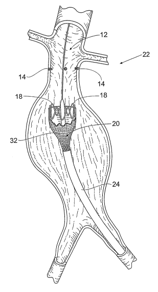
II. Type II Systems and Methods
Figs. 11 to 21 depict the systems and methods
40 that can be characterized as a Type II arrangement. In
this embodiment, the systems and methods 40 include a
stmt ring 44 that is implanted by a stmt ring
attachment assembly 42 prior to deployment of a
prosthesis 50. As shown in Fig. 11, the assembly 42 is
deployed to a targeted prosthesis attachment site 12,
which, like Fig. 1, is shown as being within an abdominal
aortic aneurysm. Fig. 11 shows the attachment assembly 42
being deployed over a guide wire.
As Fig. 12 shows, the stmt ring attachment
assembly 42 serves the function of implanted one or more
stmt rings 44 in the vessel wall at the targeted site
12. As will be described in greater detail, the stmt
rings 44 each includes an attachment element 46 that, in
use, couples to a corresponding attachment element 48 on
a prosthesis 50 deployed in the site 12.
In this arrangement (see Fig. 13), the systems
and methods 40 of Type II include a prosthesis delivery
CA 02546681 2006-03-21
WO 2005/044147 PCT/US2004/027589
- 14 -
catheter 24, like the one previously described in the
Type I arrangement. The catheter 24 is deployed to the
targeted prosthesis attachment site 12, after
implantation of the stent ring 44 or rings at the site
12, and after removal of the stmt ring attachment
assembly 42. Fig. 13 shows the catheter 24 being deployed
over a guide wire.
The catheter 24 carries a prosthesis 50 for
deployment at the targeted site 12 (see Fig. 14), e.g.,
by radial expansion of the prosthesis 50. The prosthesis
includes the attachment elements 48. After expansion
of the prosthesis 50 (or at least the proximal end of the
prosthesis 50) (see Fig. 15), the catheter 24 is
manipulated to move the attachment elements 48 on the
15 prosthesis 50 into engagement with the attachment
elements 46 on the stmt ring 44. The prosthesis 50 is
thereby anchored in place by the stmt ring 44.
A. The Stent Ring and its Attachment
Elements
20 The stmt ring 44 (see Fig. 16) can be
variously constructed. The stmt ring 44 may be elastic,
e.g., comprised of a shape memory alloy elastic stainless
steel, or the like. For elastic stmt rings 44, expanding
typically comprises releasing the stmt ring 44 from a
constraint to permit the stmt ring 44 to self-expand at
the implantation site. For example, a sheath carried by
the stmt ring attachment assembly 42 covers and
constrains the stmt ring 44 in a radially compressed
condition during while the assembly 42 is steered to the
targeted site 12. In this arrangement, self-expansion of
the stmt ring 44 is achieved by pulling back on the
sheath, to permit the stmt ring 44 to radially expand
and assume its larger diameter configuration.
Alternatively, the stmt ring 44 may be formed
CA 02546681 2006-03-21
WO 2005/044147 PCT/US2004/027589
- 15 -
from a malleable material, such as malleable stainless
steel of other metals. Expansion may then comprise
applying a radially expansive force within the stmt ring
44 to cause expansion, e.g., inflating a delivery
catheter within the stmt ring 44 in order to affect the
expansion. In this arrangement, the positioning and
deployment of the prosthesis 50 can be accomplished by
the use of an expansion means either separate or
incorporated into the st mt ring attachment assembly 42.
The expansion can be accomplished either via a balloon or
mechanical expansion device. Additionally, this expansion
stabilizes the position of the prosthesis 50 within the
artery by resisting the force of blood on the endograft
until the prosthesis can be fully deployed.
The stmt ring 44 includes an element 52 to
secure the st mt ring 44 to a vessel or body organ. The
element 52 can take various forms, e.g., hooks or barbs,
or a supra-renal stent, and/or combinations thereof. In
Fig. 16, the element 52 comprises barbs, which engage and
anchor into tissue upon expansion of the ring stmt 44.
In this arrangement, the stmt ring 44
includes an appropriate attachment element 46. As shown
in Figs. 17A to 17D, the attachment element 46 can take
similar form as the fastener attachment elements 16
previously described, e.g., a hook 46A (as shown in Fig.
17A); or a barb 46B (see Fig. 17B); or a permanent magnet
46C (see Fig. 17C); or a chemical bonding agent 46D (see
Fig. 17D). As has been explained, these forms of
attachment elements 46 are sized and configured to couple
to a compatible attachment element 48 on the prosthesis
50 deployed in the site 12.
The illustrated forms of attachment elements
46 are not exhaustive of the possible sizes and
configurations arrangements for the attachment elements
46. If given ring stent 44 has means, after the ring
CA 02546681 2006-03-21
WO 2005/044147 PCT/US2004/027589
- 16 -
st mt 44 has been deployed, to accommodate the fastening
of a later-deployed prosthesis 50, the ring stmt 44 can
be defined as having an attachment element 46. Likewise,
different styles of attachment elements 46 can be used in
conjunction with one another, provided attachment between
the prosthesis 50 and the fastener 14 occurs. For
instance, hooks and barbs may be used together.
Desirably, the ring stmt 44 and/or attachment
elements 46 includes a radio-opaque marker material 30.
The material 30 aids the visualization of the ring stent
44/attachment element 46 for alignment with and
attachment of the prosthesis 50.
B. The Prosthesis and its Attachment
Elements
The prosthesis 50 (see Fig. 18) can share the
same attributes of the prosthesis 20. It desirably
incorporates a support frame or scaffold 32, as
previously described and be deployed in the same manner.
Like the prosthesis 20, the prosthesis 50 may have a wide
variety of conventional configurations. It can be sized
and figured to be either straight or bifurcated form.
Fig. 18 shows a straight prosthesis 50 for the purpose of
illustration.
As previously described, the prosthesis 50
includes. the attachment elements 48 that couple in a
compatible fashion to the attachment elements 46 on the
stent ring 44. As before explained, the size and
configuration of the prosthesis attachment elements 48
are selected to be compatible with the size and
configuration of stmt ring attachment elements 46, to
enable coupling the attachment elements 46 and 48
together. In Fig. 18, the attachment elements 48 take
the form of magnets 36, as are also shown in Fig. 20 and
which will be described in greater detail later.
CA 02546681 2006-03-21
WO 2005/044147 PCT/US2004/027589
- 17 -
For example (see Fig. 19), when the stmt ring
attachment elements 46 comprise mechanical coupling
arrangements (e.g., the barbs 46B shown in Fig. 17B) the
compatible attachment element 48 on the prosthesis 50 can
comprise a zone in the prosthesis 20, which the barbs 46B
can penetrate to couple the couple the stent ring 44 to
the prosthesis 50. As another example of a mechanical
coupling arrangement (as shown in Fig. 15), when the
stent ring attachment elements 46 comprise the hooks 46A
(shown in Fig. 17A), the compatible attachment element 48
on the prosthesis 50 can comprise a proximal stmt
structure 34, which mechanically engages the attachment
elements 46 on the st mt ring 44 to couple prosthesis 50
to the stent ring 44.
Alternatively (see Fig. 20) , when the stmt
ring attachment elements 46 comprise magnetic coupling
arrangements (e.g., the magnet 46C in Fig. 17C), the
compatible attachment element 48 on the prosthesis 50 can
comprise a magnet 36 carried on the proximal end of the
prosthesis 50 having an opposite magnetic orientation --
or which has a ferromagnetic material that is otherwise
attracted to the stmt ring magnet 46C -- to thereby
magnetically engage the stmt ring attachment elements 46
and couple the stmt ring 44 to the prosthesis 50.
Alternatively (see Fig. 21), when the fastener
attachment elements 46 comprise chemical coupling
arrangements (e. g., the chemical material 46D in Fig.
17D), the compatible attachment element 48 on the
prosthesis 50 can comprise a compatible material 38 on
the proximal end of the prosthesis 50 that bonds to the
chemical material 46D, to thereby chemically engage the
attachment elements 46 and couple the stmt ring 44 to
the prosthesis 50.
It can be seen that the attachment mechanisms
between the fasteners 14 and prosthesis 20 in the Type I
CA 02546681 2006-03-21
WO 2005/044147 PCT/US2004/027589
- 18 -
arrangement and the attachment mechanisms between the
stmt ring 44 and prosthesis 50 in the Type II
arrangement are functionally similar.
The Type II arrangement makes possible the
precise placement of a scent ring in a desired iocati.on
within a vessel or..hol~ow body organ in preparation for
deployment of a prosthesis . The scent ring -serves a.s
positional marker .for the precise deployment of the
prosthesis in the vessel or hollow body organ. The stmt
, ring also provides a secure, permanent attachment of the
prosthesis in th.e vessel or hollow body organ.
III. Type III Systems-and Methods
Figs. 22 _to 25 depict the systems and methods
60 that can be-characterized as a Type III arrangement. .
Tn this embodiment, the systems and methods 60 .include a
prostrtesis delivery catheter 62 (see Fig. 22), like the
ones previously described with respect to the 'I-ype T anal
II arrangements. As Fig,. 22 srwws, the catheter 5? is
deplo=,..e;i. to tk?.e tarajFved 7.:rcs;~~=~?.esis attae:hment: .bite 12, , .
_2~U . whi.ch,, . l.ik.e Fi gs . 3 ., awd 1-; , i ~~.. ~; r~own .as _:bei.rxg
vA. ~.hin an
G.l:~d~,n::~.n.a:._ aortic. ane;.urysm. I'ig. 22 shows t:ne cat'.r.,eter 52
1=-eir~g d.epl oyed over- a guide wi re .
LTnl::,ke the systems and methcds 40 of t:he Types
I and II arrangements, the prosthesis delivery catheter
62 of the Type III arrangement is deployed before
implantation of fasteners 14 or a stmt ring 64 a.t the
site 12. The catheter 62 carries a prosthesis ~6 for
depl.o~~msnt at the targeted si ~-e 12 ( see Fig . 23 ) , a , g ~
by radial expansion o.f the prc~~.ehesis 66, as px~evio.~?sl.y
described.
The systf:~aa.:, and methc~~s 60 of Type II'I. include
a _,st~ert :ring at~tach:,.wnt a~aembiy 6.8 . As shown .1I1 fi.g. 24;
in t3-W 'ype ITI arrangement, the stmt ring attachment
assembly 68. is deployed within the prosthesis E6 after
deplclm:ent o:C t=he prosthesis ~6 a.nd after the prcsthesi.s
CA 02546681 2006-03-21
WO 2005/044147 PCT/US2004/027589
- 19 -
delivery catheter 62 has been withdrawn.
As Fig. 25 shows, the stmt ring attachment
assembly 68 serves the function of implanted one or more
stent rings 70 in the vessel wall at the targeted site
12. The stmt ring 70 includes elements 72 to pass
through the proximal end of the prosthesis 66 and secure
the stent ring 70 to a vessel or body organ. The
elements 72 can take various forms, e.g., hooks or barbs,
or a supra-renal stent, and/or combinations thereof, as
previously described in connection with the Type II
arrangement. The prosthesis 66 is thereby anchored in
place by the stmt ring 44.
As before described, the stmt ring 70 and/or
locations on the prosthesis 66 desirable includes a
radio-opaque marker material 30. The material 30 aids the
visualization of the stmt ring 70 and/or prosthesis 66
for alignment with and attachment of the prosthesis 50.
The Type III arrangement enables the
implantation of an anchoring device (i.e., the stmt
ring) all at once after a prosthesis has been deployed.
The embodiments of the invention are described
above in detail for the purpose of setting forth a
complete disclosure and for the sake of explanation and
clarity. Those skilled in the art will envision other
modifications within the scope and sprit of the present
disclosure.