Note: Descriptions are shown in the official language in which they were submitted.
CA 02546856 2006-05-23
WO 2005/051244 PCT/US2004/038918
EXPANDABLE SPINAL IMPLANT
FIELD OF THE INVENTION
The present invention relates generally to the field of spinal implants, and
more
particularly relates to an expandable spinal implant.
BACKGROUND
There have been numerous attempts to develop an intervertebral implant to
replace
a damaged or degenerated natural spinal disc and to maintain sufficient
stability of the disc
space between adjacent vertebrae, at least until arthrodesis is achieved.
These types of
intervertebral implants have taken many forms.
For example, one of the more prevalent designs includes spinal implants having
a
cylindrical shape. With regard to cylindrically-shaped implants, the exterior
portion of the
implant is typically threaded to facilitate insertion into the disc space.
Additionally,
intervertebral implants can either be solid, sometimes referred to as a spacer
or plug, or
can define a hollow interior designed to permit bone in-growth, sometimes
referred to as a
fusion device or fusion cage. The interior of a fusion device may be filled
with a bone
growth inducing substance to facilitate or promote bone growth into and
through the
device. It is commonly accepted that intervertebral implants that facilitate
or promote
natural bone in-growth typically achieve a more rapid and stable arthrodesis.
One area that is usually not addressed by the above-discussed intervertebral
implant designs concerns maintaining and restoring the natural anatomy of the
fused
spinal segment. Notably, once natural disc material is removed, the normal
lordotic or
kyphotic curvature of the spine is reduced or eliminated. With regard to prior
implants
having a substantially uniform outer cross section, the need to restore this
curvature is
largely neglected. Moreover, in some cases the adjacent vertebral bodies are
reamed to
form a passage having a shape corresponding to the particular shape of the
implant. In
other cases, the normal curvature is established prior to reaming followed by
insertion of
the implant. However, these techniques generally involve over-reaming of the
posterior
portion of the adjacent vertebral bodies, thereby resulting in excessive
removal of load
bearing vertebral bone which may lead to instability of the portion of the
spinal column
CA 02546856 2006-05-23
WO 2005/051244 PCT/US2004/038918
2
being treated. Also, it is typically difficult to ream through the posterior
portion of the
lower lumbar segment where lordosis is the greatest.
Accordingly, with regard to many intervertebral implant designs, limited
effort or
no effort is made to restore the lordotic curvature. As a result, the implant
is likely to
cause a kyphotic deformity as the vertebral bodies settles around the
intervertebral
implant. Additionally, with regard to intervertebral implants that attempt to
restore the
lordotic curvature, expansion of the implant is typically limited to a single
direction along
the height of the disc space, with no consideration being given to expanding
the implant in
a lateral direction to provide a larger overall area for
absorbing/distributing vertebral loads
and improved stability and/or an increased resistance to subsidence into the
adjacent
,v
vertebral bodies.
Thus, there is a general need in the industry to provide an improved
expandable
spinal implant. The present invention satisfies this need and provides other
benefits and
advantages in a novel and unobvious manner.
SUMMARY
The present invention relates generally to an expandable spinal implant. While
the
actual nature of the invention covered herein can only be determined with
reference to the
claims appended hereto, certain forms of the invention that are characteristic
of the
preferred embodiments disclosed herein are described briefly as follows.
In one form of the present invention, an expandable spinal implant is
provided,
including a body having a plurality of movable portions cooperating to define
an outer cross
section having a first transverse dimension and a second transverse dimension
and defining
first and second substantially planar surfaces disposed generally opposite one
another and
adapted to engage adjacent vertebral bodies. The spinal implant also includes
an expansion
member co-acting with the movable portions to expand the outer cross section
along the first
and second transverse dimensions.
In another form of the present invention, an expandable spinal implant is
provided,
including a body having a longiW dinal axis and a plurality of movable
portions cooperating to
define a generally rectangular outer cross section having a first transverse
dimension and a
second transverse dimension. The spinal implant also includes an expansion
member co-
acting with the movable portions to expand the outer cross section along the
first and second
transverse dimensions.
CA 02546856 2006-05-23
WO 2005/051244 PCT/US2004/038918
In another form of the present invention, an expandable spinal implant is
provided,
including a body having a longitudinal axis and a plurality of movable
portions cooperating to
define an outer cross section having a first transverse dimension and a second
transverse
dimension, with the movable portions having substantially planar inner
surfaces that cooperate
to define an inner chamber having a substantially rectangular inner cross
section and with the
inner surfaces defining an inward taper along the longitudinal axis. The
spinal implant also
includes an expansion member having a substantially rectangular outer cross
section and
engaging the inner surfaces of the movable portions to expand the movable
portions along the
first and second transverse dimensions as the expansion member is displaced
generally along
the longitudinal axis.
In another foam of the present invention, an expandable spinal implant is
provided,
including a body having a longitudinal axis and including a plurality of
movable portions
cooperating to define an outer cross section having a first transverse
dimension and a second
transverse dimension and defining first and second substantially planar
surfaces disposed
generally opposite one another and adapted to engage adjacent vertebral
bodies. The spinal
implant also includes means for expanding the outer cross section along the
first and second
transverse dimensions.
In another form of the present invention, a surgical method is provided,
including
providing an expandable spinal implant having a plurality of movable portions
extending
along a longitudinal axis and cooperating to define an outer cross section
having a first
transverse dimension and a second transverse dimension, with the movable
portions defining
first and second substantially planar surfaces disposed generally opposite one
another. The
method further includes inserting the spinal implant within an intervertebral
space with the
first and second substantially planar surfaces positioned adjacent first and
second vertebrae,
and expanding the outer cross section along each of the first and second
transverse
dimensions.
It is one object of the present invention to provide an improved expandable
spinal
implant. Further objects, features, advantages, benefits, and aspects of the
present
invention will become apparent from the drawings and description contained
herein.
BRIEF DESCRIPTION OF THE DRAWINGS
FIG. 1 is a perspective end view of an expandable spinal implant according to
one
form of the present invention, as shown in a non-expanded configuration.
CA 02546856 2006-05-23
WO 2005/051244 PCT/US2004/038918
4
FIG. 2 is a perspective end view of the spinal implant illustrated in FIG. 1,
as
shown in an expanded configuration.
FIG. 3 is a side view of the spinal implant illustrated in FIG. 1.
FIG. 4 is a proximal end view of the spinal implant illustrated in FIG. 1.
FIG. 5 is a distal end view of the spinal implant illustrated in FIG. 1.
FIG. 6 is a top plan view of the spinal implant illustrated in FIG. 1.
FIG. 7 is a cross-sectional side view of the spinal implant illustrated in
FIG. 6, as
viewed along line 7-7 of FIG. 6.
FIG. 8 is a cross-sectional view of the spinal implant illustrated in FIG. 7,
as
viewed along line 8-8 of FIG. 7.
FIG. 9 is a partial cross-sectional side view of the spinal implant
illustrated in FIG.
1, as positioned between adjacent vertebral bodies in a non-expanded
configuration and
with a surgical instrument engaged thereto.
FIG. 10 is a partial cross-sectional side view of the spinal implant
illustrated in
FIG. l, as positioned between adjacent vertebral bodies in a fully-expanded
configuration.
FIG. 11 is a top plan view of a pair fully expanded spinal implants positioned
side-
by-side in a bi-lateral arrangement within an intervertebral disc space.
DESCRIPTION OF THE PREFERRED EMBODIMENTS
For the purposes of promoting an understanding of the principles of the
invention,
reference will now be made to the embodiments illustrated in the drawings and
specific
language will be used to describe the same. It will nevertheless be understood
that no
limitation of the scope of the invention is hereby intended, and that
alterations and further
modiEcations to the illustrated devices and/or further applications of the
principles of the
invention as illustrated herein are contemplated as would normally occur to
one skilled in
the art to which the invention relates.
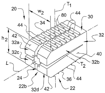
Refernng to FIGS. 1-8, shown therein is a spinal implant 20 according to one
form
of the present invention. The spinal implant 20 extends along a longitudinal
axis L and is
generally comprised of an expandable fusion cage 22 and an expansion member
24. As
will be discussed below, the expansion member 24 serves to transition the
fission cage 22
from an initial configuration, as shown in FIG. l, toward an expanded
configuration, as
shown in FIG. 2.
In the illustrated embodiment of the invention, expansion of the fusion cage
22
CA 02546856 2006-05-23
WO 2005/051244 PCT/US2004/038918
occurs along two transverse dimensions (i.e., along dimensions that are
transverse to the
longitudinal axis L), and more specifically along a first transverse axis Tl
and a second
transverse axis T2. However, it should be understood that in other embodiments
of the
invention, expansion of the fusion cage 22 may occur along any number of axes,
including
5 a single transverse axis or three or more transverse axes. As will be
discussed in greater
detail below, in the illustrated embodiment of the invention, the fusion cage
22 is
configured to expand along the first transverse axis TI to distract the disc
space and/or to
restore/maintain lordosis between the adjacent vertebral bodies. Additionally,
the fusion
cage 22 is configured to expand along the second transverse axis TZ to
distribute loading
of the fusion cage 22 across a larger and more dispersed area of the adjacent
vertebral
endplates to provide improved stability and/or an increased resistance to
subsidence.
The components of the spinal implant 20 are preferably formed of a bio-
compatible material. In one embodiment, the fusion cage 22 and/or the
expansion member
24 are formed of a material that has a modulus of elasticity substantially
similar to that of
bone. In a further embodiment, the fusion cage 22 and/or the expansion member
24 are
formed of a resorbable material that resorbs or degrades within the body over
a period of
time for partial or total replacement by bone. In a specific embodiment of the
invention,
the fusion cage 22 and/or the expansion member 24 are formed of a polymeric
material,
including, for example, a non-resorbable polymer such as polyetheretherketone
(PEEK) or
a resorbable polymers such as polylactates (PLA). However, it should be
understood that
other suitable polymeric/non-polymeric materials and/or other suitable
resorbable/non-
resorbable materials are also contemplated for use in association with the
present
invention. Examples of other suitable materials include composite polymers,
non-
reinforced polymers, carbon-reinforced polymer composites, carbon fiber, PMMA,
calcium hydroxide, ceramics, polylactide, polyglycolide, tyrosine-derived
polycarbonate,
polyanhydride, polyorthoester, polyphosphazene, calcium phosphate, calcium
hydroxide,
hydroxyapatite, bioactive glass, or any combination thereof. The use of
metallic materials
are also contemplated, including, for example, stainless steel and stainless
steel alloys,
titanium and titanium alloys, shape-memory alloys, cobalt chrome alloys, or
any
combination thereof. Additionally, the use of bone or bone substitute
materials is also
contemplated.
In one aspect of the invention, the fusion cage 22 is comprised of a fixed
base
CA 02546856 2006-05-23
WO 2005/051244 PCT/US2004/038918
6
portion 30 and plurality of movable branch portions 32 extending from the
fixed base
portion 30 generally along the longitudinal axis L. In the illustrated
embodiment of the
invention, the fixed base portion 30 includes an opening 31 extending
therethrough and
positioned generally along the longitudinal axis L adjacent the proximal end
22a of the
fusion cage, the purpose of which will be discussed below. Additionally, in
the illustrated
embodiment, the fusion cage 22 includes four movable branch portions 32,
including a
pair of upper branch portions 32a, 32b and a pair of lower branch portions
32c, 32d.
However, it should be understood that the fusion cage 22 may define any number
of
movable branch portions 32, including two, three, or five or more movable
branch
portions 32.
The branch portions 32 are coupled to the base portion 30 in such a mamier as
to
allow the branch portions 32 to move relative to one another to provide for
expansion of
the fusion cage 22. In the illustrated embodiment of the invention, the branch
portions 32
are formed integral with the base portion 30 to define a single-piece, unitary
fusion cage
22. As such, the base portion 30 flexibly interconnects the branch portions 32
in a manner
allowing expansion of the fusion cage 22 via flexible material deformation of
the branch
portions 32 and/or the base portion 30. The interconnection between the base
portion 30
and the branch portions 32 acts in a hinge-like manner during expansion of the
fusion cage
22 to provide for substantially independent movement of the branch portions
32.
Although the illustrated embodiment of the fusion cage 22 utilizes integrally
connected branch portions 32, it is also contemplated that the branch portions
32 may be
formed separately and connected together to form a multi-piece fusion cage
assembly. In
another alternate embodiment, the branch portions 32 may be pivotally attached
to the
base portion 30 or directly to one other via a hinge or pivot pin such that
the fusion cage
22 may be expanded without flexible material deformation. Other suitable means
for
coupling the branch portions 32 together to provide for expansion of the
fusion cage 22 are
also contemplated, including forming or coupling of the branch portions 32
directly to one
another without the use of a fixed base portion 30.
In a further aspect of the invention, the movable branch portions 32 cooperate
with
one another to define a generally rectangular outer transverse cross section.
In one
embodiment, the fusion cage 22 includes a first pair of substantially planar
upper and
lower surfaces 34, 36 extending generally along the second transverse axis TZ
(defined by
CA 02546856 2006-05-23
WO 2005/051244 PCT/US2004/038918
branch portions 32a, 32b and 32c, 32d, respectively) and a second pair of
substantially
planar side surfaces 38, 40 extending along the first transverse axis Tl
(defined by branch
portions 32a, 32c and 32b, 32d, respectively). In a further embodiment, the
fusion cage 22
has a substantially parallelpiped configuration including six sides, with each
side generally
defining a parallelogram. However, it should be understood that other shapes,
configurations and outer cross sections of the branch portions 32 and the
fusion cage 22
are also contemplated as falling within the scope of the present invention.
In another embodiment of the invention, the upper and lower corners of the
fusion
cage 22 adjacent the distal end 22b are tapered or beveled to facilitate
insertion of the
fusion cage 22 into an intervertebral disc space and/or distraction of the
adjacent vertebral
bodies VU, VL. Specifically, the distal end portions of the upper pair of
branches 32a, 32b
define an inwardly tapering surface 42 extending from the upper surface 34
toward the
distal end 22b of the fusion cage 22. Similarly, the distal end portions of
the lower pair of
branches 32c, 32d define an inwardly tapering surface 42 extending from the
lower
surface 36 toward the distal end 22b of the fusion cage 22. The tapered
surfaces 42 may
be particularly useful to facilitate insertion of the fusion cage 22 between
the adjacent
vertebral bodies VU, VL via an impaction or push-in technique. Although not
specifically
illustrated in the figures, it should be understood that the side or lateral
corners of the
fusion cage 22 defined by the branches 32a, 32c and 32b, 32d, respectively,
may also be
beveled to define an inwardly tapering surface extending from the side
surfaces 38, 40
toward the distal end 22b of the fusion cage 22.
In a further embodiment of the invention, the upper and lower surfaces 34, 36
defined by the branch portions 32a, 32b and 32c, 32d, respectively, define a
number of
bone anchoring elements 44 adapted for engagement with adjacent vertebral
bodies VU,
VL (FIGS. 9 and 10) to prevent or inhibit movement of the fusion cage 22 once
implanted
within the intervertebral disc space. In a specific embodiment, the bone
anchoring
elements 44 comprise a number of rows of triangular-shaped ridges or teeth
extending
across a width of the fusion cage 22 generally along the transverse axis TZ.
However, it
should be understood that other shapes, orientations and/or configurations of
ridges or
teeth are also contemplated as falling within the scope of the present
invention. It should
also be understood that other configurations of bone anchoring elements 44 are
also
contemplated for use in association with the fusion cage 22, such as, for
example, other
CA 02546856 2006-05-23
WO 2005/051244 PCT/US2004/038918
types of projections extending fiom the upper and lower surfaces 34, 36 of the
fusion cage,
including spikes, surface roughening, or threads. It should further be
understood that in
other embodiments of the invention, the upper and lower surfaces 34, 36 of the
fusion cage
22 need not necessarily include bone anchoring elements 44, but may
alternatively define
a substantially smooth configuration devoid of any surface projections or
irregularities. In
other embodiments of the invention, the side surfaces 3 8, 40 of the fusion
cage 22 may
also define bone anchoring elements in instances where the side surfaces 38,
40 may at
some point be in full or partial engagement with the adjacent vertebral bodies
VU, VL.
As illustrated in FIG. 2, upon transitioning of the fusion cage 22 toward an
expanded configuration, the upper branch portions 32a, 32b will separate or
splay apart
relative to the lower branch portion 32c, 32d to expand the fusion cage 22
along the first
transverse axis Tl. Similarly, the upper branch portions 32a, 32b will
separate or splay
apart relative to one another and the lower branch portion 32c, 32d will
separate or splay
apart relative to one another to expand the fusion cage 22 along the second
transverse axis
TZ. As a result, the fusion cage 22 is capable of expanding along two
transverse
dimensions. In one embodiment of the invention, the transverse dimensions
correspond to
an axial/veutical dimension of the disc space (e.g~, the height of the disc
space) and a
lateral/horizontal dimension of the disc space (e.g., the width or depth of
the disc space).
In the illustrated embodiment of the invention, since the movable branch
portions
32 are integrally connected with the base portion 30, expansion of the fusion
cage 22 is not
uniform along the longitudinal axis L. Instead, the fixed proximal ends of the
branch
portions 32 adjacent the base portion 30 remain relatively stationary and
therefore do not
appreciably expand along the transverse axes Tl, T2. However, the movable
distal ends of
the branch portions 32 separate or splay apart to expand the distal end
portion of the fusion
cage 22 from an initial height lal and width wl (FIG. 1) to an expanded height
h2 and width
w2 (FIG. 2). In one embodiment, expansion of the fusion cage 22 along the
transverse axis
Tl (the change in height between hl and la2) and along the transverse axis TZ
(the change in
width between w~ and w2) falls within a range of about 2-4 millimeters.
However, it
should be understood that other embodiments of the invention are also
contemplated
wherein the fusion cage 22 is configured to expand less than 2 millimeters or
greater than
4 millimeters along the transverse axes TI and T2. In a specific embodiment of
the
invention, the initial height lay and width w~ of the fusion cage 22 are each
about 10
CA 02546856 2006-05-23
WO 2005/051244 PCT/US2004/038918
9
millimeters, and the expanded height hz and width wz of the fusion cage 22 are
each about
14 millimeters. However, it should be understood that these specific
dimensions are
exemplary, and that other dimensions of the fusion cage 22 are also
contemplated.
In the illustrated embodiment of the invention, the initial height h, and
width wr of
the fusion cage 22 are substantially equal, thereby providing the fusion cage
22 with an
initial configuration having a square-shaped transverse cross section.
Likewise, the
expanded height h2 and width w~ of the fusion cage 22 are also illustrated as
being
substantially equal, thereby providing the fusion cage 22 with an expanded
configuration
adjacent the distal end 22b having a square-shaped transverse cross section.
It should be
understood, however, that in other embodiments of the invention, the initial
height hl and
width wl of the fusion cage 22 and/or the expanded height hz and width wz of
the fusion
cage 22 may differ. It should also be understood that the rate of expansion
along the
transverse axes Tl and TZ need not necessarily be equal. Instead, the fusion
cage 22 and/or
the expansion member 24 may be configured to provide unequal or varying rates
of
expansion along the transverse axes Tl and T2. Additionally, although the
illustrated
embodiment of the spinal implant 20 is configured to expand the fusion cage 22
in a non-
uniform mamier along the longitudinal axis L, it is also contemplated that the
branch
portions 32 may be interconnected in a manner that would allow for relatively
uniform
expansion of the fusion cage 22 along the longitudinal axis L, or other types
of non-
uniform expansion of the fusion cage 22, such as, for example, configurations
resulting in
a greater degree of expansion along the central region of the branch portions
32.
In the illustrated embodiment of the invention, the branch portions 32 have a
shell-
like configuration and cooperate with one another to define a hollow interior
chamber 50
(FIG. 7) extending generally along the longitudinal axis L. In one embodiment,
the
chamber 50 is sized and configured to receive the expansion member 24 therein
such that
movement of the expansion member 24 within the chamber 50 engages the
expansion
member 24 with the branch portions 32 to expand the fusion cage 22 along the
first and
second transverse axes Tl and T2. In one embodiment, axial displacement of the
expansion member 24 generally along the longitudinal axis L causes the branch
portions
32 to separate or splay apart, thereby transitioning the fusion cage 22 toward
an expanded
configuration. However, it should be understood that in other embodiments of
the
invention, relative rotational or pivotal displacement of the expansion member
24 may
CA 02546856 2006-05-23
WO 2005/051244 PCT/US2004/038918
cause the branch portions 32 to separate or splay apart to expand the fusion
cage 22.
Additionally, other types of relative displacement of the expansion member 24
are also
contemplated for use in association with the present invention to expand the
fusion cage
22, including displacement of the expansion member 24 in directions transverse
to the
5 longitudinal axis L.
As illustrated in FIGS. 7 and 8, the branch portions 32 define inner surfaces
52 that
cooperate to define the interior chamber 50. In the illustrated embodiment of
the
invention, the inner surfaces 52 are substantially planar so as to provide the
chamber 50
with a generally rectangular inner cross section that corresponds to the outer
cross section
10 of the expansion member 24 (FIG. 8). As illustrated in FIG. 8, in one
embodiment, the
branch portions 32 cooperate to define a first pair of substantially planar
upper and lower
surfaces 54, 56 (defined by branch portions 32a, 32b and 32c, 32d,
respectively) and a
second pair of substantially planar side surfaces 58, 60 (defined by branch
portions 32a,
32c and 32b, 32d, respectively). As illustrated in FIG. 7, the upper and lower
surfaces 54,
56 and the side surfaces 58, 60 (not shown) are inclined or inwardly tapered
along the
longitudinal axis L to facilitate expansion of the fusion cage 22 along both
of the
transverse axes Tl and TZ, the details of which will be discussed below.
However, it
should be understood that other shapes, configurations and cross sections of
the branch
portions 32 and the fusion cage 22 are also contemplated as falling within the
scope of the
presentinvention.
In a further embodiment of the invention, one or more of the branch portions
32
defines an inwardly extending flange or transverse projection 62 adjacent the
distal end
22b of the fusion cage 22 (FIGS. 5 and 7). In the illustrated embodiment, the
branch
portions 32a-32d each define an inwardly extending flange or transverse
projection 62 that
cooperate with one another to define a transverse shoulder 64 extending about
the inner
periphery of the chamber 50. Additionally, as illustrated in FIG. 5, the
inwardly extending
corners of each of the transverse flanges 62 each define a cut-out or notch
66, the purpose
of which will be discussed below. In the illustrated embodiment, the notch 66
has a
rectangular configuration; however, other suitable shapes and configurations
are also
contemplated as falling with the scope of the present invention.
In another embodiment of the invention, one or more of the branch portions 32
defines a retention element 72 extending from the inner surface 52 adjacent
the distal end
CA 02546856 2006-05-23
WO 2005/051244 PCT/US2004/038918
11
22b of the fusion cage 22 (FIG. 7). The retention element 72 is adapted to
engage and
retain the expansion member 24 in a select position and orientation relative
to the branch
portions 32 upon expansion of the fusion cage 22 (FIG. 10). In one embodiment,
each of
the branch portions 32a-32d includes a retention element 72 so as to define a
peripheral
retention element extending generally about the interior chamber 50. In the
illustrated
embodiment of the invention, the retention elements 72 are configured as
transverse
projections or ridges extending from the inner surfaces 52 of the branch
portions 32 in a
direction transverse to the longitudinal axis L. In a specific embodiment, the
retention
elements 72 have a triangular configuration, including an inclined or ramped
portion 74
tapering inwardly along the longitudinal axis L and a transverse shoulder
portion 76 facing
generally opposite the shoulder portion 64 defined by the distal end portions
of the
branches 32a-32d. However, other suitable shapes and configurations of the
retention
elements 72 are also contemplated as falling with the scope of the present
invention.
Additional details regarding interaction between the retention element 72 and
the
expansion member 24 will be discussed below.
In one embodiment of the invention, the branch portions 32 define a number of
bone in-growth openings 80 extending through the upper and lower outer
surfaces 34, 36
and communicating with the inner chamber 50 to permit bone growth from the
adjacent
vertebral bodies into and possibly through the fusion cage 22. In one
embodiment, the
bone in-growth openings 80 are disposed along substantially the entire length
of the
interior chamber 50 and positioned intermediate the rows of triangular-shaped
ridges or
teeth 44. Although the bone in-growth openings 80 are illustrated as having a
circular
cross section defining a relatively small diameter, it should be understood
that other
shapes, sizes andlor configurations of the bone in-growth openings are also
contemplated.
For example, in other embodiments of the invention, the bone in-growth
openings 80 may
have a larger diameter or an elongate slotted configuration. Additionally,
although the
bone in-growth openings 80 are illustrated as extending through respective
ones of the
branch portions 32, in other embodiments of the invention, one or more of the
openings 80
may be defined between the adjacent branches 32a, 32b and 32c, 32d. Moreover,
although
the bone in-growth openings 80 are illustrated as extending through the upper
and lower
outer surfaces 34, 36, it should be understood that bone in-growth openings
may also
extend through the side surfaces 38, 40 of the fusion cage 22. It should
fiirther be
CA 02546856 2006-05-23
WO 2005/051244 PCT/US2004/038918
12
understood that although the bone in-growth openings 80 are illustrated and
described as
communicating with the interior chamber 50, in other embodiments, the openings
80 need
not necessarily extend entirely through the branch portions 32.
Referring to FIGS. 7 and 8, shown therein is the expansion member 24 disposed
within the interior chamber 50 of the fusion cage 22. The expansion member 24
includes
a main body portion 90 and a stem portion 92 extending axially therefrom.
Although a
specific embodiment of the expansion member 24 is illustrated and described
herein, it
should be understood that other suitable configurations of the expansion
member 24 are
also contemplated as falling within the scope of the present invention.
In the illustrated embodiment of the expansion member 24, the main body
portion
90 has a generally rectangular outer cross section that substantially
corresponds to the
inner rectangular cross section of the inner fusion chamber 50. The main body
portion 90
includes outer surfaces that are adapted to slide along the inclined inner
surfaces 52 of the
branch portions 32 during axial displacement of the expansion member 24 along
the
interior chamber 50 to transition the fusion cage 22 to an expanded
configuration. In one
embodiment of the invention, the outer surfaces of the main body portion 90
are
substantially planar and are arranged generally parallel with the longitudinal
axis L.
However, other shapes, configurations and outer cross sections of the main
body portion
90 are also contemplated for use in association with the present invention.
The main body
portion 90 also defines an opening 96 sized and configured to receive a distal
end portion
of a surgical instrument therein to facilitate axial displacement of the
expansion member
24 along the inner chamber 50 of the fusion cage 22. In the illustrated
embodiment, the
tool receiving opening 96 has a generally circular Timer cross section to
receive a
correspondingly shaped distal end portion of a surgical instrument therein.
However,
other shapes and configurations of the opening 96 are also contemplated for
use in
association with the present invention, such as, for example, rectangular or
hexagonal
configurations.
In the illustrated embodiment of the expansion member 24, the stem portion 92
is
sized and shaped for positioning within the cut-out or notched portions 66
defined by the
distal transverse flanges 62 of the movable branches 32a-32d when the
expansion member
24 is disposed adjacent the distal end 22b of the fusion cage 22 (FIG. 10). In
one
embodiment, the stem portion 92 has a generally rectangular outer cross
section; however,
CA 02546856 2006-05-23
WO 2005/051244 PCT/US2004/038918
13
other shapes and configurations of the stem portion 92 are also contemplated
for use in
association with the present invention, such as, for example, hexagonal or
circular
configurations.
Referring now to FIG. 9, shown therein is a surgical instrument 100 engaged
with
the implant 20 for transitioning the fusion cage 22 to an expanded
configuration. In one
embodiment of the invention, the surgical instrument 100 generally includes an
outer
sleeve 102 and an inner drive shaft 104. The surgical instrument 100 may also
include a
handle (not shown) to aid in the manipulation and handling of the spinal
implant 20.
However, it should be understood that other suitable types and configurations
of surgical
instruments are also contemplated for use in association with the present
invention, and
that the elements and operation thereof may differ from the embodiment of the
surgical
instrument 100 illustrated and described herein. For example, another type of
instnunent
that may be used in association with the present invention is illustrated and
described in
U.S. Patent No. 6,436,140 to Liu et al., the entire contents of which are
hereby
incorporated herein by reference.
The outer sleeve 102 of the surgical instrument 100 has a distal end portion
that is
adapted to engage the fusion cage 22. In one embodiment, engagement between
the distal
end portion of the sleeve 102 and the fusion cage 22 is abutting engagement.
However, it
should be understood that other types of engagement are also contemplated,
such as, for
example, threaded engagement, keyed engagement, tongue-and-groove engagement,
frictional engagement, or any other suitable method of engagement. The inner
drive shaft
104 is disposed within the outer sleeve 102 and extends through the aperture
31 in the base
portion 30 of the fusion cage 22 and into engagement with the expansion member
24. In
one embodiment of the invention, engagement between the distal end portion of
the drive
shaft 104 and the expansion member 24 is abutting engagement. However, other
types of
engagement are also contemplated, such as, for example, threaded engagement,
keyed
engagement, tongue-and-groove engagement, frictional engagement, or any other
suitable
method of engagement. In a further embodiment of the invention, the distal end
portion of
the drive shaft 104 is configured to be received within the opening 96 in the
expansion
member 24. In the illustrated embodiment, the distal tip portion 108 of the
drive shaft 104
has a generally circular outer cross section that corresponds with the inner
cross section of
the opening 96 to provide secure engagement between the drive shaft 104 and
the
CA 02546856 2006-05-23
WO 2005/051244 PCT/US2004/038918
14
expansion member 24. However, other shapes and configurations of the distal
tip portion
108 are also contemplated for use in association with the present invention,
including
rectangular or hexagonal shapes.
As should be appreciated, axial displacement of the drive shaft 104 in the
direction
of arrow A will correspondingly axially displace the expansion member 24
through the
inner chamber 50 to thereby transition the fusion cage 22 toward the fully
expanded
configuration illustrated in FIG. 10. In one embodiment, the drive shaft 104
may be
displaced via threading engagement between the drive shaft 104 and the
aperture 31
extending through the fixed base portion 30 of the fusion cage 22. In this
manner,
rotational movement of the drive shaft 104 and threading engagement with the
aperture 31
results in axial movement of the drive shaft 104 generally along the
longitudinal axis L in
the direction of arrow A. In another embodiment, threading engagement between
the
inner drive shaft 104 and the outer sleeve 102 may be used to displace the
drive shaft 104
generally along the longitudinal axis L in the direction of arrow A. Other
suitable
techniques for axially displacing the drive shaft 104 are also contemplated as
falling
within the scope of the present invention. v
As discussed above, the outer surfaces of the expansion member 24 slidably
engage the inclined inner surfaces 52 of the branch portions 32 as the
expansion member
24 is axially displaced along the inner chamber 50 of the fusion cage 22. As
should be
appreciated, sliding engagement of the expansion member 24 along the inclined
surfaces
54, 56, 58 and 60 (FIG. 8) causes the branch portions 32a-32d to separate or
splay apart
along each of the transverse axes Tl and TZ to transition the fusion cage 22
from the initial
configuration illustrated in FIGS. 1 and 9 toward the fully expanded
configuration
illustrated in FIGS. 2 and 10. As the expansion member 24 is slidably
displaced along the
upper and lower inclined surfaces 54, 56, the upper and lower outer surfaces
34, 36 of the
fusion cage 22 are displaced away from another along the transverse axis Tl to
distract the
intervertebral disc space and/or to restore/maintain lordosis between the
upper and lower
vertebrae VU, VL. Lilcewise, as the expansion member 24 is slidably displaced
along the
inclined side surfaces 58, 60, the outer side surfaces 38, 40 of the fission
cage 22 are
displaced away from another along the transverse axis T2. In this manner, the
loads
transferred from the upper and lower vertebrae VU, VL to the fusion cage 22
are distributed
CA 02546856 2006-05-23
WO 2005/051244 PCT/US2004/038918
across a larger and more dispersed area of the adjacent vertebral endplates to
provide
improved stability and/or an increased resistance to subsidence.
As the expansion member 24 is advanced to a position adjacent the distal end
portion 22b of the fusion cage 22, the expansion member 24 will engage the
retention
element 74. Specifically, the expansion member 24 will slide along the ramp
portions 74
of the retention element 72 and will ultimately be positioned beyond the
retention element
72 between the transverse shoulders 64 and 76 defined by the branch portions
32a-32d and
the retention element 72, respectively (FIG. 7). As illustrated in FIG. 10,
the main body
portion 90 of the expansion member 24 is captured between the transverse
shoulders 64,
10 76 to secure the expansion member 24 in the proper orientation and position
within the
inner chamber 50 and to maintain the fusion cage 22 in the expanded
configuration. As
also illustrated in FIG. 10, the stem portion 92 of the expansion member 24 is
positioned
within the cut-out portions 66 defined by the transverse flanges 62a-62b of
the branch
portions 32a-32d. Engagement of the stem portion 92 with the transverse
flanges 62a-62d
15 provides stability between the expansion member 24 and the fusion cage 22
and also
provides added support to the distal ends of the branch portions 32.
Following expansion of the fusion cage 22, the surgical instt-ument 100 may be
disengaged from the spinal implant 20 and removed from the patient. In a
further
embodiment of the invention, a bone growth promoting material 120 (FIG. 10)
may be loaded
into the inner chamber 50 of the fusion cage 22 to facilitate or promote bone
growth from the
upper and lower vertebrae VU, VL, through the openings 80 and into and
possibly through the
fusion cage 22. In one embodiment, the bone growth promoting material 120 is
comprised of
a bone graft material, a bone morphogenic protein (BMP), or any other suitable
bone growth
promoting material or substance including but not limited to bone chips or
bone marrow, a
demineralized bone matrix (DBM), mesenchymal stem cells, and/or a LIM
mineralization
protein (LMP). It should be understood that the bone growth promoting material
120 can be
used with or without a suitable carrier.
In one embodiment, the bone growth promoting material 120 is injected into the
inner
chamber 50 via the aperture 31 extending through the fixed base portion 30. In
another
embodiment, the bone growth promoting material 120 is positioned within the
inner chamber
50 subsequent to expansion of the fusion cage 22. However, it should be
understood that the
fusion cage 22 and the expansion member 24 may alternatively be configured so
as to allow
CA 02546856 2006-05-23
WO 2005/051244 PCT/US2004/038918
16
the bone growth promoting material 120 to be loaded within the inner chamber
50 in another
manner and/or prior to or during expansion of the fusion cage 22.
Having illustrated and described the elements and operation of the spinal
implant 20,
reference will now be made to a technique for implanting the spinal implant 20
within an
intervertebral space according to one embodiment of the invention. However, it
should be
understood that other implantation techniques and procedures are also
contemplated, and that
the following technique in no way limits the scope of the present invention.
Referring to FIGS. 9 and 10, the vertebral level to be treated is identified,
followed by
the removal of at least a portion of the natural intervertebral disc via a
total or partial
discectomy. The endplates of the upper and lower vertebrae VU, VL are then
prepared using
known surgical instruments and techniques (e.g., rotating cutters, curettes,
chisels, etc.).
Notably, since the spinal implant 20 is not externally threaded, forming a
cylindrically-
shaped passage between and into the adjacent vertebrae VU, VL and tapping the
passage is not
required. Accordingly, removal or disruption of vertebral tissue from the
upper and lower
' vertebrae VU, VL is minimized.
Following preparation of the intervertebral disc space and the upper and lower
vertebrae VU, VL, the spinal implant 20 is positioned within the
intervertebral disc space via a
suitable insertion techniques such as, for example, an irnpaction or push-in
type insertion
techniques. Notably, since the spinal implant 20 is not threaded, insertion
into the disc space
can be accomplished without having to thread or otherwise rotate the spinal
implant 20 into
position. Additionally, in a preferred embodiment, the spinal implant 20 is
inserted into the
disc space while in a non-expanded conftguration to minimize neural
distraction. However, it
should be understood that in certain circumstances, it may be desirable to
transition the spinal
implant 20 to an expanded configuration either before or during insertion in
the disc space.
In a further embodiment of the invention, the spinal implant 20 may be
inserted into the disc
space in a minimally invasive manner (i.e., through a small access portal) via
the use of
endoscopic equipment, a small diameter tube or cannula, or by other suitable
minimally
invasive surgical techniques. However, it should be understood that other
conventional
surgical methods and techniques may also be used.
After the spinal implant 20 is inserted in the disc space, the fusion cage 22
is
transitioned to an expanded conftguration via axially displacing the inner
shaft 104 of the
instrument 100 in the direction of arrow A (toward the distal end 22b of the
fusion cage),
CA 02546856 2006-05-23
WO 2005/051244 PCT/US2004/038918
17
which correspondingly displaces the expansion member 24 through the inner
chamber 50.
As discussed above, axial displacement of the expansion member 24 results in
sliding
engagement between the expansion member 24 and the branch 32, thereby causing
the
branch portions 32 to separate or splay apart along each of the transverse
axes TI and TZ to
transition the fusion cage 22 to the fully expanded configuration illustrated
in FIG. 10. As
also discussed above, expansion of the fusion cage 22 along the transverse
axis Tl distracts
and/or restores/maintains lordosis between the upper and lower vertebrae VU,
VL, with the
upper vertebral bearing surface 34 being oriented at an angle relative to the
lower vertebral
bearing surface 36.
When the fusion cage 22 is fully expanded to the configuration illustrated in
FIG. 10,
the expansion member 24 is securely captured between the retention element 72
and the
transverse flanges of the branch portions 32 to lock the expansion member 24
in the proper
orientation and position and to securely maintain the fusion cage 22 in the
expanded
configuration. Although the fusion cage 22 is maintained in the expanded
configuration
solely via engagement between the expansion member 24 and the branch portions
32, it
should be understood that one or more supplemental internal fixation elements
are also
contemplated for use in association with the fusion cage 22, particularly in
instances
involving excessive vertebral loading and/or instability. It should also be
understood that
supplemental external intravertebral fixation elements and/or stabilization
techniques may
also be used if excessive residual instability is encountered following
insertion and expansion
of one or~more of the spinal implants 20 with the disc space.
Once the fusion cage 22 is fully expanded, a bone growth promoting material
120,
such as BMP and a suitable Garner, is injected or otherwise loaded into the
inner chamber 50
of the fusion cage 22 to facilitate or promote bone growth from the upper and
lower vertebrae
VU, VL, through the bone growth openings 80, and into and possibly through the
fusion cage
22. Additionally, morselized autograft bone or a similar type of material may
be positioned
adjacent the expanded fusion cage 22 to further promote fusion.
In one embodiment of the invention, access to the spinal column and insertion
of the
spinal implant 20 into the disc space is accomplished via a posterior surgical
approach.
However, it should be understood that access and insertion of the spinal
implant 20 into the
disc space may be accomplished via other surgical approaches, such as, for
example, an
anterior approach or a lateral approach. In another embodiment of the
invention, the spinal
CA 02546856 2006-05-23
WO 2005/051244 PCT/US2004/038918
18
implant 20 is used to treat the lumbar region of the spine, with the upper and
lower vertebrae
VU, VL comprising lumbar vertebrae. However, it should nevertheless be
understood that the
present invention is also applicable to other portions of the spine, including
the cervical,
thoracic or sacral regions of the spine. Additionally, as illustrated in FIG.
11, in a further
embodiment of the invention, a pair of spinal implants 20a, 20b may be
positioned side-by-
side in a bilateral arrangement within the disc space. However, it should be
understood that
unilateral placement or central placement of a single spinal implant 20 within
the disc space
is also contemplated as falling within the scope of the present invention.
While the invention has been illustrated and described in detail in the
drawings and
I O foregoing description, the same is to be considered as illustrative and
not restrictive in
character, it being understood that only the preferred embodiments have been
shown and
described and that all changes and modifications that come within the spirit
of the invention
are desired to be protected.