Note: Descriptions are shown in the official language in which they were submitted.
CA 02546965 2011-05-20
WO 2005/051240 PCT/US2004/039714
1
PATIENT SELECTABLE KNEE JOINT ARTHROPLASTY DEVICES
FIELD OF THE INVENTION
[0001] The present invention relates to orthopedic methods,
systems and devices and more particularly relates to methods, systems
and devices for articular resurfacing in the knee.
BACKGROUND OF THE INVENTION
[0002] There are various types of cartilage, e.g., hyaline cartilage
r and fibrocartilage. Hyaline cartilage is found at the articular surfaces of
bones, e.g., in the joints, and is responsible for providing the smooth
gliding motion characteristic of moveable joints. Articular cartilage is
firmly
attached to the underlying bones and measures typically less than 5mm in
thickness in human joints, with considerable variation depending on the
joint and the site within the joint.
[0003] Adult cartilage has a limited ability of repair; thus, damage to
cartilage produced by disease, such as rheumatoid and/or osteoarthritis,
or trauma can lead to serious physical deformity and debilitation.
Furthermore, as human articular cartilage ages, its tensile properties
change. The superficial zone of the knee articular cartilage exhibits an
increase in tensile strength up to the third decade of life, after which it
decreases markedly with age as detectable damage to type II collagen
occurs at the articular surface. The deep zone cartilage also exhibits a
progressive decrease in tensile strength with increasing age, although
collagen content does not appear to decrease. These observations
indicate that there are changes in mechanical and, hence, structural
organization of cartilage with aging that, if sufficiently developed, can
predispose cartilage to traumatic damage.
CA 02546965 2006-05-24
WO 2005/051240 PCT/US2004/039714
2
[0004] Once damage occurs, joint repair can be addressed through
a number of approaches. One approach includes the use of matrices,
tissue scaffolds or other carriers implanted with cells (e.g., chondrocytes,
chondrocyte progenitors, stromal cells, mesenchymal stem cells, etc.).
These solutions have been described as a potential treatment for cartilage
and meniscal repair or replacement. See, also, International Publications
WO 99/51719 to Fofonoff, published October 14, 1999; W001 /91672 to
Simon et al., published 12/6/2001; and W001/17463 to Mannsmann,
published March 15, 2001; U.S. Patent No. 6,283,980 1311 to Vibe-Hansen
et al., issued September 4, 2001, U.S. Patent No. 5,842,477 to Naughton
issued December 1, 1998, U.S. Patent No. 5,769,899 to Schwartz et al.
issued June 23, 1998, U.S. Patent No. 4,609,551 to Caplan et al. issued
September 2, 1986, U.S. Patent No. 5,041,138 to Vacanti et al. issued
August 29, 1991, U.S. Patent No. 5,197,985 to Caplan et al. issued March
30, 1993, U.S. Patent No. 5,226,914 to Caplan et al. issued July 13, 1993,
U.S. Patent No. 6,328,765 to Hardwick et al. issued December 11, 2001,
U.S. Patent No. 6,281,195 to Rueger et al. issued August 28, 2001, and
U.S. Patent No. 4,846,835 to Grande issued July 11, 1989. However,
clinical outcomes with biologic replacement materials such as allograft and
autograft systems and tissue scaffolds have been uncertain since most of
these materials do not achieve a morphologic arrangement or structure
similar to or identical to that of normal, disease-free human tissue it is
intended to replace. Moreover, the mechanical durability of these biologic
replacement materials remains uncertain.
[0005] Usually, severe damage or loss of cartilage is treated by
replacement of the joint with a prosthetic material, for example, silicone,
e.g. for cosmetic repairs, or metal alloys. See, e.g., U.S. Patent No.
6,383,228 to Schmotzer, issued May 7, 2002; U.S. Patent No. 6,203,576
to Afriat et al., issued March 20, 2001; U.S. Patent No. 6,126,690 to
CA 02546965 2006-05-24
WO 2005/051240 PCT/US2004/039714
3
Ateshian, et al., issued October 3, 2000. Implantation of these prosthetic
devices is usually associated with loss of underlying tissue and bone
without recovery of the full function allowed by the original cartilage and,
with some devices, serious long-term complications associated with the
loss of significant amount of tissue and bone can include infection,
osteolysis and also loosening of the implant.
[0006] Further, joint arthroplasties are highly invasive and require
surgical resection of the entire articular surface of one or more bones, or a
majority thereof. With these procedures, the marrow space is often
reamed to fit the stem of the prosthesis. The reaming results in a loss of
the patient's bone stock. U.S. Patent 5,593,450 to Scott et al. issued
January 14, 1997 discloses an oval domed shaped patella prosthesis. The
prosthesis has a femoral component that includes two condyles as
articulating surfaces. The two condyles meet to form a second trochlear
groove and ride on a tibial component that articulates with respect to the
femoral component. A patella component is provided to engage the
trochlear groove. U.S. Patent 6,090,144 to Letot et al. issued July 18, 2000
discloses a knee prosthesis that includes a tibial component and a
meniscal component that is adapted to be engaged with the tibial
component through an asymmetrical engagement.
[0007] A variety of materials can be used in replacing a joint with a
prosthetic, for example, silicone, e.g. for cosmetic repairs, or suitable
metal alloys are appropriate. See, e.g., U.S. Patent No. 6,443,991 131 to
Running issued September 3, 2002, U.S. Patent No. 6,387,131 B1 to
Miehlke et al. issued May 14, 2002; U.S. Patent No. 6,383,228 to
Schmotzer issued May 7, 2002; U.S. Patent No. 6,344,059 B1 to Krakovits
et al. issued February 5, 2002; U.S. Patent No. 6,203,576 to Afriat et al.
issued March 20, 2001; U.S. Patent No. 6,126,690 to Ateshian et al.
issued October 3, 2000; U.S. Patent 6,013,103 to Kaufman et al. issued
CA 02546965 2006-05-24
WO 2005/051240 PCT/US2004/039714
4
January 11, 2000. Implantation of these prosthetic devices is usually
associated with loss of underlying tissue and bone without recovery of the
full function allowed by the original cartilage and, with some devices,
serious long-term complications associated with the loss of significant
amounts of tissue and bone can cause loosening of the implant. One such
complication is osteolysis. Once the prosthesis becomes loosened from
the joint, regardless of the cause, the prosthesis will then need to be
replaced. Since the patient's bone stock is limited, the number of possible
replacement surgeries is also limited for joint arthroplasty.
[0008] As can be appreciated, joint arthroplasties are highly
invasive and require surgical resection of the entire, or a majority of the,
articular surface of one or more bones involved in the repair. Typically with
these procedures, the marrow space is fairly extensively reamed in order
to fit the stem of the prosthesis within the bone. Reaming results in a loss
of the patient's bone stock and over time subsequent osteolysis will
frequently lead to loosening of the prosthesis. Further, the area where the
implant and the bone mate degrades over time requiring the prosthesis to
eventually be replaced. Since the patient's bone stock is limited, the
number of possible replacement surgeries is also limited for joint
arthroplasty. In short, over the course of 15 to 20 years, and in some
cases even shorter time periods, the patient can run out of therapeutic
options ultimately resulting in a painful, non-functional joint.
[0009] U.S. Patent No. 6,206,927 to Fell, et al., issued March 27,
2001, and U.S. Patent No. 6,558,421 to Fell, et al., issued May 6, 2003,
disclose a surgically implantable knee prosthesis that does not require
bone resection. This prosthesis is described as substantially elliptical in
shape with one or more straight edges. Accordingly, these devices are not
designed to substantially conform to the actual shape (contour) of the
remaining cartilage in vivo and/or the underlying bone. Thus, integration of
CA 02546965 2006-05-24
WO 2005/051240 PCT/US2004/039714
the implant can be extremely difficult due to differences in thickness and
curvature between the patient's surrounding cartilage and/or the
underlying subchondral bone and the prosthesis. U.S. Patent 6,554,866 to
Aicher, et al. issued April 29, 2003 describes a mono-condylar knee joint
5 prosthesis.
[0010] Interpositional knee devices that are not attached to both the
tibia and femur have been described. For example, Platt et al. (1969)
"Mould Arthroplasty of the Knee," Journal of Bone and Joint Surgery
51B(1):76-87, describes a hemi-arthroplasty with a convex undersurface
that was not rigidly attached to the tibia. Devices that are attached to the
bone have also been described. Two attachment designs are commonly
used. The McKeever design is a cross-bar member, shaped like a 't' from
a top perspective view, that extends from the bone mating surface of the
device such that the "t" portion penetrates the bone surface while the
surrounding surface from which the "t" extends abuts the bone surface.
See McKeever, "Tibial Plateau Prosthesis," Chapter 7, p. 86. An
alternative attachment design is the Macintosh design, which replaces the
"t" shaped fin for a series of multiple flat serrations or teeth. See Potter,
"Arthroplasty of the Knee with Tibial Metallic Implants of the McKeever and
MacIntosh Design," Surg. Clins. Of North Am. 49(4): 903-915 (1969).
[0011] U.S. Patent 4,502,161 to Wall issued March 5, 1985,
describes a prosthetic meniscus constructed from materials such as
silicone rubber or Teflon with reinforcing materials of stainless steel or
nylon strands. U.S. Patent 4,085,466 to Goodfellow et al. issued March 25,
1978, describes a meniscal component made from plastic materials.
Reconstruction of meniscal lesions has also been attempted with carbon-
fiber-polyurethane-poly (L-lactide). Leeslag, et al., Biological and
Biomechanical Performance of Biomaterials (Christel et al., eds.) Elsevier
Science Publishers B.V., Amsterdam. 1986. pp. 347-352. Reconstruction
CA 02546965 2006-05-24
WO 2005/051240 PCT/US2004/039714
6
of meniscal lesions is also possible with bioresorbable materials and tissue
scaffolds.
[0012] However, currently available devices do not always provide
ideal alignment with the articular surfaces and the resultant joint congruity.
Poor alignment and poor joint congruity can, for example, lead to instability
of the joint. Further, none of these solutions take into account the fact that
roughly 80% of patients undergoing knee surgery have a healthy lateral
compartment and only need to repair the medial condyle and the patella.
An additional 10% only have damage to the lateral condyle. Thus, 90% of
patients do not require the entire condylar surface repaired.
[0013] Thus, there remains a need for compositions for joint repair,
including methods and compositions that facilitate the integration between
the cartilage replacement system and the surrounding cartilage which
takes into account the actual damage to be repaired. Further, there is a
need for an implant or implant system that improves the anatomic result of
the joint correction procedure by providing surfaces that more closely
resemble the natural knee joint anatomy of a patient. Additionally, what is
needed is an implant or implant system that provides an improved
functional joint.
SUMMARY OF THE INVENTION
[0014] The present invention provides novel devices and methods
for replacing a portion (e.g., diseased area and/or area slightly larger than
the diseased area) of a knee joint (e.g., cartilage, meniscus and/or bone)
with one or more implants, where the implant(s) achieves an anatomic or
near anatomic fit with the surrounding structures and tissues. In cases
where the devices and/or methods include an element associated with the
underlying articular bone, the invention also provides that the bone-
associated element can achieve a near anatomic alignment with the
CA 02546965 2006-05-24
WO 2005/051240 PCT/US2004/039714
7
subchondral bone. The invention also provides for the preparation of an
implantation site with a single cut, or a few relatively small cuts.
Asymmetrical components can also be provided to improve the anatomic
functionality of the repaired joint by providing a solution that closely
resembles the natural knee joint anatomy. The improved anatomic results,
in turn, leads to an improved functional result for the repaired joint. The
invention also provides a kit which includes one or more implants used to
achieve optimal joint correction.
BRIEF DESCRIPTION OF THE DRAWINGS
[0015] FIG. 1A is a block diagram of a method for assessing a joint
in need of repair according to the invention wherein the existing joint
surface is unaltered, or substantially unaltered, prior to receiving the
selected implant. FIG. 1B is a block diagram of a method for assessing a
joint in need of repair according to the invention wherein the existing joint
surface is unaltered, or substantially unaltered, prior to designing an
implant suitable to achieve the repair. FIG. 1c is a block diagram of a
method for developing an implant and using the implant in a patient.
[0016] FIG. 2A is a perspective view of a joint implant of the
invention suitable for implantation at the tibial plateau of the knee joint.
FIG. 2B is a top view of the implant of FIG. 2A. FIG. 2c is a cross-sectional
view of the implant of FIG. 2B along the lines C-C shown in FIG. 2B. FIG. 2D
is a cross-sectional view along the lines D-D shown in FIG. 2B. FIG. 2E is a
cross-sectional view along the lines E-E shown in FIG. 2B. FIG. 2F is a side
view of the implant of FIG. 2A. FIG. 2G is a cross-sectional view of the
implant of FIG. 2A shown implanted taken along a plane parallel to the
sagittal plane. FIG. 2H is a cross-sectional view of the implant of FIG. 2A
shown implanted taken along a plane parallel to the coronal plane. FIG. 21
is a cross-sectional view of the implant of FIG. 2A shown implanted taken
CA 02546965 2011-05-20
7a
subchondral bone. The invention also provides for the preparation of an
implantation site with a single cut, or a few relatively small cuts.
Asymmetrical components can also be provided to improve the anatomic
functionality of the repaired joint by providing a solution that closely
resembles the natural knee joint anatomy. The improved anatomic results,
in turn, leads to an improved functional result for the repaired joint. The
invention also provides a kit which includes one or more implants used to
achieve optimal joint correction.
Moreover, according to a first aspects the invention provides
for an implant suitable for a condyle of a femur having a superior surface
and an inferior surface wherein the superior surface opposes at least a
portion of the condyle of the femur and the trochlea and the inferior surface
opposes at least a portion of a weight bearing portion of a tibial surface
and a patella and further wherein at least a portion of one of the superior or
inferior surfaces has a three-dimensional shape that substantially matches
the shape of one of the femur and tibia surfaces.
According to a second aspect, the invention provides for a kit
for repairing a knee comprising one or more implants selected from the
following: a condylar implant having a superior surface and an inferior
surface wherein the superior surface opposes at least a portion of a
condyle of the femur and a trochlea and the inferior surface opposes at
least a portion of a weight bearing portion of a tibial surface and a patella
and further wherein at least one of the superior or inferior surfaces has a
three-dimensional shape that substantially matches the shape of one of the
femur and tibia surfaces; a condylar implant having a superior surface and
an inferior surface wherein the superior surface opposes at least a portion
of a condyle of the femur and the inferior surface opposes at least a portion
of a weight bearing portion of a tibial surface and further wherein at least
one of the superior or inferior surfaces has a three-dimensional shape that
substantially matches the shape of one of the femur and tibia surfaces a
patellar implant having a first surface that engages the femur mating
surface of the patella and a second surface that engages the trochlea; and
an implant suitable for the tibial plateau having a superior surface and in
inferior surface wherein the superior surface opposes at least a portion of a
femur and the inferior portion opposes at least a portion of the tibial
surface
and further wherein at least one of the superior or inferior surfaces has a
CA 02546965 2011-05-20
7b
three-dimensional shape that substantially matches the shape of one of the
femur and tibial surfaces.
According to a third aspect, the invention provides for a
prosthetic device for a knee joint comprising: a femoral condyle component
having a superior surface and an inferior surface and a top portion and a
bottom portion with a curved lateral edge extending therebetween; and a
trochlear groove component along the top portion of the device, wherein
the bottom portion of the femoral condyle component terminates prior to a
sulcus terminalis on the joint surface.
According to a fourth aspect, the invention provides for a
prosthetic device for a knee joint comprising: a femoral condyle component
having a top portion and a bottom portion with a curved lateral edge
therebetween; and a trochlear groove component along the top portion of
the device, wherein the bottom portion of the femoral condyle component
terminates at a sulcus terminalis on the knee joint surface.
According to a fifth aspect, the invention provides for an
implant suitable for a distal femur in a knee joint having a superior surface
and an inferior surface wherein the superior surface is configured to
communicate with the femoral surface of a tibiofemoral articulation surface
and the inferior surface is configured to communicate with the tibial surface
of the tibiofemoral articulation surface.
According to a sixth aspect, the invention provides for an
implant system comprising: a femoral component, wherein the femoral
component replaces a femoral surface of the patellofemoral articulation
surface and a tibiofemoral articulation surface; and a tibial component,
wherein the tibial component replaces a tibial surface of the tibiofemoral
articulation surface.
According to a seventh aspect, the invention provides for an
implant system comprising: a femoral component, wherein the femoral
component replaces a femoral surface of the patellofemoral articulation
surface and a tibiofemoral articulation surface; a tibial component, wherein
the tibial component replaces the tibial surface of the tibiofemoral
articulation surface; and a patellar component designed to replace a
patellar surface of the patellofemoral articulation surface.
CA 02546965 2011-05-20
7c
According to a eight aspect, the invention provides for a
prosthetic device for a knee joint comprising: a femoral condyle component
having a top portion and a bottom portion with a curved lateral edge
therebetween; and a trochlear groove component along the top portion of
the device, wherein the bottom portion of the femoral condyle component
terminates before a sulcus terminalis on the knee joint surface.
According to a ninth aspect, the invention provides for a
prosthetic device for a knee joint comprising: a femoral condyle component
having a top portion and a bottom portion with a curved lateral edge
therebetween; and a trochlear groove component along the top portion of
the device, wherein the bottom portion of the femoral condyle component
terminates near a sulcus terminalis on the knee joint surface.
According to a tenth aspect, the invention provides for a
prosthetic device for a knee joint comprising: a femoral condyle component
having a top portion and a bottom portion with a curved lateral edge
therebetween;and a trochlear groove component along the top portion of
the device, wherein the bottom portion of the femoral condyle component
terminates beyond a sulcus terminalis on the knee joint surface.
According to an eleventh aspect, the invention provides for an
implant system comprising: a femoral component, wherein the femoral
component replaces a femoral surface of the patellofemoral articulation
surface and a tibiofemoral articulation surface; and a tibial component,
wherein the tibial component replaces the tibial surface of the tibiofemoral
articulation surface, wherein at least one of the tibial component and
femoral component is asymmetric.
According to a twelfth aspect, the invention provides for an
implant system comprising: a femoral component, wherein the femoral
component replaces a femoral surface of the patellofemoral articulation
surface and a tibiofemoral articulation surface; a tibial component, wherein
the tibial component replaces the tibial surface of the tibiofemoral
articulation surface; and a patellar component designed to replace a
patellar surface of the patellofemoral articulation surface, wherein at least
one of the tibial, femoral and patellar component is asymetrical.
According to a thirteenth aspect, the invention provides for a
prosthetic device for a knee joint comprising: a femoral condyle component
CA 02546965 2012-03-30
7e
invention suitable for implantation at the tibial plateau of the knee joint.
FIG. 2B is a top view of the implant of FIG. 2A. FIG. 2c is a cross-sectional
view of the implant of FIG. 2B along the lines C-C shown in FIG. 2B. FIG. 2D
is a cross-sectional view along the lines D-D shown in FIG. 2B. FIG. 2E is a
cross-sectional view along the lines E-E shown in FIG. 2B. FIG. 2F is a side
view of the implant of FIG. 2A. FIG. 2G is a cross-sectional view of the
implant of FIG. 2A shown implanted taken along a plane parallel to the
sagittal plane. FIG. 2H is a cross-sectional view of the implant of FIG. 2A
shown implanted taken along a plane parallel to the coronal plane. FIG. 21
is a cross-sectional view of the implant of FIG. 2A shown implanted taken
CA 02546965 2006-05-24
WO 2005/051240 PCT/US2004/039714
8
along a plane parallel to the axial plane. FIG. 21 shows a slightly larger
implant that extends closer to the bone medially (towards the edge of the
tibial plateau) and anteriorly and posteriorly. FIG. 2K is a side view of an
alternate embodiment of the joint implant of FIG. 2A showing an anchor in
the form of a keel. FIG. 2L is a bottom view of an alternate embodiment of
the joint implant of FIG. 2A showing an anchor. FIG. 2M shows an anchor
in the form of a cross-member. FIG. 2N-o are alternative embodiments of
the implant showing the lower surface have a trough for receiving a cross-
bar. FIG. 2P illustrates a variety of cross-bars. FIGS. 2Q-R illustrate the
device implanted within a knee joint. FIGS. 2s(1-9) illustrate another
implant suitable for the tibial plateau further having a chamfer cut along
one edge. FIG. 2T(1-8) illustrate an alternate embodiment of the tibial
implant wherein the surface of the joint is altered to create a flat or angled
surface for the implant to mate with.
[0017] FIGS. 3A and B are perspective views of a joint implant
suitable for use on a condyle of the femur from the inferior and superior
surface viewpoints, respectively. FIG. 3c is a side view of the implant of
FIG. 3A. FIG. 3D is a view of the inferior surface of the implant; FIG. 3E is
a
view of the superior surface of the implant and FIG. 3F is a cross-section of
the implant. FIG. 3G is an axial view of a femur with the implant installed
thereon. FIG. 3H is an anterior view of the knee joint without the patella
wherein the implant is installed on the femoral condyle. FIG. 31 is an
anterior view of the knee joint with an implant of FIG. 3A implanted on the
femoral condyle along with an implant suitable for the tibial plateau, such
as that shown in FIG. 2. FIGS. 3J-K illustrate an alternate embodiment of a
joint implant for use on a condyle of a femur further having at least one
chamfer cut.
[0018] FIG. 4A illustrates an implant suitable for the femoral condyle
according to the prior art. FIGS. 4B-1 depict another implant suitable for
CA 02546965 2006-05-24
WO 2005/051240 PCT/US2004/039714
9
placement on a femoral condyle. FIG. 4B is a slightly perspective view of
the implant from the superior surface. FIG. 4c is a side view of the implant
of FIG. 4B. FIG. 4D is a top view of the inferior surface of the implant;
FIG. 4E and F are perspective side views of the implant. FIG. 4G is an axial
view of a femur with the implant installed thereon. FIG. 4H is an anterior
view of the knee joint without the patella wherein the implant is installed on
the femoral condyle. FIG. 41 is an anterior view of the knee joint with an
implant of FIG. 4B implanted on the femoral condyle along with an implant
suitable for the tibial plateau, such as that shown in FIG. 2.
[0019] FIGS. 5A-s are depictions of another implant suitable for
placement on the femoral condyle. FIG. 5A is a top view of the inferior
surface of the implant showing a chamfer cut. FIG. 5B is a slightly
perspective view of the superior surface of the implant. FIG. 5c is a
perspective side view of the implant from a first direction; FIG. 5D is a
slightly perspective side view of the implant from a second direction.
FIGS. 5E-F are side views of the implant showing the bearing loads;
FIGS. 5G and H illustrate an alternative embodiment wherein the implant
has lateral rails; FIG. 51 illustrates another embodiment wherein the implant
has an anchoring keel. FIG. 51 is an axial view of a femur with the implant
installed on the femoral condyles. FIG. 5K is an anterior view of the knee
joint without the patella wherein the implant is installed on the femoral
condyle. FIG. 5L is an anterior view of the knee joint with an implant of
FIG. 5A implanted on the femoral condyles along with an implant suitable
for the tibial plateau, such as that shown in FIG. 2. FIGS. 5M-N depicts a
device implanted within the knee joint. FIG. 5o depicts an alternate
embodiment of the device which accommodates an partial removal of the
condyle. FIGS. 5P-s illustrate alternative embodiments of the implant
having one or more chamfer cuts.
CA 02546965 2006-05-24
WO 2005/051240 PCT/US2004/039714
[0020] FIGS. 6A-G illustrate a device as shown in FIG. 5 along with a
graphical representation of the cross-sectional data points comprising the
surface map.
[0021] FIGS. 7A-c illustrate an alternate design of a device, suitable
5 for a portion of the femoral condyle, having a two piece configuration.
[0022] FIGS. 8A-i depict a whole patella (FIG. 8A) and a patella that
has been cut in order to install an implant (FIG. 8B). A top and side view of
a suitable patella implant is shown (FIGS. 8c-D), and an illustration of the
implant superimposed on a whole patella is shown to illustrate the location
10 of the implant dome relative to the patellar ridge. FIGS. 8E-F illustrate
the
implant superimposed over a patella. FIGS. 8G-J illustrate an alternate
design for the patella implant basedon a blank (FIG. 8G).
[0023] FIGS. 9A-c depict representative side views of a knee joint
with any of the devices taught installed therein. FIG. 9A depicts the knee
with a condyle implant and a patella implant. FIG. 98 depicts an alternate
view of the knee with a condyle implant and a patella implant wherein the
condyle implant covers a greater portion of the surface of the condyle in
the posterior direction. FIG. 9c illustrates a knee joint wherein the implant
is provided on the condyle, the patella and the tibial plateau.
[0024] FIGS. 1 OA-D depict a frontal view of the knee joint with any of
the devices taught installed therein. FIG. 10A depicts the knee with a tibial
implant. FIG. 10B depicts the knee with a condyle implant. FIG. 10c depicts
a knee with a tibial implant and a condyle implant. FIG. 10c depicts a knee
with a bicompartmental condyle implant and a tibial implant.
DETAILED DESCRIPTION OF THE INVENTION
[0025] The following description is presented to enable any person
skilled in the art to make and use the invention. Various modifications to
CA 02546965 2011-05-20
WO 2005/051240 PCTIUS2004/039714
11
the embodiments described will be readily apparent to those skilled in the
art, and the generic principles defined herein can be applied to other
embodiments and applications without departing from the spirit and scope
of the present invention as defined by the appended claims. Thus, the
present invention is not intended to be limited to the embodiments shown,
but is to be accorded the widest scope consistent with the principles and
features disclosed herein. To the extent necessary to achieve a complete
understanding of the invention disclosed, see the specification and drawings
of all issued patents, patent publications, and patent applications cited in
this application.
[0026] As will be appreciated by those of skill in the art, methods
recited herein may be carried out in any order of the recited events which
is logically possible, as well as the recited order of events. Furthermore,
where a range of values is provided, it is understood that every intervening
value, between the upper and lower limit of that range and any other
stated or intervening value in that stated range is encompassed within the
invention. Also, it is contemplated that any optional feature of the
inventive variations described may be set forth and claimed independently,
or in combination with any one or more of the features described herein.
[0027] The practice of the present invention can employ, unless
otherwise indicated, conventional and digital methods of x-ray imaging and
processing, x-ray tomosynthesis, ultrasound including A-scan, B-scan and
C-scan, computed tomography (CT scan), magnetic resonance imaging
(MRI), optical coherence tomography, single photon emission tomography
(SPECT) and positron emission tomography (PET) within the skill of the
art. Such techniques are explained fully in the literature and need not be
described herein. See, e.g., X-Ray Structure Determination: A Practical
Guide, 2nd Edition, editors Stout and Jensen, 1989, John Wiley & Sons,
publisher, Body CT: A Practical Approach, editor Slone, 1999, McGraw-Hill
CA 02546965 2006-05-24
WO 2005/051240 PCT/US2004/039714
12
publisher; X-ray Diagnosis: A Physician's Approach, editor Lam, 1998
Springer-Verlag, publisher; and Dental Radiology: Understanding the X-
Ray Image, editor Laetitia Brocklebank 1997, Oxford University Press
publisher. See also, The Essential Physics of Medical Imaging (2nd Ed.),
Jerrold T. Bushberg, et al.
[0028] The present invention provides methods and compositions
for repairing joints, particularly for repairing articular cartilage and for
facilitating the integration of a wide variety of cartilage repair materials
into
a subject. Among other things, the techniques described herein allow for
the customization of cartilage repair material to suit a particular subject,
for
example in terms of size, cartilage thickness and/or curvature. When the
shape (e.g., size, thickness and/or curvature) of the articular cartilage
surface is an exact or near anatomic fit with the non-damaged cartilage or
with the subject's original cartilage, the success of repair is enhanced.
The repair material can be shaped prior to implantation and such shaping
can be based, for example, on electronic images that provide information
regarding curvature or thickness of any "normal" cartilage surrounding the
defect and/or on curvature of the bone underlying the defect. Thus, the
current invention provides, among other things, for minimally invasive
methods for partial joint replacement. The methods will require only
minimal or, in some instances, no loss in bone stock. Additionally, unlike
with current techniques, the methods described herein will help to restore
the integrity of the articular surface by achieving an exact or near anatomic
match between the implant and the surrounding or adjacent cartilage
and/or subchondral bone.
[0029] Advantages of the present invention can include, but are not
limited to, (i) customization of joint repair, thereby enhancing the efficacy
and comfort level for the patient following the repair procedure; (ii)
eliminating the need for a surgeon to measure the defect to be repaired
CA 02546965 2006-05-24
WO 2005/051240 PCT/US2004/039714
13
intraoperatively in some embodiments; (iii) eliminating the need for a
surgeon to shape the material during the implantation procedure; (iv)
providing methods of evaluating curvature of the repair material based on
bone or tissue images or based on intraoperative probing techniques; (v)
providing methods of repairing joints with only minimal or, in some
instances, no loss in bone stock; (vi) improving postoperative joint
congruity; (vii) improving the postoperative patient recovery in some
embodiments and (viii) improving postoperative function, such as range of
motion.
[0030] Thus, the methods described herein allow for the design and
use of joint repair material that more precisely fits the defect (e.g., site
of
implantation) or the articular surface(s) and, accordingly, provides
improved repair of the joint.
[0031] I. ASSESSMENT OF JOINTS AND ALIGNMENT
[0032] The methods and compositions described herein can be
used to treat defects resulting from disease of the cartilage (e.g.,
osteoarthritis), bone damage, cartilage damage, trauma, and/or
degeneration due to overuse or age. The invention allows, among other
things, a health practitioner to evaluate and treat such defects. The size,
volume and shape of the area of interest can include only the region of
cartilage that has the defect, but preferably will also include contiguous
parts of the cartilage surrounding the cartilage defect.
[0033] As will be appreciated by those of skill in the art, size,
curvature and/or thickness measurements can be obtained using any
suitable technique. For example, one-dimensional, two-dimensional,
and/or three-dimensional measurements can be obtained using suitable
mechanical means, laser devices, electromagnetic or optical tracking
systems, molds, materials applied to the articular surface that harden and
D.,,... 4'5 ..a o-!
CA 02546965 2006-05-24
WO 2005/051240 PCT/US2004/039714
14
"memorize the surface contour," and/or one or more imaging techniques
known in the art. Measurements can be obtained non-invasively and/or
intraoperatively (e.g., using a probe or other surgical device). As will be
appreciated by those of skill in the art, the thickness of the repair device
can vary at any given point depending upon patient's anatomy and/or the
depth of the damage to the cartilage and/or bone to be corrected at any
particular location on an articular surface.
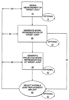
[0034] FIG. 1A is a flow chart showing steps taken by a practitioner
in assessing a joint. First, a practitioner obtains a measurement of a target
joint 10. The step of obtaining a measurement can be accomplished by
taking an image of the joint. This step can be repeated, as necessary, 11
to obtain a plurality of images in order to further refine the joint
assessment process. Once the practitioner has obtained the necessary
measurements, the information is used to generate a model representation
of the target joint being assessed 30. This model representation can be in
the form of a topographical map or image. The model representation of the
joint can be in one, two, or three dimensions. It can include a physical
model. More than one model can be created 31, if desired. Either the
original model, or a subsequently created model, or both can be used.
After the model representation of the joint is generated 30, the practitioner
can optionally generate a projected model representation of the target joint
in a corrected condition 40, e.g., from the existing cartilage on the joint
surface, by providing a mirror of the opposing joint surface, or a
combination thereof Again, this step can be repeated 41, as necessary or
desired. Using the difference between the topographical condition of the
joint and the projected image of the joint, the practitioner can then select a
joint implant 50 that is suitable to achieve the corrected joint anatomy. As
will be appreciated by those of skill in the art, the selection process 50 can
be repeated 51 as often as desired to achieve the desired result.
CA 02546965 2006-05-24
WO 2005/051240 PCT/US2004/039714
Additionally, it is contemplated that a practitioner can obtain a
measurement of a target joint 10 by obtaining, for example, an x-ray, and
then select a suitable joint replacement implant 50.
[0035] As will be appreciated by those of skill in the art, the
5 practitioner can proceed directly from the step of generating a model
representation of the target joint 30 to the step of selecting a suitable
joint
replacement implant 50 as shown by the arrow 32. Additionally, following
selection of suitable joint replacement implant 50, the steps of obtaining
measurement of target joint 10, generating model representation of target
10 joint 30 and generating projected model 40, can be repeated in series or
parallel as shown by the flow 24, 25, 26.
[0036] FIG. 1B is an alternate flow chart showing steps taken by a
practitioner in assessing a joint. First, a practitioner obtains a
measurement of a target joint 10. The step of obtaining a measurement
15 can be accomplished by taking an image of the joint. This step can be
repeated, as necessary, 11 to obtain a plurality of images in order to
further refine the joint assessment process. Once the practitioner has
obtained the necessary measurements, the information is used to
generate a model representation of the target joint being assessed 30.
This model representation can be in the form of a topographical map or
image. The model representation of the joint can be in one, two, or three
dimensions. The process can be repeated 31 as necessary or desired. It
can include a physical model. After the model representation of the joint is
assessed 30, the practitioner can optionally generate a projected model
representation of the target joint in a corrected condition 40. This step can
be repeated 41 as necessary or desired. Using the difference between the
topographical condition of the joint and the projected image of the joint, the
practitioner can then design a joint implant 52 that is suitable to achieve
the corrected joint anatomy, repeating the design process 53 as often as
CA 02546965 2006-05-24
WO 2005/051240 PCT/US2004/039714
16
necessary to achieve the desired implant design. The practitioner can also
assess whether providing additional features, such as rails, keels, lips,
pegs, cruciate stems, or anchors, cross-bars, etc. will enhance the
implants' performance in the target joint.
[0037] As will be appreciated by those of skill in the art, the
practitioner can proceed directly from the step of generating a model
representation of the target joint 30 to the step of designing a suitable
joint
replacement implant 52 as shown by the arrow 38. Similar to the flow
shown above, following the design of a suitable joint replacement implant
52, the steps of obtaining measurement of target joint 10, generating
model representation of target joint 30 and generating projected model 40,
can be repeated in series or parallel as shown by the flow 42, 43, 44.
[0038] FIG. 1c is a flow chart illustrating the process of selecting an
implant for a patient. First, using the techniques described above or those
suitable and known in the art at the time the invention is practiced, the size
of area of diseased cartilage or cartilage loss is measured 100. This step
can be repeated multiple times 101, as desired. Once the size of the
cartilage defect is measured, the thickness of adjacent cartilage can
optionally be measured 110. This process can also be repeated as desired
111. Either after measuring the cartilage loss or measuring the thickness
of adjacent cartilage, the curvature of the articular surface is then
measured 120. Alternatively, the subchondral bone can be measured. As
will be appreciated measurements can be taken of the surface of the joint
being repaired, or of the mating surface in order to facilitate development
of the best design for the implant surface.
[0039] Once the surfaces have been measured, the user either
selects the best fitting implant contained in a library of implants 130 or
generates a patient-specific implant 132. These steps can be repeated as
CA 02546965 2006-05-24
WO 2005/051240 PCT/US2004/039714
17
desired or necessary to achieve the best fitting implant for a patient, 131,
133. As will be appreciated by those of skill in the art, the process of
selecting or designing an implant can be tested against the information
contained in the MRI or x-ray of the patient to ensure that the surfaces of
the device achieves a good fit relative to the patient's joint surface.
Testing
can be accomplished by, for example, superimposing the implant image
over the image for the patient's joint. Once it has been determined that a
suitable implant has been selected or designed, the implant site can be
prepared 140, for example by removing cartilage or bone from the joint
surface, or the implant can be placed into the joint 150.
[0040] The joint implant selected or designed achieves anatomic or
near anatomic fit with the existing surface of the joint while presenting a
mating surface for the opposing joint surface that replicates the natural
joint anatomy. In this instance, both the existing surface of the joint can be
assessed as well as the desired resulting surface of the joint. This
technique is particularly useful for implants that are not anchored into the
bone.
[0041] As will be appreciated by those of skill in the art, the
physician, or other person practicing the invention, can obtain a
measurement of a target joint 10 and then either design 52 or select 50 a
suitable joint replacement implant.
[0042] II. REPAIR MATERIALS
[0043] A wide variety of materials find use in the practice of the
present invention, including, but not limited to, plastics, metals, crystal
free
metals, ceramics, biological materials (e.g., collagen or other extracellular
matrix materials), hydroxyapatite, cells (e.g., stem cells, chondrocyte cells
or the like), or combinations thereof. Based on the information (e.g.,
measurements) obtained regarding the defect and the articular surface
CA 02546965 2006-05-24
WO 2005/051240 PCT/US2004/039714
18
and/or the subchondral bone, a repair material can be formed or selected.
Further, using one or more of these techniques described herein, a
cartilage replacement or regenerating material having a curvature that will
fit into a particular cartilage defect, will follow the contour and shape of
the
articular surface, and will match the thickness of the surrounding cartilage.
The repair material can include any combination of materials, and typically
includes at least one non-pliable material, for example materials that are
not easily bent or changed.
[0044] A. METAL AND POLYMERIC REPAIR MATERIALS
[0045] Currently, joint repair systems often employ metal and/or
polymeric materials including, for example, prostheses which are anchored
into the underlying bone (e.g., a femur in the case of a knee prosthesis).
See, e.g., U.S. Patent No. 6,203,576 to Afriat, et al. issued March 20, 2001
and 6,322,588 to Ogle, et al. issued November 27, 2001, and references
cited therein. A wide-variety of metals are useful in the practice of the
present invention, and can be selected based on any criteria. For example,
material selection can be based on resiliency to impart a desired degree of
rigidity. Non-limiting examples of suitable metals include silver, gold,
platinum, palladium, iridium, copper, tin, lead, antimony, bismuth, zinc,
titanium, cobalt, stainless steel, nickel, iron alloys, cobalt alloys, such as
Elgiloy , a cobalt-chromium-nickel alloy, and MP35N, a nickel-cobalt-
chromium-molybdenum alloy, and NitinolTM, a nickel-titanium alloy,
aluminum, manganese, iron, tantalum, crystal free metals, such as
Liquidmetal alloys (available from LiquidMetal Technologies,
www.liquid metal. com), other metals that can slowly form polyvalent metal
ions, for example to inhibit calcification of implanted substrates in contact
with a patient's bodily fluids or tissues, and combinations thereof.
CA 02546965 2006-05-24
WO 2005/051240 PCT/US2004/039714
19
[0046] Suitable synthetic polymers include, without limitation,
polyamides (e.g., nylon), polyesters, polystyrenes, polyacrylates, vinyl
polymers (e.g., polyethylene, polytetrafluoroethylene, polypropylene and
polyvinyl chloride), polycarbonates, polyurethanes, poly dimethyl
siloxanes, cellulose acetates, polymethyl methacrylates, polyether ether
ketones, ethylene vinyl acetates, polysulfones, nitrocelluloses, similar
copolymers and mixtures thereof. Bioresorbable synthetic polymers can
also be used such as dextran, hydroxyethyl starch, derivatives of gelatin,
polyvinylpyrrolidone, polyvinyl alcohol, poly[N-(2-hydroxypropyl)
methacrylamide], poly(hydroxy acids), poly(epsilon-caprolactone),
polylactic acid, polyglycolic acid, poly(dimethyl glycolic acid), poly(hydroxy
butyrate), and similar copolymers can also be used.
[0047] Other materials would also be appropriate, for example, the
polyketone known as polyetheretherketone (PEEKTM). This includes the
material PEEK 450G, which is an unfilled PEEK approved for medical
implantation available from Victrex of Lancashire, Great Britain. (Victrex is
located at www.matweb.com or see Boedeker www.boedeker.com). Other
sources of this material include Gharda located in Panoli, India
(www.ghardapolymers.com).
[0048] It should be noted that the material selected can also be
filled. For example, other grades of PEEK are also available and
contemplated, such as 30% glass-filled or 30% carbon filled, provided
such materials are cleared for use in implantable devices by the FDA, or
other regulatory body. Glass filled PEEK reduces the expansion rate and
increases the flexural modulus of PEEK relative to that portion which is
unfilled. The resulting product is known to be ideal for improved strength,
stiffness, or stability. Carbon filled PEEK is known to enhance the
compressive strength and stiffness of PEEK and lower its expansion rate.
Carbon filled PEEK offers wear resistance and load carrying capability.
CA 02546965 2011-05-20
WO 20051051240 PCT/US2004/039714
[0049] As will be appreciated, other suitable similarly biocompatible
thermoplastic or thermoplastic polycondensate materials that resist
fatigue, have good memory, are flexible, and/or deflectable have very low
moisture absorption, and good wear and/or abrasion resistance, can be
5 used without departing from the scope of the invention. The implant can
also be comprised of polyetherketoneketone (PEKK).
[0050] Other materials that can be used include polyetherketone
(PEK), polyetherketoneetherketoneketone (PEKEKK), and
polyetheretherketoneketone (PEEKK), and generally a
10 polyaryletheretherketone. Further other polyketones can be used as well
as other thermoplastics.
[0051] Reference to appropriate polymers that can be used for the
implant can be made to the following documents:
PCT
15 Publication WO 02/02158 Al, dated Jan. 10, 2002 and entitled Blo-
Compatible Polymeric Materials; PCT Publication WO 02100275 Al, dated
Jan. 3, 2002 and entitled Bio-Compatible Polymeric Materials; and PCT
Publication WO 02/00270 Al, dated Jan. 3, 2002 and entitled Bio-
Compatible Polymeric Materials.
20 [0052] The polymers can be prepared by any of a variety of
approaches including conventional polymer processing methods.
{ Preferred approaches include, for example, injection molding, which is
suitable for the production of polymer components with significant
structural features, and rapid prototyping approaches, such as reaction
injection molding and stereo-lithography. The substrate can be textured or
made porous by either physical abrasion or chemical alteration to facilitate
incorporation of the metal coating. Other processes are also appropriate,
such as extrusion, injection, compression molding and/or machining
CA 02546965 2006-05-24
WO 2005/051240 PCT/US2004/039714
21
techniques. Typically, the polymer is chosen for its physical and
mechanical properties and is suitable for carrying and spreading the
physical load between the joint surfaces.
[0053] More than one metal and/or polymer can be used in
combination with each other. For example, one or more metal-containing
substrates can be coated with polymers in one or more regions or,
alternatively, one or more polymer-containing substrate can be coated in
one or more regions with one or more metals.
[0054] The system or prosthesis can be porous or porous coated.
The porous surface components can be made of various materials
including metals, ceramics, and polymers. These surface components can,
in turn, be secured by various means to a multitude of structural cores
formed of various metals. Suitable porous coatings include, but are not
limited to, metal, ceramic, polymeric (e.g., biologically neutral elastomers
such as silicone rubber, polyethylene terephthalate and/or combinations
thereof) or combinations thereof. See, e.g., U.S. Pat. No. 3,605,123 to
Hahn, issued September 20, 1971. U.S. Pat. No. 3,808,606 to Tronzo
issued May 7, 1974 and U.S. Pat. No. 3,843,975 to Tronzo issued October
29, 1974; U.S. Pat. No. 3,314,420 to Smith issued April 18, 1967; U.S.
Pat. No. 3,987,499 to Scharbach issued October 26, 1976; and German
Offenlegungsschrift 2,306,552. There can be more than one coating layer
and the layers can have the same or different porosities. See, e.g., U.S.
Pat. No. 3,938,198 to Kahn, et al., issued February 17, 1976.
[0055] The coating can be applied by surrounding a core with
powdered polymer and heating until cured to form a coating with an
internal network of interconnected pores. The tortuosity of the pores (e.g.,
a measure of length to diameter of the paths through the pores) can be
important in evaluating the probable success of such a coating in use on a
Pane 91 rf n7
CA 02546965 2006-05-24
WO 2005/051240 PCT/US2004/039714
22
prosthetic device. See, also, U.S. Pat. No. 4,213,816 to Morris issued July
22, 1980. The porous coating can be applied in the form of a powder and
the article as a whole subjected to an elevated temperature that bonds the
powder to the substrate. Selection of suitable polymers and/or powder
coatings can be determined in view of the teachings and references cited
herein, for example based on the melt index of each.
[0056] B. BIOLOGICAL REPAIR MATERIAL
[0057] Repair materials can also include one or more biological
material either alone or in combination with non-biological materials. For
example, any base material can be designed or shaped and suitable
cartilage replacement or regenerating material(s) such as fetal cartilage
cells can be applied to be the base. The cells can be then be grown in
conjunction with the base until the thickness (and/or curvature) of the
cartilage surrounding the cartilage defect has been reached. Conditions
for growing cells (e.g., chondrocytes) on various substrates in culture, ex
vivo and in vivo are described, for example, in U.S. Patent Nos. 5,478,739
to Slivka et al. issued December 26, 1995; 5,842,477 to Naughton et al.
issued December 1, 1998; 6,283,980 to Vibe-Hansen et al., issued
September 4, 2001, and 6,365,405 to Salzmann et al. issued April 2, 2002.
Non-limiting examples of suitable substrates include plastic, tissue
scaffold, a bone replacement material (e.g., a hydroxyapatite, a
bioresorbable material), or any other material suitable for growing a
cartilage replacement or regenerating material on it.
[0058] Biological polymers can be naturally occurring or produced in
vitro by fermentation and the like. Suitable biological polymers include,
without limitation, collagen, elastin, silk, keratin, gelatin, polyamino
acids,
cat gut sutures, polysaccharides (e.g., cellulose and starch) and mixtures
thereof. Biological polymers can be bioresorbable.
CA 02546965 2006-05-24
WO 2005/051240 PCT/US2004/039714
23
[0059] Biological materials used in the methods described herein
can be autografts (from the same subject); allografts (from another
individual of the same species) and/or xenografts (from another species).
See, also, International Patent Publications WO 02/22014 to Alexander et
al. published March 21, 2002 and WO 97/27885 to Lee published August
7, 1997. In certain embodiments autologous materials are preferred, as
they can carry a reduced risk of immunological complications to the host,
including re-absorption of the materials, inflammation and/or scarring of
the tissues surrounding the implant site.
[0060] In one embodiment of the invention, a probe is used to
harvest tissue from a donor site and to prepare a recipient site. The donor
site can be located in a xenograft, an allograft or an autograft. The probe is
used to achieve a good anatomic match between the donor tissue sample
and the recipient site. The probe is specifically designed to achieve a
seamless or near seamless match between the donor tissue sample and
the recipient site. The probe can, for example, be cylindrical. The distal
end of the probe is typically sharp in order to facilitate tissue penetration.
Additionally, the distal end of the probe is typically hollow in order to
accept the tissue. The probe can have an edge at a defined distance from
its distal end, e.g. at 1 cm distance from the distal end and the edge can
be used to achieve a defined depth of tissue penetration for harvesting.
The edge can be external or can be inside the hollow portion of the probe.
For example, an orthopedic surgeon can take the probe and advance it
with physical pressure into the cartilage, the subchondral bone and the
underlying marrow in the case of a joint such as a knee joint. The surgeon
can advance the probe until the external or internal edge reaches the
cartilage surface. At that point, the edge will prevent further tissue
penetration thereby achieving a constant and reproducible tissue
penetration. The distal end of the probe can include one or more blades,
CA 02546965 2006-05-24
WO 2005/051240 PCT/US2004/039714
24
saw-like structures, or tissue cutting mechanism. For example, the distal
end of the probe can include an iris-like mechanism consisting of several
small blades. The blade or blades can be moved using a manual,
motorized or electrical mechanism thereby cutting through the tissue and
separating the tissue sample from the underlying tissue. Typically, this will
be repeated in the donor and the recipient. In the case of an iris-shaped
blade mechanism, the individual blades can be moved so as to close the
iris thereby separating the tissue sample from the donor site.
[0061] In another embodiment of the invention, a laser device or a
radiofrequency device can be integrated inside the distal end of the probe.
The laser device or the radiofrequency device can be used to cut through
the tissue and to separate the tissue sample from the underlying tissue.
[0062] In one embodiment of the invention, the same probe can be
used in the donor and in the recipient. In another embodiment, similarly
shaped probes of slightly different physical dimensions can be used. For
example, the probe used in the recipient can be slightly smaller than that
used in the donor thereby achieving a tight fit between the tissue sample
or tissue transplant and the recipient site. The probe used in the recipient
can also be slightly shorter than that used in the donor thereby correcting
for any tissue lost during the separation or cutting of the tissue sample
from the underlying tissue in the donor material.
[0063] Any biological repair material can be sterilized to inactivate
biological contaminants such as bacteria, viruses, yeasts, molds,
mycoplasmas and parasites. Sterilization can be performed using any
suitable technique, for example radiation, such as gamma radiation.
[0064] Any of the biological materials described herein can be
harvested with use of a robotic device. The robotic device can use
information from an electronic image for tissue harvesting.
CA 02546965 2006-05-24
WO 2005/051240 PCT/US2004/039714
[0065] In certain embodiments, the cartilage replacement material
has a particular biochemical composition. For instance, the biochemical
composition of the cartilage surrounding a defect can be assessed by
taking tissue samples and chemical analysis or by imaging techniques.
5 For example, WO 02/22014 to Alexander describes the use of gadolinium
for imaging of articular cartilage to monitor glycosaminoglycan content
within the cartilage. The cartilage replacement or regenerating material
can then be made or cultured in a manner, to achieve a biochemical
composition similar to that of the cartilage surrounding the implantation
10 site. The culture conditions used to achieve the desired biochemical
compositions can include, for example, varying concentrations.
Biochemical composition of the cartilage replacement or regenerating
material can, for example, be influenced by controlling concentrations and
exposure times of certain nutrients and growth factors.
15 [0066] III. DEVICE DESIGN
[0067] A. CARTILAGE MODELS
[0068] Using information on thickness and curvature of the
cartilage, a physical model of the surfaces of the articular cartilage and of
the underlying bone can be created. This physical model can be
20 representative of a limited area within the joint or it can encompass the
entire joint. This model can also take into consideration the presence or
absence of a meniscus as well as the presence or absence of some or all
of the cartilage. For example, in the knee joint, the physical model can
encompass only the medial or lateral femoral condyle, both femoral
25 condyles and the notch region, the medial tibial plateau, the lateral
tibial
plateau, the entire tibial plateau, the medial patella, the lateral patella,
the
entire patella or the entire joint. The location of a diseased area of
CA 02546965 2006-05-24
WO 2005/051240 PCT/US2004/039714
26
cartilage can be determined, for example using a 3D coordinate system or
a 3D Euclidian distance as described in WO 02/22014.
[0069] In this way, the size of the defect to be repaired can be
determined. This process takes into account that, for example, roughly
80% of patients have a healthy lateral component. As will be apparent,
some, but not all, defects will include less than the entire cartilage. Thus,
in one embodiment of the invention, the thickness of the normal or only
mildly diseased cartilage surrounding one or more cartilage defects is
measured. This thickness measurement can be obtained at a single point
or, preferably, at multiple points, for example 2 point, 4-6 points, 7-10
points, more than 10 points or over the length of the entire remaining
cartilage. Furthermore, once the size of the defect is determined, an
appropriate therapy (e.g., articular repair system) can be selected such
that as much as possible of the healthy, surrounding tissue is preserved.
[0070] In other embodiments, the curvature of the articular surface
can be measured to design and/or shape the repair material. Further, both
the thickness of the remaining cartilage and the curvature of the articular
surface can be measured to design and/or shape the repair material.
Alternatively, the curvature of the subchondral bone can be measured and
the resultant measurement(s) can be used to either select or shape a
cartilage replacement material. For example, the contour of the
subchondral bone can be used to re-create a virtual cartilage surface: the
margins of an area of diseased cartilage can be identified. The
subchondral bone shape in the diseased areas can be measured. A virtual
contour can then be created by copying the subchondral bone surface into
the cartilage surface, whereby the copy of the subchondral bone surface
connects the margins of the area of diseased cartilage. In shaping the
device, the contours can be configured to mate with existing cartilage or to
account for the removal of some or all of the cartilage.
CA 02546965 2006-05-24
WO 2005/051240 PCT/US2004/039714
27
[0071] FIG. 2A shows a slightly perspective top view of a joint
implant 200 of the invention suitable for implantation at the tibial plateau
of
the knee joint. As shown in FIG. 2A, the implant can be generated using,
for example, a dual surface assessment, as described above with respect
to FIGS. 1A and B.
[0072] The implant 200 has an upper surface 202, a lower
surface 204 and a peripheral edge 206. The upper surface 202 is formed
so that it forms a mating surface for receiving the opposing joint surface; in
this instance partially concave to receive the femur. The concave surface
can be variably concave such that it presents a surface to the opposing
joint surface, e.g. a negative surface of the mating surface of the femur it
communicates with. As will be appreciated by those of skill in the art, the
negative impression, need not be a perfect one.
[0073] The upper surface 202 of the implant 200 can be shaped by
any of a variety of means. For example, the upper surface 202 can be
shaped by projecting the surface from the existing cartilage and/or bone
surfaces on the tibial plateau, or it can be shaped to mirror the femoral
condyle in order to optimize the complimentary surface of the implant
when it engages the femoral condyle. Alternatively, the superior surface
202 can be configured to mate with an inferior surface of an implant
configured for the opposing femoral condyle.
[0074] The lower surface 204 has a convex surface that matches, or
nearly matches, the tibial plateau of the joint such that it creates an
anatomic or near anatomic fit with the tibial plateau. Depending on the
shape of the tibial plateau, the lower surface can be partially convex as
well. Thus, the lower surface 204 presents a surface to the tibial plateau
that fits within the existing surface. It can be formed to match the existing
surface or to match the surface after articular resurfacing.
CA 02546965 2006-05-24
WO 2005/051240 PCT/US2004/039714
28
[0075] As will be appreciated by those of skill in the art, the convex
surface of the lower surface 204 need not be perfectly convex. Rather, the
lower surface 204 is more likely consist of convex and concave portions
that fit within the existing surface of the tibial plateau or the re-surfaced
plateau. Thus, the surface is essentially variably convex and concave.
[0076] FIG. 2B shows a top view of the joint implant of FIG. 2A. As
shown in FIG. 2B the exterior shape 208 of the implant can be elongated.
The elongated form can take a variety of shapes including elliptical, quasi-
elliptical, race-track, etc. However, as will be appreciated the exterior
dimension is typically irregular thus not forming a true geometric shape,
e.g. ellipse. As will be appreciated by those of skill in the art, the actual
exterior shape of an implant can vary depending on the nature of the joint
defect to be corrected. Thus the ratio of the length L to the width W can
vary from, for example, between 0.25 to 2.0, and more specifically from
0.5 to 1.5. As further shown in FIG. 2B, the length across an axis of the
implant 200 varies when taken at points along the width of the implant. For
example, as shown in FIG. 2B, L, # L2 # L3.
[0077] Turning now to FIGS. 2c-E, cross-sections of the implant
shown in FIG. 2B are depicted along the lines of C-C, D-D, and E-E. The
implant has a thickness t1, t2 and t3 respectively. As illustrated by the
cross-sections, the thickness of the implant varies along both its length L
and width W. The actual thickness at a particular location of the implant
200 is a function of the thickness of the cartilage and/or bone to be
replaced and the joint mating surface to be replicated. Further, the profile
of the implant 200 at any location along its length L or width W is a
function of the cartilage and/or bone to be replaced.
[0078] FIG. 2F is a lateral view of the implant 200 of FIG. 2A. In this
instance, the height of the implant 200 at a first end h, is different than
the
CA 02546965 2006-05-24
WO 2005/051240 PCT/US2004/039714
29
height of the implant at a second end h2. Further the upper edge 208 can
have an overall slope in a downward direction. However, as illustrated the
actual slope of the upper edge 208 varies along its length and can, in
some instances, be a positive slope. Further the lower edge 210 can have
an overall slope in a downward direction. As illustrated the actual slope of
the lower edge 210 varies along its length and can, in some instances, be
a positive slope. As will be appreciated by those of skill in the art,
depending on the anatomy of an individual patient, an implant can be
created wherein h, and h2 are equivalent, or substantially equivalent
without departing from the scope of the invention.
[0079] FIG. 2G is a cross-section taken along a sagittal plane in a
body showing the implant 200 implanted within a knee joint 1020 such that
the lower surface 204 of the implant 200 lies on the tibial plateau 1022 and
the femur 1024 rests on the upper surface 202 of the implant 200. FIG. 2H
is a cross-section taken along a coronal plane in a body showing the
implant 200 implanted within a knee joint 1020. As is apparent from this
view, the implant 200 is positioned so that it fits within a superior
articular
surface 224. As will be appreciated by those of skill in the art, the
articular
surface could be the medial or lateral facet, as needed.
[0080] FIG. 2i is a view along an axial plane of the body showing the
implant 200 implanted within a knee joint 1020 showing the view taken
from an aerial, or upper, view. FIG. 21 is a view of an alternate embodiment
where the implant is a bit larger such that it extends closer to the bone
medially, i.e. towards the edge 1023 of the tibial plateau, as well as
extending anteriorly and posteriorly.
[0081] FIG. 2K is a cross-section of an implant 200 of the invention
according to an alternate embodiment. In this embodiment, the lower
surface 204 further includes a joint anchor 212. As illustrated in this
CA 02546965 2006-05-24
WO 2005/051240 PCT/US2004/039714
embodiment, the joint anchor 212 forms a protrusion, keel or vertical
member that extends from the lower surface 204 of the implant 200 and
projects into, for example, the bone of the joint. As will be appreciated by
those of skill in the art, the keel can be perpendicular or lie within a plane
5 of the body.
[0082] Additionally, as shown in FIG. 2L the joint anchor 212 can
have a cross-member 214 so that from a bottom perspective, the joint
anchor 212 has the appearance of a cross or an "x." As will be appreciated
by those of skill in the art, the joint anchor 212 could take on a variety of
10 other forms while still accomplishing the same objective of providing
increased stability of the implant 200 in the joint. These forms include, but
are not limited to, pins, bulbs, balls, teeth, etc. Additionally, one or more
joint anchors 212 can be provided as desired. FIG. 2M and N illustrate
cross-sections of alternate embodiments of a dual component implant from
15 a side view and a front view.
[0083] In an alternate embodiment shown in FIG. 2M it may be
desirable to provide a one or more cross-members 220 on the lower
surface 204 in order to provide a bit of translation movement of the implant
relative to the surface of the femur, or femur implant. In that event, the
20 cross-member can be formed integral to the surface of the implant or can
be one or more separate pieces that fit within a groove 222 on the lower
surface 204 of the implant 200. The groove can form a single channel as
shown in FIG. 2N1, or can have more than one channel as shown in
FIG. 2N2. In either event, the cross-bar then fits within the channel as
25 shown in FIGS. 2N1-N2. The cross-bar members 220 can form a solid or
hollow tube or pipe structure as shown in FIG. 2P. Where two, or more,
tubes 220 communicate to provide translation, a groove 221 can be
provided along the surface of one or both cross-members to interlock the
tubes into a cross-bar member further stabilizing the motion of the cross-
CA 02546965 2006-05-24
WO 2005/051240 PCT/US2004/039714
31
bar relative to the implant 200. As will be appreciated by those of skill in
the art, the cross-bar member 220 can be formed integrally with the
implant without departing from the scope of the invention.
[0084] As shown in FIGS. 2Q-R, it is anticipated that the surface of
the tibial plateau will be prepared by forming channels thereon to receive
the cross-bar members. Thus facilitating the ability of the implant to seat
securely within the joint while still providing movement about an axis when
the knee joint is in motion.
[0085] FIG. 2s(1-9) illustrate an alternate embodiment of implant
200. As illustrated in FIG. 2s the edges are beveled to relax a sharp corner.
FIG. 2s(1) illustrates an implant having a single fillet or bevel 230. The
fillet
is placed on the implant anterior to the posterior portion of the tibial
spine.
As shown in FIG. 2s(2) two fillets 230, 231 are provided and used for the
posterior chamfer. In FIG. 2s(3) a third fillet 234 is provided to create two
cut surfaces for the posterior chamfer.
[0086] Turning now to FIG. 2s(4) a tangent of the implant is
deselected, leaving three posterior curves. FIG. 2s(5) shows the result of
tangent propagation. FIG. 2s(6) illustrates the effect on the design when
the bottom curve is selected without tangent propagation. The result of
tangent propagation and selection is shown in FIG. 2s(7). As can be seen
in FIG. 2s(8-9) the resulting corner has a softer edge but sacrifices less
than 0.5 mm of joint space. As will be appreciated by those of skill in the
art, additional cutting planes can be added without departing from the
scope of the invention.
[0087] FIG. 2T illustrates an alternate embodiment of an implant 200
wherein the surface of the tibial plateau 250 is altered to accommodate the
implant. As illustrated in FIG. 2T(1-2) the tibial plateau can be altered for
only half of the joint surface 251 or for the full surface 252. As illustrate
in
CA 02546965 2006-05-24
WO 2005/051240 PCT/US2004/039714
32
FIG. 2T(3-4) the posterior-anterior surface can be flat 260 or graded 262.
Grading can be either positive or negative relative to the anterior surface.
Grading can also be used with respect to the implants of FIG. 2T where the
grading either lies within a plane or a body or is angled relative to a plane
of the body. Additionally, attachment mechanisms can be provided to
anchor the implant to the altered surface. As shown in FIG. 2T(5-7) keels
264 can be provided. The keels 264 can either sit within a plane, e.g.
sagittal or coronal plane, or not sit within a plane (as shown in FIG. 2T(7)).
FIG. 2T(8) illustrates an implant which covers the entire tibial plateau. The
upper surface of these implants are designed to conform to the projected
shape of the joint as determined under the steps described with respect to
FIG. 1, while the lower surface is designed to be flat, or substantially flat
to
correspond to the modified surface of the joint.
[0088] Turning now to FIGS. 3A-I an implant suitable for providing an
opposing joint surface to the implant of FIG. 2A is shown. This implant
corrects a defect on an inferior surface of the femur 1024 (e.g., the
condyle of the femur that mates with the tibial plateau) and can be used
alone, i.e., on the femur 1024, or in combination with another joint repair
device. Formation of the surfaces of the devices can be achieved using
the techniques described above with respect to the implant of FIG. 2.
[0089] FIG. 3A shows a perspective view of an implant 300 having a
curved mating surface 302 and convex joint abutting surface 304. The joint
abutting surface 304 need not form an anatomic or near anatomic fit with
the femur in view of the anchors 306 provided to facilitate connection of
the implant to the bone. In this instance, the anchors 306 are shown as
pegs having notched heads. The notches facilitate the anchoring process
within the bone. However, pegs without notches can be used as well as
pegs with other configurations that facilitate the anchoring processor
cruciate stems. Pegs and other portions of the implant can be porous
CA 02546965 2006-05-24
WO 2005/051240 PCT/US2004/039714
33
coated. The implant can be inserted without bone cement or with use of
bone cement. The implant can be designed to abut the subchondral bone,
i.e. it can substantially follow the contour of the subchondral bone. This
has the advantage that no bone needs to be removed other than for the
placement of the peg holes thereby significantly preserving bone stock.
[0090] The anchors 306 could take on a variety of other forms
without departing from the scope of the invention while still accomplishing
the same objective of providing increased stability of the implant 300 in the
joint. These forms include, but are not limited to, pins, bulbs, balls, teeth,
etc. Additionally, one or more joint anchors 306 can be provided as
desired. As illustrated in FIG. 3, three pins are used to anchor the implant
300. However, more or fewer joint anchors, cruciate stems, or pins, can be
used without departing from the scope of the invention.
[0091] FIG. 3B shows a slightly perspective superior view of the
bone mating surface 304 further illustrating the use of three anchors 306 to
anchor the implant to the bone. Each anchor 306 has a stem310 with a
head 312 on top. As shown in FIG. 3c, the stem 310 has parallel walls
such that it forms a tube or cylinder that extends from the bone mating
surface 304. A section of the stem forms a narrowed neck 314 proximal to
the head 312. As will be appreciated by those of skill in the art, the walls
need not be parallel, but rather can be sloped to be shaped like a cone.
Additionally, the neck 314 need not be present, nor the head 312. As
discussed above, other configurations suitable for anchoring can be used
without departing from the scope of the invention.
[0092] Turning now to FIG. 3D, a view of the tibial plateau mating
surface 302 of the implant 300 is illustrated. As is apparent from this view,
the surface is curved such that it is convex or substantially convex in order
to mate with the concave surface of the plateau. FIG. 3E illustrates the
CA 02546965 2006-05-24
WO 2005/051240 PCT/US2004/039714
34
upper surface 304 of the implant 300 further illustrating the use of three
pegs 306 for anchoring the implant 300 to the bone. As illustrated, the
three pegs 306 are positioned to form a triangle. However, as will be
appreciated by those of skill in the art, one or more pegs can be used, and
the orientation of the pegs 306 to one another can be as shown, or any
other suitable orientation that enables the desired anchoring. FIG. 3F
illustrated a cross section of the implant 300 taken along the lines F-F
shown in FIG. 3E. Typically the pegs are oriented on the surface of the
implant so that the peg is perpendicular to the femoral condyle, which may
not result in the peg being perpendicular to the surface of the implant.
[0093] FIG. 3G illustrates the axial view of the femur 1000 having a
lateral condyle 1002 and a medial condyle 1004. The intercondylar fossa
is also shown 1006 along with the lateral epicondyle 1008 and medial
epicondyle 1010. Also shown is the patellar surface of the femur 1012.
The implant 300 illustrated in FIG. 3A, is illustrated covering a portion of
the
lateral condyle. The pegs 306 are also shown that facilitate anchoring the
implant 300 to the condyle.
[0094] FIG. 3H illustrates a knee joint 1020 from an anterior
perspective. The implant 300 is implanted over a condyle. As shown in
FIG. 1 the implant 300 is positioned such that it communicates with an
implant 200 designed to correct a defect in the tibial plateau, such as
those shown in FIGS. 2.
[0095] FIGS. 3J-K illustrate an implant 300 for placement on a
condyle. In this embodiment, at least one flat surface or chamfer cut 360 is
provided to mate with a cut made on the surface of the condyle in
preparing the joint. The flat surface 360 typically does not encompass the
entire proximal surface 304 of the implant 300.
CA 02546965 2006-05-24
WO 2005/051240 PCT/US2004/039714
[0096] FIG. 4A illustrates the design of a typical total knee
arthroplasty ("TKA") primary knee 499. Posterior cuts 498, anterior cuts
497 and distal cuts 496 are provided as well as chamfer cuts 495.
[0097] FIGS. 4B and 4c illustrate another implant 400. As shown in
5 FIG. 4B, the implant 400 is configured such that it covers both the lateral
and medial femoral condyle along with the patellar surface of the femur
1012. The implant 400 has a lateral condyle component 410 and a medial
condyle component 420 and a bridge 430 that connects the lateral condyle
component 410 to the medial condyle component 420 while covering at
10 least a portion of the patellar surface of the femur 1012. The implant 400
can optionally oppose one or more implants, such as those shown in
FIG. 2, if desired. FIG. 4c is a side view of the implant of FIG. 4B. As shown
in FIG. 4c, the superior surface 402 of the implant 400 is curved to
correspond to the curvature of the femoral condyles. The curvature can be
15 configured such that it corresponds to the actual curvature of one or both
of the existing femoral condyles, or to the curvature of one or both of the
femoral condyles after resurfacing of the joint. One or more pegs 430 can
be provided to assist in anchoring the implant to the bone. As will be
appreciated by those of skill in the art, the implant can be configured such
20 that the superior surface contacting a first condyle is configured to male
with the existing condule while a surface contacting a second condyle has
one or more flat surfaces to mate with a condyle surface that has been
modified.
[0098] FIG. 4D illustrates a top view of the implant 400 shown in
25 FIG. 4B. As is should be appreciated from this view, the inferior surface
404 of the implant 400 is configured to conform to the shape of the femoral
condyles, e.g. the shape healthy femoral condyles would present to the
tibial surface in a non-damaged joint.
CA 02546965 2006-05-24
WO 2005/051240 PCT/US2004/039714
36
[0099] FIGS. 4E and F illustrate perspective views of the implant from
the inferior surface (i.e., tibial plateau mating surface).
[0100] FIG. 4G illustrates the axial view of the femur 1000 having a
lateral condyle 1002 and a medial condyle 1004. The intercondylar fossa
is also shown 1006 along with the lateral epicondyle 1008. The implant
400 illustrated in FIG. 4B, is illustrated covering both condyles and the
patellar surface of the femur 1012. The pegs 430 are also shown that
facilitate anchoring the implant 400 to the condyle.
[0101] FIG. 4H illustrates a knee joint 1050 from an anterior
perspective. The implant 400 is implanted over both condyles. As shown in
FIG. 4i the implant 400 is positioned such that it communicates with an
implant 200 designed to correct a defect in the tibial plateau, such as
those shown in FIGS. 2.
[0102] As will be appreciated by those of skill in the art, the implant
400 can be manufactured from a material that has memory such that the
implant can be configured to snap-fit over the condyle. Alternatively, it can
be shaped such that it conforms to the surface without the need of a snap-
fit.
[0103] FIGS. 5A and 5B illustrate yet another implant 500 suitable for
repairing a damaged condyle. As shown in FIG. 5A, the implant 500 is
configured such that it covers only one of the lateral or medial femoral
condyles 510. The implant differs from the implant of FIG. 3 in that the
implant 500 also covers at least a portion of the patellar surface of the
femur 512.
[0104] Similar to the implant of FIG. 4, the implant can optionally
oppose one or more implants or opposing joint surfaces, such as those
shown in FIG. 2, and can be combined with other implants, such as the
implants of FIG. 3. FIG. 5c is a perspective side view of the implant of
CA 02546965 2006-05-24
WO 2005/051240 PCT/US2004/039714
37
FIG. 5A. As shown in FIG.5c, the superior surface 502 of the implant 500 is
curved to correspond to the curvature of the femoral condyle that it mates
with and the portion of the patellar surface of the femur that it covers. One
or more pegs 530 can be provided to assist in anchoring the implant to the
bone. Additionally, an angled surface 503 can be provided on an interior
surface 502 of the condyle component that conforms to an optionally
provided cut made on the surface of the joint surface with which the
implant mates.
[0105] FIG. 5D illustrates a perspective top view of the implant 500
shown in FIG. 5A. As is should be appreciated from this view, the inferior
surface 504 of the implant 500 is configured to conform to the projected
shape of the femoral condyles, e.g. the shape healthy femoral condyles
would present to the tibial surface in a non-damaged joint.
[0106] FIG. 5E is a view of the implant 500 showing a hatched three
point loading support area which extends from a top portion 513 to a line
(plane 17) and from a line (plane 18) to a bottom portion 515. Also
illustrated are the pegs 530 extending from the superior surface. FIG. 5F
illustrates the superior surface of the implant 500 with the pegs 530
extending from the superior surface. FIG. 5F also illustrates the hatched
cantilever loading support area, which extends from the line (plane 18) to
the top portion 513 of the implant. The loading forces and directions for
each support condition are based on physiological load encounters. Table
1 shows the Physiological Loadings taken from a study by Seth Greenwald
Table 1
Physiological Loadings'
Set-up 69199 "2" "3"
Flexion Angle 00 60 900
(degree)
Normal Force N 2,900 3,263 3,625
CA 02546965 2006-05-24
WO 2005/051240 PCT/US2004/039714
38
(lbs.) (652) (733.5) (815)
Normal Force Walking Stair Descent Stair Ascent
Case (4.0 x BW (4.5 x BW 5.0 x BW
Body Weight (BW) taken as a 60 year old male, with 173
cm height for an average body weight of 74 kg (163 lbs).
"'Tibial Plateau Surface Stress in TKA: A Factor Influencing Polymer
Failure Series III - Posterior Stabilized Designs;" Paul D. Postak, B.Sc.,
Christine S. Heim, B.Sc., A. Seth Greenwald, D. Phil.; Orthopaedic
Research Laboratories, The Mt. Sinai Medical Center, Cleveland, Ohio.
Presented at the 62nd Annual AAOS Meeting, 1995.
[0107] Using the implant 500 described in this application, the three
point loading will occur from set-up 1 (2900 N). To replicate a worst case
loading scenario, a 75/25 load distribution (75% of 2900 N = 2175 N) will
be used. The loading will be concentrated on a 6mm diameter circular
area located directly below and normal to the ped on the bearing surface.
[0108] Turning to the cantilever loading shown in FIG. 5F, the
loading will occur from set-up 3, or 90 , at a 75/25 load distribution (75% of
3625 N = 2719 N). As with the above example, the loading will be
concentrated on a 6 mm diameter circular area located at the center of the
posterior-most portion of the medial condyle normal to the flat cut surface
of the posterior condyle.
[0109] FIGS. 5G and H illustrate alternate embodiments of the
implant 500 having a rail design that provides one or more rails 521 along
medial and/or lateral sides of the implant 500. The rail 521 can be
positioned so that it extends along a portion of the medial 517 and/or
lateral 519 sides before communicating with the angled surface 503. As
will be appreciate, a single side rail 521can be provided without departing
from the scope of the invention.
[0110] FIG. 51 illustrates another embodiment of an implant 500
having a keel design. A keel 523 (or centrally formed rail) is provided on
the superior surface of the implant. In this embodiment, the keel 523 is
CA 02546965 2006-05-24
WO 2005/051240 PCT/US2004/039714
39
located on the surface of the implant, but not at the sides. As will be
appreciated, the keel can be centered, as shown, substantially centered,
or located off-center. An angled surface 503 can be provided to
communicate with a modified joint surface. Alternatively, where the joint
surface is worn or modified, the cut 503 could be configured to mate with
the worn or modified surface.
[0111] FIG. 51 illustrates the axial view of the femur 1000 having a
lateral condyle 1002 and a medial condyle 1004. The intercondylar fossa
is also shown 1006 along with the lateral epicondyle 1008 and the medial
epicondyle 1010. The patellar surface of the femur 1012 is also illustrated.
The implant 500, illustrated in FIG. 5A, is shown covering the lateral
condyle and a portion of the patellar surface of the femur 1012. The pegs
530 are also shown that facilitate anchoring the implant 500 to the condyle
and patellar surface.
[0112] FIG. 5K illustrates a knee joint 1020 from an anterior
perspective. The implant 500 is implanted over the lateral condyle. FIG. 5L
illustrates a knee joint 1020 with the implant 500 covering the medial
condyle 1004. As illustrated in FIGS. 5K and L the shape of the implant 500
corresponding to the patella surface can take on a variety of curvatures
without departing from the scope of the invention.
[0113] Turning now to FIG. 5M and N the implant 500 is positioned
such that it communicates with an implant 200 designed to correct a defect
in the tibial plateau, such as those shown in FIGS. 2.
[0114] In another embodiment of the invention, the implant 500 can
have a superior surface 502 which substantially conforms to the surface of
the condyle but which has at one flat portion corresponding to an oblique
cut on the bone as shown in FIG. 50.
CA 02546965 2006-05-24
WO 2005/051240 PCT/US2004/039714
[0115] Turning now to FIG. 5P-Q an implant 500 is shown from a
side view with a 7 difference between the anterior and posterior cuts.
[0116] FIG. 5R-s illustrate an implant 500 having a contoured
surface 560 for mating with the joint surface and an anterior cut 561 and a
5 posterior cut 562. FIG. 5s shows the same implant 500 from a slightly
different angle. FIG. 5T illustrates another implant 500 having a contoured
surface 560 for mating with the joint surface and posterior cut 562, a distal
cut 563, and a chamfer cut 564. In this embodiment no anterior cut is
provided. FIG. 5u illustrates the implant 500 of FIG. 5T from a side
10 perspective. The cuts are typically less than the cut required for a TKA,
i.e., typically less than 10mm. The design of the cuts for this implant allow
for a revision surgery to the knee, if required, at a later date.
[0117] FIGS. 6A-G illustrate the implant 500 of FIG. 5 with a graphical
representation of the cross-sections 610, 620 from which a surface shape
15 of the implant is derived. FIG. 6A illustrates a top view of the implant
500
sitting on top of the extracted surface shape 600. This view of the implant
500 illustrates a notch 514 associated with the bridge section of the
implant 512 which covers the patellar surface of the femur (or the trochlea
region) to provide a mating surface that approximates the cartilage
20 surface. As will be appreciated by those of skill in the art, the shape of
an
implant designed for the medial condyle would not necessarily be a mirror
image of the implant designed for the lateral condyle because of
differences in anatomy. Thus, for example, the notch 514 would not be
present in an implant designed for the medial condyle and the patellar
25 surface of the femur. Therefore, the implant can be designed to include all
or part of the troclea region or to exclude it entirely.
[0118] FIG. 6B illustrates a bottom view of the implant 500 layered
over another derived surface shape 601. FIG. 6c is a bottom view showing
CA 02546965 2006-05-24
WO 2005/051240 PCT/US2004/039714
41
the implant 500 extending through the extracted surface shape 600 shown
in FIG. 6A. FIG. 6D is a close-up view of the FIG. 6c showing the condylar
wing of the implant covering the extracted surface 600. FIG. 6E illustrates
a top posterior view of the implant 500 positioned over the graphical
representation of the surface shape 600. FIG. 6F is an anterior view and
FIG. 6G is a bottom-posterior view.
[0119] FIG. 7A-c illustrate an implant 700 for correcting a joint similar
to the implant 500 above. However, implant 700 consists of two
components. The first component 710 engages a condyle of the femur,
either medial or lateral depending on the design. The second component
720 engages the patellar surface of the femur. As discussed with the
previous embodiments, the surfaces of the implant 700 can be configured
such that the distal surface 722 (e.g., the surface that faces the tibial
plateau) is shaped based on a projection of the natural shape of the femur
compensating the design for valgus or varus deformities and/or flattening
of the surface of the femur. Alternatively, the distal surface can be shaped
based on the shape of the tibial plateau to provide a surface designed to
optimally mate with the tibial plateau. The proximal surface 724 (e.g., the
surface that engages the femoral condyle) can be configured such that it
mirrors the surface of the femur in either its damaged condition or its
modified condition. Likewise, the proximal surface can have one or more
flattened sections 726 that form, e.g., chamfer cuts. Additionally the
surface can include mechanisms facilitating attachment 728 to the femur,
such as keels, teeth, cruciate stems, and the like. The medial facing
portion of the condyle implant has a tapered surface 730 while the lateral
facing portion of the patellar component also has a tapered surface such
that each component presents tapered surfaces 730 to the other
component.
CA 02546965 2006-05-24
WO 2005/051240 PCT/US2004/039714
42
[0120] By dividing the surfaces of the medial and lateral
compartments into independent articulating surfaces, as shown in FIG. 7,
the implant provides improved fit of the conformal surfaces to the
subchondral bone. Additionally, the lateral-anterior portion of the femur is
shielded from stress which could cause bone loss. Also, the smaller size of
each component of the implant, enables the implant to be placed within
the joint using a smaller incision. Finally, the wear of the patellar
component is improved.
[0121] FIGS. 8A-F illustrate a patella 00 with an implants 810. The
implant 810 can have one or more pegs, cruciate stems, or other
anchoring mechanisms, if desired. As will be appreciated by those of skill
in the art, other designs can be arrived at using the teachings of this
disclosure without departing from the scope of the invention. FIG. 8A
illustrates a perspective view of an intact patella 800. FIG. 8B illustrates
the
patella 800 wherein one surface of the patella 800 has been cut for form a
smooth surface 802 to mate with an implant. FIG. 8c illustrates the patella
800 with an implant 810 positioned on the smooth surface 802. The
implant 810 has a plate structure 812 that abuts the smooth surface of the
patella 802 and a dome 814 positioned on the plate 812 so that the dome
is positioned in situ such that it will match the location of the patellar
ridge.
The implant 810 can be configured such that the edge of the plate is offset
1 mm from the actual edge of the patella, as illustrated. As wll be
appreciated by those of skill in the art, the plate 812 and dome 814 can be
formed as a single unit or formed from multiple components. FIG. 8D is a
side view of the implant 810 positioned on the patella 800. As shown, the
dome is positioned on the implant such that it is off-center. Optimal
positioning of the dome will be determined by the position of the patellar
ridge.
CA 02546965 2006-05-24
WO 2005/051240 PCT/US2004/039714
43
[0122] Turning now to FIGS. 8E-F, the implant 810 is shown
superimposed on the unaltered patella 800 in order to illustrate that the
position of the dome 814 of the implant corresponds to the location of the
patellar ridge.
[0123] FIGS. 8G-J illustrate an alternative design for the patellar
implant. FIG. 8G illustrates the implant 850 in its beginning stages as a
blank with a flat inferior surface 852 having pegs 854 extending therefrom
for anchoring to the patella. The articular or superior surface 860 has a
rounded dome 856, and a round plate section 858 that can be machined to
match the bone cut. The articular surface 860 takes on the appearance of
a "hat" or somberero, having a dome with a rim. The center of the dome
856 is also the center of the bearing surface. The rim 858 is cut to conform
to the needs of the particular patient. FIG. 81 illustrates an implant which
has been formed from the blank shown in FIGS. 8c-1. FIG. 81 shows a
plurality of possible cut lines 862, 862' for purposes of illustration.
[0124] FIGS. 9A-c illustrate a lateral view of a knee 1020 having a
combination of the implants of implanted thereof. In FIG. 9A, an implant
covering the condyle 900, is illustrated. Suitable implants can be, for
example, those shown in FIGS. 3-8, as will be appreciated the portion of
the condyle covered anterior to posterior can include the entire weight
bearing surface, a portion thereof, or a surface greater than the weight
bearing surface. Thus, for example, the implant can be configured to
terminate prior to the sulcus terminalis or after the sulcus terminalis (e.g.,
the groove on the femur that coincides with the area where load bearing
on the joint surface stops). As shown in FIGS. 9A-B, a patellar implant 900
can also be provided. FIG. 9c illustrates a knee having a condyle implant
900, a patellar implant 800 and an implant for the tibial plateau 200.
CA 02546965 2006-05-24
WO 2005/051240 PCT/US2004/039714
44
[0125] FIGS. 1 OA-D provide an alternate view of the coronal plane of
a knee joint with one or more implants described above implanted therein.
FIG. 10A illustrates a knee having a tibial implant 200 placed therein.
FIG. 10s illustrates a knee with a condyle implant 300 placed therein. As
described above, a plurality of the implants taught herein can be provided
within a joint in order to restore joint movement. FIG. 10c illustrates a knee
joint having two implants therein. First, a tibial implant 200 is provided on
the tibial plateau and a second implant 300 is provided on the facing
condyle. As will be appreciated by those of skill in the art. The implants
can be installed such that the implants present each other mating surfaces
(as illustrated), or not. For example, where the tibial implant 200 is placed
in the medial compartment of the knee and the condyle implant 300 is
placed in the lateral compartment. Other combinations will be appreciated
by those of skill in the art. Turning now to FIG. 10D, a tibial implant 200 is
provided along with a bicompartmental condyle implant 500. As discussed
above, these implants can be associated with the same compartment of
the knee joint, but need not be.
[0126] The arthroplasty system can be designed to reflect aspects
of the tibial shape, femoral shape and/or patellar shape. Tibial shape and
femoral shape can include cartilage, bone or both. Moreover, the shape of
the implant can also include portions or all components of other articular
structures such as the menisci. The menisci are compressible, in particular
during gait or loading. For this reason, the implant can be designed to
incorporate aspects of the meniscal shape accounting for compression of
the menisci during loading or physical activities. For example, the
undersurface 204 of the implant 200 can be designed to match the shape
of the tibial plateau including cartilage or bone or both. The superior
surface 202 of the implant 200 can be a composite of the articular surface
of the tibia (in particular in areas that are not covered by menisci) and the
CA 02546965 2006-05-24
WO 2005/051240 PCT/US2004/039714
meniscus. Thus, the outer aspects of the device can be a reflection of
meniscal height. Accounting for compression, this can be, for example,
20%, 40%, 60% or 80% of uncompressed meniscal height.
[0127] Similarly the superior surface 304 of the implant 300 can be
5 designed to match the shape of the femoral condyle including cartilage or
bone or both. The inferior surface 302 of the implant 300 can be a
composite of the surface of the tibial plateau (in particular in areas that
are
not covered by menisci) and the meniscus. Thus, at least a portion of the
outer aspects of the device can be a reflection of meniscal height.
10 Accounting for compression, this can be, for example, 20%, 40%, 60% or
80% of uncompressed meniscal height. These same properties can be
applied to the implants shown in FIGS. 4-8, as well.
[0128] In some embodiments, the outer aspect of the device
reflecting the meniscal shape can be made of another, preferably
15 compressible material. If a compressible material is selected it is
preferably designed to substantially match the compressibility and
biomechanical behavior of the meniscus. The entire device can be made
of such a material or non-metallic materials in general.
[0129] The height and shape of the menisci for any joint surface to
20 be repaired can be measured directly on an imaging test. If portions, or
all,
of the meniscus are torn, the meniscal height and shape can be derived
from measurements of a contralateral joint or using measurements of
other articular structures that can provide an estimate on meniscal
dimensions.
25 [0130] In another embodiment, the superior face of the implants
300, 400 or 500 can be shaped according to the femur. The shape can
preferably be derived from the movement patterns of the femur relative to
the tibial plateau thereby accounting for variations in femoral shape and
CA 02546965 2006-05-24
WO 2005/051240 PCT/US2004/039714
46
tibiofemoral contact area as the femoral condyle flexes, extends, rotates,
translates and glides on the tibia and menisci.
[0131] The movement patterns can be measured using any current
or future test know in the art such as fluoroscopy, MRI, gait analysis and
combinations thereof.
[0132] The arthroplasty can have two or more components, one
essentially mating with the tibial surface and the other substantially
articulating with the femoral component. The two components can have a
flat opposing surface. Alternatively, the opposing surface can be curved.
The curvature can be a reflection of the tibial shape, the femoral shape
including during joint motion, and the meniscal shape and combinations
thereof.
[0133] Examples of single-component systems include, but are not
limited to, a plastic, a polymer, a metal, a metal alloy, crystal free metals,
a
biologic material or combinations thereof. In certain embodiments, the
surface of the repair system facing the underlying bone can be smooth. In
other embodiments, the surface of the repair system facing the underlying
bone can be porous or porous-coated. In another aspect, the surface of
the repair system facing the underlying bone is designed with one or more
grooves, for example to facilitate the in-growth of the surrounding tissue.
The external surface of the device can have a step-like design, which can
be advantageous for altering biomechanical stresses. Optionally, flanges
can also be added at one or more positions on the device (e.g., to prevent
the repair system from rotating, to control toggle and/or prevent settling
into the marrow cavity). The flanges can be part of a conical or a
cylindrical design. A portion or all of the repair system facing the
underlying bone can also be flat which can help to control depth of the
implant and to prevent toggle.
CA 02546965 2006-05-24
WO 2005/051240 PCT/US2004/039714
47
[0134] Non-limiting examples of multiple-component systems
include combinations of metal, plastic, metal alloys, crystal free metals,
and one or more biological materials. One or more components of the
articular surface repair system can be composed of a biologic material
(e.g. a tissue scaffold with cells such as cartilage cells or stem cells alone
or seeded within a substrate such as a bioresorable material or a tissue
scaffold, allograft, autograft or combinations thereof) and/or a non-
biological material (e.g., polyethylene or a chromium alloy such as
chromium cobalt).
[0135] Thus, the repair system can include one or more areas of a
single material or a combination of materials, for example, the articular
surface repair system can have a first and a second component. The first
component is typically designed to have size, thickness and curvature
similar to that of the cartilage tissue lost while the second component is
typically designed to have a curvature similar to the subchondral bone. In
addition, the first component can have biomechanical properties similar to
articular cartilage, including but not limited to similar elasticity and
resistance to axial loading or shear forces. The first and the second
component can consist of two different metals or metal alloys. One or
more components of the system (e.g., the second portion) can be
composed of a biologic material including, but not limited to bone, or a
non-biologic material including, but not limited to hydroxyapatite, tantalum,
a chromium alloy, chromium cobalt or other metal alloys.
[0136] One or more regions of the articular surface repair system
(e.g., the outer margin of the first portion and/or the second portion) can be
bioresorbable, for example to allow the interface between the articular
surface repair system and the patient's normal cartilage, over time, to be
filled in with hyaline or fibrocartilage. Similarly, one or more regions
(e.g.,
the outer margin of the first portion of the articular surface repair system
CA 02546965 2006-05-24
WO 2005/051240 PCT/US2004/039714
48
and/or the second portion) can be porous. The degree of porosity can
change throughout the porous region, linearly or non-linearly, for where
the degree of porosity will typically decrease towards the center of the
articular surface repair system. The pores can be designed for in-growth of
cartilage cells, cartilage matrix, and connective tissue thereby achieving a
smooth interface between the articular surface repair system and the
surrounding cartilage.
[0137] The repair system (e.g., the second component in multiple
component systems) can be attached to the patient's bone with use of a
cement-like material such as methylmethacrylate, injectable hydroxy- or
calcium-apatite materials and the like.
[0138] In certain embodiments, one or more portions of the articular
surface repair system can be pliable or liquid or deformable at the time of
implantation and can harden later. Hardening can occur, for example,
within 1 second to 2 hours (or any time period therebetween), preferably
with in 1 second to 30 minutes (or any time period therebetween), more
preferably between 1 second and 10 minutes (or any time period
therebetween).
[0139] One or more components of the articular surface repair
system can be adapted to receive injections. For example, the external
surface of the articular surface repair system can have one or more
openings therein. The openings can be sized to receive screws, tubing,
needles or other devices which can be inserted and advanced to the
desired depth, for example, through the articular surface repair system into
the marrow space. Injectables such as methylmethacrylate and injectable
hydroxy- or calcium-apatite materials can then be introduced through the
opening (or tubing inserted therethrough) into the marrow space thereby
bonding the articular surface repair system with the marrow space.
CA 02546965 2006-05-24
WO 2005/051240 PCT/US2004/039714
49
Similarly, screws or pins, or other anchoring mechanisms. can be inserted
into the openings and advanced to the underlying subchondral bone and
the bone marrow or epiphysis to achieve fixation of the articular surface
repair system to the bone. Portions or all components of the screw or pin
can be bioresorbable, for example, the distal portion of a screw that
protrudes into the marrow space can be bioresorbable. During the initial
period after the surgery, the screw can provide the primary fixation of the
articular surface repair system. Subsequently, ingrowth of bone into a
porous coated area along the undersurface of the articular cartilage repair
system can take over as the primary stabilizer of the articular surface
repair system against the bone.
[0140] The articular surface repair system can be anchored to the
patient's bone with use of a pin or screw or other attachment mechanism.
The attachment mechanism can be bioresorbable. The screw or pin or
attachment mechanism can be inserted and advanced towards the
articular surface repair system-from a non-cartilage covered portion of the
bone or from a non-weight-bearing surface of the joint.
[0141] The interface between the articular surface repair system
and the surrounding normal cartilage can be at an angle, for example
oriented at an angle of 90 degrees relative to the underlying subchondral
bone. Suitable angles can be determined in view of the teachings herein,
and in certain cases, non-90 degree angles can have advantages with
regard to load distribution along the interface between the articular surface
repair system and the surrounding normal cartilage.
[0142] The interface between the articular surface repair system
and the surrounding normal cartilage and/or bone can be covered with a
pharmaceutical or bioactive agent, for example a material that stimulates
the biological integration of the repair system into the normal cartilage
CA 02546965 2006-05-24
WO 2005/051240 PCT/US2004/039714
and/or bone. The surface area of the interface can be irregular, for
example, to increase exposure of the interface to pharmaceutical or
bioactive agents.
[0143] E. PRE-EXISTING REPAIR SYSTEMS
5 [0144] As described herein, repair systems of various sizes,
curvatures and thicknesses can be obtained. These repair systems can
be catalogued and stored to create a library of systems from which an
appropriate system for an individual patient can then be selected. In other
words, a defect, or an articular surface, is assessed in a particular subject
10 and a pre-existing repair system having a suitable shape and size is
selected from the library for further manipulation (e.g., shaping) and
implantation.
[0145] F. MINI-PROSTHESIS
[0146] As noted above, the methods and compositions described
15 herein can be used to replace only a portion of the articular surface, for
example, an area of diseased cartilage or lost cartilage on the articular
surface. In these systems, the articular surface repair system can be
designed to replace only the area of diseased or lost cartilage or it can
extend beyond the area of diseased or lost cartilage, e.g., 3 or 5 mm into
20 normal adjacent cartilage. In certain embodiments, the prosthesis
replaces less than about 70% to 80% (or any value therebetween) of the
articular surface (e.g., any given articular surface such as a single femoral
condyle, etc.), preferably, less than about 50% to 70% (or any value
therebetween), more preferably, less than about 30% to 50% (or any value
25 therebetween), more preferably less than about 20% to 30% (or any value
therebetween), even more preferably less than about 20% of the articular
surface.
CA 02546965 2006-05-24
WO 2005/051240 PCT/US2004/039714
51
[0147] The prosthesis can include multiple components, for
example a component that is implanted into the bone (e.g., a metallic
device) attached to a component that is shaped to cover the defect of the
cartilage overlaying the bone. Additional components, for example
intermediate plates, meniscal repair systems and the like can also be
included. It is contemplated that each component replaces less than all of
the corresponding articular surface. However, each component need not
replace the same portion of the articular surface. In other words, the
prosthesis can have a bone-implanted component that replaces less than
30% of the bone and a cartilage component that replaces 60% of the
cartilage. The prosthesis can include any combination, provided each
component replaces less than the entire articular surface.
[0148] The articular surface repair system can be formed or
selected so that it will achieve a near anatomic fit or match with the
surrounding or adjacent cartilage or bone. Typically, the articular surface
repair system is formed and/or selected so that its outer margin located at
the external surface will be aligned with the surrounding or adjacent
cartilage.
[0149] Thus, the articular repair system can be designed to replace
the weight-bearing portion (or more or less than the weight bearing
portion) of an articular surface, for example in a femoral condyle. The
weight-bearing surface refers to the contact area between two opposing
articular surfaces during activities of normal daily living (e.g., normal
gait).
At least one or more weight-bearing portions can be replaced in this
manner, e.g., on a femoral condyle and on a tibia.
[0150] In other embodiments, an area of diseased cartilage or
cartilage loss can be identified in a weight-bearing area and only a portion
of the weight-bearing area, specifically the portion containing the diseased
CA 02546965 2006-05-24
WO 2005/051240 PCT/US2004/039714
52
cartilage or area of cartilage loss, can be replaced with an articular surface
repair system.
[0151] In another embodiment, the articular repair system can be
designed or selected to replace substantially all of the articular surface,
e.g. a condyle.
[0152] In another embodiment, for example, in patients with diffuse
cartilage loss, the articular repair system can be designed to replace an
area slightly larger than the weight-bearing surface.
[0153] In certain aspects, the defect to be repaired is located only
on one articular surface, typically the most diseased surface. For
example, in a patient with severe cartilage loss in the medial femoral
condyle but less severe disease in the tibia, the articular surface repair
system can only be applied to the medial femoral condyle. Preferably, in
any methods described herein, the articular surface repair system is
designed to achieve an exact or a near anatomic fit with the adjacent
normal cartilage.
[0154] In other embodiments, more than one articular surface can
be repaired. The area(s) of repair will be typically limited to areas of
diseased cartilage or cartilage loss or areas slightly greater than the area
of diseased cartilage or cartilage loss within the weight-bearing surface(s).
[0155] In another embodiment, one or more components of the
articular surface repair (e.g., the surface of the system that is pointing
towards the underlying bone or bone marrow) can be porous or porous
coated. A variety of different porous metal coatings have been proposed
for enhancing fixation of a metallic prosthesis by bone tissue in-growth.
Thus, for example, U.S. Pat. No. 3,855,638 to Pilliar issued December 24,
1974, discloses a surgical prosthetic device, which can be used as a bone
prosthesis, comprising a composite structure consisting of a solid metallic
CA 02546965 2006-05-24
WO 2005/051240 PCT/US2004/039714
53
material substrate and a porous coating of the same solid metallic material
adhered to and extending over at least a portion of the surface of the
substrate. The porous coating consists of a plurality of small discrete
particles of metallic material bonded together at their points of contact with
each other to define a plurality of connected interstitial pores in the
coating. The size and spacing of the particles, which can be distributed in
a plurality of monolayers, can be such that the average interstitial pore
size is not more than about 200 microns. Additionally, the pore size
distribution can be substantially uniform from the substrate-coating
interface to the surface of the coating. In another embodiment, the articular
surface repair system can contain one or more polymeric materials that
can be loaded with and release therapeutic agents including drugs or other -
pharmacological treatments that can be used for drug delivery. The
polymeric materials can, for example, be placed inside areas of porous
coating. The polymeric materials can be used to release therapeutic drugs,
e.g. bone or cartilage growth stimulating drugs. This embodiment can be
combined with other embodiments, wherein portions of the articular
surface repair system can be bioresorbable. For example, the first layer of
an articular surface repair system or portions of its first layer can be
bioresorbable. As the first layer gets increasingly resorbed, local release of
a cartilage growth-stimulating drug can facilitate in-growth of cartilage
cells
and matrix formation.
[0156] In any of the methods or compositions described herein, the
articular surface repair system can be pre-manufactured with a range of
sizes, curvatures and thicknesses. Alternatively, the articular surface
repair system can be custom-made for an individual patient.
CA 02546965 2006-05-24
WO 2005/051240 PCT/US2004/039714
54
[0157] IV. MANUFACTURING
[0158] A. SHAPING
[0159] In certain instances shaping of the repair material will be
required before or after formation (e.g., growth to desired thickness), for
example where the thickness of the required cartilage material is not
uniform (e.g., where different sections of the cartilage replacement or
regenerating material require different thicknesses).
[0160] The replacement material can be shaped by any suitable
technique including, but not limited to, casting techniques, mechanical
abrasion, laser abrasion or ablation, radiofrequency treatment,
cryoablation, variations in exposure time and concentration of nutrients,
enzymes or growth factors and any other means suitable for influencing or
changing cartilage thickness. See, e.g., WO 00/15153 to Mansmann
published March 23, 2000; If enzymatic digestion is used, certain sections
of the cartilage replacement or regenerating material can be exposed to
higher doses of the enzyme or can be exposed longer as a means of
achieving different thicknesses and curvatures of the cartilage
replacement or regenerating material in different sections of said material.
[0161] The material can be shaped manually and/or automatically,
for example using a device into which a pre-selected thickness and/or
curvature has been input and then programming the device using the input
information to achieve the desired shape.
[0162] In addition to, or instead of, shaping the cartilage repair
material, the site of implantation (e.g., bone surface, any cartilage material
remaining, etc.) can also be shaped by any suitable technique in order to
enhance integration of the repair material.
CA 02546965 2006-05-24
WO 2005/051240 PCT/US2004/039714
[0163] B. SIZING
[0164] The articular repair system can be formed or selected so that
it will achieve a near anatomic fit or match with the surrounding or adjacent
cartilage, subchondral bone, menisci and/or other tissue. The shape of the
5 repair system can be based on the analysis of an electronic image (e.g.
MRI, CT, digital tomosynthesis, optical coherence tomography or the like).
If the articular repair system is intended to replace an area of diseased
cartilage or lost cartilage, the near anatomic fit can be achieved using a
method that provides a virtual reconstruction of the shape of healthy
10 cartilage in an electronic image.
[0165] In one embodiment of the invention, a near normal cartilage
surface at the position of the cartilage defect can be reconstructed by
interpolating the healthy cartilage surface across the cartilage defect or
area of diseased cartilage. This can, for example, be achieved by
15 describing the healthy cartilage by means of a parametric surface (e.g. a
B-spline surface), for which the control points are placed such that the
parametric surface follows the contour of the healthy cartilage and bridges
the cartilage defect or area of diseased cartilage. The continuity
properties of the parametric surface will provide a smooth integration of
20 the part that bridges the cartilage defect or area of diseased cartilage
with
the contour of the surrounding healthy cartilage. The part of the
parametric surface over the area of the cartilage defect or area of
diseased cartilage can be used to determine the shape or part of the
shape of the articular repair system to match with the surrounding
25 cartilage.
[0166] In another embodiment, a near normal cartilage surface at
the position of the cartilage defect or area of diseased cartilage can be
reconstructed using morphological image processing. In a first step, the
CA 02546965 2006-05-24
WO 2005/051240 PCT/US2004/039714
56
cartilage can be extracted from the electronic image using manual, semi-
automated and/or automated segmentation techniques (e.g., manual
tracing, region growing, live wire, model-based segmentation), resulting in
a binary image. Defects in the cartilage appear as indentations that can
be filled with a morphological closing operation performed in 2-D or 3-D
with an appropriately selected structuring element. The closing operation
is typically defined as a dilation followed by an erosion. A dilation operator
sets the current pixel in the output image to 1 if at least one pixel of the
structuring element lies inside a region in the source image. An erosion
operator sets the current pixel in the output image to 1 if the whole
structuring element lies inside a region in the source image. The filling of
the cartilage defect or area of diseased cartilage creates a new surface
over the area of the cartilage defect or area of diseased cartilage that can
be used to determine the shape or part of the shape of the articular repair
system to match with the surrounding cartilage or subchondral bone.
[0167] As described above, the articular repair system can be
formed or selected from a library or database of systems of various sizes,
curvatures and thicknesses so that it will achieve a near anatomic fit or
match with the surrounding or adjacent cartilage and/or subchondral bone.
These systems can be pre-made or made to order for an individual patient.
In order to control the fit or match of the articular repair system with the
surrounding or adjacent cartilage or subchondral bone or menisci and
other tissues preoperatively, a software program can be used that projects
the articular repair system over the anatomic position where it will be
implanted. Suitable software is commercially available and/or readily
modified or designed by a skilled programmer.
[0168] In yet another embodiment, the articular surface repair
system can be projected over the implantation site using one or more 3-D
images. The cartilage and/or subchondral bone and other anatomic
CA 02546965 2006-05-24
WO 2005/051240 PCT/US2004/039714
57
structures are extracted from a 3-D electronic image such as an MRI or a
CT using manual, semi-automated and/or automated segmentation
techniques. A 3-D representation of the cartilage and/or subchondral bone
and other anatomic structures as well as the articular repair system is
generated, for example using a polygon or NURBS surface or other
parametric surface representation. For a description of various parametric
surface representations see, for example Foley, J.D. et al., Computer
Graphics: Principles and Practice in C; Addison-Wesley, 2nd edition, 1995).
[0169] The 3-D representations of the cartilage and/or subchondral
bone and other anatomic structures and the articular repair system can be
merged into a common coordinate system. The articular repair system
can then be placed at the desired implantation site. The representations of
the cartilage, subchondral bone, menisci and other anatomic structures
and the articular repair system are rendered into a 3-D image, for example
application programming interfaces (APIs) OpenGL (standard library of
advanced 3-D graphics functions developed by SGI, Inc.; available as part
of the drivers for PC-based video cards, for example from www.nvidia.com
for NVIDIA video cards or www.3dlabs.com for 3DIabs products, or as part
of the system software for Unix workstations) or DirectX (multimedia API
for Microsoft Windows based PC systems; available from
www.microsoft.com). The 3-D image can be rendered showing the
cartilage, subchondral bone, menisci or other anatomic objects, and the
articular repair system from varying angles, e.g. by rotating or moving
them interactively or non-interactively, in real-time or non-real-time.
[0170] The software can be designed so that the articular repair
system, including surgical tools and instruments with the best fit relative to
the cartilage and/or subchondral bone is automatically selected, for
example using some of the techniques described above. Alternatively, the
operator can select an articular repair system, including surgical tools and
CA 02546965 2006-05-24
WO 2005/051240 PCT/US2004/039714
58
instruments and project it or drag it onto the implantation site using
suitable tools and techniques. The operator can move and rotate the
articular repair systems in three dimensions relative to the implantation site
and can perform a visual inspection of the fit between the articular repair
system and the implantation site. The visual inspection can be computer
assisted. The procedure can be repeated until a satisfactory fit has been
achieved. The procedure can be performed manually by the operator; or it
can be computer-assisted in whole or part. For example, the software can
select a first trial implant that the operator can test. The operator can
evaluate the fit. The software can be designed and used to highlight areas
of poor alignment between the implant and the surrounding cartilage or
subchondral bone or menisci or other tissues. Based on this information,
the software or the operator can then select another implant and test its
alignment. One of skill in the art will readily be able to select, modify
and/or create suitable computer programs for the purposes described
herein.
[0171] In another embodiment, the implantation site can be
visualized using one or more cross-sectional 2-D images. Typically, a
series of 2-D cross-sectional images will be used. The 2-D images can be
generated with imaging tests such as CT, MRI, digital tomosynthesis,
ultrasound, or optical coherence tomography using methods and tools
known to those of skill in the art. The articular repair system can then be
superimposed onto one or more of these 2-D images. The 2-D cross-
sectional images can be reconstructed in other planes, e.g. from sagittal to
coronal, etc. Isotropic data sets (e.g., data sets where the slice thickness
is the same or nearly the same as the in-plane resolution) or near isotropic
data sets can also be used. Multiple planes can be displayed
simultaneously, for example using a split screen display. The operator can
also scroll through the 2-D images in any desired orientation in real time or
CA 02546965 2006-05-24
WO 2005/051240 PCT/US2004/039714
59
near real time; the operator can rotate the imaged tissue volume while
doing this. The articular repair system can be displayed in cross-section
utilizing different display planes, e.g. sagittal, coronal or axial, typically
matching those of the 2-D images demonstrating the cartilage,
subchondral bone, menisci or other tissue. Alternatively, a three-
dimensional display can be used for the articular repair system. The 2-D
electronic image and the 2-D or 3-D representation of the articular repair
system can be merged into a common coordinate system. The articular
repair system can then be placed at the desired implantation site. The
series of 2-D cross-sections of the anatomic structures, the implantation
site and the articular repair system can be displayed interactively (e.g. the
operator can scroll through a series of slices) or non-interactively (e.g. as
an animation that moves through the series of slices), in real-time or non-
real-time.
[0172] C. RAPID PROTOTYPING
[0173] Rapid protyping is a technique for fabricating a three-
dimensional object from a computer model of the object. A special printer
is used to fabricate the prototype from a plurality of two-dimensional
layers. Computer software sections the representations of the object into a
plurality of distinct two-dimensional layers and then a three-dimensional
printer fabricates a layer of material for each layer sectioned by the
software. Together the various fabricated layers form the desired
prototype. More information about rapid prototyping techniques is available
in US Patent Publication No 2002/0079601A1 to Russell et al., published
June 27, 2002. An advantage to using rapid prototyping is that it enables
the use of free form fabrication techniques that use toxic or potent
compounds safely. These compounds can be safely incorporated in an
excipient envelope, which reduces worker exposure
CA 02546965 2006-05-24
WO 2005/051240 PCT/US2004/039714
[0174] A powder piston and build bed are provided. Powder
includes any material (metal, plastic, etc.) that can be made into a powder
or bonded with a liquid. The power is rolled from a feeder source with a
spreader onto a surface of a bed. The thickness of the layer is controlled
5 by the computer. The print head then deposits a binder fluid onto the
powder layer at a location where it is desired that the powder bind. Powder
is again rolled into the build bed and the process is repeated, with the
binding fluid deposition being controlled at each layer to correspond to the
three-dimensional location of the device formation. For a further discussion
10 of this process see, for example, US Patent Publication No
2003/017365A1 to Monkhouse et al. published September 18, 2003.
[0175] The rapid prototyping can use the two dimensional images
obtained, as described above in Section I, to determine each of the two-
dimensional shapes for each of the layers of the prototyping machine. In
15 this scenario, each two dimensional image slice would correspond to a two
dimensional prototype slide. Alternatively, the three-dimensional shape of
the defect can be determined, as described above, and then broken down
into two dimensional slices for the rapid prototyping process. The
advantage of using the three-dimensional model is that the two-
20 dimensional slices used for the rapid prototyping machine can be along
the same plane as the two-dimensional images taken or along a different
plane altogether.
[0176] Rapid prototyping can be combined or used in conjunction
with casting techniques. For example, a shell or container with inner
25 dimensions corresponding to an articular repair system can be made using
rapid prototyping. Plastic or wax-like materials are typically used for this
purpose. The inside of the container can subsequently be coated, for
example with a ceramic, for subsequent casting. Using this process,
personalized casts can be generated.
CA 02546965 2006-05-24
WO 2005/051240 PCT/US2004/039714
61
[0177] Rapid prototyping can be used for producing articular repair
systems. Rapid prototyping can be performed at a manufacturing facility.
Alternatively, it may be performed in the operating room after an
intraoperative measurement has been performed.
[0178] V. SURGICAL TECHNIQUES
[0179] Prior to performing surgery on a patient, the surgeon can
preoperatively make a determination of the alignment of the knee using,
for example, an erect AP x-ray. In performing preoperative assessment
any lateral and patella spurs that are present can be identified.
[0180] Using standard surgical techniques, the patient is
anesthetized and an incision is made in order to provide access to the
portion or portions of the knee joint to be repaired. A medial portal can be
used initially to enable arthroscopy of the joint. Thereafter, the medial
portal can be incorporated into the operative incision and/or standard
lateral portals can be used.
[0181] Once an appropriate incision has been made, the exposed
compartment is inspected for integrity, including the integrity of the
ligament structures. If necessary, portions of the meniscus can be
removed as well as any spurs or osteophytes that were identified in the AP
x-ray or that may be present within the joint. In order to facilitate removal
of osteophytes, the surgeon may flex the knee to gain exposure to
additional medial and medial-posterior osteophytes. Additionally,
osteophytes can be removed from the patella during this process. If
necessary, the medial and/or lateral meniscus can also be removed at this
point, if desired, along with the rim of the meniscus.
[0182] As would be appreciated by those of skill in the art,
evaluation of the medial cruciate ligament may be required to facilitate
tibial osteophyte removal.
CA 02546965 2006-05-24
WO 2005/051240 PCT/US2004/039714
62
[0183] Once the joint surfaces have been prepared, the desired
implants can be inserted into the joint.
[0184] A. Tibial Plateau
[0185] To insert the device 200 of FIG. 2 into the medial
compartment, perform a mini-incision arthrotomy medial to the patella
tendon. Once the incision is made, expose the medial condyle and
prepare a medial sleeve to about 1 cm below the joint line using a suitable
knife and curved osteotome. After preparing the medial sleeve, place a Z-
retractor around the medial tibial plateau and remove anterior portions of
the meniscus and the osteophytes along the tibia and femur. At this point,
the knee should be flexed to about 600 or more. Remove the Z-retractor
and place the implant against the most distal aspect of the femur and over
the tibial plateau edge. Push the implant straight back. In some instances,
application of valgus stress may ease insertion of the implant.
[0186] To insert the device of FIG. 2 into the lateral compartment,
perform a mini-incision arthrotomy lateral to the patella tendon. Once the
incision is made, expose the lateral condyle and prepare a lateral sleeve to
about 1 cm below the joint line using a suitable knife and curved
osteotome. After preparing the lateral sleeve, place a Z-retractor around
the lateral tibial plateau and remove anterior portions of the meniscus and
the osteophytes along the tibia and femur. Remove the Z-retractor and
place the implant against the distal aspect of the femur and over the tibial
plateau edge. Hold the implant at a 45 angle and rotate the implant
against the lateral condyle using a lateral to medial push toward the lateral
spine. In some instances, application of varus stress may ease insertion of
the implant.
[0187] Once any implant shown in FIG. 2 is implanted, the device
should be positioned within 0 to 2mm of the AP boundaries of the tibial
CA 02546965 2006-05-24
WO 2005/051240 PCT/US2004/039714
63
plateau and superimposed over the boundary. Verification of the range of
motion should then be performed to confirm that there is minimal
translation of the implant. Once positioning is confirmed, closure of the
wound is performed using techniques known in the art.
[0188] As will be appreciated by those of skill in the art, additional
treatment of the surface of the tibial plateau may be desirable depending
on the configuration of the implant 200. For example, one or more
channels or grooves may be formed on the surface of the tibial plateau to
accommodate anchoring mechanisms such as the keel 212 shown in
FIG. 2K or the translational movement cross-members 222, 221 shown in
FIGS. 2M-N.
[0189] B. Condylar Repair Systems
[0190] To insert the device 300 shown in FIG. 3, depending on the
condyle to be repaired either an antero-medial or antero-lateral skin
incisions is made which begins approximately 1 cm proximal to the
superior border of the patella. The incision typically can range from, for
example, 6-10 cm along the edge of the patella. As will be appreciated by
those of skill in the art, a longer incision may be required under some
circumstances.
[0191] It may be required to excise excess deep synovium to
improve access to the joint. Additionally, all or part of the fat pad may also
be excused and to enable inspection of the opposite joint compartment.
[0192] Typically, osteophytes are removed from the entire medial
and/or lateral edge of the femur and the tibia as well as any osteophytes
on the edge of the patella that might be significant.
[0193] Although it is possible, typically the devices 300 do not
require resection of the distal femur prior to implanting the device.
Pond RA of A7
CA 02546965 2006-05-24
WO 2005/051240 PCT/US2004/039714
64
However, if desired, bone cuts can be performed to provide a surface for
the implant.
[0194] At this juncture, the patient's leg is placed in 900 flexion
position. I guide can then be placed on the condyle which covers the distal
femoral cartilage. The guide enables the surgeon to determine placement
of apertures that enable the implant 300 to be accurately placed on the
condyle. With the guide in place, holes are drilled into the condyle to
create apertures from 1-3mm in depth. Once the apertures have been
created, the guide is removed and the implant 300 is installed on the
surface of the condyle. Cement can be used to facilitate adherence of the
implant 300 to the condyle.
[0195] Where more than one condyle is to be repaired, e.g., using
two implants 300 of FIG. 3, or the implant 400 of FIG. 4, or where a condyle
and a portion of the patellar surface is to be repaired, e.g., using the
implant 500 of FIG. 5, the surgical technique described herein would be
modified to, for example, provide a greater incision for accessing the joint,
provide additional apertures for receiving the pegs of the implant, etc.
[0196] C. Patellar Repair System
[0197] To insert the device shown in FIG. 7, it may be appropriate to
use the incisions made laterally or medially to the patella tendon and
described above with respect to FIG. 2. First the patella is everted laterally
and the fat pad and synovium are bent back from around the periphery of
the patella. If desired, osteophytes can be removed. Prior to resurfacing
the natural patella 620, the knee should be manually taken through several
range of motion maneuvers to determine whether subluxation is present. If
subluxation is present, then it may be necessary to medialize the implant
600. The natural patella can then be cut in a planar, or flat, manner such
that a flat surface is presented to the implant. The geometric center of the
CA 02546965 2006-05-24
WO 2005/051240 PCT/US2004/039714
patella 620 is then typically aligned with the geometric center of the
implant 600. In order to anchor the implant 600 to the patella 620, one or
more holes or apertures 612 can be created in the patellar surface to
accept the pegs 610 of the implant 600.
5 [0198] VI. KITS
[0199] One ore more of the implants described above can be
combined together in a kit such that the surgeon can select one or more
implants to be used during surgery.
[0200] The foregoing description of embodiments of the present
10 invention has been provided for the purposes of illustration and
description. It is not intended to be exhaustive or to limit the invention to
the precise forms disclosed. Many modifications and variations will be
apparent to the practitioner skilled in the art. The embodiments were
chosen and described in order to best explain the principles of the
15 invention and its practical application, thereby enabling others skilled in
the
art to understand the invention and the various embodiments and with
various modifications that are suited to the particular use contemplated. It
is intended that the scope of the invention be defined by the following
claims equivalents thereof.