Note: Descriptions are shown in the official language in which they were submitted.
CA 02547435 2006-05-25
WO 2005/051424 PCT/SE2004/001753
1
TARGETING OF ERB ANTIGENS
Technical Field of the Invention
The present invention relates to a conjugate and a
novel medical composition comprising said conjugate which
binds to mammalian Erb gene products, to a kit comprising
the medical composition and an extracorporeal device, and
to methods for treatment and/or diagnosing of cancer
expressing Erb gene products.
Background Art
Proto-oncogenes that encode growth factors and their
receptors contribute to the development of breast cancer
and other human malignancies (Aronson, SA, Science, 254:
1146-1153 (1991) and, therefore, are potential targets
for novel therapeutic strategies. In particular,
increased expression of this gene has been observed in
more aggressive carcinomas of the breast, bladder, lung
and stomach.
The human epidermal growth factor receptor-2 (HER2)
encodes a cell-surface receptor and is involved in signal
transduction pathways that are responsible for normal
cell growth and differentiation (DiAgustine R & Richards
RG, J.Mammary Gland Biol Neoplasia 2:109-118 (1997) .
However, the HER2 receptor is overexpressed in 15 to 25%
of human breast cancers (Hynes NE & Stern DF, 1198:165-
184 (1994), Revillion F et.al., Eur.J.Cancer 34:791-808
(1998) and such overexpression is correlated with poor
clinical outcome in women with node-positive and node-
negative disease, including reduced disease-free and
overall survival (Hynes NE & Stern DF, Biochim.Biophys.
Acta ,1198:165-184 (1994); Slamon DJ et. al. Science,
244:707-712;Ravdin PM & Chamness GC, Gene, 159:19-27
(1995) ; Bell R. Oncology, 63( suppl.l): 39-46 (2002).
Further, current evidence suggests that HER2 is
predictive for response to standard anticancer therapies.
CA 02547435 2006-05-25
WO 2005/051424 PCT/SE2004/001753
2
See also PCT/US00/18283; PCT/US97/18385; PCT/US98/26266;
EP 1 106 183; PCT/US00/12552 and PCT/US00/17366.
HER-2 is a member of the erbB epidermal growth
factor receptor tyrosine kinase family. In the early
1980s the erbB receptor tyrosine kinases became implicat-
ed in cancer when it was found that the avian erythro-
blastosis tumor virus encoded an oncogene that was highly
homologous to the human epidermal growth factor receptor
(HER-1, also known as ErbB1 and EGFR). Subsequently a
gene called neu was identified from a chemically induced
rat neuroblastoma that was able to transform fibroblast
cell lines in culture and was shown to be related to but
distinct from the HER-1 gene (Shih, C et al.,Nature,
290:261-264 (1981), Schechter et al., Narure,312:513-516
(1984). At about the same time two other groups indepen-
dently isolated human erbB-related proto-oncogenes and
named them HER-2 (Coussens et al.,Science, 230: 1132-1139
(1985) and c-erbB2 (Semba et al., PNAS, 82: 6497-6501
(1985). These genes were then shown to be the same as
neu. King and colleagues also identified an EGFR-related
gene that was over-amplified in a human mammary carcinoma
cell line; this gene was also found to be identical to
the HER-2/neu/erbB2 gene (King, CR. et al., Science
229:974-976 (1985).
HER-1 and HER-2 differ in a number of ways: the HER-
2 gene is located on chromosome 17 whereas the HER-1 gene
has been mapped to chromosome 7, and the HER-2 mRNA and
protein are of different sizes from the HER-1 gene pro-
ducts. The erbB receptor tyrosine kinase family has two
other members, HER-3 and HER-4 (erbB4), with the four
receptors sharing an overall membrane spanning structure
composed of extracellular and transmembrane components
together with an intracellular region containing a kinase
domain flanked by tyrosine autophosphorylation sites.
There are a number of functional differences between
the domains of the different family members. For example,
CA 02547435 2006-05-25
WO 2005/051424 PCT/SE2004/001753
3
HER-2 appears to have no direct ligand and HER-3 has no
intrinsic kinase activity and therefore a number of
complex interactions between the different family members
involving dimerisation are required for signalling. The
HER-2 receptor can signal by forming heterodimers with
other members of the HER family that are bound to a lig-
and, or two HER-2 molecules can combine to form a homo-
dimer which has intrinsic kinase activity. Overexpression
of HER-2 favours the production of both activated re-
cruits of homo- and hetero-dimers. ErbB receptor kinase
activation recruits a number of adaptor proteins to the
cytoplasmic domains which in turn trigger a number of
downstream signalling cascades. The end results of HER-2
activation are effects on cell growth, division, differ-
entiation, migration and adhesion /reviewed in Yarden,Y &
Sliwkowski, MX, Nature Reviews in Molecular and Cellular
Biology, 2: 127-137 (2001).
Slamon and colleagues initially reported that the
HER-2 receptor was overexpressed in 20-30% of human
breast cancers (Slamon, DJ et al., Science 235:177-182
1987). In the vast majority of cases overexpression is
caused by amplification of the HER-2 gene (Pauletti, G et
al., Oncogene, 13:63-72 (1996). Amplification and/or
overexpression of the human HER2 gene correlates with a
poor prognosis in breast and ovarian cancers (Slamon, DJ
et al., Science, 235:177-182 (1987); and Slamon, DJ et
al., Science, 244:707-712 (1989)). Overexpression of HER2
has also been correlated with other carcinomas including
carcinomas of the stomach, endometrium, salivary gland,
lung, kidney, colon and bladder. HER-2 gene amplification
results in increased levels of mRNA as detected by
Northern blot and of the HER-2 receptor as detected by
immunohistochemistry (IHC) or Western blot analysis.
Over-amplification of the gene is most strikingly seen
using fluorescence in situ hybridisation (FISH), when
multiple copies of the HER-2 gene can be seen in the
nuclei of affected cells. This technique has become a
CA 02547435 2006-05-25
WO 2005/051424 PCT/SE2004/001753
4
useful method of detecting HER-2 gene amplification in
clinical samples.
A further related gene, called erbB3 or HER3, has
also been described. See US Pat. Nos 5.183,884 and
5,480,968 Plowman et al., Proc. Natl. Acad. Sci. USA,
87:4905-4909 (1990); Kraus et al., Proc. Natl. Acad. Sci.
USA, 86:9193-9197 (1989); EP Pat Appln No 444,961a1; and
Kraus et al., Proc. Natl. Acad. Sci. USA, 90:2900-2904
(1993). Kraus et al. (1989) discovered that markedly
elevated levels of erbB3 mRNA were present in certain
human mammary tumor cell lines indicating that erbB3,
like erbB1 and erbB2, may play a role in some human
malignancies. These researches demonstrated that some
human mammary tumor cell lines display significant eleva-
tion of steady-state ErbB3 tyrosine phosphorylation,
further indicating that this receptor may play a role in
human malignancies. Accordingly, diagnostic bioassays
utilizing antibodies, which bind to ErbB3, are described
by Kraus et al. in US Pat. Nos 5.183.884 and 5,480,968.
The role of erbB3 in cancer has also been explored
by others. It has been found to be overexpressed in
breast (Lemoine et al., Br. J. Cancer, 66:1116-1121
(1992)), gastrointestinal (Poller et al., J. Pathol.,
168:275-280 (1992), Rajkumer et al., J. Pathol., 170:271-
278 (1993), and Sanidas et al., Int. J. Cancer. 54:935-
940 (1993)), and pancreatic cancers (Lemoine et al., J.
Pathol., 168:269-273 (1992) and Friess et al. Clinical
Cancer Research, 1:1413-1420 (1995)).
ErbB3 is unique among the ErbB receptor family in
that it possesses little or no intrinsic tyrosine kinase
activity (Guy et al., Proc. Natl. Acad. Sci. USA 91:8132-
8136 (1994) and Kim et a1. J. Biol. Chem. 269:24747-55
(1994)). When Erb3 is co-expressed with ErbB2 an active
signaling complex is formed and antibodies directed
against ErbB2 are capable of disrupting this complex
(Sliwkowski et al., J. Biol. Chem., 269(20): 14661-14665
(1994)). Additionally, the affinity of ErbB3 for
CA 02547435 2006-05-25
WO 2005/051424 PCT/SE2004/001753
heregulin (HRG) is increased to a higher affinity state
when co-expressed with ErbB2. See also Levi et al.,
Journal of Neuroscience 15: 1329-1340 (1995): Morrissey
et al., Proc. Natl. Acad. Sci. USA 92:1431-1435(1995);
5 and Lewis et al., Cancer Res., 56:1457-1465 (1996) with
respect to the ErbB2-ErbB3 protein complex.
Rajkumar et al., British Journal Cancer. 70(3):459-
465 (1994). Developed a monoclonal antibody against
ErbB3, which had an agonistic effect on the anchorage-
independent growth of cell lines expressing this recep-
tor.
The class 1 subfamily of growth factor receptor
protein tyrosine kinases has been further extended to
include the HER4/p180erbB4 receptor ( See EP Pat Appln No
599,274; Plowman, et al., Proc. Natl. Acad. Sci. USA,
90:1746-1750 (1993); and Plowman et al., Nature, 366:473-
475 (1993). Plowman et al. found that increased HER4
expression closely correlated with certain carcinomas of
epithelial origin, including breast adenocarcinomas.
Accordingly, diagnostic methods for detection of human
neoplastic conditions (especially breast cancers) which
evaluate HER 4 expression are described in EP Pat Appln
No. 599,274.
The search for an activator of the HER2 oncogene has
lead to the discovery of a family of heregulin polypep-
tides. These proteins appear to result from alternative
splicing of a single gene which was mapped to the short
arm of human chromosome 8 by Lee et al., Genomics,
16:790-791 (1993); and Orr-Urtreger et al., Proc. Natl.
Acad. Sci. USA, Vol. 90 pp. 1867-1871 (1993); PCT/US79/
03546 and PCT/US97/11825.
The discovery of HER-2 overexpression in a signifi-
cant minority of human breast cancers and its adverse
prognostic significance prompted investigators to develop
agents using HER-2 as a target for treatments. Several
groups including workers at Genentech Inc. raised murine
monoclonal antibodies to the extra cellular domain of
CA 02547435 2006-05-25
WO 2005/051424 PCT/SE2004/001753
6
HER-2 and showed that some of these antibodies were cap-
able of inhibiting the growth of cell lines that over-
expressd the receptor (Hudziak, RM, et al Molecular Cell
Biology, 9:1165-1172 (1989); Fendly, BM., et al. Cancer
Research 50:1550-1558 (1990). This effect was also seen
in HER-2-overexpressing human breast cancer xenografts
where the effects of the antibody were found to be syn-
ergistic to anti-neoplastic agents such as cisplatin
(Pietras, RJ et al., Cancer Research, 9: 1829-1838 1994);
Harris, M & Smith, I , Endocrine-Related Cancer,9: 75-85
(2002) .
The Genentech researchers developed a panel of
murine monoclonal antibodies capable of inhibiting HER-2+
cell lines; the most potent of these was muMAb 4D5. This
antibody was found markedly to inhibit proliferation of
cell lines that overexpressed HER-2 but had little or no
effect on cells without elevated levels of HER-2 (Sarup,
JC. et al., Growth Regulation, 1: 72-82 ( 1991). 4D5 was
found to be a potent inhibitor of growth of human breast
cancer xenografts (Beselga & Mendelsohn, Pharmacology
Therapy, 64: 127-154 (1994) and was therefore selected
for further clinical development.
In order to reduce the potential for generating a
human anti-mouse immune response the 4D5 murine mono-
clonal antibody was subsequently humanised. Carter and
colleagues subcloned the hypervariable region of the
antibody into plasmids encoding a human K light chain and
the IgGl constant region to generate a vector encoding a
chimeric antibody which was then further humanised by
site-directed mutagenesis (Carter,P., et al., PNAS: 89,
4285-4289 (1992). The vector was transduced into Chinese
hamster ovary (CHO) cells that then secrete the antibody
into the culture medium from which it is purified. The
chimeric antibody called trastuzumab is 95% human and 5%
murine and retains the high affinity for the HER-2 epi-
tope of the parental antibody.
CA 02547435 2006-05-25
WO 2005/051424 PCT/SE2004/001753
7
Trastuzumab has a binding affinity for HER-2 that is
three times that of its parent murine antibody 4D5. Like
4D5, it has been shown to have a marked anti-prolifera-
tive effect on HER-2-overexpressing cell lines and very
little effect on cells not expressing HER-2 (Carter, P.
et al., PNAS: 89, 4285-4289 (1992). This anti-prolifera-
tive effect has also been demonstrated in vivo in breast
cancer xenograft experiments by Baselga and colleagues in
which established BT-474 tumour xenografts were inhibited
from growing by trastuzumab. In doses of less than 1
mg/kg growth was inhibited in a dose-dependent fashion
and no growth at all was seen at higher doses (Baselga,
J. et al., Cancer Research, 58: 2825-2831 (1998). In the
same study, the researchers explored the addition of
trastuzumab to either paclitaxel or doxorubicin. Chemo-
therapy alone was shown to have only modest anti-tumor
activity, whereas combined treatment with trastuzumab
resulted in a marked enhancement of the effect of chemo-
therapy with the greatest growth inhibition being seen
with paclitaxel and trastuzumab.
Pegram and colleagues examined the effect of tras-
tuzumab on a number of other chemotherapeutic agents in a
HER-2 transfected MCF7 xenograft model. Synergistic
interactions were seen with cisplatin, docetaxel, thio-
tepa, cyclophosphamide, vinorelbine and etoposide. Addic-
tive effects were seen with doxorubicine, paclitaxel,
vinblastine and methotrexate and the combination of tras-
tuzumab with 5-fluorouracil (5-FU) was found to be anta-
gonistic (Pegram, M. et al., Oncogene, 18: 2241-2251
(1999); Konecny, G, et al., Breast Cancer Research and
Treatment, 69:53-63 (2001) and reviewed in Pegram, MD. et
al., Seminars in Oncology, 27: 21-25 ( 2000). The synergy
seen in these in vivo models has led to the exploration
in clinical trials of trastuzumab in combination with
chemotherapy.
Trastuzumab ( Herceptin~ ) has been shown to provide
significant clinical benefits in patients with HER2-
CA 02547435 2006-05-25
WO 2005/051424 PCT/SE2004/001753
8
positive metastatic breast disease when administered as
monotherapy ( Cobleigh MA et al. J.Clin.Oncol. 17:2639-
2648 (1999); Vogel CL. Et.al. J.Clin.Oncol. 20:719-726
(2002) or in combination with chemotherapy Slamon DJ.
et. al. N.EngI.J.Med. 344:783-792 (2001). Trastuzumab
therapy is associated with impressive survival benefits
(Vogel CL. et. al. J.Clin.Oncol. 20:719-726 (2002); Slamon
DJ. et.al. N.EngI.J.Med. 344:783-792 (2001) , including a
45o increase in median survival when it is added to
chemotherapy (29 vs 20 months, respectively) in patients
whose tumours demonstrate IHC 3+ protein overexpression
by immunohistochemistry (IHC) compared with chemotherapy
alone ( Smith IE, Anticancer Drugs 12 (suppl. 4): S3-S10
(2001). As indicated elsewhere in this supplement,
evidence from cross-trial comparisons suggests that, in
the metastatic setting, the clinical benefits achieved
with trastuzumab are greater the earlier treatment is
given (Bell R. Oncology, 63 (suppl.l): 39-46 (2002).
WO 03/03511 (The AB Research Foundation) discloses
multidrug multiligand conjugates for targeted drug
delivery, wherein an epidermal growth factor receptor
recognizing peptide, a monoclonal antibody or a portion
thereof may be used as targeting molecules.
WO 01/00244 (Genentech, Inc.) discloses methods of
treatment using anti-ErbB antibody-maytansinoid conju-
gates, wherein the maytansinoid is directly bound to the
anti-ErbB antibody.
WO 00/02050 (Mitra Medical Technology AB and Depart-
ment of Radiation Oncology, University of Washington)
discloses a trifunctional reagent for conjugation to a
biomolecule.
An estimated 211,300 new cases of invasive breast
cancer are expected to occur among women in the United
States during 2003. It is the most frequently diagnosed
non-skin cancer in women. Breast cancer incidence rates
have continued to increase since 1980, although the rate
of increase slowed in the 1990s, compared to the 1980s.
CA 02547435 2006-05-25
WO 2005/051424 PCT/SE2004/001753
9
Furthermore, in the more recent time period, breast
cancer incidence rates have increased only in those aged
50 and over. About 1,300 new cases of breast cancer are
expected in men in 2003.
In addition to invasive breast cancer, 55,700 new
cases of in situ breast cancer are expected to occur
among women during 2003. Of these, approximately 85% will
be ductal carcinoma in situ (DCIS). The increase in
detection of DCIS cases is a direct result of increased
use of screening with mammography, with detects invasive
breast cancers before they are palpable, that is, before
they can be felt.
An estimated 40,200 deaths (39,800 women, 400 men)
anticipated from breast cancer ranks second among cancer
deaths in women. According to the most recent data,
mortality rates declined by 1.4% per year during 1989-
1995 and by 3.2o afterwards, with the largest decrease in
younger woman in both whites and African Americans. These
decreases are probably the result of both earlier
detection and improved treatment.
Despite the fact that tumors are removed by surgery,
there is always a risk of recurrence because there may be
microscopic cancer cells that have spread to distant
sites in the body. In order to decrease a patient's risk
of recurrence, many breast cancer patients are offered
chemotherapy. Chemotherapy is the use of anti-cancer
drugs that go throughout the entire body.
There are many different chemotherapy drugs, and
they are usually given in combinations for 3 to 6 months
after the patient received her surgery. Depending on the
type of chemotherapy regimen received, medication may be
given every 3 or 4 weeks and many of the drugs have to be
given systemically. Two of the most common regimens are
AC (doxorubicin and cycolphophamide) for 3 months or CMF
(cyclophosphamide, methotrexate, and fluorouracil) for 6
months.
CA 02547435 2006-05-25
WO 2005/051424 PCT/SE2004/001753
Sometimes patients have a recurrence of their
cancer, or progress to stage IV with disease outside
their breast. These patients will all need chemotherapy,
and a variety of different agents may be tried until a
5 response is achieved. Sometimes chemotherapy is given
before surgery, i.e. neoadjuvant chemotherapy. This is
usually reserved for very advanced cancers that need to
be shrunken before they can be operated on.
Breast cancer commonly receives high energy
10 radiation-therapy, which requires patients to come 5 days
a week for up to 6 weeks to a radiation therapy treatment
center. Radiation is important in reducing the risk of
local recurrence and is often offered in more advanced
cases to kill tumor cells that may be located in lymph
nodes.
Although, trastuzumab (Herceptin) has shown to
increase the "mean survival time" for breast cancer in
patients over-expressing Her-2, the most significant
effect occurs when combined with chemotherapy. However,
these combined therapies are afflicted with severe side
effects, in particular ventricular dysfunction and
congestive heart failure, which has in some cases been
fatal. The incidence and severity of cardiac dysfunction
was particularly high in patients who received Herceptin
in combination with anthracyclines and cyclophosphamid.
Radioimmunotargeting has proven to be more effect-
tive than the naked antibody for a number of cancer in-
dications (Goldenberg D.M. & Nabi,H.A., Cancer 89:104-
113, 2000 ).
Whereas the efficacy of "naked antibodies" relies on
the ability to induce host tumour response via antibody-
dependent cell toxicity (ADCC) and complement activation
or as in the case of trastuzumab (Herceptin) block and
possibly prevent further growth by interrupting the
growth signal. Radiolabelled antibodies, on the other
hand, kill tumour cells by emission of radioactive par-
ticles and may therefore be effective even when host
CA 02547435 2006-05-25
WO 2005/051424 PCT/SE2004/001753
11
immune-effector functions are impaired. Furthermore,
dependent on radionuclide characteristics, radioimmuno-
therapy is capable of destroying cells distant from
immunotargeted cells (cross firing). Consequently, even
heterogeneous tumours (tumours that express various
degrees of the antigen) can be treated, because not all
cells have to be targeted. Hence, antibodies carrying
radio nuclides only require tumour specific binding sites
in order to exert their cell-killing effect. However,
radioimmunotargeting may also be used in conjunction with
the naked antibody and/or together with chemotherapy or
external irradiation.
Several studies have explored the use of radio-
immunotargeting in breast cancer. Antigen targets have
included primarily CEA, MUC1, and L6. These and other
antibodies used in breast cancer have recently been
reviewed (Goldenberg D.M. & Nabi,H.A., Cancer 89:104-
113, 2000).
However, normal organ toxicity limits the amount of
activity that can safely be administered to patients and
thereby the absorbed dose to tumour. The first dose-
limiting organ is the bone marrow. Hemotological cancer
like localised B-cell lymphoma may be cured by external
beam radiotherapy with a dose of 30 to 44 Gy. The dose
that may be achieved with conventional radioimmuno-
therapy without the use of stem cell support is substan-
tially lower. Wiseman et al has reported a median dose
of 15 Gy in B-cell lymphoma in a phase III trial
(Wiseman G et al., Critical reviews in Oncology/Hema-
tology 39 (2001) 181-194). The response rate was 800
objective response and 34 % complete response. The
Seattle group using stem cell support has reported the
highest remission rate 80% complete remissions (Liu
Steven Y. et al., J. Clin. Onco1.16(10): 3270-3278,
1998). They estimated tumour sites to achieve 27 to 92
Gy.
CA 02547435 2006-05-25
WO 2005/051424 PCT/SE2004/001753
12
The non-haematological dose-limiting toxicity was
reversible pulmonary insufficiency, which occurred at
doses > 27 Gy to the lungs. Although the studies are not
quite comparable, they indicate a dose effect relation-
s ship in RIT. If there is a dose relationship, it may be
possible to increase efficacy if a higher dose to the
tumour can be delivered. This may be most clinically re-
levant, since complete remission following RIT has been
associated with longer duration of remission (Wahl et
al., J.Nucl. Med.39:215-265, 1998.).
An obstacle to this is the radio sensitivity of the
bone marrow. A higher absorbed dose to the bone marrow
may cause myeloablation. Thus, the dose necessary to
achieve a more effective therapy is hampered by the acc-
umulation of radioactivity in the blood circulation,
leading to toxicity of normal organs, such as bone
marrow. Various means to clear blood from cytotoxic
targeting biomolecules (e. g. therapeutic or diagnostic
monoclonal antibodies) after intravenous administration
have been reported (See review article by Schriber G.J.
and Kerr D. E., Current Medical Chemistry 2:616-629,
(1995) Goldenberg D.M., J.Nucl.Med 43: 693-713
(2002)and Carlsson et.al. Radiotherapy and Oncology 66:
107-117 (2003).
Various methods have been proposed to rapidly clear
radiolabelled antibodies from blood circulation after
the tumour has accumulated a sufficient quantity of
immunoconjugate to obtain a diagnosis or therapy. Some
of the methods employed involve enhancement of the
body's own clearing mechanism through the formation of
immune complexes. Enhanced blood clearance of radio-
labelled antibodies can be obtained by using molecules
that bind to the therapeutic antibody, such as other
monoclonal antibodies directed towards the therapeutic
antibody (Klibanov et al, J. Nucl. Med 29:1951-1956
(1988); Marshall et al, Br. J. Cancer 69: 502-507
(1994); Sharkey et al, Bioconjugate Chem. 8:595-604,
CA 02547435 2006-05-25
WO 2005/051424 PCT/SE2004/001753
13
(1997), avidin/streptavidin (Sinitsyn et al J. Nucl.
Med. 30:66-69 (1989), Marshall et al Br. J. Cancer
71:18-24 (1995), or glycosyl containing compounds which
are removed by receptors on liver cells (Ashwell and
Morell Adv. Enzymol. 41:99-128 (1974).
In the so-called avidin chase modality, avidin or
streptavidin is administered systemically after admini-
stration of the therapeutic or diagnostic antibody to
which biotin has been attached, at a time when a suffi-
cient amount of the antibody has been accumulated in the
tumour. Avidin or streptavidin will associate with the
antibodies and the so formed immunocomplex will be
cleared from the blood circulation via the reticuloendo-
thelial system(RES) and be cleared from the patient via
the liver. These procedures will improve the clearance
of biotinylated cytotoxic antibodies. An alternative
approach to the same end is the use of anti-idiotypic
antibodies. However, all these methods rely on the liver
or kidney for blood clearance and thereby expose either
or both of these vital organs as well as the urinary
bladder to high dose of cytotoxicity.
Another major drawback of the methods is the
immunogenicity of these agents, particularly the strept-
avidin, which prevent repetitive treatments once the
immune response has been developed.
Extracorporeal techniques for blood clearance are
widely used in kidney dialysis, where toxic materials
build up in the blood due to the lack of kidney
function. Other medical applications, in which an extra-
corporeal apparatus can be used, include: removal of
radioactive materials; removal of toxic levels of
metals, removal of toxins produced from bacteria or
viruses; removal of toxic levels of drugs, and removal
of whole cells (e.g cancerous cells, specific haemato-
poietic cells - e.g. B, T, or NK cells) or removal of
bacteria and viruses.
CA 02547435 2006-05-25
WO 2005/051424 PCT/SE2004/001753
14
The extracorporeal techniques used to clear a medi-
cal agent from blood circulation are particularly at-
tractive because the toxic material is rapidly removed
from the body.
Applications of these methods in the context of
immunotargeting have been previously described (Henry
Chemical Abstract 18:565 (1991); Hofheinze D. et al
Proc. Am. Assoc. Cancer Res. 28:391 (1987); Lear J. K.
et al Antibody Immunoconj. Radiopharm. 4:509 (1991);
Dienhart D. G. et al Antibody Immunoconj. Radiopharm.
7:225 (1991); DeNardo S.J. et al J. Nucl. Med 33:862-863
(1992); DeNardo G.L. et al J. Nucl. Med 34:1020-1027
(1993); DeNardo G. L. J. Nucl. Med 33:863-864 (1992);
and US patent No. 5,474,772 (Method of treatment with
medical agents).
To make the blood clearance more effective and to
enable processing of whole blood, rather than blood
plasma as the above methods refer to, the medical agents
(e. g. tumour specific monoclonal antibody carrying cell
killing agents or radio nuclides for tumour localiza-
tion) have been biotinylated and cleared by an avidin-
based adsorbent on a column matrix. A number of publica-
tions provide data showing that this technique is both
efficient and practical for the clearance of biotinylat-
ed and radionuclide labelled tumour specific antibodies
(Norrgren K. et al, Antibody Immunoconj. Radiopharm.
4:54 (1991), Norrgren K. et al J. Nucl. Med 34:448-454
(1993); Garkavij M. et al Acta Oncologica 53:309-312
(1996); Garkavij M. et al, J. Nucl. Med. 38:895-901
(1997)).
These techniques are also described in EP 0 567 514
and US 6,251,394. The device MitraDep~, developed and
manufactured by Mitra Medical Technology AB, Lund,
Sweden, is based on this technology. By using the avidin-
coated filter in conjunction with biotin labelled thera-
peutic antibodies, the blood clearance technique can be
applied equally well for chimeric or fully humanised
CA 02547435 2006-05-25
WO 2005/051424 PCT/SE2004/001753
antibodies. Experimental data reveal that during a three-
hour adsorption procedure, more than 90 per cent of the
circulating biotinylated antibodies can be removed by the
MitraDep~ system (Clinical Investigator's Brochure -
5 MitraDep~). This has been confirmed in recent clinical
studies.
In order to be adsorbed to the extracorporeal
filter, the monoclonal antibodies carrying the cytotoxic
agent (e. g. radionuclide) need to be biotinylated (biotin
10 binds irreversibly to the avidin in the filter) prior to
administration to the patient. The number of biotinyl
moieties per IgG molecule is in the range of 3-6, typi-
cally 4.
However, in most cases the same type of functions
15 (E-amino groups) on the antibodies is utilized for coup-
ling of the chelating groups and the biotinyl groups,
leading to a competition of the most accessible sites.
Chelation and/or biotinylation of an antibody
results in a heterogenous preparation, if for example a
chelated antibody has an average of 3 chelates per anti-
body, the preparation will in fact contain a mixture of
antibodies which range from 1 chelate/antibody to 7
chelates/antibody. As the chelate and biotin are linked
to the same moieties on the antibody, some antibodies
with a higher number of chelates will also have a low
number of biotin molecules and some antibodies with a
high number of chelates will have no biotin at all.
This means, statistically, that a population of the
antibodies carrying radionuclide but no biotin will
circulate in the blood, and these antibodies will not be
removed by the MitraDep~ filter.
To facilitate the labelling of the naked therapeutic
or diagnostic antibody and to ensure that the ratio of
biotin to the radiolabel is one to one, Mitra Medical
Technology AB, Lund, Sweden has developed a series of
novel water soluble structures (Tag-reagent; MitraTagTM)
containing the two types of functions, thereby enabling
CA 02547435 2006-05-25
WO 2005/051424 PCT/SE2004/001753
16
simultaneous and site specific conjugation of chelating
groups (for radiolabelling) and the biotin groups.
This later method has a number of advantages over
the consecutive labelling of radio nuclides and biotiny-
lation and is particularly attractive in cases where the
naked (non-chelated) antibody is supplied to the hospi-
tal, and where both the chelating group and the biotin
groups have to be conjugated to the antibody in addition
to the radiolabelling step.
A further development and applications of these
agents are described in US 6 251 394; PCT/SE98/01345;
PCT/SE99/01241; PCT/SE99/01241; US 09/519 998;
US 09/750,280; PCT/SE02/01191 and by Wilbur, S.D, et. al.
Bioconjugate Chemistry, 13: 1079-1092 ( 2002).
The Tag-reagent labeled with the chelating group
DOTA, is called MitraTagTM-1033, as also stated in the
definition part below.
Summary of the Invention
The object of the present invention is to solve the
above discussed problems in connection with treatment of
certain cancer diseases expressing the protooncogen Erb.
This object is achieved by the present invention as
defined in the claims and in the description below.
The present invention encompasses a conjugate
including an anti Erb antibody, a medical composition
comprising the conjugate including the anti Erb antibody,
a kit comprising the medical composition, and various
methods for the treatment and/or diagnosing of cancer
expressing the oncogene protein HER, i.e. breast cancer
and ovarian cancer in particular.
More precisely, the present invention relates in one
aspect to a conjugate comprising
a) a trifunctional cross-linking moiety, to
which is coupled
b) an affinity ligand via a linker 1,
c) a cytotoxic agent, optionally via a linker
2, and
CA 02547435 2006-05-25
WO 2005/051424 PCT/SE2004/001753
17
d) an anti Erb antibody or variants thereof
having the ability to bind to Erb antigens
expressed on mammalian tumour surfaces with
an affinity-binding constant of at least
5x10⁶M-1, wherein the affinity ligand is
biotin, or a biotin derivative having
essentially the same binding function to
avidin or streptavidin as biotin, wherein
stability towards enzymatic cleavage of the
biotinamide bond has been introduced in
linker 1.
In another aspect the present invention relates to a
medical composition comprising said conjugate and a
pharmaceutically acceptable excipient.
In a further aspect the present invention relates to
a kit for extracorporeal removal of, or at least reduc-
tion of, the concentration of the non-tissue bound
medical composition comprising the conjugate in the
plasma or whole blood of a mammalian host, wherein said
medical composition previously has been introduced in the
body of said mammalian host and kept therein a certain
time in order to be concentrated to the specific tissues
or cells by being attached thereto, said kit comprising
a) said medical composition, and
b) an extracorporeal device comprising an immobi-
lized receptor onto which the affinity ligand of the
reagent adheres.
In a further aspect, the present invention relates
to Methods according to claims 33-45 for treatment and/or
diagnosing of cancer expressing Erb gene products on the
surface of its tumour cells in a mammalian host, wherein
the medical composition is administered to the mammal in
need thereof.
Further advantages and objects of the present inven-
tion will now be described in more detail, inter alia
with reference to the accompanying drawings.
CA 02547435 2006-05-25
WO 2005/051424 PCT/SE2004/001753
18
Brief description of the Drawings
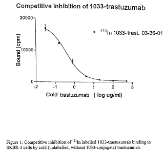
Figure 1 shows competitive inhibition of kiln
labelled 1033-trastuzumab binding to SKBR-3 cells by cold
(unlabelled, without 1033-conjugate) trastuzumab.
Figure 2 shows comparison of whole body clearance of
radioactivity in rats, injected with kiln-1033-
trastuzumab (filled triangles) or kiln-1033-rituximab
(filles squares) antibody conjugates expressed as percen-
tage ~ std.dev. The data are corrected for radioactivity
decay and background.
Figure 3 shows comparison of whole blood clearance
of radioactivity in rats, injected with kiln-1033-
trastuzumab (filled triangles) or kiln-1033-rituximab
(filles squares) antibody conjugates expressed as % of
activity at start ~ std.dev. The data are corrected for
radioactivity decay.
Figure 4 shows biodistribution of kiln-1033-
trastuzumab in rats, expressed as % of injected dose per
gram tissue ~ std.dev. The results are corrected for
radiochemical decay.
Figure 5 shows biodistribution of kiln-1033-
rituximab in rats, expressed as % of injected dose per
gram tissue ~ std.dev. The results are corrected for
radiochemical decay.
Description of Preferred Embodiments
Definitions:
When used in this context "naked antibody" means an
antibody, antibody fragments, "Single-chain Fv" anti-
body fragments or "diabodies", which does not carry any
agents or structures attached to the immunoglobulin
structure in order to enhance the effect of antibody,
hence, the effect on tumours cells of the naked anti-
bodies need to rely on the intrinsic effect of the anti-
body itself.
The term "monoclonal antibody" as used herein refers
to an antibody obtained from a population of substanti-
ally homogeneous antibodies, i.e., the individual anti-
CA 02547435 2006-05-25
WO 2005/051424 PCT/SE2004/001753
19
bodies comprising the population are identical except for
possible naturally occurring mutations that may be pre-
sent in minor amounts. Monoclonal antibodies are highly
specific, being directed against a single antigenic site.
Furthermore, in contrast to conventional (polyclonal)-
antibody preparations which typically include different
antibodies directed against different determinants
(epitopes), each monoclonal antibody is advantageous in
that they are synthesized by the hybridoma culture,
uncontaminated by other immunoglobulins. The modifier
"monoclonal" indicates the character of the antibody as
being obtained from a substantially homogeneous popula-
tion of antibodies, and is not to be construed as
requiring production of the antibody by any particular
method. For example, the monoclonal antibodies to be used
in accordance with the present invention may be made by
the hybridoma method first described by Kohler et al.,
Nature, 256:495 (1975), or made by recombinant DNA
methods (see, e.g., U.S. Patent No. 4,816,567). The mono-
clonal antibodies may also be isolated from phage anti-
body libraries using the techniques described in Clackson
et al., Nature, 352:624-628 (1991) and Marks et al., J.
Mol. Biol., 222:581-597 (1991), for example.
The monoclonal antibodies herein specifically
include "chimeric" antibodies (immunoglobulins) in which
a portion of the heavy and/or light chain is identical
with or homologous to corresponding sequences in
antibodies derived from a particular species or belonging
to a particular antibody class or subclass, while the
reminder of the chains) is identical with or homologous
to corresponding sequences in antibodies derived from
another species or belonging to another antibody class or
subclass, as well as fragments of such antibodies, so
long as they exhibit the desired biological activity
(U. S. Patent No 4,816,567; Morrison et al., Proc. Natl.
Acad. Sci. USA, 81:6851-6855 (1984)).
CA 02547435 2006-05-25
WO 2005/051424 PCT/SE2004/001753
"Humanized" forms of non-human (e. g. murine) anti-
bodies are chimeric immunoglobulins. Immunoglobulin
chains or fragments thereof (such as Fv, Fab, Fab',
F(ab')2 or other antigen-binding subsequences of anti-
s bodies) which contain a minimal sequence derived from
non-human immunoglobulin. For the most part, humanized
antibodies are human immunoglobulins (recipient antibody)
in which residues from a complementarity-determining
region (CDR) of the recipient are replaced by residues
10 from a CDR of a non-human species (donor antibody) such
as mouse, rat or rabbit having the desired specificity,
affinity, and capacity. In some instances, Fv framework
region (FR) residues of the human immunoglobulin are
replaced by corresponding non-human residues. Further-
15 more, humanized antibodies may comprise residues which
are neither in the recipient antibody nor in the imported
CDR or framework sequences. These modifications are made
to further refine and optimize antibody performance.
In general, the humanized antibody will comprise
20 substantially all of, or at least one, and typically two,
variable domains, in which all or substantially all of
the CDR regions correspond to those of a non-human
immunoglobulin and all or substantially all of the FR
regions are those of a human immunoglobulin sequence. The
humanized antibody optimally will also comprise at least
a portion of an immunoglobulin constant region (Fc),
typically that of a human immunoglobulin. For further
details, see Jones et al., Nature, 321:522-525 (1986):
Reichmann et al., Nature. 332:323-329 (1988): and Presta,
Curr. Op. Struct. Biol., 2:593-596 (1992). The humanized
antibody produced by immunizing macaque monkeys with the
antigen of interest.
"Antibody fragments" comprise a portion of an intact
antibody, generally the antigen-binding or variable
region of the intact antibody. Examples of antibody
fragments include Fab, Fab'. F(ab')2. and Fv fragments:
CA 02547435 2006-05-25
WO 2005/051424 PCT/SE2004/001753
21
diabodies; single-chain antibody molecules; and multi-
specific antibodies formed from antibody fragments.
"Single-chain Fv" antibody fragments comprise the VH
and VL domains of antibody, wherein these domains are
present in a single polypeptide chain Generally, the Fv
polypeptide further comprises a polypeptide linker
between the VH and VL domains which enables the sFv to
form the desired structure for antigen binding. For a
review of sFv, see Pluckthun in The Pharmacology of
Monoclonal Antibodies, vol. 113, Rosenbourg and Moore
eds., Springer-Verlag, New York, pp. 269-315 (1994).
The term "diabodies" refers to small antibody frag-
ments with two antigen-binding sites, which fragments
comprise a heavy-chain variable domain (VH) connected to
a light-chain variable domain (VL) in the same polypep-
tide chain (VH-VL). By using a linker that is too short
to allow paring between the two domains on the same
chain, the domains are forced to pair with the comple-
mentary domains of another chain and create two antigen-
binding sites. Diabodies are described more fully in, for
example, EP 404,097;W0 93/11161;and Hollinger et al.,
Proc. Natl. Acad. Sci. USA, 90:6444-6448 (1993).
The term "anti Erb antibody" used herein is intended
to mean an antibody with the ability of specific binding
to the various types of mammalian erb gene products
expressed on tumour cells, and with an affinity-binding
constant of at least 5x10⁻⁶M-1. The term will include, but
is not limited to, antibodies against erbl, erb2, erb3
and erb4.
The term erb or erb antigens) in this application
refers to the various types of the mammalian erb gene
products, and in particular the use of these gene pro-
ducts as targets for anti-tumour antibodies.
The term "variants" of the anti Erb antibody as used
herein means any modifications, fragments or derivatives
thereof having the same or essentially similar affinity-
binding constant when binding to the Erb antigen mole-
CA 02547435 2006-05-25
WO 2005/051424 PCT/SE2004/001753
22
cute, i.e. an affinity-binding constant of at least
5x10⁶M-1.
Any of these variants could have been modified by
the coupling of various numbers of polyethylene glycol
chains in order to optimise the half-life in body fluid
and the retention of the antibody or antibody fragments
or derivatives, in the tumor tissue. In the most preferr-
ed application the antibodies or antibody derivatives
should allow for the attachment of a sufficient number of
biotin residues to be used for extracorporeal removal
through interaction with immobilized avidin, without
significantly diminishing the binding properties of the
targeting agent.
"Treatment" refers to both therapeutic treatment and
prophylactic or preventative measures. Those in need of
treatment include those already with the disorder as well
as those in which the disorder is to be prevented.
"Mammal" for purposes of treatment refers to any
animal classified as a mammal, including humans, domestic
and farm animals, and zoo, sports, or pet animals, such
as dogs, horses, cats, cows, etc. Preferably, the mammal
is human.
A "disorder" is any condition that would benefit
from treatment with the anti-Erb antibodies. This in-
cludes chronic and acute disorders or diseases including
the pathological conditions which predispose the mammal
to the disorder in question. Non-limiting examples of
disorders to be treated herein include benign and malig-
nant tumors; leukemias and lymphoid malignancies;
neuronal, filial, astrocytal, hypothalamic and other
glandular, macrophagal, epithelial, stromal and blasto-
coelic disorders; and inflammatory, angiogenic and immo-
logic disorders.
The terms "cancer" and "cancerous" refer to or
describe the physiological condition in mammals that is
typically characterized by unregulated cell growth.
Examples of cancer include, but are not limited to,
CA 02547435 2006-05-25
WO 2005/051424 PCT/SE2004/001753
23
carcinoma, lymphoma, blastoma, sarcoma, and leukemia.
More particular examples of such cancers include
squarnous cell cancer, small-cell lung cancer, non-small
cell lung cancer, gastrointestinal cancer, pancreatic
cancer, glioblastoma, cervical cancer, ovarian cancer,
liver cancer, bladder cancer, hepatoma, breast cancer,
colon cancer, colorectal cancer, endometrial carcinoma,
salivary gland carcinoma, kidney cancer, renal cancer,
prostate cancer, vulval cancer, thyroid cancer, hepatic
carcinoma and various types of head and neck cancer.
The term "cytotoxic agent" as used herein refers to
a substance that inhibits or prevents the function of
cells and/or causes destruction of cells. The term is
intended to include radioactive isotopes (e. g. I, Y, Lu),
chemotherapeutic agents, and toxins such as, but not
limited to, active toxins of bacterial, fungal, plant or
animal origin, or fragments thereof. Some radionuclides,
like indium-111, are used as diagnostic agents and are as
such administered with low activity, but could also be
used for therapeutical purposes if given in higher doses
and are therefore also referred to as cytotoxic agents
herein.
A "chemotherapeutic agent" is a chemical compound
useful in the treatment of cancer. Examples of chemo-
therapeutic agents include Adriamycin, Doxorubicin, 5-
Fluoruracil, Cytosine arabinoside ("Ara-C"), Cyclo-
phosphamide, Thioptepa, Busulfan, Cytoxin, Taxol, Metho-
trexate, Cisplatin, Melphalan, Vinblastine, Bleomycin,
Etoposide, Ifosfamide, Mitomycin C, Mitoxantrone,
Vincristine, Vinorelbine, Carboplatin, Tenisposide,
Duanomysin, Carminomycin, Aminopterin, Dactinomycin,
Mitomycins, Esperamicins (see U.S. Pat. No. 4,675,187),
Maytansinoids, Melphalan and other related nitrogen
mustards.
The term "MitraTagTM-1033", also called for short
"1033", as used herein refers to the compound 3-(13'-
CA 02547435 2006-05-25
WO 2005/051424 PCT/SE2004/001753
24
thioureabenzyl-DOTA)trioxadiamine-1-(13"-biotin-Asp-OH)-
trioxadiamine-5-isothiocyanato-aminoisophatalate.
The following embodiments of the invention also
serve to explain the details of the invention.
All types of cancer expressing Erb gene products on
the surface of tumor cells are applicable to treatment
with a medical composition, a kit or a method according
to the present invention. In a preferred embodiment the
medical composition, the kit, or the method, is applied
to breast cancer or ovarian cancer. A most preferred
application is breast cancer of the so-called HER-2
type, that is breast cancer which over-expresses HER-2.
This type is also known as Erb-B2 or c-erb-2.
The present invention presents new medical and
pharmaceutical compositions in the treatment of certain
types of breast cancer and ovarian cancer in particular.
Furthermore, with the present invention it is poss-
ible to improve the tumour to non-tumour ratio of cyto
toxic targeting agents in the treatment of disseminated
cancer expressing the protooncogene Erb, in particular
breast cancer and ovarial cancer, by reducing the con-
centration of the cytotoxic medical agent in the blood
circulation after administrations of a cytotoxic agent
and thereby facilitating a higher dosage and hence a more
effective treatment regime without exposing the vital
organs to higher toxicity.
In one embodiment, a radiolabelled anti Erb antibody
is given in a single dose which is limited to what is
regarded as tolerable to the patient without reconstitu-
tion of the hematopoietic function, through bone marrow
transplantation, or by some other means known in the art.
The dose range will be 10-20 MBq/kg body weight of 9°Y-
anti Erb antibody ("low dose"), preferably 11-15 MBq/kg,
and the range for ~~lIn-anti Erb antibody for targeting
localisation will be 50-200 MBq/mz body surface, prefer
ably 100-150 MBq/m2body surface. In this embodiment,
CA 02547435 2006-05-25
WO 2005/051424 PCT/SE2004/001753
extracorporeal clearance of non-bound radiolabelled
therapeutic or diagnostic antibody is optional.
In another embodiment, a radiolabelled anti Erb
antibody is given in a single dose designated to deliver
5 a high amount of radioactivity to the patient. This "high
dose method" has to be combined with means to reconstitu-
ting the bone marrow or by reducing the radiation effect
on bone marrow, preferably by the use of the MitraDep~
system. For 9°Y-anti Erb antibodies, a "high dose" means a
10 single dose exceeding 20 MBq/kg body weight.
In a preferred embodiment, kiln-anti Erb antibodies
at a dose of 100-150 MBq/m2 body surface is combined with
a "high dose" (>20 MBq/kg body weight) of 9°Y-anti Erb
antibody, either given in sequence by a time interval of
15 6-8 days or given simultaneously.
In one embodiment, a radiolabelled anti Erb antibody
is given in a single dose which is limited to what is
regarded as tolerable to the patient without reconstitu-
tion of the hematopoietic function through bone marrow
20 transplantation, or by some other means. The dose range
will be 555-2220 MBq/m2body surface of 177Lu-anti Erb
antibody ("low dose"), preferably 1000-2000 MBq/m2. In
this embodiment, extracorporeal clearance of non-bound
radiolabelled therapeutic or diagnostic antibody is
25 optional.
In another embodiment, a radiolabelled anti Erb
antibody is given in a single dose designated to deliver
a high amount of radioactivity to the patient. This "high
dose method" has to be combined with means known in the
art to reconstitute the bone marrow or by reducing the
radiation effect on bone marrow, preferably by the use of
the MitraDep~ system. For l~~Lu-anti Erb antibodies, "high
dose" means a single dose exceeding 2220 MBq/m2 body
surface .
The advantages of 177Lu compared to 9°Y are the
following:
CA 02547435 2006-05-25
WO 2005/051424 PCT/SE2004/001753
26
9°Y is a pure beta-emitter and can not be imaged by
external gamma cameras (immunoscintigraphy) and therefore
requires the use of kiln for imaging. Conversely, 17'Lu
emits gamma radiation in addition to beta particle
emission. As a result, 177Lu can be imaged directly, with-
out need for a combination with kiln. Therefore, only one
radiopharmaceutical is required for localisation and
therapy when 17'Lu is used, which will simplify the treat-
ment regime and lower the cost as well as reduce the
irradiation burden on the patient.
9°Y has a shorter physical half-life (2.67 days) and
a longer range (12.0 mm) than 1"Lu. The longer half-life
(6.7 days) and shorter range (2.2 mm) of 177Lu offers
benefits by allowing a longer time for the antibody-
radionuclide to localise to the tumour and the longer
half-life also combines well with the long intracellular
half-life. In addition, the shorter range of 17'Lu would
cause less bystander radiation (cross-fiering) to tissues
adjacent to the tumour tissue at the possible cost of
less efficacy in bulkier lesions. The longer range of 9°Y
offers benefits in being better able to radiate bulkier
lesions.
Breast cancer is staged into five different groups
based on the prognosis. Breast cancer happens when cells
in the breast begin to grow out of control and can then
invade nearby tissues or spread throughout the body. The
tumors that can spread throughout the body or invade
nearby tissues are considered cancer and are called
malignant tumors. Theoretically, any of the types of
tissue in the breast can form a cancer, but usually it
comes from either the ducts or the glands.
In order to guide treatment and offer some insight
into prognosis, breast cancer is staged into five
different groups.
Stage 0 (called carcinoma in situ)
Lobular carcinoma in situ (LCIS) refers to abnormal
cells lining a gland in the breast. This is a risk factor
CA 02547435 2006-05-25
WO 2005/051424 PCT/SE2004/001753
27
for the future development of cancer, but this is not
felt to represent a cancer itself.
Ductal carcinoma in situ (DCIS) refers to abnormal
cells lining a duct. Women with DCIS have an increased
risk of getting invasive breast cancer in the breast.
Treatment options are similar to patients with Stage I
breast cancers.
Stage I - early stage breast cancer when the tumor is
less than 2 cm across and has not spread beyond the
breast.
Stage II - early stage breast cancer where the tumor is
either less than 2 cm across and has spread to the lymph
nodes under the arm; or the tumor is between 2 and 5 cm
(with or without spread to the lymph nodes under the
arm); or the tumor is greater than 5 cm and has not
spread outside the breast.
Stage III - locally advanced breast cancer where the
tumor is greater than 5 cm across and has spread to the
lymph nodes under the arm; or the cancer is extensive in
the underarm lymph nodes; or the cancer has spread to
lymph nodes near the breastbone or to other tissues near
the breast.
Stage IV - metastatic breast cancer where the cancer has
spread outside the breast to other organs in the body.
Although patients representing all five groups could
be eligible to treatment according to present invention,
in a most preferred embodiment the malignancy represents
Stage III and IV.
In the present invention an immunotargeting agent
(immunoconjugate) is an agent which carries a cytotoxic
moiety that, contrary to common cytotoxic medical agents,
binds specifically and with high affinity to tumor cells
expressing the protooncogene Erb, and which could be
administered to a human being. In a preferred application
the immunotargeting agents are antibodies, which could be
of different isotypes and could originate from any
species. Preferred antibodies are humanised monoclonal
CA 02547435 2006-05-25
WO 2005/051424 PCT/SE2004/001753
28
antibodies. Furthermore, of particular interest are
those, which in addition to the above-described proper-
ties bind the erb receptor with an affinity of at least
about 50 nM, more preferably at least about 10 nM.
Of particular interest are derivatives of monoclonal
antibodies. The latter include fragments such as the Fab,
Fab', F(ab')2, Flab") and Fv fragments and the like. They
also include genetically engineered hybrids or chemically
synthesized peptides based on the specificity of the
antigen binding region of one or several target specific
monoclonal antibodies, e.g. chimeric or humanized
antibodies, single chain antibodies etc. The biomolecule
binding moiety, which is an IgG reactive moiety, is bound
or conjugated to the anti Erb antibody, either covalently
or non-covalently with an affinity-binding constant of at
least 5x10⁸ M-1.
In order to enhance the effect or to introduce diag-
nostic properties, tumour specific monoclonal antibodies
are used as a carrier (immunoconjugates) of various
cytotoxic agents, such as, but not limited to,
radionuclides, chemotherapeutical agents, synthetic or
natural occurring toxins, immunosuppressive or
immunostimulating agents, radiosensitizers, enhancers for
X-ray or MRI or ultrasound, non-radioactive elements,
which can be converted to radioactive elements by means
of external irradiation after that the anti Erb antibody
carrying said element has been accumulated to specific
cells or tissues, or photoactive compounds or compounds
used in photo imaging or photodynamic therapy, or any
other molecule having the same or a similar effect, di-
rectly or indirectly, on cancer cells or cancer tissues,
and enzymes used in pro-drug protocols. The cytotoxic
agent is preferably a radionuclide, such as a gamma-
emitter e.g. iodine-131 or metal ion conjugate, where the
metal is selected from a beta-particle emitter, such as
yttrium, lutetium or rhenium. U.S. Patent No. 4,472,509,
Gansow et al., discloses the use of diethylenetriamine-
CA 02547435 2006-05-25
WO 2005/051424 PCT/SE2004/001753
29
pentaacetic acid (DTPA) chelating agents for the binding
of radio metals to monoclonal antibodies. The patent is
particularly directed to a purification technique for the
removal of non-bonded and adventitiously bonded (non-
chelated) metal from radiopharmaceuticals but is illu-
strative of art recognized protocols for preparation of
radionuclide labelled antibodies.
According to such general procedures, an antibody
specifically reactive with the target tissue associated
antigen is reacted with a quantity of a selected bifunc-
tional chelating agent having protein binding and metal
binding functionalities to produce a chelator/antibody
conjugate. In conjugating the antibodies with the chela-
tors, an excess of chelating agent is reacted with the
antibodies, the specific ratio being dependent upon the
nature of the reagents and the desired number of chelat-
ing agents per antibody. It is a requirement that the
radionuclides be bound by chelation (for metals) or
covalent bonds in such a manner that they do not become
separated from the biotinylated/radiolabelling compound
under the conditions that the biomolecule conjugate is
used (e. g. in patients).
When the cytotoxic agent is a radionuclide, parti-
cularily metallic radionuclides, it is bound to the tri-
functional cross-linking moiety via a cytotoxic agent
binding moiety.
Thus, the most stable chelates or covalent bonding
arrangements are preferred. Examples of such binding/-
bonding moieties, i.e. the cytotoxic agent binding
moiety, form aryl halides and vinyl halides for radio-
nuclides of halogens; and comprise NZSZ and N3S chelates
for Tc and Re radionuclides; amino-carboxy derivatives
such as EDTA, triethylenetetraaminehexaacetic acid and
DTPA or derivatives thereof, said DTPA derivatives being
Me-DTPA, CITC-DTPA and cyclohexyl-DTPA, and cyclic
amines, such as NOTA, DOTA, and TETA, and derivatives
CA 02547435 2006-05-25
WO 2005/051424 PCT/SE2004/001753
(Yuangfang and Chuanchu, Pure & Appl. Chem. 63, 427-463,
1991) for In, Y, Pb, Bi, Cu, Sm, and Lu radionuclides.
Beta radiation emitters, which are useful as cyto-
toxic agents, include radionuclides, such as scandium-46,
5 scandium-47, scandium-48, copper-67, gallium-72, gallium-
73, yttrium-90, ruthenium-97, palladium-100, rhodium-101,
palladium-109, samarium-153, lutetium-177, rhenium-186,
rhenium-188, rhenium-189, gold-198, and radium-212. The
most useful gamma emitters are iodine-131 and indium-
10 m114. Other metal ions useful with the invention include
alpha radiation emitting materials such as bismuth-212,
bismuth-213, and astate-211 as well as positron emitters
such as gallium-68 and zirconium-89.
In another embodiment of the invention, radio-
15 nuclide-labelled targeting agents are useful not only in
the treatment of cancer expressing erb antigens, but also
for imaging of such cancers. Imaging can be conducted by
the use of (3-emitting radionuclides utilizing the brems-
strahlung or by y-emitting radionuclides for imaging. In
20 another preferred embodiment 177Lu, which is both a ~i and
y emitter, is used as the cytotoxic agent for both treat-
ment and diagnosing of cancer.
In a preferred embodiment on average 2-4 molecules
of the part a)-c) of the conjugate, preferably MitraTagT"',
25 are linked to each molecule of the anti Erb antibody, and
in the most preferred embodiment the average number of
such molecules per anti Erb antibody is 2.5-3.5.
At a suitable time after administration, "cytotoxic
targeting agents" will be cleared from the blood system
30 by extracorporeal means. To facilitate the extracorporeal
depletion an apparatus for extracorporeal circulation of
whole blood or plasma will be connected to the patient
through tubing lines and blood access device(s). Such an
apparatus should provide conduits for transporting the
blood to an adsorption device and conduits for returning
the processed blood or plasma to the patient. In the case
plasma is processed through the adsorption device, a
CA 02547435 2006-05-25
WO 2005/051424 PCT/SE2004/001753
31
plasma separation device is needed as well as means of
mixing the concentrated blood with processed plasma. The
later is normally achieved by leading the two components
into an air-trap where the mixing occurs.
In the case where whole blood is processed, an ordi-
nary dialysis machine can constitute the base for such an
apparatus. Dialysis machines are normally equipped with
all the necessary safeguards and monitoring devices to
meet patient safety requirements and allow easy handling
of the system. Hence, in a preferred embodiment whole
blood is processed and a standard dialysis machine is
utilised with only minor modifications of the hardware.
However, such a machine requires a new program fitted to
the new intended purpose.
In addition to the apparatus, special blood line
tubings suitable for the intended flow and distance from
the patient and the machine are needed. These line tub-
ings could be made of any material compatible with blood
or plasma and would include material used in ordinary
tubings used in dialysis.
Blood access could be achieved through peripheral
vein catheters, or if higher blood flow is needed,
through central vein catheters such as, but not limited
to, subclavian or femoral catheters.
For affinity adsorbents, the matrix may be of vari-
ous shapes and chemical compositions. It may for example
constitute a column house filled with particulate poly-
mers, the latter of natural origin or artificially made.
The particles may be macroporous or their surface may be
grafted, the latter in order to enlarge the surface area.
The particles may be spherical or granulated and be based
on polysaccharides, ceramic material, glass, silica,
plastic, or any combination of these or alike material. A
combination of these could, for example, be solid par-
ticles coated with a suitable polymer of natural origin or
artificially made. Artificial membranes may also be used.
These may be flat sheet membranes made of cellulose, poly-
CA 02547435 2006-05-25
WO 2005/051424 PCT/SE2004/001753
32
amide, polysulfone, polypropylene or other types of
material which are sufficiently inert, biocompatible, non-
toxic and to which the receptor could be immobilized
either directly or after chemical modification of the mem-
brane surface. Capillary membranes like the hollow fibers
made from cellulose, polypropylene or other materials
suitable for this type of membranes may also be used. A
preferred embodiment is a particulate material based on
agarose and suitable for extracorporeal applications.
In one embodiment Molecularly Imprinted Polymers
(MIPs) are used. In such a case the conjugate does not
contain any affinity ligands. These are normally cross-
linked polymers prepared in the presence of a template
molecule. The template can either be molecular structures
conjugated to the targeting molecule (chelating groups
such as DOTA or DTPA derivatives) or particular
structures more or less specific of the targeting mole-
cule (e.g.the antibody structure).
In another embodiment the matrix is coated by lig-
ands which exhibit a specific interaction to the agent
(e. g. radio active anti Erb antibody) to be removed from
the blood circulation. Such ligands can be chosen from a
group comprising monoclonal antibodies including frag-
ments or engineered counterparts thereof, aptamers, pep-
tides, oligodeoxynucleosides including fragments thereof,
intercalation reagents including dyestof, oligo-
saccharides and chelating groups interacting with metals
bound to the agent to be removed.
In another embodiment an affinity ligand is attached
to the anti Erb antibody and the adsorption device con-
tains an immobilized receptor binding specifically to the
affinity ligand. Any type of affinity ligand/immobilized
receptor combinations such as "antibodies and antigens/-
haptens" and "protein and co-factors" could be used in
this application, provided that they exhibit a suffici-
ently high binding affinity and selectively to the tumor
markers and that the affinity ligand-receptor interaction
CA 02547435 2006-05-25
WO 2005/051424 PCT/SE2004/001753
33
is not interfered with by blood or other body fluids or
tissues being in contact with the immunotargeting agent
and/or the device.
In one of the most preferred applications, the affi-
nity ligand/immobilized receptor combination is biotin or
biotin derivatives and biotin binding molecules, and in
particular where the affinity ligand is biotin or deriva-
tives thereof and the immobilized receptor is avidin or
streptavidin or any other biotin binding molecule. The
affinity ligand pairs of biotin/avidin and biotin/strept-
avidin are often used with biomolecules. The very strong
interaction ( i . a . K = 1013-1015 M-1 ) of biotin with the
proteins avidin and streptavidin (Green, Methods Enzymol.
184, 51-67, 1990; Green, Adv. Prot. Chem. 29, 85-133,
1975) provides a foundation for their use in a large
number of applications, both for in vitro and in vivo
uses. A further application of the invention is the
simultaneous removal of several different biotinylated
"anti-cancer agents" through the same extracorporeal
procedure.
One embodiment of the conjugate according to the
present invention is in part schematically shown below,
wherein the anti Erb reactive moiety is trastuzumab.
The structural requirements for this 1033-conjugate
include the biotin containing moiety (the affinity
ligand), a linker 1 between biotin and the rest of the
molecule, a trifunctional cross-linking moiety, a cyto-
toxic agent binding moiety, and a linker 2 between the
cytotoxic agent binding moiety and the rest of the mole-
rule. The structural requirements of the 1033-conjugate
can be split into three parts based on functional
requirements. Those parts are the biotin containing
moiety, the cytotoxic agent binding moiety, and the
trifunctional cross-linking moiety. Formula 1 shows a
generalized structure of the inventive conjugate (without
any cytotoxic agent bound thereto).
CA 02547435 2006-05-25
WO 2005/051424 PCT/SE2004/001753
34
Formula I: Generalized structure for the inventive con-
jugate intended to bind a metallic radionuclide and con-
taining trastuzumab as the anti Erb antibody.
biotin trifunctional cytotoxic agent
linker 1 . I cross-linking I linker 2
molecule binding moiety
moiety
I0'
HN~NH
R~ O O H H -. ~ ~ 02H
S/~\y~N\r~~H ~ \ H~N~N \ / N~ J 02H
O R2 / S N
S~NH HO C ~N~
'NH
J
HOpC
trastuzumab
Structural requirements of the biotin containing moiety:
There are three aspects of the biotin containing moiety,
i.e. the affinity ligand, of the above structure that are
important in this context. Those are: (1) blockage of
biotinidase cleavage, (2) retention of high biotin bind-
ing affinity, and (3) attainment of a reasonable aqueous
solubility. To provide those attributes, biotin conju-
gates must be composed of a biotin molecule and an
appropriate linker, which are coupled to a cross-linking
moiety.
Biotin conjugates must be prepared by conjugation
with the carboxylate on the pentanoic acid side chain (n
- 3). Conjugation at other locations in the biotin mole-
cule results in complete loss of binding with avidin and
streptavidin. This would render the biotin molecule use-
less for this application. The preferred form of conjuga-
tion is formation of an amide bond with the carboxylate
group (as depicted in the general formula). Since binding
CA 02547435 2006-05-25
WO 2005/051424 PCT/SE2004/001753
of biotin with avidin and streptavidin takes place in a
deep pocket (e. g. 9A), shortening (n<3) or lengthening
(n>3) of the pentanoic acid side chain results in low
binding affinity, which is not desired for this applica-
5 tion.
Blocking of the biotinidase activity is achieved by
attaching appropriate substituents on the biotinamide
amine (i.e. R1) or on an atom adjacent, i.e. less than
three carbon atoms apart, to that amine (i.e. RZ).
10 Biotinidase is an enzyme that cleaves (hydrolyzes) the
amide bond of biotin carboxylate conjugates. This enzyme
is very important in recycling biotin in animals and man.
Metabolism of biotin in (several different) protein
carboxylases releases biotin-s-N-lysine (biocytin), and
15 biotinidase specifically cleaves that amide bond to
release free biotin. Biotinidase is also capable of
cleaving (non-specifically) other biotinamide bonds. In
this application, it is important that biotinidase does
not cleave biotin from the conjugates, since otherwise
20 the desired outcome will not be achieved. Thus, the
useful biotin conjugate structures incorporate functional
groups (R1 or RZ) that block the enzymatic activity of
biotinidase. While it is likely that any structure for R1
will block biotinidase, its structure is generally
25 limited to a methyl (CH3) group, as this group completely
blocks biotinidase activity. The N-methyl group
decreases the binding affinity of biotin with avidin and
streptavidin significantly, but it still has use in this
application. Larger groups for R1 (e. g. ethyl, aryl,
30 etc.) are not useful due to the loss of binding affinity.
The alternative to having a substituent R1 is to have a
substituent Rz on the atom (e.g. methylene) adjacent to
the biotinamide amine. Much larger and more varied sub-
stituents can be used in this position without signifi-
35 cant effect on the binding affinity of biotin. Biotini-
dase is not completely blocked when RZ is CH3 or CHZCH3,
although the rate of cleavage is slowed considerably
CA 02547435 2006-05-25
WO 2005/051424 PCT/SE2004/001753
36
(i.e. to 25°s and 10% respectively). Complete blockage of
biotinidase activity is attained when RZ are -CHzOH and
-C02H functionalities. In the case of the -CHZOH (hydroxy-
methyl) functionality, such a blocking may be achieved by
the introduction of a serinyl group. In the case of the
C02H (carboxy) functionality, such a blocking may be
achieved by the introduction of an a or (3 aspartyl group.
The important consideration is that there is no decrease
in binding affinity when these groups are incorporated as
R2. Larger functional groups can also be used as RZ to
block biotinidase activity, but a decrease in binding
affinity results. The larger functional groups as RZ are
useful in this application if they do not cause a
decrease in binding affinity greater than that obtained
when R1 is CH3.
The biotin affinity and aqueous solubility of the
biotin moiety in the structure of Formula I is affected
by the linker moiety used. The length and nature of the
linker moiety (linker 1) will be dependent to some degree
on the nature of the molecule that it is conjugated with.
The linker moiety serves the function of providing a
spacer between the biotin moiety and the rest of the
conjugate such that the biotin binding is not affected by
steric hindrance from the protein (or other conjugated
molecule). The length (number of atoms in a linear chain)
of linker 1 may vary from o = 4-20 for conjugates with
small molecules (e. g. steroids) to o > 20 for large con-
jugate molecules (e.g. IgG molecule). The nature of the
atoms in linker 1 (linear chain or branch from it) will
also vary to increase water solubility. For example,
linkers that contain more than 4 methylene units are im-
proved by incorporation of oxygen or sulfur atoms (form-
ing ethers or thioethers) or by having appended ionizable
functionalities (e.g. sulfonates, carboxylates, amines or
ammonium groups).
Structural requirements of the cytotoxic agent binding
moiety: Various radionuclide chelating and bonding
CA 02547435 2006-05-25
WO 2005/051424 PCT/SE2004/001753
37
agents can be used in the structure of Formula I. In
Formula I, a "benzyl-DOTA" moiety is used as an example.
Depending on the nature of the cytotoxic agent binding
moiety, a linker moiety (linker 2) is required. Some
radionuclide chelation and/or bonding moieties have low
aqueous solubility, so addition of a linker molecule
containing functional groups which improve water solu-
bility is important. In the DOTA chelate, the primary
function of the linker moiety is to improve the water
solubility of the conjugated molecule. The nature of the
atoms in linker 2 (linear chain or branch from it) will
vary to increase water solubility. For example, linkers
that contain more than 4 methylene units are improved by
incorporation of oxygen or sulfur atoms (forming ethers
or thioethers) or by having appended ionizable
functionalities (e.g. sulfonates, carboxylates, amines or
ammonium groups). The length (number of atoms in a linear
chain) of linker 2 may also vary (e.g. p = 1 - 20)
depending on the nature of hetero atoms incorporated or
functional groups appended to the linear chain.
Structural requirements of the trifunctional cross-
linking moiety: Various trifunctional molecules can be
used as the cross-linking moiety. Any molecule that has
three functional groups that can be reacted with func-
tional groups on the linkers (linker 1 and 2) and on the
protein is a candidate for the trifunctional cross-link-
ing moiety. Aside from the requirement that the trifunc-
tional cross-linking moiety not impart insolubility of
e.g. the structure of Formula I in aqueous solutions, the
only other structural limitations on the trifunctional
cross-linking molecule is that the structure be such that
it can be modified in a manner that allows a sequential
addition of the biotin containing moiety, and the cyto-
toxic agent binding moiety, and conjugation with the anti
Erb antibody. As an example, a trifunctionalized benzene
ring (aminoisophthalic acid) is used in the 1033
structure.
CA 02547435 2006-05-25
WO 2005/051424 PCT/SE2004/001753
38
A preferred structure, 1033-trastuzumab, is shown in
Formula II below, wherein n = 3, o = 3, p = 3, R1 = H and
R2 = COOH (without any cytotoxic agent bound).
Formula II: Specific structure of 1033-trastuzumab
z
N
0
U
0
1
ZJ N
0
z
U
/ I O
Z
zx
N
Z=
O
0
0
Z=
0
N
~
o ,~.r
L
0
o ..
-o
z=
0
==o
o z=
0
x
z
N ~O
~
Z
Z
CA 02547435 2006-05-25
WO 2005/051424 PCT/SE2004/001753
39
Specific examples of the conjugate according to the
present invention are 177Lu-1033-trastuzumab, i.e. 177Lu-3-
(13'-thioureabenzyl-DOTA)trioxadiamine-1-(13"-biotin-Asp-
OH)trioxadiamine-5-isothiocyanato-aminoisophtalate-
trastuzumab; 9°Y-1033-trastuzumab; kiln-1033-trastuzumab;
1033-trastuzumab, wherein thioureabenzyl-DOTA has been
replaced with maytansinoid; and 1033-trastuzumab, wherein
thioureabenzyl-DOTA has been replaced with doxorubicin.
In another embodiment of the present invention more
than one affinity ligand, preferably two, and/or more
than one cytotoxic agent, preferably two, are included in
the conjugate. In such a case, the cross-linking moiety
is more than trifunctional.
Examples
The following examples shall not be construed as
limiting the invention, but should be regarded as evi-
dence for the applicability of the invention.
Example 1 - Conjugation and radiolabelling of trastuzumab
In this and subsequent examples, Indium-111 has in
some instances been used as a substitute for Yttrium-90,
because the former is a gamma-emitter and possesses less
radiation hazard than Yttrium-90. The monoclonal anti-
body, trastuzumab, was conjugated with 3-(13'-thiourea-
benzyl-DOTA)-trioxadiamine-1-(13"-biotin-Asp-OH)-trioxa-
diamine-5-isothiocyanato-aminoisophtalate (MitraTagTM-
1033), for short also called "1033" in the following,
using the method described by Wilbur D.S et al in Bio-
conjugate Chem. 13:1079-1092, 2002. A 10 mg quantity of
the monoclonal antibody was dialysed against 1L metal
free HEPES with 3 buffer changes over 3 days at 4°C. A
solution of MitraTagTM-1033 (800 fig) was made in water
and was added to the antibody solution. After incubation
overnight at room temperature, the antibody-conjugate
was dialysed against 1L metal free 250 mM ammonium
acetate buffer pH 5.3 with a minimum of 4 buffer changes
over 4 days at 4°C. The average number of MitraTagTM-1033
CA 02547435 2006-05-25
WO 2005/051424 PCT/SE2004/001753
per monoclonal antibody was determined to 2.2 by the
HABA method. The demetalated conjugated antibody was
stored at 4-8°C until used in radiolabelling experi-
ments.
5 Two mg (400 ~l) 1033-antibody in 250 mM ammonium
acetate (pH 5.3) was mixed with 30 ~l of the radionuclide
to be studied (liilnCl3; 9°YC13; 177LuC13) in 40 mM HC1. The
labeling was conducted at 45°C for 15 minutes. 43 ~1 of
DTPA was added to stop the reaction. The quality of the
10 radio conjugate was determined by TLC and HPLC.
Example 2 - Binding of the 1033-conjugated trastuzumab to
an avidin adsorbent
The fraction of kiln-labelled 1033-trastuzumab radio
conjugate binding to the avidin adsorbent utilised in the
15 MitraDep~ device was analysed utilising microcolumns.
About 97 0 of the radioactivity in the radiolabelled
1033-conjugate sample was bound to the microcolumn with
the avidin adsorbent.
Example 3 - Analyses of the affinity of the binding to
20 the target antigen
The influence of the conjugation process on the
binding affinity (strength) of trastuzumab to the target
antigen was studied utilizing a competitive inhibition
assay. Briefly, increasing amounts of trastuzumab were
25 mixed with a constant amount of kiln-labelled 1033-
trastuzumab. The mixtures were added to fixed SK-BR3
cells in 96 plate wells. After incubation for 2 hours at
room temperature, the wells were washed, and the radio-
activity bound to the cells was measured in an automatic
30 NaI(Tl) scintillation well counter.
The amount of bound radioactivity was plotted
against the concentration of trastuzumab (figure 1), and
the concentration required for 50 o inhibition (ICS°) was
calculated. The ICS° is a measure of the relative affinity
35 (avidity) of the tested antibody; a decrease of affinity
is seen as an increased IC5° concentration. To be a sig-
CA 02547435 2006-05-25
WO 2005/051424 PCT/SE2004/001753
41
nificant change in affinity it is often stated that the
difference in ICso should be at least 10-fold.
1 ~g/ml (6.7 nM) of kiln-1033-trastuzumab is inhi-
bited by 0.03 - 500 ~g/ml cold non-conjugated
trastuzumab. The ICSO was determined to 0.4 ~,g/ml (2.5
nM). From ICSO, the dissociation constant was calculated
to 0.3 nM. According to information published by the
manufacturer of trastuzumab the dissociation constant is
0.1 nM.
A slight decrease in affinity was seen for the 1033-
trastuzumab conjugate. It has been shown in clinical
studies that a 10-fold difference in affinity does not
result in any significant difference in tumour uptake.
Therefore, it was concluded that conjugation of
trastuzumab with up to 2.2 conjugates per antibody would
not diminish the binding properties of the antibody in
V1.V0.
Example 4 - Pharmacokinetics of MitraTagTM-1033 conjugate
of trastuzumab.
The pharmacokinetics and biodistribution data of
iiiln-1033-trastuzumab is compared to the data obtained
with kiln-1033-rituximab as clinical data is available
for this radio conjugate. Both antibodies are humanized
human monoclonal IgGl antibodies.
Fifteen (15) rats of the Spraque Dawley strain were
injected intravenously with approximately 100 ug/rat of
1033-antibody conjugate labelled with 3-4 MBq kiln.
Whole body (WB) imaging was performed using a scin-
tillation camera (General Electric 400T, GE, Milwaukee,
WI, USA) equipped with a medium-energy collimator. Images
were stored and analysed with Nuclear MAC 2.7 software.
From the images, the total numbers of counts in the
entire body were obtained. After radioactivity decay
correction and background subtraction, the counts were
used for the calculation of activity retention (%) in the
body. See Figure 2.
CA 02547435 2006-05-25
WO 2005/051424 PCT/SE2004/001753
42
When whole body retention of kiln-1033-trastuzumab
was compared to that of kiln-1033-rituximab, no signi-
ficant difference was seen.
To define pharmacokinetics of kiln-1033-trastuzumab
and compare it with lilln-1033-rituximab, about 0.2 ml
blood was obtained from the periorbital venous plexa on
the following occasions: 10 min, 2.5, 8, 24, 48 and 96
hours after injection. The radioactivity was measured in
an automatic NaI(T1) scintillation well counter and
expressed in per cent of injected activity per gram blood
(%/g) corrected for kiln decay (figure 3) . When blood
clearance of kiln-1033-trastuzumab was compared to that
of kiln-1033-rituximab, no significant difference was
seen.
Example 5 - Biodistribution of conjugates to organs and
tissues
In dissections, performed 2.5, 8, 24, 48, and 96
hours after injection, organs and tissues of interest
were removed, weighed and measured for radioactivity
content. The radioactivity was measured in an automatic
NaI(T1) scintillation well counter, and the counts were
corrected for decay. The distribution to various organs
was compared to that of kiln-1033-rituximab. The
distribution of the injected activity is shown in figure
4 (iilln-1033-trastuzumab) and figure 5 (kiln-1033-
rituximab).
A higher uptake in the kidneys and lungs, and a
lower in the lungs were seen for kiln-1033-trastuzumab
compared to kiln-1033-rituximab. The higher uptake in the
lungs for kiln-1033-trastuzumab is mainly observed
shortly after injection, ending up at about the same
level after 48 hours.
Example 6 - Treatment regime with 9°y/liiln-trastuzumab
in breast cancer expressing HER-2 according to a
preferred embodiment of the invention
~ On day 0 all patients will receive 1-4 mg/body weight
of trastuzumab immediately followed by a therapeutic
CA 02547435 2006-05-25
WO 2005/051424 PCT/SE2004/001753
43
dose of 9°Y-1033-trastuzumab (>lOMBq/kg body weight).
Patients may, optionally, be administered a dose of
100-150 MBq/m2 body surface (1.1-3.9 mCi/m2 body sur-
face) kiln-1033-trastuzumab, which will be used for
imaging and for dosimetry.
At one occasion during day 1-3, patients are treated
with MitraDep~, allowing at least 3 blood volumes to
pass the MitraDep~ device.
Optionally, and as a safety measure, prior to
administration of 9°Y-1033-trastuzumab, bone marrow may
be harvested to allow for bone marrow rescue if
required.
Optionally, the treatment can be repeated with 9°Y-
1033-trastuzumab 2-6 times a year and in a most
preferred embodiment 2-4 times a year, provided that
no dose limiting toxicity has occurred and that the
patient has recovered from previous treatment with
respect to radiation toxicity.
~ Optionally, the patient is receiving therapeutic or
sub-therapeutic doses of trastuzumab (Herceptin) be-
fore or after receiving 9°Y-1033-trastuzumab and/or
trastuzumab (Herceptin) is given in connection with
the administration of 9°Y-1033-trastuzumab.
Example 7 - Treatment regime with 177Lu-trastuzumab in
breast cancer expressing HER-2 according to another
preferred embodiment of the invention
~ At day -7 to day -1 all patients will once receive 6-8
mg/kg body weight of trastuzumab.
~ On day 0 patients receive a therapeutic dose of 177Lu-
1033-trastuzumab (>555 MBq/mz body surface). Patients
may, optionally, be investigated by immunoscintography
for imaging and for dosimetry.
~ At one occasion during day 1-4, patients are treated
with MitraDep~, allowing at least 3 blood volumes to
pass the MitraDep~ device.
~ Optionally, and as a safety measure, prior to
administration of 1'7Lu-1033-trastuzumab, bone marrow
CA 02547435 2006-05-25
WO 2005/051424 PCT/SE2004/001753
44
may be harvested to allow for bone marrow rescure if
required.
Optionally, the treatment can be repeated with 17'Lu
1033-trastuzumab 2-6 times a year and in a most pre
y ferred embodiment 2-4 times a year provided that no
dose limiting toxicity has occurred and that the
patient has recovered from previous treatment with
respect to radiation toxicity.
Optionally, the patient is receiving therapeutic or
sub-therapeutic doses of trastuzumab (Herceptin) be-
fore, or after, receiving 177Lu-1033-trastuzumab and/or
trastuzumab (Herceptin) is given in direct connection
with the administration of 1'7Lu-1033-trastuzumab.