Note: Descriptions are shown in the official language in which they were submitted.
CA 02547605 2006-05-25
WO 2004/096318 PCT/US2004/013408
POLYMER COATED DEVICE FOR ELECTRICALLY MEDIATED DRUG
DELIVERY
BACKGROUND OF THE INVENTION
Cardiovascular disease is commonly accepted as being
one of the most serious health risks facing our society
today. Diseased and obstructed coronary arteries can
restrict the flow of blood and cause tissue ischemia and
necrosis. After over two decades of investigation, the
exact etiology of sclerotic cardiovascular disease is still
in question, the treatment of narrowed coronary arteries is
more defined. Surgical construction of coronary artery
bypass grafts (CABG) is often the method of choice when
there are several diseased segments in one or multiple
arteries. Open heart surgery is, of course, very traumatic
for patients. In many cases, less traumatic, alternative
methods are available for treating cardiovascular disease
percutaneously. These alternate treatment methods
generally employ various types of percutaneous transluminal
angioplasty (PTCA) balloons or excising devices
(atherectomy) to remodel or debulk diseased vessel
segments. A further alternative treatment method involves
percutaneous, intraluminal installation of expandable,
tubular stents or prostheses in sclerotic lesions.
A recurrent problem with the previous devices and PTCA
procedures a.s their failure to maintain patency due to the
growth of injured vascular tissue. This is known as
"restenosis" and may be a result of the original injury to
the vessel wall occurring during the angioplasty procedure.
Pathologically restenosis represents a neointimal
proliferative response characterized by smooth muscle cell
CA 02547605 2006-05-25
WO 2004/096318 PCT/US2004/013408
hyperplasia that results .in reblockage of the vessel lumen
necessitating repeat PTCA procedures up to 35-50~ of all
cases. It has been generally accepted that certain
therapeutic agents or medicaments may be capable of
selectively ' inhibiting the growth of these
hyperproliferating smooth muscle cells and- thereby reduce
the rate of restenosis after the primary interventional
procedure.
Heretofore, various devices have been. disclosed which
may be used to deliver a therapeutic agent or medicament to
a blood vessel while undergoing angioplasty. Balloon
angioplasty catheters have been used to place and deliver
various therapeutic agents or medicaments Within human
vessels. For example, in U.S. Patent Nos. 5112,305,
5,746,716, 5,681,281, 5,873,852, 5,713,863 and 6,102,904
disclose and claim a balloon catheter system with various
injector plates mounted on the balloon for delivering a
drug into an arterial segment.
Alternatively a standard angioplasty balloon may be
coated with a substrate or polymeric material Which either
incorporates, or is then used to bond, certain medicaments
or theraputic agents. These agents are then delivered to
the desired therapeutic site by inflation of the balloon
and diffusion of the medicament or therapeutic agent into
the vessel wall. Only limited quantities of therapeutic
agents can be delivered because of "Wash-out" of the drug
into the circulation during balloon placement and due to
the limited time the inflated balloon can be left in place
due to ischemia caused by the balloon.
In addition, previously disclosed methods of
delivering drug to a site of treatment are described which
CA 02547605 2006-05-25
WO 2004/096318 PCT/US2004/013408
utilize iontophoretic or electrophoretic means as disclosed
in Patent No. 5,499,971. Using these iontophoretic or
electrophoretic means passive diffusion of the drug or
medicament is enhanced by placing the medicament or
therapeutic agent in close proximity to the site of
treatment and then using electrical energy to augment
delivery of the drug into the tissues or cells. These
methods generally place the drug inside a balloon mounted
distally on a catheter whereby the balloon is composed of a
semi-porous material through which the drug can diffuse.
Additional devices have been disclosed which attempt
to improve the depth of penetration into tissue by pressure
driving a solution of the drug into the vessel wall through
small orifices in the balloon material. There is, however,
some evidence that high pressure "jetting" of a drug
solution out of small pores close to the vessel lumen can
in fact cause vessel Wall injury. The development of double
skinned, microporous (or weeping) balloons obviated this
"jetting" effect to some extent, but diffusion of the drug
into the vessel wall is still slow, and much of the drug
can be lost through subsequent "Washout effects". This
method leads to limited amounts of drugs or therapeutic
agents delivered to the tissues or cells. Furthermore, in
all of these methods the balloon must be expanded and
thereby restricts blood flow to the distal arterial
segments While the balloon is in the expanded configuration
thus limiting the time the drug delivering balloon can be
clinically utilized.
There are also several disadvantages using either a
stent or balloon catheter to deliver a therapeutic agent or
medicament to a vascular segment. Regarding the
therapeutic agent eluting stents, once the stent is
3
CA 02547605 2006-05-25
WO 2004/096318 PCT/US2004/013408
deployed, there is no means outside of invasive surgical
excision, to remove the eluting stent from the vascular
segment. Therefore, stents or implanted prostheses with
therapeutic agent eluting properties must be precisely
calibrated to deliver an exact quantity of the therapeutic
agent or medicament to the vascular segment upon stent
deployment. Balloon catheters employed to deliver a
therapeutic agent or medicament to a vascular segment have
limitations including potential balloon rupture and
ischemia due to balloon inflation limiting distal blood
flow to the artery. This leads to tissue ischemia and
potential necrosis. Even "perfusion" type angioplasty
balloons used to delivery a therapeutic agent or medicament
to the affected artery provide far less than physiological
blood flow during balloon inflation and dwell times are
limited by ischemia and tissue necrosis.
Additional devices have been disclosed which utilize
catheter based multiple injecton ports to inject the drug
directly into the vessel walls. Disadvantages of this
system include potential injury to vessel walls, non
uniform drug delivery and the requirement that the drug
must be carried either in the solubilized' form or in fine
suspensions which is a particular problem for drugs that
are not water-soluble).
Recent studies have demonstrated the effectiveness of
a number of agents (e. g., paclitaxel, rapamycin,
Actinomycin D) on the prevention of unwanted cellular
proliferation. These agents have proven efficacy in the
treatment of cancer transplant rejection and restenosis
following angioplasty. A major advantage of these agents
is their high lipid solubility that causes tissue levels of
these agents to remain high for an extended period of time
4
CA 02547605 2006-05-25
WO 2004/096318 PCT/US2004/013408
since they cannot be rapidly cleared. However, the delivery
of these lipophillic medicaments generally present
formulation and transport challenges in aqueous media.
Furthermore, they are less likely to permeate across
hydrophilic boundries and cell membranes into tissue.
Recently various genetic agents such as DNA, RNA, and
antisense oligonucleotides have shown promises in treating
certain disease states. In-vivo delivery of these genetic
agents is currently carried out with viral vectors or viral
compounds may oftern lead to very undesirable side effects.
Thus, it can be seen that there is a need for a new
and improved apparatus and method to selectively deliver a
therapeutic agent or medicament to an arterial segment or
other selected sites in a body, and which overcomes these
disadvantages.
2n general, it is an object of this present invention
to provide a catheter coated with a hydrogel copolymer
encapsulating one or more medicaments that is capable of
delivering, by an active means, the medicaments) to the
vessel segment or obstruction.
In general, it is an object of this present invention
to provide a catheter system whereby the catheter can be
charged with an electrical energy and the electrical energy
will facilitate the release of medicaments present in an
encapsulated state a.n a hydrogel or polymer present on a
portion of the catheter and augmenting transport of the
medicaments into surrounding tissues.
In general, a.t is an object of this present invention
to provide a method whereby the medicament is released from
5
CA 02547605 2006-05-25
WO 2004/096318 PCT/US2004/013408
the hydrogel and transported into the surrounding tissues
through the electrophoretic, iontophoretic or electro-
osmotic processes, or the combination of the above
processes. The delivery of the medicaments is can be
without a charged carrier or with one or more charged
carriers. The charged carriers can be charged surfactants,
polyelectrolytes, liposomes or other charged entities
including, but no exclusively, small ions.
Another object of the invention is to provide a method
to deliver high concentrations of agents that are poorly
soluble or insoluble in aqueous media to selected sites in
the body including arteries, veins or other tubular
structures, prosthetic devices such as grafts, and tissues
such as, but not limited to, brain, myocardium, colon,
liver, breast and lung.
Another object of the invention is to provide an
apparatus and a method to deliver a wide range of
medicaments with different degrees of solubility, molecular
sizes and chemical structures These medicaments can be
charged or neutral. The medicaments can include, but not
exclusively, genetic agents
Another object of the invention is to provide an
apparatus and a method that can control the active release
or diffusion of a medicament or therapeutic agent to
minimize potential systemic effects and promote and
maximize the delivery of the medicament or therapeutic
agent into the surrounding tissue
Another object of the invention is to provide an
apparatus and a method to promote and maximize the
penetration of a medicament or therapeutic agent into the
6
CA 02547605 2006-05-25
WO 2004/096318 PCT/US2004/013408
surrounding tissues uniformly throughout the diseased area
and to facilitate the binding to the tissue and thus
promote a therapeutic effect.
Another object of the invention is to provide a
apparatus and method that can promote the active release or
diffusion of a medicament or therapeutic agent while
simultaneously dilating an obstruction within a blood
vessel or organ.
Another object of the invention is to provide a
apparatus and method that can promote the active release or
diffusion of a medicament or therapeutic agent while
simultaneously allowing perfusion of blood or liquid to
occur through the apparatus delivering the medicament or
therapeutic agent.
SUMMARY OF THE INVENTION
The present invention relates to a catheter with an
expandable distal end. The catheter is manufactured with
materials of construction -that allow the transfer of
electrical energy from the proximal end of the catheter to
the distal expandable end. The catheter also has a means
for controlling or manipulating the expandable distal end
to expand and contract into various configurations.
The distal end of the catheter is processed by a
specific method of manufacturing Whereby the ,expandable
distal end is coated with one or more layers of a hydrogel
copolymer at least one layer of Which coating encapsulates
one or more medicaments and zero or more charged carriers
CA 02547605 2006-05-25
WO 2004/096318 PCT/US2004/013408
to facilitate the electrophoretic and electro-osmostic
mobilities of the medicaments.
Electrophoresis, or iontophoresis, process describes
the migration of a charged entity under the influence of an
electrical field. Gel electrophoresis refers to use a
porous Water-swellable material as a supporting medium in
this process With the specific purpose of minimizing the
convection currents and diffusion in the free solutions.
The supgorting medium of a hydrogel is often used when
critical separations of biologic components are required.
The charged entities moving in the electrical field can be
biological molecules such as proteins, DNA, carbohydrates,
or oligonucleotides Often that the aqueous solution i.s
buffered with various electrolytes. The electrolytes can
be small ions, polyelectrolytes, charged or uncharged
surfactants. All charged entities, regardless of the
origin, migrate to the opposite electrode upon the
application of the electrical field.
The successful migration of the desired medicaments
out of the encapsulating hydrogel and into surrounding
tissue in this invention, depends on many factors Migration
is dependent on the charge and mass of the migrating
entity. Other factors affecting migration and tissue
penetration include the seJ.ection of the hydrogel and its
chemical and physical characteristics, the selection of
buffer systems, the selection of charged carriers, and the
selection of electrical energy ...and time of discharge. In
this invention, the proper selection of the electrophoretic
conditions causes the medicaments encapsulated in the
hydrogel to transport out of the gel and into the
surrounding tissues. The medicaments may either be
8
CA 02547605 2006-05-25
WO 2004/096318 PCT/US2004/013408
transported out of the gel by themselves or through the use
of a charged carrier.
The medicaments are thought to be transported out of
the hydrogel via several different mechanisms. If a
medicament itself is charged, for example, proteins, DNA
and RNA molecules which are negatively charged, the
application of an electrical energy will cause these
molecules to migrate to the opposite electrode and thus
released from the hydrogel. If a medicament is uncharged
the medicament is likely to migrate by electro-osmotic
processes.
Electro-osmosis is a term applied to the process in
Which a liquid containing ions moves relative to a charged
stationary surface. Electro-osmosis refers to the bulk
movement of an aqueous solution past a stationary solid
surface, due to an externally applied electrical field.
Electro-osmosis generally requires the existence of a
charged double-layer at the solid-liquid interface. This
charged double layer results from an attraction between
bound surface charges and ions in the passing fluid. The
phenomenon of electro-osmosis has been applied in numerous
ways, including as a means to augment the anodic delivery
of (in particular) large, positively charged drugs, the
transport numbers of which are often extremely small (and
whose iontophoretic enhancement therefore depends heavily
upon electro-osmosis) and to promote the migration of
uncharged, yet polar, molecules, the passive permeation of
which is typically very limited.
The present invention relates to the selection of
charged carriers to facilitate electrophoretic mobility of
medicaments, especially hydrophobic and water-insoluble
9
CA 02547605 2006-05-25
WO 2004/096318 PCT/US2004/013408
medicaments. The charged carriers are surface active
agents including; but not exclusively, surfactants,
polyelectrolyts and phospholipids.... These surface active
agents may also form micelles or vesicles promoting the
lipophillic medicaments to be solubilized inside the
micelles. The medicament may thus be released from the
hydrogel as the charged micelles are transported out of the
hydrogel upon the application of the electrical energy.
Another potential mechanism of medicament release from
the hydrogel involves binding of the charged carrier onto
the medicament molecules. This renders the medicament
charged and capable of migrating in the electrical field.
It is well known that surface active surfactants, either
charged or uncharged, are very likely to bind strongly to
biological molecules, such as proteins or DNA, and
synthetic polyelectrolytes. The binding. of a charged
surfactant to a medicament molecule effectively increases
the charge of the medicament and thus facilitate its
electrical mobility.
The present invention relates to the delivery of
charged or uncharged medicaments within the body of a
patient. The invention uses electrophoretic mediated drug
delivery with a specially designed catheter. The catheter
has a metal mesh on its distal end that expands against a
solid, tubular or hollow structure. The mesh is coated with
one or more layers of hydrogel at least one layer of which
contains at least one drug or medicament and zero or more
charged carriers or surface active agents. When current is
applied, the charged medicament or the charged carrier
carrying one or more medicaments along with it, moves
electrophoretically towards the opposite electode. This
10
CA 02547605 2006-05-25
WO 2004/096318 PCT/US2004/013408
method is thus capable of moving drugs or medicaments that
are either charged or, uncharged using electrical energy.
The hydrogel used in the present invention is a block
copolymer consisting of alternating hydrophilic and
hydrophobic blocks. A hydrogel is a material that swells in
water and has certain structural integrity, and resists
flow, upon swelling. A hydrogel forms a network type of
structure in Water consisting of pores and crosslinks, and
is often characterized by its water content, or
alternatively the pore size. The pores serve as the water
channels for the charged particles to move during the
electrophoretic process while the crosslinks serve to
maintain the network stability. The greater the water
content, the bigger the pores.
The hydrogel copolymer used in the current invention
possesses the following characteristics. Tt forms strong
adhesion With the catheter metal surface. The adhesion must
sustain the repeatedly contraction and expansion of the
catheter mesh during application. Furthermore, the
hydxogel must possess certain tensile and mechanical
properties that preserve the coating integrity during the
contraction and expansion operations of the catheter mesh.
The water content, and thus the pore size, of the
hydrogel is one of the critical factors affecting the
electrophoretic behaviors of the charged particles. The
water content of the hydrogel may be controlled by
controlling the ratio of the hydrophilic block to the
hydrophobic block during hydrogel fabrication. Other
chemical characteristics, such as the degree of
hydrophilicity, and physical characteristics such as
mechanical strength, may be also be controlled by the ratio
11
CA 02547605 2006-05-25
WO 2004/096318 PCT/US2004/013408
and the size of the hydrophilic and the hydrophobic blocks
in the block copolymer. The flexibility to adjust the
chemical nature of the hydrogl network makes it possible to
encapsulate a wide range of medicaments of different
chemical and physical properties.
Furthermore, the hydrogel block copolymer is a
thermoplastic copolymer soluble in certain polar solvents.
Conventional coating methods can be used to apply the
viscous polymer solution to the catheter surface to form a
thin layer of coating. The polymer solution coagulates and
precipitates in the presence of a non-solvent. If water is
used as coagulant, only the hydrophobic blocks collapse and
coagulate, but the hydrophilic blocks swell in Water, in
the presence of water, the polar solvent is replaced and
the polymer solution turns into a swollen gel network
structure. The coating may be one layer or multiple layers
with the same or different hydrogel copolymer. Each layer
may or may not contain medicaments. In the event of a
multiple layer hydrogel coating, the introduction of the
water to completely coagulate the copolymer is at the final
step.
There are several Ways a medicament, or medicaments,
may be introduced into the hydrogel and thus onto the
catheter. For example, the hydrogel coating may be
preformed on the catheter surfaces and the coated catheter
immersed into a concentrated drug solution. The hydrogel
coating will absorb medicaments until it reaches an
equilibrium state. Alternatively, the medicaments, either
in the solid form or in a solution form, may be mixed
directly into the polymer solution prior to coating, which
12
CA 02547605 2006-05-25
WO 2004/096318 PCT/US2004/013408
is then followed by the coating and coagulation steps
outlined above. This latter compounding method allows very
high concentrations of medicaments to be placed into the
hydrogel coating.
The delivering of medicaments by the present invention
and methods generally comprises the steps of advancing a
catheter or medical device generally including a distal
expansion member and advancing the expansion member to an
obstruction Within a vessel or to the desired site of
treatment. At this time the clinician applies forces on
the expansion, member causing the expansion member to become
fully expanded wherein the expansion member contacts the
surrounding tissue. An electrical means is then employed
which actively activates the encapsulated medicaments and
zero or more charged carriers within the hydrogel causing
the medicaments to be transported out of the hydrogel and
delivered into the surrounding tissues.
BRIEF DESCRIPTION OF THE DRAWINGS
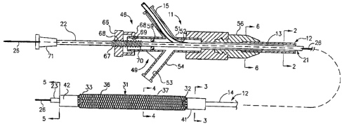
Figure 1 is a side-elevational view partially in
section of a medicament delivery device incorporating a
copolymer hydrogel encapsulating a therapeutic agent.
Figure 2 is a cross-sectional view taken along the
line 2-2 of Figure 1.
Figure 3 is a cross-sectional view taken along the
line 3-3 of Figure 1.
13
CA 02547605 2006-05-25
WO 2004/096318 PCT/US2004/013408
Figure 4 is a cross-sectional view taken along the
line 4-4 of Figure 1.
Figure 5 is a cross-sectional view taken along the
line 5-5 of Figure 1 demonstrating the electrical
connection means.
Figure 6 is a cross-sectional view taken along the
line 6-6 of Figure 1.
Figure 6a is a cross-sectional view taken along the
line 6-6 of Figure 1 also demonstrating the electrical
connection means.
Figure 7 is a greatly enlarged view of a portion of
the dilatation and medicament delivery device in a
partially expanded state.
Figures 8a-8f depict a variety of electric waveforms
for use in iontophoresis and electrophoresis with the
catheter and distal mesh of the present invention.
Figure 9 is a side-elevational view of the distal
extremity of the device shown in Figures 1-9 showing the
distal extremity With the expansion member in an expanded
condition showing the hydrogel with encapsulated
medicaments coated on the distal expansion member.
Figure 10 is a cross sectional view of the flexible
elongated elements demonstrating the active electrically
mediated dispensing of the hydrogel encapsulated
therapeutic agent or medicament into the vessel wall.
14
CA 02547605 2006-05-25
WO 2004/096318 PCT/US2004/013408
Figure 11 is another cross sectional side view of the
flexible elongated elements and a vessel demonstrating the
electrically mediated dispensing of the liposome or
micelle-encapsulated therapeutic agent or medicament into
the vessel wall.
Figure 12 is a representation of the chemical
structure of the multi-block copolymer With two functional
groups used to fabricate the hydrogel.
Figure 13 is a representation of the hydrogel formed
network that encapsulates therapeutic agents or
medicaments.
DETAThED DESCRIPTION OF THE DRAWINGS
In general, the present invention relates generally to
devices that are used to dilate and dispense a medicament
or therapeutic agent to an obstruction within a stenotic
segment of a vessel or other tubular structure. The device
is comprised of an cylindrical expansion member to be
disposed in an obstruction in a vessel carrying flowing
blood. The cylindrical expansion member has first and
second ends and an intermediate portion between the first
and second ends. The cylindrical expansion member also has
a flow passage extending therethrough with a diameter and a
longitudinal central axis. The diameter of the flow passage
is a variable with movement of the first and second ends
relative to each other along the longitudinal central axis
from a diametrically contracted position to a diametrically
expanded condition. The cylindrical expansion member is
comprised of a plurality of flexible elongate elements each
of Which extends helically about the longitudinal extending
15
CA 02547605 2006-05-25
WO 2004/096318 PCT/US2004/013408
central axis. The flexible elongate elements are coated
With a copolymer hydrogel encapsulating a therapeutic
agent, medicaments, drugs, pharmaceuticals, plasmids,
genes, double and single stranded DNA or other agents. For
the purposes of this application, the terms copolymer
hydrogel encapsulating a medicament or therapeutic agent,
drugs, pharmaceuticals, plasmids, genes or other agents,
will be used to encompass all the particular agents
described herein. It is also contemplated that the
copolymer hydrogel encapsulated medicament or therapeutic
agent may be incorporated with a non-medicament substrate
that has been previously or simultaneously coated on the
flexible elongate elements.
Means are provided for engaging the first and second
ends of said cylindrical expansion member for retaining
said first and second ends in contracted positions. Means
are provided for causing relative axial movement of the
first and second ends towards each other to cause the
intermediate cylindrical portion of the expansion member to
contract longitudinally and to expand diametrically by
causing the flexible elongate elements in the intermediate
portion of the cylindrical member to move closer to each
other expanding the diametric dimensions of the cylindrical
expansion member thereby allowing it to contact the vessel
wall and enable it to dilate an obstruction within the
vessel. Flexible elongate elements at the first and second
ends of the cylindrical expansion member remain contracted
around and within first and second means and are thereby
prevented from moving closer which maintains spacing
between the flexible elongate members so that blood in the
vessel can continue to flow through the first and second
ends and through the flow passage in the cylindrical
expansion member while the cylindrical expansion member is
16
CA 02547605 2006-05-25
WO 2004/096318 PCT/US2004/013408
in engagement with vessel wall and dilating an obstruction
within the vessel.
More in particular as shown in Figures 1-7 of the
drawings, the mechanical dilatation and medicament delivery
device 11 shown therein consists of a first or outer
flexible elongate tubular member 12 having proximal and
distal extremities 13 and 14 with the flow passage 16
extending from the proximal extremity 13 to the distal
extremity 14. Figures 2, 3, 5, and 6 are provided to
demonstrate the electrical conduction means extending from
the proximal connector and engaged to the distal expansion
member 31. A second or inner flexible tubular member 21 is
coaxially and slidably disposed within the flow passage 16
of the first or outer flexible elongate tubular member 12
and is provided with proximal and distal extremities 22 and
23 with a flaw passage 24 extending from the proximal
extremity 22 to the distal extremity 23. Since the
flexible elongate elements of the dilating member are made
of a metallic material such as stainless steel, elgiloy or
other conductive material, an electrical lead can be
connected to the mesh to make it part of the circuit. The
electrical lead can either run along or Within one of the
lumens of the catheter or can be in the form of a braid
that is made of a conductive material and have generally
functions to provide reinforcement to the catheter shaft.
A second electrode could be placed on the distal tip of the
catheter via a small band With its electrical lead running
down one of the lumens to the proximal end of the catheter.
Alternatively, the electrical lead could be engaged to the
patient's skin or could be the guidewire over which the
catheter is routinely advanced.
17
CA 02547605 2006-05-25
WO 2004/096318 PCT/US2004/013408
A guide wire 26 of a conventional type is adapted to
be introduced through the flow passage 24 in the inner
flexible elongate tubular member for use in guiding the
mechanical dilatation and medicament delivery device 11 as
a over-the-wire design as hereinafter described. The guide
wire 26 can be of a suitable size as for example 0.010"-
0.035" and can have a suitable length ranging from 150 to
300 centimeters. Fox example, the first or outer flexible
elongate tubular member 12 can have an outside diameter of
0.6-3 millimeters With a wall thickness of 0.12 millimeters
to provide a flow passage of 0.75 millimeters in diameter.
Similarly, the second or inner flexible elongate tubular
member 21 can have a suitable outside diameter as for
example 0.6 millimeters with a wall thickness of 0.12
millimeters and a flow passage 24 of 0.45 millimeters in
diameter. The flexible elongate tubular members 12 and 21
can be formed of a suitable plastic as for example a
polyimide, polyethylene, Nylon or polybutylterphalate
(PBT) .
In accordance with the present invention an
essentially cylindrically shaped expansion member 31 is
provided which has a first or proximal end 32 and a second
or distal end 33 with a central or inner flow passage 34
extending from the proximal end 32 to the distal end 33
along a longitudinally extending central axis and has a
diameter which is a variable as hereinafter described. The
cylindrically shaped expansion member 31 is comprised of a
plurality of flexible elongate elements or filaments 36
34 each of which extends helically about the longitudinally
extending central axis. The flexible elongate elements 36
are formed of suitable materials which can be utilized in
the human blood as for example stainless steel, Nitinol,
AermetTM, ElgiloyT~ or certain other plastic fibers. The
I$
CA 02547605 2006-05-25
WO 2004/096318 PCT/US2004/013408
flexible elongate elements 36 can have a suitable diameter
as for example 0.001 to 0.010 inches or can be configured
as a round, elliptical, flat or triangular wire ribbon. A
plurality of the flexible elongate elements 36 have a first
common direction of rotation about the central axis as
shown in Figures 1 and 7 are axially displaced relative to
each other and cross a further plurality of the flexible
elongate elements 36 also axially displaced relative to
each o her but having a second common direction of rotation
opposite to that of the first direction of rotation to form
a double helix or, braided or mesh-like cylindrical
expansion member with the crossing of flexible elongate
elements 36 occurring in the area of contact between the
flexible elongate elements to form openings or interstices
37 therebetween. Thus the flexible elongate elements 36
form an expansion member 31 which provides a central or
inner flow passage 34 which is variable in diameter upon
movement of the first and second ends of the expansion
member 31 relative to each other along the longitudinally
extending central axis.
Means is provided for constraining the first and
second or proximal and distal ends 32 and 33 of the
expansion member 31 and consists of a first or proximal
collar 41 and a second or distal collar 42. The first and
second collars 41 and 42 are formed of a suitable material
such as a polyimide.
Typically the distance between the first and second
collars 41 and 42 can range from between 5 to 150
millimeters. Typically the distal end 23 of the secand or
inner flexible elongate tubular member 21 extends
approximately 5-170 millimeters beyond the distal extremity
19
CA 02547605 2006-05-25
WO 2004/096318 PCT/US2004/013408
14 of the first or outer flexible elongate tubular
member 12.
It can be seen that by moving the first or outer
flexible elongate tubular member 22 and the second inner
flexible elongate tubular member 21 axially with respect to
each other, the first and second ends of the expansion
member 31 are moved towards each other causing the elongate .
elements or filaments 36 of an intermediate portion of the
cylindrical expansion member between the first and second
ends to move closer to each other to cause these flexible
elongate elements to move into apposition with each other
and to expand in a first radial direction the intermediate
portion of the cylindrical expansion member 31 (Figure 7)
and to cause the diameter of the central flow passage 34 to
increase. The portions ~of the expansion member 31
immediately adjacent the first and second collars 41 and 42
remain restrained by the collars 41 and 42 causing the
flexible elongate elements 36 immediately adjacent to the
collars 41 and 42 to curve comically toward and remain
crossed and unable to come into close apposition and
thereby provide openings or interstices 37 therebetween,
which remain relatively constant in shape and size so that
blood can flow from the first and second ends 32 and 33
through the central ox inner flow passage 34 as hereinafter
described.
The essentially cylindrical shape of the expansion
member When expanded in a radial direction provides an
enlarged surface of contact between the expansion member
and the vessel wall or obstruction. This enlarged surface
of contact enables the cylindrical expansion member to
deliver an increased amount of medicament or, therapeutic
agent Which is incorporated within the hydrogel coated on
20
CA 02547605 2006-05-25
WO 2004/096318 PCT/US2004/013408
the surface of the flexible elongate elements that comprise
the expansion member. This delivery of medicament or
therapeutic agent may be by the various well known means
previously described electrically active diffusion,
pressure, iontophoresis, electroporesis or electro-osmosis.
One example of the means provided in the mechanical
dilatation and medicament delivery device 11 for causing
relative movement between the first or outer flexible
elongate tubular member 12 and the second or inner flexible
elongate tubular member 21 and consists of a linear
movement mechanism 46. The linear movement mechanism 46
includes a Y-adapter 49 that is provided with a central arm
51 having a lumen 52 through which the second or inner
flexible elongate tubular member 21 extends.
It should be appreciated that even though one
particular linear movement mechanism 46 has been provided
for advancing and retracting the flexible elongate members
12 and 21 with respect to each other, other mechanisms also
can be utilized if desired to provide such relative
movement. Other possible designs that could be employed
are scissors-jack, racket-type or straight slide
mechanisms.
The distal expansion member of the catheter is coated
With one or more layers of a hydrogel copolymer material or
similar substrate, into which are encapsulated one or more
medicaments or therapeutic agents and zero or more
electrophoretic carriers. These charged electrophoretic
carriers may include by example, sodium lauryl sulfate,
phophatidyl choline of various hydrocarbon chain lenglits,
bile salts, phospholipids or any of the other charged
21
CA 02547605 2006-05-25
WO 2004/096318 PCT/US2004/013408
molecules which would augment the electrophoretic and
electro-osmotic processes previously described.
Once the site of obstruction or treatment is reached
and the distal cylindrical expansion member is expanded and
in contact with the surrounding tissue or vessel wall an
electrical charge is applied to the mesh thus driving the
therapeutic agent or medicament out of the hydrogel coating
and into the target tissue. Tn this case, the second
14 electrode is placed on the skin of the patient which would
act to complete the electrical circuit. The electrical
charge applied to the distal mesh and hydrogel coating with
encapsulated medicament may be used to cause
electrophoretic or electro-osmotic migration of the
therapeutic agent ox medicament into the target tissues.
As shown in Figures 8a-8f, the present invention can
employ flow of electrical current in the form of various
waveforms to perfarm the electrophoretic and electro-
osmotic procedures. Possible waveforms contemplated far
the present invention include square waves, rectangular
waves, saw-toothed waves, sinusoidal waves that do not
reverse polarity, rectified sinusoidal waves, and other
waveform shapes which may reverse polarity but provide a
net flow of current in the desired'direction.
Electrical current could also be coordinated with the
patient's elctracardiogram such that electrical current is
provided only during certain phases of cardiac
depolarization. This "gating" of the electrical current
would avoid the potential danger of discharging electrical
current to the heart during vulnerable phases of
depolarization which may lead to cardiac arrhythmias.
22
CA 02547605 2006-05-25
WO 2004/096318 PCT/US2004/013408
As seen in Figure l0 the flexible elongated elements
36,are designed to be electrically conductive and cause the
therapeutic agent or medicament 40 to dispense or migrate
.into the vessel wall 17.
Figure 1.1 is a cross sectional side view of the
flexible elongated elements 36 demonstrating the passive or
electrically active dispensing of the therapeutic,agent or
medicament 40 into the vessel wall 17.
Referring. to Figure 12, the hydrogel copolymer 100
used in this invention is a mufti-block copolymer having
two functional groups. The first functional group is a
hydrophobic nitrile groups 110 and the second function
group a hydrophilic amide groups 120. The
polyacrylonitrile-polyacrylamide copolymer is converted
from the linear pvlyacrylonitrile. Upon conversion, the
linear homopolymer becomes an alternating copolymer. After
conversion and in the presence of water, the nitrite groups
collapse and the amide groups hydrate and the polymer forms
a hydrogel network structure 130, with pores 132 (amide
groups) and crosslinks 134 (chain entanglement and nitrite
groups) .
The conversion process allows the precise control of
the degree of conversion and the size of each block. The
degree of conversion and the size of each block, among
other factors, influence the chemical and physical
properties of the resulting hydrogel. The hydrogel for
delivery of medicaments in this applications may have a
degree of conversion from approximately 55~ to 85~ which
corresponds to approximately 60~ to 950 of water content.
23
CA 02547605 2006-05-25
WO 2004/096318 PCT/US2004/013408
The converted block copolymer is dissolved in a
suitable solvent at a certain concentration (polymer
precursor solution) to generate the desirable viscosity for
coating. At this time, the polymer solution is coated onto
the catheter. Alternatively, the medicaments or
therapeutic agents along With zero or more electrophoretic
carriers are mixed with polymer precursor solution before
coating. The medicaments or the eleetrophoretic carriers
can be either in the solid form, liquid form or solution
form in a suitable solvent. The methodology for thoroughly
blending and mixing the medicaments or therapeutic agents
or electrophoretic carriers is well known to those skilled
art or can be determined by reference to standard
references.
Therapeutic agents 40 that can be employed may be
anticoagulants, such as D-Phe-Pro-Arg chloromethyl ketone,
an RGD . peptide-containing compound, heparin, an
antithrombin compound, a platelet receptor antagonist, an
anti-thrombin antibody, an anti-platelet receptor antibody,
aspirin, a prostaglandin inhibitor, a platelet inhibitor, a
tissue factor inhibitor, or a tick anti-platelet peptide.
The therapeutic agents 40 can also be a promoter of
vascular cell growth, such as a growth factor stimulator, a
growth factor receptor agonist, a transcriptional
activator, and a translational promoter. Alternatively,
the therapeutic agent 40 can be an inhibitor of vascular
cell growth, such as a growth factor inhibitor, a growth
24
CA 02547605 2006-05-25
WO 2004/096318 PCT/US2004/013408
factor receptor antagonist, a transcriptional repressor, a
translational repressor, an ant,isense DNA, an antisense
RNA, a single-stranded DNA molecule, a double-stranded DNA
molecule, a single-stranded RNA molecule, a double-stranded
RNA molecule, a replication inhibitor, an inhibitory
antibody, an antibody directed against growth factoxs, a
bifunctional molecule consisting of a growth factor and a
cytotoxin, or a bifuncti.onal molecule consisting of an
antibody and a cytotoxin.
The therapeutic agent 40 can be a cholesterol-lowering
agent such as atorvastatin, simvistatin, pravastatin and
lovastatin, a vasodilating agent, or other agents that
interfere with endogenous vasoactive mechanisms.
Additionally, the therapeutic agent 40 can be a smooth
muscle inhibitor, such as: an agent that modulates
intracellular calcium binding proteins; a receptor blocker
for contractile agonists; an inhibitor of the sodium/
hydrogen antiporter; a protease inhibitor; a vasodilator; a
phasphodiesterase inhibitor; a phenothiazine; a compound
that increases or mimics endogenous nitrous oxide; a gxowth
factor receptor agonist; an anti-mitotic agent; an
immunosuppressive agent; or a protein kinase inhibitor.
In addition, the therapeutic agent 40 can be a smooth
muscle inhibitor selected from the group consisting of an
agent that modulates intracellular calcium binding
proteins, a receptor Mocker for contractile agon~,sts, an
inhibitor of the sodium/hydrogen antiporter, a protease
25
CA 02547605 2006-05-25
WO 2004/096318 PCT/US2004/013408
inhibitor, a nitrovasodilator, a phosphodiesterase
inhibitor, a phenothiazine, a growth factor receptor
agonist, a growth factor receptor antagonist, an anti
mitotic agent, an immunosuppressive agent, a steroid such
as estrogen, hydrocortisone or dexamethasone, and a
protein kinase inhibitor, and combinations thereof.
Furthermore, the therapeutic agents 40 employed can be
compounds that inhibit cellular proliferation such as
Paclitaxel, Rapamycin, Actinomycin D, Methotrexate,
Doxorubicin, cyclophosphamide, and 5-fluorouracil, 6-
mercapatopurine, 6-thioguanine, cytoxan, cytarabinoside,
cis-platin, alcohol, arsenic trioxide, bleomycin,
captothecin, capecitabine, carmustine, celecoxib,
daunorubucin, docetaxel, etoposide, exemestane,
fludarabine, gemcitabine, hydroxyurea, idarubicin,
irinotecan, ifosfamide, letrozole, leucovorin,
mitoxantrone, pamidronate, pentostatin, porfirmer sodium,
streptozotocin, tamoxifen, ~ temozolamide, tenopside,
topotecantoremifene, tretinoin, valrubicin, vinorelbine,
zoledronate, altretamine, anastrozole, bexarotene,
carboplatin, everolimus, chlorarnbucil, busulfan, and any
other drug that can inhibit cell proliferation, and
combinations thereof.
To perform as a hydrogel coated device for electrical
mediated drug delivery, the distal expansion member will be
coated as described in more detail below.
26
CA 02547605 2006-05-25
WO 2004/096318 PCT/US2004/013408
The viscous polymer precursor solution, either by
itself or mixed With medicaments with or without charged
electrophoretic carriers, is spread onto a 'clean plastic
plate. The mixture is gently mixed using~a metal spatula or
a glass rod. Upon mixing, the viscous solution is drawn
down to a predetermined thickness within a certain surface
area., The thickness is related to the desired final coating
thickness on the catheter the desired length of coating on
the catheter. The distal cylindrical dilating mesh is then
rolled over the polymer precursor. A single layer or
multiple layers of viscous polymer precursor containing
medicaments and electrophoretic carriers are then deposited
onto the catheter mesh surface.
Other coating methods may also be employed to deposit a
uniform and defined layer of polymer solution onto the
surface of the catheter mesh. The conventional coating
technology is well known to those skilled in the art or can
be determined by reference to standard references.
The coated catheter is then dipped into an appropriate
solution to coagulate the polymer to form hydrogel. The
solution can be,aqueous solution containing electrolytes or
other coagulants such as ethanol and Water mixtures. The
coagulation step converts a viscous polymer precursor
solution into a water-swellable hydrogel. Sometimes, a
27
CA 02547605 2006-05-25
WO 2004/096318 PCT/US2004/013408
secondary solution is used to remove remaining polar solvent
or other non-water coagulants such as ethanol.
The resulting hydrogel polymer structure is a network
130 consisting of pores 132 and crosslinks 134 With the
medicaments and zero or more eleetrophoretic carriers
residing in this structure. The pores 132 are connected
together through the polymer chain entanglement 134 and
through the collapsed hydrophobic nitrile groups 110 which
have strong association through their dipolar interactions.
The dipolar interactions of the nitrile groups 110
provide the structural integrity of the gel network and the
mechanical strength of the hydrogel. The pores 132 (the
hydrated amide groups) serve as water channels to transport
medicaments and electrophoretic carriers upon the
application of electrical energy. Figure 13 shows the
encapsulated medicaments or therapeutic agent as single
structure 136, multiple structure 138, precipitates in a
solution 140, or as piece of DNA or RNA 142.
The coating can be a one-step operation to coat one
layer of hydrogel. However, additional layers serving
different purposes may also be added. The additional layers
of hydrogel may be of the same kind, or of a different
kind, of polymers depending on the desired application For
example, a very thin layer of hydrogel may be applied to
the catheter mesh ' surface to promote adhesion.
Alternatively, a secondary layer formed of the same or a
different h dro el ma be a lied to cover the
y g y pp primary
coating containing the drugs or medicaments in a manner
similar to that described above. This coating may contain
zero or more additional drugs or medicaments and zero or
28
CA 02547605 2006-05-25
WO 2004/096318 PCT/US2004/013408
more electrophoretic carriers. Depending on application
requirements, multiple layers of polymer coating may be
used.
S One of the purposes of the secondary coating is to
retard the passive diffusion of the previously encapsulated
drugs or medicaments and electrophoretic carriers into the
bloodstream or tissues Which may occur prior to the active
'delivery of the drug or medicaments using electricity.
Operation and use of the polymer coated device for
electrically mediated drug delivery 11 may now be briefly
described as follows. Let it be assumed that the patient
in whom the medical procedure is to be performed utilizing
the medicament delivery device 11 has one or more stenoses
Which at least partially occlude one or more arterial
vessels supplying blood to the heart and that it is desired
to enlarge the flow passages through these stenoses.
Typically the polymer coated device for electrically
mediated drug delivery 11 would be supplied by the
manufacturer with the cylindrical expansion member 31 in
its most contracted position to provide the lowest possible
configuration in terms of diameter and so that the diameter
approximates the diameter of the outer flexible elongate
tubular member 12 and previously coated With a therapeutic
agent or medicament 40 and zero or more electrophoretic
charged carriers. A means for ,'maintaining the coated
expansion member exposed to water Will be employed.
Preferably, the coated expansion member 35 should have
a diameter that is only slightly greater than the tubular
member 12, as for example by 1.0 - 2.3 millimeters. The
first and second collars 41 and 42 also have been sized so
they only have a diameter that is slightly greater than the
29
CA 02547605 2006-05-25
WO 2004/096318 PCT/US2004/013408
outer diameter of the outer flexible elongate tubular
member 12. To bring the cylindrical expansion member 31 to
its lowest configuration, the linear movement mechanism 46
has been adjusted so that there is a maximum spacing
between the distal extremity 23 of the inner flexible
elongate tubular member 21 and the distal extremity 14 of
the outer flexible elongate tubular member 12. In this
position of the expansion member 31, the flexible elongate
elements 36 cross each other at nearly right angles so that
the interstices or openings 37 therebetween are elongated
with respect to the longitudinal axis.
The polymer coated device for electrical mediated drug
delivery 11 is then inserted into a guiding catheter (not
shown) typically used in such a procedure and introduced
into the femoral artery and having its distal extremity in
engagement with the ostium of the selected coronary artery.
The guide wire 26 is then advanced in a conventional
manner by the physician undertaking the procedure and is
advanced into. the vessel containing a stenosis. The
progress of the distal extremity of the guide wire 26 is
observed fluoroscopically and is advanced until its distal
extremity extends distally of the stenosis. With the
expansion member 31 in its diametrically contracted
position and the hydrogel encapsulated medicament or
therpeutic agent coated thereon, the polymer coated device
for electrical mediated drug delivery 11 is advanced over
the guide wire 26 until the distal end a.s centered within
the region of interest.
After the hydrogel-coated cylindrical expansion member
31 is in a desired position in the stenosis, the
cylindrical expansion member 31 is expanded from its
30
CA 02547605 2006-05-25
WO 2004/096318 PCT/US2004/013408
diametrically contracted position to an expanded position
by moving the distal extremities 14 and 23 closer to each
other by operation of the screw mechanism 46. This can be
accomplished by holding one distal extremity stationary and
moving the other distal extremity towards it or by moving
both distal extremities closer to each other
simultaneously.
When the hydrogel coated' distal cylindrical
expansion member 31 is fully expanded it i.s almost a solid
tubular mass which has significant radial strength to fully
expand a stenosis or alternatively a stent or prosthesis.
Since the expansion member is coated With a hydrogel
encapsulating a therapeutic agent or medicament this drug
or medicament can be delivered to the vessel during the
time of device expansion while blood is permitted to flow
unobstructed to the distal vessel.
Now, an electrical charge can be provided to the
cylindrical expansion member. This charge will then tend to
drive the encapsulated medicaments or therapeutic agents
and the zero or .more electrophoretic carriers into the
tissue through electrophoretic, iontophoretic or electro-
osmotic means. The process is known to facilitate or assist
the transport of the encapsulated medicament or therapeutic
agents and the electrophoretic carriers across the
selectively permeable membranes and enhance tissue
penetration. Since the present invention involves the use
of electrical energy, there are many possible waveforms
contemplated for use. As depicted in Figs 8a-8f, square
waves 61, rectangular waves 63, saw toothed waves 64,
sinusoidal Waves that do not reverse polarity 65, rectified
sinusoidal Waves, 72 and modified rectangular or other
waves 73. The primary characteristic of the preferred
waveforms is that they all provide a net flow of current to
31
CA 02547605 2006-05-25
WO 2004/096318 PCT/US2004/013408
the coated expansion member 35. It must be appreciated by
those skilled a.n the art, that the waveforms with
frequencies and duty cycles must be capable of delivering
the desired current under varying impedances encountered by
the expansion member 35 and the surrounding vessel wall 17
and fluids.
After a predetermined time, the electrical current can
be altered to achieve another purpose or terminate.
.After delivery of the medicaments or therapeutic agent
to the lesion has been carried out for an appropriate
length of time, the expansion member 31 can be moved from
its expanded position to a contracted position.
After the expansion member 31 has been reduced to its
contracted or minimum diameter, the polymer coated device
for electrically mediated drug delivery 11 can be removed
along With the guide Wire 26 after which the guiding
catheter (not shown) can be removed and the puncture site
leading to the femoral artery closed in a conventional
manner.
Although, the procedure hereinbefore described was for
treatment of a single stenosis or region of. interest, it
should be appreciated that if desired during the same time
another stenosis or region of interest need be treated, the
catheter may be advanced to this second area of interest
and the procedure repeated. Alternatively, another polymer
coated device for electrical mediated drug delivery 11 may
be re-inserted in the same or other vessels or regions of
interest of the patient and can be treated in a similar
manner.
32
CA 02547605 2006-05-25
WO 2004/096318 PCT/US2004/013408
Described below are some examples of experiments
conducted using the present invention.
Example 1. Local Delivery of Paclitaxel
Preparation of compounded paclitaxol,sodium dodecyl
sulfate (SDS) and hydrogel polymer solution precursor for
coating.
Prepare stock solutions of paclitaxol and SDS in the
solvent of dimethyl sulfoxide (DMSO) at the concentration
of 0.5mg/ul for paclitael in DMSO and 0.6mg/ul for SDS in
DMSO. Weigh 433mg of hydrogel solution onto a clean plastic
plate having two elevated spacers at 0.5mm height and
separated by ~cm. Add 81.3u1 of SDS stock solution, or
48.8mg of SDS, and 9'7.5u1 of Paclitaxel stock solution, or
48.8mg of Paclitaxel onto the hydrogel polymer solution.
Mix well with a spatula. Upon mixing, spread the viscous
mix evenly with the spatula or a glass rod within the
confines of the two spacers. Use both gloved hands to hold
both ends of the catheter mesh portion and roll the mesh
gently over the viscous polymer mix once, or twide if
necessary. This coating is designated to contain 5%
paclitaxel and 5% SDS using the initial weight of polymer
mix as a reference.
When the coating is completed, the mesh portion of the
catheter is dipped into 3 ml of trisborate buffer for 3
minutes to coagulate the polymer solution into gel. After
33
CA 02547605 2006-05-25
WO 2004/096318 PCT/US2004/013408
3 minutes, the mesh portion of the catheter a.s removed from
the coagulation solution and ready for use.
Example 2. Local Delivery of E2F Decoy
Preparation of compounded E2F Decoy, sodium dodecyl
sulfate (SDS) and hydrogel polymer solution precursor for
coating.
Prepare SDS stock solution in the solvent of dimethyl
sulfoxide (DMSO) at the concentration of 0.6mg/ul. Weigh
747mg of hydrogel solution onto a clean plastic plate
having two elevated spacers at 0.5mm height and separated
by 5cxn. Add 69.8mg of E2F Decoy and 70u1 of DMSO. Mix
well. Add 35ul,of SDS stock solution (or 2lmg of SDS). Mix
well With the spatula. Upon mixing, spread the viscous mix
evenly With the spatula or a glass rod Within the confines
of the two spacers. Use both gloved hands to hold both
ends of the catheter mesh portion and roll the mesh gently
over the viscous polymer mix once, or twice if necessary.
This coating is designated to contain 7.5% paclitaxel and
2.25% SDS using the initial weight of polymer mix as a
reference.
When the coating is completed, the mesh portion of the
catheter is dipped into l3ml of ethanol/water solution
containing 75% ethanol and 25% water for 1 minutes.
Following the coagulation, the mesh is dipped into a second
solution for 2 minutes. The second solution is l3ml of
saline containing 20% glycerol. After completing the
immersion for 2 minutes in the second solution, the mesh
portion of the catheter is removed from the coagulation
solution and ready for use.
34
CA 02547605 2006-05-25
WO 2004/096318 PCT/US2004/013408
In an experiment to measure the amount of taxol
released, the catheter Was placed in an electrolytic cell
and discharged at a current of 10 milliamps and a voltage
of 10 volts. The solution~was analyzed for paclitaxel
concentration. There was minimal paclitaxel release
Without power, but a significant increase of the release of
paclitaxel with pov~rer. In another experiment, catheters
loaded with a fluorescent paclitaxel compound (Oregon Green
paclitaxel) were placed into isolated coronary arteries.
The catheter Was expanded and current (10 millamps, 10
volts) for 10 minutes . The arterial segment With no power
had the fluorescent compound limited to the luminal surface
that was in contact with the catheter, consistent with
little or no movement of the paclitaxel. In the arterial
segment treated with power, the fluorescent compound was
seen throughout the artery, consistent with movement of the
fluorescent compound through the arterial wall.
In an experiment to measure the amount of taxol
released, the catheter Was placed in an electrolytic cell
and discharged at a current of 10 milliamps and a voltage
of 10 volts. The solution was analyzed for paclitaxel
concentration. There was minimal paclitaxel release
without power, but a significant increase of the release of
paclitaxel with power. In another experiment, catheters
loaded with a fluorescent paclitaxel compound (Oregon Green
paclitaxel) were placed into isolated caronary arteries.
The catheter was .expanded and current (10 millamps, 10
volts) for 10 minutes. The arterial segment with no power
had the fluorescent compound limited to the luminal surface
that Was in contact With the catheter, consistent With
little or no movement of the paclitaxel. In the arterial
segment treated with power, the fluorescent compound Was
seen throughout the artery, consistent with movement of the
fluorescent campound through the arterial wall.
CA 02547605 2006-05-25
WO 2004/096318 PCT/US2004/013408
Example 3. Ionto~horetic Release of Paclitaxel
In an experiment to measure the amount of taxol released, a
mesh segment that Was coated with a hydrogel that was
loaded with paclitaxel according to the procedure described
in Example 1 was placed in an electrolytic cell and
discharged at a current of 10 milliamps and a voltage of 10
volts for 10 minutes. The solution Was then analyzed for
paclitaxel concentration by high performance liquid
chromatography. As shown in Figure 1, there Was minimal
paclitaxel release without power, but a significant
increase of the release of paclitaxel with power.
Example 4. Iontophoretic Delivery of Pacltaxel into
Isolated Coronary Arteries
Catheters with mesh segments that were coated with a
hydrogel that Was loaded With a fluorescent paclitaxel
compound (Oregon Green paclitaxel) were placed into
isolated coronary arteries. The catheter was expanded and
current (10 millamps, 10 volts) for 10 minutes. As seen in
Figure 2, the arterial segment With no power had the
fluorescent compound limited to the luminal surface that
Was in contact with the catheter, consistent with little or
no movement of the paclitaxel. In the arterial segment
treated with power, the fluorescent compound Was seen
throughout the artery, consistent with movement of the
fluorescent compound throughout the arterial wall.
35
36
CA 02547605 2006-05-25
WO 2004/096318 PCT/US2004/013408
Example 5. Iontophoretic Release of E2F Decoy
In an experiment to measure the amount of E2F decoy
released, a mesh segment that was coated with a hydrogel
that was loaded with E2F decoy according to the procedure
described in Example 4 was placed in an electrolytic cell
and discharged at a current of 10 milliamps and a voltage
of 10 volts for 10 minutes. The solution Was then analyzed
for E2F decoy concentration by ultravioilet
spectrophotometry. As shown in Figure 3, there Was minimal
E2F decoy release without power, but a significant increase
of the release of E2F decoy With power.
Example 6. Iontophoretic Delivery of E2F Decoy into
Isolated Coronary Arteries
Catheters With mesh segments that were coated with a
hydrogel that was loaded with a fluorescent E2F Decoy
compound (Bodipy-labeled E2F Decoy) were placed into
isolated coronary arteries. The catheter was expanded and
current (l0,millamps, 10 volts) for 10 minutes. As seen in
Figure 4, the arterial segment with no power had the
fluorescent compound limited to the luminal surface that
Was in contact with the catheter, consistent With little or
no movement of the E2F Decoy. In the arterial segment
treated with power, the fluorescent compound was seen
throughout the artery, consistent with movement of the
fluorescent compound throughout the arterial wall.
35
37