Note: Descriptions are shown in the official language in which they were submitted.
CA 02547998 2006-06-02
WO 2005/062040 PCT/US2004/038298
METHOD AND SYSTEM FOR ANALYZING REACTIONS USING AN
INFORMATION SYSTEM
COPYRIGHT NOTICE
Pursuant to 37 C.F.R. 1.71 (e), applicants note that this disclosure contains
material that is subject to and for which is claimed copyright protection,
such as, but
not limited to, source code listings, screen shots, user interfaces, user
instructions,
and any other aspects of this submission for which copyright protection is or
may be
available in any jurisdiction. The copyright owner has no objection to the
facsimile
reproduction by anyone of the patent document or patent disclosure, as it
appears in
the records of the Patent and Trademark Office. All other rights are reserved,
and all
other reproduction, distribution, creation of derivative works based on the
contents,
public display, and public performance of the application or any part thereof
are
prohibited by applicable copyright law.
-,_
BACKGROUND OF THE INVENTION
1. FIELD OF THE INVENTION
The present invention relates to analysis of data of nucleic acid
amplification reactions. More specifically, the invention relates to an
information
system and method for making determinations regarding chemical and/or
biological
reactions. The invention also involves an alternate method of quantifying
nucleic
acids in a sample comprising amplification of a target nucleic acid and
analysis of
data obtained during the amplification reaction. The invention further
involves a
diagnostic system and/or kit using real-time nucleic acid amplification
including, but
not limited to, PCR analysis.
2. DISCUSSION OF THE ART
In many different industrial, medical, biological, and/or research fields, it
is
desirable to determine the quantity of a nucleic acid of interest. Some
methods of
CA 02547998 2006-06-02
WO 2005/062040 PCT/US2004/038298
quantifying nucleic acids of interest involve amplifying them and observing a
signal
proportional to the quantity of amplified products made; other methods involve
generating a signal in response to the presence of a target nucleic acid,
which signal
accumulates over the duration of the amplification reaction. As used herein,
nucleic
acid amplification reaction refers both to amplification,of a portion of the
sequence of
a target nucleic acid and to amplification and accumulation of a signal
indicative of
the presence of a target nucleic acid, with the former often being preferred
to the
latter. The quantification of nucleic acids is made more difficult or less
accurate or
both because data captured during amplification reactions are often
significantly
obscured by signals that are not generated in response to the target nucleic
acid
(i.e., noise). Furthermore, the data captured by many monitoring methods can
be
subject to variations and lack of reproducibility due to conditions that can
change
during a reaction or change between different instances of a reaction. In view
of the
above, there is a need to develop improved means of quantifying a nucleic
acid.
Where quantification of nucleic acids is enabled by amplification reactions,
there is
also a need to improve current methods of detecting suspect or invalid
amplification
reactions. There further remains a need to improve current abilities to
distinguish
between amplification reactions that do not detect a target nucleic acid
(i.e., negative
reactions) from weak signals obtained from amplification reactions suffering
from low
quantities of a target nucleic acid in a sample, a degree of inhibition of the
amplification reaction, or other causes. The present invention provides
improvements in these areas as is disclosed below.
A non-exhaustive list of references providing background information
regarding the present invention follows:
Livak, K. and Schmittgen, T., Analysis of Relative Gene Expression Data
Using Real-Time Quantitative PCR and the 22DDCT Method, METHODS
25: 402-4.08 (2001 ) doi:10.1006/meth.2001.1262.
Bustin SA, Absolute quantification of mRNA using real-time reverse
transcription PCR assays, Journal of Molecular Endocrinology 25: 169-193
( 2000)
Bustin SA., Quantification of mRNA using real-time reverse transcription
PCR: trends and problems, J Mol Endocrinol. 29: 23-29 (2002).
2
CA 02547998 2006-06-02
WO 2005/062040 PCT/US2004/038298
While the inventors cannot guarantee that the following website will remain
available
and do not necessarily endorse any opinions expressed therein, an interested
person may wish to refer to the website www.wzw.tum.de/gene-
quantification/index.shtml for useful background information.
The discussion of any works, publications, sales, or activity anywhere in
this submission, including in any documents submitted with this application,
is not
intended to be an admission of any manner that any such work constitutes prior
art,
unless explicitly stated to the contrary. Similarly, the discussion of any
activity, work,
or publication herein is not an admission that such activity, work, or
publication was
known in any particular jurisdiction.
Real-time PCR is an amplification reaction used for the quantification of
target nucleic acids in a test sample. Conventionally, skilled artisans
typically view
the amplification reaction as comprising three distinct phases. First, there
is a
background or baseline phase, in which the target nucleic acid is being
amplifiied but
the signal proportional to the quantity of the target nucleic acid cannot be
detected
because it is too small to be observed relative to signals independent of the
target
(sometimes called "background" or "background signal"). Next, there is a
logarithmic
phase in which the signal grows substantially logarithmically because the
signal is
substantially proportional to the quantity of target nucleic acid in the
amplification
reaction and is greater than the background signal. Finally, the growth in the
signal
slows during a "plateau" phase reflecting less than logarithmic amplification
of the
target nucleic acid. As is known in the art, the time at which the logarithmic
phase
crosses a threshold value, which is a value somewhat greater than the value of
the
background signal, is reproducibly related to the log of the concentration of
the target
nucleic acid. This prior art method is generically referred to as the Ct
method,
perhaps so named for the Cycle at which the signal crosses the threshold. Ct
analysis is reasonably reproducible and accurate, but suffers from some
drawbacks,
which need not be discussed here to understand the present invention.
U. S. Patent No. 6,303,305 discloses a method of quantification of nucleic
acids employing PCR reactions. The method disclosed employs the nth derivative
of
the growth curve of a fluorescent nucleic acid amplification reaction. This
method
effectively avoids the need to perform a baseline correction, but provides no
reliable
3
CA 02547998 2006-06-02
WO 2005/062040 PCT/US2004/038298
method of determining reactive from non-reactive samples, and does not
reasonably
suggest how to use an nth derivative calculation to assess the validity of the
results
obtained. In addition, nucleic acid amplification signals resulting from any
artifacts in
the system (e.g., crosstalk or positive bleedover - defined infra) cannot be
distinguished from true positive responses using the methods disclosed therein
and
can lead to false positive results. However, the first derivative calculation
disclosed
by U. S. Patent No. 6,303,305 provides an efficiency related value that is
useful in
the context of the present invention. The skilled artisan can refer to U. S.
Patent No.
6,303,305 for additional details relating to calculation of a first derivative
of a nucleic
acid amplification signal growth curve. U. S. Patent No. 6,303,305 is
incorporated by
reference only in the United States of America, and other jurisdictions
permitting
incorporation by reference, to the extent it discloses the calculation of the
first
derivative of a nucleic acid amplification growth curve. However, U. S. Patent
No.
6,303,305 does not disclose or suggest the uses of this efficiency related
value
described in this disclosure (below).
Co-owned US Provisional Patent Application No. 60/527,339, filed
December 6, 2003, discloses a method for analyzing a nucleic acid
amplification
reaction in which the log of the signal from an amplification reaction is
examined for
the maximum gradient or slope. This value, which for any data set corresponds
to a
point a certain period of time or number of cycles after the initiation of the
amplification reaction, is called the MGL of the reaction. The MGL is useful
in certain
embodiments of the present invention, particularly in those that distinguish
qualitatively those samples comprising little target nucleic acid from those
samples
that do not contain target nucleic acid. U. S. Patent Application No.
60/527,389, filed
December 6, 2003 is incorporated herein by reference in its entirety.
SUMMARY OF THE INVENTION
The present invention provides a method for determining whether a
sample contains a nucleic acid of interest, for quantifying this nucleic acid,
and for
4
CA 02547998 2006-06-02
WO 2005/062040 PCT/US2004/038298
assessing the validity or quality of the data used to reach the preceding
qualitative
and quantitative determinations.
The method of this invention method comprises contacting a sample with
amplification or detection reagents or both in order to amplify the nucleic
acid (as the
term "amplified" is used herein). The amplification reaction generates signals
indicative of the quantity of the target nucleic acid present in the sample,
which
signals are recorded at numerous points during the amplification reaction. The
signal can be measured and recorded as a function of time value, or in the
alternative, cycle number.
Suitable "efficiency related transforms" viewed or calculated as a function
of time are determined for the amplification reaction, and the point in the
amplification reaction of the maximum of the efficiency related transform, the
magnitude of the maximum of the efficiency related transform, or the width (or
similar
parameter) of a peak in the plot of the efficiency related transform as a
function of
time can be used to obtain information about the reaction. This point in the
reaction
represents the point in time or the amplification cycle at which the maximum
of the
efficiency related transform occurs. Advantageously, the maximum of the
efficiency
related transform for a particular reaction, as well as the duration and
magnitude of
substantial changes in the calculated efficiency related transform, have
consistently
reproducible relationships to the initial concentration of a target nucleic
acid in a
sample, to the reliability of the data and information generated by the assay,
to the
presence or absence of a bona fide target nucleic acid, and to other
parameters of
the reaction. Advantageously, these relationships hold even in the presence of
substantial noise and unpredictable variations in the signals) generated by
the
amplification reaction. As used herein, the term "maximum", as applied to
efficiency
related transforms, is intended to include the minimum of the efficiency
related
transform when the reciprocal of the efficiency related transform is used. One
can
use the inverse ratio, in which, in the case of a curve, the curve will start
at a value of
approximately 1 in the baseline region, decrease during the growth region, and
return approximately to one in the plateau region. The use of this transform
would
allow one to use the magnitude and the position of the trough instead of the
magnitude and position of the peak for analysis. This transform is implemented
in a
5
CA 02547998 2006-06-02
WO 2005/062040 PCT/US2004/038298
manner that essentially equivalent to the ratio method in which the maximum of
the
efficiency related transform for a particular reaction is employed.
In all embodiments, signals from the amplification reaction are measured
at intervals of time appropriate for the amplification reaction during the
amplification
reaction. These signals can be referred to as time-based or periodic
measurements,
such that' every measurement of the signal generated for a particular reaction
can be
expressed as a function of time. In some embodiments, the amplification
reaction is
cyclical (e.g., as in PCR). Because cycles often have a substantially uniform
duration, it is frequently convenient to substitute a "cycle number" for a
time
measurement. Accordingly, in some embodiments of the present invention, a
region
of data identified by one or more methods on an information processing system
as
described herein can correspond to a cycle number. However, some cyclical
amplification reactions have cycles of non-uniform duration. For these
amplification
reactions, it may be preferable to measure time in non-uniform measures. For
example, the theoretical extent of amplification in a PCR reaction having
cycles of
varying duration will be linked more directly to the number of cycles
performed rather
than the duration of the reaction. Accordingly, the skilled artisan will
readily
appreciate that the time-based measurements can easily be scaled to reflect
the
underlying amplification reaction. As is known in the art, it is often useful
to
interpolate data and results between cycle numbers, which gives rise to the
concept
of a fractional cycle number "FCN." Similarly, in reactions where measurements
are
based on time, events can be measured in fractional time units.
In further embodiments, the invention advantageously involves a system
or method or both for analyzing a reaction sample, such as a PCR reaction
sample,
that uses a substantial set of available reaction kinetics data to identify a
region of
interest, rather than using a very limited data set, such as where a reaction
curve
crosses a threshold.
In certain embodiments, an identified region can be used to determine one
or more qualitative results, or quantitative data analysis results, or both.
The
reaction point of the maximum of the efficiency related transform can be used
to
determine the concentration of a target nucleic acid in a sample or to
determine
qualitatively whether any target analyte is present in a test sample. These
and other
6
CA 02547998 2006-06-02
WO 2005/062040 PCT/US2004/038298
values can be compared with reference quantities in generally the same way
that a
threshold cycle number (Ct) or fractional threshold cycle number can be used
in the
prior art.
The reaction point corresponding to the maximum of the efficiency related
transform can be understood as indicating or being derived from a cycle number
that
is located at a relatively consistent point with respect to reaction
efficiency, such as
at a maximum of reaction efficiency or a region consistently related to a
maximum of
reaction efficiency or consistently related to some other reaction
progression.
Different methods can be used to determine a reaction point related to a
maximum of
reaction efficiency. This value can comprise adjusted FCN values (e.g.,
FCNMRAaj.
and FCNi"t. aa~.)~ as described below. In certain embodiments of this
invention,
methods of the invention can determine FCN values for multiple reaction
signals,
such as a target and/or a control and use those values in determining reaction
parameters, including, but not limited to, quantity of target nucleic acid
initially
present in a sample and the validity of the results generated by an
amplification
reaction.
The present invention can identify a value indicative of the reaction
efficiency (at times, herein, generally referred to as an "efficiency related
value"
(ERV)) at one or more regions on a signal growth curve. A specific efficiency
related
value is referred to as a MaxRatio value or MR. MaxRatio refers to one
possible
method for calculating an efficiency related value as further discussed
herein. This
is one example of a method for determining an ERV and illustrative examples
herein
that refer to MR should also be understood to include other suitable methods
for
determining an efficiency related value, including, but not limited to, the
maximum
gradient of the log of the growth curve, as described in co-owned U. S. Patent
Application No. 60/527,389, filed December 6, 2003, the maximum first
derivative of
the signal obtained from the amplification reaction (e.g., as disclosed in U.
S. Patent
No. 6,303,305), and the maximum difference between two sequential signals
obtained from the amplification reaction. Thus, this invention is involved
with an
analytical method that identifies two values for a reaction curve: (1 ) one
value related
to a cycle number or time value and (2) one value indicating an efficiency
related
value. The invention can use those two values in analysis of reaction data
7
CA 02547998 2006-06-02
WO 2005/062040 PCT/US2004/038298
performed using an information-handling system and method of using the system.
An example of two such values are FCN and MR specific embodiments discussed
below.
This invention is also involved with a method and system that uses two
values as discussed above that are determined from a reaction under
examination to
compare that reaction to one or more criteria data sets. A criteria comparison
can be
used to determine and/or correct any results and/or quantifications as
described
herein. Criteria data can be derived by generating pairs of cycle number
related
values-efficiency related values (e.g., FCN-MR pairs) from multiple
calibration
reactions of known quantity or known concentration or both.
This invention also involves one or more techniques for performing
efficiency analysis of reaction data. This analysis can be used separately
from or in
conjunction with the cycle number related value-efficiency related value
analysis
discussed herein. Efficiency analysis can be used to find a region of interest
for
making a determination about reaction data, such as, for comparison to
calibration
data sets, in a way similar to Ct analysis as understood in the art.
The present invention also provides a method for analyzing a nucleic acid
amplification reaction, in which a sample containing a nucleic acid is
contacted with
amplification agents and placed under suitable amplification conditions to
amplify a
portion of the nucleic acid in the sample. During the amplification reaction,
signals
that are proportional to the amount of the target nucleic acid present are
periodically
measured at a suitable interval. Conveniently, the interval can correspond to
the
duration of a cycle for those amplification reactions that are cyclical. The
signals are
then manipulated to determine an efficiency related transform for the
amplification
reaction. Any suitable efficiency related transform can be used for the
invention.
Efficiency related transforms preferred in the context of the present
invention include
the slope of the line, which can be determined by many techniques, including,
but
not limited to, difference calculations on sequential data points, determining
the first
derivative of a line fitted to the growth curve of the reaction signal, and
defiermining
the gradient, slope, or derivative of the log of the growth curve (i.e., Log
(growth
curve)). More preferably, the efficiency related transform is the ratio of
sequential
data points, sometimes referred to herein as the ratio curve. When the
efficiency
8
CA 02547998 2006-06-02
WO 2005/062040 PCT/US2004/038298
related transform for the reaction is known, a plot of the efficiency related
transform
as a function of time (preferably expressed in the units used to measure the
signal)
(or mathematical manipulation yielding information similar to a plot) can be
used to
identify a peak value. However, a plot is not required. The width of the peak
in the
selected range of acceptable peak widths can be determined by any suitable
technique or method. However, a preferred method for determining the
acceptable
peak width involves statistically analyzing the degree of variance in peak
widths
obtained from objectively normal amplification reactions that are very similar
to or
even identical to the amplification method analyzed by the method of this
invention.
In the reaction analyzed, an unknown test sample is usually used in place of
samples used to characterize the amplification reaction or an analyte assay.
If the
peak width of the analyzed amplification reaction falls within the prescribed
range of
acceptable peak widths, the reaction is declared normal; if the peak width of
the
analyzed amplification reaction does not fall within the prescribed range of
acceptable peak widths, the reaction is identified as having provided sub-
optimal,
aberrant, or otherwise questionable signals. The width of the leading half of
the
efficiency related transform peak is evaluated. This evaluation is a more
forgiving
measurement of amplification reaction validity, and therefore may be preferred
in
some instances, but generally not in all instances.
The invention further involves an information system and/or program able
to analyze captured data. Data can be captured as image data from observable
features of the data, and the information system can be integrated with other
components for capturing, preparing, andlor displaying sample data.
Representative
examples of systems in which the invention can be employed include, but are
not
limited to, the BioRad~ i-Cycler~, the Stratagene~ MX4000°, and the ABI
Prism
7000° systems. Similarly, the present invention provides a computer
product
capable of executing the method of this invention.
Various embodiments of the present invention provide methods and/or
systems that can be implemented on a general purpose or special purpose
information handling system by means of a suitable programming language, such
as
Java, C++, C#, Cobol, C, Pascal, Fortran, PL1, LISP, assembly, etc., and any
suitable data or formatting specifications, such as HTML, XML, dHTML, TIFF,
JPEG,
9
CA 02547998 2006-06-02
WO 2005/062040 PCT/US2004/038298
tab-delimited text, binary, etc. For ease of discussion, various computer
software
commands useful in the context of the present invention are illustrated in
MATLAB~
commands. The MATLAB software is a linear algebra manipulator and viewer
package commercially available from The Mathworks, Natick, Massachusetts
(USA).
Of course, in any particular implementation (as in any-software development
project),
numerous implementation-specific decisions can be made to achieve the
developer's
specific goals, such as compliance with system-related and/or business-related
constraints, which will vary from one implementation to another. Moreover, it
will be
appreciated that such a developmental effort might be complex and time-
consuming,
but would nevertheless be a routine undertaking of software engineering for
those of
ordinary skill in the art having the benefit of this disclosure.
The invention will be better understood with reference to the following
drawings and detailed descriptions. For purposes of clarity, this discussion
refers to
devices, methods, and concepts in terms of specific examples. However, the
invention and aspects thereof may have applications to a variety of types of
devices
and systems.
Furthermore, it is well known that logic systems and methods such as
those described herein can include a variety of different components and
different
functions in a modular fashion. Different embodiments of the invention can
include
different combinations of elements and functions and may group various
functions as
parts of various elements. For purposes of clarity, the invention is described
in terms
of systems that include many different components and combinations of novel
components and known components. No inference should be taken to limit the
invention to combinations requiring all of the novel components in any
illustrative
embodiment of this invention.
As used herein, "the invention" should be understood to include one or
more specific embodiments of the invention (unless explicitly indicated to the
contrary). Many variations according to the invention will be understood from
the
teachings herein to those of ordinary skill in the art.
10
CA 02547998 2006-06-02
WO 2005/062040 PCT/US2004/038298
BRIEF DESCRIPTION OF THE DRAWINGS
FIG. 1 is a plot of discrete captured reaction data values from 43 readings
(e.g., cycles) taken from a nucleic acid amplification reaction that can be
used in an
analysis method according to embodiments of this invention.
FIG. 2 is a plot illustrating captured reaction data showing target and
control data sets that have been normalized according to embodiments of this
invention.
FIG. 3 is a plot illustrating reaction data showing target and control data
that have been scaled according to embodiments of this invention.
FIG. 4 is a plot illustrating captured reaction data showing target and
control data after digital filtering according to embodiments of this
invention.
FIG. 5 is a plot illustrating captured reaction data showing target and
control data with slope values removed according to embodiments of this
invention.
FIG. 6 is a plot illustrating ratio transform of reaction target and control
data according to embodiments of this invention.
FIG. 7 is a plot illustrating shifted ratio transform of reaction target and
control data according to embodiments of this invention.
FIG. 8 is a plot illustrating interpolated transformed reaction data showing
target and control data that have been interpolated according to embodiments
of this
invention.
FIG. 9 is a plot illustrating interposed reaction data showing identification
of the FCN and MR points according to embodiments of this invention.
FIG. 10 is a flow chart for performing a characterization of reaction data
according to embodiments of this invention.
FIG. 11 is a plot illustrating methods for determining criteria data according
to embodiments of this invention.
FIG. 12 is a plot illustrating two sets of reaction data that illustrate how
reaction curves for same concentration initial samples can vary due to
different
reaction anomalies.
11
CA 02547998 2006-06-02
WO 2005/062040 PCT/US2004/038298
FIG. 13 illustrates peak efficiency calculations for the data sets in FIG. 12.
The figure illustrates the desirability of using an offset efFiciency
transform according
to specific embodiments of the present invention.
FIG. 14 illustrates data for an HIV assay run with eight replicates of known
concentrafiion samples at 50, 500, 5,000, 50,000, 500,000 and 5,000,000 copies
per
mL.
FIG. 15 is a plot illustrating four linear standard curves generated from
three-point calibration data using four different cycle number related values
(e.g.,
FCN, FCN2, FCNMR Adj.~ and FCNinc. Adj.) according to embodiments of this
invention.
FIG. 16 compares calculated concentrations to known concentrations for
the data illustrated in °FIG. 14 using the four curves illustrated in
Fig. 15 according to
embodiments of this invention.
FIG. 17 illustrates results using a one-point calibration according to
embodiments of this invention.
FIG. 18 illustrates experimental HBV results using MR analysis with a one-
point calibration according to embodiments of this invention.
FIG. 19 illustrates experimental HBV results using MR analysis and FCNMR
adj. with a one-point calibration according to embodiments of this invention.
FIG. 20 illustrates experimental HBV results using Ctanalysis and a one-
point calibration according to embodiments of this invention.
FIG. 21 illustrates experimental HIV results using MR analysis and one-
point calibration, e.g. using 103 and 10' copies/mL responses as calibrators,
according to embodiments of this invention.
FIG. 22 is a plot illustrating two types of criteria data according to
embodiments of this invention wherein the lower horizontal line represents
criteria
data suitable for differentiating negative reactions from positive reactions.
FIG. 23 is a plot illustrating FCN-MR for HIV data from 50 copies/mL to
5,000,000 copies/mL analyzed by a statistics software package to apply a curve
fit to
the data and to determine confidence intervals according to embodiments of
this
invention.
12
CA 02547998 2006-06-02
WO 2005/062040 PCT/US2004/038298
FIG. 24 is a plot illustrating internal control data analysed by a statistics
software package to determine confidence intervals according to embodiments of
this invention.
FIG. 25 is a flow chart illustrating a logic analysis tree for assessment of
assay validity through analysis of pairs of cycle number related value -
efficiency
related value for both the internal control and the target amplification
reactions
according to embodiments of this invention.
FIG. 26 is a flow chart illustrating a logic analysis tree for reporting
target
results with validity criteria assessment using the pairs of cycle number
related value
- efficiency related value according to embodiments of this invention.
FIG. 27 illustrates the calculation of peak width measurements according
to embodiments of this invention.
FIG. 28 illustrates experimental HIV results using the full peak width
measurement according to embodiments of this invention.
FIG. 29 illustrates experimental HIV results using the full peak width
measurement to identify an abnormal response according to embodiments of this
invention.
FIG. 30 illustrates an example of a user interface displaying an FCN-MR
plot according to embodiments of this invention.
FIG. 31 illustrates an example of a user interface displaying a shifted,ratio
plot according to embodiments of this invention.
FIG. 32 is a block diagram showing a representative example of a logic
device in which various aspects of the present invention may be embodied.
DETAILED DESCRIPTION OF THE INVENTION
As used herein, the expression "efficiency related value" means a value that
has a consistent relationship to the efficiency of an amplification reaction.
The
expression "efficiency related transform" means a mathematical transformation
involving the response in an amplification reaction that is used to determine
an
efficiency related value. The expression "reaction point" means a point during
a
13
CA 02547998 2006-06-02
WO 2005/062040 PCT/US2004/038298
reaction at which an efficiency related value occurs. The reaction point can
be a
point in time measured from the beginning of the reaction. Alternatively, the
reaction
point can be a point that denotes a cycle measured from the beginning of the
reaction. The term "derivative" means the slope of a curve at a given point in
the
curve.
The present invention is directed to the analysis of a sample containing an
analyte. The analyte can be a nucleic acid. In the context of the present
invention,
copies of a portion of the analyte are made (hereinafter "amplified") in a
manner that
generates a detectable signal during amplification. The signal is indicative
of the
progress of the amplification reaction, and preferably is related either to
the quantity
of analyte and copies of the analyte present in a test sample, or is related
to the
quantity of the copies of the analyte produced by the reaction. The
amplification is
preferably configured to allow logarithmic accumulation of the target analyte
(e.g., as
in a PCR reaction), and in a more preferred embodiment, the amplification is a
PCR
reaction in which data are collected at regular time intervals and/or at a
particular
point in each PCR cycle.
Many systems have been developed that are capable of amplifying and
detecting nucleic acids. Similarly, many systems employ signal amplification
to allow
the determination of quantities of nucleic acids that would otherwise be below
the
limits of detection. The present invention can utilize any of these systems,
provided
that a signal indicative of the presence of a nucleic acid or of the
amplification of
copies of the nucleic acid can be measured in a time-dependent or cycle-
dependent
manner. Some preferred nucleic acid detection systems that are useful in the
context of the present invention include, but are not limited to, PCR, LCR,
3SR,
NASBA, TMA, and SDA.
Polymerise Chain Reaction (PCR) is well-known in the art and is
essentially described in Saiki et al., Science 230; 1350-1354 (1985); Saiki et
al.,
Science 239:487-491 (1988); Livak et al., U.S. Patent Nos. 5,538,848;
5,723,591;
and 5,876,930, and other references. PCR can also be used in conjunction with
reverse transcriptase (RT) and/or certain multifunctional DNA polymerises to
transform an RNA molecule into a DNA copy, thereby allowing the use of RNA
14
CA 02547998 2006-06-02
WO 2005/062040 PCT/US2004/038298
molecules as substrates for PCR amplification by DNA polymerase. Myers et al.
Biochem. 30: 7661-7666 (1991 )
Ligation chain reactions (LCR) are similar to PCR with the major
distinguishing feature that, in LCR, ligation instead of polymerization is
used to
5' amplify target sequences. LCR is described inter alia in Backman et al.,
European
Patent 320 308; Landegren et al., Science 241:1077 (1988); Wu et al., Genomics
4:560 (1989). In some advanced forms of LCR, specificity can be increased by
providing a gap between the oligonucleotides, which gaps must be filled in by
template-dependent polymerization. This can be especially advantageous if all
four
dNTPs are not needed to fill the gaps between the oligonucleotide probes and
all
four dNTPS are not supplied in the amplification reagents. Similarly, rolling
circle
amplification (RCA) is described by Lisby, Mol. Biotechnol. 12(1 ):75-99
(1999)),
Hatch et al., Genet. Anal. 15(2):35-40 (1999) and others, and is useful in the
context
of the present invention.
Isothermal amplification reactions are also known in the art and useful in
the context of the present invention. Examples of isothermal amplification
reactions
include 3SR as described by Kwoh et al., Proc. Nat. Acad. Sci. (USA) 86: 1173-
1177
(1989) and further developed in the art; NASBA as described by Kievits et al.,
J.
Virol. Methods 35:273-286 (1991 ) and further developed in the art; and Strand
Displacement Amplification (SDA) method as initially described by Walker et
al.,
Proc. Nat. Acad. Sci. (USA) 89:392-396 (1992) and U.S. Patent No. 5,270,184
and
further developed in the art.
Thus, many amplification or detection systems requiring only that signal
gains indicative of the quantity of a target nucleic acid can be measured in a
time-
dependent or cycle-dependent manner are useful in the context of the present
invention. Other systems having these characteristics are known to the skilled
artisan, and even though not discussed above, are useful in the context of the
present invention.
Analysis of the data collected from the amplification reaction can provide
answers to one or more of the following questions:
(1 ) Was the target sequence found?
CA 02547998 2006-06-02
WO 2005/062040 PCT/US2004/038298
(2) If yes, what was the initial level or quantity of the target sequence?
(3) Is the result correct?
(4) Did the reaction series run correctly?
(5) Was there inhibition of the desired or expected reaction?
(6) Is the sample preparation recovery acceptable?
(7) Is the calibration to any reference data, if used, still valid?
According to some embodiments of this invention, one or more of these
questions can be answered by identifying a region of interest (e.g., an FCN)
and an
efficiency related value (e.g., an MR) of a target and/or internal control
reaction. In
other embodiments, one or more of these questions can be answered by comparing
such values to data sets herein referred to as criteria data, criteria curves,
and/or
criteria data sets. In additional embodiments, one or more of these questions
can be
answered by comparing such values obtained for an internal control, e.g., a
2"d
amplification control reaction, in the same reaction mixture as its criteria
data. In still
further embodiments, one or more of these questions can be answered by
comparing such values obtained for the target reaction to such values obtained
for
an internal control reaction in the same reaction mixture as their respective
criteria
data.
For clarity, the invention will be illustrated with reference to real-time PCR
reactions, which are one class of measuring and monitoring techniques of high
interest in automated and manual systems for detecting and quantifying human
nucleic acids, animal nucleic acids, plant nucleic acids, and nucleic acids of
human,
non-human animal, and plant pathogens. Real-time PCR is also well adapted to
detection of bio-warfare agents and other living or viral organisms in the
environment. Real-time PCR combines amplification of nucleic acid (NA)
sequence
targets with substantially simultaneous detection of the amplification
product.
Optionally, detection can be based on fluorescent probes or primers that are
quenched or are activated depending on the presence of a target nucleic acid.
The
intensity of the fluorescence is dependent on the concentration or amount of
the
target sequence in a sample (assuming, of course, that the quantity of the
target is
above a minimal detectable limit and is less than any saturation limit). This
16
CA 02547998 2006-06-02
WO 2005/062040 PCT/US2004/038298
quench/fluoresce capability of the probe allows for homogeneous assay
conditions,
i.e., all the reagents for both amplification and detection are added together
in a
reaction container, e.g., a single well in a multi-well reaction plate.
Electronic
detection systems, target-capture based systems, and aliquot-analysis systems
and
techniques are other forms of detection systems useful in the context of the
present
invention so long as a given system accumulates data indicative of the
quantity of
target present in a sample during various time points of a target
amplification
reaction.
In PCR reactions, the quantity of target nucleic acid doubles at each cycle
until reagents become limiting or are exhausted, there is significant
competition, an
inadequate supply of reactants, or other factors that accumulate over the
course of a
reaction. At times during which a PCR reaction causes doubling (exactly) of
the
target in a particular cycle, the reaction is said to have an efficiency (e)
of 1 (e.g., a
=1 ). After numerous cycles, detectable quantities of the target can be
created from
very small and initially undetectable quantity of target. Typically, PCR
cycling
protocols consist of between around 30-50 cycles of amplification, but PCR
reactions
employing more or fewer cycles are known in the art and useful in the context
of the
present invention.
In the real-time PCR reactions described below to illustrate the present
invention, the reaction mixture includes an appropriate reagent cocktail of
oligonucleotide primers, fluorescent dye-labeled oligonucleotide probes
capable of
being quenched when not bound to a complementary target nucleic acid,
amplification enzymes, deoxynucleotide triphosphates (dNTPs), and additional
support reagents. Also, a second fluorescent dye-labeled oligonucleotide probe
for
detection of an amplifiable "control sequence" or "internal control" and a
"reference
dye", which optionally may be attached to an oligonucleotide that remains
unamplified throughout a reaction series, can be added to the mixture for a
real-time
PCR reaction. Thus, some real-time PCR systems use a minimum of three
fluorescent dyes in each sample or reaction container (e.g., a well). PCR
systems
using additional fluorescent probes) for the detection a second target nucleic
acid
are known in the art and are useful in the context of the present invention.
17
CA 02547998 2006-06-02
WO 2005/062040 PCT/US2004/038298
Systems that plot and display data for each of one, or possibly more,
reactions (e.g., each well in a multi-well plate) are also useful in the
context of the
present inventions. These systems optionally calculate values representing the
fluorescence intensity of the probe as a function of time or cycle number (CN)
or both
as a two-dimensional plot (y versus x). Thus, the plotted fluorescence
intensity can
optionally represent a calculation from multiple dyes (e.g., the probe dye
and/or the
control dye normalized by the reference dye) and can include subtraction of a
calculated background signal. In PCR systems, such a plot is generally
referred to
as a PCR amplification curve and the data plotted can be referred to as the
PCR
amplification data.
In PCR, data analysis can be made difFicult by a number of factors.
Accordingly, various steps can be performed to account for these factors. For
example, captured light signals can be analyzed to account for imprecision in
the
light detection itself. Such imprecision can be caused by errors or
difficulties in
resolving the fluorescence of an individual dye among a plurality of dyes in
mixture of
dyes (described below as "bleedover"). Similarly, some amount of signal can be
present (e.g., "background signal") and can increase even when no target is
present
(e.g., "baseline drift"). Thus, a number of techniques for removing the
background
signal, preferably including the baseline drift, trend analysis, and
normalization are
described herein and/or are known in the art. These techniques are useful but
are
not required in the context of the present invention. (Baseline drift or
trending can be
caused by many sources, such as, for example, dye instability, lamp
instability,
temperature fluctuations, optical alignment, sensor stability, or combinations
of the
foregoing. Because of these factors and other noise factors, automated methods
of
identifying and correcting the baseline region are prone to errors.)
Typically in PCR, the answers of interest are generally determined from a
growth curve, which characteristically starts out as nearly flat during the
early
reaction cycles when insufficient doubling has occurred to cause a detectable
signal,
and then rises exponentially until one or more reaction limiting conditions,
such as
exhaustion of one or more reactants, begins to influence the amplification
reaction or
the detection process.
18
CA 02547998 2006-06-02
WO 2005/062040 PCT/US2004/038298
A number of methods have been proposed and have been used in
research and other settings to analyze PCR-type reaction data. Typically,
these
methods attempt to detect when the reaction curve has reached a particular
point,
generally during a period of exponential or near-exponential signal growth
(also
known as "the log-linear phase"). While not wishing to be bound by any theory,
the
inventors believe that the earliest points) in which the log linear phase can
be
observed above the baseline or background signal provides the most useful
information about the reaction and that the slope of the log-linear phase is a
reflection of the amplification efficiency. Some prior art references
erroneously
suggest that for the slope to be an indicator of real amplification (rather
than signal
drift), there has to be an inflection point, which is the point on the growth
curve where
the log-linear phase ends. The inflection point can also represent the
greatest rate
of change along the growth curve. In some reactions where inhibition occurs,
the
end of the exponential growth phase may occur before the signal emerges from
the
background.
in running a PCR analysis, it is generally desired to determine one or more
assay results regarding the initial amount/concentration of the target
molecules. For
discussion purposes, results may be expressed by answers to at least one of
four
questions:
(1 ) Was the target molecule present at all in the initial sample (e.g., a
positive/negative detection result)?
(2) What was the absolute quantity of the initial target present?
(3) What is the confidence (e.g., sometimes expressed as a confidence
value that the answers to questions 1 or 2 are correct)?
(4) What is the relative amount of the target present in two different
samples?
A number of methods have been proposed and can be used in research and other
settings to answer one or more of these questions.
Data for PCR reactions is often collected one time in each cycle for each
dye that is measured (i.e., fluorescence determined) in a reaction. While such
data
19
CA 02547998 2006-06-02
WO 2005/062040 PCT/US2004/038298
is useful in the context of the present invention, more precise quantification
can be
carried out by interpolation between the data points acquired at each cycle.
In this
way, the data can be analyzed to generate "fractional cycle numbers", and
points of
interest can be determined to be coincident with a particular cycle number or
at a
reaction point between any pair of cycle numbers.
One problem with methods that rely on thresholds, particularly in
diagnostic settings where it is desirable to fix thresholds, is that theses
methods can
be susceptible to errors due to the presence of noise factors, particularly
systematic
noise factors, such as, for example, "crosstalk" and "bleedover". Crosstalk
can
generally be understood as occurring when a signal from an assay in one
location
(such as one well in a multi-well plate) causes an anomaly in a signal in a
different,
usually adjacent assay location. Bleedover can generally be understood as
occurring in situations where more than one signal or data set is detected
from the
reaction. While detection dyes for a reaction are selected to be largely
independent
from each other and to have individual fluorescence emission spectra, the
emission
spectra sometimes overlap such that the emission spectrum from one dye will
bleedover into the emission spectrum of a different dye.
Both crosstalk and bleedover can have the effect of either increasing or
decreasing the calculated measurement of interest. Furthermore, in both cases,
there can be situations where the curve itself can have an anomaly due to
either or
both of these phenomena. Systematic noise factors such as crosstalk and
bleedover
can be especially difficult to deal with when performing a baseline
correction.
In some systems of the prior art, in order to detect low-level signals for
either qualitative results or quantitative results, a low threshold is
generally required.
However, the use of a low threshold causes discrimination between a false
positive
signal due to crosstalk and a correct positive signal to be particularly
difficult,
because either can cause the PCR curve to rise above an amplification
threshold,
thereby suggesting that a target analyte is present. Positive and negative
bleedover
can also present problems. Positive bleedover can produce a false-positive
results
or cause falsely elevated estimates of the initial quantity of target in a
sample, while
negative bleedover can cause falsely depressed estimates of the initial
quantity of
target in a sample or falsely indicate the absence of a target in a test
sample.
CA 02547998 2006-06-02
WO 2005/062040 PCT/US2004/038298
The method or system of this invention can reproducibly identify a region
in a reaction curve or data, preferably using an information processing
system, which
can then be used to provide results based on the amplification reaction data.
The
invention can identify this region regardless of the base level of the signal,
even in
the presence of substantial noise. The invention can furthermore identify a
value
that is representative of efficiency at that region. This value can be used in
determining primary results or in adjusting results or in determining
confidence
values as described herein, or all of the foregoing.
The invention can be illustrated by a specific example, shown below. In
this example, an information processing system is used to analyze data
representing
the growth curve of an amplification reaction. In the amplification, a "peak"
is
generated by one step in the data analysis. The location of this peak
(measured in
time units or in cycles from the initiation of the amplification reaction) is
referred to as
the fractional cycle number (FCN) and the maximum value of the peak is
referred to
as the ERV (efficiency related value). These values can be used in a method to
identify an efficiency related value region and to determine an efficiency
related
value at this peak. Both of these values can be understood as being derived
from a
method that analyzes the shape of the reaction curve regardless of the
intensity of
the amplification signal, which intensity of amplification signal can vary
from reaction
to reaction and from instrument to instrument, despite starting with identical
samples.
The reaction curve is a representation of the reaction wherein a signal
substantially
indicative of the quantity of target in a reaction is plotted as a function of
time or,
when appropriate, cycle number. The FCN can be understood as being
consistently
related to a point of maximum growth efficiency of a reaction curve, and the
ERV can
be understood as being consistently related to the efficiency at that point.
In some embodiments of this invention, analytical methods can optionally,
and advantageously, be employed without use of baseline correction. In some
systems and methods of this invention, a reference dye is not needed.
The present invention allows objective quantification of the quantity of a
target present in a test sample without the need to calculate a subjective and
variable threshold or a Ct value, as employed in some techniques of the prior
art.
Furthermore, the invention can use information that is available for
determining the
21
CA 02547998 2006-06-02
WO 2005/062040 PCT/US2004/038298
degree of inhibition in a reaction by analyzing the shape of the PCR
amplification
curve, including data that previously has generally been ignored, such as data
in
cycles after a Ct.
General methods for generating and using data pairs determined from
reaction curve data will be understood from the examples below. For clarity,
these
examples refer to a specific set of data and specific functions for analyzing
that data,
though the invention is not limited to the examples discussed.
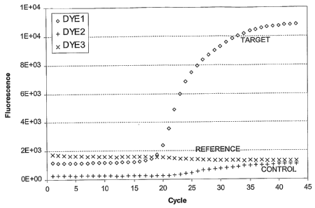
Example 1 - Captured Data
By way of example, a typical real-time PCR reaction detection system
generates a data file that stores the signal generated from one or more
detection
dyes. FIG. 1 illustrates a plot of captured reaction data that can be used in
an
analytical method according to the present invention. In this example, one dye
signal (DYE1 ) provides the captured target data, another dye signal (DYE2)
provides
captured internal control data, and a further dye signal (DYE3) provides
optional
captured reference data. These data represent data from a single reaction,
taken
from a standard output file. This particular plot can be understood to
represent initial
data to which some type of multi-component algorithm has been applied. In this
plot,
the x-axis provides an indication of cycle number (e.g., 1 to 45) and the y-
axis
indicates dye intensity detected, in relative fluorescence units. In this
figure, the
three different capture data sets are illustrated as continuous curves.
However, the
actual captured data values are generally discrete signal values captured at
each
cycle number. Thus, an initial data set as illustrated in FIG. 1 may consist
of three
sets (target, control, and reference) of suitable discrete values (e.g., about
50 values
in this case).
Example 2 - Normalization
Although optional, normalization can be performed on the captured data in
several different ways. One method involves dividing the target and control
values at
each cycle reading by the corresponding reference dye signal. Alternatively,
the
22
CA 02547998 2006-06-02
WO 2005/062040 PCT/US2004/038298
divisor can be the average reference value over all cycles or an average over
certain
cycles. In another alternative embodiment, the divisor can be the average of
the
target dye or the control dye or the target dye and the control dye over one
or more
earlier (baseline) cycles, when no amplification signal is detected. Any known
normalization method can be employed in a data analysis. The invention can be
used with data that has already been normalized by a PCR system. FIG. 2 is a
plot
of captured reaction data showing target and control data sets that have been
normalized according to the present invention. In this example, as a result of
the
normalization, the y-axis scale represents a pure number. In this case, the
number
is between about 0 and 9. Other normalization methods are known in the art and
can convert this number to between about 0 and 100 or to any other desired
range.
Because normalization is optional, the present invention can be used to
analyze reaction data without the use of a normalization or reference dye.
Alternatively, the target signal or the control signal or both can be used for
normalization.
23
CA 02547998 2006-06-02
WO 2005/062040 PCT/US2004/038298
Example 3 - Scaling
Scaling is optional but can be performed to make it easier for a human
operator to visualize the data. Scaling does not affect analytical results.
Scaling can
be carried out in addition to normalization, in the absence of normalization,
or before
or after normalization.
One method of scaling involves dividing each data set value by the
average of the values during some early cycles, generally in the baseline
region
before any positive data signal is detected. In this example, readings 4
through 8
were averaged and normalization was performed first. FIG. 3 is a plot of
reaction
data showing target and control data that have been scaled. In this example,
scaling
forces the early values of the target and control to one, and because the
early values
are less than one, the division forces the later values to slightly larger
pure numbers.
Example 4 - Digital Filtering
One or more digital filtering methods can be applied to the captured data
to "clean up" the signal data sets and to improve the signal to noise ratio.
Many
different filtering algorithms are known. The present invention can employ a
four-
pole filter with no zeros. This eliminates the potential for overshoot of the
filtered
signal. As an example, this can be implemented with the MATLAB function
"filtfilt"
provided with 'the MATLAB Signal Processing Toolbox, which both forward and
backward filters to eliminate any phase lag (time delays). An example of
parameters
and MATLAB function call is as follows:
b=0.3164;
a=[1.0000 -1.0000 0.3750 -0.0625 0.0039];
data(:,:,assay)=filtfilt(b,a,data(:,:,assay));
data(:,:,ic)=filtfilt(b,a,data(:,:,ic));
In this example, "b" and "a" contain the filter coefficients.
"data(:,:,assay)"
and "data(:,:,ic)" contain the captured data that may or may not have been
24
CA 02547998 2006-06-02
WO 2005/062040 PCT/US2004/038298
normalized, scaled, or both. In this case, the filtered data is both
normalized and
scaled. FIG. 4 is a plot of captured reaction data showing target and control
data
after digital filtering. The values are not changed by the digital filtering,
but the data
set is "smoothed" somewhat.
Example 5 - Slope Removal/Baselining
An optional slope removal method can be used to remove any residual
slope that is present in the early baseline signal before any detectable
actual signal
is produced. This procedure may also be referred to as baselining, but in some
embodiments, the offset is not removed, only the slope. According to this
invention,
for slope removal, both the target (DYE1 ) and control (DYE2) signals are
examined
simultaneously. Whichever signal comes up first defines the forward regression
point, and the method generally goes back 10 cycles. If 10 cycles back is
before
cycle 5, then cycle 5 is used as the initial regression point to avoid any
earlier signal
transients. A linear regression line is calculated using the signal data
between these
points and the slope of the regression for each dye is subtracted from that
dye's.
signal. In this case, the slope removal is applied to the normalized, scaled,
and
filtered data discussed above. FIG. 5 is a plot of captured reaction data
showing
target and control data with slope values removed. In each of these figures,
very
little slope was present in early cycles; therefore, the slope removal does
not
substantially affect the captured data values.
Example 6 - Transform Calculation
An embodiment of the method of this invention is the MaxRatio
method. In this method, the ratio between sequential measurements is
calculated,
thereby yielding a series of ratios, each of which can be indexed to a time
value or
cycle number. Many suitable means of calculating these ratios exist, and any
suitable means can be used. The simplest way of performing this ratio
calculation
utilizes the following function: Rcztio(n) = s(n + 1)
s(ta)
CA 02547998 2006-06-02
WO 2005/062040 PCT/US2004/038298
where n represents the cycle number and s(n) represents the signal at cycle n.
This
calculation provides a curve that starts at approximately 1 in the baseline
region ofi
the response, increases to a maximum during the growth region, and returns to
approximately 1 in the plateau region. A MATLAB expression that performs this
calculation efficiently is the following:
Ratio = s(2:end,:)./s(1:end-1,:),
where "s" represents the signal response matrix, with each column
representing a separate response.
FIG. 6 shows an example of this ratio transform. Because of the intrinsic
background fluorescence, the ratio does not reach 2 as would be expected of a
PCR
reaction if the signal were doubling. Regardless, the magnitude of the peak is
independent of multiplicative intensity variations and is proportional to the
rate of
growth or efficiency at that point. The method of calculating ratios is simple
and
efficiently calculated. Other equivalent calculations could be made. An
example
would involve calculating the forward and reverse ratios and then averaging
them.
On can use the inverse of the ratio, in which case the curve will begin at a
value of
approximately 1 in the baseline region, decrease in the growth region, and
return to
a value of approximately 1 in the plateau region. One would then use the
magnitude
and location of the trough instead of a peak for analysis. This transform can
be
implemented in a manner essentially equivalent to the ratio method.
Although the MaxRatio algorithm is usable as described, it is convenient to
shift the curve by subtracting a constant, e.g., about one (1 ), from each
point. This
operation provides a transformation of the original response, which starts
near zero
in the baseline region, rises to a peak in the growth region of the curve, and
returns
near zero in the plateau region. This shifted ratio calculation is described
by the
following function: Ration) = S S~ ~1) -1. FIG. 7 shows the output of this
shifted ratio
calculation. The reaction point and magnitude of the peak of the shifted ratio
curve is
then determined. The reaction point (i.e., distance along the x-axis)
specifies the
26
CA 02547998 2006-06-02
WO 2005/062040 PCT/US2004/038298
FCN value of the MR and the magnitude specifies the efficiency related value
MR
(Maximum of the Ratio).
Example 7 - Interpolation
In order to enhance cycle number resolution, an interpolation can be
performed. Many ways of accomplishing this operation are known in the art. One
method of interpolating in the context of the invention is cubic spline
interpolation,
which provides a smooth interpolation, so that even the second derivative of
the
captured data sets will be continuous. The invention can be used to
interpolate the
entire data series. The invention can be used to determine a region of
interest and
then to interpolate only in that region to achieve sub-periodic, or sub-cycle,
resolution. An example of a MATLAB command for performing a cubic spline
interpolation is as follows:
out=interp1 (x,in,x2,'spline')
where "x" represents the period (or cycle) numbers (1,2,3...), "in" represents
the
uninterpolated signal at those cycles, "x2" represents the higher resolution
period (or
cycle) vector (1.00,1.01,1.02,...) and "out" represents the interpolated
signal that
corresponds to the fractional cycles in "x2".
FIG. 8 is a plot of captured reaction data showing target and control data
that have
been interpolated to provide function continuity.As a result of an
interpolation, the
number of values in the data set will generally increase substantially, for
example
from 43 values to 4201 values.
It should be understood that the steps described above can be performed
in different orders, such as, for example, filtering first, followed by
baselining before
scaling. However, if the interpolation is performed before the ratio
calculation, care
must be taken to select the appropriate interpolated response values for the
ratio
calculation. It is important that the interval between ratio values remain the
same.
Thus, if cycles are used as the period of measurement, and interpolation
increases
27
CA 02547998 2006-06-02
WO 2005/062040 PCT/US2004/038298
the time resolution to 0.01 cycles, then the shifted ratio at x = 2.35 would
be
R=s(3.35)/s(2.35) -1.
Example 8 - Finding Peaks to Determine FCN and ERV (e g~ , MR) of Target and
Control
Another step is to select peaks in the data series. This operation involves
the steps of (1 ) finding local peaks and (2) selecting from local peaks one
or more
peaks for further analysis, optionally using criteria data (defined infra).
A peak-finding algorithm identifies where the slope of the curve changes
from positive to negative, which represents a local maximum. The algorithm
identifies the locations and the magnitude of the peaks. An example of a
MATLAB
function to do this calculation is as follows:
28
CA 02547998 2006-06-02
WO 2005/062040 PCT/US2004/038298
function [ind,peaks] = findpeaks(y)
FINDPEAKS Find peaks in real vector.
ind = findpeaks(y) finds the indices (ind) which are
local maxima in the sequence y.
% [ind,peaks] = findpeaks(y) returns the value of the
peaks at these locations, i.e. peaks=y(ind);
Y = Y(~)'
switch lengthy)
case 0
ind = ~;
case 1
ind=1;
otherwise
dy = diff(y);
not plateau ind = find(dy~=0);
ind = find( ([dy(not plateau ind) 0]<0) &
([0 dy(not plateau ind)]>0) );
ind = not plateau_ind(ind);
end
if nargout > 1
peaks = y(ind);
end
FIG. 9 is a of an efficiency calculation showing identified FCN and MR
values of the target and internal control dyes and a criteria curve according
to
embodiments of the present invention. For the target data, FINDPEAKS located
one
peak at' cycle axis x = 19.42 with a magnitude of 0.354. For the internal
control data,
FINDPEAKS found peaks at: x = 2.03, 5.29, 7.67, 12.83, 22.70, 37.86, with
respective magnitudes 0.0027, 0.0027, 0.0022, 0.0058, 0.1738, 0.0222.
29
CA 02547998 2006-06-02
WO 2005/062040 PCT/US2004/038298
Example 9 - Selecting Peaks to Determine FCN and ERV (e.g., MR) of Target and
Control
In the method discussed above, a number of local maximum peaks are
often identified for both the target data and the control data. Various
methods can
be used for selecting which of these local maximum peaks will be used for
determining an FCN and ERV.
Typically, and in particular during well-behaved reactions, the highest peak
or maximum peak is selected. In many situations, this selection provides the
most
reproducible reaction point from which to perform further calculations as
discussed
herein. However, in some situations, a first peak, or first peak above a
particular
cutoff or after a particular number of cycles is preferable. Thus, in
particular
examples, a Max Peak or First Peak selection can be employed where Max Peak
finds the largest peak in the shifted ratio curve while First Peak finds the
first peak
that is higher than some selected value.
Once criteria data are determined, these data can also be used to
determine which peak to select for an ERV determination during actual
operation,
particularly for weak or noisy signals.
In FIG. 9, for example, for the DYE2 data, the peak-finding algorithm found
six local peaks, but the fifth peak was the maximum peak and was also the only
one
that was above the criteria curve. Thus, in this example, an FCN determined
for
DYE2 is 22.70 and the MR determined for DYE 2 is 0.1733.
An information appliance or system apparatus can also be used to perform
the methods of this invention. FIG. 10 is a flow chart for performing a
reaction data
characterization according to embodiments of the present invention. Further
details
of this general method will be understood from the discussion below.
The analytic methods described herein can be applied to reactions
containing either known or unknown target concentrations. In one embodiment,
known target nucleic acid concentrations will be included in calibration wells
in a
reaction carried out in a multi-well reaction plate, and the ERV and value of
the
reaction point will be used from these known concentration samples to perform
CA 02547998 2006-06-02
WO 2005/062040 PCT/US2004/038298
quantification. Known concentrations may also be used to develop criteria data
as
further described herein.
Example 10 - Determining Criteria Curve/Criteria Data Sets
In other embodiments, efficiency related values (e.g., MR values) can be
plotted as a function of their reaction point values (FCN values) for a number
of data
sets of known concentration in order to generate a characteristic criteria
curve for a
particular assay. The criteria curve is characteristic of a particular assay
formulation
and detection protocol and can be used to reliably determine positive/negative
results, to determine whether a particular result should be discarded as
unreliable, to
determine a confidence measure of a result, or any combination of the
foregoing. In
general, pairs of reaction data that lie below a criteria curve indicate non-
reactive
samples, or non-functional reactions, such as reactions encountering
significant
inhibition.
Criteria data can be used to select which peaks to report or to use in
reaction analysis, or both. Criteria data provide an automatic and reliable
method for
discriminating between negative results (e.g., target not present at all) and
results
showing low amount of target.
FIG. 11 is a plot in which the MR of six sets of reactions of known
concentration (i.e., standards or calibrators) and one set of negative
reactions are
plotted as a function of the calculated FCN value of the MR value. This plot
allows a
criteria curve to be selected. A criteria curve, which was described
previously, is any
curve or line that separates positive results from negative results. The
criteria curve
is preferably selected so that it is relatively close to and above the
negative reaction
data (in the x-y space of the plot). In FIG. 11, pairs of MR-FCN data from a
number
of samples of known concentrations determined under the same or similar assay
conditions are plotted together with pairs of MR-FCN data from samples that do
not
contain the target of the assay, which samples are also referred to as
negatives.
Although the negatives should exhibit no amplification response, the
analytical
method does determine an MR-FCN data pair for these samples. These data for
negative samples usually correspond to noise driven maxima on the response
31
CA 02547998 2006-06-02
WO 2005/062040 PCT/US2004/038298
output, which is generally a random response. The MR value determined from
noise
is very low and far removed from the responses from samples of known
concentrations. MR-FCN pairs for negative reactions can cluster if there is a
systematic noise source, such as bleedover, in which case the MR-FCN pairs may
falsely appear to be positive reaction signals. In characterizing the MR-FCN
response of true positives versus true negatives, one can identify a clear
region of
separation between these two sets of data, which is represented by the broken
line
or curve in FIG. 11, the criteria curve. In this figure, each circle
represents a FCN-
MR data pair. In this case, each of the clusters of circles represents
multiple
responses at known concentrations of the target. There are eight different
replicates
at six known concentrations within this example. From the right of the plot,
for
example, these known concentrations can represent concentrations of 50
copies/ml,
5x10 copies/ml, 5x10³ copies/ml, 4x10⁴ copies/ml, 5x10⁵ copies/ml, and 5x10⁶
copies/ml. These criteria data clusters can be used to generate a criteria
curve.
Multiple, relatively simple criteria data sets can be used to provide
characteristic criteria curves for a number of assays. One useful approach
involves
taking the mean of the MR values for the set of negative responses and adding
to
this value a multiple of the standard deviation of the MR values for the
negative
responses. For the example shown in FIG. 11, the criteria curve was set to be
a
horizontal line equal to the mean plus 10 standard deviations of the MR values
for
the negative responses. The criteria value in this example was calculated to
be
about 0.026. In some systems, other considerations can make modification of
the
criteria value (e.g., an FCN-MR value) desirable to account for potential
signal
anomalies, such as, for example, crosstalk or positive bleedover. Crosstalk
can
result from signal in a positive well of a multi-well instrument and influence
the signal
from a different well. As much as 2% crosstalk has been observed in certain
instruments. For this reason, the criteria may be increased so as to avoid
classifying
true negative samples as positive samples. For the assay data represented in
FIG.
11, the highest MR values for positive assays are about 0.50. Two percent of
this
value is 0.010. Increasing the criteria by 0.010 should eliminate false
positives due
to crosstalk. Because the highest MR values in this assay only occur with
samples
of higher concentration that have smaller FCN values, the criteria may be
increased
32
CA 02547998 2006-06-02
WO 2005/062040 PCT/US2004/038298
only at smaller FCN values, where crosstalk is likely to occur. This modified
criteria
set can be described by a series of data pairs (Xn, Yn), which describe a
multi-
element curve. For example, the modified criteria curve shown in FIG, 11 can
be
specified by the criteria data set:
(X~, Y~) _ (1, 0.036)
(X2, Y2) _ (20, 0.036)
(X3, Y3) _ (25, 0.026)
(X4, Y4) _ (45, 0.026)
As a further example, the criteria curve shown in FIG. 10 can be specified by
the
criteria data set:
(X~,Y~)_(1,0.10)
(X2, Y~) _ (10, 0.10)
(X3, Y3) _ (20, 0.05)
(X4, Y4) _ (40, 0.05)
Criteria curves and/or criteria data sets, including sets having different
shapes or more complex shapes or both, can be determined without undue
experimentation. The intended use of the PCR application will call for
different ,
approaches to establishing criteria lines. The skilled artisan will readily
appreciate
that when high sensitivity is desired in an assay, a low criteria line is
used. For
example, if an assay is designed for differentiating sequence variants, such
as
population consensus sequence (i.e., a "wild type" sequence) versus
polymorphic or
variant sequences (e.g., a "single nucleotide polymorphism"), then a criteria
line of
higher value can be used, because the detection of limiting quantities of
target
nucleic acid is not usually required in the determination of sequence
variants.
The particular example shown in FIG. 11 does not exhibit positive
bleedover from the internal control (IC) signal response to the assay signal
response. If positive (C signs( response to assay b(eedover were to be
present, a
similar modification to the criteria could be made. Because the IC signal
response
33
CA 02547998 2006-06-02
WO 2005/062040 PCT/US2004/038298
should only occur over a narrow range of FCN values, the criteria could be
increased
only in that limited range.
Generally, as further discussed herein, a FCN-MR response is determined
for samples of known concentration across the target concentration range of
interest
to define the "normal" response. Additional studies in a population of samples
that
challenge the assay reaction may be run to see how much deterioration in MR is
acceptable before the assay performance is compromised. These types of
characterization analyses can be used to establish criteria data or sets of
criteria
data independently of the standard deviation or other characteristics of the
noise or
baseline observed when samples that do not contain target nucleic acid are
treated
under amplification conditions.
According to other embodiments of the invention, criteria data also can be
determined in ways similar to determining a Ct, for Ct analysis as has been
done in
the prior art. A particular assay under design can be performed a number of
times to
characterize it's typical MR-FCN response. From this typical response, the
criteria
data set can be defined. However, unlike Ct analysis, in FCN-MR, the response
is
independent of intensity of signal and is easily reproducible, even across
instruments
of a particular type that produce highly variable results with identical
samples.
Example 11 - Alternative Region of Interest
It has been empirically found that the FCN value of an efficiency related
value as determined above can be advantageously adjusted to provide an even
more reproducible quantification value. For example, FIG. 12 is a plot of two
sets of
reaction data that illustrate how reaction curves for samples having the same
initial
concentration can vary due to different reaction anomalies. This figure
illustrates two
responses for samples containing equal quantities of an HIV target nucleic
acid.
However, in one response, the signal obtained from the reaction falls off
early due to
an anomaly in the reaction. This fall off can cause a FCN value determined
from the
maximum of the shifted ratio curve to vary substantially between the two
samples, as
illustrated in FIG. 13. However, the figure also shows that the two gradient
curves
34
CA 02547998 2006-06-02
WO 2005/062040 PCT/US2004/038298
are more substantially similar at early time or cycle number, which is plotted
on the
x-axis of the graph.
Thus, the invention involves determining an offset from the cycle number
of maximum efficiency value (herein referred to as an FCN2 value), which is
the
location of another point on a reaction curve that can be used for analysis as
described herein. In further embodiments, an Efficiency Related Value
Threshold
(ERVT) or Ratio Threshold (RT) value can be selected and used to determine a
cycle number region of interest. An ERVT or RT can be an automatically or
empirically determined value for a particular assay. The RT value can be set
near to
or at a criteria data level that is determined at the latter cycles during
assay
calibration.
One embodiment of a method of this invention starts at the FCN value on
the shifted ratio curve and determines an earlier reaction point where the
curve
crosses the RT value. This reaction point is reported as an FCN2 value. It is
believed that the FCN2 value provides improved linearity in samples having low
copy
numbers, in contrast with FCN values for certain assays, such as reactions
where
non-specific product formation reduces the efficiency of product formation in
samples
having low copy numbers.
FIG. 13 illustrates the desirability of using an offset efficiency value. This
figure shows the shifted ratio curves for the responses shown in FIG. 12 and
an RT
line at 0.03. For this example, the FCN and FCN2 values are shown in Table 1.
TABLE 1
Response FCN FCN2 MR
Well41 28.81 22.85 0.129
Well42 28.06 22.92 0.097
Difference 0.75 0.07 0.032
In this example, the curve of one response flattens out early and differs in
shape from the curve of the other response, and the shifted ratio curve shows
a
difference. The early flattening can cause the earlier peak. In this example,
the
CA 02547998 2006-06-02
WO 2005/062040 PCT/US2004/038298
FCN2 values are more closely matched than the FCN values. In general, FCN and
FCN2 values have been found to be more precise (lower standard deviations)
than
Ctvalues. While these examples focus on use of the MR, it will be appreciated
that
other measures of the efficiency of the amplification reaction can be employed
in the
FCN and FCN2 embodiments of the present invention. Other efficiency related
transforms useful in the context of the present invention include, but are not
limited
to, (a) use of first derivative, (b) use of the differences between sequential
periodic
data points, and (c) use of the slope or gradient of the log of the growth
curve.
Example 12 - Quantification Usinq MR-FCN Analysis
C~uantification is often desired in various types of reaction analysis. In
PCR reactions, for example, quantification generally refers to an analysis of
a
reaction to estimate a starting amount or concentration of a target having an
unknown concentration. The invention involves methods or systems or both for
using an efficiency related value and a cycle number value (e.g., FCN) to
perform a
quantification. Specifically, the ERV of a test sample is compared to one or
more of
the ERV of at least one calibrator, preferably at least two calibrators, and,
optionally,
3, 4, 5, or 6 calibrators, each of which contains a known quantity of a target
nucleic
acid.
In further embodiments, quantification can generally be understood as
involving one or more calibration data captures and one or more quantification
data
captures. The calibration data and quantification are related using a
quantification
relationship or equation.
In calibration, a relationship between captured data, or a value derived
from captured data (such as an FCN, FCN2, or MR, or combination of the
foregoing),
and one or more known starting concentration reactions is used to establish
one or
more parameters for a quantification equation. These parameters can then be
used
to determine the starting concentrations of one ~or more unknown reactions.
Various methods and techniques are known in the art for performing
quantification and/or calibration in reaction analysis. For example, in
diagnostic PCR
-settings, it is not uncommon to analyze test samples in a 96-well reaction
plate. In
36
CA 02547998 2006-06-02
WO 2005/062040 PCT/US2004/038298
each 96-well reaction plate, some wells are dedicated to calibration reactions
with
samples having known initial concentrations of target. The calibration values
determined for these samples can then be used to quantify the samples of
unknown
concentration in the well.
Two general types of calibration methods are referred to as one-point
calibration and standard curve (e.g., multiple points) calibration. Examples
of these
types are set forth below. Any suitable calibration method, however, can be
used in
the context of the present invention.
When there is no inhibition or interference, the PCR reaction proceeds
with the target sequence showing exponential growth, so that after N cycles of
replication, the initial target concentration has been amplified according to
the
relationship:
Conch oc Conco (1 + e) N
which can also be expressed as:
Conc oc Conc x
o N (1 + e) N
where Conch represents the concentration of amplified target after N reaction
cycles,
Conco represents the initial target concentration before amplification, N
represents
the cycle number and a represents the efficiency of the target amplification.
Quantitative data analysis is used to analyze real time PCR reaction curves so
as to
determine Conco to an acceptable degree of accuracy. Previous Ct analysis
methods attempt to determine a cycle number at a reaction point where the
Conch is
the same for all reactions under analysis. The FCN value determined by the
methods of the invention provides a good estimate for the cycle number N for
an
assay in which no significant inhibition or signal degradation over the
dynamic range
37
CA 02547998 2006-06-02
WO 2005/062040 PCT/US2004/038298
of input target concentrations is demonstrated. The following proportionality
relationship between a starting concentration and FCN can be used:
Coraco (FCN) ~ 1 FcN
~1+e)
where Conco(FCN) represents the estimate of the initial target concentration
determined by using the FCN value as determined by the methods of this
invention.
In other words, the lower the starting concentration of target, the higher the
FCN
value determined for the PCR reaction. This relationship can be used for both
calibration data and for quantification data.
This proportionality relationship can also be expressed as an equivalence,
such as
Conco (FCN) = K x 1 FcN
~1+e)
where K represents a calibration proportionality constant.
For calibration data, Conco (FCN) represents a known concentration, such as
500,000 copies of target nucleic acid/mL; the exponent FCN is a FCN cycle
number
determined as described above; and a represents the efficiency value for a
reaction,
with a = 1 indicating a doubling each cycle. These factors combine to form a
relationship to allow for determination of the proportionality constant.
Determination
of the proportionality constant can ''only be made if there is a priori
knowledge of the
efficiency, e, of the amplification reaction. This a priori knowledge enables
a one-
point calibration. For quantification data, FCN values are determined for
reactions
involving samples having unknown concentrations of target. The FCN values are
then converted to concentration values by use of the above equation. If the
efficiency, e, is not known a priori, then a standard curve quantification
method can
be used. In this case, for calibration data, different samples having
different levels of
38
CA 02547998 2006-06-02
WO 2005/062040 PCT/US2004/038298
known concentration are amplified, and the FCN values of the samples are
determined. These FCN values can be plotted against the log (base 10) of the
known concentrations to describe a log (concentration) vs. FCN response. For
an
assay that demonstrates no significant inhibition or signal degradation over
the
dynamic range of input target concentrations, this response is typically well-
fitted by
a linear curve. The following equation describes the form of this standard
curve:
Logo (Conco(FCN)) = m x FCN + b
where Log~o(Conco(FCN)) represents the log (base 10) of the initial target
concentration, m represents the slope of the linear standard curve, and b
represents
the intercept of the linear standard curve.
By using two or more known concentration calibration samples, a linear
regression
can be applied to determine the slope, m, and intercept, b, of the standard
curve.
For quantification data, FCN values are determined for reactions involving
test
samples of unknown concentration, which values are then converted to log
(concentration) values by use of the above linear equation. Results can be
reported
in either log (concentration) or concentration units by the appropriate
conversion.
It should be noted that the one-point calibration equation is easily
converted to this linear standard curve form:
Conco (FCN) = K x 1 FcN
~l+e)
Logo (Conco (FCN)) _ -logo (1+e) x FCN + logo (f~. The linear coefficient m
can
be used to calculate the efficiency of the particular PCR reaction.
Example 13 - Quantification Adjustments
When PCR reactions are subjected to inhibition, the resulting real-time
PCR signal intensity can be depressed or delayed. The effect of this signal
39
CA 02547998 2006-06-02
WO 2005/062040 PCT/US2004/038298
degradation on an efficiency related value such as MR is a reduction in that
value.
In addition, the efifect ofi signal degradation on the firactional cycle
number is
generally to identify the FCN at an earlier cycle number than would be
expected for
the uninhibited reaction. These factors cause the plot ofi log (concentration)
as a
function of FCN to be less well described by a linear curve fitting function.
Although
higher order curve fitting functions can be applied for a standard curve, a
linear fit
requires fewer calibration levels and is simpler to calculate.
Some of these problems can be addressed in a sfiandard curve analysis by
incorporating an ERV or Intensity value into the quantification relationships
as
discussed above. Thus, the equations above can be rewritten a:
Ihtet~stty
Corzco (FCNI,Ztensity Adj ) °~ . (1 + e)FCN
Co72c0 (FCNMR Adj ) °~ MR FCN
~1+e)
where Intensity represents the response intensity (above background) at the
determined FCN value, MR represents the MR value as described previously.
Conco (FCN~~,tensityadj) represents the estimate of the initial concentration
of the target
determined by using the FCN value adjusted by using the Intensity value and
Conco ~FCNMRAdj~ represents the estimate of the initial concentration of the
target
determined by using the FCN value adjusted by using the MR value.
These expressions take advantage ofi the relationship observed between
the intensity at the selected FCN cycle or the MR determined at the selected
FCN
cycle, or both, and the change to the FCN value in the presence of inhibition,
as
discussed above. The net efFect is that the right hand side ofi the
proportionality
expressions above is relatively insensitive to inhibition and other factors
that afFect
the PCR amplification curve, and, therefore, provide significant robustness as
expressions for determining the concentration values of the target.
The following discussion further explains the properties and relationships
ofi FCN, FCNintensi~Adj~ and FCNMRAdj~ Assuming the efficiency is 1, the
previous can
be simplified to:
CA 02547998 2006-06-02
WO 2005/062040 PCT/US2004/038298
Cortco (FCN) ac 1 ~FCN
Intensity
CO72C0 (FCNhatensityAdj ) °~ 2FCN
Conco (FCNMRAdj ) °~ MR 2FCN
Taking the Log base two of the expressions yields:
Loge (Conco (FCN)) °c FCN
Loge (COYlGO (FCNIy2tensityAdj )) °~ FCN - Loge (Intensity)
Loge (Conco (FCNMR Adj )) °~ FCN - Loge (MR)
From the right sides of the expressions come the values for compensating for
intensity or MR to adjust the FCN value by means of the following formulas:
FCN i~c. aa~. = FCN - Logy (Intensity)
FCN MR. aa~. = FCN - Logy (MR).
This calculation then provides quantification by using adjusted FCN values
analogous to using FCN values or Ctvalues. It should be noted that the use of
these
adjusted FCN values provide significant robustness to inhibition and other
factors
that affect PCR amplification, such as Ct values used in determining the
concentrations of the target in the unknown samples. The plot of Log
(concentration)
vs. these adjusted FCN values is generally well fitted by a linear standard
curve.
Thus, the present invention provides a method for determining the quantity of
a
target nucleic acid in a sample comprising involving the steps of (a) finding
the
period of time or cycle number of an amplification reaction corresponding to a
maximum of an efficiency related value, preferably of an MR, and (b) adjusting
that
value by subtracting a logarithm of the Intensity or a logarithm of the MR,
and (c)
comparing the value obtained to calibration data obtained using the same
methodology.
41
CA 02547998 2006-06-02
WO 2005/062040 PCT/US2004/038298
Example 14 - Standard Curve Calibration
Development of a standard curve from known concentrations and use
thereof for quantification is well known in the art and can be further
understood from
the following example. In a typical case, a number of calibration reactions
(such as
in wells in which the initial concentrations are known) are used during each
amplification or series of amplifications to perform the calibration
operation. One
problem that arises with attempting to quantify a target nucleic acid in a
sample
through a large range of possible initial concentrations is that
quantification of lower
quantities of target nucleic acid in any particular reaction becomes more
difficult. For
example, FIG. 14 illustrates data for an assay designed to quantify the amount
of
HIV in test samples. The reactions were performed with eight replicates of six
known concentrations of target nucleic acid, which were 50; 500; 5,000;
50,000;
500,000; and 5,000,000 copies per mL. The assay data show significant signal
suppression in reactions where the copy number is low (the curves farthest to
the
right). While quantity of the four highest concentrations of target nucleic
acid (the
curve sets to the left) yielded precise results with low coefficients of
variability, the
two lowest concentrations produced less precise curves. The imprecision caused
by
the difficulties in quantifying low concentrations of target nucleic acids in
assays
having a dynamic range of 100,000 to 1 or more can be addressed by the
following
methods of this invention.
Because calibration runs in a reaction plate are relatively expensive, it is
conventional to collect a minimal acceptable number of calibration data sets.
For
example, in one implementation, the average of two replicates each of the 500;
50,000; and 5,000,000 copy/mL samples are run along with the diagnostic
assays,
thereby requiring perhaps six wells in a 96 well plate to be used for
calibration
reactions.
Because the relationship between the cycle numbers and the log of the
calibrator concentration is substantially linear, a linear regression can be
performed
between a log (e.g., logo) of the calibrator concentrations and the cycle
number.
This regression can easily be performed via the Excel program and other
42
CA 02547998 2006-06-02
WO 2005/062040 PCT/US2004/038298
mathematical analysis software. FIG. 15 illustrates four linear standard
curves
generated from three-point calibration data using four different cycle number
related
values (e.g., FCN, FCN2, FCNMR Adj.~ and FCN,nt. aa~.).
In each of the curve fit equations, the x-axis displays values of the Logo
[Target] actual or known concentration. Thus, solving for x provides an
expression
for converting from cycle number related values to Logo (Target) calculated
concentration of the assay. If the assay response is not linear with Log
(Target), a
higher order or more complex regression, or a larger number of calibration
reactions,
or both, can be used. In this example, the following equations were
determined:
FCN = -3.0713*Log~o (Conco) + 31.295
FCN2 = -3.0637*Log~o (Conco) + 25.006
FCN MR adj = -3.2344*Log~o (Conco) + 33.271
FCN ant. aa~ _ -3.2870*Log~o (Conco) + 32.775
Example 15 - Comparing Quantification Using Different Cycle Number
Related Values
In order to examine the different characteristics of calibrations using the
different cycle number related values described above, quantification can be
performed on various samples having known concentrations, and the
concentrations
calculated compared with the known concentrations. In one example of such a
comparison, the standard curves having the parameters generated above were
used
to carry out quantification of the assay responses shown in FIG. 14. The mean
of
the calculated concentrations of the eight replicates at each known
concentration
was compared to the known concentration value. FIG. 16 compares logo of the
known concentration values (x-axis) to the means of the logo of each of the
calculated concentrations for the eight samples at each concentration.
As indicated by FIG. 16, the 50 copies/mL samples (log (concentration) _
1.7) are slightly over-quantified (i.e., higher than the actual concentration)
using
FCN, while the accuracy for the FCN method (of the MR) at the higher
concentrations is very good. FCN2 is more accurate at the lowest
concentration, but
43
CA 02547998 2006-06-02
WO 2005/062040 PCT/US2004/038298
somewhat under-quantified (i.e., lower than the actual concentration), and
exhibit
less linearity and accuracy at some higher concentrations. FCNMRAaj. showed
very
accurate and linear quantification throughout the concentration range. FCN~nt.
Adj.
also showed substantial improvement in accuracy and linearity compared to FCN,
except for very slight under-quantification at the lowest concentration.
Accordingly,
all four methods work well, but some are better than others for particular
situations.
Therefore, the skilled artisan can easily select an appropriate method for any
particular application to obtain excellent results.
Example 16 - Quantification Using One-Point Calibration
A one-point calibration can be used for quantification. In this case, two
wells at the 50,000 copies/mL concentration (Log (4.7)) were used for
calibration. In
order to calculate the calibration constant, the following equation is used:
K = Corr.co * 2FCN , where K represents the calibration constant, Conco
represents the
known concentration of the calibrator, FCN represents the fractional cycle
number of
the calibrator, and the efficiency of the reaction, e, as described earlier,
is assumed
to be 1. Similar calibration constants can be generated using the
proportionality
relationships such as FCN2, FCNMRAaj. and FCN i~t.Aa~.
In this case, the constant was generated for two wells and the average
was used. Once the calibration constant is generated, the concentration for
each
assay is calculated with the following equation: Cofac = KFC~FCN . FIG. 17
illustrates
resulting from a one-point calibration.
As can be seen, the FCN results are elevated at the lowest two
concentrations and accurate from log (Conc) equals 3.7 and above. FCN2 shows
improved accuracy at low concentrations compared to FCN, but under-quantifies
at
log (Target) equal to 5.7 and 6.7. FCN-MR adjusted shows good linearity over
the
entire range with slight over-quantification at the two lowest concentrations.
FCN-
Intensity adjusted also shows good linearity with very slight under-
quantification at
the lowest two concentrations. Accordingly, each of these embodiments works
well
and the skilled artisan can readily select from among these options.
44
CA 02547998 2006-06-02
WO 2005/062040 PCT/US2004/038298
As discussed above, an FCN-MR analysis can be used to characterize a
particular reaction as positive or negative or to compare the reaction to
criteria data,
or both. These values can be used to quantify a reaction. A variety of
quantification
methods can benefit from FCN-MR analysis rather than Ct analysis.
In one embodiment, a FCN value, a FCN2 value, or a FCN adjusted value
can be used in any way that a Ct value has been used in the prior art.
Typically, but
not necessarily, FCN-adjusted, FCN2-adjusted, or FCN-adjusted analysis can be
applied to various sets of calibration data to thereby develop reference data
curves
or an equation for comparing the result of a reaction in which the
concentration of
target is unknown to the results of reactions in which the concentration of
target is
known. Thus, the present invention can be used to develop reference data and
to
perform a comparison wherein two values (e.g., FCN-MR) are used both for
developing reference data and also for making a comparison to that data.
While experiments using the MR method regularly used different
preprocessing steps on the captured data set before processing the data set
with a
ratio function, most of these steps are not required. In particular,
experimental
results have indicated that scaling, normalization by a reference dye,
baselining
(both offset and slope correction), and filtering are not required. However,
filtering
has generally been found to be desirable as it improves performance in the
presence
of noise. Slope correction (for the baseline region) has also been found to be
desirable as it slightly improves discrimination between samples that do not
contain
target nucleic acid and those that contain very little target nucleic acid or
suffer from
significant inhibition of the amplification reaction. Generally, however, when
FCNintensity ads is used, it is preferable to use a normalization technique,
such as, but
not limited to, scaling or normalization to a reference dye.
Example 17 - MR Algorithm Applied to HBV Data Using a One-Point Calibration
HBV assays of control solutions ranging from 10 copies/reaction to 109
copies/reaction and negatives were processed on an ABI Prism 7000 with six
replicates at each concentration. The captured data was processed using only a
digital filter. FCN values were then calculated using a MR algorithm as
described
CA 02547998 2006-06-02
WO 2005/062040 PCT/US2004/038298
above. The concentrations were calculated by means of a one-point calibration
using the three of the responses at 109 copies/reaction as a reference
calibrator.
Even without normalization, scaling, or baselining, the resulting ,
quantification was very good, with the exception of an acceptable amount of
over-
quantification of the 10 copies/reaction and 100 copies/reaction samples
(i.e., the
Log (Target) = 1 and 2 samples). There was a very clear distinction between
the
negatives and the 10 copies/reaction assays, with no false positives or false
negatives. Additional results indicated that when the same data was quantified
with
Ct analysis, the 10 copies/reaction and 100 copies/reaction assays are also
slightly
over-quantified, and the precision at all concentrations above 10
copies/reaction is
better with the MR analysis. In this case, the Ct results were normalized,
baselined,
and calibrated by means of a two-point calibration with three replicates each
at
concentrations 103 and 10' copies/reaction.
FIG. 19 illustrates an example of the same HBV data using MR analysis
and with FCNMR ads., correction. Again, the quantification was performed by
means of
a one-point calibration with three responses at the 109 copies/reaction with
no
normalization, scaling, or baselining. As can be seen, the over-quantification
of the
low concentrations is significantly reduced, i.e., the quantitatjve results
are
significantly improved.
Example 18 - MR Algorithm Applied to HIV Data
In this example, HIV assays of control solution were performed at
concentrations of negatives, 50 copies/mL, and 100 copies/mL, through 106
copies/mL in replicates of six. The responses were processed by means of the
MR
algorithm using FCNMRAd~. with normalizing and baselining. FIG. 21 illustrates
results the example using MR analysis and two-point calibration, e.g., using
two
replicates of the 102 and 105 copies/mL responses as calibrators. There was
clear
differentiation between the negatives and the 50 copies/mL assays with no
false
positives or false negatives. As can be seen, there is good linearity and
precision.
46
CA 02547998 2006-06-02
WO 2005/062040 PCT/US2004/038298
Example 19 - Validity Determination Using Target and IC (FCN, MR) Pairs
It has been found that pairs of reaction time or cycle number values and
efficiency related values (e.g., pairs of FCN-MR values) can provide valuable
information about a nucleic acid amplification reaction, e.g., a PCR reaction,
which
can be further enhanced by considering data pairs for both the internal
control and
target amplification reactions. While pairs for a target reaction alone carry
important
information about reaction efficiency and can be used for comparison with
criteria
data, additional factors that arise in processing samples or in the samples
themselves may be better analyzed by considering control data as well.
For example, in processing specimens for use in PCR or other suitable
amplification reactions, the sample can carry various inhibitors into the
reaction,
which might be detectable through assessment of target data only. However,
abnormal recovery of target nucleic acid during sample preparation typically
would
not be detected by analysis of a single amplification reaction. Furthermore, a
target
nucleic acid may possess polymorphic sequences that could impair detection of
the
target nucleic acid, e.g., if a probe is used that binds to a polymorphic
region of the
sequence. Mismatches caused by the polymorphic sequence in this region would
affect the detected signal, and, consequently, the amplification might not
appear as
abnormal or inhibited using the evaluation of data pairs for a single
amplification.
Co-analysis of an internal control together with analysis of the target
amplification
responses can provide accurate quantification of the target nucleic acid in
such
samples when other methods would typically indicate an invalid reaction.
Thus, pairs of reaction time or cycle number values and efficiency related
values can be used together to assess the validity of a given reaction, such
as in a
given container or well. One could design the internal control (IC)
amplification
reaction to be comparable in robustness to the target amplification reaction,
or
slightly less robust. Robustness in this context means the sensitivity of the
reaction
performance to factors that can affect the PCR processing pathway, such as
inhibition that results from sample preparation or the samples themselves, or
to
variability in transferring of the reaction mixture by pipette, such as
transferring
inaccurate amounts of amplification reagents by pipette.
47
CA 02547998 2006-06-02
WO 2005/062040 PCT/US2004/038298
Example 20 - Multiple Criteria Data Curves
Multiple criteria curves for the pairs of cycle number value - efficiency
related value (e.g., FCN-MR pairs) can be developed and can have different
uses or
levels of importance, particular for use with validity determination. For
example, a
first criteria curve can be selected so as to be able to discriminate reactive
amplification signals from non-reactive responses. A second criteria curve can
be
selected so as to be more constraining than the first type, so that it would
be useful
in identifying sample responses that lead to accurate quantification in
contrast to
those having partial inhibition that might have lower confidence in
quantification.
FIG. 22 is a plot illustrating two types of criteria data, wherein the lower
horizontal
line represents criteria data suitable for differentiating negative from
reactive
reactions. The second set of lines represents criteria data indicating the
normal
range for the FCN-MR pair responses. These criteria can be used to distinguish
high confidence in quantification in contrast to a lower confidence that might
be
associated with a value outside this range due to partial reaction inhibition.
For example, the first type of criteria data that differentiates reactive and
non-reactive amplification reaction can be referred to as "MR criteria data."
These
data act as a cutoff threshold - reactive responses will have MR values that
exceed
the MR criteria data, whereas negative samples will have MR values that will
not
exceed the criteria value or criterion line. The criteria data is preferably
set so that
noise in the response signal does not exceed the criteria, nor will such
biases as
cross-talle or bleed-over.
The second type of criteria data is referred to as the MR normal range.
This range would be the range of MR values for a given FCN over which
quantification of the sample is accurate. If a signal response is suppressed,
the MR
value observed will drop. As the MR value decreases due to inhibition, the FCN
value can shift to earlier cycles, whereas a threshold based Ct might shift to
later
cycles. The MR normal range would be the range for MR values in a criteria
data set
for which a chosen value related to a cycle number would provide an accurate
48
CA 02547998 2006-06-02
WO 2005/062040 PCT/US2004/038298
quantitative result for the sample when used to determine the concentration of
target
in the sample from the assay standard curve.
The "MR normal range" can be developed using a Bivariate Fit of the
MR by FCN as will be understood in the art. FIG. 23, for example, shows a FCN-
MR
plot for HIV data from 50 copies/mL to 5,000,000 copies/mL. The data was
analyzed
by means of a statistics software package (such as JMP (SAS Institute, Inc.))
to
apply a cubic curve fit to the data. This cubic curve fit is represented by
the solid line
in middle of the figure. The upper and lower dashed curves represent the
confidence interval generated using a confidence interval individual analysis
option
with an alpha level of 0.001. TABLES 2A, 2B, and 2C illustrate sample data
input
and output related to FIG. 23.
49
CA 02547998 2006-06-02
WO 2005/062040 PCT/US2004/038298
TABLE 2A
Summary of Fit
RSquare 0.971668
RSquare Adj 0.969737
Root Mean Square 0.023918
Error
Mean of Response 0.401317
Observations 48
(or Sum
Wgts)
Polynomial Fit Degree=3
MR = 0.6710196 - 0.0101107 FCN - 0.0039387 (FCN-18.3056)~2 - 0.0004202 (FCN-
18.3056)~3
TAC21 C ~1~
Analysis of Variance
Source DF Sum of Mean SquareF Ratio
Squares
Model 3 0.86331003 0.287770 503.0120
Error 44 0.02517212 0.000572 Prob >
F
C. Total47 0.88848215 <.0001
Tnm c ~r
Parameter Estimates
Term ' Estimate Std Error t Ratio Prob>~t~
Intercept 0.6710196 0.036617 18.33 <.0001
FCN -0.010111 ' 0.002006-5.04 <.0001
(FCN-18.3056)~2-0.003939 0.000198 -19.92 <.0001
(FCN-18.3056)~3-0.00042 0.000047 -8.93 .0001
A statistically derived confidence interval, as shown, is a systematic
approach to
determining which data points represent "normal" responses and should
therefore be
quantified. Data points lying outside this interval are exceptional and are
preferably
identified to a human operator by a software program so that further
investigation
can be made.
In alternative embodiments, such a curve can be simplified in the form of
one or more straight-line segments. This simplification can in some cases be
CA 02547998 2006-06-02
WO 2005/062040 PCT/US2004/038298
performed by a technician viewing the raw data or may be derived from an alpha
interval as discussed above.
A similar statistical fit can be performed on the internal control (IC) data.
FIG. 24, for example, shows a plot of MR as a function of FCN for IC data,
namely IC
data associated with the data shown in FIG. 23. This data can be used to
determine
an IC criteria, which, for example, can be a single value that is five
standard
deviations below the mean of the MR values of the IC or can be a range or box
of
values, for example, based on the mean ~5 standard deviations of the MR and
FCN
values.
Thus, the present invention also provides a method for analyzing an
amplification reaction, the method comprising establishing a "confidence
corridor",
which is a range of selected values provided in pairs in which the first value
is a
maximum efficiency related value (which is preferably the MR), and the second
value
is a time value or cycle number value at a reaction point (which optionally
can be
fractional). The method further comprises determining whether a maximum
efficiency value occurring at any particular periodic time value or cycle
number value
at a reaction point (which optionally can be fractional) falls within the
selected range.
If the value does not, then further investigation, or disregarding the
results, is
indicated. Any suitable method can be used to establish the selected
confidence
corridor. Preferred methods include setting the confidence corridor about 1,
2, 3, 5,
10, or any other suitable number of standard deviations from the mean of data
obtained from a set of reactions used to characterize the assay. Another
suitable
method involves modifying the confidence corridor by observing known aberrant
or
discrepant results and modifying the confidence corridor to exclude a portion
of
those aberrant or discrepant results in future assays. The use of the
confidence
corridor of the present invention can be applied to target nucleic acid
quantification,
analysis of any of standards, calibrators, controls, or to combinations of the
foregoing.
Example 21 - Validity Analysis
51
CA 02547998 2006-06-02
WO 2005/062040 PCT/US2004/038298
FIG. 25 is a flow chart illustrating a logic analysis tree for assessment of
assay validity through analysis of pairs of cycle number (e.g., FCN) minus of
ERV
(e.g., MR) for both the internal control and the target amplification
reactions. FIG. 26
is a flow chart illustrating a logic analysis tree for reporting target
results with validity
criteria assessment using pairs of cycle number (e.g., FCN) minus ERV (e.g.,
MR).
In the flow charts, FCN is used for clarity of illustration, but as noted
elsewhere
herein, other methods can be used to generate the reaction point value, for
example,
Ct method, FCN2, FCNMR Adj. or FCN,~t. Ads,. or other suitable method.
Thus, a validity check can optionally proceed as a series of questions
regarding the internal control (IC) and/or target data.
In FIG. 25, the left-most arrow blocks provide general descriptions of the
steps of the method. Details of methods) can be understood further by
considering
the following. The method analyzes a cycle number/ERV pair from both a target
and
control (IC) reaction. Initially, if (1) the IC MR is above the IC MR criteria
data, and
then if (2) the IC FCN is within the normal range, and further if (3) the IC
MR is within
the normal range, then reaction validity is confirmed.
As shown in the figure, an invalid result can be fiurther characterized or
explained by considering one or more characteristics of the target MR.
FIG. 26 illustrates a method for analyzing the target data for valid reactions
to further characterize a valid result as indicating (1 ) a non-reactive
target sample,
(2) a target at a concentration of less than the detecting limit of the assay,
(3) a
target present but with a quantification inhibited, possibly due to sub-type
mismatch,
or (4) a valid, quantifiable target reaction.
Thus, by combining the analysis based on multiple targets and using both
cycle number and efficiency related values, one can distinguish an inhibited
sample
from a sample that suffered from poor nucleic acid recovery during sample
preparation. The analysis makes use of pre-established knowledge of the assay
that
is contained in the internal control and target criteria data.
Example 22 - Validity Determination Using Peak Width
52
CA 02547998 2006-06-02
WO 2005/062040 PCT/US2004/038298
In contrast to the conventional Ct analyses in the prior art, which only
presents a single value describing an amplification response, an efficiency
related
value analysis (and preferably an MR analysis) can provide an efficiency
related
transform curve with data corresponding to the time value or cycle number
value of
the entire amplification reaction or any portion thereof. It has been
discovered that
within a specific assay formulation, normal assay responses generate highly
reproducible efficiency related transform curves. One characteristic in
particular is
the width of the peak of the efficiency related transform curve. It has been
found that
the width of the peak of the efficiency related transform, e.g., as defined by
its width
at the half maximum height, varies very little even when the magnitude of the
fluorescence intensity varies greatly.
Any suitable method can be used to determine the width of the peak of
the efficiency related value. FIG. 27 depicts one suitable method for
determining the
width of an efficiency related value peak. In FIG. 27, the full peak width is
the width
in cycles of the peak at it half maximum level. The HIV responses in FIG. 14
show
normalized fluorescence for samples of higher concentration at approximately
8,
while the normalized fluorescence for the samples of low concentration is as
low as
about 1. Using the shifted ratio method to calculate an efficiency related
transform
for each amplification reaction and computing the full peak width provides the
results
shown in FIG. 28. Even with an eight-fold change in final fluorescence
intensity, the
peak widths are surprisingly conserved within a narrow range. Accordingly, the
present invention provides amplification reaction validity criteria, wherein
an
amplification reaction is deemed valid when the width of the peak of an
efficiency
related value is contained within a selected range characteristic of the
amplification
reaction. In FIG. 28, the dashed horizontal lines in bold type represent the
mean of
the width measurements plus and minus 10 standard. deviations. Width
measurements that are not within the range of about 5.5 and 8.0 (as shown in
FIG.
28) are considered invalid or at least suspect. The skilled artisan can
readily vary
the parameters describing the acceptance interval, depending on the
requirements
of the particular assay and without undue experimentation.
Peak width can be used to detect an abnormal assay response. The
full peak width calculation was applied to the assay data that contained the
abnormal
53
CA 02547998 2006-06-02
WO 2005/062040 PCT/US2004/038298
response shown in FIG. 12. The results are presented in FIG. 29. As can be
seen,
normal responses for this data set produce full peak widths between about 6
and 9
cycles whereas full peak width of well 42 is 17.42. Accordingly, the
amplification
reaction of well 42 is abnormal and is disregarded.
The full peak width calculation will be affected by abnormal variations in
amplification response that occur both before and after the reaction point
value (e.g.,
the FCN) of the efficiency related value. Abnormal variations that occur after
the
reaction point value of the efficiency related value are not considered for an
assay
validity test, because they cannot affect assay quantification by the MR
method.
This option can readily be achieved using the half peak width calculation
illustrated in
FIG. 27 or its equivalent. In the illustrated example, only the width in
periodic time
units from about the half-maximum efficiency related transform up to about the
reaction point value of the maximum efficiency related value is used. Of
course,
other suitable methods for measuring peak widths and half peak widths are
known in
the art.
Example 23 - Software Embodiments
The systems of this invention can be incorporated into a multiplicity of
suitable computer products or information instruments. Some details of a MR
software implementation are provided below. Specific user interface
descriptions
and illustrations are taken to illustrate specific embodiments only and any
number of
different user interface methods known in the information processing art can
be used
in systems embodying this invention. The invention can also be used in systems
where virtually all of the options described below are preset, calculated, or
provided
by an information system, and, consequently, provide little or no user
interface
options. In some cases, details and/or options of a prototype system are
described
for exemplification purposes; many of these options and/or details may not be
relevant or available for a production system.
Furthermore, software embodiments can include various functionalities,
such as, for example, processing reactions with one or two target reactions,
or one
or more internal control reactions, or reference data, or combinations of the
54
CA 02547998 2006-06-02
WO 2005/062040 PCT/US2004/038298
foregoing. A software system suitable for use in this invention can provide
any
number of standard fife handling functions such as open, close, printing,
saving, etc.
FIG. 30 illustrates a user interface for processing PCR data according to
this invention. In this interface, the selection of appropriate dyes)
corresponding to
the target assay, internal control, and reference responses are selected from
popup
lists in the upper left portion of the window. Tabs for selecting different
viewing
options are positioned in the middle of the window and are arranged
horizontally.
FIG. 30 shows that the flab displaying the MR-FCN plot has been selected. FIG.
31
illustrates a user interface showing the same data for well 1, but displaying
the
shifted ratio curve. Other tabs allow viewing of the raw fluorescence data,
normalized fluorescence, and baselined data for all the responses. In
addition, a tab
allows inspection of each response individually. Fields to the right of the
plot show
calculated response values such as MR, FCN, Ct, and standard deviation in the
baseline region. Below these calculated values are radio buttons allowing the
user
to display either the assay data or the internal control data.
Embodiment in a Programmed Information Appliance
FIG. 32 is a block diagram showing an example of a logic device in which
various aspects of the present invention may be embodied. As will be
understood
from the teachings provided herein, the invention can be implemented in
hardware or
software or both. In some embodiments, different aspects of the invention can
be
implemented in either client-side logic or server-side logic. Moreover, the
invention
or components thereof can be embodied in a fixed media program component
containing logic instructions or data, or both, that when loaded into an
appropriately
configured computing device can cause that device to perform according to the
invention. A fixed media component containing logic instructions can be
delivered to
a viewer on a axed medium for physically loading into a viewer's computer or a
fixed
medium containing logic instructions can reside on a remote server that a
viewer can
access through a communication medium in order to download a program
component.
CA 02547998 2006-06-02
WO 2005/062040 PCT/US2004/038298
FIG. 32 shows an information instrument or digital device 700 that can be
used as a logical apparatus for performing logical operations regarding image
display or analysis, or both, as described herein. Such a device can be
embodied as
a general-purpose computer system or workstation running logical instructions
to
perform according to various embodiments of the present invention, Such a
device
can also be customized and/or specialized laboratory or scientific hardware
that
integrates logic processing into a machine for performing various sample
handling
operations. In general, the logic processing components of a device according
to the
present invention are able to read instructions from media 717 or network port
719,
or both. The central processing unit can optionally be connected to server 720
having fixed media 722. Apparatus 700 can thereafter use those instructions to
direct actions or perform analysis as described herein. One type of logical
apparatus
that can embody the invention is a computer system as illustrated in 700,
containing
CPU 707, optional input devices 709 and 711, storage media 715, e.g., disk
drives,
and optional monitor 705. Fixed media 717, or fixed media 722 over port 719,
can
be used to program such a system and can represent disk-type optical or
magnetic
media, magnetic tape, solid state dynamic or static memory, etc. The invention
can
also be embodied in whole or in part as software recorded on this fixed media.
Communication port 719 can also be used to initially receive instructions that
are
used to program such a system and represents any type of communication
connection.
FIG. 32 shows additional components that can be part of a diagnostic
system. These components include a viewer or detector 750 or microscope,
sample
handler 755, UV or other light source 760 and filters 765, and a CCD camera or
capture device 780 for capturing signal data. These additional components can
be
components of a single system that includes logic analysis and/or control.
These
devices may also be essentially stand-alone devices that are in digital
communication with an information instrument such as 700 via a network, bus,
wireless communication, etc., as will be understood in the art. Components of
such
a system can have any convenient physical configuration and/or appearance and
can be combined into a single integrated system. Thus, the individual
components
shown in FIG. 42 represent just one example system.
56
CA 02547998 2006-06-02
WO 2005/062040 PCT/US2004/038298
The invention can also be embodied in whole or in part within the circuitry
of an application specific integrated circuit (ASIC) or a programmable logic
device
(PLD). In such a case, the invention can be embodied in a computer
understandable
descriptor language, which may be used to create an ASIC, or PLD, that
operates as
described herein.
Other Embodiments
The invention has now been described with reference to specific
embodiments. Other embodiments will be apparent to those of skill in the art.
In
particular, a viewer digital information appliance has generally been
illustrated as a
computer workstation such as a personal computer. However, the digital
computing
device is meant to be any information appliance suitable for performing the
logic
methods of the invention, and could include such devices as a digitally
enabled
75 laboratory systems or equipment, digitally enabled television, cell phone,
personal
digital assistant, etc. Modification within the spirit of the invention will
be apparent to
those skilled in the art. In addition, various different actions can be used
to effect
interactions with a system according to specific embodiments of the present
invention. For example, a voice command may be spoken by an operator, a key
may
be depressed by an operator, a button on a client-side scientific device may
be
depressed by an operator, or selection using any pointing device may be
effected by
the user.
It is understood that the examples and embodiments described herein are
for illustrative purposes and that various modifications or changes in light
thereof will
be suggested by the teachings herein to persons skilled in the art and are to
be
included within the spirit and purview of this application and scope of the
claims.
All publications, patents, and patent applications cited herein or filed with
this application, including any references filed as part of an Information
Disclosure
Statement, are incorporated by reference in their entirety.
57