Note: Descriptions are shown in the official language in which they were submitted.
CA 02548001 2006-05-31
WO 2005/056108 PCT/US2004/040521
METHOD AND APPARATUS FOR DETERMINING AN EFFICACIOUS
ATRIOVENTRICULAR DELAY INTERVAL
The present invention relates to the field of cardiac therapy. In particular,
the
present invention provides methods and apparatus for optimizing an
atrioventricular (A-V)
interval based on measuring or obtaining one or more physiologic parameters of
a patient.
The parameters may be obtained using echocardiographic equipment and the like
to
enhance cardiac therapy delivery, such as a dual chamber pacing therapy and
cardiac
resynchronization therapy (CRT), among others.
Those skilled in the art of diagnosing cardiac ailments have long understood
that
certain patients, in particular heart failure (HF) patients, suffer
uncoordinated mechanical
activity wherein the myocardial depolarization and contraction of the atria
and ventricles
(i.e., right and left) occur in an uncoordinated fashion. Such uncoordinated
motion can
cause a decrease in stroke volume and/or cardiac output (CO), among other
detrimental
effects. Recently a variety of techniques have been proposed and practiced for
minimizing
such uncoordinated motion.
These prior art techniques for minimizing uncoordinated myocardial motion
include CRT optimization. One known way to attempt to optimize CRT delivery
involves
Doppler echocardiographic imaging of ventricular contractions while adjusting
interventricular pacing stimulus delivery (i.e., V-V timing). The optimized V-
V timing is
the interventricular timing that produces the least amount of visibly
perceptible
dyssynchrony. For successful CRT delivery, the A-V intervals typically are
programmed
to a magnitude less than the intrinsic atrial-to-ventricular (P-R) interval
for a given subject
to help ensure bi-ventricular CRT delivery.
An apparatus for delivering CRT includes implantable pulse generator (IPG)
with
or without high-energy cardioversion/defibrillation therapy capability. An IPG
adapted
for CRT delivery typically includes three medical electrical leads coupled to
myocardial
tissue. A first lead typically coupled to the right atrium, a second lead
typically coupled to
the right ventricle, and a third lead typically coupled to the left ventricle
(often via the
coronary sinus or great vein). That is, the third lead couples to a location
on the free wall
of the left ventricle.
CA 02548001 2006-05-31
WO 2005/056108 PCT/US2004/040521
-2-
Thus, as is known in the art, based at least in part on acute
echocardiographic
measurement an IPG configured for CRT delivery provides only a limited ability
to adjust
operative A-V and to a slightly greater degree, V-V intervals. Thus, a need
exists in the
art for appropriately optimizing electrical cardiac pacing stimulus delivery
between the
atria and the left ventricle (LV) and/or the right ventricle (RV) in an effort
to enhance
hemodynamics and other benefits of optimized pacing therapy delivery. When
successfully and optimally delivered, certain pacing therapies, such as bi-
ventricular CRT,
are known to increase CO and may, over time, cause a phenomenon known in the
art as
"reverse remodeling" of the LV and RV (and/or other beneficial) physiologic
changes to
the patient's heart.
The present invention addresses the above described needs by providing means
for
predicting appropriately timed electrical stimulation of one or both
ventricular chambers
based on inter-atrial delay and/or characteristics (measured or estimated) of
the left
ventricular (LV) chamber (e.g., filling characteristics, end-diastolic volume
or "LVEDV,"
end-systolic volume or "LVESV," etc.). The present invention provides for
quickly and
easily optimizing the atrio-ventricular (A-V) pacing intervals to enhance
cardiac
resynchronization therapy (CRT) delivery, among other advantages.
Although some practitioners optimize the A-V interval in all of their patients
following their reception of a CRT device, the majority of practices send
their patient for
optimization only if they do not clinically respond to the therapy with a
nominal device
setting. A major issue that remains is that of reimbursement for the
optimization
procedures, since in the U.S. echocardiographic optimizations are typically
only
reimbursed for needed A-V optimization following a three-month (90-day) post-
implant
time-frame. Additionally, the inventive approach presented herein complements
the
practice wherein patients are initially screened using echocardiography to
determine if
they would respond to CRT (presence of mechanical dyssynchrony). During the
same
echocardiography session, the inter-atrial mechanical delay and LV volume
measurements
(or estimates) can readily be utilized to program A-V timing for a CRT device
following
implant.
One feature of the present invention provides an algorithmic approach to
determining which patients may benefit from a programmed A-V interval
different than a
CA 02548001 2006-05-31
WO 2005/056108 PCT/US2004/040521
-3-
nominal setting (e.g., other than 100 ms), and provides a suggested A-V
interval for these
patients. A premise behind the invention is an assumption that a patient has
an A-V of
100 ms (the average in the MIRACLE trial). Then, by adding or subtracting from
that
nominal, assumed value - based on current or recently obtained patient cardiac
information
(e.g., dimensions, inter-atrial delays, etc.) computation (or look-up) of a
corrected,
operative A-V interval results.
By way of background, the MIRACLE trial data was acquired in blinded fashion
in
which patients were individually optimized based on maximizing trans-mitral
filling. The
final histogram of the programmed A-V delays for the entire population
resembles a
Normal distribution centered at an A-V interval of 100 ms. The standard
deviation of the
A-V delay was 20 ms. Due in part to measurement uncertainties, the inventors
posit that
alteration of an A-V interval by 20 ms or less has minimal impact on patient
outcome or
clinical response, although refining, or tuning, operative A-V intervals by
less than 20 ms
is considered within the metes and bounds of the present invention. That is,
considerable
debate exists regarding the importance of A-V intervals, with one extreme of
the debate
essentially believing in leaving the device settings at a nominal setting
(e.g., 100 ms), and
the other extreme of the debate believing that periodic automatic A-V interval
adjustment
is necessary to account for rest and elevated cardiac states. From
retrospective analysis of
the MIRACLE and MIRACLE ICD data, 66% of patients were set at 100 +/- 20 ms
(mean +/- 1 std. dev.). A relevant question therefore becomes whether patients
at the
extremes can be identified and pacing interval timing programmed more
appropriately.
The approach of setting the A-V timing by placing patients into discrete
"bins" of A-V
settings may be of clinical importance versus a nominal A-V = 100 ms approach.
Based
on retrospective data analysis of the MIRACLE trial database, LV size and
inter-atrial
delays were major factors in determining the final optimal A-V timing
interval. Patients
with long inter-atrial mechanical delays had significantly longer A-V delays.
Patients with
smaller LV dimensions at baseline had significantly longer A-V delays.
The algorithm operates using data regarding incidence (and duration) of inter-
atrial
delays (mechanical or electrical), estimate of relative LV size, and
optionally filling
characteristics of the atrial and/or ventricular chambers. The algorithm can
be used to
calculate an operative A-V delay interval based on an original nominal setting
(e.g.,
setting of 80 ms, 90 ms, 100 ms, 110 ms, etc.) and either adding or
subtracting increments
CA 02548001 2006-05-31
WO 2005/056108 PCT/US2004/040521
-4-
of A-V timing based on the physiologic information collected for a given
patient.
Alternatively, the operative A-V delay interval can be generated iteratively.
In its simplest
form, a constant amount could be added or subtracted from the nominal setting
if one or
more of the parameters of interest puts the patient in the upper or lower
quartile of cardiac
performance.
In addition, in a more advanced version of the algorithm according to the
present
invention, a linear or higher order formula can be employed to compute the
amount of
shortening or lengthening of the A-V interval based on the extent (or
magnitude) of
ventricular size or inter-atrial delay. These two measures and others can be
employed in
combination and need not be sequentially implemented (as depicted
hereinabove). Such
use could include multiparametric equations or more simply for example, a
multidimensional so-called "lookup table" (LUT) or other data structure
capable of
correlating discrete parameters in which an optimal A-V interval (or
adjustment thereof) is
listed or "mapped" for each combination of the parameters (e.g., inter-atrial
delay,
ventricular size, chamber filling time, etc.). Such a LUT can be used to
correlate discrete
heart rate (or ranges of heart rate) to further refine, or tune, the operative
A-V delay
interval. Thus implemented the algorithm can be embodied in software on a
programmer
and prompt the clinician or user for echocardiographic- or electrical-derived
data relating
to the inter- or infra-atrial delay, LV dimensions (e.g., LVEDV, LVESV, etc.).
This data
would then be processed by a processor running the program to generate an
optimal A-V
interval based on a model derived from a physiologically similar patient
population.
In one embodiment, one generalized technique according to the present
invention
utilizes baseline echocardiographic data (or any baseline physiologic data) to
predict
optimal device programming based on a known model derived from a specific
patient
population, such a clinical trial (e.g., the MIRACLE trial, MIRACLE ICD
trial).
In one form of the present invention, an inter-atrial mechanical delay is
measured
automatically by electrode pairs operatively coupled to an implantable medical
device
(e.g., P-wave duration from a far field ECG, infra-atrial conduction delay if
two atrial
leads available) the device then calculates a suggested A-V interval based on
the detected
inter-atrial delay. According to this form of the invention, continuous or
interative A-V
interval tuning can be performed while a patient performs activities of daily
living (ADL)
such as sleeping, sustained physical exertion, driving, etc. With respect to
measuring
CA 02548001 2006-05-31
WO 2005/056108 PCT/US2004/040521
-5-
inter-atrial delay a right atrial (RA) lead and a LV lead disposed through the
coronary
sinus with at least one electrode adjacent the left atria (LA) can be used to
sample and
adjust A-V interval timing based on essentially real-time data acquisition.
In one form of the invention, a properly-timed single ventricular pacing
stimulus
produces bi-ventricular synchrony (sometimes called "fusion-based CRT
delivery")
Depending at least in part upon the conduction status of a patient, such
fusion-based
pacing may require what was termed pre-excitation of one ventricle (e.g., the
LV) as
further described in the co-pending application serial no. 10/803,570 to
Burnes and
Mullen, cross-referenced above and incorporated by reference in its entirety
herein.
Thus, the present invention provides novel methods and apparatus implemented
to
minimize uncoordinated cardiac motion, among other advantages.
With respect to the closed-loop CRT optimization methods and apparatus, in
addition to detecting (diagnosing) cardiac mechanical dysfunction using
echocardiographic techniques and using data that correlates LVEDV, LVESV,
filling
characteristics and/or inter-atrial delay with A-V interval provides
automatically
optimized, dynamically-adjustable CRT pacing modalities. In essence, one basic
embodiment of the present invention provides A-V interval timing to maximize
the
benefits afforded by chronic CRT delivery.
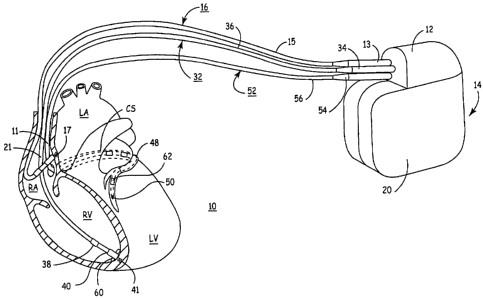
FIG. 1 depicts an exemplary implantable, multi-chamber cardiac pacemaker
coupled to a patient's heart via transvenous endocardial leads.
FIG. 2 is a schematic block diagram of the mufti-chamber pacemaker of FIG. 1
capable of delivering a resynchronization therapy.
FIG. 3 is a flow chart providing an overview of a method for optimizing
cardiac
pacing intervals.
FIG. 4 is a flow chart summarizing steps included in a method for determining
an
optimal A-V interval.
As indicated above, the present invention is directed toward providing a
method
and apparatus for optimizing ventricular function and selecting cardiac pacing
intervals for
the purposes of restoring ventricular synchrony based on inter- and/or infra-
atrial delay,
ventricular filling characteristics and/or physiologic dimensions of one or
both ventricles.
CA 02548001 2006-05-31
WO 2005/056108 PCT/US2004/040521
-6-
The present invention is useful in optimizing atrial-ventricular, inter-atrial
and inter-
ventricular pacing intervals during cardiac resynchronization therapy (CRT)
used for
treating heart failure. The present invention is also useful in selecting
pacing parameters
used during temporary pacing applied for treating post-operative uncoordinated
cardiac
chamber (e.g., atrial and/or ventricular) motion. As such, the present
invention may be
embodied in an implantable cardiac pacing system including a dual chamber or
multichamber pacemaker and associated set of medical electrical leads.
Alternatively, the
present invention may be embodied in a temporary pacing system including an
external
pacing device with associated temporary pacing leads.
FIG. 1 depicts an exemplary implantable, multi-chamber cardiac pacemaker 14 in
which the present invention may be implemented. The multi-chamber pacemaker 14
is
provided for restoring ventricular synchrony by delivering pacing pulses to
one or more
heart chambers as needed to control the heart activation sequence. The
pacemaker 14 is
shown in communication with a patient's heart 10 by way of three leads
16,32,52. The
heart 10 is shown in a partially cut-away view illustrating the upper heart
chambers, the
right atrium (RA) and left atrium (LA) and septal wall (SW) disposed
therebetween, and
the lower heart chambers, the right ventricle (RV) and left ventricle (LV) and
the septal
wall (SW) disposed therebetween, and the coronary sinus (CS) extending from
the
opening in the right atrium laterally around the atria to form the great
cardiac vein 48
including branches thereof.
The pacemaker 14, also referred to herein from time to time as an implantable
pulse generator (IPG) or an implantable cardioverter-defibrillator (ICD), is
implanted
subcutaneously in a patient's body between the skin and the ribs. Three
transvenous-
endocardial leads 16,32,52 connect the IPG 14 with the RA, the RV and the LV,
respectively. Each lead has at least one electrical conductor and pace/sense
electrode. A
remote indifferent can electrode 20 is formed as part of the outer surface of
the housing of
the IPG 14. The pace/sense electrodes and the remote indifferent can electrode
20 can be
selectively employed to provide a number of unipolar and bipolar pace/sense
electrode
combinations for pacing and sensing functions.
The depicted bipolar endocardial RA lead 16 is passed through a vein into the
RA
chamber of the heart 10, and the distal end of the RA lead 16 is attached to
the RA wall by
an attachment mechanism 17. The attachment mechanism may be active or passive
as is
CA 02548001 2006-05-31
WO 2005/056108 PCT/US2004/040521
_7_
known in the art and as may be later developed. A helix or tined lead may be
used as is
known in the art, to adapt the distal end of a lead for relatively permanent
fixation to
myocardial tissue. The bipolar endocardial RA lead 16 is formed with an in-
line
connector 13 fitting into a bipolar bore of IPG connector block 12 that is
coupled to a pair
of electrically insulated conductors within lead body 15 and connected with
distal tip RA
pace/sense electrode 17 and proximal ring RA pace/sense electrode 21 provided
for
achieving RA pacing and sensing of RA electrogram (EGM) signals.
In accordance with a triple chamber embodiment of the present invention, a
coronary sinus lead 52 capable of stimulating the left ventricle is preferably
of a relatively
small size and diameter such that it may be passed through the coronary sinus
and entering
a vessel branching from the great cardiac vein and able to be steered to a
left ventricular
pacing site.
The depicted positions of the leads and electrodes shown in FIG. 1 in or about
the
right and left heart chambers are approximate and merely exemplary.
Furthermore, it is
recognized that alternative leads and pace/sense electrodes that are adapted
for placement
at pacing or sensing sites on or in or relative to the RA, LA, RV and LV may
be used in
conjunction with the present invention.
Bipolar, endocardial RV lead 32 passes through the RA into the RV where its
distal ring and tip RV pace/sense electrodes 38,40 are adapted for fixation to
myocardial
tissue by a distal attachment mechanism 41. The RV lead 32 is formed with an
in-line
connector 34 fitting into a bipolar bore of IPG connector block 12 that is
coupled to a pair
of electrically insulated conductors within lead body 36 and connected with
distal tip RV
pace/sense electrode 41 and proximal ring RV pace/sense electrode 38 provided
for RV
pacing and sensing of RV EGM signals.
In the illustrated embodiment of a triple chamber IPG capable of delivering
CRT, a
unipolar or bipolar or multipolar endocardial LV CS lead 52 is passed through
the RA,
into the CS and further into a cardiac vein to extend the distal LV CS
pace/sense electrode
50 alongside the LV chamber to achieve LV pacing and sensing of LV EGM
signals. The
LV CS lead 52 is coupled at the proximal end connector 54 fitting into a bore
of IPG
connector block 12. A small diameter unipolar lead body 56 is selected in
order to lodge
the distal LV CS pace/sense electrode 50 deeply in a cardiac vein branching
from the great
cardiac vein 48.
CA 02548001 2006-05-31
WO 2005/056108 PCT/US2004/040521
_g_
In a four chamber embodiment, LV CS lead 52 could bear a proximal LA CS
pace/sense electrode positioned along the lead body to lie in the larger
diameter coronary
sinus adjacent the LA for use in pacing the LA or sensing LA EGM signals. In
that case,
the lead body 56 would encase an insulated lead conductor extending proximally
from the
more proximal LA CS pace/sense electrodes) and terminating in a bipolar
connector 54.
FIG. 2 is a schematic block diagram of an exemplary multi-chamber IPG 14, such
as that shown in FIG. 1, that provides delivery of a resynchronization therapy
and is
capable of processing atrial and/or ventricular signal inputs. The IPG 14 is
preferably a
microprocessor-based device. Accordingly, microprocessor-based control and
timing
system 102, which varies in sophistication and complexity depending upon the
type and
functional features incorporated therein, controls the functions of IPG 14 by
executing
firmware and programmed software algorithms stored in associated RAM and ROM.
Control and timing system 102 may also include a watchdog circuit, a DMA
controller, a
block mover/reader, a CRC calculator, and other specific logic circuitry
coupled together
by on-chip data bus, address bus, power, clock, and control signal lines in
paths or trees in
a manner known in the art. It will also be understood that control and timing
functions of
IPG 14 can be accomplished with dedicated circuit hardware or state machine
logic rather
than a programmed microcomputer.
The IPG 14 includes interface circuitry 104 for receiving signals from sensors
and
pace/sense electrodes located at specific sites of the patient's heart
chambers and
delivering cardiac pacing to control the patient's heart rhythm and
resynchronize
depolarization of chambers of a patient's heart. The interface circuitry 104
therefore
includes a therapy delivery system 106 intended for delivering cardiac pacing
impulses
under the control of control and timing system 102. Delivery of pacing pulses
to two or
more heart chambers is controlled in part by the selection of programmable
pacing
intervals, which can include atrial-atrial (A-A), atrial-ventricular (A-V),
and ventricular-
ventricular (V-V) intervals.
Physiologic input signal processing circuit 108 is provided for receiving
cardiac
electrogram (EGM) signals for determining a patient's heart rhythm.
Physiologic input
signal processing circuit 108 additionally can receive signals related to
infra- or inter-atrial
delay and processes these signals and provides signal data to control and
timing system
102 for further signal analysis and/or storage. For purposes of illustration
of the possible
CA 02548001 2006-05-31
WO 2005/056108 PCT/US2004/040521
-9-
uses of the invention, a set of lead connections are depicted for making
electrical
connections between the therapy delivery system 106 and the input signal
processing
circuit 108 and sets of pace/sense electrodes located in operative relation to
the RA, LA,
RV and/or LV.
Control and timing system 102 controls the delivery of bi-atrial, bi-
ventricular, or
mufti-chamber cardiac pacing pulses at selected intervals intended to improve
heart
chamber synchrony. The initial delivery of pacing pulses by IPG 14 may be
programmed
to nominal settings or provided according to programmable pacing intervals,
such as
programmable conduction delay window times as generally disclosed in U.S. Pat.
No.
6,070,101 issued to Struble et al., incorporated herein by reference in its
entirety, or
programmable coupling intervals as generally disclosed in above-cited U.S.
Pat. No.
6,473,645 issued to Levine. Selection of the programmable pacing intervals
while a
patient is ambulatory is preferably based on infra-, inter-atrial delay and/or
based upon
clinical evidence of ventricular filling characteristics or dimensions of
ventricular chamber
(i.e., chamber volume) as described herein.
The therapy delivery system 106 can optionally be configured to include
circuitry
for delivering cardioversion/defibrillation therapy in addition to cardiac
pacing pulses for
controlling a patient's heart rhythm. Accordingly, as previously mentioned
medical
electrical leads in communication with the patient's heart can also
advantageously include
high-voltage cardioversion or defibrillation shock electrodes.
A battery 136 provides a source of electrical energy to power components and
circuitry of IPG 14 and provide electrical stimulation energy for delivering
electrical
impulses to the heart. The typical energy source is a high energy density, low
voltage
battery 136 coupled with a power supply/POR circuit 126 having power-on-reset
(POR)
capability. The power supply/POR circuit 126 provides one or more low voltage
power
(Vlo), the POR signal, one or more reference voltage (VREF) sources, current
sources, an
elective replacement indicator (ERI) signal, and, in the case of a
cardioversion/defibrillator
capabilities, high voltage power (Vhi) to the therapy delivery system 106. Not
all of the
conventional interconnections of these voltages and signals are shown in FIG.
2.
Current electronic mufti-chamber pacemaker circuitry typically employs clocked
CMOS digital logic ICs that require a clock signal CLK provided by a
piezoelectric crystal
132 and system clock 122 coupled thereto as well as discrete components, e.g.,
inductors,
CA 02548001 2006-05-31
WO 2005/056108 PCT/US2004/040521
-10-
capacitors, transformers, high voltage protection diodes, and the like that
are mounted with
the ICs to one or more substrate or printed circuit board. In FIG. 2, each CLK
signal
generated by system clock 122 is routed to all applicable clocked logic via a
clock tree.
The system clock 122 provides one or more fixed frequency CLK signal that is
independent of the battery voltage over an operating battery voltage range for
system
timing and control functions and in formatting uplink telemetry signal
transmissions in the
telemetry I/O circuit 124.
The RAM registers included in microprocessor-based control and timing system
102 may be used for storing data compiled from sensed EGM signals, wall motion
signals,
and/or relating to device operating history or other sensed physiologic
parameters for
uplink telemetry transmission upon receipt of a retrieval or interrogation
instruction via a
downlink telemetry transmission. Criteria for triggering data storage can be
programmed
via down linked instructions and parameter values. Physiologic data, including
septal wall
motion data, may be stored on a triggered or periodic basis or by detection
logic within the
physiologic input signal processing circuit 108. In some cases, the IPG 14
includes a
magnetic field sensitive switch 130 that closes in response to a magnetic
field, and the
closure causes a magnetic switch circuit 120 to issue a switch closed (SC)
signal to control
and timing system 102 which responds in a magnet mode. For example, the
patient may be
provided with a magnet 116 that can be applied over the subcutaneously
implanted IPG 14
to close switch 130 and prompt the control and timing system to deliver a
therapy and/or
store physiologic data. Event related data, e.g., the date and time and
current pacing
parameters, may be stored along with the stored physiologic data for uplink
telemetry in a
later interrogation session.
Uplink and downlink telemetry capabilities are provided to enable
communication
with either a remotely located external medical device or a more proximal
medical device
on or in the patient's body. Stored EGM data (and data derived therefrom), as
well as real-
time generated physiologic data and non-physiologic data can be transmitted by
uplink RF
telemetry from the IPG 14 to the external programmer or other remote medical
device 26
in response to a downlink telemetered interrogation command. As such, an
antenna 128 is
connected to radio frequency (RF) transceiver circuit 124 for the purposes of
uplink/downlink telemetry operations. Telemetering both analog and digital
data between
antenna 128 and an external device 26, also equipped with an antenna 118, may
be
CA 02548001 2006-05-31
WO 2005/056108 PCT/US2004/040521
-11-
accomplished using numerous types of telemetry systems known in the art for
use in
implantable devices.
The physiologic input signal processing circuit 108 includes at least one
electrical
signal amplifier circuit for amplifying, processing and in some cases
detecting sensed
events from characteristics of the electrical sense signal or sensor output
signal. The
physiologic input signal processing circuit 108 may thus include a plurality
of cardiac
signal sense channels for sensing and processing cardiac signals from sense
electrodes
located in relation to a heart chamber. Each such channel typically includes a
sense
amplifier circuit for detecting specific cardiac events and an EGM amplifier
circuit for
providing an EGM signal to the control and timing system 102 for sampling,
digitizing
and storing or transmitting in an uplink transmission. Atrial and ventricular
sense
amplifiers include signal processing stages for detecting the occurrence of P-
waves and R-
waves, respectively, and providing atrial sense or ventricular sense event
signals to the
control and timing system 102. Timing and control system 102 responds in
accordance
with its particular operating system to deliver or modify a pacing therapy, if
appropriate,
or to accumulate data for uplink telemetry transmission in a variety of ways
known in the
art. Thus the need for pacing pulse delivery is determined based on EGM signal
input
according to the particular operating mode in effect. The operative A-V
intervals for
pacing pulse delivery can vary based on heart rate, sensed level activity
(e.g., via a
piezoelectric crystal, accelerometer, etc.), detected inter-atrial delay,
filling characteristics
and/or measured ventricular chamber volume.
FIG. 3 is a flow chart providing an overview of a method for optimizing
cardiac
pacing intervals according to the present invention. Method 200 begins at step
205,
wherein LVEDV, LVESV, filling characteristics and/or measured infra-atrial
delay (e.g.,
measured via ECG, or electrogram - EGM, or using Doppler ultrasound, or
mechanically
monitored or detected). As is known in the art, these values are readily
obtained using
known echocardiographic techniques. At step 210, an optimal A-V interval is
determined
based upon the values obtained in step 205. Depending on the dual chamber or
multichamber pacing system being used, a right A-V interval or a left A-V
interval or both
may be determined. For the embodiment shown in FIG. 1, an optimal RA to LV
interval
is determined. However, in other embodiments, the left atrial-left ventricular
interval is
optimized based on the value obtained in step 205 to ensure optimal filling of
the LV. At
CA 02548001 2006-05-31
WO 2005/056108 PCT/US2004/040521
-12-
step 215, the A-V interval is automatically adjusted to the optimal A-V
interval
determined at step 210.
Optionally, at step 220 the optimal V-V interval is determined for bi-
ventricular or
atrio-biventricular pacing modes. A method for optimizing the V-V interval can
be used
that relies upon accelerometer sensors coupled to the LV or the ventricular
septum and the
like (as described and depicted in the co-pending applications incorporated
hereinabove).
At optional step 225, the V-V interval is automatically adjusted to the
optimal V-V
interval determined at step 220. After adjusting the V-V interval, an optional
step 230 may
be performed to re-optimize the A-V interval. Verification of the
provisionally
determined optimal A-V interval is made by re-determining the optimal A-V
interval
during biventricular pacing at the newly optimized V-V interval. The A-V
interval may be
re-adjusted accordingly if a different A-V interval is identified as being
optimal during
pacing at the optimal V-V interval.
FIG. 4 is a flow chart summarizing steps included in a method for determining
an
optimal A-V interval for use in method 200 of FIG. 3. Method 300 begins at
step 305 by
setting the A-V interval to a desired nominal value. For example, a nominal A-
V interval
setting of 100 ms may be used. At step 310, any infra-atrial delay present is
monitored
and characterized using, for example, non-invasive echocardiographic
equipment, surface-
based ECG equipment and/or internal electrogram (EGM) monitoring techniques.
For
example, in the embodiment depicted at FIG. 4, inter-atrial delay is declared
present if a P-
wave duration exceeds about 100 ms or the RA activates more than 60 ms prior
to the LA
activation. However, other values and techniques may be used. As depicted in
FIG. 4, in
the event that intertribal delay is deemed present, then at step 315 the A-V
interval is
incremented upward (as depicted 40 ms is added to the A-V interval). If no
inter-atrial
delay is present then at step 320 no change to the A-V interval occurs and the
method
proceeds to step 325. At step 325, an LVEDV value is obtained (e.g., measured
or
otherwise determined). If the LVEDV value exceeds a threshold value (i.e., 275
ml as
shown in FIG. 4), then at step 330 the A-V interval is decremented by an
amount (e.g., 40
ms). If the LVEDV value does not exceed the threshold value, then at step 335
no change
is made. Also, a different or additional initial A-V interval may be used than
the 100 ms
value described above. In addition, the method depicted in FIG. 4 may be
iteratively
applied, periodically or otherwise anytime that one or more of LVEDV, LVESV
and/or
CA 02548001 2006-05-31
WO 2005/056108 PCT/US2004/040521
-13-
inter-atrial delay information is available for a given patient. Furthermore,
one or more
mechanical sensors may be used to confirm that physiologically appropriate A-V
intervals
are being used.
In a patient with intact atrioventricular conduction, the method depicted and
described with respect to FIG. 4 may include patient's intrinsic A-V interval
as a factor in
setting the initial A-V interval (at step 305). This may be very useful in the
event that the
patient is receiving so-called fusion pacing based on intrinsic atrial
activation. In order to
allow intrinsic A-V conduction, the A-V interval is set at a maximum setting
or a setting
longer than the intrinsic A-V conduction time. The intrinsic A-V conduction
time may be
determined by measuring the interval from an atrial pacing pulse to a
subsequently sensed
R-wave. Remaining test A-V intervals may be applied at decreasing increments
from the
intrinsic A-V interval. Alternatively, test A-V intervals may be applied
randomly ranging
from 0 ms to the intrinsic A-V interval. If atrioventricular conduction is not
intact, a set of
test A-V intervals may be selected over a predefined range, for example a
range from 0 ms
to on the order of 250 ms.
While not depicted, sustaining a stable heart rate during the data acquisition
interval is performed may be beneficial. Heart rate instability, such as the
presence of
ectopic heart beats or other irregularities, can produce anomalous mechanical
(motion)
data. As such, the heart rate preferably stays within a specified range. In
one
embodiment, heart rate stability may be verified by determining the average
and standard
deviation of the cardiac cycle length during the data acquisition period. The
cardiac cycle
length may be determined as the interval between consecutive ventricular
events including
ventricular pacing pulses and any sensed R-waves. If the average cardiac cycle
length or
its standard deviation falls outside a predefined range, the data is
considered unreliable.
When method 300 is executed by an external pacing system, the obtained data
relating to LVEDV, LVESV and/or inter-atrial (electrical or mechanical) delays
may be
displayed in real-time or stored and presented following an optimization
procedure. When
method 300 for identifying an optimal A-V interval is executed by an implanted
device,
the obtained data may be stored for later uplinking to an external device for
display and
review by a physician.
The optional steps 220,225,230 of FIG. 3 for determining an optimal V-V
interval
are now briefly described. The optimal A-V interval is programmed to an
optimal setting
CA 02548001 2006-05-31
WO 2005/056108 PCT/US2004/040521
-14-
determined according to method 300 of FIG. 4. The V-V interval is set to a
test interval
and a range of test intervals are predefined and may be delivered in a random,
generally
increasing, or generally decreasing fashion. A range of test intervals may
include intervals
that result in the right ventricle being paced prior to the left ventricle and
intervals that
result in the left ventricle being paced prior to the right ventricle. A set
of exemplary test
intervals includes right ventricular pacing 20 ms and 40 ms prior to left
ventricular pacing,
simultaneous left and right ventricular pacing (a V-V interval of 0 ms), and
left ventricular
pacing 20 ms and 40 ms prior to the right ventricle. After each of a plurality
of test V-V
intervals are applied, the optimal V-V interval is identified as having the
least amount of
extraneous or dysschronous motion. When the V-V interval is determined using
an
external pacing system in a clinic having echocardiographic imaging and
measurement
equipment, ventricular volumes, ventricular wall motion and/or septal wall
motion data
may be displayed in real-time or stored and presented during optimization
procedures.
When identifying an optimal V-V interval using an implanted device, the volume
data
and/or wall motion data may be stored for later uplinking to an external
device for display
and review by a physician. After identifying the optimal V-V interval, the V-V
interval
setting may be automatically adjusted or programmed.
When the methods of the present invention are implemented in an implantable
device, stored data available through uplink telemetry to an external device
can be
displayed and/or reviewed by a physician. When such methods are implemented in
an
external device, a display of cardiac function data may be updated
periodically an intra- or
inter-atrial delay characteristic changes.
Thus, a method and apparatus have been described for optimizing a cardiac
therapy. The methods described herein may advantageously be applied in
numerous
cardiac monitoring or therapy modalities including chronic or acute
applications
associated with implantable or external devices. In addition, certain of the
methods and
apparatus operated according to the present invention can be operated using
computer
processors operating pursuant to instructions stored on a computer readable
medium.
Accordingly, all diverse types of computer readable medium and other
substrates capable
of producing control signals for operating structure according to the
invention are included
with the scope of the invention.