Note: Descriptions are shown in the official language in which they were submitted.
CA 02548097 2006-06-02
WO 2005/056084 PCT/US2004/041091
SPECIFICATION
TITLE OF INVENTION
PLEDGET-HANDLING SYSTEM AND METHOD
FOR DELIVERING HEMOSTASIS PROMOTING
MATERIAL TO A BLOOD VESSEL PUNCTURE SITE
BY FLUID PRESSURE
RELATED PATENT APPLICATIONS
[0001] This application is a continuation in part of the following prior co-
copending
U.S. patent applications 1) Serial Number 10/256,493 filed September 26, 2002
and
titled SYSTEM AND METHOD FOR DELIVERING HEMOSTASIS PROMOTING
MATERIAL TO A BLOOD VESSEL PUNCTURE SITE BY FLUID PRESSURE and
2) Serial Number 10/007,204 filed November 8, 2001 and titled SYSTEM AND
METHOD FOR DELIVERING HEMOSTASIS PROMOTING MATERIAL TO A
BLOOD VESSEL PUNCTURE SITE BY FLUID PRESSURE .
FIELD OF THE INVENTION
[0002] The invention relates to a system and method for delivering hemostasis
promoting material to a blood vessel puncture site by fluid pressure, and more
particularly, the invention relates to an improved system and method for
delivery of
absorbable sponge material for sealing of a blood vessel puncture site.
DESCRIPTION OF THE RELATED ART
[0003] A large number of diagnostic and interventional procedurals involve the
percutaneous introduction of instrumentation into a vein or artery. For
example,
coronary angioplasty, angiography, atherectorny, stenting of arteries, and
many other
procedures often involve accessing the vasculature through a catheter placed
in the
femoral artery or other blood vessel. Once the procedure is completed and the
catheter
or other instrumentation is removed, bleeding from the punctured artery must
be
controlled.
CA 02548097 2006-06-02
WO 2005/056084 PCT/US2004/041091
[0004] Traditionally, external pressure is applied to the skin entry site to
stem bleeding
from a puncture wound in a blood vessel. Pressure is continued until
hemostasis has
occurred at the puncture site. In some instances, pressure must be applied for
up to an
hour or more during which time the patient is uncomfortably immobilized. In
addition, a
risk of hematoma exists since bleeding from the vessel may continue beneath
the skin
until sufficient clotting effects hemostasis. Further, external pressure to
close the
vascular puncture site works best when the vessel is close to the skin surface
and may be
unsuitable for patients with substantial amounts of subcutaneous adipose
tissue since the
skin surface may be a considerable distance from the vascular puncture site.
[0005] More recently, devices have been proposed to promote hemostasis
directly at a
site of a vascular puncture. One class of such puncture sealing devices
features an
intraluminal anchor which is placed within the blood vessel and seals against
an inside
surface of the vessel puncture. The intraluminal plug may be used in
combination with a
sealing material positioned on the outside of the blood vessel, such as
collagen. Sealing
devices of this type are disclosed in U.S. Patent Nos. 4,852,568; 4,890,612;
5,021,059;
and 5,061,274.
[0006] Another approach to subcutaneous blood vessel puncture closure involves
the
delivery of non-absorbable tissue adhesives, such cyanoacrylate, to the
perforation site.
Such a system is disclosed in U.S. Patent No. 5,383,899.
[0007] The application of an absorbable material such as collagen or a non-
absorbable
tissue adhesive at the puncture site has several drawbacks including: 1)
possible injection
of the material into the blood vessel causing thrombosis; 2) a lack of
pressure directly on
the blood vessel puncture which may allow blood to escape beneath the material
plug
into the surrounding tissue; and 3) the inability to accurately place the
absorbable
material plug directly over the puncture site.
[0008] The use of an anchor and plug system addresses these problems to some
extent
but provides other problems including: 1) complex and difficult application;
2) partial
occlusion of the blood vessel by the anchor when placed properly; and 3)
complete
blockage of the blood vessel or a branch of the blood vessel by the anchor if
placed
improperly. Another problem with the anchor and plug system involves reaccess.
Reaccess of a particular blood vessel site sealed with an anchor and plug
system is not
possible until the anchor has been completely absorbed because the anchor
could be
dislodged into the blood stream by an attempt to reaccess.
2
CA 02548097 2006-06-02
WO 2005/056084 PCT/US2004/041091
[0009] A system which addresses many of these problems is described in U.S.
Patent
No. 6,162,192 which delivers a hydrated pledget of absorbable sponge material
to a
location outside the blood vessel to facilitate hemostasis. However, this
system involves
the removal of the introduces sheath used during the intravascular procedure
and the
insertion of a dilator and introduces into the tissue tract vacated by the
introduces sheath
to place the absorbable sponge. It would be desirable to reduce the number of
steps
involved in delivery of a hemostasis promoting material by allowing the
material to be
delivered through an introduces sheath already in place within the tissue
tract and used in
the intravascular procedure.
[0010] Accordingly, it would be desirable to provide a system for accurately
locating
the blood vessel wall at a puncture site and for properly placing a hemostasis
plug over
the puncture site where the locating and placing steps are performed through
the
introduces sheath already in place in the blood vessel.
SUMMARY OF THE INVENTION
[0011] The present invention relates to a system for delivering hemostasis
promoting
material to a blood vessel puncture site through a sheath already in place in
the blood
vessel.
[0012] In accordance with one aspect of the invention, a pledget handling
system is
provided. The pledget handling system facilitates consistent hydration of the
pledget,
provides for effective staging of the pledget, and prevents early pledget
delivery.
[0013] In accordance with another aspect of the invention, a system for
delivering
hemostasis promoting material includes an introduces sheath, a control tip
coupled to the
introduces sheath; and, seal means disposed around a portion of said control
tip to
prevent blood from passing into and through the introduces sheath. The seal
means also
protects against unwanted transmission of materials from the sheath into the
blood
vessel.
BRIEF DESCRIPTION OF THE DRAWING FIGURES
[0014] The invention will now be described in greater detail with reference to
the
preferred embodiments illustrated in the accompanying drawings, in which like
elements
bear like reference numerals, and wherein:
FIG. 1 is an exploded side view of a first embodiment of a system for
delivering
hemostasis promoting material to a blood vessel puncture site by fluid
pressure.
FIG. 2 is an assembled side view of the system of FIG. 1.
CA 02548097 2006-06-02
WO 2005/056084 PCT/US2004/041091
FIG. 3 is a side cross sectional view of a portion of the system of FIG. 2.
FIG. 4 is an exploded side view of a further system for delivering hemostasis
promoting material to a blood vessel puncture site by fluid pressure with the
material
delivered to a side branch of the sheath.
FIG. 5 is an assembled side view of the system of FIG. 4.
FIG. 6 is a side cross sectional view of a portion of the system of FIG. S
including a
proximal end of the introducer sheath and control tip.
FIG. 7 is a side cross sectional view of a portion of the system of FIG. 5
including
an exhaust valve, a hydration chamber, and a syringe.
FIG. 8 is a side cross sectional view of a portion of the system of FIG. 1
with a
pledget of hemostasis promoting material positioned in the hydration chamber.
FIG. 9 is a side cross sectional view of a portion of the system of FIG. 1
with the
sponge hydrated and advanced in preparation for delivery.
FIG. 10 is a side cross sectional view of a blood vessel puncture site with an
introducer sheath and guidewire positioned in the blood vessel puncture.
FIG. 11 is a side cross sectional view of the blood vessel puncture site with
the
hemostasis promoting material delivery system connected to the introducer
sheath and
bleed back visible from the vent tube.
FIG. 12 is a side cross sectional view of the blood vessel puncture site with
the
hemostasis promoting material delivery system and introducer sheath withdrawn
to a
desired position for delivery of the hemostasis promoting material.
FIG. 13 is a side cross sectional view of the blood vessel puncture site with
the
hemostasis promoting material delivered to the blood vessel puncture site by
fluid
pressure.
FIG. 14 is a side cross sectional view of the blood vessel puncture site with
the
hemostasis promoting material delivery system and guidewire removed from the
introducer sheath.
FIG. 15 is a side cross sectional view of the blood vessel puncture site with
the
introducer sheath withdrawn.
FIG. 16 is an alternative embodiment of a system for delivering hemostasis
promoting material to a blood vessel puncture site by fluid pressure.
FIG. 16a is a detail of the embodiment shown in FIG 16 illustrating the
operation
thereof.
4
CA 02548097 2006-06-02
WO 2005/056084 PCT/US2004/041091
FIG. 16b is another detail of the embodiment shown in FIG 16 illustrating the
operation thereof.
FIG. 16c is another detail of the embodiment shown in FIG 16 illustrating the
operation thereof.
FIG. 17 is an alternative embodiment for delivering hemostasis promoting
material
including an introducer sheath, a control tip coupled to and extending from
the introducer
sheath; and, seal means disposed around a portion of said control tip.
FIG. 18 is an alternative embodiment.
FIG. 19 is an alternative embodiment.
FIG. 20 is an alternative embodiment.
FIG. 21 is an alternative embodiment.
FIG. 22 is an alternative embodiment.
FIG. 23 is an alternative embodiment.
FIG. 24 is an alternative embodiment.
FIG. 25 is an alternative embodiment.
DETAILED DESCRIPTION OF THE PREFERRED EMBODIMENTS
[0015] A system for delivering hemostasis promoting material of the present
invention
allows the hemostasis promoting material to be delivered to a blood vessel
puncture site
by fluid pressure. The system allows the hemostasis promoting material to be
delivered
through an introducer sheath which is already in place within a tissue tract.
This system
includes a control tip which is insertable through the introducer sheath to
locate and
occlude the blood vessel puncture site and a hydration chamber for receiving
and
delivering the hemostasis promoting material to the blood vessel puncture
site.
[0016] Although the present invention is particularly designed for delivering
a
hemostasis promoting material in the form of an absorbable sponge through the
introducer sheath by fluid pressure, it should be understood that the system
may also be
used for delivering other hemostasis promoting materials which are useful for
sealing a
puncture site. The use of an absorbable hydrated sponge material allows the
delivery of
more absorbable sponge material down through a smaller sheath by allowing the
sponge
material to be hydrated and compressed. Once delivered, the absorbable sponge
rapidly
expands to fill the entire width of the tissue tract and provides hemostasis
at the puncture
site.
CA 02548097 2006-06-02
WO 2005/056084 PCT/US2004/041091
[0017] In the context of the present invention, "pledget" means a piece of
sponge
formed into a generally elongated shape having a size which allows delivery in
a
hydrated state through an introduces sheath, delivery cannula or introduces to
a site of a
puncture in a blood vessel.
[0018] "Sponge" means a biocompatible material which is capable of being
hydrated
and is resiliently compressible in a hydrated state. Preferably, the sponge is
non-
immunogenic and may be absorbable or non-absorbable.
[0019] "Absorbable sponge" means sponge which, when implanted within a human
or
other mammalian body, is absorbed or resorbed by the body.
[0020] "Hydrate" means to partially or fully saturate with a fluid, such as
saline, water,
blood contrast agent, thrombin, ionic solutions, therapeutic agents, or the
like.
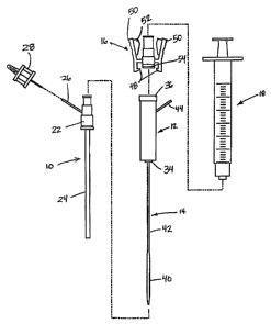
[0021] The system of FIG. 1 includes an introduces sheath 10, a hydration
chamber 12
with an attached control tip 14, a coupler 16, and a syringe 18. The
introduces sheath 10
is an intravascular access sheath as is conventionally used for procedures
such as
coronary angioplasty and stenting procedures. The introduces sheath 10
includes a
proximal hub 22 connected to a tubular sheath 24. A vent tube 26 is in fluid
communication with an interior of the hub 22 for purposes of providing a
visual bleed
back indication which will be discussed in further detail below. In the
embodiment
illustrated in FIG. 1, a vent cap 28 is provided for opening and closing the
vent tube 26
manually.
[0022] The hydration chamber 12 is configured to receive a pledget of
absorbable
sponge material for hydration of the pledget and delivery of the pledget
through the
introduces sheath 10. A proximal end of the hydration chamber 12 includes a
flange 36
or other connecting element for receiving the coupler 16. A distal end 34 of
the
hydration chamber 12 connects to the proximal hub 22 of the introduces sheath
12. The
control tip 14 has an enlarged distal end 40 configured to be received in the
puncture in
the blood vessel and to control blood flow through the puncture in the blood
vessel. The
enlarged distal end 40 is connected to a smaller diameter control tip tube 42
which
extends from the enlarged distal end through the distal end of the hydration
chamber 12
and out a side of the hydration chamber 12 to a proximal end 44 of the control
tip. The
enlarged distal end 40 of the control tip performs the multiple functions of
controlling
blood flow through the blood vessel puncture, providing an indication of the
position of
the distal end of the introduces sheath, and guiding the hemostasis promoting
material
delivery system over a guidewire.
6
CA 02548097 2006-06-02
WO 2005/056084 PCT/US2004/041091
[0023] The coupler 16 allows the syringe 18 to be connected to the hydration
chamber
12_ Removal of the coupler 16 from the hydration chamber 12 allows the pledget
of
absorbable sponge material to be easily inserted into the hydration chamber in
its dry
form. Upon.connection of the coupler 16 to the hydration chamber 12 the
conventional
syringe 18 will be connected to the coupler 16 for injection of fluid into the
hydration
chamber. The coupler 16 includes a seal 54 and two or more locking tabs 48
which lock
over the flange 36 of the hydration chamber and are releasable by pressing on
two wings
50 of the coupler. Stops 52 on the interior surfaces of the wings 50 prevent
the coupler
16 from being removed from the hydration chamber 12 when a syringe 18 is
mounted on
the coupler. It should be understood that many other coupler designs may also
be used
without departing from the present invention.
[0024] In use, the system of FIGS. 1, 2, and 3 is assembled with a sponge
placed inside
the hydration chamber 12 and a syringe 18 containing water, saline solution,
or other
fluid attached to the hydration chamber by the coupler 16. The sponge is
hydrated and
staged or moved, to a position at the distal end of the hydration chamber as
will be
described in further detail below. The syringe 18 is preferably capable of
generating a
high pressure with a relatively low plunger force such as a 1 cc syringe.
[0025] The introducer sheath 10 is placed in the blood vessel puncture of a
patient in a
conventional manner for performance of the intravascular procedure. After the
intravascular procedure, the introducer sheath 10 and a guidewire (not shown)
are
maintained in place extending into the blood vessel. The control tip 14 is
threaded over
the proximal end of the guidewire and the hydration chamber 12 and control tip
14 are
advanced into the introducer sheath until the hydration chamber distal end 34
is engaged
with the hub 22 of the introducer sheath 10. Bleed back is observed by a
variety of
methods which will be described below with respect to FIG. 3. In the
embodiment of
FIG. 3, the vent cap 28 is removed from the vent tube 26 to observe bleed
back. The
introducer sheath 10, hydration chamber 12, and control tip 14, are withdrawn
together
slowly from the puncture site until the bleed back observed from the vent tube
26 stops.
The bleed back stops when the enlarged distal end 40 of the control tip 44 is
positioned
in the blood vessel puncture preventing blood from escaping from the puncture.
The
distance d between the distal end of the tubular sheath 24 and the enlarged
distal end 40
of the control tip 14 is selected so that the point at which bleed back stops
indicates that
the distal end of the introducer sheath 10 is located at a desired delivery
location for
delivery of the hemostasis promoting material to the blood vessel puncture
site. The
7
CA 02548097 2006-06-02
WO 2005/056084 PCT/US2004/041091
distance d will be selected to correspond to the size of the pledget to be
delivered to the
puncture site and will be selected such that the hemostasis promoting material
is located
in the tissue tract adjacent the blood vessel without extending into the lumen
of the blood
vessel.
[0026] FIG. 3 illustrates a vent tube 26 with a vent cap 28 for observing
bleed back.
When the vent cap 28 is removed from the vent tube 26 blood is able to pass
from the
distal end of the introducer sheath 10 through the introducer sheath and out
of the vent
tube. The vent tube 26 has a relatively small diameter which is selected to
provide a
very noticeable spurt or stream of blood to indicate bleed back has occurred.
In contract,
the observance of bleed back from a larger tube such as the introducer sheath
would
result in an oozing or dripping bleed back indication which is difficult for
the user to use
as a precise indicator of position. According to one preferred embodiment, the
vent tube
26 has an inner diameter of about 0.4mm to about 2mm, preferably about lmm.
[0027] FIGS. 4-7 illustrate an alternative embodiment of a system for
delivering
hemostasis promoting material in which a hydration chamber 312 is connected to
a side
port 320 of an introducer sheath 310. The vent tube 326 is connected to
another port of
the side port 320. The stop cock 322 is movable between an open delivery
position
shown in FIG. 10 and a closed bleed back position shown in phantom in FIG. 4.
In the
closed bleed back position, bleed back is allowed through the vent tube 326.
In the open
delivery position the hemostasis promoting material is delivered from the
hydration
chamber 312 to the introducer sheath. As in the embodiment described above in
connection with FIGS. 1-3, a syringe 318 is coupled to and decoupled from a
hydration
chamber 312 by coupler 316.
[0028] As shown in the cross sectional view of FIG. 7, when the stop cock 322
is in the
open delivery position, the hemostasis promoting material will pass from the
hydration
chamber 312 through the stop cock 322 and the side port 320 and into the
introducer
sheath 310 for delivery to the blood vessel puncture site.
[0029] FIG. 6 illustrates the connection of the control tip 314 to a proximal
plug 330
which is connectable by a coupler 316 to the hub 332 of the introducer sheath
310. The
hemostasis promoting material is delivered through the side port 320 of FIG. 6
and into
the hub 332 of the introducer sheath 310 and then is delivered through the
introducer
sheath to the puncture site.
[0030] FIGS. 8-15 illustrate the preparation and use of the system for
delivering
hemostasis promoting material to a blood vessel puncture site. Although FIGS.
8-15
8
CA 02548097 2006-06-02
WO 2005/056084 PCT/US2004/041091
illustrate the procedure which is used with the embodiment of FIGS. 1-3, a
similar
procedure would be used with the other embodiments described below. FIGS. 8
and 9
illustrate the hydration and staging of a pledget 20 of sponge material in the
hydration
chamber 12. Once the pledget 20 is inserted into the hydration chamber 12 and
the
coupler 16 and syringe 18 have been connected to the proximal end of the
hydration
chamber, the pledget is ready to be hydrated and staged. For the staging
procedure a
staging tube 100 is used to position a distal end of the pledget 20 and
prevent the pledget
from being expelled from the hydration chamber 12. The staging tube 100
includes a
tube 102 having a longitudinal slit (not shown) and preferably including a
handle 104.
The staging tube 100 uses a longitudinal slit to allow the staging tube to be
mounted onto
the shaft of the control tip 14 since the staging tube 100 will not fit over
the enlarged
distal end 40 of the control tip. Once the staging tube 100 is placed over the
shaft of the
control tip 14, it is advanced into the distal end of the hydration chamber 12
to the first
position shown in FIG. 8. In the position illustrated in FIG. 8 saline or
other fluid is
injected at high pressure into the hydration chamber 12 by the syringe 18 to
hydrate the
pledget 20. The staging tube 100 is then moved to the position illustrated in
FIG. 9 and
additional fluid is injected by the syringe 18 to advance the pledget 20 into
the distal end
of the hydration chamber.
[0031] It should be noted that in embodiments of the invention employing a
vent tube
in a hydration chamber, the pledget 20 should be staged with a distal end of
the pledget
positioned proximally of the inlet to the vent tube to prevent the pledget
from blocking
the bleed back vent. Once the pledget 20 has been hydrated and staged at a
desired
position in the hydration chamber 12, the hemostasis promoting material
delivery system
is ready to deliver the pledget to the puncture site.
[0032] FIG. 10 illustrates a blood vessel 106 with a puncture 108 and
overlying tissue
109. In FIG. 10, the introducer sheath 10 and a guidewire 30 are in position
in the blood
vessel puncture 108 following an intravascular procedure.
[0033] In the step illustrated in FIG. 11, the control tip 14 has been
inserted over the
guidewire 30 and into the introducer sheath 10 and the distal end 34 of the
hydration
chamber 12 has been connected to the hub 22 of the introducer sheath. The vent
cap 28
is then removed from vent tube 26 and the spurt of blood B called bleed back
is observed
from the vent tube.
[0034] In the next step illustrated in FIG. 12, the combination of the
introducer sheath
10, the hydration chamber 12, and the control tip 14, and slowly withdrawn
from the
9
CA 02548097 2006-06-02
WO 2005/056084 PCT/US2004/041091
puncture site until bleed back is no longer visible from the vent tube 26.
When bleed
back is no longer present this indicates that the enlarged distal end 40 of
the control tip
14 is located in the blood vessel puncture 108 and is preventing blood from
passing
through the blood vessel puncture and into the introducer sheath 10.
[0035] FIG. 13 illustrates a step of injecting the hemostasis promoting
material or
pledget 20 to the blood vessel puncture site by fluid pressure applied by the
syringe 18.
The hemostasis promoting material substantially fills the tissue tract at a
space between
the puncture in the blood vessel and the location of a distal end of the
introducer sheath
10. The pledget material, once delivered, rapidly expands to fill the tissue
tract and
promotes hemostasis of the blood vessel puncture.
[0036] As shown in FIG. 14, the hydration chamber 12, the control tip 14, and
the
guidewire 30 are then removed from the puncture site with the introducer
sheath 10 held
in place to stabilize the hemostasis promoting material 20 during removal of
the
remaining structures. The introducer sheath 10 is then removed leaving the
hemostasis
promoting material in the tissue tract as shown in FIG. 15. Alternatively, the
hydration
chamber 12, control tip 14, guidewire 30, and introducer sheath 10 may be
withdrawn
together from the puncture site.
[0037] FIGS. 16, 16a, 16b and 16c illustrate another alternative embodiment.
This
embodiment includes a pledget handling system 400 for hydrating, staging and
delivering a pledget 20. The pledget handling system 400 includes a body 402
which
includes a substantially cylindrical section 404, sized to fit between the
thumb 411 and
forefinger 410, and two end sections 406 and 408 which substantially close the
ends of
the cylindrical section 404. A valve 412 is mounted in the body 402, and the
valve 412
includes a rotatable control member 414 enclosed in a housing 416, and a
control lever
418 is connected to the control member 414 to permit a user to rotate the
control member
414. The control member 414 comprises a solid portion 470 which is
substantially
cylindrical, and a port 472 is formed through the solid portion 470. A relief,
shown as a
semi-cylindrical cut-out, 474 is formed in the edge of the solid portion 470.
The control
lever 418 is includes detents (not shown) which provide audible and tactile
indication in
the form of a clicking sound and feel to notify a user that the lever has
moved from one
position to another. The distal end of the valve 412 is connected to a
coupling member
422 which permits coupling to a proximal hub of an introducer sheath (not
shown). The
proximal hub and introducer sheath are substantially the same as the proximal
hub 332
CA 02548097 2006-06-02
WO 2005/056084 PCT/US2004/041091
and introducer sheath 310 shown in FIG. 4. A bleed back vent 420 is connected
to valve
412.
[0038] A control tip 424 extends through the coupling member 422, and the
proximal
end of the control tip 426 is connected to the cylindrical section 404. The
distal end of
the control tip 424 is not shown and is substantially the same as the distal
end of control
tip 14 discussed above. The proximal end of valve 412 is connected to an
elongated
staging chamber 430 comprised of a hose , which is partially contained within
the body
402 and forms an S shape. A first connector 432 is connected to the staging
chamber 430
and protrudes from the end section 406 of body 402. Alternatively, instead of
connector
432 a second connector 434 can be connected to the staging chamber 430 to
extend
through the cylindrical section 404. The connectors 432 and 434 are
substantially the
same and are constructed to permit a user to couple the distal end of the
hydration
chamber 312 in fluid flow communication with the staging chamber 430. The
connectors 432 and 434 each include a one-way valve 436, but alternatively,
instead of
the one-way valves 436, manually operated valves such as gate valves or stop
cocks can
be used. The proximal end of the hose 438 is connected to a syringe 440, which
is
mounted to the body 402.
[0039] The operation of the embodiment shown in FIG. 16 is as follows. The
hydration chamber 312 is supplied to the user containing a dry pledget 20, and
pre-
attached and the user then connects the hydration chamber 312 to connector 432
or 434.
The user fills a syringe 318 with fluid (e.g. 3 or 4 cc's) and then connects
syringe 318 to
the hydration chamber 312. The user then uses the syringe 318 to push fluid
into the
staging chamber 430 and the delivery syringe 440 and fill the staging chamber
430 and
delivery syringe 440, (which requires about 1 cc of fluid.)
[0040] During the steps above the control lever is in position A shown in
solid lines in
FIG. 16 so that the valve 412 is in the closed position illustrated in FIG. 16
and FIG. 16a.
The user then uses the syringe 318 to apply fluid pressure to hydrate the
pledget in the
hydration chamber 312. After hydrating the pledget, the user then moves the
control
lever 418 to position B which causes the rotatable member 414 to rotate to the
staging
position (FIG. 16b.) It should be understood that the valve provides an
audible and
tactile click to notify the user that the valve has engaged in the staging
position. In the
staging position the valve permits a low rate of flow through the cut-out 474
and out of
valve 412 via a vent, not shown, but cut-out 474 is sufficiently small so as
not to permit
passage of the pledget. The pledget 20 travels from the hydration chamber 312
into the
11
CA 02548097 2006-06-02
WO 2005/056084 PCT/US2004/041091
staging chamber 430 and to a position adjacent the valve 412, as shown in FIG.
16b. At
this time the pledget 20 is staged and the system is ready for placement
(ready for
delivery).
[0041] The user then removes the syringe 318 and staging chamber 312 from
connector 432 or 434 and places the control tip 424 into an introducer sheath
10 which is
already in the patient as previously discussed. The user then moves the
control tip in the
distal direction and checks for bleed back from the bleed back vent 420 to
properly
position the control tip as discussed above. The user then grasps the pledget
handling
system 400 with the thumb 411 and forefinger 410 as shown in FIG. 16 rotates
the lever
418 to position C. This causes the control member 414 to rotate to a position
in which
full flow is permitted between the proximal and distal sides of the valve 412.
With the
other hand the user applies fluid pressure with syringe 440 which causes the
pledget to
pass through the valve 412 and the introducer sheath 10 and be placed in the
patient
substantially as shown and described above in connection with FIGS. 13-15. It
should
be understood that because the coupling member rigidly connects the introducer
sheath
and the pledget handling system 400, the user can easily use one hand to
operate the
lever 418 while holding the introducer sheath steady.
[0042] Certain aspects of the staging chamber 430 should be understood. The
length
of the staging chamber 430 should be greater than or equal to the length of
the pledget
20. The S-shaped configuration of the staging chamber 430 facilitates a device
length
that is shorter than one having a straight stager. Staging position B is also
the position in
which the user determines bleedback wherein blood flows out of the coupling
member
422, through valve 412 and out bleedback vent tube 420.
[0043] Although the present invention has been described and illustrated with
bleed
back provided between the introducer sheath 10 and the control tip 14, an
alternative way
of obtaining bleed back involves providing a hole 438 in the control tip and
bleed back
through the internal lumen of the control tip. According to this alternative
bleed back
system, a bleed back hole 438 is provided in the enlarged distal end 40 of the
control tip
14 at a location close to the proximal end of the enlarged portion. The bleed
back hole
438 communicates with the lumen of the control tip body and allows bleed back
to be
viewed at the proximal end 44 of the control tip which extends out of the side
wall of the
hydration chamber 12. A system according to this design is taught in U.S.
patent
application number 091859,682, filed May 18, 2001, which was published May 23,
2002
as publication number US 2002/0062104 A1.
12
CA 02548097 2006-06-02
WO 2005/056084 PCT/US2004/041091
[0044] It is preferred that the distance d between the distal end of the
introducer sheath
and the enlarged distal end 40 of the control tip 14 in each of the foregoing
embodiments
be selected so that the point at which bleed back stops is the desired
delivery location for
delivering the hemostasis promoting material to the blood vessel puncture.
Alternatively, the introducer sheath 10, hydration chamber 12, and control tip
14 may be
withdrawn an additional predetermined amount to the desired delivery location
after
bleed back stops.
[0045] The system discussed above as taught in U.S. patent application number
09/859,682, filed May 18, 2001, can be susceptible to certain problems in that
blood can
leak between the edges of the blood vessel puncture 108 and the enlarged
distal end of
the control tip 40 and flow through the introducer sheath 10. If such leakage
occurs it
can be difficult for the user to conclusively determine when bleedback stops
and starts,
thus making positioning of the device difficult. The following alternative
embodiments
can reduce or eliminate this problem.
[0046] The alternative embodiments shown in FIGS. 17-21 include a flexible
seal
around the control tip 14 which is sufficiently flexible and resilient to
deform to fit
through the introducer sheath and then expand upon emerging from the
introducer sheath
to prevent the leakage of blood between the edges of the puncture 108 and the
enlarged
end of the control tip 40 and through the introducer sheath. In FIG. 17 a
flexible seal
440 includes a plurality of cylindrically shaped ridges 442 connected to each
other by
cylindrically shaped sections 444. The upper and lower ends of the seal 440
fit tightly to
the enlarged distal end 40 of the control tip 14, and the diameters of the
ridges 442 are
larger than the inside diameter of the introducer sheath 10, and preferably
greater than or
equal to the outside diameter of the sheath 10. Thus, when enlarged distal end
40 of the
control tip 14 is pushed through the introducer sheath 10, the ridges 442 are
compressed
to slide through the sheath 10, and when the ridges 442 emerge from the distal
end of the
introducer sheath 10 the ridges expand to have a diameter larger than the
inside diameter
of the introducer sheath 10, as shown in Fig. 17. Accordingly, when the
enlarged distal
end of the control tip emerges distally from the distal end of an introducer
sheath already
positioned within the blood vessel lumen, blood can flow into the sheath to be
observed
by the user. The sheath and control tip are withdrawn together until the
ridges 442
emerge from the introducer sheath and are positioned within the blood vessel
puncture
108, The ridges block the flow of blood from the blood vessel puncture into
and through
the introducer sheath. Moreover, as the ridges 442 emerge from the end of the
introducer
13
CA 02548097 2006-06-02
WO 2005/056084 PCT/US2004/041091
sheath 10 their rapid expansion causes a slight vibration through the control
tip 14 to
provide tactile feed back to the user indicating that the ridges have emerged.
Also, it
should be noted that the flexible nature of the ridges 442 facilitates their
compression
and removal through the sheath 10.
[0047] In the embodiment shown in FIG. 18 the flexible seal 440 comprises a
conical
member 446 connected to a cylindrical member 448. The cylindrical member 448
includes a cylindrical slot 450 in the interior thereof to cooperate with a
raised cylindrical
section 452 on the control tip 14 to keep the flexible seal in a fixed
position along the
length of the control tip 14. The conical member 446 is hollow and flexible to
be easily
pushed through the introducer sheath 10 and provide a fluid-tight seal within
the vessel
puncture and later easily removed through the sheath 10.
[0048] In the embodiment shown in FIG. 19 the control tip 14 includes a
cylindrical
slot 460 and the flexible seal 440 comprises a flexible member shaped like an
O-ring
gasket but having longer and more tapered edges than those of an O-ring.
[0049] In the embodiment in FIG. 20 the flexible seal 440 includes an upper,
conical
portion 462 having a plate-shaped portion 464 around the periphery thereof.
The plate-
shaped portion 464 is shaped like a dinner plate in that the outside edge is
somewhat
higher than the more central portion. This shape can be useful in allowing the
flexible
seal 440 to slide from the proximal toward the distal end of the introducer
sheath while
causing it to lock in a fixed position against the inner surfaces of the
puncture 108 under
pressure of blood from the blood vessel. As in the FIG. 18 embodiment the FIG.
20
embodiment includes a cylindrical slot 450 in the interior thereof to
cooperate with a
raised cylindrical section 452 on the control tip 14 to keep the flexible seal
in a fixed
position along the length of the control tip 14.
[0050] The embodiment in FIG. 21 is similar to the embodiment in FIG. 20,
except
that in the FIG. 21 embodiment a cylindrical slot 466 is formed in the
interior of the seal
440 and the slot is filled with glue to glue the seal 440 to the control tip
14. It should
also be understood that in this embodiment the periphery of the plate-shaped
part 464
can flex upward and downward. This shape can be useful in allowing the
flexible seal
440 to slide from through the introducer sheath either from the proximal
toward the
distal end or in the other direction.. .
[0051] The embodiment shown in FIG. 22 is similar to the embodiment shown in
FIG.
21. However, the FIG. 22 embodiment includes a bleed back hole 438 formed in
the
control tip 14 and a bleed back port 476 formed in the flexible seal 441.
14
CA 02548097 2006-06-02
WO 2005/056084 PCT/US2004/041091
[0052] FIGS. 23-25 illustrate collapsable and expandable foam or expandable
polymer
regions on a control tip. FIG. 23 shows a conical tip with a proximal foam
region 478
having an insertion diameter 480 less then or equal to the internal diameter
of the sheath
and an expanded diameter 482 greater than or equal to the outside diameter of
the sheath.
In this way the control tip can collapse (or be pre-collapsed) to pass through
the sheath
and into the blood vessel, and expand to a diameter greater than or equal to
the size of
the hole created by the sheath outside diameter. In this way, maximum puncture
control
can be achieved.
[0053] Alternative embodiments are shown in FIGS. 24 and 25 and other designs
are
possible. The foam may be non-absorbable, such as polyurethane foam, or may be
absorbable, such as gelatin or collagen sponge. Further, the foam may be
collapsed
radially by the user to the sheath diameter as it is inserted into the sheath.
It thereafter
freely expands to the expanded diameter once inside the lumen of the blood
vessel. In
this way, it will provide maximum control of the puncture when positioned
within the
puncture, while still accommodating withdrawal through the sheath. Any of the
embodiments shown in FIGS. 23-25 can be housed within an insertion aid which
pre-
collapses them to a diameter smaller than the expanded diameter and preferably
less than
or equal to the inside diameter or less than or equal to the insertion
diameter. Any of the
collapsible control tips can be coated with an absorbable capsule (e.g.
gelatin or
mannitol) in an insertion configuration to facilitate easy insertion, rapid
capsule
dissolution once in the lumen, and expansion of the collapsible member to the
expanded
diameter. Expansion may be driven by the elastic memory of the material, i.e.
as a
urethane foam pad or elastomer recovers when released from constraint.
Expansion may
be triggered by fluid absorption, i.e. a sponge swelling as it resorbs fluid.
Expansion
may be triggered by "heat memory". That is, an elastomer that has one shape at
a ftrst
temperature (i.e. insertion diameter at room temperature) and a second
radially larger
shape at a second temperature (i.e. body temperature.)
[0054] Although the present invention has been described as a system for
delivering
hemostasis promoting material to a blood vessel puncture site which is
delivered over a
guidewire to the puncture site, the system may also be used without a
guidewire in which
case the lumen of the control tip may be omitted.
[0055] The entire system illustrated in the drawings may be provided in a kit
or the
parts may be provided individually for use with known introduces sheaths and
syringes.
CA 02548097 2006-06-02
WO 2005/056084 PCT/US2004/041091
[0056] The hydration chamber 12 may be designed to be received interchangeably
on
one or more of a variety of different sheaths having different hub
configurations. For
example, some of the known introducer sheaths have hubs which include internal
flanges, external flanges, internal threads, external threads, and/or locking
detents. The
hubs of some of these known sheaths are designed for connection to a
correspondingly
shaped dilator.
[0057] One example of a hemostasis promoting material for use in the systems
of the
present invention is commercially available Gelfoam from UpJohn. However,
other
forms of gelatin foam sponge may also be used which are modified from the
commercially available Gelfoam to achieve reduced friction between the
delivery system
and the gelatin foam sponge. Once such modification is to change an amount of
cross
linking agent added to the gelatin to improve the delivery properties of the
sponge.
[0058] For all of the embodiments of the control tip herein, during insertion,
when the
flexible seal 440 is in a collapsed state, the outer diameter of the central
portion of the
enlarged distal end 40 is between about 5 French and about 9 French, when used
with a
SF to 9F sheath respectively. The expanded diameter of the flexible seal 440
shown in
FIGS. 17, 18 and 19 are preferably greater than or equal to the outside
diameter of the
sheath 10. The expanded diameter of the flexible seal 440 shown in FIGS. 20,
21 and 22
are preferably significantly larger than the outside diameter of the sheath
10, and may
range from about 3 mm to 10 mm depending upon the type of sheath used. The
length of
the enlarged control head, between the distal most end and the proximal end of
the
proximal tapered portion, is between about 1.5 inches (3.8 cm) and about 3
inches (7.6
cm), preferably between about 1.5 inches and about 2 inches (6.4 cm), and more
preferably about 1.875 inches (4.8 cm). Control heads of these dimensions are
well
suited for controlling puncture sites as described herein, particularly
puncture sites used
during Seldinger-type vascular access.
[0059] The transverse cross sectional profile of the foregoing structures can
be any
desired shape, including square, oval, triangular, and preferably circular.
The materials
out of which the introducer sheaths, hydration chamber, control tip, and
couplers are
constructed are preferably selected to be relatively rigid and biocompatible,
and more
preferably are biocompatible polymers, biocompatible metals and metal alloys,
and
combinations thereof.
[0060] While the invention has been described in detail with reference to the
preferred
embodiments thereof, it will be apparent to one skilled in the art that
various changes and
16
CA 02548097 2006-06-02
WO 2005/056084 PCT/US2004/041091
modifications can be made and equivalents employed, without departing from the
presentinvention.
17