Note: Descriptions are shown in the official language in which they were submitted.
CA 02548407 2006-06-07
WO 2005/060626 PCT/US2004/041400
TITLE
METAL ION MEDIATED FLUORESCENCE SUPERQUENCHING
ASSAYS, KITS AND REAGENTS
BACKGROUND
This application claims the benefit of: U.S. Provisional Patent Application
Serial No. 60!528,792, filed December 12, 2003; U.S. Provisional Patent
Application Serial No. 60/550,733, filed March 8, 2004; and U.S. Provisional
Patent Application Serial No. 60/604,813, filed August 27, 2004. Each of the
aforementioned applications is incorporated by reference herein in its
entirety.
Technical Field
The present application relates generally to reagents, kits and assays for the
detection of biological molecules and, in particular, to reagents, kits and
assays for
the detection of biological molecules wluch combine metal ion binding and
fluorescent polymer superquenching.
Background of the Technolo~y
The enzyme linked immunosorbant assay (i.e., ELISA) is the most widely
used and accepted technique for identifying the presence and biological
activity of
a wide range of proteins, antibodies, cells, viruses, etc. An ELISA is a multi-
step
"sandwich assay" in which the analyte biomolecule is first bound to an
antibody
attached to a surface. A second antibody then binds to the biomolecule. In
some
cases, the second antibody is attached to a catalytic enzyme which
subsequently
CA 02548407 2006-06-07
WO 2005/060626 PCT/US2004/041400
"develops" an amplifying reaction. In other cases, this second antibody is
biotinylated to bind a third protein (e.g., avidin or streptavidin). This
protein is
attached either to an enzyme, which creates a chemical cascade for an
amplified
colorimetric change, or to a fluorophore for fluorescent tagging.
Despite its wide use, there are many disadvantages to ELISA. For example,
because the mufti-step procedure requires both precise control over reagents
and
development time, it is time-consuming and prone to "false positives".
Further,
careful washing is required to remove nonspecific adsorbed reagents.
Fluorescence resonance energy transfer (i.e., FRET) techniques have been
applied to both polymerise chain reaction-based (PCR) gene sequencing and
immunoassays. FRET uses homogeneous binding of an analyte biornolecule to
activate the fluorescence of a dye that is quenched in the off state. In a
typical
example of FRET technology, a fluorescent dye is linked to an antibody (F-Ab),
and this diad is bound to an antigen linked to a quencher (Ag-Q). The bound
complex (F-Ab:Ag-Q) is quenched (i.e., non-fluorescent) by energy transfer. In
the
presence of identical analyte antigens which are untethered to Q (Ag), the Ag-
Q
duds are displaced quantitatively as determined by the equilibrium binding
probability determined by the relative concentrations, [Ag-Q~l[Age. This
limits the
FRET technique to a quantitative assay where the antigen is already
well-characterized, and the chemistry to link the antigen to Q must be worked
out
for each new case.
Other FRET substrates and assays are disclosed in U.S. Patent
No. 6,291,201 as well as the following articles: Anne. et al.~ "High
Throughput
Fluorogenic Assay for Determination of Botulinum Type B Neurotoxin Protease
-2-
CA 02548407 2006-06-07
WO 2005/060626 PCT/US2004/041400
Activity", Analytical Biochemistry, 291, 253-261 (2001); Curnmin s,~,et al., A
Peptide Based Fluorescence Resonance Energy Transfer Assay for Bacillus
Anthracis Lethal Factor Protease", Proc. Natl. Acad. Scie. 99, 6603-6606
(2002);
Mock, et al., "Progress in Rapid Screening of Bacillus Anthracis Lethal
Activity
Factor", Proc. Natl. Acad. Sci. 99, 6527-6529 (2002); Sportsman et al., Assay
Drug
Dev. Technol., 2004, 2, 205; and Rodems et al., Assay Drug Dev. Technol.,
2002,
l, 9.
Other assays employing intramolecularly quenched fluorescent substrates
are disclosed in the following articles: Zhon-,g et al., Development of an
Internally
Quenched Fluorescent Substrate for Escherichia Coli Leader Peptidase",
Analytical
Biochemistry 255, 66-73 (1998); Rosse, et al., "Rapid Identification of
Substrates
for Novel Proteases Using a Combinatorial Peptide Library", 3. Comb. Chem., 2,
461-466 (2000); and Thompson, et al., "A BODIPY Fluorescent Microplate Assay
for Measuring Activity of Calpains and Other Proteases", Analytical
Biochemistry,
279, 170-178 (2000).
Assays have also been developed wherein changes in fluorescence
polarization have been measured and used to quantify the amount of an analyte.
See, fox example, Levine, et al., "Measurement of Specific Protease Activity
Utilizing Fluorescence Polarization", Analytical Biochemistry 247, 83-88
(1997).
See also Schade, et al., "BODIPY-a-Casein, a pH-Independent Protein Substrate
for Protease Assays Using Fluorescence Polarization", Analytical Biochemistry
243, 1-7 (1996).
-3_
CA 02548407 2006-06-07
WO 2005/060626 PCT/US2004/041400
There still exists a need, however, to rapidly and accurately detect and
quantify biologically relevant molecules such as enzymes and nucleic acids
with
lugh sensitivity.
SUMMARY
According to a first embodiment, a complex is provided which comprises:
a biotinylated polypeptide, wherein the polypeptide comprises one or more
phosphate groups; and
a metal cation associated with a phosphate group of the polypeptide.
According to a second embodiment, a method of detecting the presence
andlor amount of a kinase or phosphatase enzyme analyte in a sample is
provided.
The method according to this embodiment comprises:
a) incubating the sample with a biotinylated polypeptide, wherein, for a
kinase enzyme analyte, the polypeptide comprises one or more groups which are
phosphorylatable by the analyte or, wherein for a phosphatase enzyme analyte,
the
polypeptide comprises one or more groups which are dephosphorylatable by the
analyte;
b) adding to the sample a metal cation, wherein either the metal cation is a
quencher or wherein the method further comprises adding to the sample a
quencher
which can associate with the metal cation;
c) adding to the sample a fluoresces comprising a plurality of fluorescent
species associated with one another such that the quencher is capable of
amplified
superquenching of the fluoresces when the quencher is associated with the
fluoresces, wherein the fluoresces is associated with a biotin binding
protein; and
-4-
CA 02548407 2006-06-07
WO 2005/060626 PCT/US2004/041400
d) detecting fluorescence;
wherein the detected fluorescence indicates the presence and/or amount of
analyte in the sample.
According to a third embodiment, a method of screening a compound. as an
inhibitor of kinase or phosphatase enzyme activity is provided. The method
according to this embodiment comprises:
a) incubating in a sample a biotinylated polypeptide with a kinase or
phosphatase enzyme in the presence of the compound, wherein, for a kinase
enzyme assay, the polypeptide comprises one or more groups which are
phosphorylatable by the analyte and wherein, fox a phosphatase enzyme assay,
the
polypeptide comprises one or more groups which are dephosphorylatable by the
analyte;
b) adding to the sample a metal cation, wherein either the metal canon is a
quencher or wherein the method further comprises adding to the sample a
quencher
which can associate with the metal cation;
c) adding to the sample a fluorescer comprising a plurality of fluorescent
species associated with one another such that the quencher is capable of
amplified
superquenching of the fluorescer when the quencher is associated with the
fluorescer, wherein the fluorescer is associated with a biotin binding
protein; and
d) detecting fluorescence from the sample in the presence of the
compound;
wherein the amount of fluorescence detected in the presence of the
compound indicates the inhibitory effect of the compound on kinase or
phosphatase enzyme activity.
-5-
CA 02548407 2006-06-07
WO 2005/060626 PCT/US2004/041400
According to a fourth embodiment, a bioconjugate is provided which
comprises:
a polypeptide comprising one or more phosphorylatable or
dephosphorylatable groups; and
a quenching moiety conjugated to the polypeptide. The quenching moiety
can be rhodamine or another dye with similar spectral characteristics.
According to a fifth embodiment, a bioconjugate as set forth above can
further comprise one or more phosphate groups and a cleavage site, wherein the
quenching moiety and the phosphate groups are on opposite sides of the
cleavage
site. Preferably, no phosphate groups are present on the side of the cleavage
site to
which the quenching moiety is conjugated.
According to a sixth embodiment, a method of detecting the presence
andlor amount of a protease enzyme in a sample is provided which comprises:
a) incubating the sample with a bioconjugate comprising a cleavage site
1 S and one or more phosphate groups as set forth above, wherein the protease
enzyme
cleaves the polypeptide at the cleavage site;
b) adding to the sample a fluorescer comprising a plurality of fluorescent
species associated with one another such that the quenching moiety is capable
of
amplified superquenching of the fluorescer when the quenching moiety is
associated with the fluorescer, wherein the fluorescer further comprises one
or
more anionic groups and wherein at least one metal cation is associated with
an
anionic group of the fluorescer; and
c) detecting fluorescence from the sample;
-6-
CA 02548407 2006-06-07
WO 2005/060626 PCT/US2004/041400
wherein the detected fluorescence indicates the presence and/or amount of
protease enzyme in the sample.
According to a seventh embodiment, a kit for detecting the presence and/or
amount of a kinase or protease enzyme analyte in a sample is provided which
comprises:
a first component comprising a bioconjugate as set forth above; and
a second component comprising a fluoresces, the fluoresces comprising a
plurality of fluorescent species associated with one another such that the
quenching
moiety of the bioconjugate is capable of amplified superquenching of the
fluoresces when the quenching moiety is associated with the fluoresces,
wherein
the fluoresces further comprises one or more anionic groups and wherein at
least
one metal canon is associated with an anionic group of the fluoresces.
According to an eighth embodiment, a method of detecting the presence
and/or amount of an enzyme analyte in a sample is provided which comprises:
a) incubating the sample with a bioconjugate as set forth above, wherein
the polypeptide of the bioconjugate comprises groups which are
phosphorylatable
or dephosphorylatable by the enzyme analyte;
b) adding to the sample a fluoresces comprising a plurality of fluorescent
species associated with one another such that the quenching moiety is capable
of
amplified superquenching of the fluoresces when the quenching moiety is
associated with the fluoresces, wherein the fluoresces further comprises one
or
more anionic groups and wherein at least one metal cation is associated with
an
anionic group of the fluoresces; and
c) detecting fluorescence from the sample;
CA 02548407 2006-06-07
WO 2005/060626 PCT/US2004/041400
Wherein the detected fluorescence indicates the presence and/or amount of
analyte in the sample.
According to a ninth embodiment, a kit for detecting the presence of an
analyte in a sample is provided which comprises:
a first component comprising a quencher; and
a second component comprising a biotinylated polypeptide, wherein the
polypeptide can be modif ed by the analyte and wherein the polypeptide
modified
by the analyte associates with the quencher.
According to a tenth embodiment, a method of detecting the presence
and/or amount of a phosphodiesterase enzyme in a sample is provided which
comprises:
a) incubating the sample with a bioconjugate comprising a quencher
conjugated to cyclic AMP or cyclic GMP;
b) adding to the sample a fluorescer comprising a plurality of fluorescent
species associated with one another such that the quencher is capable of
amplified
superquenching of the fluoresces when the quencher is associated with the
fluoresces, wherein the fluoresces further comprises one or more anionic
groups
and wherein at least one metal canon is associated with an anionic group of
the
fluoresces; and
c) detecting fluorescence from the sample;
wherein the amount of detected fluorescence indicates the presence and/or
amount of phosphodiesterase enzyme in the sample.
According to an eleventh embodiment, a method of detecting kinase
enzyme activity of a polypeptide substrate is provided which comprises:
_g_
CA 02548407 2006-06-07
WO 2005/060626 PCT/US2004/041400
a) incubating the polypeptide substrate and a quencher labeled polypeptide
comprising one or more phosphorylatable groups with a sample comprising a
kinase enzyme;
b) adding to the sample a fluorescer comprising a plurality of fluorescent
species associated with one another such that the quencher is capable of
amplified
superquenching of the fluorescer when the quencher is associated with~the
fluorescer, wherein the fluorescer further comprises one or more anionic
groups
and wherein at least one metal cation is associated with an anionic group of
the
fluorescer; and
. c) detecting fluorescence from the sample;
wherein phosphorylation of the polypeptide substrate results in an increase
in fluorescence; and
wherein the amotult of fluorescence detected indicates the presence and/or
amount of kinase enzyme activity of the polypeptide substrate.
According to a twelfth embodiment, a method of detecting the presence
and/or amount of a nucleic acid analyte in a sample is provided which
comprises:
a) incubating the sample with a polynucleotide comprising a quencher
conjugated to the polypeptide in a first terminal region of the polynucleotide
and a
phosphate group in a second terminal region of the polynucleotide, wherein at
least
a portion of the first and second terminal regions of the polynucleotide can
hybridize together to form a hairpin structure and wherein a central region of
the
polynucleotide between the terminal regions comprises a nucleic acid sequence
which can hybridize to the nucleic acid analyte thereby disrupting the hairpin
-9-
CA 02548407 2006-06-07
WO 2005/060626 PCT/US2004/041400
structure and resulting in separation of the quencher and the phosphate group
of the
polynucleotide;
b) adding to the sample a fluorescer comprising a plurality of fluorescent
species associated with one another such that the quencher is capable of
amplified
superquenching of the fluorescer when the quencher is associated with the
fluorescer, wherein the fluorescer further comprises one or more anionic
groups
and wherein at least one metal cation is associated with an anionic group of
the
fluorescer; and
c) detecting fluorescence from the sample;
wherein the detected fluorescence indicates the presence and/or amount of
nucleic acid analyte in the sample.
According to a thirteenth embodiment, a method of detecting the presence
and/or amount of a nucleic acid analyte in a sample is provided which
comprises:
a) labeling nucleic acids in the sample with a quencher;
b) incubating the sample with a polynucleotide comprising a phosphate
group in a first terminal region of the polynucleotide, wherein the
polynucleotide
comprises a nucleic acid sequence which can hybridize to the nucleic acid
analyte;
c) adding to the sample a fluorescer comprising a plurality of fluorescent
species associated with one another such that the quencher is capable of
amplified
superquenching of the fluorescer when the quencher is associated with the
fluorescer, wherein the fluorescer further comprises one or more anionic
groups
and wherein at least one metal cation is associated with an anionic group of
the
fluorescer; and
-10-
CA 02548407 2006-06-07
WO 2005/060626 PCT/US2004/041400
d) detecting fluorescence from the sample;
wherein hybridization of the nucleic acid analyte to the polynucleotide
results in a decrease in fluorescence; and
wherein decreased fluorescence indicates the presence and/or amount~of
nucleic acid analyte in the sample.
According to a fourteenth embodiment, a method of detecting the presence
and/or amount of a nucleic acid analyte in a sample is provided which
comprises:
a) incubating the sample with a first polynucleotide comprising a phosphate
group in a terminal region thereof and a second polynucleotide comprising a
quencher conjugated to the second polynucleotide in a terminal region thereof,
wherein the second polynucleotide and the nucleic acid analyte can hybridize
to the
first polynucleotide;
b) adding to the sample a fluorescer comprising a plurality of fluorescent
species associated with one another such that the quencher is capable of
amplified
superquenching of the fluorescer when the quencher is associated with the
fluorescer, wherein the fluorescer further comprises one or more anionic
groups
and wherein at least one metal canon is associated with an anionic group of
the
fluorescer; and
c) detecting fluorescence from the sample;
wherein hybridization of the nucleic acid analyte to the first polynucleotide
results in an increase in fluorescence; and
wherein the amount of fluorescence detected indicates the presence and/or
amount of nucleic acid analyte in the sample.
-11-
CA 02548407 2006-06-07
WO 2005/060626 PCT/US2004/041400
According to a fifteenth embodiment, a method of detecting the presence
and/or amount of a polypeptide analyte in a sample is provided which
comprises:
a) incubating the sample with: a nucleic acid aptamer comprising a
phosphate group in a terminal region thereof, wherein the nucleic acid aptamer
can
bind to the polypeptide analyte; and a polynucleotide comprising a quencher,
wherein the polynucleotide can hybridize to the nucleic acid aptamer;
b) adding to the sample a fluorescer comprising a plurality of fluorescent
species associated with one another such that the quencher is capable of
amplified
superquenching of the fluorescer when the quencher is associated with the
fluorescer, wherein the fluorescer further comprises one or more anionic
groups
and wherein at least one metal cation is associated with an anionic group of
the
fluorescer; and
c) detecting fluorescence from the sample;
wherein binding of the polypeptide analyte to the nucleic acid aptamer
results in an increase in fluorescence; and
wherein the amount of fluorescence detected indicates the presence and/or
amount of polypeptide analyte in the sample.
According to a sixteenth embodiment, a complex is provided which
comprises:
a polypeptide comprising a biotin moiety wherein one or more amino acid
residues of the polypeptide are phosphorylatable or dephosphorylatable; and
a biotin binding protein conjugated to a quenching moiety;
wherein the biotin moiety of the polypeptide is associated with the biotin
binding protein via protein-protein interactions; and
-12-
CA 02548407 2006-06-07
WO 2005/060626 PCT/US2004/041400
wherein the quenching moiety is capable of amplified super-quenching of a
fluorescer when associated therewith.
According to a seventeenth embodiment, a method of detecting the
presence and/or amount of a kinase or phosphatase enzyme analyte in a sample
is
provided which comprises:
a) incubating the sample with a complex as set forth above, wherein for a
kinase enzyme analyte, the polypeptide comprises one or more groups which are
phosphorylatable by the analyte and, wherein for a phosphatase enzyme analyte,
the polypeptide comprises one or more groups which are dephosphorylatable~by
the
analyte;
b) adding to the sample a fluorescer comprising a plurality of fluorescent
species associated with one another such that the quencher is capable of
amplified
superquenching of the fluorescer when the quencher is associated with the
fluorescer, wherein the fluorescer fux-ther comprises one or more anionic
groups
and wherein at least one metal cation is associated with an anionic group of
the
fluorescer; and
c) detecting fluorescence from the sample;
wherein the amount of fluorescence detected indicates the presence and/or
amount of analyte in the sample.
According to a eighteenth embodiment, a method of detecting the presence
and/or amount of a kinase or phosphatase enzyme analyte in a sample is
provided
which comprises:
a) incubating the sample with a biotinylated polypeptide comprising either
one or more groups which are phosphorylatable by the analyte for a kinase
enzyme
-13-
CA 02548407 2006-06-07
WO 2005/060626 PCT/US2004/041400
analyte assay or one or more groups which are dephosphorylatable by the
analyte
for a phosphatase enzyme analyte assay;
b) adding to the incubated sample a biotin binding protein conjugated to a
quenching moiety;
c) adding to the sample a fluoresces comprising a plurality of fluorescent
species associated with one another such that the quenching moiety is capable
of
amplified superquenching of the fluoresces when the quenching moiety is
associated with the fluoresces, wherein the fluoresces further comprises one
or
more anionic groups and wherein at least one metal cation is associated with
an
I O anionic group of the fluoresces; and
d) detecting fluorescence from the sample;
wherein the detected fluorescence indicates the presence and/or amount of
analyte in the sample.
BRIEF DESCRIPTION OF THE DRAWINGS
IS Figures 1A and 1B show the chemical structures ofpolymers which can be
used in metal ion mediated fluorescence superquenching assays.
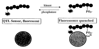
Figure 2 is a schematic of an assay for enzyme mediated phosphorylation or
dephosphorylation activity based on metal ion mediated fluorescence
superquenching.
20 Figure 3 is a Stern-Voliner plot for the quenching of a gallium sensor by a
Rhodamine labeled phosphorylated peptide.
Figures 4A and 4B are graphs showing endpoint and kinetic assays for
Protein Kinase A (PKA).
-14-
CA 02548407 2006-06-07
WO 2005/060626 PCT/US2004/041400
Figure 5 is a graph showing Protein I~inase A (PKA) assay response in the
presence of an inhibitor.
Figure 6 is a graph demonstrating ECSO and limit of detection for protein
tyrosine phosphatase 1B (PTB-1B) phosphatase assay.
Figure 7 is a graph showing the inhibition of protein tyrosine phosphatase
1B (PTB-1B) activity.
Figure ~ is a schematic of a protease assay based on metal ion mediated
fluorescence superquenching.
Figure 9 is a schematic of a blocking kinase assay using protein and peptide
substrates based on metal ion mediated superquenching.
Figure 10 is a graph showing a fluorescence turn-on blocking kinase assay
using PKCa as an example.
Figure 11 is a schematic of a phosphodiesterase assay employing metal ion-
mediated superquenching.
Figure 12 is a graph showing the results of monitoring Trypsin activity in a
real time or kinetic assay format.
Figure 13 illustrates the detection of phosphorylated polypeptides according
to one embodiment.
Figure 14 is a graph showing relative fluorescence as a function of protein
kinase A (PKA) concentration in an assay using a biotinylated peptide
substrate
(BT) according to one embodiment.
Figure 15 is a chart showing the relative fluorescence response to
phosphorylated and non-phosphorylated histone.
-15-
CA 02548407 2006-06-07
WO 2005/060626 PCT/US2004/041400
Figure 16 is a graph showing relative fluorescence as a function of protein
tyrosine phosphatase-1B (PTP-1B) concentration in an assay using a
biotinylated
peptide substrate (BT) according to a further embodiment.
Figure 17 illustrates an assay wherein a quencher-tether conjugate (QT)
associates with a metal ion and fluorescent polymer ensemble resulting in
amplified superquenching of the fluorescent polymer.
Figure 18 is a graph showing a phosphopeptide calibrator curve for a metal
ion mediated superquenching assay.
Figure 19 shows a Protein Kinase-A concentration curve obtained from a
metal ion mediated superquenching assay.
Figure 20 is a schematic for a kinase enzyme activity sensor based on metal
ion mediated fluorescence superquenching via association of a streptavidin
quencher molecule added in a second step to kinase reaction.
Figures 21A and 21B are graphs comparing endpoint assays for PKA using
the two-step approach with biotinylated substrates and a quencher (i.e.,
Rhodamine) labeled substrate wherein Figure 21A shows RFIJ as a function of
PKA concentration and Figure 21B shows % phosphorylation as a function of PKA
concentration.
Figure 22 is a bar chart illustrating the results of a screen using seven (7)
different biotinylated peptide substrates which Were each reacted with 3
different
enzymes (i.e., PTP-1B, PI~Ca and PKA).
-16-
CA 02548407 2006-06-07
WO 2005/060626 PCT/US2004/041400
DETAILED DESCRIPTION
The quencher-tether-ligand (QTL) approach to biosensing takes advantage
of superquenching of fluorescent polyelectrolytes by electron and energy
transfer
quenchers. The QTL assay platform utilizes the light harvesting ability of
conjugated polymers along with their highly delocalized excited state to
provide
amplified fluorescent signal modulation in response to the presence of very
small
quantities of electron and energy transfer species. This novel technology has
been
applied to the highly sensitive detection of proteins, small molecules,
peptides,
proteases and oligonucleotides by associating the signal modulation phenomenon
with antigen-receptor, substrate-enzyme and oligonucleotide-oligonucleotide
binding interactions. [1-9]
In one approach, the fluorescent polymer, P, is co-located with biotin-
binding protein either in solution or on a solid support, and forms an
association
complex with a quencher-tether-biotin (QTB) bioconjugate through biotin-biotin
binding protein interactions. The QTB bioconjugate includes a quencher, Q,
linked
through a reactive tether to biotin, which strongly binds the biotin binding
protein
co-located with the polymer, P. The reaction of the QTB bioconjugate with the
target analyte modifies the polymer fluorescence in a readily detectable way.
As described herein, an alternate way of associating the QTL bioconjugate
with a fluorescent polymer has been developed which uses the self organizing
capability of fluorescent polyelectrolytes either as individual molecules in
solution
or as an assembly on a support to complex with metal ions. The thus complexed
metal ions can associate with selectivity to coordinating groups (e.g.,
phosphate
groups) incorporated into the QTL bioconjugate thus providing the basis for
-17-
CA 02548407 2006-06-07
WO 2005/060626 PCT/US2004/041400
selective detection of, fox example, proteins, small molecules, peptides,
proteases,
kinases, phosphatases and oligonucleotides. [10-11]
The efficiency with which an acceptor molecule (i. e., quencher) can quench
the efficiency of a donor molecule is dependent on the distance that separates
the
two entities. Tn constructing assays, the tethering of molecules (to bring the
acceptor and donor together) can be accomplished by common strategies such as
covalent linkage, and the biotin-avidin interaction. Covalent linkage is an
excellent approach for resonance energy transfer because it places the
quencher
directly onto the acceptor making them one molecule. The distance between the
two can therefore be as small as a single bond length. The interaction between
biotin and a biotin binding protein (BBP) such as avidin, on the other hand,
provides extensive versatility because nearly any molecule can be covalently
linked
to biotin. However, biotin binding proteins are generally larger that 60
kilodaltons,
and as a result when the acceptor and donor are brought together through a
biotin-
BBP interaction, the distance between the acceptor and donor can be
significant.
As a general replacement for the biotin-BBP interaction, we have proposed
a metal-ion phosphate interaction for the co-location of acceptors and donors
in
superquenching assays. As with the biotin-BBP interaction this strategy is
generally applicable because many molecules can be phosphorylated. In
addition,
this strategy is a general improvement over the biotin-avidin interaction
because
the end-to-end distance of the tether (i.e., the coordination distance between
the
metal ion and the phosphate) is significantly shorter. The affinity of metal
ions fox
ligands such as phosphate groups is significantly lower than that of the
biotin-BBP
interaction (K.~ = 10 5-'versus 1013-ls).
-1 ~-
CA 02548407 2006-06-07
WO 2005/060626 PCT/US2004/041400
According to one embodiment, a novel sensor comprising fluorescent
polyelectrolytes either as individual molecules in solution or as an assembly
on a
support complexed to metal ions is provided. The metal ions of the sensor can
further associate with selectivity to ligands (e.g., phosphate groups)
incorporated
S into the QTL bioconjugate and provide the basis for selective detection of
the same
molecules described above (e.g., proteins, small molecules, peptides,
proteases,
kinases, phosphatases and oligonucleotides) including, but not limited to, end-
point
and kinetic modes. As will be developed below, for,some assays the
coordinating
group-metal ion binding provides an alternative to biotin-biotin binding
protein
association. In other examples the coordinating group is attached or removed
from
the quencher portion of the QTL so as to provide for a quench, or a recovery
(or
both) of sensor fluorescence.
Various embodiments described herein employ fluorescent polymer-QTL
superquenching and metal ion-phosphate ligand specific binding to provide
1S improved assays for kinase, phosphatase and protease activity. Metal ion
mediated
superquenching of fluorescent polymers provides a general platform for the
measurement of kinase, phosphatase and protease enzyme activity using peptide
and protein substrates as well as a more general approach for carrying out
assays
based on DNA hybridization and assays for proteins employing aptamers,
antibodies and other ligands.
Conjugated polymers in the poly(phenyleneethynylene) (PPE) family can be
prepared with a variety of functional groups appended on the aromatic rings.
Among the polymers synthesized with pendant anionic groups are those shown in
Figures 1A and 1B. FIG. 1A shows the molecular structure of sulfo poly p-
-19-
CA 02548407 2006-06-07
WO 2005/060626 PCT/US2004/041400
phenyleneethynylene (PPE-Di-COOK conjugated polymer. Figure 1B shows the
molecular structure of sulfo poly p-phenyleneethynylene (PPE) conjugated
polymer. Both of these polymers can associate with cationic microspheres in
water
to form stable polymer coatings. The polymer coated microspheres exhibit
strong
fluorescence. The overall charge on the polymer-coated microspheres can be
tuned
by varying the degree of polymer loading and by varying the structure of the
polymer.
It has been found that fluorescent polymer coated microspheres can
associate with metal rations and that the loading of metal rations may depend
on
the loading level of the polymer on the microsphere. Certain metal ions such
as
Fe3+ and Cuz+ can quench the polymer fluorescence while others such as Ga3+~
do
not. In some embodiments, Ga3+ is used to mediate superquenching of
microsphere-bound polymer fluorescence under conditions where, in the absence
of the metal ions, little or no quenching would occur.
For example, a phosphorylated peptide containing a dye:
Rhodamine-LRRA(pS)LG SEQ ID N0:1
wherein pS designates phosphorylated serine, which should serve as a good
energy
transfer quencher for the polymer was found to have little or no quenching of
the
fluorescence of polymer-coated microspheres. After the polymer-coated
microspheres are "charged" by the addition of Ga3~, however, addition of the
same
peptide to the suspensions results in a pronounced quenching of the polymer
fluorescence. In contrast, peptides containing only a phosphorylated residue
or
only the quencher dye, such as the peptide represented by:
-20-
CA 02548407 2006-06-07
WO 2005/060626 PCT/US2004/041400
Rhodamine-LRRASLG SEQ m N0:2
produce little effect on the polymer fluorescence under the same conditions.
The
specific association of a phosphorylated biomolecule with the metal ion
charged
polymer can be the basis of a number of assays as described below.
Figure 2 shows schematically a sensor based on metal ion mediated
superquenching which can be used in kinase or phosphatase activity assays.
Figure 2 shows how the phosphorylation or dephosphorylation of rhodamine
peptide substrates by target enzymes can be detected by the addition of the
QTL
sensor. The peptide products are labeled with a rhodamine quencher and brought
to the surface of the polymer by virtue of specific phosphate binding to the
Ga3+
metal ion. The resulting quench of polymer fluorescence is concomitant with
phosphorylation or dephosphorylation of the polypeptide substrate. This type
of
assay can be used for enzymes which moderate phosphorylation or
dephosphorylation for biologicqal substrates including, but not limited to,
peptides,
proteins, lipids, carbohydrates and nucleotides or small molecules.
KizzaselPlzosplzatase Assays
Phosphorylation and dephosphorylation of proteins mediate the regulation
of cellular metabolism, growth, differentiation and cell proliferation.
Aberration in
enzymatic function can lead to diseases such as cancer and inflammation. More
than 500 kinases and phosphatases are thought to be involved in the regulation
of
cellular activity and many among them are targets for drug therapy.
Protein Kinase A (PKA) is a cAMP dependent protein kinase and functions
as an effector of many cAMP-elevating first messengers such as hormones and
-21-
CA 02548407 2006-06-07
WO 2005/060626 PCT/US2004/041400
neurotransmitters. The ubiquitous distribution of PISA and it's flexible
substrate
recognition properties make PK.A a central element in many processes of living
cells, such as in the inhibition of lymphocyte cell proliferation and immune
response, mediation of long-term depression in the hippocampus and sensory
nerve
transmission. Protein Tyrosine Phosphatase-1B (PTP-1B) has recently been shown
to be a negative regulator of the insulin signaling pathway suggesting that
inhibitors to PTP-1B might be beneficial in the treatment of type 2 diabetes.
Of the kinases, 90% phosphorylate serine residues, 10% phosphorylate
threonine residues and 0.1 % phosphorylate tyrosine residues. Although it has
become possible to develop anti-phosphotyrosine antibodies, antibodies against
phospho-serine and threonine residues are of low affinity and often specific
to only
one kinase. Currently, non-antibody-based high-throughput screening (HTS)~
assays are based on methods such as time-resolved fluorescence (TRF),
fluorescence polarization assays (FP) or fluorescence resonance energy
transfer
(FRET). These assays require specialized equipment and/or suffer from low
fluorescence intensity change as a function of enzyme activity.
We sought to enhance sensitivity in the measurement of enzymatic activity
by amplifying the fluorescence signal using superquenching as described above.
The sensor platform can comprise a modified anionic polyelectrolyte fluorescer
such as the poly(phenylenethylene) (PPE) derivative shown in Figure 1A. The
PPE
fluorescer can be immobilized by adsorption on positively charged
microspheres.
This polymer exhibits photoluminescence with high quantum efficiency and has
been used fox detection of protease activity. [9] In this platform, a reactive
peptide
sequence was used which is flanked by a N-terminal quencher and a C-terminal
-22-
CA 02548407 2006-06-07
WO 2005/060626 PCT/US2004/041400
biotin. The peptide binds to PPE coated rnicrospheres that are co-located with
biotin binding proteins, resulting in a near total quenching of PPE
fluorescence.
Enzyme mediated cleavage of the peptide leads to a reversal of fluorescence
quenching that was linear with enzymatic activity. It has been demonstrated
that a
single energy acceptor dye can quench the photoluminescence from approximately
49 repeat units per quencher. [9]
Fluorescent polymer superquenching can be adapted to the biodetection of
kinase/phosphatase enzyme activity as illustrated in Figure 2. As shown in
Figure 2, multivalent metal ions can strongly associate with anionic
conjugated
polymers in solution, resulting in modification and/or quenching of polymer
fluorescence. Since the overall charge on a polymer-microsphere ensemble can
be
tuned, these ensembles can afford a platform whereby metal ions associate with
the
polymer without strongly quenching the polymer fluorescence while retaining
the
ability to complex with specific ligands. The approach is similar to that used
in
I 5 immobilized metal ion affinity chromatography (IMAC) whereby metal ions
can
specifically trap phosphorylated compounds by coordination with the phosphate
oxygen at low pH. See, for example, Mor~an et al., Assay Drug Dev. Technol.,
2004, 2, 171.
As described herein, gallium can associate with fluorescers (including, but
not limited to, anionic conjugated polymers such as those shown in Figures 1A
and
1B and other fluorescers comprising a plurality of fluorescent species)
without
quenching the polymer emission. The gallium can exist as monomeric Ga3~ or as
a
multimeric ensemble such as a polyoxo species. The fluorescer-associated
gallium
can also associate With phosphorylated peptides such that, when the peptide
-23-
CA 02548407 2006-06-07
WO 2005/060626 PCT/US2004/041400
contains a dye such as rhodamine, metal ion mediated polymer superquenching
occurs. The fluorescer can be associated with a surface of a solid support
such as a
microsphere. This approach provides the basis for a sensitive and selective
kinaselphosphatase assay as illustrated in Figure 2.
In the case of the fluorescence quench (turn off) kinase assay, the quench of
polymer fluorescence is linear with enzyme activity. As described in the
following
example, the assay can be carried out a near physiological pH and allows
flexibility
in constructing real time or end point assays. The assays are instantaneous,
"mix
and read" and require no wash steps or complex sample preparation.
Example 1 below shows robust assays for protein kinase A (PISA) and
protein tyrosine phosphatase IB (PTB-IB) enzyme activities. The assays
routinely
deliver Z' values greater than 0.9 at substrate conversion of 10 - 20 %. In
the
example shown below, the kinase assay provides fluorescence signal attenuation
as
a function of enzyme activity while the phosphatase assay provides signal
enhancement with increasing enzyme activity. Since, for peptides such as
SEQ ID NO:1, the quencher may exhibit sensitized fluorescence as a consequence
of the quenching of polymer fluorescence, the assays can exhibit signal
enhancement or reduction in the same sample, depending on the wavelengths
monitored. Accordingly, ratiometric measurements can be made. Additionally,
detection can be carried out by monitoring fluorescence polarization in the
quencher of the peptide. For protein kinase, phosphatase and protease assays.
based
on metal ion mediated superquenching, both end point and kinetic assays may be
earned out.
-24-
CA 02548407 2006-06-07
WO 2005/060626 PCT/US2004/041400
Example 1- Assays for Protein Kinase a (PKA) and Tyrosine Phosphatase
Activity IB (PTRIB)
The following peptides were used as enzyme substrates and as phospho-
peptide calibrators.
For detection of PKA activity:
Rhodamine-LRRASLG SEQ m NO:2
and the calibrator peptide:
Rhodamine-LRRA(pS)LG SEQ ID NO:1
were synthesized by Anaspec.
For detection of phosphatase activity:
Rhodamine-KVEKIGEGT(pY)GVVYK SEQ m N0:3
and the calibrator peptide:
Rhodamine-KVEKIGEGTYGVVYK SEQ m NO:4
were synthesized by American Peptide Company.
Recombinant PKA was purchased from Promega. Enzyme PTP-1B as well
as inhibitor RK682 were purchased from Biomol. A Staurosporine inhibitor for
PKA was purchased from Sigma. Polystyrene amine functionalized beads were
obtained from Interfacial Dynamics.
The performance of sensor beads was determined by adding 15 ~L of a 1
~,M peptide solution (either rhodamine-phospho-peptide or rhodamine-non-
phospho-peptide) in assay buffer to 15 ~,L of sensor in a detector buffer. The
fluorescence of the mixture was measured using a SpectraMax Gemini XS plate
reader (Molecular Devices, Inc.) in well scan mode and with excitation at 450
nm
with a 475 nm cutoff filter and emission at 490 nm.
-25-
CA 02548407 2006-06-07
WO 2005/060626 PCT/US2004/041400
The polymer whose structure is shown in Figure 1A was chosen as a sensor
for kinase/phosphatase assays based upon the discovery that di- or trivalent
metal
ions can strongly associate with anionic polymers such as those shown in
Figures
1A and 1B in solution. No quench of emission was observed when GaCl3 in a
concentration of 340 ~,M was added to a solution comprising microspheres
coated
with PPE-Di-COOH. At higher concentrations of GaCl3, quenching of fluorescent
emissions was observed. However, when using an optimal concentration of Ga3~,
it was found that rhodamine labeled phospho-peptides provided a strong quench
of
polymer fluorescence whereas little modulation of fluorescence was observed
when non phosphorylated rhodamine labeled peptides were used.
Figure 3 shows a Stern Volmer plot obtained for Rhodamine labeled PTP-
1B phosphopeptide substrate. The Stern Volmer constant (KS~) provides a
quantitative measure of quenching where Fo is the intensity of fluorescence in
the
absence of quencher and F the fluorescence intensity in the presence of
quencher.
The KS~ determined here is relatively large (i.e., 2 x 10' M-~). The 50%
quench
gives (PRU/Q)50 = 50, demonstrating the occurrence of superquenching.
As shown above, assays have been developed using quencher labeled
substrates. Upon phosphorylation of the substrate, the peptide associates to
the
sensor via the phosphate groups and quenches fluorescence. Since the metal-ion
coordinating groups specifically bind to phosphates, phosphorylated serine,
threonine or tyrosine residues can be detected.
Fluorescent superquenching-based assays for serine and tyrosine enzymes,
namely Protein Kinase A (PKA), and Protein Tyrosine Phosphatasel-B (PTP-1B)
are described below.
-26-
CA 02548407 2006-06-07
WO 2005/060626 PCT/US2004/041400
Figure 4A shows an endpoint measurement of PKA enzyme activity in
which an increase in polymer quench correlates with enzyme concentration.
Unlike Fe3+ coordination assays, which require very low pH, this platform is
functional at near physiological pH and thus allows researchers the
flexibility of
choice in performing real time assays or endpoint assays. A real time assay,
that
includes the detector mix as part of the enzymatic reaction mix requires
approximately 10 fold higher concentrations of enzyme for 50 % substrate
phosphorylation than an endpoint assay which is shown in Figure 4B.
The sensitivity of the assay was tested by using a known inhibitor of PKA
activity, Staurosporine. The results are shown in Figure 5. As shown in Figure
5,
the ICSO obtained using 1 ~,M substrate in a reaction with 6.5 ~.M ATP and 200
mU
PKA was 59 mU and is in agreement with published values (18.4 mU).
The format was tested for detection of protein tyrosine phosphatase activity
1B (PTP-1B) on a peptide substrate of different length and sequence
composition
than the one used for PKA. Figure 6 shows results of ECSO and LOD of enzyme
concentration curves measured as endpoint assays or in realtime using PTP-1B
on
125 nM substrate. An inhibitor curve using the known inhibitor RK-682 yields
an
excellent ICSa of 26.4 nM.
The statistical parameters that can be delivered with this assay were
determined by evaluating known amounts of phospho peptide calibrator peptide
in
replicates of 8 (Figure 6). The data are excellent and show that this assay is
suitable to determine as little as 5 - 10 % substrate conversion with Z'
factors of
0.8 and 0.9 respectively.
-27-
CA 02548407 2006-06-07
WO 2005/060626 PCT/US2004/041400
The performance of this PKA assay has been compared with a
commercially available FRET assay, an ATP consmnption assay and an IMAC-
based assay. All assays were performed to produce optimal performance in an
enzyme concentration curve and where possible using the identical peptide. The
IMAC-based assay delivers the lowest sensitivity in an enzyme concentration
curve
(1 ng compared to 20 pg). In this assay, which is closest to the QTL
LightspeedTM
assay in principle, the sensor to detector follows a 1:1 ratio as opposed to
the 1:50
ratio in the present format. These results clearly demonstrate the enhanced .
sensitivity obtainable with superquenching.
Additional assays have been developed using substrates for Akt-1 and
PKCa,. No significant dependency of fluorescence quench on substrate length or
peptide sequence content was observed when using these different substrates.
In
this regard, the metal ion mediated superquenching assay can be considered
generic
and offers a major advantage over FRET peptides in which quenching is highly
dependent on the distance between the donor and acceptor.
Protease Assays
Protease enzymes cleave amide bonds on their substrate. The use of
peptide or protein substrates that contain a quencher and a phosphate group on
either side of the cleavage site along with the metal ion-fluorescent polymer
ensemble affords the development of highly sensitive assays for the detection
of
protease enzyme activity.
One embodiment of a protease assay is illustrated in Figure 8. As shown in
Figure 8, when the intact substrate binds the sensor, the sensor fluorescence
is
-28-
CA 02548407 2006-06-07
WO 2005/060626 PCT/US2004/041400
quenched by the promixity of the quencher dye. Cleavage of the substrate by
the
enzyme into fragments separates the quencher from the phosphate group
resulting
in separation of the quencher and polymer. Tlus separation leads to reduced
quench of polymer fluorescence (z.e., enhanced signal from the sensor) in the
presence of enzyme acitivity.
Protease activity can be monitored either real-time or at the end-point in
homogeneous or heterogeneous formats. In a homogeneous real-time assay, the
substrate can reside on the surface of the polymer-microsphere ensemble. In a
homogeneous end-point assay, the substrate and the enzyme can react in
solution
and, at the end of a specified incubation period, the sensor can be added to
the
sample to stop the reaction. Protease activity can be monitored
ratiometrically
when a fluorescent dye is used as the quencher. In a heterogeneous end-point
format, biotinylated substrates can be used which contain phosphate groups and
a
quencher on the same side of a cleavage site.. Following cleavage, the peptide
species are separated by binding of the biotin species whereas the quencher-
labeled
portion is transferred and can thereby quench the fluorescer.
Exazzzple 2 - A Protease Assay Based ofz Metal Ion Mediated Fluorescence
Superquenclzizzg
The peptide substrate for trypsin in this assay is
Rhodamine-LRRApSLG (SEQ m NO:l).
Trypsin cleaves the peptide at the two arginines. The assay performed in this
example used the following parameters:
Microsphere-Fluorescer-Gallium ensemble (QTL sensor);
_29_
CA 02548407 2006-06-07
WO 2005/060626 PCT/US2004/041400
3 ~.M final Rh-LRR.ApSLG (SEQ m NO:l);
1 U/~,L trypsin;
40 x 106 microspheres (MS)/15 ~L;
~eX 43 0;
a,em 490; and
~,~0 475nm.
The assay was conducted for 1 hr at approximately 22 °C in a 384-well
white plate.
The results of this assay are shown below in Table 1.
Table 1 - Results of Protease Assay Based on Metal Ion Mediated Fluorescence
Superqueuclziug
QTL Sensor alone 88842
No enzyme control 7771
Sample 42138
Signal Increase 343 67
SignallBackground 5.42
Z' 0.68
Si al/Noise 9.69
Figure 12 is a graph showing the results of monitoring Trypsin activity in a
"real time" (i.e., kinetic) assay format. As can be seen from Figure 12, there
is a
time-dependent increase in Trypsin activity. Correspondingly, the fluorescence
signal enhancement occurs with time.
-30-
CA 02548407 2006-06-07
WO 2005/060626 PCT/US2004/041400
Blockihg Assays Using U~zlabeled Peptides ahd Protei~zs
The basis for the assays described above and shown in Figure 2 can be
adapted to a blocking assay in which a "generic" phosphorylated dye labeled
peptide or other substrate containing both a dye and a metal ion binding
phosphate
(e.g., gallium) quenches the polymer beads containing fluorescent polymer and
metal ion in the absence of additional phosphorylated substrates but is
"blocked"
when a peptide or protein substrate is phosphorylated.
The principle of the assay is shown in Figure 9 which illustrates
schematically a blocking kinase assay based on metal ion mediated
superquenching. The assay is most conveniently carried out by adding the
sensor
to a mixture of enzyne and analyte following incubation for reaction. Any .
phosphorylated analyte will associate with the sensor as demonstrated in
Figure 9,
without quenching the polymer fluorescence. Addition of the "generic"
phosphorylated dye labeled peptide will result in a quenching of the polymer
fluorescence, limited by the extent of "free" phosphate binding sites on the
"blocked" microspheres. The assay functions as a fluorescence "turn-on" assay
and offers the additional advantage that no prior derivitization of the
substrate need
to be done in developing the assay. Figure 10 shows experimental data for a
bloclcing assay ("fluorescence turn-on") for PKCa with Myelin Basic Protein
(MBP).
The detection of kinase activity on natural protein substrates has several
advantages over using peptide substrates as set forth below.
~ Of the 518 known human kinases (or 2500 isoforms), peptide substrates
-31-
CA 02548407 2006-06-07
WO 2005/060626 PCT/US2004/041400
have been established for only approximately 50 kinases but the target
proteins are
identified in most cases. Some enzymes may require non-continuous amino acids
of a target for effective substrate recognition, binding and phosphorylation,
in
which case an artificial peptide sequence can not be constructed even if the
involved amino acids are identified.
~ The phosphorylation of natural target proteins is expected to be much
more efficient than phosphorylation of peptide substrates. This is important
for
purpose of cost (of peptide substrates) but also makes identification of
inhibitors in
HTS more accurate.
~ The phosphorylation of natural target proteins is more specific than the
phosphorylation of artificial substrates. Future attempts to dissect kinase
activity
in cells will be impeded by the cross recognition of peptide substrates but
should
work on protein substrates.
~ Current non-radioactive and non-antibody based assays that allow for
detection of phosphorylation of proteins axe based on ATP consumption by
secondary enzyme Luciferase. Such assays are prone to false negative results
in
inhibitor screens, as a result of inhibition of the secondary enzyme,
Luciferase. FP
assays require a large change in molecular motion to obtain a signal,
therefore only
proteins of small molecular weight can be detected.
Exa~rzple 3
Phosphorylation of myelin basic protein (MBP) by kinase PKCa was
performed in a standard reaction and QTL sensors as described above in Example
2
were added. Phosphorylated MBP binds to the QTL sensor by virtue of specific
-32-
CA 02548407 2006-06-07
WO 2005/060626 PCT/US2004/041400
phosphate binding to the metal coordinating ions and inhibits association of
dye-
labeled phospho peptide (tracer) in a concentration dependent manner. The
resulting fluorescence correlates with the extent of mbp phosphorylation.
This principle is demonstrated in the following example. A concentration
of 1 ~.g mbp was phosphorylated using serially diluted kinase PKCa enzyme for
1
hour at room temperature in a white 384-well Optiplate. Following incubation,
50
x 106 QTL Sensor beads were added for 10 minutes at approximately 22 °C
and
subsequently 1 p,M dye labeled peptide tracer added. Plates were incubated for
30
minutes at approximately 22 °C and the fluorescence signal monitored
using
excitation at 450 nm, emission at 490 mn with a 475 rnn cutoff filter in a
Gemini
XS Plate reader (Molecular Devices, Inc.). The fluorescence "turn on" is shown
schematically in Figure 9.
Phosphodiesterase Euzyfrze Activity Monitored by Metal Iou Mediated
Fluorescence Superque>zclzihg
The 3',5'-cyclic nucleotide phosphodiesterases (PDEs) comprise a family
of metallophosphohydrolases that specifically cleave the 3' bond of cyclic
adenosine monophosphate (CAMP) and/or cyclic guanosine monophosphate
(cGMP) to produce the corresponding 5'-nucleotide. Eleven families of PDEs
with
varying selectivities for cAMP and cGMP have been identified in mammalian
tissues.
PDEs are essential modulators of cellular cAMP and/or cGMP levels.
Cyclic-AMP or cGMP are intracellular second messengers that play crucial roles
in
intracellular signal transduction involved in important cellular processes.
PDEs
-33-
CA 02548407 2006-06-07
WO 2005/060626 PCT/US2004/041400
have been targets for drug discovery to treat a variety of diseases. For
example,
Sidenafil, a selective inhibitor of PDE 5, has been commercialized as a drug
(i. e.,
Viagra~, a registered trademark of Pfizer, Inc.). Several PDE 4 inhibitors are
in
clinical trials as anti-inflammatory drugs treating diseases such as asthma.
As described above, the QTL sensor shows a high binding affinity towards
phosphate groups as demonstrated in the kinase and phosphatase assays. The PDE
assay uses a dye-labeled cAMP or cGMP as a substrate to assay the activity of
the
phosphodiesterase. Dyes including, but not limited to, rhodamine, azo or
fluorescein can be coupled to cAMP or cGMP without inhibiting reactivity
towards
PDEs. Since cAMP or cGMP exists as a phosphodiester, which does not bind
strongly to the gallium-polymer surface, there is little initial quenching of
the
polymer fluorescence. During hydrolysis catalyzed by the PDE, the
phosphodiester
on these substrates is converted to a phosphate group. The dye then is brought
to
the vicinity of the microsphere surface through gallium-phosphate specific
interactions, resulting in quenching of the polymer fluorescence. Figure 11 is
a
schematic depicting a phosphodiesterase assay.
Nucleic Acid Assays
The metal-phosphate mediated binding can be used to generate
superquenching assays for DNA and RNA detection. A number of different
approaches based on hybridization of a nucleic acid species to a target
nucleic acid
species which can be in solution or immobilized on a solid support can be
used. A
first approach utilizes an oligonucleotide that is phosphorylated at one of
its
termini. The phosphate allows for metal-phosphate mediated co-location of the
-34-
CA 02548407 2006-06-07
WO 2005/060626 PCT/US2004/041400
DNA strand with the conjugated fluorescent polymer. If a phosphate group is
attached to the 5'-terminus of the oligonucleotide, a complementary target
bearing
a quencher at the 3'-terminus can be hybridized to the phosphorylated strand.
The
termini can also be reversed while retaining a functional system. In this
hybridized
conformation, the quencher would be oriented towards the conjugated polymer to
facilitate superquenching. Hence, in the presence of the quencher labeled
target,
the fluorescence of the polymer is quenched. Such a system can be easily
envisioned as an assay for unlabeled DNA by allowing unlabeled and labeled DNA
strands to compete for binding to their phosphorylated complementary strand.
A second approach follows a strategy that is similar to the approach used by
molecular beacons. A hairpin oligonucleotide bearing a phosphate at one of its
termini and a quencher at another can be designed so that the terminal regions
of
the oligonucleotide are complementary to each other and form a hybridized
stem,
while the central region of the oligonucleotide is complementary to a target
oligonucleotide and forms a single stranded loop when no target is present.
Such
an oligonucleotide will form a "hairpin" structure which brings the phosphate
and
the quencher into close proximity by virtue of stem hybridization. When the
phosphorylated hairpin oligonucleotide is bound to the metal-polymer complex
by
virtue of the phosphate metal interaction, a quench will be induced because of
the
orientation of the quencher towards the polymer. If the phosphate/quencher
functionalized oligonucleotide is hybridized to a target that binds to the
loop region
of the hairpin, the loop region becomes a rigid rod which disrupts the
secondary
structure of the stem region. This would cause the acceptor and donor pair to
be
forced apart thereby reducing the quenching of the polymer.
-35-
CA 02548407 2006-06-07
WO 2005/060626 PCT/US2004/041400
Direct assays for proteins and other targets can also be conducted through a
number of routes using the binding properties of DNA aptamers. A
phosphorylated DNA aptamer can be bound to the surface of a metal-coated
conjugated polymer surface. In the presence of the target molecule (small
molecules in size, up to proteins in size) the aptamer conformation of the
oligonucleotide should be stabilized (lower OG). In the absence of its
selected
target, the aptamer strand rnay bear a weak self structure. If the self
structure of
the aptamer can be penetrated by a complementary oligonucleotide that is
labeled
with a quencher, an assay can be generated. In such an assay, when the
aptamer's
target is absent, the complementary oligonucleotide-quencher may hybridize to
the
aptamer. This hybrid can be of the form listed above (i.e., phosphate at 5'-
terminus, and quencher at 3'-terminus; or vice-versa), thus the quencher will
be
oriented to quench the conjugated polymer. In the presence of the aptamer's
target,
the aptamer self structure will be stabilized and the oligonucleotide quencher
will
not be able to hybridize to the aptamer. Hence, in the presence of the
aptamer's
target, the polymer will fluoresce and in the absence of the aptamer's target
the
fluorescence will be quenched.
General Phosplzate Modification or Cousunzptiosz
In any system containing a phosphate tethered through any means to a
quencher, the modification of the phosphate through chemical' means can
convert
the phosphate to another functionality thus preventing phosphate-metal
mediated
binding to the metal-polymer complex. Likewise, the binding of the phosphate
to
other elements may prevent the binding of that same phosphate to a metal
polymer
-36-
CA 02548407 2006-06-07
WO 2005/060626 PCT/US2004/041400
complex. In these cases, the quencher will not be co-located with the
conjugated
polymer and fluorescence will be present. As a general example, complex A,
which contains a phosphate tethered through any means to a quencher, can
quench
the metal polymer complex. If present with a molecule B which bears an
affinity
for complex A and which also contains elements which will either chemically
modify or bind to the phosphate contained in complex A, complex A will not be
capable of binding and thereby quenching the metal polymer complex.
Assays, Reagents and Kits Employing Biotin-Tether (BT) Conjugates
According to one embodiment, a kit for conducting an assay for a target
analyte is provided. The kit comprises two separate components: a quencher (Q)
and a biotin-tether conjugate (BT). The tether (T) of the BT conjugate can
comprise, for example, a protein or polypeptide substrate. According to this
embodiment, the tether acquires the capacity to associate with the quencher
upon
interaction with and modification by the target analyte to form a modified
tether
(T'). Following modification of the tether, a QT'B bioconjugate is formed as a
result of the interaction of the BT conjugate with the target analyte followed
by
association of the modified BT conjugate (BT') with the quencher (Q). The kit
may also comprise a fluorescer component (P). The fluorescer component
comprises a plurality of fluorescent species associated in such a maimer that
the
quencher is capable of amplified superquenching of the fluorescer when
associated
therewith. The fluorescer can be a fluorescent polymer. The fluorescer can be
associated with a solid support such as a microsphere, bead or nanoparticle.
The
solid support can also comprise a biotin binding protein such that interaction
of the
-37-
CA 02548407 2006-06-07
WO 2005/060626 PCT/US2004/041400
biotin moiety on the QT'B complex with the biotin binding protein on the solid
support results in quenching of fluorecence.
As set forth above, the tether of the BT conjugate can be recognized and
modified by association or reaction to the target analyte to form the BT'
conjugate.
Modification of the tether renders the modified BT conjugate (BT') capable of
binding the quencher (Q) to form the QT'B complex. This sequence of events can
be followed by a modulation of the polymer fluorescence. In particular, a
change
in fluorescence can be used to indicate the presence and/or the amount of a
target
analyte in a sample. Moreover, in the absence of a specific association or
reaction
of the BT conjugate with an enzyme or other target analyte, the fluorescence
of P is
unaffected by association to the BT conjugate. Accordingly, methods of using a
quencher (Q) and a biotin-tether conjugate (B'I) as set forth above to
determine the
presence andlor amount of a taxget analyte in a sample are also provided.
According to one embodiment, the interaction of the tether (T) of the BT
conjugate with a target analyte may result in the removal of a
quencher~binding
component on the tether. In this embodiment, the capacity of the BT conjugate
to
bind the quencher (Q) is eliminated as a result of the interaction with the
analyte to
form the modified conjugate (BT'). Again, this sequence of events can be
followed quantitatively via the modulation of polymer fluorescence. In certain
embodiments, the reaction of BT and the target analyte may be catalytic,
resulting
in an amplified modulation of polymer fluorescence.
According to a further embodiment, polymer superquenching may be
mediated by a metal-ion. According to this embodiment, a QT conjugate (wherein
Q is an electron or energy transfer quencher and T is a reactive tether) can
react
-3 ~-
CA 02548407 2006-06-07
WO 2005/060626 PCT/US2004/041400
with a target analyte to introduce, modify or remove a functional group on the
tether. The functional group can be a functional group which is capable of
associating with a metal ion associated to or co-located (e.g., on a surface
of a solid
support) with a fluorescent polymer. The modified QT conjugate (QT') is
therefore capable of associating with the ensemble comprising the fluorescent
polymer and the metal ion. Consequently, modification of the tether results in
a
change in the polymer fluorescence. This method may be employed in highly
sensitive assays for kinase, phosphatase and other enzymes as target analytes.
Modifiable Tether Based QTB Approach for the Biadetectiosa of Post-
Trataslatiotaal Modification Events
This approach employs a synthetic biotinylated peptide substrate or tether
(hereinafter referred to as a "BT conjugate") which upon interaction with a
target
analyte is modified to form a BT' conjugate. In one embodiment, the BT
conjugate
is incapable of complexing to the non-fluorescent quencher (Q) whereas the
modifed conjugate (BT') readily binds to the quencher. This type of
interaction
leads to a fluorescence "turn-off ' assay where the polymer fluorescence
decreases
with increasing substrate conversion.
In another embodiment, the BT conjugate can readily associate with the
dark quencher. However, the BT conjugate loses the ability to associate after
interaction with the target analyte to form the modified conjugate (BT'). This
type
of interaction results in a fluorescence "turn-on" assay.
In a further embodiment, the quencher in the above embodiments can also
be a fluorescent moiety. The use of a fluorescent moiety as a quencher can
provide
-39-
CA 02548407 2006-06-07
WO 2005/060626 PCT/US2004/041400
sensitized emission of fluorescence. In all of these embodiments, the QTB
bioconjugate can form a complex with the polymer-receptor ensemble to modulate
the polymer fluorescence efficiently by the superquenching process.
The quencher moiety used in the assay for post-translational modification
interaction combines the properties of association to the functional group
that is
modified on the substrate and amplified superquenching of the fluorescence of
the
conjugated polymer when present in close proximity. In one embodiment, the
quencher can be a transition metal or an organometallic species such as an
iron (~
iminodiacetic acid (mA) type chelate, wherein the fernc iron can both
associate
strongly to a phosphopeptide and superquench the fluorescent polymer by
electron
transfer. In another embodiment, the quencher may consist of two distinct
moieties, one that promotes association of the quencher to the modified
functional
group and another that causes polymer quench by energy transfer.
The sensor can comprise a conjugated fluorescent polymer that is co-
located with biotin binding protein either on a solid support or in solution.
The
polymer can be a charged polymer, a neutral polymer, or a "virtual" polymer
composed of fluorescent dyes assembled on a non-conjugated backbone or on an
oppositely charged surface of a solid support such as a bead or nanoparticle.
Modifiable Tether Based (QT'B) Approaclz for Biodetection afzd Bioassay of
Kifzase afzd Phosplzatase E~zzyyfzes
The QT'B format can be used for the detection and quantitation of kinase or
phosphatase enzyme activity in a sample. For example, this assay can be used
to
monitor the phosphorylation or the dephosphorylation, respectively, of
biotinylated
-40-
CA 02548407 2006-06-07
WO 2005/060626 PCT/US2004/041400
peptide substrates by target kinases such as PKA and phosphatases such as PTP-
1B. The use of a QT'B format for the sensing of kinase or phosphatase activity
is
shown in Figure 13.
The QTL sensor can comprise a highly fluorescent conjugated
polyelectrolyte co-located with biotin-binding protein, either coated on the
surface
of a solid support (e.g., a microsphere) as shown in Figure 13 or present as a
complex in solution. A biotinylated peptide or protein substrate that is known
to
be specifically phosphorylated by a target kinase (e.g., PKA) or
dephosphorylated
by a target phosphatase (e.g., PTP-1B) can be incubated with the appropriate
enzyme for a given time period.
As shown in Figure 13, a non-phosphorylated BT conjugate can be added to
a sample and incubated with the sample to monitor kinase enzyme activity.
After
incubation of the conjugate with the sample, addition of the polymer sensor
and
quencher to the sample can result in quenching of polymer fluorescence. The
decrease in fluorescence is a linear fiulction of enzymatic activity.
Figure 14 is a graph showing the measurement of protein kinase A (PISA)
activity using a QT'B assay. W Figure 14, fluorescence (RFU) is plotted as a
function of PKA concentration (mU/weil). As can be seen from Figure 14,
increasing concentrations of PKA result in decreased fluorescence.
Figure 15 is a chart illustrating the detection of protein kinase C activity
using whole protein substrate, Histone 1. As can be seen from Figure 15, lower
levels of polymer fluorescence are observed for non-phosphorylated histone
substrate (2) compared to phosphorylated histone substrate (1).
-41-
CA 02548407 2006-06-07
WO 2005/060626 PCT/US2004/041400
As also shown in Figure 13, phosphatase enzyme activity in a sample can
be monitored by incubation of the sample with a phosphorylated BT conjugate.
The addition of the polymer sensor and quencher to the incubated sample can
result
in an increase in polymer fluorescence as a function of PTP-1B activity.
Figure 16 is a graph illustrating the detection of protein tyrosine
phosphatase-1B (PTP-1B) activity using a QT'B assay. In Figure 16,
fluorescence
(RFU) is plotted as a function of PTP-1B concentration (mUlwell). As can be
seen
from Figure 16, increasing concentrations of PTP-1B result in increased
fluorescence.
For the detection of PISA kinase activity, a Kemptide peptide substrate can
be used. This substrate contains a biotin at the N-terminus and a serine that
can be
phosphorylated by PISA.
For the detection of PTP-1B phosphatase activity, a phosphorylated
substrate with an N-terminal biotin can be used. This substrate can undergo de-
phosphorylation upon interaction with PTP-1B.
Unlike FRET (fluorescence resonance energy transfer) assays where the
quench is an equimolar event between the donor and acceptor, the QTL kinase
and
phosphatase assays described above employ a functionally superior platform
that
combines the well-established phosphate-metal complex interactions with the
phenomenon of conjugated polymer superquenching by electron and energy
transfer quenchers, resulting in amplification of the fluorescence signal and
enhanced sensitivity in the measurement of enzymatic activity.
-42-
CA 02548407 2006-06-07
WO 2005/060626 PCT/US2004/041400
Metal Ioh Mediated Polyssaer SuperquehclZifag Based Bioassays
It has previously been shown that anionic conjugated polymers associate
strongly with metal cations and organic cations, sometimes with concurrent
quenching of the polymer fluorescence. [1, 4] The association occurs as a
consequence of coulombic and hydrophobic interactions. Previous studies have
also shown that the association between polymer and counterions can be
controlled
or tuned by pre-association of the polymer with a charged support such as
polystyrene microspheres, silica or clay or with another charged polymer. [4-
6]
Anionic polymers, an example of which is shown in Figure 1A, can
associate with metal ions in a process which causes little modification of the
polymer fluorescence. As an example of this approach, a polymer having the
structure shown in Figure 1A was first coated onto cationic polystyrene
microspheres and then treated with Ga3+. This process is illustrated in Figure
17.
As can be seen from Figure 17, the Ga3+ associates with the polymer but does
not
quench its fluorescence. The ensemble consisting of the solid support (e.g.,
the
beads), the polymer and the metal ions (e.g., Ga3+) provides a new sensor
platform
that takes advantage of the previously demonstrated ability of metal ions to
associate with organic phosphates.
Metal ion affinity chromatography (1MAC) is a common technique in the
purification of phosphorylated species. Metal ions such as Fe(III~, Ga(III),
Al(III),
Zr(IV), Sc(ffl) and Lu(ITI) (hard Lewis acids) can be immobilized on the
surface of
resin beads such as Agarose, Sepharose etc., through association with
covalently
linked iminodiacetic acetic acid (IDA) or nitrilotriacetic acid (NTA) or other
ligands. The bound metal ions can in turn bind to phosphorylated species such
as
-43-
CA 02548407 2006-06-07
WO 2005/060626 PCT/US2004/041400
proteins or peptides. In addition to the applications of 1MAC in the isolation
of
proteins, IMAC related technology can be used as a sensing format for protein
kinase enzymes by monitoring changes in fluorescence polarization of a
fluorescent-labeled substrate upon forming the phosphate metal complex
subsequent to phosphorylation.
As shown in Figure 17, the solid support associated Ga3+ retains the ability
to complex with phosphorylated substrates generated by kinase enzymes (or
dephosphorylated by a phosphatase enzyme). The solid support associated Ga3+
can therefore be used to provide the basis for a QTL assay. In the example
shown,
the substrate has been functionalized with a quencher that can reduce the
fluorescence of the fluorescent polymer by either energy or electron transfer
.
quenching when brought into the vicinity of the polymer by association with
the
metal ion (e.g., Ga3+).
An exemplary sensing format employs an anionic polyeletrolyte having a
structure as shown in Figure 1A (hereinafter refered to as "PPE"), a 0.55 ~,m
cationic polystyrene microsphere, gallium chloride, and a rhodamine labeled
phosphorylated peptide. This sensing format is illustrated schematically in
Figure 17.
The anionic PPE polymer was first immobilized on the solid support (i.e.,
0.55 ~,m cationic polystyrene microspheres) through deposition in water. The
polymer coated microspheres were then treated with gallium chloride in aqueous
solution at a pH of 5.5. Excess Ga3+ was than washed away.
A dye labeled phosphorylated substance generated from either enzyme
phosphorylation reaction (e.g., kinase), protease cleavage reaction, or a
single
-44-
CA 02548407 2006-06-07
WO 2005/060626 PCT/US2004/041400
DNA/RNA sequence, or through a competitive reaction may associate with the
gallium polymer sensor and modulate the fluorescence from the polymer. .
Figure 18 shows the fluorescence of a gallium polymer sensor as a function
of the degree of phosphorylation in a peptide substrate. In Figure 18,
relative
fluorecence is plotted as a function of the degree of phosphorylation
(%phosphopeptide).
Figure 19 demonstrates an actual kinetic assay for the level of protein
kinase A enzyme in a sample in which the enzyme mediated phosphorylation of
the
substrate occurs in the presence of the gallium polymer sensor. In Figure 19,
relative fluorecence is plotted as a function of protein kinase A (PKA)
concentration (mU/R.x).
The fluorescence change can be monitored in a variety of formats. The
general assay may be used to monitor enzyme mediated reactions for a variety
of
substrates as both a kinetic and end-point assay.
Applicatiozz of QT'B Sezzsifzg Approach to lfzlzibito,~ Scveetzizzg fov Drug
Discovery
The use of conjugated polymers that exhibit superquenching in the presence
of electron or energy transfer quenchers in assays for kinase and phosphatase
enzyme activity can be adapted to screen large compound libraries for drugs
that
alleviate the effects of pharmacologically relevant enzymes and other
biomolecules. Addition of a known inhibitor of enzyme activity will interfere
with
the reaction of enzyme with substrate and thus modulate the signal response
otherwise seen in the absence of the inhibitor. The extent of signal
modulation
-45-
CA 02548407 2006-06-07
WO 2005/060626 PCT/US2004/041400
seen for a given concentration of the inhibitor is a measure of the strength
of the
inhibitor.
The QT'B-based assays can be conducted in microtiter plates of various
well densities to accelerate the drug discovery process. In one embodiment, a
library of compounds can be screened in a kinase or phosphatase assay to look
for
inhibition of the phosphorylation or dephosphorylation reaction respectively.
Assays, Reagehts ahd Kits Employiizg a Biotihylated Tetlzer (BT) a~zd a
Cofzjugate of a Quefzclzer aizd a Biotiiz Bihdiszg Proteifz
As set forth above, QTL bioconjugates associated with fluorescent
polymers have been developed which employ the self organizing capability of
fluorescent polyelectrolytes either as individual molecules in solution or as
an
assembly on a support to complex with metal ions. The thus complexed metal
ions
can associate with selectivity to coordinating groups (e.g., phosphate groups)
on a
bioconjugate comprising a quencher (Q) thus providing the basis for selective
detection of proteins, small molecules, peptides, proteases and
oligonucleoti~es.
[10-11]
The approach described above utilizes a bioconjugate which is labeled with
a quencher. The bioconjugate, however, can also be assembled in a two-step
process wherein a biotinylated substrate is enzymologically reacted in a first
step
and a detection molecule containing a biotin binding protein molecule (e.g.,
streptavidin) coupled to a quencher is added in a second step. Upon addition
of a
sensor, an association of phosphate to metal ion occurs and quench is mediated
by
the bound biotin binding protein/quencher conjugate.
-46-
CA 02548407 2006-06-07
WO 2005/060626 PCT/US2004/041400
This "snap-on" approach may also be used in a one-step assay by pre-
associating the biotinylated substrate with the streptavidin quencher and
using the
assembled bioconjugate to react directly with the enzyme. The use of this one-
step
snap-on assay approach may, however, compromise assay speed andJor
sensitivity.
Metal Iota Mediated Superquefaclzing
Conjugated polymers in the poly(phenyleneethynylene) (PPE) family can be
prepared with a variety of functional groups appended to the aromatic rings.
Among the pendant anionic groups that have been used are those shown
schematically in Figure 1A which shows the molecular structure of a sulfo poly
p-
phenyleneethynylene (PPE-Di-COOH) conjugated polymer. This polymer can
associate with cationic microspheres in water to form a stable polymer coat.
The
coated microspheres exhibit strong fluorescence. The overall charge on the
polymer-coated microspheres can be tuned by the degree of polymer loading and
by varying the structure of the polymer.
It has been found that the polymer coated microspheres can associate with
metal cations and that the loading of metal cations may depend on the loading
level
of the polymer on the microsphere. Certain metal ions such as Fe3~" and
Cu2+.can
quench the polymer fluorescence while others such as Ga3~ do not. Non-
quenching
metal ions mediate superquenching of microsphere-bound polymer fluorescence
under conditions where otherwise, in the absence of the metal ions, little or
no
quenching would occur. After the polymer-coated microspheres are "charged" by
the addition of Ga3+, the addition of the phosphorylated peptide to the
suspension
results in a pronounced quenching of the polymer fluorescence. It was shown
that
_q.7_
CA 02548407 2006-06-07
WO 2005/060626 PCT/US2004/041400
association of the phosphate on the peptide with the Ga3+ brings the quencher
into
close proximity with the polymer and mediates the fluorescence quenching.
The polymer quench of a phosphorylated biomolecule with the metal ion
charged polymer can be achieved in a two-step process is described below.
Figure 20 shows schematically the metal ion mediated superquenching achieved
by
subsequent addition of a quencher to an enzymatically reacted biotinylated
substrate and an example for a kinase assay. Figure 20 is a schematic
illustrating
the phosphorylation or dephosphorylation of biotin peptide substrates by
target
enzymes detected by addition of streptavidin-quencher following QTL sensor.
The
peptide products are brought to the surface of the polymer by virtue of
specific
phosphate binding to Ga3+ metal ion. The resulting quench of polymer
fluorescence is concomitant with phosphorylation or dephosphorylation.
Bioassays Based oh Metal Iou Mediated Superqueuchiug - I~inaselPlaosphatase
Assays
Phosphorylation and dephosphorylation of proteins mediates the regulation
of cellular metabolism, growth, differentiation and cell proliferation.
Aberration in
enzymatic function can lead to diseases such as cancer and inflammation. More
than 500 kinases and phosphatases are thought to be involved in the regulation
of
cellular activity and are possible targets for drug therapy.
Assays exhibiting enhanced sensitivity in the measurement of enzymatic
activity by amplifying the fluorescence signal using superquenching have been
described. [10-11] The sensor platform used in these assays comprises a
modified
auonic .polyelectrolyte derivative which is immobilized by adsorption on
positively
-48-
CA 02548407 2006-06-07
WO 2005/060626 PCT/US2004/041400
charged microspheres. An exemplary modified anionic polyelectrolyte is the
derivative of poly(phenyleneethynylene) (PPE) shown in Figure 1A. Fluorescent
polymer superquenching has been adapted to the detection of kinase/phosphatase
activity as shown in Figure 20. Di- or trivalent metal ions can strongly
associate
with anionic conjugated polymers in solution, resulting in modification and/or
quenching of polymer fluorescence. Since the overall charge on a polymer-
microsphere ensemble can be tuned, ensembles were constructed to afford a
platform whereby metal ions can associate with the polymer without strongly
quenching the polymer fluorescence while retaining the ability to complex with
, specific ligands. For example, it has been found that PPE-associated Ga3+
can also
associate with phosphorylated peptides such that when the peptide contains a.
dye
such as rhodamine, metal ion mediated polymer superquenching occurs. Here we
describe the application of the platform for the detection of biotinylated
peptide
substrates.
In applications using, for example, scintillation proximity (SPA) or
streptavidin membrane supports (SAMs), wash steps are required to separate
unbound radioactive ATP or unbound anti-phospho antibodies from the reaction
mixture. To retain converted substrate, biotinylated peptides have been used
and
immobilized via streptavidin or other biotin-binding proteins on various
matrixes.
As set forth below, metal-ion mediated superquenching can be used to screen
the
activity of kinases on individual substrates or biotin- peptide libraries.
This
approach enables researchers to:
-49-
CA 02548407 2006-06-07
WO 2005/060626 PCT/US2004/041400
1) test substrate specificities of enzyme mutants;
2) evaluate enzyme purity of proprietary enzymes by comparing
phosphorylation patterns;
3) monitor for enhanced emission that provides a fluorescence turn-on
assay for kinases; and
4) thereby use enhanced emission with appropriate dye-quenchers that
shifts detection to the red in order to improve screening of visible auto-
fluorescent
compounds in libraries.
As an example, streptavidin-coupled fluorescein quenchers can be added to
enzymatically reacted biotinylated peptide substrates. This approach provides
the
basis for sensitive and selective kinaselphosphatase assays as illustrated in
Figure 20. The assays are instantaneous "mix and read" assays which require no
wash steps or complex sample preparation.
After incubation of the biotinylated peptide substrate with enzyme in the
sample, a conjugate of a quencher and a biotin binding protein (e.g.,
streptavidin) is
added and allowed to associate with the incubated sample (e.g., for 15 minutes
at
room temperature).
Example 4 below illustrates a robust assay for protein kinase A (PISA) and
the comparable performance of the one-step and two-step approaches. In
Exaanple
4, the kinase assay functions as a fluorescence "turn. off' assay. Since the
quencher
may exhibit sensitized fluorescence as a consequence of the quenching of
polymer
fluorescence, the assays can be used as either turn on or turn off, depending
on
wavelength monitored. Fuxther, monitoring simultaneously the fluorescence of
the
polymer and quencher provides for a sensitive ratiometric assay.
-50-
CA 02548407 2006-06-07
WO 2005/060626 PCT/US2004/041400
Example 4 - Assays for Protein Kinase A (PKA) Activity
The peptides used as enzyme substrates and as phospho-peptide calibrators
are described below. For detection of PISA activity in a one-step mode,
Rhodamine-LRR.ASLG SEQ m N0:2
and the calibrator peptide
Rhodamine-LRRA(pS)LG SEQ ID NO:1
were synthesized by Anaspec.
For detection of PKA activity in a two-step mode
biotin-LRR.ASLG SEQ m NO:S
and
biotin-LRR.A(pS)LG SEQ ID NO:6
were purchased from Anaspec. Recombinant PKA was purchased from Promega.
Streptavidin-coupled fluorescein was obtained from Molecular Probes.
Polystyrene functionalized beads were obtained from Interfacial Dynamics.
The performance of the one-step versus the two-step approach was
determined by reacting 1 ~,M peptide (either Rhodamine-peptide or biotin-
peptide)
in assay buffer for 60 minutes at CRT. For the two-step process 5 ~L of
streptavidin-fluorescein was added and incubated for 15 minutes at CRT.
Lastly,
15 ~,L of sensor in detector buffer were added. The fluorescence of the
mixture
was measured using a SpectraMax Gemini XS plate reader (Molecular Devices,
Inc.) in well scan mode and with excitation at 450 nm with a 475 nm cutoff
filter
and emission at 490 nm.
As shown in Figures 21A and 21B, the assays perform using either
synthetic substrates with an N-terminal quencher or using biotinylated
substrates to
-51-
CA 02548407 2006-06-07
WO 2005/060626 PCT/US2004/041400
which a streptavidin-fluorescein conjugate is added. Upon phosphorylation of
the
substrate, the peptide associates to the sensor via the phosphate groups and
quenches the fluorescence.
Figures 21A and 21B are graphs showing an enzyme concentration curve
for PKA using rhodamine-labeled substrates or biotinylated substrates in a two
step
approach. The RFU generated in the assays are shown in Figure 21A and the
Phosphorylation following backcalculation from a standard curve are shown in
Figure 21B. In Figures 21A and 21B, a concentration of 1 ~M substrate was
phosphorylated using serially diluted kinase PISA enzyme for 1 hour at room
temperature in a white 384-well Optiplate. Following incubation, 5 pmol
streptavidin-rhodamine conjugate was added and incubated for 15 minutes at
approximately 22 °C followed by the addition of approximately 100x106
QTL
Sensor beads and incubation for 10 minutes at approximately 22 °C.
Plates were
incubated for 30 minutes at approximately 22 °C and the fluorescence
signal
monitored using excitation at 450 nm, emission at 490 nm with a 475 nm cutoff
filter in a Gemini XS Plate reader (Molecular Devices, Inc.).
Example S - Assays for Screehi~zg Substrates for PKA, PKCtror PTP-IB
For substrate screening, 1 ~,M biotin-peptide was reacted in assay buffer for
60 minutes at approximately 22 °C. Control reactions contained no
enzyme.
Subsequently 5 ~,L of streptavidin-fluorescein conjugate was added and
incubated
for 15 minutes at approximately 22 °C. Lastly, 15 ~L of sensor in
detector buffer
was added. The fluorescence of the mixture was measured using a SpectraMax
-52-
CA 02548407 2006-06-07
WO 2005/060626 PCT/US2004/041400
Gemini XS plate reader (Molecular Devices, Inc.) in well scan mode and with
excitation at 4S0 nm with a 47S nm cutoff filter and emission at 490 nm.
Figure 22 is a bar chart illustrating the screening of seven (7) different
biotinylated substrates for kinase or phosphatase with enzymes PTP-1B, PKCa
and
S PKA. Reactions were run with or without enzyme and the difference in RFU was
computed and plotted. As can be seen from Figure 22, phosphorylation dependent
quench of fluorescence was detected only in reactions containing the
appropriate
substrate and not in reactions containing nonspecific substrates.
According to one embodiment, the quenching sensitivity of the amplified
superquenching as measured by the Stern-Volrner quenching constant is at least
500. According to further embodiments, the quenching sensitivity of the
amplified
superquenching as measured by the Stern-Volmer quenching constant is at least
1000, 2000, 5000, 10,000, 100,000 or 1x10⁶.
Exemplary fluorescers include fluorescent polymers. Exemplary
1 S fluorescent polymers include luminescent conjugated materials such as, for
example, a poly(phenylene vinylene) such as polyp-phenylene vinylene) (PPV),
polythiophene, polyphenylene, polydiacetylene, polyacetylene, polyp-
naphthalene
vinylene), poly(2,S-pyridyl vinylene) and derivatives thereof such as poly(2,S-
methoxy propyloxysulfonate phenylene vinylene) (MPS-PPV), poly(2,S-methoxy
butyloxysulfonate phenylene vinylene) (MBS-PPV) and the like. For water
solubility, derivatives can include one or more pendant ionic groups such as
sulfonate and methyl azmnonium. Exemplary pendant groups include:
-S3-
CA 02548407 2006-06-07
WO 2005/060626 PCT/US2004/041400
-O-(CHZ)n OS03 (M+)
wherein n is an integer (e.g., n=3 or 4) and M+ is a ration (e.g., Na+ or
Li+);
-(CHz)ri OS03-(M~)
where n is an integer (e.g., n=3 or 4) and M+ is a ration (e.g., Na+ or Li*);
-O-(CHz)ri ~(CH3)3(X )
where n is an integer (e.g., n=3 or 4) and X- is an anion (e.g., Cl-); and
-(CHZ)ri ~(CH3)3(X )
where n is an integer (e.g., n=3 or 4) and X- is an anion (e.g., Cl-).
While the foregoing specification teaches the principles of the present
application, with examples provided for the purpose of illustration, it will
be
appreciated by one skilled in the art from reading this disclosure that
various .
changes in form and detail can be made without departing from the true scope
of
the disclosure.
-54-
CA 02548407 2006-06-07
WO 2005/060626 PCT/US2004/041400
CITED REFERENCES
[1] Chen, L. et al, Proc. Natl. Acad. Sci. 1999, 96, 12287.
[2] Chen, L. et al, Chem. Phys. Lett. 2000, 330, 27.
[3] Chen, L. et al, J. Arn. Cl2em. Soc. 2000,122, 9302.
[4] Jones, R.M. et al, Langmuir 2000,17, 2568.
[5] Jones, R.M. et al, J. Am. Chem. Soc. 2001,123, 6726.
[6] Jones, R.M. et al, Proc. Natl. Acad. Sci. 2001, 98, 14769.
[7] Kushon, S. A. et al, Langmuir 2002, 18, 7245.
[8] Lu, L. et al, J. Am. Chem. Soc. 2002,124, 483.
[9] Kumaraswamy, S. et al, Proc. Natl. Acad. Sci. 2004,101, 7511.
[10] Xia, W. et al, Assay and Drug Dev. Techn., 2004, 2, 183
[ 11 ] Xia, W. et al, Amer icarz Laboratory, 2004, 36, 15.
OTHER REFERENCES
Zhou, W. et al, J. Am. Soc. Mass Spectrom 2000, 273.
Breuer, W. et al, J. Biol. Chem. 1995 270, 24209.
Rininsland et al., Proc. Natl. Acad. Sci. 2004, 101, 15295.
-55-