Note: Descriptions are shown in the official language in which they were submitted.
CA 02548440 2006-06-02
1
DESCRIPTION
METHOD OF MEASURING BLOOD COMPONENT, SENSOR USED
IN THE METHOD, AND MEASURING DEVICE
Technical Field
[0001] The present invention relates to a method of measuring a blood
component, a sensor used in the method, and a measuring device.
Background Art
[0002] Conventionally, sensors for measuring a blood component have been
used for clinical tests, self-measurement of blood glucose level by diabetics,
etc. The configuration of the sensor for measuring a blood component is
such that, for example, a cover is disposed on an insulating substrate having
a working electrode and a counter electrode on its surface with a spacer
intervening between the cover and the insulating substrate. On the working
electrode and. the counter electrode, a reagent containing an oxidoreductase,
a
mediator (an electron carrier), and the like is provided, thereby forming an
analysis portion. The analysis portion communicates with one end of a
channel for leading blood to the analysis portion. The other end of the
channel is open toward the outside of the sensor so as to serve as a blood
supply port. Blood component analysis (e.g., analysis of blood glucose level)
using the sensor configured as above is carried out in the following manner,
for example. First, the sensor is set in a dedicated measuring device (a
meter). Then, a fingertip or the like is punctured with a lancet to cause
bleeding, and the blood supply port of the sensor is brought into contact with
the blood that has come out. The blood is drawn into the channel of the
sensor by capillary action and flows through the channel to be led to the
analysis portion where the blood comes into contact with the reagent. Then,
a redox reaction occurs between a component in the blood and the
oxidoreductase so that a current flows via the mediator. The current is
CA 02548440 2014-05-15
73466-125
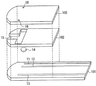
=
2
detected, and the measuring device calculates an amount of the blood
component based on the detected current and displays the value obtained by
the calculation.
= [0003] In the above-described manner, the sensor can measure the blood
component. However, since the obtained measured value might be affected
by a hematocrit (Hct), it might be necessary to measure a Hat value and then
correct the amount of the blood component based on this Hct value in order to
=
obtain an accurate measured value. For example, there has been a sensor
that corrects an amount of a blood component by measuring a Het value by .
=
the use of two working electrodes and one reference electrode (see Patent
Document 1). Other than this, there has been a method in which a Hct
=
value is measured using a mediator (see Patent Document 2). However, the
conventional technique has a problem concerning the accuracy and the
reliability of the measured Hct value so that the amount of the blood
component cannot be corrected sufficiently and accurately.
Patent Document 1: JP 2003-501627 A
Patent Document 2: Japanese Patent No. 3369183
=
Disclosure of Invention
[0004] The present invention relates to
=
a method of measuring a blood component, by which an amount of
the blood component can. be.corrected sufficiently and accurately by
=
measuring a Hct value with high accuracy and high reliability and also to
provide a sensor used. in the method and a measuring device.
26
[0005] The measurement method
= according to the present invention is a method of measuring a component
in
= blood, including: causing a redox reaction between the component in the
blood
'
and an oxidoreductase in the presence of a mediator; detecting an oxidation
= 30 current or a reduction current caused through the redox reaction by an
CA 02548440 2012-08-14
73466-125
3
electrode system; and calculating an amount of the component based on a
value of the detected current. The method further includes measuring a Het
value of the blood and correcting the amount of the component using this Hct
value. The measurement of the Hct value includes: providing an electrode
system having a working electrode and a counter electrode; providing a
mediator on the counter electrode but not on the working electrode; supplying
the blood to the electrode system; applying a voltage to the electrode system
in this state to cause an oxidation current or a reduction current to flow
between the electrodes; detecting the oxidation current or the reduction
current; and calculating the Hct value based on a value of the detected
current.
[00061 Furthermore, the sensor according to the present invention is a
sensor for measuring a component in blood by causing a redox reaction of the
component and detecting an oxidation current or a reduction current caused
through the redox reaction by an electrode. The sensor includes: a first
analysis portion including a first electrode system on which at least an
oxidoreductase that acts upon the component and a mediator are provided;
and a second analysis portion induding a second electrode system that
includes a working electrode and a counter electrode, in which a mediator is
provided on the counter electrode but not on the working electrode for
measuring a
Hct value. In the first analysis portion, the component in the blood is
measured by causing a
redox reaction between the component and the mddoreductase in the
presence of the mediator and detecting by the first electrode system an
oxidation current or a reduction current caused to flow when a voltage is
applied. On the other hand, in the second analysis portion, the Hct value of
the blood is measured by supplying the blood to the second electrode system,
applying a voltage to the blood in this state to cause an oxidation current or
a
reduction current to flow independent of the mediator between the working
electrode and
the counter electrode, and detecting a value of the oxidation current or the
reduction current.
CA 02548440 2014-05-15
73466-125
4
[0007] The measuring device according to the present invention is a measuring
device for
measuring a component in blood, including: the sensor of the present
invention; means for
holding the sensor; means for applying a voltage to the first electrode system
of the sensor;
means for detecting an oxidation current or a reduction current flowing
through the first
electrode system; means for calculating an amount of the component from a
value of the
detected current; means for applying a voltage to the second electrode system
of the sensor;
means for detecting an oxidation current or a reduction current flowing
through the second
electrode system; and means for calculating a Hct value of the blood from a
value of the
detected current.
Effects of the Invention
[0008] As described above, the present invention is characterized by the
measurement of a
Hct value. That is, by providing a mediator only on a counter electrode in the
measurement of
a Hct value, the current reflecting the Hct value can be measured easily with
high accuracy
and high reliability. Thus, according to the measurement method, the sensor,
and the
measuring device of the present invention, the amount of the blood component
can be
corrected sufficiently and accurately because it is corrected based on the Hct
value measured
with high accuracy and high reliability. As a result, it is possible to obtain
a highly accurate
and highly reliable corrected value of the amount of the blood component.
[0008a] Specific device aspects of the invention relate to:
1. A measuring device for measuring a component in blood, comprising: a
sensor,
comprising: a first analysis portion for measuring the component in the blood
sample,
comprising an electrode system, an oxidoreductase that acts upon the
component, and a
mediator are provided; and a second analysis portion for measuring a Hct value
of the blood
sample, comprising an electrode system that Comprises a working electrode and
a counter
electrode, a mediator being provided on the counter electrode but not on the
working electrode
for measuring the Hct value, wherein the first analysis portion measures the
component in the
blood with a redox reaction between the component and the oxidoreductase in
the presence of
CA 02548440 2014-05-15
73466-125
4a
the mediator by application of a voltage to the electrode system of the first
analyzing portion
and detecting an oxidation current or a reduction current caused to flow, the
second analysis
portion measures the Hct value of the blood sample by application of a voltage
to the blood
supplied to the second analysis portion, and detecting a value of the
oxidation current or the
reduction current, and the oxidoreductase and the mediator of the first
analysis portion are
present on the counter electrode of the second analysis portion; a holder that
holds the sensor;
a voltage source for the first analysis portion of the sensor; a detector that
detects an oxidation
current or a reduction current of the first analysis portion; a calculator
that calculates an
amount of the component from a value of the detected current of the first
analysis portion; a
voltage source for the second analysis portion of the sensor; a detector that
detects an
oxidation current or a reduction current of the second analysis portion; a
calculator that
calculates a Hct value of the blood from a value of the detected oxidation or
reduction current
of the second analysis portion, the calculated amount of the component being
corrected using
the Hct value; and a controller configured to direct application of a voltage
by the voltage
source for the first analysis portion, communicate with the detector of the
first analysis
portion, direct application of a voltage by the voltage source for the second
analysis portion,
and communicate with the detector of the second analysis portion.
2. The device according to 1 above, wherein the working electrode and the
counter electrode in the second analysis portion are provided on a common
insulating base
material so as to be coplanar and spaced apart from each other.
3. The device according to 1 above, wherein the sensor further comprises a
channel for leading blood to the sensor, wherein the second analysis portion
is on an upstream
side and the first analysis portion is on a downstream side with respect to
flow of the blood
supplied from one end of the channel.
4. The device according to 1 above, wherein the sensor further
comprises a
channel for leading blood to the sensor, wherein the working electrode of the
second analysis
CA 02548440 2014-05-15
73466-125
4b
portion is on an upstream side and the counter electrode of the second
analysis portion is on a
downstream side with respect to flow of the blood supplied from one end of the
channel.
5. The device according to 1 above, wherein the mediator of the second
analysis
portion is a ferricyanide.
6. The device according to 5 above, wherein the ferricyanide is
potassium
ferricyanide.
7. The device according to 1 above, wherein the working electrode of the
second
analysis portion, on which the mediator is not provided, is coated with a
polymeric material.
8. The device according to 7 above, wherein the polymeric material is
carboxymethylcellulose.
9. The device according to 1 above, wherein the voltage source is
controlled to
apply a voltage for measuring the Hct value that is equal to or higher than a
voltage causing
electrolysis of water.
10. The device according to 9 above, wherein the applied voltage for
measuring
the Hct value is in a range of 1 to 10 V.
11. The device according to 10 above, wherein the applied voltage for
measuring
the Hct value is in a range of 1 to 6.5 V.
12. The device according to 1 above, wherein the first analysis portion
comprises a
working electrode and a counter electrode.
13. The device according to 12 above, wherein at least one of the
electrodes of the
first analysis portion serves as the counter electrode of the second analysis
portion.
14. The device according to 13 above, wherein only the working electrode
of the
first analysis portion serves as the counter electrode of the second analysis
portion.
CA 02548440 2014-05-15
73466-125
4c
15. The device according to 1 above, wherein the mediator of the first
analysis
portion is a ferricyanide.
16. The device according to 15 above, wherein the ferricyanide is potassium
ferricyanide.
17. The device according to 1 above, wherein the sensor further comprises
an
insulating substrate on which the first and second analysis portions are
provided, and a
channel for leading the blood to the analysis portions is provided on the
insulating substrate,
with one end of the channel being open toward an outside of the sensor so as
to serve as a
blood supply port.
18. The device according to 17 above, wherein there is only one blood
supply port,
and the channel branches so that ends of branched portions communicate with
the first and =
second analysis portions, respectively.
19. The device to according to 17 above, wherein the second analysis
portion is
located in the channel, and the first analysis portion is located farther from
the blood supply
port than the second analysis portion.
20. The device according to 17 above, wherein the sensor further comprises,
a
spacer and a cover, wherein the cover is disposed on the insulating substrate
via the spacer.
21. The device according to 1 above, which measures at least one component
selected from the group consisting of glucose, lactic acid, uric acid,
bilirubin, and cholesterol.
22. The device according to 21 above, which measures glucose, and the
oxidoreductase is at least one of glucose oxidase and glucose dehydrogenase.
23. The device according to 1 above, wherein a polymeric material,
an enzyme
stabilizer, and a crystal homogenizing agent are provided on the electrode
system of the first
analysis portion.
CA 02548440 2014-05-15
73466-125
4d
24. The device according to 17 above, wherein the sensor further comprises
a
blood detecting electrode, wherein the blood detecting electrode is located
farther from the
blood supply port than at least one of the analysis portions so that whether
or not the blood is
supplied to the at least one of the analysis portions can be detected by the
blood detecting
electrode.
25. The device according to 1 above, wherein the voltage source for the
second
analysis portion applies the voltage for measuring Hct for a period in the
range of from 0.01
to 10 seconds.
Brief Description of Drawings
[0009] [FIG. 1] FIG. 1 is an exploded perspective view showing an example of a
sensor
according to the present invention.
[FIG. 2] FIG. 2 is a sectional view of the sensor.
[FIG. 3] FIG. 3 is a plan view of the sensor.
[FIG. 4] FIG. 4 is an exploded perspective view of another example of a sensor
according to the present invention.
[FIG. 5] FIG. 5 is a sectional view of the sensor.
[FIG. 6] FIG. 6 is a plan view of the sensor.
CA 02548440 2006-06-02
[FIG. 7] FIG. 7A shows how a reagent layer is provided in still
another example of a sensor according to the present invention; FIG. 7B is a
graph showing changes in response current (i.tA) obtained in Hct
measurement over time during voltage application in the example; and FIG.
5 70 is a graph showing changes in difference in sensitivity (%) over time
during the voltage application in the example.
[FIG. 8] FIG. 8A shows how a reagent layer is provided in still
another example of a sensor according to the present invention; FIG. 8B is a
graph showing changes in response current (.IA) obtained in Hct
measurement over time during voltage application in the example; and FIG.
80 is a graph showing changes in difference in sensitivity (%) over time
during the voltage application in the example.
[FIG. 9] FIG. 9A shows how a reagent layer is provided in still
another example of a sensor according to the present invention; FIG. 9B is a
graph showing changes in response current ( A) obtained in Hct
measurement over time during voltage application in the example; and FIG.
90 is a graph showing changes in difference in sensitivity (%) over time
during the voltage application in the example.
[FIG. 10] FIG. 10A shows how a reagent layer is provided in still
another example of a sensor according to the present invention; FIG. 10B is a
graph showing changes in response current ( A) obtained in Hct
measurement over time during voltage application in the example; and FIG.
10C is a graph showing changes in difference in sensitivity (%) over time
during the voltage application in the example.
[FIG. 11] FIG. 11A shows how a reagent layer is provided in still
another example of a sensor according to the present invention; FIG. 11B is a
graph showing changes in response current ( A) obtained in Hct
measurement over time during voltage application in the example; and FIG.
110 is a graph showing changes in difference in sensitivity (%) over time
during the voltage application in the example.
CA 02548440 2006-06-02
6
[FIG. 121 FIG. 12A shows how a reagent layer is provided in still
another example of a sensor according to the present invention; FIG. 12B is a
graph showing changes in response current (pA) obtained in Hct
measurement over time during voltage application in the example; and FIG.
12C is a graph showing changes in difference in sensitivity (%) over time
during the voltage application in the example.
[FIG. 13] FIG. 13A shows how a reagent layer is provided in a sensor
according to a comparative example; FIG. 13B is a graph showing changes in
response current ( A) obtained in Hct measurement over time during voltage
application in the comparative example; and FIG. 13C is a graph showing
changes in difference in sensitivity (%) over time during the voltage
application in the comparative example.
[FIG. 141 FIG. 14A shows how a reagent layer is provided in a sensor
according to another comparative example; FIG. 14B is a graph showing
changes in response current ( A) obtained in Hct measurement over time
during voltage application in the comparative example; and FIG. 14C is a
graph showing changes in difference in sensitivity (%) over time during the
voltage application in the comparative example.
[FIG. 151 FIG. 15A shows how a reagent layer is provided in a sensor
according to still another comparative example; FIG. 15B is a graph showing
changes in response current ( A) obtained in Hct measurement over time
during voltage application in the comparative example; and FIG. 15C is a
graph showing changes in difference in sensitivity (%) over time during the
voltage application in the comparative example.
[FIG. 16] FIG. 16A is a graph showing changes in response current
( A) obtained in Hct measurement over time during voltage application (0.5
V) in still another example of a sensor according to the present invention;
and
FIG. 16B is a graph showing changes in difference in sensitivity (%) over time
during the voltage application in the example.
[FIG. 17] FIG. 17A is a graph showing changes in response current
CA 02548440 2006-06-02
7
(1.1A) obtained in Hct measurement over time during voltage application (1.0
V) in still another example of a sensor according to the present invention;
and
FIG. 17B is a graph showing changes in difference in sensitivity (%) over time
during the voltage application in the example.
[FIG. 18] FIG. 18Ais a graph showing changes in response current
(1,1A) obtained in Hct measurement over time during voltage application (1.5
V) in still another example of a sensor according to the present invention;
and
FIG. 18B is a graph showing changes in difference in sensitivity (%) over time
during the voltage application in the example.
[FIG. 19] FIG. 19Ais a graph showing changes in response current
(IAA) obtained in Hct measurement over time during voltage application (2.0
V) in still another example of a sensor according to the present invention;
and
FIG. 19B is a graph showing changes in difference in sensitivity (%) over time
during the voltage application in the example.
[FIG. 20] FIG. 20Ais a graph showing changes in response current
( A) obtained in Hct measurement over time during voltage application (2.5
V) in still another example of a sensor according to the present invention;
and
FIG. 20B is a graph showing changes in difference in sensitivity (%) over time
during the voltage application in the example.
[FIG. 21] FIG. 21A is a graph showing changes in response current
( A) obtained in Hct measurement over time during voltage application (3.0
V) in still another example of a sensor according to the present invention;
and
FIG. 21B is a graph showing changes in difference in sensitivity (%) over time
during the voltage application in the example.
[FIG. 22] FIG. 22A is a graph showing changes in response current
( A) obtained in Hct measurement over time during voltage application (3.5
V) in still another example of a sensor according to the present invention;
and
FIG. 22B is a graph showing changes in difference in sensitivity (%) over time
during the voltage application in the example.
[FIG. 23] FIG. 23A is a graph showing changes in response current
CA 02548440 2006-06-02
8
( A) obtained in Hct measurement over time during voltage application (4.0
V) in still another example of a sensor according to the present invention;
and
FIG. 23B is a graph showing changes in difference in sensitivity (%) over time
during the voltage application in the example.
[FIG. 24] FIG. 24A is a graph showing changes in response current
(pA) obtained in Hct measurement over time during voltage application (4.5
V) in still another example of a sensor according to the present invention;
and
FIG. 24B is a graph showing changes in difference in sensitivity (%) over time
during the voltage application in the example.
[FIG. 25] FIG. 25A is a graph showing changes in response current
( A) obtained in Hct measurement over time during voltage application (5.0
V) in still another example of a sensor according to the present invention;
and
FIG. 25B is a graph showing changes in difference in sensitivity (%) over time
during the voltage application in the example.
[FIG. 26] FIG. 26A is a graph showing changes in response current
([iA) obtained in Hct measurement over time during voltage application (5.5
V) in still another example of a sensor according to the present invention;
and
FIG. 26B is a graph showing changes in difference in sensitivity (%) over time
during the voltage application in the example.
[FIG. 271 FIG. 27A is a graph showing changes in response current
( A) obtained in Hct measurement over time during voltage application (6.0
V) in still another example of a sensor according to the present invention;
and
FIG. 27B is a graph showing changes in difference in sensitivity (%) over time
during the voltage application in the example.
[FIG. 281 FIG. 28A is a graph showing changes in response current
( A) obtained in Hct measurement over time during voltage application (6.5
V) in still another example of a sensor according to the present invention;
and
FIG. 28B is a graph showing changes in difference in sensitivity (%) over time
during the voltage application in the example.
[FIG. 291 FIG. 29 is a perspective view showing an example of a
CA 02548440 2006-06-02
9
measuring device according to the present invention.
[FIG. 30] FIG. 30 is a plan view showing still another example of a
sensor according to the present invention.
[FIG. 31] FIG. 31 is a plan view showing the configuration of the
measuring device according to the above example.
Explanation of reference numerals
[0010] 11, 12, 13, 21, 22, 23, 24, 81, 82, 111, 112, 113, 114 electrode
14, 25, 83 reagent portion (reagent layer)
15, 26, 84 channel
16, 27, 85 air vent hole
101, 201, 801 insulating substrate
102, 202, 802 spacer
103, 203, 803 cover
121 sensor
122 sample supply port
130, 123 measuring device
124 display portion
125 attachment portion
131 CPU
132 LCD
133 reference voltage source
134 A/D conversion circuit
135 current/voltage conversion circuit
136 switching circuit
137a, 137b, 137c, 137d connector
Description of the Invention
[0011] Hereinafter, the present invention will be described in detail.
[0012] In the method of measuring a blood component and the sensor
according to the present invention, the mediator used for the Hct
measurement or in the second analysis portion is not particularly limited.
CA 02548440 2006-06-02
Examples of the mediator indude a ferricyanide, p-benzoquinone,
p-benzoquinone derivatives, phenazine methosulfate, methylene blue,
ferrocene, and ferrocene derivatives. Among these, a ferricyanide is
preferable, and potassium ferricyanide is more preferable. The amount of
5 the mediator to be blended is not particularly limited, but is, for
example, 0.1
to 1000 mM, preferably 1 to 500 mM, and more preferably 10 to 200 mM per
one measurement or one sensor.
[0013] In the method of measuring a blood component and the sensor
according to the present invention, the electrode that is used for the Hct
10 measurement or in the second analysis portion and on which the mediator
is
not provided preferably is coated with a polymeric material in order to
prevent adhesion of impurities, oxidation of the electrode, and the like.
Examples of the polymeric material include carboxymethyl cellulose (CMC),
hydroxyethyl cellulose, hydroxypropyl cellulose, methyl cellulose, ethyl
cellulose, ethyl hydroxyethyl cellulose, carboxyethyl cellulose, polyvinyl
alcohol, polyvinylpyrrolidone, polyamino acid such as polylysine, polystyrene
sulfonate, gelatin and derivatives thereof, polyacrylic acid and salts
thereof,
polymethacrylic acid and salts thereof, starch and derivatives thereof, maleic
anhydride polymer and salts thereof, and agarose gel and derivatives thereof.
They may be used individually or two or more of them may be used together.
The method of coating the electrode with a polymeric material is not
particularly limited. For example, the coating can be achieved by providing
a polymeric material solution, applying the solution to the electrode surface,
and then removing a solvent contained in the coating layer of the solution by
drying.
[0014] In the method of measuring a blood component and the sensor
according to the present invention, a voltage applied between the working
electrode and the counter electrode that are used for the Hct measurement or
in the second analysis portion preferably is equal to or higher than a voltage
causing electrolysis of water, more preferably in the range from 1 to 10 V,
and
CA 02548440 2006-06-02
11
still more preferably in the range from 1 to 6.5 V. By applying a voltage that
is equal to or higher than a voltage causing electrolysis of water, a current
depending on a hematocrit alone can be measured with a still higher
sensitivity. As a result, it is possible to obtain a stable current that is
not
affected by other redox substances present in blood and thus does not vary
depending on a specimen (an individual). The voltage is applied for, for
example, 0.001 to 60 seconds, preferably 0.01 to 10 seconds, and more
preferably 0.01 to 5 seconds.
[0015] In the method of measuring a blood component and the sensor
according to the present invention, it is preferable that the shortest
distance
between the working electrode and the counter electrode that are used for the
Hct measurement or in the second analysis portion is at least 0.05 mm.
When the distance between the electrodes is at least 0.05 mm as described
above, the reliability of the measured value is improved. More preferably,
the distance between the electrodes is at least 0.1 mm, still more preferably
at least 0.5 mm.
[0016] In the method of measuring a blood component according to the
present invention, the correction using the Hct value preferably is carried
out
based on a previously prepared calibration curve or calibration table for
showing the relationship between a Hct value and an amount of the blood
component.
[0017] In the method of measuring a blood component according to the
present invention, the order of carrying out the blood component
measurement and the Hct measurement is not particularly limited.
However, in the case where the same electrode is used in both the
measurements as will be described later, it is preferable that the blood
component is measured first and the Hct value is measured thereafter. Note
here that the case where the electrode that is used as a working electrode in
the blood component measurement is used as a counter electrode in the Hct
measurement corresponds to the above case. On this electrode, a mediator
CA 02548440 2006-06-02
12
(e.g., potassium ferricyanide) that initially is in an oxidized state is
provided.
This mediator is reduced through the enzyme reaction caused in the blood
component measurement and is oxidized again for the purpose of measuring
the blood component. Thus, after the blood component measurement,
ferricyanide ions are present dominantly at the interface of the electrode.
On the other hand, it is preferable that a large amount of ferricyanide ions
are present in the vicinity of a counter electrode used for the Hct
measurement in order to suppress an electrolytic reduction reaction occurring
at the counter electrode from being a rate-determining step. On this account,
it is preferable that the electrode used as a working electrode in the blood
component measurement is used as a counter electrode in the Hct
measurement after the completion of the blood component measurement.
[0018] In the method of measuring a blood component according to the
present invention, it is preferable that the electrode system for detecting
the
oxidation current or the reduction current in the measurement of the blood
component includes a working electrode and a counter electrode.
[0019] Preferably, the method of measuring a blood component according to
the present invention further includes measuring a temperature of a
measurement environment, and the amount of the blood component is
corrected using the measured temperature. This is because the enzyme
reaction is affected by the temperature of the measurement environment. In
this case, it is preferable that the correction using the temperature is
carried
out based on a previously prepared calibration curve or calibration table.
[0020] In the method of measuring a blood component and the sensor
according to the present invention, the blood component to be measured is,
for example, glucose, lactic acid, uric acid, bilirubin, cholesterol, or the
like.
Furthermore, the oxidoreductase is selected as appropriate depending on the
blood component to be measured. Examples of the oxidoreductase include
glucose oxidase, lactate oxidase, cholesterol oxidase, bilirubin oxidase,
glucose
dehydrogenase, and lactate dehydrogenase. The amount of the
CA 02548440 2006-06-02
13
oxidoreductase is, for example, 0.01 to 100 U, preferably 0.05 to 10 U, and
more preferably 0.1 to 5 U per one sensor or one measurement. Among
these, the blood component to be measured preferably is glucose, and the
oxidoreductase to be used in this case preferably is glucose coddase or
glucose
dehydrogenase.
[0021] In the sensor for measuring a blood component according to the
present invention, it is preferable that the first electrode system includes a
working electrode and a counter electrode. Furthermore, in the sensor of the
present invention, it is preferable that, in the first electrode system and
the
second electrode system, at least one of the electrodes or all the electrodes
provided in the first electrode system also serve as the counter electrode in
the second electrode system. It is more preferable that, in the first
electrode
system and the second electrode system, only the working electrode in the
first electrode system also serves as the counter electrode in the second
electrode system.
[0022] In the sensor for measuring a blood component according to the
present invention, the mediator provided on the first electrode system is not
particularly limited, and examples thereof include a ferricyanide,
p-benzoquinone, p-benzoquinone derivatives, phenazine methosulfate,
methylene blue, ferrocene, and ferrocene derivatives. Among these, a
ferricyanide is preferable, and potassium ferricyanide is more preferable.
The amount of the mediator to be blended is not particularly limited, but is,
for example, 0.1 to 1000 mM, preferably 1 to 500 mM, and more preferably 10
to 200 mM per one measurement or one sensor.
[0023] The sensor for measuring a blood component according to the present
invention preferably is configured so that it further includes an insulating
substrate, the first analysis portion, the second analysis portion, and a
channel for leading the blood to the analysis portions are formed on the
insulating substrate, and one end of the channel is open toward the outside of
the sensor so as to serve as a blood supply port. In this case, the sensor may
CA 02548440 2006-06-02
14
be configured so that there is only one blood supply port and the channel
branches so that ends of branched portions communicate with the analysis
portions, respectively. Alternatively, the sensor may be configured so that
the second analysis portion is located in the channel and the first analysis
portion is located farther from the blood supply port than the second analysis
portion.
[0024] Preferably, the sensor for measuring a blood component according to
the present invention is configured so that it further includes a spacer and a
cover and the cover is disposed on the insulating substrate via the spacer.
[0025] In the sensor for measuring a blood component according to the
present invention, it is preferable that a polymeric material, an enzyme
stabilizer, and a crystal homogenizing agent further are provided on the first
electrode system.
[0026] The polymeric material serves to prevent adhesion of impurities to
the electrode surface and oxidation of the electrode surface as well as to
protect the electrode surface. Examples of the polymeric material include
CMC, hydroxyethyl cellulose, hydroxypropyl cellulose, methyl cellulose,
ethykellulose, ethyl hydroxyethyl cellulose, carboxyethyl cellulose, polyvinyl
alcohol, polyvinylpyrrolidone, polyarnino acid such as polylysine, polystyrene
sulfonate, gelatin and derivatives thereof, polyacrylic acid and salts
thereof,
polymethacrylic acid and salts thereof, starch and derivatives thereof, maleic
anhydride polymer and salts thereof, and agarose gel and derivatives thereof.
They may be used individually or two or more of them may be used together.
Among these, CMC is preferable. The ratio of the polymeric material to an
entire reagent solution for preparing a reagent portion is, for example, 0.001
to 10 wt%, preferably 0.005 to 5 wt%, and more preferably 0.01 to 2 wt%.
[0027] As the enzyme stabilizer, sugar alcohol can be used, for example.
Examples of the sugar alcohol include chain polyhydric alcohols and cyclic
sugar alcohols, such as sorbitol, maltitol, xylitol, mannitol, lactitol,
reduced
paratinose, arabinitol, glycerol, ribitol, galactitol, sedoheptitol,
perseitol,
CA 02548440 2006-06-02
volemitol, styracitol, polygalitol, iditol, talitol, allitol, isylitol,
hydrogenated
glucose syrup, and isylitol. Note here that stereoisomers, substitution
products, and derivatives of these sugar alcohols also may be used as the
enzyme stabilizer. These sugar alcohols may be used individually or two or
5 more of them may be used together. Among these, maltitol is preferable.
The amount of the enzyme stabilizer to be blended is, for example, in the
range from 0.1 to 500 mM, preferably from 0.5 to 100 mM, and more
preferably from 1 to 50 mM per one measurement or one sensor.
[0028] The crystal homogenizing agent serves to homogenize the crystal
10 condition of the reagent portion. As the crystal homogenizing agent, an
amino acid can be used, for example. Examples of the amino acid include
glycine, alanine, valine, leucine, isoleucine, serine, threonine, methionine,
asparagine, glutamine, arginine, lysine, histidine, phenylalanine, tryptophan,
proline, sarcosine, betaine, taurine, and salts, substitution products, and
15 derivatives of these amino acids. They may be used individually or two
or
more of them may be used together. Among these, glycine, serine, proline,
threonine, lysine, and taurine are preferable, and taurine is more preferable.
The amount of the crystal homogenizing agent to be blended is, for example,
0.1 to 1000 mM, preferably 10 to 500 mM, and more preferably 20 to 200 mM
per one measurement or one sensor.
[0029] Preferably, the sensor for measuring a blood component according to
the present invention is configured so that it further includes a blood
detecting electrode, and the blood detecting electrode is located farther from
the blood supply port than at least one of the analysis portions so that
whether or not blood is supplied surely to the at least one of the analysis
portions can be detected by the blood detecting electrode. It is more
preferable that the blood detecting electrode is located farther from the
blood
supply port than both the analysis portions.
[0030] Next, the measuring device according to the present invention
preferably further includes means for correcting the amount of the blood
CA 02548440 2006-06-02
16
component using the Hct value. Furthermore, in the measuring device of
the present invention, the voltage applied to the second electrode system
preferably is equal to or higher than a voltage causing electrolysis of water,
more preferably in the range from 1 to 10 V, and still more preferably from 1
to 6.5 V.
[0031] FIG. 29 is a perspective view showing an example of a measuring
device according to the present invention to which a sensor according to the
present invention is attached. As shown in FIG. 29, this measuring device
123 has a sensor attachment portion 125 at one end, and a sensor 121 is
attached to this portion so as to be held by the measuring device. The
reference numeral 122 denotes a sample supply port of the sensor 121. This
measuring device 123 has a display portion 124 at a substantially center
portion thereof, and the result of the measurement is displayed in this
display portion 124.
[0032] The measuring device according to the present invention preferably
includes a connector, a switching circuit, a current/voltage conversion
circuit,
an A/D conversion circuit, a reference voltage source, a CPU, and a liquid
crystal display portion (LCD). By providing these components, the following
operations become possible: applying a voltage to the first electrode system
and the second electrode system in the sensor of the present invention;
detecting the value of a current flowing between these electrode systems;
calculating an amount of the blood component or a Hct value based on the
thus-detected current value; correcting the amount of the blood component
based on the Hct value; and displaying the thus-obtained corrected value.
With regard to the circuit configuration of a measuring device according to
the present invention, an example thereof will be described later.
[0033] Hereinafter, examples of a sensor for measuring a blood component
according to the present invention will be described with reference to the
drawings.
Example 1
CA 02548440 2006-06-02
17
[0034] FIGs. 1, 2, and 3 show one example of a sensor for measuring a blood
component according to the present invention. FIG. 1 is an exploded
perspective view of the sensor, FIG. 2 is a sectional view of the sensor, and
FIG. 3 is a plan view of the sensor. In these three drawings, the same
components are given the same reference numerals.
[0035] As shown in the drawings, in this sensor, three electrodes 11, 12, and
13 are formed on an insulating substrate 101. Each of the electrodes can be
switched between a working electrode and a counter electrode. The surface
of the electrode 13 is coated with a polymeric material such as CMC. On an
electrode portion formed by the electrodes 11 and 12, a reagent layer 14 is
disposed. The reagent layer 14 contains an oxidoreductase such as glucose
dehydrogenase and a mediator, and optionally contains a polymeric material,
an enzyme stabilizer, and a crystal homogenizing agent. The type and the
blending ratio of these reagents are as described above. A cover 103 is
disposed on the insulating substrate 101 so as to cover an entire area
excluding one end portion (the end portion on the right in the drawings) with
a spacer 102 intervening therebetween. This sensor has a channel 15 for
leading blood to the electrode 13 and the electrodes 11 and 12. This channel
15 branches into two portions so that the channel as a whole forms a T-shape,
and ends of the branched portions communicate with the electrode portions,
respectively. The channel extends to the other end portion (the end portion
on the left in the drawings) of the sensor and the tip thereof is open toward
the outside of the sensor so as to serve as a blood supply port. The three
electrodes 11, 12, and 13 are connected to leads, respectively. These leads
extend to the above-described one end portion of the sensor with the tip of
each lead not being covered with the cover but being exposed. The cover 103
has two air vent holes 16 at portions corresponding to the ends of the
branched portions of the channel 15.
[0036] In the present invention, the material of the insulating substrate is
not particularly limited, and may be, for example, polyethylene terephthalate
CA 02548440 2006-06-02
18
(PET), polycarbonate (PC), polyimide (PI), polyethylene (PE), polypropylene
(PP), polystyrene (PS), polyvinyl chloride (PVC), polyoxymethylene (P0M),
monomer-cast nylon (MC), polybutylene terephthalate (PBT), polymethyl
methacrylate (PMMA), an ABS resin (ABS), or glass. Among these,
polyethylene terephthalate (PET), polycarbonate (PC), and polyimide (PT) are
preferable, and polyethylene terephthalate (PET) is more preferable. The
size of the insulating substrate is not particularly limited. For example, the
insulating substrate may have an overall length of 5 to 100 m, a width of 2 to
50 mm, and a thickness of 0.05 to 2 mm; preferably an overall length of 7 to
50 mm, a width of 3 to 20 mm, and a thickness of 0.1 to 1mm; and more
preferably an overall length of 10 to 30 mm, a width of 3 to 10 mm, and a
thickness of 0.1 to 0.6 mm.
[00371 The electrodes and the leads on the insulating substrate may be
formed by, for example, forming a conductive layer with gold, platinum,
palladium, or the like by sputtering or vapor deposition and then processing
the conductive layer into a particular electrode pattern with a laser.
Examples of the laser include YAG lasers, CO2 lasers, and excimer lasers.
Note here that the electrode pattern is not limited to those shown in the
examples or the like, and there is no limitation regarding the electrode
pattern as long as it can achieve the effect of the present invention. The
coating of the surface of the electrode 13 can be achieved by, for example,
preparing a solution of the polymeric material, dropping or applying this
solution with respect to the electrode surface, and then drying it. The drying
may be, for example, natural drying, air drying, hot air drying, or heat
drying.
[0038] The reagent portion 14 can be formed in the following manner, for
example. First, 0.1 to 5.0 U/sensor of PQQ-GDH, 10 to 200 mM of potassium
ferricyanide, 1 to 50 mM of maltitol, and 20 to 200 mM of taurine are
dissolved in a 0.01 to 2.0 wt% CMC aqueous solution to prepare a reagent
solution. The reagent solution is dropped on the electrodes 11 and 12 formed
CA 02548440 2006-06-02
19
on the substrate and then is dried, thus forming the reagent portion 14. The
drying may be natural drying or forced drying using warm air, for example.
However, if the temperature of the warm air is too high, there is a
possibility
that the enzyme contained in the solution might be deactivated. Thus, the
temperature of the warm air preferably is around 50 C.
[0039] In the present invention, the material of the spacer is not
particularly
limited. For example, the same material as that of the insulating substrate
can be used. The size of the spacer also is not particularly limited. For
example, the spacer may have an overall length of 5 to 100 mm, a width of 2
to 50 mm, and a thickness of 0.01 to 1 mm; preferably an overall length of 7
to 50 mm, a width of 3 to 20 mm, and a thickness 0.05 to 0.5 mm; and more
preferably an overall length of 10 to 30 mm, a width of 3 to 10 mm, and a
thickness of 0.05 to 0.25 mm. The spacer has a T-shaped cut-away portion
that serves as the channel for leading blood. The size of the cut-away
portion is as follows, for example: the length from the blood supply port to
the
branching part is 0.5 to 20 mm, the length from the branching part to the end
of the branched portion is 1 to 25 mm, and the width is 0.1 to 5 mm;
preferably the length from the blood supply port to the branching part is 1 to
10 mm, the length from the branching part to the end of the branched portion
is 1.5 to 10 mm, and the width is 0.2 to 3mm; and more preferably the length
from the blood supply port to the branching part is 1 to 5 mm, the length from
the branching part to the end of the branched portion is 1.5 to 5 mm, and the
width is 0.5 to 2 mm. The cut-away portion may be formed, for instance, by
using a laser, a drill, or the like, or by forming the spacer using a die that
can
form the spacer provided with the cut-away portion.
[00401 In the present invention, the material of the cover is not particularly
limited. For example, the same material as that of the insulating substrate
can be used. It is more preferable that a portion of the cover corresponding
to the ceiling of the sample supply channel is subjected to a treatment for
imparting hydrophilicity. The treatment for imparting hydrophilicity may
CA 02548440 2006-06-02
be carried out by, for example, applying a surfactant or introducing a
hydrophilic functional group such as a hydroxyl group, a carbonyl group, or a
carboxyl group to the surface of the cover by plasma processing or the like.
The size of the cover is not particularly limited. For example, the cover may
5 have an overall length of 5 to 100 mm, a width of 3 to 50 mm, and a
thickness
of 0.01 to 0.5 mm; preferably an overall length of 10 to 50 mm, a width of 3
to
20 mm, and a thickness of 0.05 to 0.25 mm; and more preferably an overall
length of 15 to 30 mm, a width of 5 to 10 mm, and a thickness of 0.05 to 0.2
mm. The cover preferably has an air vent hole. The shape of the air vent
10 hole may be, for example, circular, oval, polygonal, or the like, and
the
maximum diameter thereof may be, for example, 0.01 to 10 mm, preferably
0.025 to 5 mm, and more preferably 0.025 to 2 mm. The cover may have a
plurality of air vent holes. The air vent hole may be formed, for instance, by
perforating the cover with a laser, a drill, or the like, or by forming the
cover
15 using a die that can form the cover provided with the air vent hole.
Then, by
laminating the insulating substrate, the spacer, and the cover in this order
and integrating them, the sensor can be obtained. The integration can be
achieved by adhering these three components with an adhesive or through
heat-sealing. As the adhesive, an epoxy adhesive, an acrylic adhesive, a
20 polyurethane adhesive, a thermosetting adhesive (a hot melt adhesive or
the
like), a UV curable adhesive, or the like can be used, for example.
[0041] Measurement of a blood glucose level using this sensor can be carried
out in the following manner, for example. First, a fingertip or the like is
punctured with a dedicated lancet to cause bleeding. On the other hand, the
sensor is set in a dedicated measuring device (a meter). The blood supply
port of the sensor set in the measuring device is brought into contact with
the
blood that has come out, so that the blood is led inside the sensor by
capillary
action. Then, the sensor analyzes the blood according to the following steps.
[0042] (Step 1: Detecting specimen (blood))
A voltage is applied between the electrode 11 and the electrode 13,
CA 02548440 2006-06-02
21
and whether or not the blood is supplied to the sensor is detected by
detecting
the change in current accompanying the supply of the blood. After the
supply of the blood has been confirmed, the subsequent step is started. Note
here that the voltage applied in Step 1 is 0.05 to 1 V, for example.
[0043] (Step 2: Measuring glucose)
After allowing glucose in the blood to react with the glucose
oxidoreductase for a certain period of time, a voltage is applied between the
electrode 11 as a working electrode and the electrode 12 as a counter
electrode, thereby oxidizing a reduced mediator generated on the electrode 11
through the enzyme reaction. The oxidation current caused at this time is
detected. The glucose is allowed to react with the oxidoreductase for, for
example, 0 to 60 seconds, preferably 0.5 to 30 seconds, and more preferably 1
to 10 seconds. In Step 2, the voltage applied is, for example, 0.05 to 1 V,
preferably 0.1 to 0.8 V, and more preferably 0.2 to 0.5 V, and the voltage
application time is, for example, 0.01 to 30 seconds, preferably 0.1 to 10
seconds, and more preferably 1 to 5 seconds.
[0044] (Step 3: Measuring Hct value)
By applying a voltage between the electrode 13 as a working electrode
and the electrode 11 as a counter electrode, a current depending on a Hct
value can be detected based on an electrolytic oxidation reaction of blood
components. Note here that the detected current can be converted into a
Hct value using a previously prepared calibration curve or calibration curve
table. In this correction, a Het value determined using a previously
prepared calibration curve showing the relationship between a current and a
Hct value may be used or alternatively, the detected current may be used as
it is. In Step 3, the voltage applied is, for example, 1 to 10 V, preferably 1
to
6.5 V, and more preferably 2 to 3 V, and the voltage application time is, for
example, 0.001 to 60 seconds, preferably 0.01 to 10 seconds, and more
preferably 0.01 to 5 seconds. In Step 3, the oxidation current depending on a
Hct value can be detected without being affected by any reagent because no
=
CA 02548440 2006-06-02
22
mediator is provided on the electrode 13 as a working electrode, and the
electrode 13 and the electrode 11 are spaced apart from each other by a
certain distance with no reagent such as a mediator being provided in this
space so that only blood is present in this space. Preferably, Step 3 is
performed after the completion of Step 2. Although the electrode 11 is used
as a counter electrode in the present example, the measurement also can be
achieved when the electrode 12 is used as a counter electrode. Also, it is
possible to use both the electrodes 11 and 12 as counter electrodes. Note
here that when the surface of the electrode 13 is not coated with a polymeric
material or the like, it is still possible to carry out the measurement.
[0045] (Step 4: Correcting blood component)
The amount of glucose obtained in Step 2 is corrected using the Hct
value detected in Step 3. The correction preferably is carried out based on a
calibration curve (including a calibration table) prepared previously. The
corrected amount of glucose is displayed on or stored in the measuring device.
Instead of determining the Hct value and then correcting the amount of
glucose as described above, the current depending on the Hct value, which
has been detected in Step 3, may be used as it is to correct the amount of
glucose.
Example 2
[0046] FIGs. 4, 5, and 6 show another example of a sensor for measuring a
blood component according to the present invention. FIG. 4 is an exploded
perspective view of the sensor, FIG. 5 is a sectional view of the sensor, and
FIG. 6 is a plan view of the sensor. In these three drawings, the same
components are given the same reference numerals.
[0047] As shown in the drawings, in this sensor, four electrodes 21, 22, 23,
and 24 are formed on an insulating substrate 201. These electrodes can be
switched between a working electrode and a counter electrode. The surface
of the electrode 24 is coated with a polymeric material in the manner as
described above. On an electrode portion formed by the electrodes 21, 22,
CA 02548440 2006-06-02
23
and 23, a reagent layer 25 is provided. The reagent layer 25 contains an
oxidoreductase such as glucose dehydrogenase and a mediator, and. optionally
contains a polymeric material, an enzyme stabilizer, and a crystal
homogenizing agent. The type and the blending ratio of these reagents are
as described above. A cover 203 is disposed on the insulating substrate 201
so as to cover an entire area excluding one end portion (the end portion on
the
right in the drawings) with a spacer 202 intervening therebetween. This
sensor has a channel 26 for leading blood to the reagent portion 25. This
channel 26 extends linearly (I-shape). The channel 26 extends to the other
end portion (the end portion on the left in the drawings) of the sensor and
the
tip thereof is open toward the outside of the sensor so as to serve as a blood
supply port. The four electrodes are arranged in series in the channel, and
the electrode 22 is located farthest from the blood supply port. The four
electrodes 21, 22, 23, and 24 are connected to leads, respectively. These
leads extend to the above-described one end portion of the sensor with the tip
of each lead not being covered with the cover but being exposed. The cover
203 has an air vent hole 27 at a portion corresponding to the rear side of the
channel 26.
[0048] In the present example, the material, the size, and the like of the
insulating substrate are not particularly limited, and may be the same as in
Example 1. Furthermore, the electrodes, the leads, the manner of coating
the electrode surface with a polymeric material, and the reagent portion also
are the same as in Example 1. Still further, the material and the size of the
spacer and the method of processing the spacer also are the same as in
Example 1. In the present example, the spacer has an I-shaped cut-away
portion that serves as the channel for leading blood. The size of the
cut-away portion is as follows, for example: the overall length is 0.5 to 50
mm
and the width is 0.1 to 5 mm; preferably the overall length is 1 to 10 mm and
the width is 0.2 to 3 mm; and more preferably the overall length is 1 to 5 mm
and the width is 0.5 to 2 mm. The cut-away portion may be formed, for
CA 02548440 2006-06-02
24
instance, by using a laser, a drill, or the like, or by forming the spacer
using a
die that can form the spacer provided with the cut-away portion. The
material and the size of the cover, the treatment for imparting hydrophilicity
to the cover, and the air vent hole provided in the cover are the same as in
Example 1. Also, the method for producing the sensor of the present
example is the same as that for producing the sensor of Example 1.
[0049] Measurement of a blood glucose level using this sensor can be carried
out in the following manner, for example. First, a fingertip or the like is
punctured with a dedicated lancet to cause bleeding. On the other hand, the
sensor is set in a dedicated measuring device (a meter). The blood supply
port of the sensor set in the measuring device is brought into contact with
the
blood that has come out, so that the blood is led inside the sensor by
capillary
action. Then, the sensor analyzes the blood according to the following steps.
[0050] (Step 1: Detecting specimen (blood))
Whether or not the blood is supplied to the end of the channel is
detected by applying a voltage between the electrode 24 and the electrode 22.
After the supply of the blood to the end of the channel has been confirmed,
the subsequent step is started. In the case where the blood is not supplied to
the end of the channel, the measuring device recognizes it as the lack of the
amount of the specimen and displays an error message. The voltage applied
in Step 1 is, for example, 0.05 to 1 V. In this case, the specimen can be
detected by detecting the change in current between the electrode 22 and any
one of other electrodes (21, 23, and 24).
[0051] (Step 2: Measuring glucose)
After allowing glucose in the blood to react with the glucose
oxidoreductase for a certain period of time, a voltage is applied between the
electrode 21 as a working electrode and the electrode 23 as a counter
electrode, thereby oxidizing a reduced mediator generated on the electrode 21
through the enzyme reaction. The oxidation current caused at this time is
detected. The glucose is allowed to react with the oxidoreductase for, for
CA 02548440 2006-06-02
example, 0 to 60 seconds, preferably 0.5 to 30 seconds, and more preferably 1
to 10 seconds. In Step 2, the voltage applied is, for example, 0.05 to 1 V,
preferably 0.1 to 0.8 V, and more preferably 0.2 to 0.5 V, and the voltage
application time is, for example, 0.01 to 30 seconds, preferably 0.1 to 10
5 seconds, and more preferably 1 to 5 seconds.
[0052] (Step 3: Measuring Hct value)
By applying a voltage between the electrode 24 as a working electrode
and the electrode 21 as a counter electrode, a current depending on a Hct
value can be detected. Based on the detected current, the Hct value of the
10 blood can be determined. The thus-determined Hct value is used for the
correction in the measurement of glucose. In this correction, a Hct value
determined using a previously prepared calibration curve showing the
relationship between a current and a Hct value may be used or alternatively,
the detected current may be used as it is. In Step 3, the voltage applied is,
15 for example, 1 to 10 V, preferably 1 to 6.5 V, and more preferably 2 to
3 V, and
the voltage application time is, for example, 0.001 to 60 seconds, preferably
0.01 to 10 seconds, and more preferably 0.01 to 5 seconds. In Step 3, the
oxidation current depending on a Hct value can be detected without being
affected by any reagent because no mediator is provided on the electrode 24
20 as a working electrode, and the electrode 24 and the electrode 21 are
spaced
apart from each other by a certain distance with no reagent such as a
mediator being provided in this space so that only blood is present in this
space. Preferably, Step 3 is performed after the completion of Step 2.
Although the electrode 21 alone is used as the counter electrode in the
25 present example, the present invention is not limited thereto. It should
be
noted that the electrode 23 alone, the electrode 22 alone, the combination of
the electrode 21 and the electrode 22, the combination of the electrode 21 and
the electrode 23, the combination of the electrode 22 and the electrode 23,
the
combination of the electrode 21, the electrode 22, and the electrode 23 also
may be used as the counter electrode. Also, it should be noted that when the
CA 02548440 2006-06-02
26
surface of the electrode 13 is not coated with a polymeric material or the
like,
it is still possible to achieve the measurement.
[0053] (Step 4: Correcting blood component)
The amount of glucose obtained in Step 2 is corrected using the Hct
value detected in Step 3. The correction preferably is carried out based on a
calibration curve (including a calibration table) prepared previously. The
corrected amount of glucose is displayed on or stored in the measuring device.
Example 3
[0054] In the present example, six types of sensors (3-1 to 3-6) were
produced so that they were different from each other in the arrangement of a
reagent layer containing a mediator with respect to a working electrode or a
counter electrode used for Hct measurement, and the response current and
the difference in sensitivity were measured using these sensors. Also, as
sensors according to Comparative Example 1, three types of sensors (3-7 to
3-9) were produced so that they were different from each other in the
arrangement of a reagent layer containing a mediator with respect to a
working electrode or a counter electrode used for Hct measurement, and the
response current and the difference in sensitivity were measured using these
sensors. The preparation of the specimens (blood), the measurement of
glucose, and the correction of the blood component were carried out in the
same manner as in Example 2. The above-described respective sensors were
produced basically in the same manner as in Example 2 except for the
arrangement of the reagent layer. The reagent layer was produced by
dissolving potassium ferricyanide (amount: 60 niM) and taurine (80 mM) in a
CMC aqueous solution (0.1 wt%) to prepare a reagent solution, dropping the
reagent solution on the electrodes, and then drying it. The distance between
the working electrode and the counter electrode was set to be at least 0.1 mm.
Furthermore, three types of blood samples whose Het values were adjusted to
be 25, 45, and 65, respectively, were provided. With regard to each of these
three blood samples, a current flowing between the electrodes of the sensor
CA 02548440 2006-06-02
27
when a voltage of 2.5 V was applied for 3 seconds was measured using the
sensor, and the response current value and the difference in sensitivity in
the
Hct value measurement were determined. FIGs. 7 to 15 show the
arrangement patterns of the reagent layers in the respective sensors and the
measurement results. In FIGs. 7 to 15, FIGs. 7A to 15A show the
arrangement pattern of the reagent layer 25, FIGs. 7B to 15B are graphs
each showing changes in response current ( A) over time during the
application of the voltage (V), and FIGs. 7C to 15C are graphs each showing
changes in difference in sensitivity (%) over time during the application of
the
voltage (V). In FIGs. 7 to 15, the same components as those shown in FIGs.
4 to 6 are given the same reference numerals.
[0055] (3-1)
As shown in FIG. 7A, in the sensor of this example, the reagent layer
25 was provided so as to extend to the outside of the counter electrode 21
used for the Hct measurement, so that the reagent layer 25 was present on
the surface of the counter electrode 21 and at a portion on the counter
electrode side between the electrodes used for the blood component
measurement. The graphs of FIGs. 7B and 7C show the results of the
measurement of the current flowing between the electrodes of this sensor.
As shown in FIGs. 7B and 7C, according to this sensor, the difference in
sensitivity did not depend on the voltage application time, so that the
response current reflecting the Hct value could be detected definitely and
favorably.
[0056] (3-2)
As shown in FIG. 8A, in the sensor of this example, the reagent layer
25 was provided only on the surface of the counter electrode 21. The graphs
of FIGs. 8B and 8C show the results of the measurement of the current
flowing between the working electrode 24 and the counter electrode 21 of this
sensor. As shown in FIGs. 8B and 8C, according to this sensor, the
difference in sensitivity did not depend on the voltage application time, so
CA 02548440 2006-06-02
28
that the response current reflecting the Hct value could be detected
definitely
and favorably.
[0057] (3-3)
As shown in FIG. 9A, in the sensor of this example, the reagent layer
25 was provided so as to extend to the outside of the counter electrode 21, so
that the reagent layer 25 was present on the surface of the counter electrode
21 and between the electrodes. Note here that no redox substance was
present on the working electrode 24. The graphs of FIGs. 9B and 9C show
the results of the measurement of the current flowing between the electrodes
of this sensor. As shown in FIGs. 9B and 9C, according to this sensor, the
difference in sensitivity did not depend on the voltage application time, so
that the response current reflecting the Hct value could be detected
definitely.
[0058] (3-4)
As shown in FIG. 10A, in the sensor of this example, the positions of
the working electrode 24 and the counter electrode 21 that were used for the
Hct measurement were switched so that the reagent layer 25 was formed on
the surface of the counter electrode 21 and at a portion on the counter
electrode side between the electrodes used for the blood component
measurement. The graphs of FIGs. 10B and 10C show the results of the
measurement of the current flowing between the electrodes of this sensor.
As shown in FIGs. 10B and 10C, according to this sensor, the difference in
sensitivity did not depend on the voltage application time, so that the
response current reflecting the Hct value could be detected definitely.
However, the difference in sensitivity was slightly smaller than those
exhibited by the sensors according to the examples (3-1), (3-2), and (3-3).
[0059] (3-5)
As shown in FIG. 11A, in the sensor of this example, the reagent layer
25 was provided so as to extend to the outside of the counter electrode 21, so
that the reagent layer 25 was present on a part of the surface of the counter
electrode 21 and at a portion between the electrodes. The graphs of FIGs.
CA 02548440 2006-06-02
29
11B and 11C show the results of the measurement of the current flowing
between the electrodes of this sensor. As shown in FIGs. 11B and 11C,
according to this sensor, for one second immediately after the start of the
voltage application (i.e., one second between third to fourth seconds in the
drawings), the response current reflecting the Hct value could be detected
definitely.
[0060] (3-6)
As shown in FIG. 12A, in the sensor of this example, the reagent
layer 25 was provided so as to extend to the outside of the counter electrode
21, so that the reagent layer 25 was present on a part of the surface of the
counter electrode 21. Note here that no redox substance was present
between the electrodes. The graphs of FIGs. 12B and 120 show the results
of the measurement of the current flowing between the electrodes of this
sensor. As shown in FIGs. 12B and 12C, according to this sensor, for one
second immediately after the start of the voltage application (i.e., one
second
between third to fourth seconds in the drawings), the response current
reflecting the Hct value could be detected definitely.
[0061] (3-7)
As shown in FIG. 13A, in the sensor of this comparative example, the
reagent layer 25 was provided so as to lie over the working electrode 24, the
counter electrode 21, and the entire region between these electrodes. The
graphs of FIGs. 13B and 13C show the results of the measurement of the
current flowing between the electrodes of this sensor. As shown in FIGs.
13B and 13C, according to this sensor, the response current reflecting the Hct
value could not be detected definitely.
[0062] (3-8)
As shown in FIG. 14A, in the sensor of this comparative example, the
reagent layers 25 were provided so as to lie over the working electrode 24 and
the counter electrode 21, respectively, and these reagent layers 25 were also
present at a part of the region between these electrodes. The graphs of FIGs.
CA 02548440 2006-06-02
14B and 14C show the results of the measurement of the current flowing
between the electrodes of this sensor. As shown in FIGs. 14B and 14C,
according to this sensor, the response current reflecting the Hct value could
not be detected definitely.
5 [0063] (3-9)
As shown in FIG. 15A, in the sensor of this comparative example, the
reagent layer 25 was not provided. The graphs of FIGs. 15B and 15C show
the results of the measurement of the current flowing between the electrodes
of this sensor. As shown in FIGs. 15B and 15C, according to this sensor, the
10 response current reflecting the Hct value could not be detected.
Example 4
[0064] In the present example, the response current and the difference in
sensitivity in the Hct measurement were measured at various applied
voltages in the range from 0.5 to 6.5 V. The preparation of the specimens
15 (blood), the measurement of glucose, and the correction of the blood
component were carried out in the same manner as in Example 2. The
sensor used for this measurement was produced in the same manner as in
Example 3. Note here that the reagent layer 25 was provided on the counter
electrode 21 but not on the working electrode 24 (see FIG. 7A). Furthermore,
20 the response current and the difference in sensitivity were measured in
the
same manner as in Example 3. The results of the measurement are shown
in the graphs of FIGs. 16 to 28. In FIGs. 16 to 28, FIGs. 16A to 28A are
graphs each showing changes in response current (0) over time during the
application of the voltage (V), and FIGs. 16B to 28B are graphs each showing
25 changes in difference in sensitivity (%) over time during the
application of the
voltage (V).
[0065] As shown in FIG. 16, even when the applied voltage was 0.5 V, it was
possible to detect the response current reflecting the Hct value. However, as
shown in FIGs. 17 to 28, the response current could be measured still more
30 definitely when the applied voltage was in the range from 1 to 6.5 V.
CA 02548440 2006-06-02
31
Furthermore, as shown in FIGs. 17 to 21, the most preferable results were
obtained when the applied voltage was in the range from 1 to 3 V. When the
applied voltage was 5 V or more, the distortion of the waveform occurred with
the passage of time. However, within a short time immediately after the
start of the voltage application, the response current reflecting the Hct
value
could be detected definitely. Although the present example is directed to the
case where the current based on a Hct value was measured with various
applied voltages under fixed conditions, the present invention is not limited
thereto. It should be noted that even when the applied voltage is outside the
range shown in the present example, it is still possible to detect the
response
current reflecting the Hct value definitely by setting other conditions such
as
the distance between the electrodes and the amount and the type of the redox
substance as appropriate, and the amount of the blood component can be
corrected based on the thus-detected Hct value.
Example 5
[0066] FIG. 30 is a plan view showing still another example of a sensor of
the present invention. This sensor has an electrode pattern different from
those of the sensors according to Examples 1 to 4. As shown in FIG. 30, this
sensor includes, on an insulating substrate, two electrodes 111 and 112
composing a second analysis portion used for Hct measurement on an
upstream side and two electrodes 113 and 114 composing a first analysis
portion used for blood component measurement on a downstream side with
respect to the flow of blood. In this sensor, reagent layers (not shown) are
provided on the first analysis portion and the second analysis portion,
respectively. The reagent layer provided on the first analysis portion
contains an oxidoreductase such as glucose dehydrogenase and a mediator
and optionally contains a polymeric material, an enzyme stabilizer, and a
crystal homogenizing agent, and the arrangement thereof is not particularly
limited. On the other hand, the reagent layer provided on the second
analysis portion contains a mediator and optionally contains a polymeric
CA 02548440 2006-06-02
32
material. In the second analysis portion, the reagent layer is provided only
on the counter electrode. Other than the above, the configuration of the
sensor according to the present example is the same as that of the sensor
according to Example 1 or 2.
[0067] Next, FIG. 31 shows an example of the configuration of a measuring
device according to the present invention. For example, the sensor shown in
Example 2 can be attached to this measuring device. As shown in FIG. 31,
this measuring device 130 includes four connectors 137a to 137d, a switching
circuit 136, a current/voltage conversion circuit 135, an A/D conversion
circuit
134, a reference voltage source 133, a CPU 131, and a liquid crystal display
(LCD) 132 as main components. Note here that the reference voltage source
133 may be grounded. The electrodes 21, 22, 23, and 24 of the sensor are
connected to the current/voltage conversion circuit 135 and the reference
voltage source 133 via the connectors 137a to 137d and the switching circuit
136. The current/voltage conversion circuit 135 is connected to the CPU 131
via the A/D conversion circuit 134.
[0068] In this measuring device, measurement of the amount of a blood
component can be carried out in the following manner, for example.
[0069] First, in accordance with an instruction from the CPU 131, the
switching circuit 136 connects the electrode 21 serving as a working electrode
for blood component measurement to the current/voltage conversion circuit
135 via the connector 137a and connects the electrode 22 serving as a
detecting electrode for detecting the supply of blood to the reference voltage
source 133 via the connector 137b. When a constant voltage is applied
between the electrode 21 and the electrode 22 from the current/voltage
conversion circuit 135 and the reference voltage source 133 in accordance
with an instruction from the CPU 131 and blood is supplied to the sensor in
this state, a current flows between the electrodes 21 and 22. This current is
converted into a voltage by the current/voltage conversion circuit 135, and
the
value of this voltage is converted into a digital value by the A/D conversion
CA 02548440 2006-06-02
33
circuit 134 and is output to the CPU 131. Based on this digital value, the
CPU 131 detects the supply of the blood.
[0070] After the supply of the blood has been detected, the amount of the
blood component is measured. The measurement of the amount of the blood
component is carried out in the following manner, for example. First, in
accordance with an instruction from the CPU 131, the switching circuit 136
connects the electrode 21 serving as a working electrode for blood component
measurement to the current/voltage conversion circuit 135 via the connector
137a and connects the electrode 23 serving as a counter electrode for blood
component measurement to the reference voltage source133 via the connector
137c.
[00711 The current/voltage conversion circuit 135 and the reference voltage
source 133 are turned off, for example, while glucose in the blood is allowed
to
react with the oxidoreductase for a certain period of time, and after a lapse
of
a certain period of time, a constant voltage is applied between the electrodes
21 and 23 in accordance with an instruction from the CPU 131. A current
flows between the electrodes 21 and 23, and this current is converted into a
voltage by the current/voltage conversion circuit 135. The value of this
voltage is converted into a digital value by the A/D conversion circuit 134
and
is output to the CPU 131. The CPU 131 converts this digital value to the
amount of the blood component.
[0072] After the amount of the blood component has been measured, a Hct
value is measured. The measurement of a Hct value is carried out in the
following manner, for example. First, in accordance with an instruction from
the CPU 131, the switching circuit 136 connects the electrode 24 serving as a
working electrode for Hct measurement to the current/voltage conversion
circuit 135 via the connector 137d and connects the electrode 21 serving as a
counter electrode for Hct measurement to the reference voltage source 133.
[0073] Then, in accordance with an instruction from CPU 131, a constant
voltage is applied between the electrodes 24 and 21 from the current/voltage
CA 02548440 2006-06-02
34
conversion circuit 135 and the reference voltage source 133. The current
flowing between the electrodes 24 and 21 is converted into a voltage by the
current/voltage conversion circuit 135, and the value of this voltage is
converted into a digital value by the AID conversion circuit 134 and is output
to the CPU 131. The CPU 131 converts the digital value into a Hct value.
[0074] Using the Hct value and the amount of the blood component obtained
in the above measurements, the amount of the blood component is corrected
using the Hct value with reference to a calibration curve or a calibration
curve table prepared previously, and the corrected amount of the blood
component is displayed in the LCD 132.
[0075] Although the present invention has been described with reference to
the examples where glucose is measured, the present invention is not limited
thereto. As already described above, the present invention also is useful for
the measurement of other blood components, such as lactic acid and
cholesterol. Moreover, according to the measurement method and the sensor
of the present invention, a current response corresponding to the type of a
sample supplied to the sensor is obtained. This allows the type of a sample
to be identified based on the result obtained. Therefore, according to the
measurement method and the sensor of the present invention, it is possible to
identify, for example, a standard solution for calibrating the sensor, blood
plasma, and blood easily.
Industrial Applicability
[0076] As specifically described above, according to a method of measuring a
blood component, a sensor used in the method, and a measuring device of the
present invention, a Hct value can be measured electrochemically and easily
with high accuracy and high reliability and the amount of the blood
component can be corrected based on the Hct value. Therefore, the
measurement method, the sensor, and the measuring device of the present
invention can be used suitably to all the technical fields in which the
measurement of a blood component is required, such as biology, biochemistry,
CA 02548440 2006-06-02
and medical science, and are particularly suitable in the field of clinical
tests.