Note: Descriptions are shown in the official language in which they were submitted.
CA 02548942 2006-06-05
WO 2005/054469
PCT/CA2004/002084
ANTI-SARS MONOCLONAL ANTIBODIES
FIELD OF THE INVENTION
The present invention relates generally to the field of therapeutic or
medical treatments and methods of diagnosis and detection. More specifically,
the
present invention relates to a plurality of anti-SARS monoclonal antibodies.
BACKGROUND OF THE INVENTION
The SARS-Coronavirus (SARS-HCoV) has been implicated as the
causative agent of SARS (severe acute respiratory syndrome) in humans. This
virus has caused multiple deaths in various affected countries throughout the
world. The SARS coronavirus spike protein has only 30% identity at the amino
acid
level to the spike proteins of the previously characterised coronaviruses.
Recently,
the genome of SARS isolates implicated in the 2003 Toronto outbreak were
sequenced in their entirety (Marco et al., 2003, Science 300: 1399-1404; Rota
et
al., 2003, Science 300: 1394-1399). The production of mAbs specific to this
agent
is critical for diagnostic development, vaccine research and studies of viral
pathogenesis. Assays that detect the presence of virally encoded proteins or
nucleic acids may be preferable for diagnosis of SARS infections as the
development of serum antibodies is quite protracted (Li et al., 2003, N. Engl.
J.
Med. 349: 508-509).
Coronaviruses acre enveloped, single stranded RNA viruses that replicate
in the host cell cytoplasm [Fields, B.N., Knipe, D.M., Howley, P.M., and
Griffin,
D.E. (2001) Fields Virology (Lippincott Williams & Wilkins, Philadelphia, ed.
4)].
The coronaviruses form a single genus of the family Coronaviridae and the
virions
are large (80-160 nm in diameter), pleomorphic but generally spherical
particles.
Virions of most coronaviruses contain three major proteins: the phosphorylated
nucleocapsid protein N; a small membrane-embedded glycoprotein (M); and a
large club-shaped peplomer glycoprotein (S) which appears in EM micrographs as
protruding spikes 20 nm in length. The M protein is synthesized on ribosomes
bound to the endoplasmic reticulum and accumulates in the Golgi apparatus. The
subcellular localization of M protein to the Golgi is believed to determine
the site of
virus budding from the infected cell. The S protein mediates many of the
biological
1
CA 02548942 2006-06-05
WO 2005/054469
PCT/CA2004/002084
properties of the virus, including attachment to cell receptors, penetration,
and cell-
fusion, and it is the major target for virus-neutralizing antibodies (Collins
et at.,
1982, Virology 61:1814-1820; Talbot et al., 1984 Virology 132: 250-260; Wege
and
Dorrier, 1984, J. Gen. Virol. 65: 217-227; Laude et at., 1986, J. Gen. Virol.
67: 119-
130; Jimenez et at., 1986, J. Virol. 60: 131-139; Godet et at., 1994, J.
Virol. 68:
8008-8016). A proportion of the S glycoprotein that is not incorporated into
budding
virions is transported to the plasma membrane of the cell where it remains
bound
to the cell surface (Gerna et al., 1982, J. Gen. Virol. 60: 385-390).
Coronaviruses infect a wide range of mammalian hosts to produce a variety
of disease outcomes including respiratory disease, enteritis and encephalitis.
Antigenic similarities between various coronaviruses have been demonstrated to
reside in the S protein and have been used to study evolution of this virus
family
[Brian, D.A., Hogue, B., Lapps, W., Potts, B. and Kapke, P. (1983) Proc. 4th
Int.
Symp. Neonatal Diarrhea (S.D. Acres, Saskatoon, Canada ed.)]. For most
coronaviruses causing enteric and respiratory diseases the pathophysiological
events leading to clinical symptoms are due to the acute cytocidal infection
of the
target cells. These infections can be limited by the local immune response
resulting in the production of secretory antibodies specific for the S protein
(Enjuanes et at., 1995, Dev. Biol. Stand, 84: 145-152). In contrast, many
coronaviruses are maintained and spread in the population as inapparent and
subclinical infections. The sequence of events leading to chronic disease is
unknown but likely depends on the expression of viral genes, the functional
impairment of host cells and the interaction with the host immune response.
There is a critical need to elucidate the immunologic basis for protection
against SARS virus. The immunogenetics of antibody responses to protective
epitopes is of particular importance and will lead to a clearer understanding
of the
nature of protective antibody responses to SARS. Lastly, the production of
protective monoclonal antibodies may lead to the development of new
recombinant
therapeutic antibodies in order to provide rapid protection in SARS patients.
In the
present work we describe the development of murine mAbs against the SARS
HCoV involved in the Toronto SARS outbreak. The mAbs were analysed for
pertinent immunochemical properties and for their ability to neutralize the
SARS
virus in vitro.
2
CA 02548942 2006-06-05
WO 2005/054469
PCT/CA2004/002084
SUMMARY OF THE INVENTION
According to a first aspect of the invention, there is provided a SARS
detecting monoclonal antibody selected from the group consisting of: F26G1,
F26G2, F26G4, F26G5, F26G6, F26G8, F26G12, F26G13, F26G14, F26G16,
F26G17, F2603, F26G7, F26G9, F26G10, G26G18 and F26G19.
According to a second aspect of the invention, there is provided a
SARS neutralizing monoclonal antibody selected from the group consisting of
F26G3, F26G7, F26G9, F26G10, F26G18 and F26G19.
According to a third aspect of the invention, there is provided a kit
comprising at least one monoclonal antibody selected from the group consisting
of:
F26G1, F26G2, F26G4, F26G5, F26G6, F26G8, F26G12, F26G13, F26G14,
F26G16, F26G17, F26G3, F26G7, F26G9, F26010, G26G18 and F26G19.
According to a fourth aspect of the invention, there is provided a
pharmaceutical composition comprising a SARS neutralizing monoclonal antibody
selected from the group consisting of F26G3, F26G7, F26G9, F26G10, F26G18,
F26G19 and combinations thereof and a suitable excipient.
According to a fifth aspect of the invention, there is provided a
method of preparing a chimeric antibody comprising:
introducing an expression vector which comprises a nucleic acid
encoding a constant region domain of a human light or heavy chain and a
nucleic
acid encoding a light chain variable region. selected from the group
consisting of
= G1-light (SEQ ID No.1); G3-light (SEQ ID No.2); G6-light (SEQ ID No.3);
G7-light
(SEQ ID No.4); G8-light (SEQ ID No.5); G10-light (SEQ ID No.6), G15-light (SEQ
ID No.7) and G18-light(SEQ ID No.8) or a heavy chain variable region selected
from the group consisting of G1-heavy (SEQ ID No.9); G3-heavy (SEQ ID No.10);
G6-heavy (SEQ ID No.11); G15-heavy (SEQ ID No.12) and G18-heavy (SEQ ID
No.13) into a suitable host cell;
growing the host cell under conditions promoting expression of the
chimeric antibody; and
recovering the chimeric antibody.
According to a sixth aspect of the invention, there is provided a
method of preparing a humanized antibody comprising:
3
CA 02548942 2006-06-05
WO 2005/054469
PCT/CA2004/002084
providing a nucleic acid comprising a light chain variable region
selected from the group consisting of 01-light (SEQ ID No.1); G3-light (SEQ ID
No.2); G6-light (SEQ ID No.3); 07-light (SEQ ID No.4); G8-light (SEQ ID No.5);
010-light (SEQ ID No.6); G15-light (SEQ ID No.7) and G18-light(SEQ ID No.8) or
a heavy chain variable region selected from the group consisting of G1-heavy
(SEQ ID No.9); 03-heavy (SEQ ID No.10); G6-heavy (SEQ ID No.11); 015-heavy
(SEQ ID No.12) and 018-heavy (SEQ ID No.13),
modifying said nucleic acid such that at least one but fewer than
about 30 of the amino acid residues of said variable region has been changed
and/or deleted without disrupting antigen binding;
introducing said nucleic acid into a suitable host cell;
growing the host cell under conditions promoting expression of the
humanized antibody; and
recovering the humanized antibody.
According to a seventh aspect of the invention, there is provided a
pharmaceutical composition comprising a chimeric antibody as described above
and a suitable carrier.
According to an eighth aspect of the invention, there is provided a
pharmaceutical composition comprising a humanized antibody described above
and a suitable carrier.
According to a ninth aspect of the invention, there is provided a
method of preparing a vaccine comprising:
recovering from a preparation of live, attenuated or recombinant
SARS virus, antigens recognized by one or more monoclonal antibodies selected
from the group consisting of F26G1, F26G2, F26G4, F26G5, F26G6, F26G8,
F26G12, F26G13, F26G14, F26G16, F26017, F26G3, F26G7, F26G9, F26G10,
026018 and F26G19.
According to a tenth aspect of the invention, there is provided a
nucleic acid molecule encoding a peptide comprising a light chain variable
region
selected from the group consisting of G1-light (SEQ ID No.1); G3-light (SEQ ID
No.2); 06-light (SEQ ID No.3); 07-light (SEQ ID No.4); 08-light (SEQ ID No.5);
G10-light (SEQ ID No.6); 015-light (SEQ ID No.7) and 018-light(SEQ ID No.8) or
a heavy chain variable region selected from the group consisting of 01-heavy
4
CA 02548942 2012-09-27
- 5 -
(SEQ ID No.9); G3-heavy (SEQ ID No.10); G6-heavy (SEQ 1D No.11); G15-heavy
(SEQ ID No.12) and G18-heavy (SEQ ID No.13).
According to an aspect of the invention, there is provided a SARS neutralizing
monoclonal antibody having a light chain variable region encoded by a nucleic
acid
molecule as set forth in SEQ ID No. 32 and a heavy chain variable region
encoded by
a nucleic acid molecule as set forth in SEQ ID No. 23.
According to another aspect of the invention, there is provided a SARS
detecting monoclonal antibody having a light chain variable region encoded by
a
nucleic acid molecule as set forth in SEQ ID No. 32 and a heavy chain variable
region
encoded by a nucleic acid molecule as set forth in SEQ ID No. 23.
According to a further aspect of the invention, there is provided a kit
comprising a monoclonal antibody having a light chain variable region encoded
by a
nucleic acid molecule as set forth in SEQ ID No. 32 and a heavy chain variable
region
encoded by a nucleic acid molecule as set forth in SEQ ID No. 23.
According to yet another aspect of the invention, there is provided a
pharmaceutical composition comprising a SARS neutralizing monoclonal antibody
having a light chain variable region encoded by a nucleic acid molecule as set
forth in
SEQ ID No. 32 and a heavy chain variable region encoded by a nucleic acid
molecule
as set forth in SEQ ID No. 23 and a suitable excipient.
According to a further aspect of the invention, there is provided a method of
preparing a vaccine comprising: recovering from a preparation of live,
attenuated or
recombinant SARS virus, antigens recognized by a monoclonal antibody having a
light chain variable region encoded by a nucleic acid molecule as set forth in
SEQ ID
No. 32 and a heavy chain variable region encoded by a nucleic acid molecule as
set
forth in SEQ ID No. 23.
According to another aspect of the invention, there is provided a nucleic acid
molecule encoding a peptide comprising a light chain variable region as set
forth in
SEQ ID No. 32 or a heavy chain variable region as set forth in SEQ ID No. 23.
CA 02548942 2012-09-27
- 5a -
BRIEF DESCRIPTION OF THE DRAWINGS
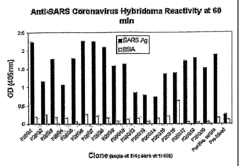
Figure 1: ELISA results of monoclonal antibody on whole inactivated SARS
virus and BSA. Hybridoma supernatants were tested at a 114 dilution in
PBS,0.2%
BSA on pre-blocked plates, coated with 18 ng per well of inactivated virus.
Positive
clones were identified as having positive binding (color) in wells which were
at least 4
¨ fold higher than the background level reactivity on BSA. Antigen Legend:
Black bars
- native, purified SARS-HCov ; White bars - BSA (bovine serum albumin).
Figure 2: lmmunofluorescence staining of SARS HCoV-infected Vero cells with
neutralizing and non-neutralizing SARS mAbs.; A. F26G6, non-neutralizing mab
specific for the spike protein. B. F26G3, neutralizing mAb. C. F26G7,
neutralizing
mAb. D. F26G9, neutralizing mAb. E. Irrelevant mAb, F25G1. F. Irrelevant mAb
F25G1 in bright field.
Figure 3. Immunohistochemical analysis of binding of mAb F26G6 to (A) SARS
infected but not (6) uninfected VERO cells.
Figure 4. Western immunoblot of monoclonal antibody on whole inactivated
SARS virus and infected vero cell lysates. The positive and preimmune control
sera
were from the corresponding immune mouse and tested at 1/2000 dilution in IBS,
0.2% BSA. Lanes marked 1 were loaded with purified virus ; 2, with infected
Vero cell
lysate.
Figure 5. This figure depicts Competion ELISA performed with F26G6
(antiOSpike)
and F26G15 (anti-NP) mAbs on whole purified SARS virus as antigen. A dilution
of
each mAbs was shoosen that would produce approximately 50% maximum OD
readin. Human normal and convalescent SARS-infected sera was diluted as shown
at
the bottom of the graph and used as a competitor for binding to the SARS
antigen. A
goat antimurine secondary antibody conjugated to HRP (preabsorbed with
multiple
species including human to remove any potantial crossreactivity) was used to
detect
murine mAb binding. Abrogation or reduction of the signal indicates the
presence of
human antibody to the same antigen/epitope in the human human serum. This
indicates that the individual was exposed or
CA 02548942 2006-06-05
WO 2005/054469 PCT/CA2004/002084
infected to the SARS corona virus. Our data also indicates that NP reactivity
may
be an earlier predictor of SARS infection as some sera inhibit NP bing mAb
F26G15 but do not inhibit spike specific mAb F26G6. NHS=Normal human Sera
tested at highest concentration 1/25; "S" are SARS patient convalescent sera.
Figure 6. Sequence data showing clones are unique and the id of the CDR
regions that play a role in neutralization (see PDF files for improved
resolution)
The data shows that none of the VH or VL genes of the anti-SARS neutralizing
or
Western immunoblot positive mAbs are the same. This means that each
hybridoma was derived from a uniques B celll and target SARS using different
proteins. (ie not the same clone picked several times)
Figure 7. SARS-specific monoclonal antibodies, Heavy chains (VH) amino
acid sequences.
Figure 8. SARS-specific monoclonal antibodies, Lignt chains (VL) amino
acid sequences.
Figure 9. SARS-specific monoclonal antibodies, Heavy chains (VH)
nucleotide sequences.
Figure 10. SARS-specific monoclonal antibodies, Lignt chains (VL)
nucleotide sequences.
Figure 11. Distribution of SARS CoV in spleen, liver and lung from mice
infected via IP (A), IN (B) and OR (C). Organs were collected on days 1, 3, 5
and 7
p.i. and viral RNA was detected by nested RT-PCR using a primer set against
the
polymerase. Number of animals that were positive by RT-PCR is shown as a
percentage (n=2).
Figure 12. Number of mice that were RT-PCR positive in the spleen, lung
and brain following IN inoculation of SARS CoV (A) or IP injection of
neutralizing
antibodies followed by IN inoculation of SARS CoV 2 hours later (B). N=5 per
group and time point. Blood, liver and kidney have been omitted due to non-
existent or insignificant levels of viral RNA.
Figure 13. Titres for lung samples collected from SARS CoV infected mice
(IN and IN+AB groups) at various time points p.i., determined by TCID50.
Values
are expressed in TCID50/gram of tissue.
DESCRIPTION OF THE PREFERRED EMBODIMENTS
6
CA 02548942 2006-06-05
WO 2005/054469 PCT/CA2004/002084
Unless defined otherwise, all technical and scientific terms used
herein have the same meaning as commonly understood by one of ordinary skill
in
the art to which the invention belongs. Although any methods and materials
similar
or equivalent to those described herein can be used in the practice or testi
ng of the
present invention, the preferred methods and materials are now described. All
publications mentioned hereunder are incorporated herein by reference.
DEFINITIONS
As used herein, "neutralizing antibody" refers to an antibody, for
example, a monoclonal antibody, capable of disrupting a formed viral particle
or
As used herein, "diagnostic antibody" or "detection antibody" or
"detecting antibody" refers to an antibody, for example, a monoclonal
antibody,
capable of detecting the presence of an antigenic target within a sample. As
will be
appreciated by one of skill in the art, such diagnostic antibodies preferably
have
high specificity for their antigenic target.
As used herein, "humanized antibodies" refer to antibodies with
reduced immunogenicity in humans.
As used herein, "chimeric antibodies" refer to antibodies with reduced
immunogenicity in humans built by genetically linking a non-human Variable
region
to human constant domains.
Described herein is the isolation, identification and characterization of
a plurality of anti-SARS monoclonal antibodies.
As discussed herein, some of the monoclonal antibodies have been
shown to have SARS neutralizing activity, meaning that said monoclonal
antibodies, humanized or chimeric versions thereof or immunoreactive fragments
thereof could be used as therapeutics for treating, preventing or ameliorating
=
symptoms associated with SARS infection in patients in need of such treatment.
Also described herein are methods of producing anti-SARS mAbs,
for example, humanized or chimeric anti-SARS mAbs. It is of note that these
mAbs
may be produced in a variety of systems; for example, germline cells or
transgenic
7
CA 02548942 2006-06-05
WO 2005/054469
PCT/CA2004/002084
plants. In these embodiments, an expression vector comprising a nucleic acid
encoding an anti-SARS mAb or a humanized or chimeric version thereof or an
immunoreactive fragment thereof is transformed into a suitable host and the
host is
grown under conditions promoting expression of the mAb which is then
recovered.
The mAbs may then be purified using means known in the art and used to develop
pharmaceuticals, as discussed below.
As described herein, some of the monoclonal antibodies are useful
for detection of SARS virus within biological samples for example, but by no
means limited to, infected cells, directly on viral particle in infected cell
lysates, in
purified virus fractions, serum, whole blood, naso-pharengeal swabs, stool, or
bronchio-alveolar lavage. As will be appreciated by one of skill in the art,
individual
detection monoclonal antibodies or combinations thereof may be packaged in a
kit
along with instructions for use, as described below.
The SARS detection monoclonal antibodies may be selected from
the group consisting of: F26G1, F26G2, F26G4, F26G5, F26G6, F26G8, F26G12,
F26G13, F26G14, F26G16, F26G17, F26G3, F26G7, F26G9, F26010, 026018
and F26G19.
The SARS neutralizing monoclonal antibodies may be selected from
the group consisting of F26G3, F26G7, F26G9, F26G10, F26018 and F26G19.
DNA and amino acid sequences for the above-referenced
monoclonal antibodies may be found in Figures 7-10 and also in the attached
sequence listing, wherein amino acid sequences of: F26G3-VH is SEQ ID No. 1;
F26G7-VH is SEQ ID No. 2; F26G9-VH is SEQ ID No. 3; F26G10-VH is SEQ ID
No. 4; F26G18-VH is SEQ ID No. 5; F26G19-VH is SEQ ID No. 6; F2601-VH is
SEQ ID No. 7; F26G6-VH is SEQ ID No. 8; F26G8-VH is SEQ ID No. 9; F26G3-VL
is SEQ ID No. 10; F26G7-VL is SEQ ID No. 11; F2609-VL is SEQ ID No. 12;
F26G10-VL is SEQ ID No. 13; F26G18-VL is SEQ ID No. 14; F26G19-VL is SEQ
ID No. 15; F26G1-VL is SEQ ID No. 16; F26G6-VL is SEQ ID No. 17; and F26G8-
VL is SEQ ID No. 18; and wherein DNA sequences of: F26G3-VH is SEQ ID No.
19; F26G7-VH is SEQ ID No. 20; F26G9-VH is SEQ ID No. 21; F26G10-VH is
SEQ ID No. 22; F26G18-VH is SEQ ID No. 23; F26G19-VH is SEQ ID No. 24;
F26G1-VH is SEQ ID No. 25; F26G6-VH is SEQ ID No. 26; F2608-VH is SEQ ID
No. 27; F26G3-VL is SEQ ID No. 28; F26G7-VL is SEQ ID No. 29; F26G9-VL is
8
CA 02548942 2006-06-05
WO 2005/054469
PCT/CA2004/002084
SEQ ID No. 30; F26G10-VL is SEQ ID No. 31; F26G18-VL is SEQ ID No, 32;
F26G19-VL is SEQ ID No. 33; F2601-VL is SEQ ID No. 34; F26G6-VL is SEQ ID
No. 35; and F26G8-VL is SEQ ID No. 36.
As will be appreciated by one of skill in the art, the monoclonal
antibodies may be used individually or in any combination thereof.
As will be appreciated by one of skill in the art, detection antibodies
must show high specificity and avidity for their antigenic target. As such,
showing
that a monoclonal antibody reacts with the antigenic target derived from a
highly
purified or in vitro prepared sample does not guarantee that the antibody has
sufficient specificity for use with biological sample. That is, the monoclonal
antibody must have sufficient specificity that it will not produce false
positives or
react with antigens from related, non-SARS coronaviridae.
Examples of suitable tests for determining utility as a diagnostic or as
a neutralizing mAb include but are by no means limited to negative
neutralization
and/or negative detection of a non-SARS coronavirus, C-ELISA data showing
competition of binding with the mouse mAbs that is being detected thereby
showing that the mAbs can be used to show that an immune response to SARS
has occurred in patient/animal sera, meaning that they were exposed/infected
(abrogation of binding by human antibodies). Alternatively, biological
material such
as blood, mucus or stool with could be spiked or enriched with the virus and
the
monoclonal antibodies used to detect added virus in the sample, which would in
turn determine limits of detection as well as other parameters of the
monoclonal
antibodies. Biological samples from experimentally infected animals could also
be
used to determine the utility of the mAbs at different stages of the infection
cycle.
Yet another method, although less desirable, would be testing of the patient
material from the outbreak as this is scarce and hence valuable material.
In use, at least one of the detection antibodies is mixed with a
biological sample under suitable conditions to promote binding of the at least
one
detection antibody with the antigenic target if the antigenic target is
present in the
biological sample. Binding of the detection antibody to an antigenic target
within
the sample is then detected using means known in the art, for example, by use
of
a labelled secondary antibody or other means discussed herein and/or known in
9
CA 02548942 2006-06-05
WO 2005/054469
PCT/CA2004/002084
the art.
As will be apparent to one of skill in the art, a combination of
detection antibodies may be used. Furthermore, at least one of the detection
antibodies or combinations thereof may be packaged in a kit for detecting SARS
virus in biological samples. The kit may include instructions and additional
reagents, for example, secondary antibodies, buffers, detection reagents and
the
like. Antibodies of the kit could be used for example in a capture ELISA
wherein
one or more mAb is coated onto a surface to catch and present SARS antigen
from biological samples, then another prelabelled mAb is added to detect the
presence of the antigen; as a control for indirect ELISA wherein a surface is
coated
with SARS antigen and the presence of antibody binding to the antigen is
detected; for immunoflourescence; or for competition ELISA wherein SARS
antigen is coated on a surface, and the ability of human or other
infected/exposed
animal serum antibody to prevent binding of one or more of the mAbs to the
SARS
antigen is measured.
The neutralizing antibodies were previously shown to react with a
conformational epitope of the native virus which is abrogated upon
denaturation of
the virus. However, as will be appreciated by one of skill in the art, this
does not
guarantee that the neutralizing antibodies will be effective in either
preventing virus
formation or disrupting intact virus particles in vivo, that is, that the
neutralizing
antibodies will have therapeutic activity.
For example Maruyama et al demonstrated in vitro neutralization using
monoclonal antibodies to Ebola virus and Parren et al confirmed this
observation in
guinea pigs; however in non-human primates there was no protection afforded by
the monoclonal antibody. Furthermore, Jones et al. conducted extensive studies
to identify which monoclonal antibodies were protective against infection with
the
bacteria Burkholderia pseudomalei. Whilst the in vitro neutralization is an
excellent
screening assay, the definitive test for neutralization is the in vivo
protection assay.
(Maruyama et al., J Virol. 1999; 73(7):6024-30; Parren et al., J Virol. 2002;
76(12):6408-12; Jones et al., J Med Microbiol. 2002,51(12):1055-62).
It has also been shown in HIV that in vitro neutralizing antibodies
may not protect against primary isolate in vivo (Poignard et al., J Virol.
2003
Jan;77(1):353-65). In addition, mAbs that recognize the same region (epitope)
but
CA 02548942 2006-06-05
WO 2005/054469
PCT/CA2004/002084
in different ways may have different neutralization properties, that is, one
may
neutralize while another may not, clearly indicating that neutralization is
entirely
empirical and needs to be tested. (Parren et al., J Virol. 1998
Dec;72(12):10270-
4).
In another embodiment of the invention, a nucleic acid sequence
encoding the neutralizing antibody as described above is subjected to
humanization techniques or converted into a chimeric human molecule for
generating a variant neutralizing antibody which has reduced immunogenicity in
humans. Humanization techniques are well known in the art ¨ see for example US
Patent 6,309,636 and US Patent 6,407,213. Chimerics are also well known, see
for example US Patent 6,461,824, US Patent 6,204,023, US Patent 6,020,153 and
US Patent 6,120,767.
In one embodiment of the invention, chimeric antibodies are
prepared by preparing an expression vector which comprises a nucleic acid
encoding a constant region domain of a human light or heavy chain genetically
linked to a nucleic acid encoding a light chain variable region selected from
the
group consisting of G1-light (SEQ ID Nlo.1); G3-light (SEQ ID No.2); G6-light
(SEQ
ID No.3); 07-light (SEQ ID No.4); 08-light (SEQ ID No.5); 010-light (SEQ ID
No.6); 015-light (SEQ ID No.7) and G18-light(SEQ ID No.8) or a heavy chain
variable region selected from the group consisting of 01-heavy (SEQ ID No.9);
03-heavy (SEQ ID No.10); 06-heavy (SEQ ID No.11); 015-heavy (SEQ ID No.12)
and 018-heavy (SEQ ID No.13). It is of note that all of these sequences are
shown
in Figures 7-10.
In another embodiment of the invention, there are provided
recombinant anti-SARS antibodies comprising at least one modified variable
region, said region selected from the group consisting of 01-light (SEQ ID
No.1);
G3-light (SEQ ID No.2); 06-light (SEQ ID No.3); 07-light (SEQ ID No.4); 08-
light
(SEQ ID No.5); 010-light (SEQ ID No.6); 015-light (SEQ ID No.7); 018-light(SEQ
ID No.8); 01-heavy (SEQ ID No.9); 03-heavy (SEQ ID No.10); 06-heavy (SEQ ID
No.11); 015-heavy (SEQ ID No.12) and 018-heavy (SEQ ID No.13) in which at
least one but fewer than about 30 of the amino acid residues of said variable
region has been changed or deleted without disrupting antigen binding. It is
of note
that all of these sequences are shown in Figures 7-10.
11
CA 02548942 2006-06-05
WO 2005/054469
PCT/CA2004/002084
= In yet other embodiments, immunoreactive fragments of any of the
above-described monoclonal antibodies, chimeric antibodies or humanized
antibodies are prepared using means known in the art, for example, by
preparing
nested deletions using enzymatic degradation or convenient restriction
enzymes.
It is of note that in all embodiments describing preparation of
humanized antibodies, chimeric antibodies or irnmunoreactive fragments of
monoclonal antibodies, these antibodies are screened to ensure that antigen
binding has not been disrupted. This may be accomplished by any of a variety
of
means known in the art, but one convenient method would involve use of a phage
display library.
The nucleotide sequence encoding the variable regions of the light
and heavy chains of antigen specific hybridomas represent the specificity of
the
anitbody. Specifically the most important regions are the CDRs (of the light
and
heavy chains): L1, L2, L3 and H1 H2 H3 respectively. It will be apparent to
one of
skill in the art that the most importance CDR domains are those that are most
variable in nature and thus are recruited most specifically by a given antigen
like
SARS. These are Ll and H3. Residues in H3 and other CDR comprise the
paratope which interacts with the epitope on the pathogen. Amino acid residues
in
H3 have have been shown to directly interact/bind to residues of the epitope
in
crystal structure determinations. (Bossart-Whitaker et al., J Mol Biol. 1995
Nov
3;253(4):559-75; Chavali et al.,.Structure (Camb). 2003 Jul;11(7):875-85;
Afonin
et al., Protein Sci. 2001 Aug;10(8):1514-21; Karpusas et al., J Mol Biol. 2003
Apr
11;327(5):1031-41; Krykbaev et al., J Biol Chem. 2001 Mar 16;276(11):8149-58.
Epub 2000 Nov 01; Beiboer et al., J Mol Biol. 2000 Feb 25;296(3):833-49;
Haruyama et al., Biol Pharm Bull. 2002 Dec;25(12):1537-45).
It is of note that as discussed herein, the above-described
neutralizing antibody or humanized variant thereof may be formulated into a
pharmaceutical treatment for providing passive immunity for individuals
suspected
of or at risk of SARS infection comprising a therapeutically effective amount
of said
antibody. The pharmaceutical preparation may include a suitable excipient or
carrier. See, for example, Remington: The Science and Practice of Pharmacy,
1995, Gennaro ed. As will be apparent to one knowledgeable in the art, the
total
dosage will vary according to the weight, health and circumstances of the
12
CA 02548942 2006-06-05
WO 2005/054469
PCT/CA2004/002084
individual as well as the efficacy of the antibody.
In another embodiment of the invention, a vaccine is prepared by
recovering from a preparation of live, attenuated or recombinant SARS virus,
antigens recognized by one or more monoclonal antibodies selected from the
group consisting of F26G1, F26G2, F26G4, F26G5, F26G6, F26G8, F26G12,
F26G13, F26G14, F26G16, F26G17, F26G3, F26G7, F26G9, F26G10, G26G18
and F26G19.
The invention will now be described according to examples; however, the
invention is not limited to or by the examples.
Immunization and Virus Antigen Preparation:
For immunizations 5-6 week old female BALB/C mice were used (Charles
River). The mice were injected subcutaneously (S.C.) with 50-ug of beta-
propiolactone-inactivated SARS-coronavirus (Tor-3 strain) with an equal part
of
Complete Freund's Adjuvant [CFA, H37 Ra; Difco]), on day 1. The virus had been
expanded after plaque purification in Vero-6 cell monolayers and partially
purified
through a sucrose cushion. Highly purified SARS-coronavirus (101-3) was
prepared the same as above except that the viral particles were further
purified
using gradient centrifugation. (Highly purified SARS CoV was prepared as
follows
briefly, 500 ml of supernatant from SARS CoV infected Vero-6 cells was
concentrated first on top of a cushion of iodixanol in a SW32 rotor (Beckman).
Subsequently, the virus was mixed to form a suspension of 20% iodixanol and
centrifuged in a NVT 90 rotor (Beckman) for 3.5 hours at 400,000g. Fractions
were
collected from the bottom of the self-generated gradient, tested by Western
immunoblot with convalescent patient serum, and the SARS CoV positive
fractions
were pooled and dialysed against PBS. The dialysed virus preparation was
further
concentrated by ultracentifugation for 1.5 hours at 150,000g. On day 30 the
mice
received 50 pg of purified SARS virus antigen S.C. in Incomplete Freund's
Adjuvant (IFA) in a total volume of 100 pl. On days 48 and 63, the mice
received 5
pg of the same antigen in a total volume of 100 pl S.C. with IFA. Mice
received a
final booster injection with 5 pg of purified SARS urus in 200 pl PBS to the
intra-
peritoneal cavity 3 days prior to hybridoma fusion. Mice were euthanised by
anaesthesia overdose and exsanguinated by cardiac puncture. The spleens were
subsequently excised under aseptic conditions.
13
CA 02548942 2006-06-05
WO 2005/054469
PCT/CA2004/002084
Preparation of Infected Cell lysate
Infected Vero cells were scraped off of 162 cm2 tissue culture flasks
(Corning) and centrifuged for clarification. A borate saline mixture (0.05 M
boric
acid, 0.12 M, NaCI, 0.024 M NaOH) was used to wash the cell pellet twice and
the
pellet was resuspended in 2 ml borate saline + 1 % triton x-100 for each T162
flask. The pellet was kept at 4 C using a water bath and sonicated for ten
minutes
at 50% power. The debris was pelleted via centrifugation at 10,000 X g for ten
minutes and the supernatant collected and stored at -20 C in aliquots for
later use.
Generation of mAbs:
Immunization of mice, removal of spleens, preparation of spleen and
myeloma cells, and the fusion for hybridoma production were performed
according
to NCFAD standard operating procedures under IS017025. Ampules of the
myeloma cell line P3X63Ag8.653 (ATCC) were thawed one week prior to fusion
and grown in BD Cell Mab Quantum yield media in the presence of 8-Azaguanine
(Sigma). Cells were in log-phase growth at the time of fusion. Hybridoma
fusion
was performed essentially as originally described (Kohler and Milstein, 1975,
Nature 256: 495-497) with the following modifications. Briefly, spleens were
harvested 3 days after a final boost and the splenocytes were prepared by
splenic
perfusion as follows. A 10 cc syringe with a 21 gauge sterile disposable
needle
was used to perforate the spleens under aseptic conditions. The spleen cells
were
perfused out of the spleen with injections of serum free BD cell Mab Quantum
Yield medium (BD-Pharmingen). Two identically immunized mouse spleens were
used to produce these hybridoma clones. The fusion was performed using the
P3X63Ag8.653 myeloma line in log phase growth. The PEG1500, 1 ml, (Roche)
was added drop-wise over one minute while gently tapping the tube containing
the
thoroughly washed myeloma-splenocyte pellet. The PEG was slowly diluted out
over three minutes with serum free BD-Cell Mab Quantum Yield media (BD-
Pharmingen). The cells were resuspended and mixed into 90 ml of Clonacell
Medium D (HAT) media (Stemcell, Vancouver) containing 5 ml HCF, and plated
out according to the manufacturers instructions. The plates were kept in a 37
C
incubator under 5% CO2 overlay for about 10-18 days in humidified chambers.
14
CA 02548942 2006-06-05
WO 2005/054469
PCT/CA2004/002084
Visible colonies were picked from the plates after about 2 weeks growth and
placed into 96 well plates containing 150-200 pl of complete hybridoma medium
(BD-Quantum Yield) with 1 X HT (Sigma), 4% Hybridoma cloning factor (Igen) and
10% FBS (Wisent). Supernatants were screened 4 days later via ELISA on
purified
virus as antigen. Isotyping was performed using a commercial dipstick test
(Roche)
according to the manufacturer's instructions. Hybridoma culture supernatants
were
concentrated 5-10 fold using stirred cell nitrogen concentrators (Amicon) with
a 30
kilodalton cutoff membrane (Millipore).
Immunoassays
Enzyme linked immunosorbent assay
Tissue culture supernatants were assayed for binding to purified SARS
coronavirus in an ELISA assay when the cultured cells were confluent in the
culture plates. The Costar 3690 96-well 1/2 well ELISA plates (Corning) were
coated with either Bovine serum albumin or purified SARS-coronavirus (18 - 37
ng/well) in PBS overnight at 4 C and then blocked with 0.4% BSA in PBS, for 2
hours at 37 C. The supernatant (30 p1/well) was incubated neat for 1 hour at
37 C.
The ELISA plates were washed ten times with dH20 and patted dry on a paper
towel. A pan-goat anti-mouse IgG-HRP antibody (Southern Biotechnology
Associates) was diluted to 1:2000 in 0.2% BSA in PBS, applied to the ELISA
plates for 45 minutes at 37 C, and then washed as described above. Positive
binding was detected with commercial ABTS used according to the manufacturers
instructions (Roche). The OD was read at 405nm at 15 and 60 minute intervals
after addition of the developing reagent. Mouse immune and preimmune sera was
diluted to 1:2000 in 1.5 ml EppendorfTM tubes (Falcon) in 2%-BSA PBS for use
as
controls.
Western lmmunoblots
Whole virions or SARS-infected Vero cells at a total protein concentration of
1 ug per lane were loaded in criterion pre-cast gels (BIO-RAD) and
electrophoresed at 200 V for 30 minutes. The proteins were transferred to
Immobilon nylon membranes (Millipore) for 2 hours at room temperature at 100
volts, or at 27 volts overnight at 4 C. Blots were blocked in 3% BSA-TBS,
rinsed
15 =
CA 02548942 2006-06-05
WO 2005/054469
PCT/CA2004/002084
three times with TBS, and reacted with monoclonal antibody overnight at 4 C.
The
antibody supernatants were reacted neat and concentrated supernatants were
diluted 1:50 in 0.2% BSA-PBS. Blots were washed three times with TBS-tween-20
(0.05%) for five minutes before being incubated with secondary antibody (same
as
above) at 1:1000 in TBS, 0.2% BSA for 1 hour. The blots were washed as above
and developed using DAB (Pierce) insoluble substrate.
lmmunofluorescence Staining of Vero cells infected with SARS-corona virus
Monolayers of SARS-infected Vero cells were stained as follows. Glass
slides were coated with infected Vero cell monolayers and fixed with acetone.
The
slides were irradiated with 20 kilogreys from a cobalt gamma irradiator,
removed
from biocontainment, and then stored at -80 C. Dilutions of antibodies and
test
sera were made initially in 96 well plates (Falcon). Samples were allowed to
incubate for 45 minutes in a 37 C incubator, and were washed with distilled
water.
Fluorescein labelled secondary antibodies (Sigma) diluted in PBS were added to
the slides and incubated for 45 minutes at 37 C, washed as above, and air
dried.
Slides were coated with mounting medium and stored at 4 C until examined.
Virus Neutralization
Plaque reduction virus neutralization assay (NML)
A standard plaque reduction neutralization test was performed as previously
described (Godet et al., 1994, J. Virol. 68: 8008-8016). Briefly, mixtures of
pre-
titred (100 PFUs) SARS coronavirus and serial 2-fold dilutions of hybridoma
supernatant were incubated at 37 C for 1 hr and added to six well plates
containing Vero cell monolayers. After a 37 C incubation for 1 hr, a nutrient-
agar
Overlay was added and the plates placed in a CO2 incubator for approximately 3
days. A second overlay was then added which contained neutral red as a vital
stain. Plates were then checked periodically over the next few days for plaque
formation. The highest dilution tested that produced a plaque reduction of at
least
90% was definedas the titration end point.
Cytopathic effect (CPE) reduction virus neutralization assay(NCFAD)
The ELISA positive monoclonal antibodies were screened for cross-
16
CA 02548942 2006-06-05
WO 2005/054469
PCT/CA2004/002084
neutralization with other coronaviruses using microtiter format CPE reduction
assay: concentrated monoclonal antibodies (hybridoma supernatants) were
diluted
1:20 in cell culture medium and incubated with 100 TCID50 of either SARS HCoV
(Tor-3), or transmissible gastroenteritis virus (TGEV, Diamond strain; kindly
provided by Dr. Susy Carman, LSD, University of Guelph) for 1 hr at 37 C. The
virus-antibody mix was then transferred onto cell monolayers in 96-well plates
(Costar, Corning, NY). Vero V-76 cells were used for the SARS WCoV, ST cells
for
the TGEV. The plates were incubated until CPE developed in virus back
titration
controls.
Development of mAbs to the SARS-virus
We developed a panel of mAbs to the SARS HCoV. ELISA screening on
purified SARS coronavirus identified a panel of 17 IgG/K type mAbs (Figure 1
a,
table 1). The general binding reactivity of these mAbs is decreased on heat
denatured purified virus preparations indicating destruction of epitopes.
There is a
similar decrease in binding by many of these mAbs when tested on SARS-HCoV
infected vero cell lysates as antigen. Heat denaturation had little effect on
the
binding of mAb F26G16 which also maintains a high OD on infected lysates. This
mAb does however show higher background on the irrelavant antigen bovine
serum albumin (BSA) (figure 1 a) and has inconsistent reactivity in
immunoblots
with heat denatured viral lysate (table 1). Immunoblot methods are less
sensitive
than ELISA especially when using the lower quality infected cell lysate as
antigen.
Unfortunately preparation of highly purified viral antigen requires enormous
efforts
under containment which emphasizes the need for a quality recombinant antigen
assay.
Western immunoblot analysis identified mAbs to the SARS spike protein. A
total of five mAbs react with the SARS-spike protein in Western innmunoblots,
using the whole purified virus or virus infected cell lysate (Figure 1b). The
antigen
identity of the remaining 11 Western immunoblot negative mAbs could not be
determined which suggests that these mAbs target conformational epitopes that
are destroyed in the Western blot sample preparation and membrane transfer
process. These data led us to test for biological activity in virus
neutralization
assays.
17
CA 02548942 2006-06-05
WO 2005/054469
PCT/CA2004/002084
lmmunochemical and Biological Characterization of binding
Neutralizing antibodies to the SARS virus recognize epitopes via interaction
with both conformational and linear epitopes. We identified mAbs that
neutralize in
vitro cell culture infectivity of the SARS-virus. Concentrated culture
supernatants
from four of the eleven Western immunoblot negative (conformational) mAbs were
significantly neutralizing compared to irrelevant isotype-matched concentrated
mAbs to other antigens (Table 1). SARS virus infectivity was neutralized with
mAbs F26G3, G7, G9, G10, G18 and G19. No cross-neutralization was observed
for the animal coronavirus TGEV. The remaining mAbs in our panel showed no
decrease in virus growth. This result reveals that we have developed mAbs
specific for epitopes on the SARS coronavirus.
lmmunoblot analysis reveals a spectrum of conformational requirements for
binding. We examined the effects of different denaturing treatments on binding
activity of a subset of neutralizing and some non-neutralizing mAbs using
immunodot blot assays on infected lysates compared to uninfected lysates. A
series of conditions were tested including exposure to heat, detergent, a
reducing
agent, and combinations thereof. The Immunodotblot reactivities of this panel
of
mAbs reveals important immunochemical requirements for their respective
epitopes, and are summarized in table 1. In general the conformational
requirements of the neutralizing antibodies are higher than the non-
neutralizing
and they are less tolerant of denaturation of the epitopes. None of the mAbs
react
with mock-infected lysates as assayed in lmmunodotblots. This suggests that
the
majority of the neutralizing mAbs likely target surface exposed protein
epitopes on
the native viral particle, which has been identified as spike protein via
Western
analysis for mAbs F26G18 and F26G19. ,This is consistent with binding data
observed in ELISA on heat denatured virus infected lysate compared to native
infected lysate. In this case, regardless of Western reactivity, the non-
neutralizing
clones retain more ability to bind to heat denatured antigens compared to
neutralizing mAbs (lower mean percent reduction in OD per group p<0.001,
students T test). There are exceptions, however, in that it is difficult to
use
traditional classifications to describe the binding properties of these mAbs
as being.
conformational or linear according to biological activity. Interestingly,
clone
=
18 '
CA 02548942 2006-06-05
WO 2005/054469
PCT/CA2004/002084
F26G18 binds to spike protein in Western blot and neutralizes the SARS virus
and
thus the binding of F26G6 cannot be termed strictly conformational in nature.
This
is in contrast to neutralizing mAbs produced against other enveloped viruses
(Zwick et al., 2001, J. Virol. 75: 6692-6699; Wilson et al., 2000, Science
287: 1664-
1666) that require the antigen to have native conformation for binding. It
will be
, important to verify, under optimized conditions (Opstelten et al.,
1995, J. Cell Biol.
131: 339-349) the use of viral lysates designed for maximal recovery of
coronavirus proteins and to this end the production of high quality
recombinant
protein antigens will provide useful insights.
SARS-virus reactivity was confirmed for the four Western immunoblot
negative, virus neutralizing mAbs (F26G3, G7, G9, G10) using an
immunofluorescence assay. In order to independently confirm recognition of
native
SARS antigens we tested these mAbs via immunofluorescence relative to a non-
neutralizing mAb F26G6, which we know recognizes Spike protein in
immunohistochemical staining of infected Vero cells. The neutralizing mAbs
F26G3, G7, G9, and G10 specifically recognize SARS-HCoV infected but not
uninfected Vero cells in immunofluorescence (Fig. 2). Irrelevant, isotype
matched
mAbs, produced in an identical fashion, do not react with SARS-virus infected
Vero
cells. These data are consistent with the appearance of coronavirus antigens
on
the surface of the infected cell during replication (Talbot et al., 1984,
Virology 132:
250-260) although the fixation process may allow for reactivity of these mAbs
with
internal antigens as well. Collectively, these data demonstrate that these
mAbs will
be useful for developing antigen detection systems for diagnostics.
Conclusions
Linear epitopes on the spike protein and conformational epitopes on as of
yet unknown antigen(s) provide neutralizing targets on the SARS virus. These
data
clearly show that the spike protein is a putative protective antigen, as it is
the
target of neutralizing mAbs F26G18 and G19. Moreover, these mAbs could be
used to identify protective epitopes for vaccine formulations (Enjuanes et
al., 1995,
Dev. Biol. Stand. 84: 145-152). Studies are underway to determine the identity
of
the additional unknown antigen(s) recognized by the other neutralizing mAbs
with
more conformational epitopes. Molecular studies have revealed that the RT PCR
19
CA 02548942 2006-06-05
WO 2005/054469
PCT/CA2004/002084
amplified V-genes of the hybridoma clones that express these neutralizing mAbs
contain distinct sequences. Therefore, the hybridomas expressing the
neutralizing
mAbs were derived from independently rearranged and clonally selected B cells
in
vivo, and are not derived from the same clone. This is the first description
of
SARS-HCoV specific and neutralizing mAbs and these antibodies should prove
useful for the development of new diagnostic tests, studies on antigenic
variation,
and vaccine development in the global fight against SARS, as discussed above.
Virus, cells and monoclonal antibodies
Vero E6 (African Green Monkey kidney) cells were cultured in
Dulbecco's modified Eagle's medium (DMEM, Sigma) with 10% heat inactivated
fetal bovine serum (FBS, Gibco BRL), 1% penicillin/streptomycin and 1% L-
glutamine. Cells were incubated in the presence of 5% CO2 at 37 C.
The Tor3 strain of SARS CoV was isolated at the National
Microbiology Laboratory from a patient infected during the initial SARS
outbreak in
Toronto 2003 (Weingartl et at., 2004, Emerg Infect Dis 10: 179-184). The virus
stock had been expanded after plaque purification in Vero E6 cell monolayers
and
partially purified through a sucrose cushion (5 x 10⁶ pfu/rnI). Preparation of
the
infectious SARS CoV was performed under BSL-3 containment conditions. All
animal experiments and processing of infected tissues were conducted under
BSL4 containment conditions. Monoclonal antibodies were generated from mice
immunized with inactivated SARS CoV Tor3 strain.
Animal Studies
Female BALB/c mice 6 to 8 weeks old were obtained from Charles River
(Quebec, Canada). In the first mouse study, BALB/c mice were infected with the
Tor3 strain of SARS CoV by one of three routes: intraperitoneal (IP),
intranasal
instillation (IN) or oral gavage (OR).
IN, IP and OR groups received 20 pl, 200 pl and 100 pl of diluted virus
(containing 5x10⁴ plaque forming units (PFU)) respectively, all animals
received
the same number of PFUs. At one hour and 1, 3, 5, 7 and 9 days post infection
(p.i.), mice were anaesthetized with halothane and sacrificed by cardiac
puncture.
Blood, spleen, liver, kidney and lungs were harvested. Organs were immediately
homogenized in DMEM immediately and an aliquot was removed for RNA
CA 02548942 2006-06-05
WO 2005/054469
PCT/CA2004/002084
extraction. Remaining homogenates were stored at ¨80 C for virus isolation.
In a follow-up study, two groups of female BALB/c mice (6 to 8 weeks old,
approximately 20 g in weight) were injected IP, a single time, with a cocktail
of 4
neutralizing antibodies (Berry et al., 2004, J Virol Methods 120: 87-96). We
administered 10 pg of each antibody to the mice; the final dose of antibody
was
therefore 40 pg/mouse. Two hours following antibody treatment, animals were
anaesthetized with halothane and were inoculated IN with 5x10⁵ PFU of the Tor3
strain in 100 pl. At 1, 2, 3,4, 5,6, 7 and 14 days following infection, mice
from the
antibody treated group (IN + AB) and untreated group (IN) were weighed then
anaesthetized with halothane and sacrificed by cardiac puncture. Blood,
spleen,
liver, kidney, lung and brain were harvested. Organs were weighed then
homogenized in to ml of DMEM, aliquots were transferred to AVL RNA extraction
buffer (Qiagen) and stored at ¨20 C. The remainder of each homogenate was
stored at ¨80 C for virus isolation. All animal experiments were performed
under
an approved animal use document and according to the guidelines of the
Canadian Council on Animal Care.
RNA Extraction
RNA from the first animal experiment was extracted using the Trizol LS
protocol (Invitrogen). RNA from the second animal study was extracted from
tissue homogenates using Qiagen viral RNA Minikit (Qiagen). Homogenate was
transferred to AVL extraction buffer and RNA was extracted following the
Qiagen
protocol.
Nested RT-PCR and Real-time RT-PCR
For the first mouse study, nested RT-PCR was performed using a primer
set targeting the polymerase gene (L). RT-PCR was performed using a one-step
RT-PCR kit (Qiagen) and primers CorV Forward1 and CorV 389 Reverse1 (Table
3) in a Bionnetra thermocycler. Nested PCR was done in a Biometra thermocycler
using Taq DNA polymerase (Invitrogen) and primers
CorV 154 Forward2 and CorV 310 Reverse2 (Table 3) with 4% of the
amplicons obtained from the first round reaction. All amplicons from first and
second round amplifications were verified for size. All positive amplicons
from the
nested round were sequenced using an ABI 3100 Genetic Analyzer.
21
CA 02548942 2006-06-05
WO 2005/054469
PCT/CA2004/002084
For the second mouse study, it was necessary to use real-time RT-PCR
due to the large number of samples collected. RT-PCR master mixes were made
using the Taqman one-step RT-PCR mastermix (Applied Biosystems) and primers
targeting the nucleoprotein gene (Table 3) in an applied biosystems 7700
thermocycler.
Virus Isolation
Virus isolation was performed on selected tissue homogenates based on
PCR data. Frozen homogenates were thawed from ¨80 C and centrifuged at
10,000xg for 5 minutes. Following centrifugation, supernatant was collected
and
mixed with 500 !.LI of DMEM (no supplements), and filtered using 0.22 p,IVI
filter
(Millipore). Each supernatant was used to infect one 25 cm2 flask of Vero E6
cells
by incubation at 37 C for 1 hour with intermittent rocking. Five ml of DMEM
containing 2% FBS, 1% penicillin/streptomycin and 1% L-glutamine was added to
each flask. Cells were incubated at 37 C with 5% CO2 and cytopathic effect
(CPE)
was monitored up to day 10 p.i. If CPE was present, supernatant was removed
for
testing in nested RT-PCR, followed by sequencing of amplicons.
Determination of viral load in the lung by TCID50
Tissue samples that demonstrated CPE upon first passage were chosen for
TCID50 determination. Homogenized tissues in DMEM were filter sterilized using
a 0.22 pM filter (Millipore) and diluted 1:100 in DMEM. Ten-fold serial
dilutions
from 10-2 to 10-8 were prepared in DMEM and used to infect Vero E6 cells at 80-
90% confluency in 24-well plates. Media was removed from the cells and 250 pl
of
each dilution of virus was added to each of four wells. Virus was adsorbed to
cells
for 1 hour at 37 C, then 1 ml of DMEM with 2% FBS, 1% penicillin/streptomycin
and t..-glutamine was added per well. Infected cells were incubated at 37 C
with
5% CO2 and were monitored for CPE up to day 10 p.i. The dilution of virus that
caused cytopathic effect (CPE) in 50% of the well was calculated by Spearman
Karber method (Spearman, 1908, But J Psycho! 2: 227). Virus titres are
expressed as the 50% tissue culture infectious dose (TCID50) per gram of
tissue.
Results
SARS CoV replication in mice infected by different routes
In order to establish a small animal model for efficacy testing of antivirals,
vaccines and therapeutic antibodies, BALB/c mice were infected with the Tor3
22
CA 02548942 2006-06-05
WO 2005/054469
PCT/CA2004/002084
strain of SARS CoV, 5x10⁴ PFU, by one of three routes: intraperitoneal (IP),
intranasal (IN) or Oral (OR). Animals were observed closely for clinical signs
or
symptoms over a period of nine days (1st study) and 14 days (2nd study) post
virus challenge. Mice were serially sacrificed at different times p.i. and
blood and
organs were harvested for the detection of viral genomic RNA by RT-PCR and the
presence of infectious virus by TCID50. In general, mice did not show any
signs of
disease, particularly not of respiratory illness.
Intranasally infected animals
demonstrated aggressive behaviour on days 3 and 4 p.i., however, no change in
weight and grooming behaviour.
Independent of the route of infection, none of the animals were viremic at
any time p.i. but virus spread systemically as indicated by replication in
several
organs, particularly spleen, liver and lungs (Figure 11). Of the three
infection
routes, the IP route was most efficient in initiating systemic infection more
rapidly.
Since the IP route does not mimic human SARS CoV transmission, of the routes
that are biologically relevant for human transmission (IN and OR), IN
infection was
most successful with highest titres in spleen and lung. Despite the fact that
OR
infection did result in systemic infection, virus replication was only short
lived
compared to the IP and IN route. Viral RNA was not detected in any of the
groups
or tissues at day 9 p.i. indicating that the animals had cleared SARS CoV by
that
time. All RT-PCR positive amplicons were sequenced and confirmed to be SARS
CoV.
Spleen and lung tissue samples from the biologically relevant routes (IN and
oral) were selected for virus isolation to confirm the presence of viable
virus in
these tissues. Following infection of Vero E6 cells with tissue homogenates,
CPE
was observed on day 4. PCR amplification from RNA extracted from tissue
culture
=
supernatants followed by sequence determination confirmed the isolation of
SARS
CoV. Thus, we confirmed establishing a systemic infection with SARS CoV in
mice
by three different routes of inoculation. Infection by oral gavage is
interesting since
earlier reports suggest the possibility that SARS CoV can infect humans via
the
fecal/oral route (Tang et al., 2004, CMAJ 170: 47-54; Chan et al., Emerg
Infect Dis
10: 825-831).
Neutralizing antibodies reduce virus titre
Having established a proper animal model with a relevant challenge route,
23
CA 02548942 2006-06-05
WO 2005/054469 PCT/CA2004/002084
we next tested the neutralizing activity of several monoclonal antibodies
raised
against SARS CoV (Berry et al., 2004). We chose to use a 10-fold higher virus
dose in a larger volume to infect the animals IN to assure a more reliable
lower
respiratory tract infection. Prior to IN infection of mice with SARS CoV (dose
5x10⁵
PFU), a cocktail of 4 neutralizing monoclonal antibodies (single dose) were
administered IP. Animals were followed up by clinical observation and were
sacrificed at different times post challenge. Tissue samples, collected post
Mortem
were tested for the presence of viral nucleic acid by real-time RT-PCR and
infectious virus by TCI D50.
In accordance with the previous experiment, none of the infected animals
demonstrated typical SARS illness. As demonstrated before, there was no
detectable viremia, however there was systemic spread of infection,
particularly to
the spleen (day 2-6) and the lungs (day 1-14) in the untreated control group
(Figure 12A). In comparison, the antibody treated group showed a dramatic
decrease in viral replication in the spleen and lungs from day 3 on (Figure
12B).
Viral replication was also observed in the brain on days 1 and 2 in the
untreated
group and only on day 1 in the antibody treated group.
To better define the neutralizing efficacy reduction in titre between
the antibody treated (IN + Ab) and untreated groups (IN), titres were
determined by
TCID50 on lung homogenates. Mice that received the cocktail of neutralizing
antibodies showed a two-log reduction in virus titre on day 1 and 3 p.i.
(Figure 13).
By day 4, the IN+AB group showed a reduction in titre by one-log in comparison
to
the IN-group. Furthermore, the viral load data was in concordance with the
viral
titre data and showed between one and three logs of decrease of viral RNA in
the
same samples.
Discussion
This study has demonstrated that SARS CoV established a systemic
infection in mice following three different routes of virus infection without
detectable
levels of viremia. This is in contrast to the results of Subbaro et al., who
recovered
virus only from the upper and lower respiratory tract following intranasal
infection
but not from the internal organs (Subbarai et al., 2004, J Virol 78: 3572-
3577). In
our study, the main target organs for viral replication were determined to be
spleen
and lung and, thus, are similar to those in humans (To and Lo, 2004, J Pathol
203:
24
CA 02548942 2006-06-05
WO 2005/054469 PCT/CA2004/002084
740-743; Wentworth et al., 2004, Emerg Infect Dis 10: 1293-1296). The virus
replicated in the respiratory tract and spread systemically infected mice
continued.
to gain weight and showed no signs of disease other than a marked increase is
aggressive behaviour in IN infected mice on days 3 and 4 post infection viral
RNA
was detected in the brains of infected mice on days 1 and 2 post infection
perhaps
indicating limited 'infection via the olfactory bulb followed by inflammation
on days 3
and 4 resulting in the observed aggression. We are confident that the mouse is
a
viable model for testing of antiviral, vaccines and immunotherapeutics as we
are
able to reliably induce systemic infection. However, as found by others groups
protection can only be assessed by measuring reduction in virus replication
(Subbarao et at., 2004), as mice are not a model for severe disease as none of
the
infected animals displayed typical SARS illness.
We attempted to determine if the SARS CoV could establish an infection in
mice following oral inoculation. This was done in response to published data
and
our own observations indicating that viral RNA could be detected in human
stool
samples for up to 35 days, far longer than in the nasal swabs (Chan et at.,
2004).
In addition, the outbreak in Amoy Gardens, Hong Kong, appeared to be
associated
with fecal transmission raising the possibility of a fecal/oral transmission
route for
human SARS CoV infection (Ng, 2003, Lancet 362: 570-572; Department of
Health, Hong Kong government. Outbreak of SARS at Amoy Gardens, available at
http://www.info.gov.hk/info/ap/pdf/amoy_e.pdf). In our hands, the virus was
clearly
capable of initiating a systemic infection following oral infection with virus
spread to
the lungs, liver and spleen of the orally infected mice. We determined that
infection via the intranasal (IN) route resulted in a!Tiore sustained and
widespread
respiratory and systemic infection than was observed following, either IP or
oral
infection and therefore, we chose to use this route for our subsequent work.
The current study demonstrated that IP administration of a single dose of a
cocktail of neutralizing monoclonal antibodies prior to mucosal challenge
reduced
virus replication by two-logs in the first critical days p.i. . The antibody
treated
group showed a complete abolishment of viral RNA in all three tissues (spleen,
liver, lung) 5 days after challenge while viral RNA was detected in the
untreated
group for up to 14 days in the lung. The ability of a single dose of
neutralizing
antibodies to inhibit virus replication in the lungs is promising since this
is the
CA 02548942 2006-06-05
WO 2005/054469 PCT/CA2004/002084
primary site of SARS replication and disease manifestation in humans. It is
likely
that consecutive treatments would enhance the efficacy particularly in humans
at
present his would be difficult to test experimentally as both NHP and mice are
capable of clearing infection with the SARS-CoV independently of treatment and
Previous studies have shown that infection as well as transfer of
hyperimmune serum protects mice from IN challenge with SARS CoV (Subbarao
et al., 2004). Although hyperimmune sera may work experimentally in mice,
there
are several problems associated with the use of polyclonal human sera in human
patients such as difficulty in finding immune donors and risks related to the
use of
human blood products (Traggiai et al., 2004, Nat Med 10: 871-875). Recently,
Traggiai and colleagues (Traggiai et al., 2004) demonstrated that human
monoclonal antibodies offer an alternative. Mice were given between 50 and
800p.g of human monoclonal antibodies IP and then challenged IN 2 days later
with SARS CoV (104 TCID50). Animals that received 2001_tg of the human
monoclonal antibodies were protected from viral replication in the lower
respiratory
tract, determined by TCID50 (Traggiai et al., 2004). However, RT-PCR detection
was not employed to determine the levels of viral genome present in the
tissues,
typically a much more sensitive approach. Furthermore, only one time point was
examined (2-days p.i.) and in our experience, even when using group sizes of 5
mice, it is possible that virus detection in the lung by RT-PCR or virus
titration is
negative at one time but positive later. We have shown that when administering
the antibody cocktail containing a total of 401.1g only 2 hours prior to
challenge we
can achieve a 2-log decrease in virus titre in the lung following infection
with a 50x
higher dose of SARS CoV (5x10⁵ PFU). It is likely that possible the dose of
antibodies, pre-treating earlier and/or multiple treatments to increase the
tissue
levels at the time of challenge will substantially improve the performance of
the
therapy. Furthermore, while a synergistic effect of these SARS-neutralizing
monoclonal antibodies has not yet been demonstrated, the use of a cocktail of
monoclonal antibodies should limit the potential deleterious effects of
antigenic
variation and escape from neutralization. Examples of synergistic effects of
26
CA 02548942 2012-09-27
- 27 -
monoclonal antibodies have been observed in the neutralization of HIV-1 in
vitro
(Zwick et al., 2001, J Viral 75: 12198-12208). Human monoclonal antibody
therapy
has also been studied in ferrets resulting in protection from SARS CoV
challenge.
However, at this time there appears to be little advantage in testing
antibodies in this
animal model (ter Meulen et al., 2004, Lancet 363: 2102-2103).
In conclusion, we have demonstrated for the first time that SARS CoV can
cause systemic infection in mice when delivered by the IP, OR and IN routes.
Despite the absence of any detectable viremia, viral RNA and infectious virus
was
primarily detected in lung and spleen.
Furthermore, we have shown that
administration of mouse monoclonal antibodies significantly reduces the viral
load in
primary target organs and protects animals from IN challenge. Thus,
therapeutic
antibodies have to be considered as a potential treatment option for SARS Coy
infections in humans.
CA 02548942 2006-06-05
WO 2005/054469 PCT/CA2004/002084
Table 1: mAbs to the SARS HCoV Coronavirus
Neutralizing Protein Conformational Requirement of
Clones Classl Titre2 Target4 Epitope in lmmuno-dot blot5 IFA5
Epitope7
NML NCFAD3 N H D HD R HR A
-F26G1 G2a/k 0 0 Spike + +/- + + -+/- - + L, E
-F26G2 G2a/k 0 0 U nd nd nd nd nd nd nd - C
F26G4 G2a/k 0 0 U nd nd nd nd nd nd -nd C
F26G5 -G2a/k 0 0 Spike + + +/- +/- + + -+/- L, E
-F26G6 G2b/k 0 0 Spike + + + +/- + + ++ L, E
F26G8 G2a/k 0 0 Spike + + + +/- + + -+ L, E
-F26G12 G2a/k 0 0 U nd nd nd nd nd nd nd - C
-F26G13 G2b/k 0 0 U nd nd nd. nd nd nd nd +/- C, E
F26G14 G2b/k 0 0 U nd nd nd nd nd nd -nd + C, E
F26G16 G1/k 0 0 U + - + - - =- C
F26G17 G2b/k nd 0 U nd nd nd nd nd nd nd nd C
F26G3 G2a/k >1/40 >1/20 U + + - - + C, E, P
_ _
F26G7 G2b/k >1/80 >1/20 U + - + - +/- - + C, E, P
F26G9 G2a/k >1/80 >1/20 U + - +/- - - - + C, E, P
F26010 G2a/k >1/80 >1/20 U + +I- - - - - +4. C, E, P
F26G18 G2b/k nd >1/20 Spike + +/- + + + - nd L, P
-F26G19 G2a/k nd >1/20 Spike + - + +/- - nd L, P
1 Only IgG class antibodies were used for this study.
2 Virus neutralization tests were performed in independent containment *
laboratories (NML, National Microbiology Laboratory; NCFAD, National Centre
for
Foreign Animal Disease) laboratories independently.
3 Only a single dilution of 1/20 was tested in microwell format.
4 Protein specificity tests, shown here were determined by Western immunoblot
with purified virus and infected cell lysate under denaturing conditions
(Figure 1).
5 Immunodot blot was performed using whole infected cell lysate separated into
6
different aliquots and then treated under various conditions described in
methods.
N, native ; H, heat denatured, 95 C for 5 minutes ; D, SDS treated ( 2%) ;
H+D,
heated in the presence of SDS (2%): R, treated with reducing agent,
betarmercaptoethanol (5%) ; H+R, heated in the presence of reducing agent,
betamercaptoethanol (5%); A, treated with heat, SDS (2%) and reducing agent
28
CA 02548942 2006-06-05
WO 2005/054469
PCT/CA2004/002084
betamercaptoethanol (5%).
6 lmmunfluoresence on whole cell slides infected with SARS coronavirus (see
Fig.
2) ; ++ strong positive reaction; + positive reaction; +/- weak positive
reaction; -
negative reaction.
7 Epitope properties described as follows: L, linear or continuous epitope; E,
surface exposed; C, conformational epitope; P, protective epitope in vitro;
nd, not
determined; neutralizing clones are embolded; U, Unknown
29
CA 02548942 2006-06-05
WO 2005/054469 PCT/CA2004/002084
Table 2
ELISA REACTIVITY
Bio-Activity triAb Western Viral Denatured O.D. Reduction
Meand
Reactivity Lysatea Lysate Fold Percent
F28G2 - 0.793 0.424 1.7 43
F2664 - 0.751 0.363 2.1 52
F2665 - 1.224 0.383 3.2 69
F26612 - 0.533 0.338 2.9 37
F26613 - 1.048 0.481 2.2 54
F26614 - 1.448 0.633 2.3 56
F26616 - 2.037 1.534 1.3 25 51
non-neutralizing
F26017 - 1.986 0.560 3.5 73
F2661 + 1.709 0.584 2.9 66
F2606 + 1.600 0.600 2.7 62
F2668 + 1.408 0.497 2.8 29
F26615 + 1.134 0.604 1.9 47
F2663- 1.253 0.276 4.5 78
F2667- 1.917 0.382 5.0 80
F2669- 1.345 0.278 4.8 79 78*
neutralizing F26610 - 1.259 0.290 4.3 77
F26618 + 1.807 0.501 3.6 72
F26619 + 1.505 0.253 8.0 83
'Native gradient purified virus coated at 32 rig/well total protein
bDenatured Virus was also coated at 32 ng/well after heating at 100 C for 10
minutes.
Fold reduction in OD at 405nm
dMean calculated based on groups of non-neutralizing or neutralzing monoclonal
antibodies
'1340.001, students 1-Test
'
This table depicts further ELISA characterisation of the nature of the
epitopes.
The neutralizing mAbs in general have a higher dependence on integrity of the
native structure for binding.
CA 02548942 2006-06-05
WO 2005/054469
PCT/CA2004/002084
Table 3 Oligonucleotides used to amplify SARS CoV viral RNA
Target Size of
Primer Name Gene Purpose Sequence 5' to 3' Amplicon
CorV 1
Forward poi RT-PCR cagagccatgcctaacatg 389 bp
CorV 389
Reversal pot RT-PCR aatgtttacgcaggtaagcg
CorV 154 Nested
Forward2 pot PCR tgttaaaccaggtggaac 310 bp
CorV 310 Nested
Reverse2 pol PCR cctgtgttgtagattgcg
Forward Real-time
Primer np PCR accagaatggaggacgcaatg NA
Reverse Real-time
Primer np PCR gctgtgaaccaagacgcagtattat
TaqMan Real-time
MGB probe np PCR (FAM)-accccaaggtttaccc NA
FAM is 6-carboxyfluorescein reporter dye
31