Note: Descriptions are shown in the official language in which they were submitted.
CA 02548990 2012-04-27
Toll-Like Receptor 4 (TLR 4)-Neutralizing Antibodies
Field of the Invention
This invention relates generally to the generation of neutralizing monoclonal
antibodies, and in particular, to monoclonal antibodies that recognize the
Toll-like Receptor
4/MD-2 receptor complex, to monoclonal antibodies that recognize both the Toll-
like
Receptor 4/MD-2 receptor complex and Toll-like Receptor 4 when not complexed
with MD-
2, and to methods of using the monoclonal antibodies as therapeutics. This
invention also
relates to soluble chimeric proteins, methods of expressing and purifying
soluble chimeric
proteins, and methods of using soluble chimeric proteins as therapeutics, in
screening assays
and in the production of antibodies.
Background of the Invention
Toll receptors, first discovered in Drosophila, are type I transmembrane
protein
having leucine-rich repeats (LRRs) in the extracellular portion of the
protein, and one or two
cysteine-rich domains. The mammalian homologs of the Drosophila Toll receptors
are
known as "Toll-like receptors" (TLRs). TLRs play a role in innate immunity by
recognizing
microbial particles and activating immune cells against the source of these
microbial particles.
Currently, ten types of Toll-like receptors have been identified in humans,
TLRs 1-10.
These TLRs are characterized by the homology of their intracellular domains to
that of the IL-
1 receptor, and by the presence of extracellular leucine-rich repeats. The
different types of
TLRs are activated by different types of microbial particles. For example,
TLR4 is primarily
activated by lipopolysaccharide (LPS), while TLR2 is activated by lipoteichoic
(LTA),
lipoarabinomannan (LAM); lipoprotein (BLP), and peptideglycans (PGN). Toll
receptor
homologs, such as RP105, have also been identified.
Myeloid differentiation protein-2 (MD-2), a TLR4 accessory protein, has been
identified and characterized. This protein has been found to interact directly
with TLR4, and
MD-2 has the ability to enable post-translational modifications of TLR4, as
well as facilitate
its transport to the cell surface. TLR4 and MD-2 form a complex on the cell
surface.
1
CA 02548990 2006-06-09
WO 2005/065015 PCT/1B2004/004433
Lipopolysaccharide (LPS), a component of gram-negative bacteria, is a
microbial
particle capable of strongly activating the innate immune system. LPS delivers
signals to
immune cells via its multi-chain receptor, comprising the TLR4/MD-2 complex as
the
principle signaling component.
Accordingly, there exists a need for methods and compositions that modulate
signaling that is mediated by the TLR4/MD-2 complex.
Summary of the Invention
The invention provides monoclonal antibodies recognizing the TLR4/MD-2
receptor
expressed on the cell surface. The antibodies are capable of blocking LPS-
induced IL-8
production. In some cases, the monoclonal antibodies of the invention also
recognize TLR4
when not complexed with MD-2 (e.g., soluble TLR4 proteins, TLR4 expressed on
the cell
surface). Exemplary monoclonal antibodies include 18H10, 16G7, 15C1 and 7E3.
The antibodies of the invention contain a heavy chain variable region having
the
amino acid sequence of SEQ ID NOS: 2, 12, 22 or 32 and a light chain variable
region having
the amino acid sequence of SEQ ED NOS: 7, 17, 27 or 37. Preferably, the three
heavy chain
CDRs include an amino acid sequence at least 90%, 92%, 95%, 97% 98%, 99% or
more
identical a sequence selected from the group consisting of DSYIH (SEQ ID
NO:3);
WTDPENVNSIYDPRFQG (SEQ 113 NO:4), GYNGVYYAMDY (SEQ ID NO:5); DYWIE
(SEQ ID NO:13); EILPGSGSTNYNEDFKD (SEQ ID NO:14); EERAYYFGY (SEQ ID
NO:15); GGYSWH (SEQ 1D NO:23); YIHYSGYTDFNPSLKT (SEQ ID NO:24);
KDPSDGFPY (SEQ NO:25); TYNIGVG (SEQ ID NO:33); HIWWNDNIYYNTVLKS
(SEQ ID NO:34); and MAEGRYDAMDY (SEQ ID NO:35) and a light chain with three
CDR
that include an amino acid sequence at least 90%, 92%, 95%, 97% 98%, 99% or
more
identical to a sequence selected from the group consisting of the amino acid
sequence of
SASSSVIYMH (SEQ ID NO:8); RTYNLAS (SEQ ID NO:9); HQWSSFPYT (SEQ ID
NO:10); RSSQSLENSNGNTYLN (SEQ ID NO:18); RVSNRFS (SEQ ID NO:19);
LQVTHVPPT (SEQ ID NO:20); RASQSISDHLH (SEQ ID NO:28); YASHAIS (SEQ ID
NO:29); QNGHSFPLT (SEQ ID NO:30); RASQDITNYLN (SEQ ID NO:38); YTSKLHS
2
CA 02548990 2006-06-09
WO 2005/065015 PCT/1B2004/004433
(SEQ 1D NO:39); and QQGNTFPWT (SEQ ID NO:40). The antibody binds to the
TLR4/MD-2 complex, to TLR4 when not complexed with MD-2, or to both.
The present invention also provides methods of treating or preventing
pathologies
associated with aberrant TLR4/MD-2 activation and/or aberrant LPS activity
(e.g., aberrant
IL-8 production), or alleviating a symptom associated with such pathologies,
by administering
a monoclonal antibody of the invention to a subject in which such treatment or
prevention is
desired. The subject to be treated is, e.g., human. The monoclonal antibody is
administered
in an amount sufficient to treat, prevent or alleviate a symptom associated
with the pathology.
The amount of monoclonal antibody sufficient to treat or prevent the pathology
in the subject
is, for example, an amount that is sufficient to reduce LPS-induced production
of IL-8. As
used herein, the term "reduced" refers to a decreased production of IL-8 in
the presence of a
monoclonal antibody of the invention, wherein the production is, for example,
local IL-8
production (e.g., at a site of inflamed tissue) or systemic IL-8 production.
LPS-induced
production of IL-8 is decreased when the level of IL-8 production in the
presence of a
monoclonal antibody of the invention is greater than or equal to 5%, 10%, 20%,
25%, 30%,
40%, 50%, 60%, 70%, 75%, 80%, 90%, 95%, 99%, or 100% lower than a control
level of IL-
8 production (i.e., the level of IL-8 production in the absence of the
monoclonal antibody).
Level of IL-8 production is measured, e.g., using the human whole blood or
huTLR4/MD2
transfected HEK293 cellular assays described herein. Those skilled in the art
will appreciate
that the level of IL-8 production can be measured using a variety of assays,
including, for
example, commercially available ELISA kits.
Pathologies treated and/or prevented using the monoclonal antibodies of the
invention
include, for example, sepsis induced by microbial products, acute
inflammation, chronic
inflammation (e.g., chronic inflammation associated with allergic conditions
and asthma),
autoimmune diseases (e.g., lBD and atherosclerosis) and diseases in which
mechanical stress
induces the expression of endogenous soluble stress factors (e.g., Hsp60,
fibronectin, heparan
sulphate, hyaluronan, gp96,13-Defensin-2 and surfactant protein A).
Pathologies in which
mechanical stress induces the expression of endogenous soluble stress factors
include, for
example, osteoartluitis and rheumatoid arthritis. Pathologies associated with
mechanical
stress can also occur in subjects and patients placed on respirators,
ventilators and other
3
CA 02548990 2006-06-09
WO 2005/065015 PCT/1B2004/004433
=
respiratory-assist devices. Such pathologies include, for example, ventilator-
induced lung
injury ("VILI"), also referred to as ventilation-associated lung injury
("VALI").
Pharmaceutical compositions according to the invention can include an antibody
of the
invention and a carrier. These pharmaceutical compositions can be included in
kits, such as,
for example, diagnostic kits.
The present invention also provides soluble chimeric toll receptor proteins
(also
referred to herein as toll-like receptor proteins), methods for expressing
toll receptor proteins,
and methods for purifying such proteins in a soluble form.
The present invention provides chimeric polypeptides in which a toll-like
receptor
polypeptide, or a biologically active derivative thereof, is operably linked
to an MD accessory
polypeptide, or a biologically active derivative thereof. The toll-like
receptor polypeptide is a
polypeptide selected from the group consisting of TLRs 1-10 and RP105.
The MD accessory polypeptide is, for example, MD-1 or MD-2. The toll-like
receptor
polypeptide is, in some instances, operably linked to the MD accessory
polypeptide using a
flexible glycine-serine linker, which renders the toll receptor both stable
during expression
and soluble during purification. For example, a chimeric polypeptide of the
invention
includes the extracellular portion of a toll receptor fused at its C terminus
to the N terminus of
a mature MD protein (L e. , MD-1 or MD-2) via a flexible glycine/serine
linker.
The present invention also provides methods for producing soluble chimeric
fusion
proteins by coupling a toll-like receptor polypeptide, or a biologically
active derivative
thereof, to an MD accessory polypeptide, or a biologically active derivative
thereof. The
present invention also provides methods for producing soluble chimeric fusion
proteins by
constructing a vector that includes a nucleic acid sequence encoding a toll-
like receptor
polypeptide (or a biologically active derivative thereof) coupled to a nucleic
acid sequence
encoding an MD accessory polypeptide (or a biologically active derivative
thereof);
transfecting a cell with this vector; culturing the cell under conditions that
permit production
of a fusion protein having a toll-like receptor polypeptide coupled to an MD
accessory
polypeptide; and isolating that fusion protein. The MID accessory polypeptide
is, for example,
MD-1 or MD-2, and the toll-like receptor polypeptide can be a polypeptide
selected from the
group consisting of TLRs 1-10 and RP105. The toll-like receptor polypeptide is
operably
4
CA 02548990 2006-06-09
WO 2005/065015 PCT/1B2004/004433
linked to the MD accessory polypeptide by a flexible glycine-serine linker,
which renders the
toll receptor both stable during expression and soluble during purification.
The present invention also provides methods of treating or preventing
pathologies
associated with aberrant toll-like receptor function, or alleviating a symptom
associated with
these pathologies, by administering a soluble chimeric polypeptide of the
invention to a
subject in which such treatment or prevention or alleviation is desired in an
amount sufficient
to treat or prevent or alleviate the pathology, or a symptom thereof, in the
subject. The
subject to be treated is, e.g., human. The amount of soluble chimeric
polypeptide sufficient to
treat or prevent the pathology in the subject is an amount that is sufficient
to modulate (e.g.,
reduce or prevent) the activation of a toll-like receptor in the subject to be
treated. Activation
of a toll-receptor is reduced or decreased when the level of toll-receptor
activation in the
presence of a chimeric protein of the invention is greater than or equal to
5%, 10%, 20%,
25%, 30%, 40%, 50%, 60%, 70%, 75%, 80%, 90%, 95%, 99%, or 100% lower than a
control
level of toll-like receptor activation (i.e., the level of activation the
absence of the chimeric
protein). The level of toll-receptor activation is measured using any of a
variety of techniques
known in the art. For example, the level of TLR4 activation can be measured by
detecting the
level of LPS-induced IL-8 production. Those skilled in the art will appreciate
that the level of
toll-receptor activation can also be measured, for example, by detecting
activation, if any, of
NF-kappa B or INK (c-jun terminal kinase), which initiate the transcription of
genes encoding
pro-inflammatory cytokines (e.g., IL1 -alpha , ILl-beta , 1L6, and TNF-alpha).
Activation of
INK and/or NF-kappa B can be detected by measuring the levels of one or more
pro-
inflammatory cytokines.
In some embodiments, the pathology to be treated is sepsis, acute
inflammation,
chronic inflammation or an autoimmune disease. For example, the pathology is
any one of a
variety of types of arthritis.
The present invention also includes antibodies that immunospecifically bind to
the
soluble chimeric polypeptides of the invention, such as, for example,
monoclonal antibodies
or humanized antibodies.
Pharmaceutical compositions according to the invention can include a soluble
chimeric polypeptide of the invention and a carrier, and/or an antibody of the
invention and a
CA 02548990 2006-06-09
WO 2005/065015 PCT/1B2004/004433
carrier. These pharmaceutical compositions can be included in kits, such as,
for example,
diagnostic kits.
The invention also provides methods of screening for a ligand that binds a
toll-like
receptor and modulates toll-like receptor activity. According to these methods
of the
invention, these ligands are identified by providing a chimeric polypeptide of
the invention
that has a property or function that is ascribable to that polypeptide;
contacting the chimeric
polypeptide with a candidate compound; and determining whether the candidate
compound
alters the property or function ascribable to the polypeptide, wherein an
alteration in the
property or function ascribable to the polypeptide in the presence of the
candidate compound
indicates that the candidate compound is a ligand that modulates toll-like
receptor activity.
One skilled in the art will appreciate that the chimeric polypeptides and
antibodies of
the invention have a variety of uses. For example, the chimeric proteins of
the invention are
used as therapeutic agents to prevent the activation of TLRs in disorders such
as, for example,
sepsis, acute inflammation, chronic inflammation, autoimmune diseases and
various forms of
arthritis. The chimeric proteins of the invention are also used as inununogens
in more
efficient methods of generating binding and blocking anti-TLR antibodies,
and/or these
chimeric polypeptides can be used as reagents in assays that screen for small
molecular
weight binders and blockers of TLRs activity. The chimeric proteins and/or
antibodies of the
invention are also used as reagents in diagnostic kits or as diagnostic tools,
or these chimeric
proteins and/or antibodies can be used in competition assays to generate
therapeutic reagents.
Brief Description of the Drawings
FIG. 1 is a graph depicting the binding of one monoclonal antibody of the
invention,
18H10, to the TLR4/MD-2 complex. Specificity of binding is shown by flow
cytometry
using mock transfected or TLR4/MD-2 transfected cells. The results using mock-
transfected
cells are shown in the filled graph (left), while the results using TLR4/MD-2
transfected cells
are shown as in the outline graph (right).
FIG. 2 is a graph depicting inhibition of lip opolysaccharide (LPS)-induced IL-
8
production in TLR4/MD-2 transfected HEK 293 cells by the monoclonal antibody
18H10.
The cells were incubated with either 18H10, HTA 125 (a commercially available
anti-human
6
CA 02548990 2006-06-09
WO 2005/065015 PCT/1B2004/004433
TLR4 non-blocking MAb) or an antibody control at the indicated concentrations
and
subsequently incubated with LPS (15 ng/ml). IL-8 levels were assessed 16 hours
post LPS
treatment.
FIG. 3 is a series of graphs depicting inhibition of LPS-induced IL-8
production in
human whole blood by the monoclonal antibody 18H10. Whole blood was drawn from
3
healthy volunteers, treated with heparin and diluted 1:4 in RPMI medium. The
following
antibodies were added at the concentrations indicated: control monoclonal
antibody; HTA125
and 18H10. LPS was subsequently added for a final concentration of 10 ng/ml,
and IL-8
levels were measured 6 hours post LPS treatment.
FIG. 4 is a series of graphs depicting the specificity of the 18H10 monoclonal
antibody for MD-2. The specificity of the 18H10 antibody is shown by flow
cytometry
analysis of HEK 293 cells transiently transfected with either human TLR4 and
human MD-2
(Panels A, E and I); rabbit TLR4 and rabbit MD-2 (Panels B, F and J); human
TLR4 and
rabbit MD-2 (Panels C, G and K); or rabbit TLR4 and human MD-2 (Panels D, H
and L).
Cells were incubated with either cLFLAGTM antibody (to detect TLR4
expression); a-C-myc
antibody (to detect MD-2 expression) or the 181110 monoclonal antibody,
followed by an
APC-coupled a -mouse (H+L) antibody.
FIG. 5A is a graph demonstrating the lack of specificity of 181110 for
recombinant
soluble MD-2 purified from baculovirus-infected insect cell supernatants as
determined by
ELISA. Protein was coated directly on 96-well plates (5 pg/m1) followed by
purified MAb at
the indicated concentration and anti-mouse IgG (H+L) HRP.
FIG 5B is a graph demonstrating that MD-2 must be associated with TLR4 for the
181110 antibody to recognize it. Lysates (Panel 1, i.e., upper panel) or
suPernatants (Panel 2,
i.e., lower panel) from HEK 293 cells, transiently transfected as indicated,
were incubated in
wells coated with anti-FLAG M2. Binding of a biotinylated form of 181110 was
detected
using streptavidin-HRP. Biotinylated 12D4 (an anti-TLR4 MAb) with streptavidin-
HRP or a
polyclonal rabbit Ab raised against soluble MD-2 with an anti rabbit IgG-HRP
controlled the
presence of TLR4 and MD-2 respectively. In this experiment, TLR4 had a FLAG
tag at the
N-terminus and was expressed using the vector pCNDA3.1(-)hygro (Invitrogen).
MD-2 had
FLAG and 6 x Histidine tags at the C terminus and was expressed using the
vector pCDNA3
(Invitrogen). Mock cells were transfected with empty plasmid.
7
CA 02548990 2006-06-09
WO 2005/065015 PCT/1B2004/004433
FIGS. 6A-6F are a series of illustrations depicting the VH nucleotide sequence
(SEQ
ID NO:1) (FIG. 6A), the VH amino acid sequence (SEQ ID NO:2) (FIG. 6B), the VL
nucleotide sequence (SEQ ID NO:6) (FIG. 6D), and the VL amino acid sequence
(SEQ ID
NO:7) for 181110 (FIG. 6E). The VH complementarity determining regions (CDRs)
(SEQ ID
NOs:3, 4 and 5) (FIG. 6C) and the VL CDRs (SEQ ID NOs: 8,9 and 10) (FIG. 6F)
are
highlighted in the underlined, italic text in FIGS. 6B and 6E.
FIG. 7 is a graph depicting that the VH and VL nucleotide sequence of 181110
expressed as a chimeric MAb ("chimeric 18H10") is capable of binding
specifically to the
human TLR4/MD-2 complex on the surface of transfected CHO cells. MAb binding
to the
TLR4/MD-2 transfected CHO cells is shown by flow cytometry using chimeric
181110 or an
isotype matched control MAb at the concentrations indicated.
FIG. 8 is a graph depicting inhibition of lipopolysaccharide (LPS)-induced IL-
8
production in TLR4/MD-2 transfected HEK 293 cells by the chimeric 181110 MAb.
Cells
were incubated with 181110, or chimeric 181110 at the indicated concentrations
and
subsequently incubated with LPS (15 ng/ml). IL-8 levels were assessed 16 hours
post LPS-
treatment. Inhibition of LPS-induced IL-8 production by the chimeric 181110
was similar to
the inhibition by the 18H10 mouse MAb of the invention.
FIG. 9 is a graph depicting the binding of an monoclonal antibody of the
invention,
16G7, to the TLR4/MD-2 complex. Specificity of binding is shown by flow
cytometry using
mock-transfected or TLR4/MD-2 transfected cells. The results using mock
transfected cells
are shown in the filled graph (left), while the results using TLR4/MD-2
transfected cells are
shown as in the outline graph (right).
FIG. 10 is a graph depicting inhibition of lipopolysaccharide (LPS)-induced IL-
8
production in TLR4/MD-2 transfected HEK 293 cells by the monoclonal antibody
16G7. The
cells were incubated with the 16G7 monoclonal antibody, the HTA 125 anti-TLR4
MAb or an
antibody control at the indicated concentrations and subsequently incubated
with LPS (15
ng/ml). IL-8 levels were assessed 16 hours post LPS treatment.
FIG. 11 is a series of graphs depicting inhibition of LPS-induced IL-8
production in
human whole blood by the monoclonal antibody 16G7. Whole blood was drawn from
3
healthy volunteers, treated with heparin and diluted 1:4 in RPMI medium. The
following
antibodies were added at the concentrations indicated: Isotype matched
control; HTA125
8
CA 02548990 2006-06-09
WO 2005/065015 PCT/1B2004/004433
(anti-human TLR4 non-blocking monoclonal antibody); 16G7 and 28C5 (anti-human
CD14
blocking monoclonal antibody). LPS was subsequently added for a final
concentration of 10
ng/ml.
FIG. 12 is a series of graphs depicting the specificity of the 16G7 monoclonal
antibody for TLR4. The specificity of the 16G7 antibody is shown by flow
cytometry
analysis of HEK 293 cells transiently transfected with either rabbit TLR4 and
rabbit MD-2
(Panels A, E and I); human TLR4 and human MD-2 (Panels B, F and J); rabbit
TLR4 and
human MD-2 (Panels C, G and K); or human TLR4 and rabbit MD-2 (Panels D, H and
L).
Cells were incubated with either aFLAGTM antibody (to detect TLR4 expression);
a-C-myc
antibody (to detect MD-2 expression) or the 16G7 monoclonal antibody, followed
by an
APC-coupled a -mouse (H+L) antibody.
FIGS. 13A-13F are a series of illustrations depicting the VH nucleotide
sequence
(SEQ ID NO:11) (FIG. 13A), the VH amino acid sequence (SEQ ID NO:12) (FIG.
13B), the
VL nucleotide sequence (SEQ ID NO:16) (FIG. 13D), and the VL amino acid
sequence (SEQ
ID NO:17) (FIG. 13E) for 16G7. The VH complementarity determining regions
(CDRs)
(SEQ ID NOs: 13, 14 and 15) (FIG. 13C) and the VL CDRs (SEQ ID NOs: 18, 19 and
20)
(FIG. 13F) are highlighted in the underlined, italic text in FIGS. 13B and
13E.
FIG. 14 is a graph depicting the binding of a monoclonal antibody of the
invention,
15C1, to the TLR4/MD-2 complex. Specificity of binding is shown by flow
cytometry using
mock transfected or TLR4/MD-2 transfected cells. The results using mock-
transfected cells
are shown in the filled graph (left), while the results using TLR4/MD-2
transfected cells are
shown as in the outline graph (right).
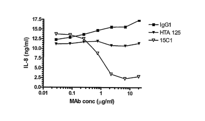
FIG. 15 is a graph depicting inhibition of lipopolysaccharide (LPS)-induced IL-
8
production in TLR4/MD-2 transfected HEK 293 cells by the monoclonal antibody
15C1. The
cells were incubated with the 15C1 monoclonal antibody, HTA 125 (anti-human
TLR4 non-
blocking monoclonal antibody) and an isotype-matched control (IgG1) at the
indicated
concentrations and subsequently incubated with LPS (15 ng/ml). IL-8 levels
were assessed
16 hours post LPS treatment.
FIG. 16 is a series of graphs depicting inhibition of LPS-induced IL-8
production in
human whole blood by the monoclonal antibody 15C1. Whole blood was drawn from
3
healthy volunteers, treated with heparin and diluted 1:4 in RPMI medium. The
following
9
CA 02548990 2006-06-09
WO 2005/065015 PCT/1B2004/004433
antibodies were added at the concentrations indicated: Isotype matched control
(IgG1);
HTA125 (anti-human TLR4 non-blocking monoclonal antibody); 15C1 and 28C5 (anti-
human CD14 blocking monoclonal antibody). LPS was subsequently added for a
final
concentration of 10 ng/ml.
FIG. 17 is a series of graphs depicting the specificity of the 15C1 monoclonal
antibody for TLR4. The specificity of the 15C1 antibody is shown by flow
cytometry
analysis of HEK 293 cells transiently transfected with either mock vector,
i.e., empty vector
(Panel A), human TLR4 (Panel B), human TLR4 and human MD-2 (Panel C), rabbit
TLR4
and rabbit MD-2 (Panel D), human TLR4 and rabbit MD-2 (Panel E), or rabbit
TLR4 and
human MD-2 (Panel F). Cells were incubated with the 15C1 Monoclonal antibody
(10
1,1g/m1), followed by an APC-coupled a -mouse (H+L) antibody.
FIGS. 18A-18F are a series of illustrations depicting the VH nucleotide
sequence
(SEQ ID NO:21) (FIG. 18A), the VII amino acid sequence (SEQ ID NO:22) (FIG.
18B), the
VL nucleotide sequence (SEQ ID NO:26) (FIG. 18D), and the VL amino acid
sequence (SEQ
ID NO:27) (FIG. 18E) for 15C1. The VII complementarity determining regions
(CDRs)
(SEQ ID NOs: 23, 24 and 25) (FIG. 18C) and the VL CDRs (SEQ ID NOs: 28, 29 and
30)
(FIG. 18F) are highlighted in the underlined, italic text in FIGS. 18B and
18E.
FIG. 19 is a graph depicting that the VII and VL nucleotide sequence of 15C1
expressed as a chimeric MAb ("chimeric 15C1") is capable of binding
specifically to the
human TLR4/MD-2 complex on the surface of transfected CHO cells. MAb binding
to the
TLR4/MD-2 complex is shown by flow cytometry using chimeric 15C1 or an isotype
matched control monoclonal antibody at the indicated concentration.
FIG. 20 is a graph depicting inhibition of lipopolysaccharide (LPS)-induced IL-
8
production in TLR4/MD-2 transfected HEK 293 cells by the chimeric 15C1 MAb.
Cells were
incubated with 15C1 or chimerical 15C1 at the concentrations indicated and
subsequently
incubated with LPS (15 ng/ml). IL-8 levels were assessed 16 hours post LPS
treatment.
Inhibition of LPS-induced IL-8 production by the chimeric 15C1 was similar to
the inhibition
by the 15C1 mouse MAb of the invention.
FIG. 21 is a graph depicting the binding of a monoclonal antibody of the
invention,
7E3, to the TLR4/MD-2 complex. Specificity of binding is shown by flow
cytometry using
mock transfected or TLR4/MD-2 transfected cells. The results using mock-
transfected cells
CA 02548990 2006-06-09
WO 2005/065015 PCT/1B2004/004433
are shown in the filled graph (left), while the results using TLR4/MD-2
transfected cells are
shown as in the outline graph (right).
FIG. 22 is a graph depicting inhibition of lipopolysaccharide (LPS)-induced IL-
8
production in TLR4/MD-2 transfected HEK 293 cells by the monoclonal antibody
7E3. The
cells were incubated with the 7E3 monoclonal antibody, HTA 125 (anti-human
TLR4 non-
blocking monoclonal antibody) and an isotype-matched control (IgG1) at the
indicated
concentrations and subsequently incubated with LPS (15 ng/rnl). IL-8 levels
were assessed
16 hours post LPS treatment.
FIG. 23 is a series of graphs depicting inhibition of LPS-induced IL-8
production in
human whole blood by the monoclonal antibody 7E3. Whole blood was drawn from 3
healthy volunteers, treated with heparin and diluted 1:4 in RPMI medium. The
following
antibodies were added at the concentrations indicated: Isotype matched control
(IgG1);
HTA125 (anti-human TLR4 non-blocking monoclonal antibody); 7E3 and 28C5 (anti-
human
CD14 blocking monoclonal antibody). LPS was subsequently added for a final
concentration
of 10 ng/ml.
FIG. 24 is a series of graphs depicting the specificity of the 7E3 monoclonal
antibody
for the TLR4/MD-2 complex. The specificity of the 7E3 antibody is shown by
flow
cytometry analysis of HEK 293 cells transiently transfected with either mock
vector (Panel
A), human TLR4 (Panel B), human TLR4 and human MD-2 (Panel C), rabbit TLR4 and
rabbit MD-2 (Panel D), human TLR4 and rabbit MD-2 (Panel E),or rabbit TLR4 and
human
MD-2 (Panel F). Cells were incubated with the 7E3 monoclonal antibody (10
gimp,
followed by an APC-coupled a -mouse (H+L) antibody. =
FIGS. 25A-25F are a series of illustrations depicting the VH nucleotide
sequence
(SEQ ID NO:31) (FIG. 25A), the VH amino acid sequence (SEQ ID NO:32) (FIG.
25B), the
VL nucleotide sequence (SEQ ID NO:36) (FIG. 25D), and the VL amino acid
sequence (SEQ
ID NO:37) (FIG. 25E) for 7E3. The VII complementarily determining regions
(CDRs) (SEQ
ID NOs: 33, 34 and 35) (FIG. 25C) and the VL CDRs (SEQ ID NOs: 38, 39 and 40)
(FIG.
25F) are highlighted in the underlined italic text in FIGS. 25B and 25E.
FIG. 26 is a graph illustrating that the VH and VL nucleotide'sequence of 7E3
expressed as a chimeric MAb ("chimeric 7E3") is capable of binding
specifically to the
human TLR4/MD-2 complex on the surface of transfected CHO cells. Monoclonal
antibody
11
CA 02548990 2006-06-09
WO 2005/065015 PCT/1B2004/004433
binding toTLR4/MD-2 transfected CHO cells is shown by flow cytometry using
chimeric 7E3
or an isotype matched control MAb at the indicated concentrations.
FIG. 27 is a graph depicting inhibition of lipopolysaccharide (LPS)-induced IL-
8
production in TLR4/MD-2 transfected HEK 293 cells by the chimeric 7E3 MAb.
Cells were
incubated with chimeric 7E3 or an isotype matched MAb control at the indicated
concentrations and subsequently incubated with LPS (15 ng/ml). IL-8 levels
were assessed
16 hours post LPS-treatment.
FIG. 28 is an illustration depicting the construction of a TLR4/MD-2 fusion
protein
cDNA according to the present invention.
FIG. 29 is an illustration depicting the expression of a TLR4/MD-2 chimeric
protein
of the invention in Sf9 cell lysates and supernatant.
FIG. 30 is an illustration depicting the purification of a TLR4/MD-2 chimeric
protein
according to the invention from infected Sf9 cell lysates.
FIG. 31 is a graph depicting the inhibit of lipopolysaccharide- (LPS) induced
IL-8
production using a soluble chimeric TLR4/MD-2 protein according to the present
invention.
FIG. 32A illustrates a nucleic acid sequence encoding the accessory protein MD-
1
(SEQ ID NO:41).
FIG. 32B depicts an amino acid sequence of a mature MD-1 accessory protein in
a
preferred embodiment of the invention (SEQ ID NO:42).
FIG. 33A illustrates a nucleic acid sequence encoding the accessory protein MD-
2
(SEQ ID NO:43).
FIG. 33B depicts an amino acid sequence of a mature MD-2 accessory protein
(SEQ
ID NO:44).
Detailed Description of the Invention
The present invention provides monoclonal antibodies that neutralize the
activation of
the TLR4/MD-2 receptor complex. In particular, the invention provides
monoclonal
antibodies that recognize the TLR4/MD-2 receptor complex expressed on the cell
surface.
These monoclonal antibodies block LPS-induced IL-8 production. In addition,
some
monoclonal antibodies of the invention also recognize TLR4 when not complexed
with MD-
12
CA 02548990 2012-04-27
2. Exemplary antibodies of the invention include, for example, the 18H10
antibody (Figures
6A-6F), the 16G7 antibody (Figures 13A-13F), the 15C1 antibody (Figures 18A-
18F) and the
7E3 antibody (Figures 25A-25F).
The present invention also provides soluble chimeric toll receptor proteins
(also
referred to herein as toll-like receptor proteins), methods for expressing
toll receptor proteins,
and methods for purifying such proteins in a soluble form. The chimeric
proteins are useful,
e.g., in generating antibodies.
TLRs recognize microbial particles and activate immune cells against the
source of
these microbial particles. (See Takeda et at., Annu. Rev. Immunol., 21: 335-76
(2003)).
TLR4 and MD-2 have been shown to form a complex on the cell surface, and the
presence of
MD-2 appears essential for the responsiveness of TLR4 to various ligands,
including LPS.
LPS is a gram-negative bacterial outer membrane glycolipid that is capable of
strongly
activating the innate immune system. LPS has been implicated as one of the
major factors
activating the immune system during the severe generalized inflammation
resulting from
gram-negative infection. (Lakhani et at., Curr. Opin. Pediatr. 15: 278-282
(2003)).
LPS delivers signals to immune cells via its multi-chain receptor in which the
TLR4/MD-2 complex is the principle signaling component. LPS has been shown to
exert its
effects on the immune system via signaling through TLR4. LPS rapidly binds to
the
lipopolysaccharide-binding protein (LBP) in the bloodstream, and in this form,
LPS interacts
with the GPI-anchored cell surface protein CD14. LPS is then transferred to
TLR4, which
transduces an intracellular activation signal. Another protein, MD-2, has been
found to be
necessary for signal transduction via TLR4 to occur. MD-2 interacts directly
with TLR4 and
plays an important role in its post-translational modification and
intracellular trafficking. In
addition, MD-2 has been shown to directly bind LPS, which demonstrates the
importance of
this accessory protein in the LPS receptor complex. (See Miyake K., Int.
Immunopharmacol.
3:119-128 (2003)).
Accordingly, neutralization of LPS signaling mediated by the TLR4/MD-2 complex
is
a potential therapeutic strategy in the treatment of disorders such as, for
example, acute
systemic inflammation and sepsis induced by gram-negative bacterial infection.
13
CA 02548990 2006-06-09
WO 2005/065015 PCT/1B2004/004433
Definitions:
Unless otherwise defined, scientific and technical terms used in connection
with the
present invention shall have the meanings that are commonly understood by
those of ordinary
skill in the art. Further, unless otherwise required by context, singular
terms shall include
pluralities and plural terms shall include the singular. Generally,
nomenclatures utilized in
connection with, and techniques of, cell and tissue culture, molecular
biology, and protein and
oligo- or polymicleotide chemistry and hybridization described herein are
those well known
and commonly used in the art. Standard techniques are used for recombinant
DNA,
oligonucleotide synthesis, and tissue culture and transformation (e.g.,
electroporation,
lipofection). Enzymatic reactions and purification techniques are performed
according to
manufacturer's specifications or as commonly accomplished in the art or as
described herein.
The foregoing techniques and procedures are generally performed according to
conventional
methods well known in the art and as described in various general and more
specific
references that are cited and discussed throughout the present specification.
See e.g.,
Sambrook et al. Molecular Cloning: A Laboratory Manual (2d ed., Cold Spring
Harbor
Laboratory Press, Cold Spring Harbor, N.Y. (1989)). The nomenclatures utilized
in
connection with, and the laboratory procedures and techniques of, analytical
chemistry,
synthetic organic chemistry, and medicinal and pharmaceutical chemistry
described herein are
those well known and commonly used in the art. Standard techniques are used
for chemical
syntheses, chemical analyses, pharmaceutical preparation, formulation, and
delivery, and
treatment of patients.
As utilized in accordance with the present disclosure, the following terms,
unless
otherwise indicated, shall be understood to have the following meanings:
As used herein, the term "antibody" refers to immunoglobulin molecules and
immunologically active portions of immunoglobulin (Ig) molecules, i.e.,
molecules that
contain an antigen binding site that specifically binds (immunoreacts with) an
antigen. By
"specifically bind" or "immunoreacts with" or "immunospecifically bind" is
meant that the
antibody reacts with one or more antigenic determinants of the desired antigen
and does not
react with other polyp eptides or binds at much lower affinity (Kd > 10-6).
Antibodies include,
14
CA 02548990 2006-06-09
WO 2005/065015 PCT/1B2004/004433
but are not limited to, polyclonal, monoclonal, chimeric, dAb (domain
antibody), single chain,
Fab, Fab' and F(ab')2 fragments, scFvs, and an Fab expression library.
The basic antibody structural unit is known to comprise a tetramer. Each
tetramer is
composed of two identical pairs of polypeptide chains, each pair having one
"light" (about 25
kDa) and one "heavy" chain (about 50-70 kDa). The amino-terminal portion of
each chain
includes a variable region of about 100 to 110 or more amino acids primarily
responsible for
antigen recognition. The carboxy-terminal portion of each chain defines a
constant region
primarily responsible for effector function. In general, antibody molecules
obtained from
humans relate to any of the classes IgG, IgM, IgA, IgE and IgD, which differ
from one
another by the nature of the heavy chain present in the molecule. Certain
classes have
subclasses as well, such as IgGi, IgG2, and others. Furthermore, in humans,
the light chain
may be a kappa chain or a lambda chain.
The term "monoclonal antibody" (MAb) or "monoclonal antibody composition", as
used herein, refers to a population of antibody molecules that contain only
one molecular
species of antibody molecule consisting of a unique light chain gene product
and a unique
heavy chain gene product. In particular, the complementarity determining
regions (CDRs) of
the monoclonal antibody are identical in all the molecules of the population.
MAbs contain
an antigen binding site capable of immunoreacting with a particular epitope of
the antigen
characterized by a unique binding affinity for it.
The term "antigen-binding site," or "binding portion" refers to the part of
the
immunoglobulin molecule that participates in antigen binding. The antigen
binding site is
formed by amino acid residues of the N-terminal variable ("V") regions of the
heavy ("H")
and light ("L") chains. Three highly divergent stretches within the V regions
of the heavy and
light chains, referred to as "hypervariable regions," are interposed between
more conserved
flanking stretches known as "framework regions," or "FRs". Thus, the term "FR"
refers to
amino acid sequences which are naturally found between, and adjacent to,
hypervariable
regions in immunoglobulins. In an antibody molecule, the three hypervariable
regions of a
light chain and the three hypervariable regions of a heavy chain are disposed
relative to each
other in three dimensional space to form an antigen-binding surface. The
antigen-binding
surface is complementary to the three-dimensional surface of a bound antigen,
and the three
hypervariable regions of each of the heavy and light chains are referred to as
CA 02548990 2006-06-09
WO 2005/065015 PCT/1B2004/004433
"complementarity-determining regions," or "CDRs." The assignment of amino
acids to each
domain is in accordance with the definitions of Kabat Sequences of Proteins of
Immunological Interest (National Institutes of Health, Bethesda, Md. (1987 and
1991)), or
Chothia & Lesk J. Mol. Biol. 196:901-917 (1987), Chothia et al. Nature 342:878-
883 (1989).
As used herein, the term "epitope" includes any protein determinant capable of
specific binding to an immunoglobulin, an scFv, or a T-cell receptor. The term
"epitope"
includes any protein determinant capable of specific binding to an
immunoglobulin or T-cell
receptor. Epitopic determinants usually consist of chemically active surface
groupings of
molecules such as amino acids or sugar side chains and usually have specific
three
dimensional structural characteristics, as well as specific charge
characteristics. For example,
antibodies may be raised against N-terminal or C-terminal peptides of a
polypeptide. An
antibody is said to specifically bind an antigen when the dissociation
constant is < 1 M;
preferably < 100 nM and most preferably 5. 10 nM.
As used herein, the terms "immunological binding," and "immunological binding
properties" refer to the non-covalent interactions of the type which occur
between an
immunoglobulin molecule and an antigen for which the immunoglobulin is
specific. The
strength, or affinity of immunological binding interactions can be expressed
in terms of the
dissociation constant (K,d) of the interaction, wherein a smaller Kid
represents a greater affinity.
Immunological binding properties of selected polypeptides can be quantified
using methods
well known in the art. One such method entails measuring the rates of antigen-
binding
site/antigen complex formation and dissociation, wherein those rates depend on
the
concentrations of the complex partners, the affinity of the interaction, and
geometric
parameters that equally influence the rate in both directions. Thus, both the
"on rate constant"
(Kon) and the "off rate constant" (Koff) can be determined by calculation of
the concentrations
and the actual rates of association and dissociation. (See Nature 361:186-87
(1993)). The
ratio of Koff aon enables the cancellation of all parameters not related to
affinity, and is equal
to the dissociation constant IQ. (See, generally, Davies et al. (1990) Annual
Rev Biochem
59:439-473). An antibody of the present invention is said to specifically bind
to the Toll-like
Receptor 4 (TLR4)/MD-2 complex or to TLR4 when not complexed to MD-2, when the
equilibrium binding constant (Kd) is 11M, preferably 100 nM, more preferably
10 nM,
16
CA 02548990 2006-06-09
WO 2005/065015 PCT/1B2004/004433
and most preferably 100 pM to about 1 pM, as measured by assays such as
radioligand
binding assays or similar assays known to those skilled in the art.
The term "isolated polynucleotide" as used herein shall mean a polynucleotide
of
genomic, cDNA, or synthetic origin or some combination thereof, which by
virtue of its
origin the "isolated polynucleotide" (1) is not associated with all or a
portion of a
polynucleotide in which the "isolated polynucleotide" is found in nature, (2)
is operably
linked to a polynucleotide which it is not linked to in nature, or (3) does
not occur in nature as
part of a larger sequence.
The term "isolated protein" referred to herein means a protein of cDNA,
recombinant
RNA, or synthetic origin or some combination thereof, which by virtue of its
origin, or source
of derivation, the "isolated protein" (1) is not associated with proteins
found in nature, (2) is
free of other proteins from the same source, e.g., free of marine proteins,
(3) is expressed by a
cell from a different species, or (4) does not occur in nature.
The term "polypeptide" is used herein as a generic term to refer to native
protein,
fragments, or analogs of a polypeptide sequence. Hence, native protein
fragments, and
analogs are species of the polypeptide genus. Preferred polypeptides in
accordance with the
invention comprise the human heavy chain immunoglobulin molecules represented
in Figures
6B, 13B, 18B and 25B and the human light chain immunoglobulin molecules
represented in
Figures 6E, 13E, 18 and 25, as well as antibody molecules formed by
combinations
comprising the heavy chain immunoglobulin molecules with light chain
immunoglobulin
molecules, such as kappa light chain immunoglobulin molecules, and vice versa,
as well as
fragments and analogs thereof.
The term "naturally-occurring" as used herein as applied to an object refers
to the fact
that an object can be found in nature. For example, a polypeptide or
polynucleotide sequence
that is present in an organism (including viruses) that can be isolated from a
source in nature
and which has not been intentionally modified by man in the laboratory or
otherwise is
naturally-occurring.
The term "operably linked" as used herein refers to positions of components so
described are in a relationship permitting them to function in their intended
manner. A
control sequence "operably linked" to a coding sequence is ligated in such a
way that
17
CA 02548990 2006-06-09
WO 2005/065015 PCT/1B2004/004433
expression of the coding sequence is achieved under conditions compatible with
the control
sequences.
The term "control sequence" as used herein refers to polynucleotide sequences
which
are necessary to effect the expression and processing of coding sequences to
which they are
ligated. The nature of such control sequences differs depending upon the host
organism in
prokaryotes, such control sequences generally include promoter, ribosomal
binding site, and
transcription termination sequence in eukaryotes, generally, such control
sequences include
promoters and transcription termination sequence. The term "control sequences"
is intended
to include, at a minimum, all components whose presence is essential for
expression and
processing, and can also include additional components whose presence is
advantageous, for
example, leader sequences and fusion partner sequences. The term
"polynucleotide" as
referred to herein means a polymeric boron of nucleotides of at least 10 bases
in length, either
ribonucleotides or deoxynucleotides or a modified form of either type of
nucleotide. The term
includes single and double stranded forms of DNA.
The term oligonucleotide referred to herein includes naturally occurring, and
modified
nucleotides linked together by naturally occurring, and non-naturally
occurring
oligonucleotide linkages. Oligonucleotides are a polynucleotide subset
generally comprising
a length of 200 bases or fewer. Preferably oligonucleotides are 10 to 60 bases
in length and
most 'preferably 12, 13, 14, 15, 16, 17, 18, 19, or 20 to 40 bases in length.
Oligonucleotides
are usually single stranded, e.g., for probes, although oligonucleotides may
be double
stranded, e.g., for use in the construction of a gene mutant. Oligonucleotides
of the invention
are either sense or antisense oligonucleotides.
The term "naturally occurring nucleotides" referred to herein includes
deoxyribonucleotides and ribonucleotides. The term "modified nucleotides"
referred to herein
includes nucleotides with modified or substituted sugar groups and the like.
The term
"oligonucleotide linkages" referred to herein includes Oligonucleotides
linkages such as
phosphorothioate, phosphorodithioate, phosphoroselerlo ate,
phosphorodiselenoate,
phosphoroanilothioate, phoshoraniladate, phosphoronmidate, and the like. See
e.g.,
LaPlanche et al. Nucl. Acids Res. 14:9081 (1986); Stec et al. J. Am. Chem.
Soc. 106:6077
(1984), Stein et al. Nucl. Acids Res. 16:3209 (1988), Zon et al. Anti Cancer
Drug Design
6:539 (1991); Zon et al. Oligonucleotides and Analogues: A Practical Approach,
pp. 87-108
18
CA 02548990 2006-06-09
WO 2005/065015 PCT/1B2004/004433
(F. Eckstein, Ed., Oxford University Press, Oxford England (1991)); Stec et
al. U.S. Patent
No. 5,151,510; Uhlmann and Peyman Chemical Reviews 90:543 (1990). An
oligonucleotide
can include a label for detection, if desired.
The term "selectively hybridize" referred to herein means to detectably and
specifically bind. Polynucleotides, oligonucleotides and fragments thereof in
accordance with
the invention selectively hybridize to nucleic acid strands under
hybridization and wash
conditions that minimize appreciable amounts of detectable binding to
nonspecific nucleic
acids. High stringency conditions can be used to achieve selective
hybridization conditions as
known in the art and discussed herein. Generally, the nucleic acid sequence
homology
between the polynucleotides, oligonucleotides, and fragments of the invention
and a nucleic
acid sequence of interest will be at least 80%, and more typically with
preferably increasing
homologies of at least 85%, 90%, 95%, 99%, and 100%. Two amino acid sequences
are
homologous if there is a partial or complete identity between their sequences.
For example,
85% homology means that 85% of the amino acids are identical when the two
sequences are
aligned for maximum matching. Gaps (in either of the two sequences being
matched) are
allowed in maximizing matching gap lengths of 5 or less are preferred with 2
or less being
more preferred. Alternatively and preferably, two protein sequences (or
polypeptide
sequences derived from them of at least 30 amino acids in length) are
homologous, as this
term is used herein, if they have an alignment score of at more than 5 (in
standard deviation
units) using the program ALIGN with the mutation data matrix and a gap penalty
of 6 or
greater. See Dayhoff, M.O., in Atlas of Protein Sequence and Structure, pp.
101-110 (Volume
5, National Biomedical Research Foundation (1972)) and Supplement 2 to this
volume, pp. 1-
10. The two sequences or parts thereof are more preferably homologous if their
amino acids
are greater than or equal to 50% identical when optimally aligned using the
ALIGN program.
The term "corresponds to" is used herein to mean that a polynucleotide
sequence is
homologous (i.e., is identical, not strictly evolutionarily related) to all or
a portion of a
reference polynucleotide sequence, or that a polypeptide sequence is identical
to a reference
polypeptide sequence. In contradistinction, the term "complementary to" is
used herein to
mean that the complementary sequence is homologous to all or a portion of a
reference
polynucleotide sequence. For illustration, the nucleotide sequence "TATAC"
corresponds to
a reference sequence "TATAC" and is complementary to a reference sequence
"GTATA".
19
CA 02548990 2006-06-09
WO 2005/065015 PCT/1B2004/004433
The following terms are used to describe the sequence relationships between
two or
more polynucleotide or amino acid sequences: "reference sequence", "comparison
window",
"sequence identity", "percentage of sequence identity", and "substantial
identity". A
"reference sequence" is a defined sequence used as a basis for a sequence
comparison a
reference sequence may be a subset of a larger sequence, for example, as a
segment of a full-
length cDNA or gene sequence given in a sequence listing or may comprise a
complete cDNA
or gene sequence. Generally, a reference sequence is at least 18 nucleotides
or 6 amino acids
in length, frequently at least 24 nucleotides or 8 amino acids in length, and
often at least 48
nucleotides or 16 amino acids in length. Since two polynucleotides or amino
acid sequences
may each (1) comprise a sequence (i.e., a portion of the complete
polynucleotide or amino
acid sequence) that is similar between the two molecules, and (2) may further
comprise a
sequence that is divergent between the two polynucleotides or amino acid
sequences,
sequence comparisons between two (or more) molecules are typically performed
by
comparing sequences of the two molecules over a "comparison window" to
identify and
compare local regions of sequence similarity. A "comparison window", as used
herein, refers
to a conceptual segment of at least 18 contiguous nucleotide positions or 6
amino acids
wherein a polynucleotide sequence or amino acid sequence may be compared to a
reference
sequence of at least 18 contiguous nucleotides or 6 amino acid sequences and
wherein the
portion of the polynucleotide sequence in the comparison window may comprise
additions,
deletions, substitutions, and the like (i.e., gaps) of 20 percent or less as
compared to the
reference sequence (which does not comprise additions or deletions) for
optimal alignment of
the two sequences. Optimal alignment of sequences for aligning a comparison
window may
be conducted by the local homology algorithm of Smith and Waterman Adv. Appl.
Math.
2:482 (1981), by the homology alignment algorithm of Needleman and Wunsch J.
Mol. Biol.
48:443 (1970), by the search for similarity method of Pearson and Lipman Proc.
Natl. Acad.
Sci. (U.S.A.) 85:2444 (1988), by computerized implementations of these
algorithms (GAP,
BESTFIT, PASTA, and TFASTA in the Wisconsin Genetics Software Package Release
7.0,
(Genetics Computer Group, 575 Science Dr., Madison, Wis.), Geneworks, or
MacVector
software packages), or by inspection, and the best alignment (i.e., resulting
in the highest
percentage of homology over the comparison window) generated by the various
methods is
selected.
CA 02548990 2006-06-09
WO 2005/065015 PCT/1B2004/004433
The term "sequence identity" means that two polynucleotide or amino acid
sequences
are identical (i.e., on a nucleotide-by-nucleotide or residue-by-residue
basis) over the
comparison window. The term "percentage of sequence identity" is calculated by
comparing
two optimally aligned sequences over the window of comparison, determining the
number of
positions at which the identical nucleic acid base (e.g., A, T, C, G, U or I)
or residue occurs in
both sequences to yield the number of matched positions, dividing the number
of matched
positions by the total number of positions in the comparison window (i.e., the
window size),
and multiplying the result by 100 to yield the percentage of sequence
identity. The terms
"substantial identity" as used herein denotes a characteristic of a
polynucleotide or amino acid
sequence, wherein the polynucleotide or amino acid comprises a sequence that
has at least 85
percent sequence identity, preferably at least 90 to 95 percent sequence
identity, more usually
at least 99 percent sequence identity as compared to a reference sequence over
a comparison
window of at least 18 nucleotide (6 amino acid) positions, frequently over a
window of at
least 24-48 nucleotide (8-16 amino acid) positions, wherein the percentage of
sequence
identity is calculated by comparing the reference sequence to the sequence
which may include
deletions or additions which total 20 percent or less of the reference
sequence over the
comparison window. The reference sequence may be a subset of a larger
sequence.
As used herein, the twenty conventional amino acids and their abbreviations
follow
conventional usage. See Immunology - A Synthesis (2nd Edition, E.S. Golub and
D.R. Gren,
Eds., Sinauer Associates, Sunderland7 Mass. (1991)). Stereoisomers (e.g., D-
amino acids) of
the twenty conventional amino acids, unnatural amino acids such as a-, a-
disubstituted amino
acids, N-alkyl amino acids, lactic acid, and other unconventional amino acids
may also be
suitable components for polypeptides of the present invention. Examples of
unconventional
amino acids include: 4 hydroxyproline, 7-carboxyglutamate, s-N,N,N-
trimethyllysine, c -N-
acetyllysine, 0-phosphoserine, N- acetylserine, N-formylmethionine, 3-
methylhistidine, 5-
hydroxylysine, a-N-methylarginine, and other similar amino acids and imino
acids (e.g., 4-
hydroxyproline). In the polypeptide notation used herein, the left-hand
direction is the amino
terminal direction and the right-hand direction is the carboxy-terminal
direction, in
accordance with standard usage and convention.
Similarly, unless specified otherwise, the left-hand end of single- stranded
polynucleotide sequences is the 5' end the left-hand direction of double-
stranded
21
CA 02548990 2006-06-09
WO 2005/065015 PCT/1B2004/004433
polynucleotide sequences is referred to as the 5' direction. The direction of
5' to 3' addition of
nascent RNA transcripts is referred to as the transcription direction sequence
regions on the
DNA strand having the same sequence as the RNA and which are 5' to the 5' end
of the RNA
transcript are referred to as "upstream sequences", sequence regions on the
DNA strand
having the same sequence as the RNA and which are 3' to the 3' end of the RNA
transcript are
referred to as "downstream sequences".
As applied to polypeptides, the term "substantial identity" means that two
peptide
sequences, when optimally aligned, such as by the programs GAP or BESTFIT
using default
gap weights, share at least 80 percent sequence identity, preferably at least
90 percent
sequence identity, more preferably at least 95 percent sequence identity, and
most preferably
at least 99 percent sequence identity.
Preferably, residue positions which are not identical differ by conservative
amino acid
substitutions.
Conservative amino acid substitutions refer to the interchangeability of
residues
having similar side chains. For example, a group of amino acids having
aliphatic side chains
is glycine, alanine, valine, leucine, and isoleucine; a group of amino acids
having aliphatic-
hydroxyl side chains is senile and threonine; a group of amino acids having
amide- containing
side chains is asparagine and glutamine; a group of amino acids having
aromatic side chains is
phenylalanine, tyrosine, and tryptophan; a group of amino acids having basic
side chains is
lysine, arginine, and histidine; and a group of amino acids having sulfur-
containing side
chains is cysteine and methionine. Preferred conservative amino acids
substitution groups
are: valine-leucine-isoleucine, phenylalanine-tyrosine, lysine-arginine,
alanine valine,
glutamic- aspartic, and asparagine-glutamine.
As discussed herein, minor variations in the amino acid sequences of
antibodies or
immunoglobulin molecules are contemplated as being encompassed by the present
invention,
providing that the variations in the amino acid sequence maintain at least
75%, more
preferably at least 80%, 90%, 95%, and most preferably 99%. In particular,
conservative
amino acid replacements are contemplated. Conservative replacements are those
that take
place within a family of amino acids that are related in their side chains.
Genetically encoded
amino acids are generally divided into families: (1) acidic amino acids are
aspartate,
glutamate; (2) basic amino acids are lysine, arginine, histidine; (3) non-
polar amino acids are
22
CA 02548990 2006-06-09
WO 2005/065015 PCT/1B2004/004433
alanine, valine, leucine, isoleucine, proline, phenylalanine, methionine,
tryptophan, and (4)
uncharged polar amino acids are glycine, asparagine, glutamine, cysteine,
serine, threonine,
tyrosine. The hydrophilic amino acids include arginine, asparagine, asp
artate, glutamine,
glutamate, histidine, lysine, serine, and threonine. The hydrophobic amino
acids include
alanine, cysteine, isoleucine, leucine, methionine, phenylalanine, proline,
tryptophan, tyrosine
and valine. Other families of amino acids include (i) serine and threonine,
which are the
aliphatic-hydroxy family; (ii) asparagine and glutamine, which are the amide
containing
family; (iii) alanine, valine, leucine and isoleucine, which are the aliphatic
family; and (iv)
phenylalanine, tryptophan, and tyrosine, which are the aromatic family. For
example, it is
reasonable to expect that an isolated replacement of a leucine with an
isoleucine or valine, an
aspartate with a glutamate, a threonine with a serine, or a similar
replacement of an amino
acid with a structurally related amino acid will not have a major effect on
the binding or
properties of the resulting molecule, especially if the replacement does not
involve an amino
acid within a framework site. Whether an amino acid change results in a
functional peptide
can readily be determined by assaying the specific activity of the polypeptide
derivative.
Assays are described in detail herein. Fragments or analogs of antibodies or
immunoglobulin
molecules can be readily prepared by those of ordinary skill in the art.
Preferred amino- and
, carboxy-termini of fragments or analogs occur near boundaries of
functional domains.
Structural and functional domains can be identified by comparison of the
nucleotide and/or
amino acid sequence data to public or proprietary sequence databases.
Preferably,
computerized comparison methods are used to identify sequence motifs or
predicted protein
conformation domains that occur in other proteins of known structure and/or
function.
Methods to identify protein sequences that fold into a known three-dimensional
structure are
known. Bowie et al. Science 253:164 (1991). Thus, the foregoing examples
demonstrate that
those of skill in the art can recognize sequence motifs and structural
conformations that may
be used to define structural and functional domains in accordance with the
invention.
Preferred amino acid substitutions are those which: (1) reduce susceptibility
to
proteolysis, (2) reduce susceptibility to oxidation, (3) alter binding
affinity for forming protein
complexes, (4) alter binding affinities, and (4) confer or modify other
physicochemical or
functional properties of such analogs. Analogs can include various muteins of
a sequence
other than the naturally-occurring peptide sequence. For example, single or
multiple amino
23
CA 02548990 2006-06-09
WO 2005/065015 PCT/1B2004/004433
acid substitutions (preferably conservative amino acid substitutions) may be
made in the
naturally- occurring sequence (preferably in the portion of the polypeptide
outside the
domain(s) forming intermolecular contacts. A conservative amino acid
substitution should
not substantially change the structural characteristics of the parent sequence
(e.g., a
replacement amino acid should not tend to break a helix that occurs in the
parent sequence, or
disrupt other types of secondary structure that characterizes the parent
sequence). Examples
of art-recognized polypeptide secondary and tertiary structures are described
in Proteins,
Structures and Molecular Principles (Creighton, Ed., W. H. Freeman and
Company, New
York (1984)); Introduction to Protein Structure (C. Branden and J. Tooze,
eds., Garland
Publishing, New York, N.Y. (1991)); and Thornton et at. Nature 354:105 (1991).
The term "polypeptide fragment" as used herein refers to a polypeptide that
has an
amino terminal and/or carboxy-terminal deletion, but where the remaining amino
acid
sequence is identical to the corresponding positions in the naturally-
occurring sequence
deduced, for example, from a full length cDNA sequence. Fragments typically
are at least 5,
6, 8 or 10 amino acids long, preferably at least 14 amino acids long' more
preferably at least
20 amino acids long, usually at least 50 amino acids long, and even more
preferably at least
70 amino acids long. The term "analog" as used herein refers to polypeptides
which are
comprised of a segment of at least 25 amino acids that has substantial
identity to a portion of a
deduced amino acid sequence and which has specific binding to TLR4/MD2 complex
or
TLR4 alone, under suitable binding conditions. Typically, polypeptide analogs
comprise a
conservative amino acid substitution (or addition or deletion) with respect to
the naturally-
occurring sequence. Analogs typically are at least 20 amino acids long,
preferably at least 50
amino acids long or longer, and can often be as long as a full-length
naturally-occurring
polypeptide.
Peptide analogs are commonly used in the pharmaceutical industry as non-
peptide
drugs with properties analogous to those of the template peptide. These types
of non-peptide
compound are termed "peptide mimetics" or "peptidomimetics". Fauchere, J. Adv.
Drug Res.
15:29 (1986), Veber and Freidinger TINS p.392 (1985); and Evans et al. J. Med.
Chem.
30:1229 (1987). Such compounds are often developed with the aid of
computerized
molecular modeling. Peptide mimetics that are structurally similar to
therapeutically useful
peptides may be used to produce an equivalent therapeutic or prophylactic
effect. Generally,
24
CA 02548990 2006-06-09
WO 2005/065015 PCT/1B2004/004433
peptidomimetics are structurally similar to a paradigm polypeptide (i.e., a
polypeptide that has
a biochemical property or pharmacological activity), such as human antibody,
but have one or
more peptide linkages optionally replaced by a linkage selected from the group
consisting of:
CH2NH--, --CH2S-, --CH=CH--(cis and trans), --COCH2--, CH(OH)CH2--,
and -CH2S0--, by methods well known in the art. Systematic substitution of one
or more
amino acids of a consensus sequence with a D-amino acid of the same type
(e.g., D-lysine in
place of L-lysine) may be used to generate more stable peptides. In addition,
constrained
peptides comprising a consensus sequence or a substantially identical
consensus sequence
variation may be generated by methods known in the art (Rizo and Gierasch Ann.
Rev.
Biochem. 61:387 (1992)); for example, by adding internal cysteine residues
capable of
forming intramolecular disulfide bridges which cyclize the peptide.
The term "agent" is used herein to denote a chemical compound, a mixture of
chemical compounds, a biological macromolecule, or an extract made from
biological
materials.
As used herein, the terms "label" or "labeled" refers to incorporation of a
detectable
marker, e.g., by incorporation of a radiolabeled amino acid or attachment to a
polypeptide of
biotinyl moieties that can be detected by marked avidin (e.g., streptavidin
containing a
fluorescent marker or enzymatic activity that can be detected by optical or
calorimetric
methods). In certain situations, the label or marker can also be therapeutic.
Various methods
of labeling polypeptides and glycoproteins are known in the art and may be
used. Examples
of labels for polypeptides include, but are not limited to, the following:
radioisotopes or
3H, 14C, 15N, 35s, 90y, 99Tc, 111in, 1251, 1311m,
radionuclides (e.g., .0
fluorescent labels (e.g., FITC,
rhodamine, lanthanide phosphors), enzymatic labels (e.g., horseradish
peroxidase, p-
galactosidase, luciferase, alkaline phosphatase), chemiluminescent, biotinyl
groups,
predetermined polypeptide epitopes recognized by a secondary reporter (e.g.,
leucine zipper
pair sequences, binding sites for secondary antibodies, metal binding domains,
epitope tags).
In some embodiments, labels are attached by spacer arms of various lengths to
reduce
potential steric hindrance. The term "pharmaceutical agent or drug" as used
herein refers to a
chemical compound or composition capable of inducing a desired therapeutic
effect when
properly administered to a patient.
CA 02548990 2006-06-09
WO 2005/065015 PCT/1B2004/004433
Other chemistry terms herein are used according to conventional usage in the
art, as
exemplified by The McGraw-Hill Dictionary of Chemical Terms (Parker, S., Ed.,
McGraw-
Hill, San Francisco (1985)).
The term "antineoplastic agent" is used herein to refer to agents that have
the
functional property of inhibiting a development or progression of a neoplasm
in a human,
particularly a malignant (cancerous) lesion, such as a carcinoma, sarcoma,
lymphoma, or
leukemia. Inhibition of metastasis is frequently a property of antineoplastic
agents.
As used herein, "substantially pure" means an object species is the
predominant
species present (i.e., on a molar basis it is more abundant than any other
individual species in
the composition), and preferably a substantially purified fraction is a
composition wherein the
object species comprises at least about 50 percent (on a molar basis) of all
macromolecular
species present.
Generally, a substantially pure composition will comprise more than about 80
percent
of all macromolecular species present in the composition, more preferably more
than about
85%, 90%, 95%, and 99%. Most preferably, the object species is purified to
essential
homogeneity (contaminant species cannot be detected in the composition by
conventional
detection methods) wherein the composition consists essentially of a single
macromolecular
species.
The term patient includes human and veterinary subjects.
Antibodies
Monoclonal antibodies of the invention have the ability to inhibit LPS-induced
IL-8
production. Inhibition is determined, for example, in the human whole blood
and
huTLR4/MD2 transfected HEK 293 cellular assays described herein. Exemplary
monoclonal
antibodies include, for example, the antibodies referred to herein as "18H10",
"16G7",
"15C1" and "7E3". The 18H10 antibody recognizes the TLR4/MD-2 complex, but
does not
recognize an MD-2 protein when not complexed with TLR4. The 16G7, 15C1 and 7E3
monoclonal antibodies recognize the TLR4/MD-2 complex. 15C1 and 16G7 also
recognize
TLR4 when not complexed with MD-2.
26
CA 02548990 2012-04-27
Also included in the invention are antibodies that bind to the same epitope as
the
antibodies described herein. Those skilled in the art will recognize that it
is possible to
determine, without undue experimentation, if a monoclonal antibody has the
same specificity
as a monoclonal antibody of the invention (e.g., monoclonal antibody 18H10,
16G7, 15C1
and/or 7E3) by ascertaining whether the former prevents the latter from
binding to the
TLR4/MD-2 complex or to TLR4 when not complexed to MD-2. If the monoclonal
antibody
being tested competes with the monoclonal antibody of the invention, as shown
by a decrease
in binding by the monoclonal antibody of the invention, then it is likely that
the two
monoclonal antibodies bind to the same, or a closely related, epitope. Another
way to
determine whether a monoclonal antibody has the specificity of a monoclonal
antibody of the
invention is to pre-incubate the monoclonal antibody of the invention with the
TLR4/MD-2
complex or a soluble TLR4 protein (with which it is normally reactive), and
then add the
monoclonal antibody being tested to determine if the monoclonal antibody being
tested is
inhibited in its ability to bind the TLR4/MD-2 complex or to bind TLR4 and
TLR4
complexed with MD-2. If the monoclonal antibody being tested is inhibited
then, in all
likelihood, it has the same, or functionally equivalent, epitopic specificity
as the monoclonal
antibody of the invention. Screening of monoclonal antibodies of the
invention, can be also
carried out by measuring LPS-induced IL-8 production and determining whether
the test
monoclonal antibody is able to neutralize LPS-induced IL-8 production.
Various procedures known within the art may be used for the production of
polyclonal
or monoclonal antibodies directed against the TLR4/MD-2 complex, or to TLR4
when not
complexed to MD-2, or against derivatives, fragments, analogs homologs or
orthologs
thereof (See, for example, Antibodies: A Laboratory Manual, Harlow E, and Lane
D, 1988,
Cold Spring Harbor Laboratory Press, Cold Spring Harbor, NY.
Antibodies are purified by well-known techniques, such as affinity
chromatography
using protein A or protein G, which provide primarily the IgG fraction of
immune serum.
Subsequently, or alternatively, the specific antigen which is the target of
the immunoglobulin
sought, or an epitope thereof, may be immobilized on a column to purify the
immune specific
antibody by immunoaffinity chromatography. Purification of immunoglobulins is
discussed,
27
CA 02548990 2006-06-09
WO 2005/065015 PCT/1B2004/004433
for example, by D. Wilkinson (The Scientist, published by The Scientist, Inc.,
Philadelphia
PA, Vol. 14, No. 8 (April 17, 2000), pp. 25-28).
The antibodies of the invention (e.g., 18E110, 16G7, 15C1 and 7E3) are
monoclonal
antibodies. Monoclonal antibodies that neutralize LPS-signaling that is
mediated by the
TLR4/MD-2 complex are generated, e.g., by immunizing BALB/c mice with
combinations of
cell transfectants expressing high levels of TLR4 and MD-2 on their surface
and a
recombinant soluble chimeric protein comprising both TLR4 and MD-2 tethered by
a flexible
linker sequence. Hybridomas resulting from myeloma/B cell fusions are then
screened for
reactivity to this TLR4/MD-2 complex.
Monoclonal antibodies are prepared, for example, using hybridoma methods, such
as
those described by Kohler and Milstein, Nature, 256:495 (1975). In a hybridoma
method, a
mouse, hamster, or other appropriate host animal, is typically immunized with
an immunizing
agent to elicit lymphocytes that produce or are capable of producing
antibodies that will
specifically bind to the immunizing agent. Alternatively, the lymphocytes can
be immunized
in vitro.
The immunizing agent will typically include the protein antigen, a fragment
thereof or
a fusion protein thereof. Generally, either peripheral blood lymphocytes are
used if cells of
human origin are desired, or spleen cells or lymph node cells are used if non-
human
mammalian sources are desired. The lymphocytes are then fused with an
immortalized cell
line using a suitable fusing agent, such as polyethylene glycol, to form a
hybridoma cell
(Goding, Monoclonal Antibodies: Principles and Practice, Academic Press,
(1986) pp.
59-103). Immortalized cell lines are usually transformed mammalian cells,
particularly
myeloma cells of rodent, bovine and human origin. Usually, rat or mouse
myeloma cell lines
are employed. The hybridoma cells can be cultured in a suitable culture medium
that
preferably contains one or more substances that inhibit the growth or survival
of the unfused,
immortalized cells. For example, if the parental cells lack the enzyme
hypoxanthine guanine
phosphoribosyl transferase (HGPRT or HPRT), the culture medium for the
hybridomas
typically will include hypoxanthine, aminopterin, and thymidine ("HAT
medium"), which
substances prevent the growth of HGPRT-deficient cells.
Preferred immortalized cell lines are those that fuse efficiently, support
stable high
level expression of antibody by the selected antibody-producing cells, and are
sensitive to a
28
CA 02548990 2006-06-09
WO 2005/065015 PCT/1B2004/004433
medium such as HAT medium. More preferred immortalized cell lines are murine
myeloma
lines, which can be obtained, for instance, from the Salk Institute Cell
Distribution Center,
San Diego, California and the American Type Culture Collection, Manassas,
Virginia.
Human myeloma and mouse-human heteromyeloma cell lines also have been
described for
the production of monoclonal antibodies. (See Kozbor, J. Immunol., 133:3001
(1984);
Brodeur et al., Monoclonal Antibody Production Techniques and Applications,
Marcel
Dekker, Inc., New York, (1987) pp. 51-63)).
The culture medium in which the hybridoma cells are cultured can then be
assayed for
the presence of monoclonal antibodies directed against the antigen.
Preferably, the binding
specificity of monoclonal antibodies produced by the hybridoma cells is
determined by
immunoprecipitation or by an in vitro binding assay, such as radioimmuno assay
(RIA) or
enzyme-linked immunoabsorbent assay (ELISA). Such techniques and assays are
known in
the art. The binding affinity of the monoclonal antibody can, for example, be
determined by
the Scatchard analysis of Munson and Pollard, Anal. Biochem., 107:220 (1980).
Moreover, in
therapeutic applications of monoclonal antibodies, it is important to identify
antibodies having
a high degree of specificity and a high binding affinity for the target
antigen.
After the desired hybridoma cells are identified, the clones can be subcloned
by
limiting dilution procedures and grown by standard methods. (See Goding,
Monoclonal
Antibodies: Principles and Practice, Academic Press, (1986) pp. 59-103).
Suitable culture
media for this purpose include, for example, Dulbecco's Modified Eagle's
Medium and
RPMI-1640 medium. Alternatively, the hybridoma cells can be grown in vivo as
ascites in a
mammal.
The monoclonal antibodies secreted by the subclones can be isolated or
purified from
the culture medium or ascites fluid by conventional immunoglobulin
purification procedures
such as, for example, protein A-Sepharose, hydroxylapatite chromatography, gel
electrophoresis, dialysis, or affinity chromatography.
Monoclonal antibodies can also be made by recombinant DNA methods, such as
those
described in U.S. Patent No. 4,816,567. DNA encoding the monoclonal antibodies
of the
invention can be readily isolated and sequenced using conventional procedures
(e.g., by using
oligonucleotide probes that are capable of binding specifically to genes
encoding the heavy
and light chains of murine antibodies). The hybridoma cells of the invention
serve as a
29
CA 02548990 2006-06-09
WO 2005/065015 PCT/1B2004/004433
preferred source of such DNA. Once isolated, the DNA can be placed into
expression
vectors, which are then transfected into host cells such as simian COS cells,
Chinese hamster
ovary (CHO) cells, or myeloma cells that do not otherwise produce
immunoglobulin protein,
to obtain the synthesis of monoclonal antibodies in the recombinant host
cells. The DNA also
can be modified, for example, by substituting the coding sequence for human
heavy and light
chain constant domains in place of the homologous murine sequences (see U.S.
Patent No.
4,816,567; Morrison, Nature 368, 812-13 (1994)) or by covalently joining to
the
immunoglobulin coding sequence all or part of the coding sequence for a
non-immunoglobulin polyp eptide. Such a non-immunoglobulin polypeptide can be
substituted for the constant domains of an antibody of the invention, or can
be substituted for
the variable domains of one antigen-combining site of an antibody of the
invention to create a
chimeric bivalent antibody.
Fully human antibodies are antibody molecules in which the entire sequence of
both
the light chain and the heavy chain, including the CDRs, arise from human
genes. Such
antibodies are termed "human antibodies", or "fully human antibodies" herein.
Monoclonal
antibodies can be prepared by using trioma technique; the human B-cell
hybridoma technique
(see Kozbor, et al., 1983 Immunol Today 4: 72); and the EBV hybridoma
technique to
produce monoclonal antibodies (see Cole, et al., 1985 In: MONOCLONAL
ANTIBODIES AND
CANCER THERAPY, Alan R. Liss, Inc., pp. 77-96). Monoclonal antibodies may be
utilized and
may be produced by using human hybridomas (see Cote, et al., 1983. Proc Natl
Acad Sci
USA 80: 2026-2030) or by transforming human B-cells with Epstein Barr Virus in
vitro (see
Cole, et al., 1985 In: MONOCLONAL ANTIBODIES AND CANCER THERAPY, Alan R. Liss,
Inc.,
pp. 77-96).
In addition, human antibodies can also be produced using additional
techniques,
including phage display libraries. (See Hoogenboom and Winter, J. Mol. Biol.,
227:381
(1991); Marks et al., J. Mol. Biol., 222:581 (1991)). Similarly, human
antibodies can be made
by introducing human immunoglobulin loci into transgenic animals, e.g., mice
in which the
endogenous immunoglobulin genes have been partially or completely inactivated.
Upon
challenge, human antibody production is observed, which closely resembles that
seen in
humans in all respects, including gene rearrangement, assembly, and antibody
repertoire.
This approach is described, for example, in U.S. Patent Nos. 5,545,807;
5,545,806; 5,569,825;
CA 02548990 2006-06-09
WO 2005/065015 PCT/1B2004/004433
5,625,126; 5,633,425; 5,661,016, and in Marks et al., Bio/Technology 10, 779-
783 (1992);
Lonberg et al., Nature 368 856-859 (1994); Morrison, Nature 368, 812-13
(1994); Fishwild et
al, Nature Biotechnology 14, 845-51 (1996); Neuberger, Nature Biotechnology
14, 826
(1996); and Lonberg and Huszar, Intern. Rev. Immunol. 13 65-93 (1995).
Human antibodies may additionally be produced using transgenic nonhuman
animals
Which are modified so as to produce fully human antibodies rather than the
animal's
endogenous antibodies in response to challenge by an antigen. (See PCT
publication
W094/02602). The endogenous genes encoding the heavy and light inu-
nunoglobulin chains
in the nonhuman host have been incapacitated, and active loci encoding human
heavy and
light chain immunoglobulins are inserted into the host's genome. The human
genes are
incorporated, for example, using yeast artificial chromosomes containing the
requisite human
DNA segments. An animal which provides all the desired modifications is then
obtained as
progeny by crossbreeding intermediate transgenic animals containing fewer than
the full
complement of the modifications. An example of such a nonhuman animal is a
mouse termed
the XenomouseTM as disclosed in PCT publications WO 96/33735 and WO 96/34096.
This
animal produces B cells which secrete fully human immunoglobulins. The
antibodies can be
obtained directly from the animal after immunization with an immunogen of
interest, as, for
example, a preparation of a polyclonal antibody, or alternatively from
immortalized B cells
derived from the animal, such as hybridomas producing monoclonal antibodies.
Additionally,
the genes encoding the immunoglobulins with human variable regions can be
recovered and
expressed to obtain the antibodies directly, or can be further modified to
obtain analogs of
antibodies such as, for example, single chain Fv (scFv) molecules.
An example of a method of producing a nonhuman host, exemplified as a mouse,
lacking expression of an endogenous immunoglobulin heavy chain is disclosed in
U.S. Patent
No. 5,939,598. It can be obtained by a method, which includes deleting the J
segment genes
from at least one endogenous heavy chain locus in an embryonic stern cell to
prevent
rearrangement of the locus and to prevent formation of a transcript of a
rearranged
immunoglobulin heavy chain locus, the deletion being effected by a targeting
vector
containing a gene encoding a selectable marker; and producing from the
embryonic stem cell
a transgenic mouse whose somatic and germ cells contain the gene encoding the
selectable
marker.
31
CA 02548990 2006-06-09
WO 2005/065015 PCT/1B2004/004433
One method for producing an antibody of interest, such as a human antibody, is
disclosed in U.S. Patent No. 5,916,771. This method includes introducing an
expression
vector that contains a nucleotide sequence encoding a heavy chain into one
mammalian host
cell in culture, introducing an expression vector containing a nucleotide
sequence encoding a
light chain into another mammalian host cell, and fusing the two cells to form
a hybrid cell.
The hybrid cell expresses an antibody containing the heavy chain and the light
chain.
In a further improvement on this procedure, a method for identifying a
clinically
relevant epitope on an immunogen, and a correlative method for selecting an
antibody that
binds immtmospecifically to the relevant epitope with high affinity, are
disclosed in PCT
publication WO 99/53049.
The antibody can be expressed by a vector containing a DNA segment encoding
the
single chain antibody described above.
These can include vectors, liposomes, naked DNA, adjuvant-assisted DNA. gene
gun,
catheters, etc. Vectors include chemical conjugates such as described in WO
93/64701, which
has targeting moiety (e.g a ligand to a cellular surface receptor), and a
nucleic acid binding
moiety (e.g. polylysine), viral vector (e.g. a DNA or RNA viral vector),
fusion proteins such
as described in PCT/US 95/02140 (WO 95/22618) which is a fusion protein
containing a
target moiety (e.g. an antibody specific for a target cell) and a nucleic acid
binding moiety
(e.g. a protamine), plasmids, phage, etc. The vectors can be chromosomal, non-
chromosomal
or synthetic.
Preferred vectors include viral vectors, fusion proteins and chemical
conjugates.
Retroviral vectors include moloney mmine leukemia viruses. DNA viral vectors
are
preferred. These vectors include pox vectors such as orthopox or avipox
vectors, herpesvirus
vectors such as a herpes simplex I virus (HSV) vector (see Geller, A. I. et
al., J. Neurochem,
64:487 (1995); Lim, F., et al., in DNA Cloning: Mammalian Systems, D. Glover,
Ed. (Oxford
Univ. Press, Oxford England) (1995); Geller, A. I. et al., Proc Natl. Acad.
Sci.: U.S.A.
90:7603 (1993); Geller, A. I., et al., Proc Natl. Acad. Sci USA 87:1149
(1990), Adenovirus
Vectors (see LeGal LaSalle et al., Science, 259:988 (1993); Davidson, et al.,
Nat. Genet 3:219
(1993); Yang, et al., J. Virol. 69:2004 (1995) and Adeno-associated Virus
Vectors (see
Kaplitt, M. G.. et al., Nat. Genet. 8:148 (1994).
32
CA 02548990 2006-06-09
WO 2005/065015 PCT/1B2004/004433
Pox viral vectors introduce the gene into the cells cytoplasm. Avipox virus
vectors
result in only a short term expression of the nucleic acid. Adenovirus
vectors, adeno-
associated virus vectors and herpes simplex virus (HSV) vectors are preferred
for introducing
the nucleic acid into neural cells. The adenovirus vector results in a shorter
term expression
(about 2 months) than adeno-associated virus (about 4 months), which in turn
is shorter than
HSV vectors. The particular vector chosen will depend upon the target cell and
the condition
being treated. The introduction can be by standard techniques, e.g. infection,
transfection,
transduction or transformation. Examples of modes of gene transfer include
e.g., naked DNA,
CaPO4 precipitation, DEAE dextran, electroporation, protoplast fusion,
lipofection, cell
microinjection, and viral vectors.
The vector can be employed to target essentially any desired target cell. For
example,
stereotaxic injection can be used to direct the vectors (e.g. adenovirus, HSV)
to a desired
location. Additionally, the particles can be delivered by
intracerebroventricular (icy) infusion
using a minipump infusion system, such as a SynchroMed Infusion System. A
method based
on bulk flow, termed convection, has also proven effective at delivering large
molecules to
extended areas of the brain and may be useful in delivering the vector to the
target cell. (See
Bobo et al., Proc. Natl. Acad. Sci. USA 91:2076-2080 (1994); Morrison et al.,
Am. J. Physiol.
266:292-305 (1994)). Other methods that can be used include catheters,
intravenous,
parenteral, intraperitoneal and subcutaneous injection, and oral or other
known routes of
administration.
These vectors can be used to express large quantities of antibodies that can
be used in
a variety of ways. For example, to detect the presence of the TLR4/MD-2
complex and/or
TLR4 in a sample. The antibody can also be used to try to bind to and disrupt
TLR4/MD-2
complex-related signaling.
Techniques can be adapted for the production of single-chain antibodies
specific to an
antigenic protein of the invention (see e.g., U.S. Patent No. 4,946,778). In
addition, methods
can be adapted for the construction of Fab expression libraries (see e.g.,
Huse, et al., 1989
Science 246: 1275-1281) to allow rapid and effective identification of
monoclonal Fab
fragments with the desired specificity for a protein or derivatives,
fragments, analogs or
homologs thereof. Antibody fragments that contain the idiotypes to a protein
antigen may be
produced by techniques known in the art including, but not limited to: (i) an
F(ab.)2 fragment
33
CA 02548990 2006-06-09
WO 2005/065015 PCT/1B2004/004433
produced by pepsin digestion of an antibody molecule; (ii) an Fab fragment
generated by
reducing the disulfide bridges of an F(ab')2 fragment; (iii) an Fab fragment
generated by the
treatment of the antibody molecule with papain and a reducing agent and (iv)
F, fragments.
The invention also includes Fv, Fab, Fab' and F(ab)2 anti-TLR4/MD2 complex
fragments or anti-
TLR4 fragments, single chain anti-TLR4/MD2 or anti-TLR4 antibodies, bispecific
anti-
TLR4/MD2 or anti-TLR4 antibodies and heteroconjugate anti-TLR4/MD2 or anti-
TLR4
antibodies.
Bispecific antibodies are antibodies that have binding specificities for at
least two
different antigens. In the present case, one of the binding specificities is
for the TLR4/MD2
complex and/or TLR4 when not complexed with MD-2. The second binding target is
any
other antigen, and advantageously is a cell-surface protein or receptor or
receptor subunit.
Methods for making bispecific antibodies are known in the art. Traditionally,
the
recombinant production of bispecific antibodies is based on the co-expression
of two
immunoglobulin heavy-chain/light-chain pairs, where the two heavy chains have
different
specificities (Milstein and Cuello, Nature, 305:537-539 (1983)). Because of
the random
assortment of immunoglobulin heavy and light chains, these hybridomas
(quadromas)
produce a potential mixture of ten different antibody molecules, of which only
one has the
correct bispecific structure. The purification of the correct molecule is
usually accomplished
by affinity chromatography steps. Similar procedures are disclosed in WO
93/08829,
published 13 May 1993, and in Traunecker et al., EMBO J., 10:3655-3659 (1991).
Antibody variable domains with the desired binding specificities (antibody-
antigen
combining sites) can be fused to immunoglobulin constant domain sequences. The
fusion
preferably is with an immunoglobulin heavy-chain constant domain, comprising
at least part
of the hinge, CH2, and CH3 regions. It is preferred to have the first heavy-
chain constant
region (CH1) containing the site necessary for light-chain binding present in
at least one of
the fusions. DNAs encoding the immunoglobulin heavy-chain fusions and, if
desired, the
immunoglobulin light chain, are inserted into separate expression vectors, and
are
co-transfected into a suitable host organism. For further details of
generating bispecific
antibodies see, for example, Suresh et al., Methods in Enzymology, 121:210
(1986).
According to another approach described in WO 96/27011, the interface between
a
pair of antibody molecules can be engineered to maximize the percentage of
heterodimers
34
CA 02548990 2006-06-09
WO 2005/065015 PCT/1B2004/004433
which are recovered from recombinant cell culture. The preferred interface
comprises at least
a part of the CH3 region of an antibody constant domain. In this method, one
or more small
amino acid side chains from the interface of the first antibody molecule are
replaced with
larger side chains (e.g. tyrosine or tryptophan). Compensatory "cavities" of
identical or
similar size to the large side chain(s) are created on the interface of the
second antibody
molecule by replacing large amino acid side chains with smaller ones (e.g
alanine or
threonine). This provides a mechanism for increasing the yield of the
heterodimer over other
unwanted end-products such as homodimers.
Bispecific antibodies can be prepared as full length antibodies or antibody
fragments
(e.g. F(ab')2 bispecific antibodies). Techniques for generating bispecific
antibodies from
antibody fragments have been described in the literature. For example,
bispecific antibodies
can be prepared using chemical linkage. Brennan et al., Science 229:81 (1985)
describe a
procedure wherein intact antibodies are proteolytically cleaved to generate
F(ab')2 fragments.
These fragments are reduced in the presence of the dithiol complexing agent
sodium arsenite
, to stabilize vicinal dithiols and prevent intermolecular disulfide
formation. The Fab'
fragments generated are then converted to thionitrobenzoate (TNB) derivatives.
One of the
Fab'-TNB derivatives is then reconverted to the Fab'-thiol by reduction with
mercaptoethylamine and is mixed with an equimolar amount of the other Fab'-TNB
derivative to form the bispecific antibody. The bispecific antibodies produced
can be used as
agents for the selective immobilization of enzymes.
Additionally, Fab' fragments can be directly recovered from E. coli and
chemically
coupled to form bispecific antibodies. Shalaby et al., J. Exp. Med. 175:217-
225 (1992)
describe the production of a fully humanized bispecific antibody F(ab')2
molecule. Each Fab'
fragment was separately secreted from E. coli and subjected to directed
chemical coupling in
vitro to form the bispecific antibody. The bispecific antibody thus formed was
able to bind to
cells overexpressing the ErbB2 receptor and normal human T cells, as well as
trigger the lytic
activity of human cytotoxic lymphocytes against human breast tumor targets.
= Various techniques for making and isolating bispecific antibody fragments
directly
from recombinant cell culture have also been described. For example,
bispecific antibodies
have been produced using leucine zippers. Kostelny et al., J. Immunol.
148(5):1547-1553
(1992). The leucine zipper peptides from the Fos and Jun proteins were linked
to the Fab'
CA 02548990 2006-06-09
WO 2005/065015 PCT/1B2004/004433
portions of two different antibodies by gene fusion. The antibody homodimers
were reduced
at the hinge region to form monomers and then re-oxidized to form the antibody
heterodimers.
This method can also be utilized for the production of antibody homodimers.
The "diabody"
technology described by Hollinger et al., Proc. Natl. Acad. Sci. USA 90:6444-
6448 (1993)
has provided an alternative mechanism for making bispecific antibody
fragments. The
fragments comprise a heavy-chain variable domain (VH) connected to a light-
chain variable
domain (VL) by a linker which is too short to allow pairing between the two
domains on the
same chain. Accordingly, the VH and VL domains of one fragment are forced to
pair with the
complementary VL and VH domains of another fragment, thereby forming two
antigen-binding sites. Another strategy for making bispecific antibody
fragments by the use
of single-chain Fv (sFv) dimers has also been reported. See, Gruber et al., J.
Immunol.
152:5368 (1994).
Antibodies with more than two valencies are contemplated. For example,
trispecific
antibodies can be prepared. Tutt et al., J. Immunol. 147:60 (1991).
Exemplary bispecific antibodies can bind to two different epitopes, at least
one of
which originates in the protein antigen of the invention. Alternatively, an
anti-antigenic arm
of an immunoglobulin molecule can be combined with an arm which binds to a
triggering
molecule on a leukocyte such as a T-cell receptor molecule (e.g. CD2, CD3,
CD28, or B7), or
Fc receptors for IgG (FcyR), such as FcyRI (CD64), FcyRII (CD32) and FcyRIII
(CD16) so as
to focus cellular defense mechanisms to the cell expressing the particular
antigen. Bispecific
antibodies can also be used to direct cytotoxic agents to cells which express
a particular
antigen. These antibodies possess an antigen-binding arm and an arm which
binds a cytotoxic '
agent or a radionuclide chelator, such as EOTUBE, DPTA, DOTA, or TETA. Another
bispecific antibody of interest binds the protein antigen described herein and
further binds
tissue factor (TF).
Heteroconjugate antibodies are also within the scope of the present invention.
Heteroconjugate antibodies are composed of two covalently joined antibodies.
Such
antibodies have, for example, been proposed to target immune system cells to
unwanted cells
(see U.S. Patent No. 4,676,980), and for treatment of HIV infection (see WO
91/00360; WO
92/200373; EP 03089). It is contemplated that the antibodies can be prepared
in vitro using
known methods in synthetic protein chemistry, including those involving
crosslinking agents.
36
CA 02548990 2006-06-09
WO 2005/065015 PCT/1B2004/004433
For example, immunotoxins can be constructed using a disulfide exchange
reaction or by
forming a thioether bond. Examples of suitable reagents for this purpose
include
iminothiolate and methyl-4-mercaptobutyrimidate and those disclosed, for
example, in U.S.
Patent No. 4,676,980.
It can be desirable to modify the antibody of the invention with respect to
effector
function, so as to enhance, e.g., the effectiveness of the antibody in
treating diseases and
disorders associated with aberrant LPS signaling. For example, cysteine
residue(s) can be
introduced into the Fc region, thereby allowing interchain disulfide bond
formation in this
region. The homodimeric antibody thus generated can have improved
internalization
capability and/or increased complement-mediated cell killing and antibody-
dependent cellular
cytotoxicity (ADCC). (See Caron et al., J. Exp Med., 176: 1191-1195 (1992) and
Shopes, J.
Immunol., 148: 2918-2922 (1992)). Alternatively, an antibody can be engineered
that has
dual Fc regions and can thereby have enhanced complement lysis and ADCC
capabilities.
(See Stevenson et al., Anti-Cancer Drug Design, 3: 219-230 (1989)).
The invention also pertains to immunoconjugates comprising an antibody
conjugated
to a cytotoxic agent such as a toxin (e.g., an enzymatically active toxin of
bacterial, fungal,
plant, or 'animal origin, or fragments thereof), or a radioactive isotope
(i.e., a radioconjugate).
Enzymatically active toxins and fragments thereof that can be used include
diphtheria
A chain, nonbinding active fragments of diphtheria toxin, exotoxin A chain
(from
Pseudomonas aeruginosa), ricin A chain, abrin A chain, modeccin A chain, alpha-
sarcin,
Aleurites fordii proteins, dianthin proteins, Phytolaca americana proteins
(PAPI, PAPII, and
PAP-S), momordica charantia inhibitor, curcin, crotin, sap aonaria officinalis
inhibitor,
gelonin, mitogellin, restrictocin, phenomycin, enomycin, and the
tricothecenes. A variety of
radionuclides are available for the production of radioconjugated antibodies.
Examples
include 212Bi, 1311, 131k, 90y, and 186Re.
Conjugates of the antibody and cytotoxic agent are made using a variety of
bifunctional protein-coupling agents such as N-succinimidy1-3-(2-
pyridyldithiol) propionate
(SPDP), iminothiolane (IT), bifunctional derivatives of imido esters (such as
dimethyl
adipimidate HCL), active esters (such as disuccinimidyl suberate), aldehydes
(such as
glutareldehyde), bis-azido compounds (such as bis (p-azidobenzoyl)
hexanediamine),
bis-diazonium derivatives (such as bis-(p-diazoniumbenzoy1)-ethylenediamine),
diisocyanates
37
CA 02548990 2012-04-27
,
(such as tolyene 2,6-diisocyanate), and bis-active fluorine compounds (such as
1,5-difluoro-2,4-dinitrobenzene). For example, a ricin immunotoxin can be
prepared as
described in Vitetta et al., Science 238: 1098 (1987). Carbon-14-labeled
1-isothiocyanatobenzy1-3-methyldiethylene triaminepentaacetic acid (MX-DTPA)
is an
exemplary chelating agent for conjugation of radionucleotide to the antibody.
(See
W094/11026).
Those of ordinary skill in the art will recognize that a large variety of
possible
moieties can be coupled to the resultant antibodies of the invention. (See,
for example,
"Conjugate Vaccines", Contributions to Microbiology and Immunology, J. M.
Cruse and R.
E. Lewis, Jr (eds), Carger Press, New York, (1989).
Coupling may be accomplished by any chemical reaction that will bind the two
molecules so long as the antibody and the other moiety retain their respective
activities. This
linkage can include many chemical mechanisms, for instance covalent binding,
affinity
binding, intercalation, coordinate binding and complexation. The preferred
binding is,
however, covalent binding. Covalent binding can be achieved either by direct
condensation of
existing side chains or by the incorporation of external bridging molecules.
Many bivalent or
polyvalent linking agents are useful in coupling protein molecules, such as
the antibodies of
the present invention, to other molecules. For example, representative
coupling agents can
include organic compounds such as thioesters, carbodiimides, succinimide
esters,
diisocyanates, glutaraldehyde, diazobenzenes and hexamethylene diamines. This
listing is not
intended to be exhaustive of the various classes of coupling agents known in
the art but,
rather, is exemplary of the more common coupling agents. (See Killen and
Lindstrom, Jour.
Immun. 133:1335-2549 (1984); Jansen et al., Immunological Reviews 62:185-216
(1982);
and Vitetta et al., Science 238:1098 (1987).
Preferred linkers are described in the literature. (See, for example,
Ramakrishnan, S.
et al., Cancer Res. 44:201-208 (1984) describing use of MBS (M-
maleimidobenzoyl-N-
hydroxysuccinimide ester). See also, U.S. Patent No. 5,030,719, describing use
of
halogenated acetyl hydrazide derivative coupled to an antibody by way of an
oligopeptide
linker. Particularly preferred linkers include: (i) EDC (1-ethyl-3-(3-
dimethylamino-propyl)
carbodiimide hydrochloride; (ii) SMPT (4-succinimidyloxycarbonyl-alpha-methyl-
alpha-(2-
38
CA 02548990 2006-06-09
WO 2005/065015 PCT/1B2004/004433
pridyl-dithio)-toluene (Pierce Chem. Co., Cat. (21558G); (iii) SPDP
(succinimidy1-6 [3-(2-
pyridyldithio) propionamido]hexanoate (Pierce Chem. Co., Cat #21651G); (iv)
Sulfo-LC-
SPDP (sulfosuccinimidyl 6 [3-(2-pyridyldithio)-propianamide] hexano ate
(Pierce Chem. Co.
Cat. #2165-G); and (v) sulfo-NHS (N-hydroxysulfo-succinimide: Pierce Chem.
Co., Cat.
#24510) conjugated to EDC.
The linkers described above contain components that have different attributes,
thus
leading to conjugates with differing physio-chemical properties. For example,
sulfo-NHS
esters of alkyl carboxylates are more stable than sulfo-NHS esters of aromatic
carboxylates.
NHS-ester containing linkers are less soluble than sulfo-NHS esters. Further,
the linker SMPT
contains a sterically hindered disulfide bond, and can form conjugates with
increased stability.
Disulfide linkages, are in general, less stable than other linkages because
the disulfide linkage
is cleaved in vitro, resulting in less conjugate available. Sulfo-NHS, in
particular, can enhance
the stability of carbodimide couplings. Carbodimide couplings (such as EDC)
when used in
conjunction with sulfo-NHS, forms esters that are more resistant to hydrolysis
than the
carbodimide coupling reaction alone.
The antibodies disclosed herein can also be formulated as immunoliposomes.
Liposomes containing the antibody are prepared by methods known in the art,
such as
described in Epstein et al., Proc. Natl. Acad. Sci. USA, 82: 3688 (1985);
Hwang et al., Proc.
Natl Acad. Sci. USA, 77: 4030 (1980); and U.S. Pat. Nos. 4,485,045 and
4,544,545.
Liposomes with enhanced circulation time are disclosed in U.S. Patent No.
5,013,556.
Particularly useful liposomes can be generated by the reverse-phase
evaporation
method with a lipid composition comprising phosphatidylcholine, cholesterol,
and
PEG-derivatized phosphatidylethanolamine (PEG-PE). Liposomes are extruded
through
filters of defined pore size to yield liposomes with the desired diameter.
Fab' fragments of the
antibody of the present invention can be conjugated to the liposomes as
described in Martin et
al., J. Biol. Chem., 257: 286-288 (1982) via a disulfide-interchange reaction.
Use of antibodies against the TLR4/MD2 complex and antibodies against TLR4
It will be appreciated that administration of therapeutic entities in
accordance with the
invention will be administered with suitable carriers, excipients, and other
agents that are
39
CA 02548990 2012-04-27
incorporated into formulations to provide improved transfer, delivery,
tolerance, and the like.
A multitude of appropriate formulations can be found in the formulary known to
all
pharmaceutical chemists: Remington's Pharmaceutical Sciences (15th ed, Mack
Publishing
Company, Easton, PA (1975)), particularly Chapter 87 by Blaug, Seymour,
therein. These
formulations include, for example, powders, pastes, ointments, jellies, waxes,
oils, lipids, lipid
(cationic or anionic) containing vesicles (such as Lipofectin"), DNA
conjugates, anhydrous
absorption pastes, oil-in-water and water-in-oil emulsions, emulsions carbowax
(polyethylene
glycols of various molecular weights), semi-solid gels, and semi-solid
mixtures containing
carbowax. Any of the foregoing mixtures may be appropriate in treatments and
therapies in
accordance with the present invention, provided that the active ingredient in
the formulation is
not inactivated by the formulation and the formulation is physiologically
compatible and
tolerable with the route of administration. See also Baldrick P.
"Pharmaceutical excipient
development: the need for preclinical guidance." Regul. Toxicol Pharmacol.
32(2):210-8
(2000), Wang W. "Lyophilization and development of solid protein
pharmaceuticals." Int. J.
Pharm. 203(1-2):1-60 (2000), Charman WN "Lipids, lipophilic drugs, and oral
drug delivery-
some emerging concepts." J Pharm Sci.89(8):967-78 (2000), Powell et al.
"Compendium of
excipients for parenteral formulations" PDA J Pharm Sci Technol. 52:238-311
(1998) and the
citations therein for additional information related to formulations,
excipients and carriers
well known to pharmaceutical chemists.
Therapeutic formulations of the invention, which include a monoclonal antibody
of
the invention, are used to treat or alleviate a symptom associated with an
immune-related
disorder. The present invention also provides methods of treating or
alleviating a symptom
associated with an immune-related disorder. A therapeutic regimen is carried
out by
identifying a subject, e.g., a human patient suffering from (or at risk of
developing) an
immune-related disorder, using standard methods.
Antibodies of the invention, which are capable of inhibiting LPS-induced IL-8
production, are useful therapeutic tools in the treatment of disorders, such
as, for example,
acute inflammation and sepsis induced by microbial products (e.g., LPS) and
exacerbations
arising from this acute inflammation, such as, for example, chronic
obstructive pulmonary
disease and asthma (see O'Neill, Curr. Opin. Pharmacol. 3: 396-403 (2003).
Such antibodies
are also useful in treating
CA 02548990 2012-04-27
neurodegenerative autoimmune diseases. (Lehnardt etal., Proc. Natl. Acad. Sci.
USA 100:
8514-8519(2003)).
In addition, the antibodies of the invention are also useful as therapeutic
reagents in
the treatment of diseases, such as, for example, osteoarthritis, which are
caused by mechanical
stress, which, in turn, induces endogenous soluble "stress" factors that
trigger TLR4.
Endogenous soluble stress factor include e.g, Hsp60 (see Ohashi et al., J.
Immunol. 164: 558-
561 (2000)) and fibronectin (see Okamura etal., J. Biol. Chem. 276: 10229-
10233 (2001) and
heparan sulphate, hyaluronan, gp96,13-Defensin-2 or surfactant protein A (see
e.g., Johnson et
al., Crit. Rev. Immunol., 23(1-2):15-44 (2003)). The antibodies of the
invention are also
useful in the treatment of a variety of disorders associated with mechanical
stress, such as for
example, mechanical stress that is associated with subjects and patients
placed on respirators,
ventilators and other respiratory-assist devices. For example, the antibodies
of the invention
are useful in the treatment of ventilator-induced lung injury ("VILI"), also
referred to as
ventilation-associated lung injury ("VALI-).
Other disease areas in which inhibiting TLR4 function could be beneficial
include, for
example, chronic inflammation (e.g., chronic inflammation associated with
allergic conditions
and asthma), autoimmune diseases (e.g., inflammatory bowel disorder) and
atherosclerosis
(see O'Neill, Curr. Opin. Pharmacol. 3: 396-403 (2003)).
Symptoms associated with these immune-related disorders include, for example,
inflammation, fever, general malaise, fever, pain, often localized to the
inflamed area, rapid
pulse rate, joint pain or aches (arthralgia), rapid breathing or other
abnormal breathing
patterns, chills, confusion, disorientation, agitation, dizziness, cough,
dyspnea, pulmonary
infections, cardiac failure, respiratory failure, edema, weight gain,
mucopurulent relapses,
cachexia, wheezing, headache, and abdominal symptoms such as, for example,
abdominal
pain, diarrhea or constipation.
Efficaciousness of treatment is determined in association with any known
method for
diagnosing or treating the particular immune-related disorder. Alleviation of
one or more
symptoms of the immune-related disorder indicates that the antibody confers a
clinical
benefit.
41
CA 02548990 2006-06-09
WO 2005/065015 PCT/1B2004/004433
Methods for the screening of antibodies that possess the desired specificity
include,
but are not limited to, enzyme linked immunosorbent assay (ELISA) and other
immunologically mediated techniques known within the art.
Antibodies directed against the TLR4/MD-2 complex or to TLR4 when not
complexed
to MD-2 (or a fragment thereof) may be used in methods known within the art
relating to the
localization and/or quantitation of the TLR4/MD-2 complex or TLR4 (e.g., for
use in
measuring levels of the TLR4/MD-2 complex or TLR4 within appropriate
physiological
samples, for use in diagnostic methods, for use in imaging the protein, and
the like). In a
given embodiment, antibodies specific to the TLR4/MD-2 complex, or TLR4, or
derivative,
fragment, analog or homolog thereof, that contain the antibody derived antigen
binding
domain, are utilized as pharmacologically active compounds (referred to
hereinafter as
"Therapeutics").
An antibody specific for the TLR4/MD-2 complex or TLR4 can be used to isolate
the
TLR4/MD-2 complex or a TLR4 polypeptide by standard techniques, such as
immunoaffinity,
chromatography or imnumoprecipitation. Antibodies directed against the TLR4/MD-
2
complex or a TLR4 protein (or a fragment thereof) can be used diagnostically
to monitor
protein levels in tissue as part of a clinical testing procedure, e.g., to,
for example, determine
the efficacy of a given treatment regimen. Detection can be facilitated by
coupling (i.e.,
physically linking) the antibody to a detectable substance. Examples of
detectable substances
include various enzymes, prosthetic groups, fluorescent materials, luminescent
materials,
bioluminescent materials, and radioactive materials. Examples of suitable
enzymes include
horseradish peroxidase, alkaline phosphatase, 13-galactosidase, or
acetylcholinesterase;
examples of suitable prosthetic group complexes include streptavidin/biotin
and avidin/biotin;
examples of suitable fluorescent materials include umbelliferone, fluorescein,
fluorescein
isothiocyanate, rhodamine, dichlorotriazinylamine fluorescein, dansyl chloride
or
phycoerythrin; an example of a luminescent material includes luminol; examples
of
bioluminescent materials include luciferase, luciferin, and aequorin, and
examples of suitable
radioactive material include 1251, 131-,
"S or 3H.
Antibodies of the invention, including polyclonal, monoclonal, humanized and
fully
human antibodies, may be used as therapeutic agents. Such agents will
generally be
employed to treat or prevent a disease or pathology associated with aberrant
TLR4 signaling
42
CA 02548990 2006-06-09
WO 2005/065015 PCT/1B2004/004433
in a subject. An antibody preparation, preferably one having high specificity
and high affinity
for its target antigen, is administered to the subject and will generally have
an effect due to its
binding with the target. Administration of the antibody may abrogate or
inhibit or interfere
with the signaling function of the target (e.g., the TLR4/MD-2 complex).
Administration of
the antibody may abrogate or inhibit or interfere with the binding of the
target (e.g., TLR4)
with an endogenous ligand (e.g., TLR4 or the MD-2 accessory protein) to which
it naturally
binds. For example, the antibody binds to the target and neutralizes LPS-
induced IL-8
production.
A therapeutically effective amount of an antibody of the invention relates
generally to
the amount needed to achieve a therapeutic objective. As noted above, this may
be a binding
interaction between the antibody and its target antigen that, in certain
cases, interferes with
the functioning of the target. The amount required to be administered will
furthermore
depend on the binding affinity of the antibody for its specific antigen, and
will also depend on
the rate at which an administered antibody is depleted from the free volume
other subject to
which it is administered. Common ranges for therapeutically effective dosing
of an antibody
or antibody fragment of the invention may be, by way of nonlimiting example,
from about 0.1
mg/kg body weight to about 50 mg/kg body weight. Common dosing frequencies may
range,
for example, from twice daily to once a week.
Antibodies specifically binding the TLR4/MD-2 complex or a TLR4 protein or a
fragment thereof of the invention can be administered for the treatment of
disorders associated
with aberrant LPS signaling in the form of pharmaceutical compositions.
Principles and
considerations involved in preparing such compositions, as well as guidance in
the choice of
components are provided, for example, in Remington : The Science And Practice
Of
Pharmacy 19th ed. (Alfonso R. Gennaro, et al., editors) Mack Pub. Co., Easton,
Pa.: 1995;
Drug Absorption Enhancement : Concepts, Possibilities, Limitations, And
Trends, Harwood
Academic Publishers, Langhorne, Pa., 1994; and Peptide And Protein Drug
Delivery
(Advances In Parenteral Sciences, Vol. 4), 1991, M. Dekker, New York.
Where antibody fragments are used, the smallest inhibitory fragment that
specifically
binds to the binding domain of the target protein is preferred. For example,
based upon the
variable-region sequences of an antibody, peptide molecules can be designed
that retain the
ability to bind the target protein sequence. Such peptides can be synthesized
chemically
43
CA 02548990 2006-06-09
WO 2005/065015 PCT/1B2004/004433
and/or produced by recombinant DNA technology. (See, e.g., Marasco et al.,
Proc. Natl.
Acad. Sci. USA, 90: 7889-7893 (1993)). The formulation can also contain more
than one
active compound as necessary for the particular indication being treated,
preferably those with
complementary activities that do not adversely affect each other.
Alternatively, or in addition,
the composition can comprise an agent that enhances its function, such as, for
example, a
cytotoxic agent, cytokine, chemotherapeutic agent, or growth-inhibitory agent.
Such
molecules are suitably present in combination in amounts that are effective
for the purpose
intended.
The active ingredients can also be entrapped in microcapsules prepared, for
example,
by coacervation techniques or by interfacial polymerization, for example,
hydroxymethylcellulose or gelatin-microcapsules and poly-(methylmethacrylate)
microcapsules, respectively, in colloidal drug delivery systems (for example,
liposomes,
albumin microspheres, microemulsions, nano-particles, and nanocapsules) or in
macroemulsions.
The formulations to be used for in vivo administration must be sterile. This
is readily
accomplished by filtration through sterile filtration membranes.
Sustained-release preparations can be prepared. Suitable examples of
sustained-release preparations include semipermeable matrices of solid
hydrophobic polymers
containing the antibody, which matrices are in the form of shaped articles,
e.g., films, or
microcapsules. Examples of sustained-release matrices include polyesters,
hydrogels (for
example, poly(2-hydroxyethyl-methacrylate), or poly(vinylalcohol)),
polylactides (U.S. Pat.
No. 3,773,919), copolymers of L-glutamic acid and y ethyl-L-glutamate, non-
degradable
ethylene-vinyl acetate, degradable lactic acid-glycolic acid copolymers such
as the LUPRON
DEPOT TM (injectable microspheres composed of lactic acid-glycolic acid
copolymer and
leuprolide acetate), and poly-D-(-)-3-hydroxybutyric acid. While polymers such
as
ethylene-vinyl acetate and lactic acid-glycolic acid enable release of
molecules for over 100
days, certain hydrogels release proteins for shorter time periods.
An antibody according to the invention can be used as an agent for detecting
the
presence of the TLR4/MD-2 complex or a TLR4 protein (or a protein fragment
thereof) in a
sample. In some embodiments, the antibody contains a detectable label.
Antibodies are
polyclonal, or more preferably, monoclonal. An intact antibody, or a fragment
thereof (e.g.,
44
CA 02548990 2006-06-09
WO 2005/065015 PCT/1B2004/004433
Fab, scFv, or F(ab)2) is used. The term "labeled", with regard to the probe or
antibody, is
intended to encompass direct labeling of the probe or antibody by coupling
(i.e., physically
linking) a detectable substance to the probe or antibody, as well as indirect
labeling of the
probe or antibody by reactivity with another reagent that is directly labeled.
Examples of
indirect labeling include detection of a primary antibody using a
fluorescently-labeled
secondary antibody and end-labeling of a DNA probe with biotin such that it
can be detected
with fluorescently-labeled streptaviclin. The term "biological sample" is
intended to include
tissues, cells and biological fluids isolated from a subject, as well as
tissues, cells and fluids
present within a subject. Included within the usage of the term "biological
sample", therefore,
is blood and a fraction or component of blood including blood serum, blood
plasma, or
lymph. That is, the detection method of the invention can be used to detect an
analyte
mRNA, protein, or genomic DNA in a biological sample in vitro as well as in
vivo. For
example, in vitro techniques for detection of an analyte mRNA include Northern
hybridizations and in situ hybridizations. In vitro techniques for detection
of an analyte
protein include enzyme linked immunosorbent assays (ELISAs), Western blots,
immunoprecipitations, and immunofluorescence. In vitro techniques for
detection of an
analyte genomic DNA include Southern hybridizations. Procedures for conducting
- immunoassays are described, for example in "ELISA: Theory and Practice:
Methods in
Molecular Biology", Vol. 42, J. R. Crowther (Ed.) Human Press, Totowa, NJ,
1995;
"Immunoassay", E. Diamandis and T. Christopoulus, Academic Press, Inc., San
Diego, CA,
1996; and "Practice and Theory of Enzyme Immunoassays", P. Tijssen, Elsevier
Science
Publishers, Amsterdam, 1985. Furthermore, in vivo techniques for detection of
an analyte
protein include introducing into a subject a labeled anti-analyte protein
antibody. For
example, the antibody can be labeled with a radioactive marker whose presence
and location
in a subject can be detected by standard imaging techniques.
Chimeric polypeptides
The chimeric peptides of the invention include a first and second domain
operably
linked together. The first domain includes at least a portion of a toll-like
receptor
polypeptide, while the second domain includes at least a portion of an MD
accessory protein.
CA 02548990 2012-04-27
The first and second domains can occur in any order in the peptide, and the
peptide can
include one or more of each domain. The chimeric protein comprises at least
one biologically
active portion of a toll-like receptor polypeptide or MD accessory protein.
The chimeric
peptide is soluble. By soluble is meant the ability to dissolve in a fluid.
A "toll-like receptor polypeptide" refers to a polypeptide having an amino
acid
sequence corresponding to at least a portion of a toll-like receptor
polypeptide. A toll-like
receptor polypeptide includes, for example, TLRs 1-10 and RP105. The toll-like
receptor
polypeptide, and/or nucleic acids encoding the toll-like receptor polypeptide,
can be
constructed using toll-like receptor polypeptide encoding sequences that are
known in the art
and are described in, e.g. GenBank Accession Nos. (CAH72620; CAH72619; NP
003254;
NP 003255; NP 003259; NP 006059; NP 057646; NP 003256; AAH33651; CAD99157;
AAM23001; BAB43955; AAF05316; AAK26744; AAF78037; AAF78036; AAF78035;
BAB19259; AAF64061; AAF60188; AAF89753; AAF07823; BAA78631; AAC34135;
AAC34134; AAC34133; AAC34137). Within the chimeric protein the toll-like
receptor
polypeptide can correspond to all or a portion of a toll-like receptor
polypeptide. Preferably
the toll-like receptor polypeptide includes the extracellular portion of the
polypeptide.
An "MD accessory protein" refers to a polypeptide having an amino acid
sequence
corresponding to at least a portion of a MD accessory protein. The MD protein
is, e.g., MD-1
or MD-2. The MD accessory protein, and/or nucleic acids encoding the MD
accessory
protein, can be constructed using MD accessory protein encoding sequences that
are known in
the art and are described in, e.g. GenBank Accession Nos. GenBank Accession
Nos. 095711
(MD-1); AAC98152 (MD-1); Q9Y6Y9 (MD-2); NP 056179 (MD-2); AAH20690 (MD-2);
and BAA78717 (MD-2). Exemplary MD accessory protein and nucleic acid sequences
are is
shown in Figures 32A, 32B, 33A and 33B. Within the chimeric protein the MD
accessory
protein can correspond to all or a portion of a MD accessory protein.
The chimeric protein may be linked to one or more additional moieties. For
example,
the chimeric protein may additionally be linked to a GST fusion protein in
which the
glycoprotein Iba fusion protein sequences are fused to the C-terminus of the
GST (i.e.,
glutathione S-transferase) sequences. Such fusion proteins can facilitate the
purification of
chimeric protein.
46
CA 02548990 2006-06-09
WO 2005/065015 PCT/1B2004/004433
In another embodiment, the chimeric protein is includes a heterologous signal
sequence (i.e., a polypeptide sequence that is not present in a polypeptide
encoded by a toll-
like receptor polypeptide or MD accessory protein nucleic acid) at its N-
terminus. For
example, the native toll-like receptor polypeptide signal sequence can be
removed and
replaced with a signal sequence from another protein. In certain host cells
(e.g., mammalian
host cells), expression and/or secretion of chimeric protein can be increased
through use of a
heterologous signal sequence.
An chimeric protein of the invention can be produced by standard recombinant
DNA
techniques. For example, DNA fragments coding for the different polypeptide
sequences are
ligated together in-frame in accordance with conventional techniques, e.g., by
employing
blunt-ended or stagger-ended termini for ligation, restriction enzyme
digestion to provide for
appropriate termini, filling-in of cohesive ends as appropriate, alkaline
phosphatase treatment
to avoid undesirable joining, and enzymatic ligation. In another embodiment,
the fusion gene
can be synthesized by conventional techniques including automated DNA
synthesizers.
Alternatively, PCR amplification of gene fragments can be carried out using
anchor primers
that give rise to complementary overhangs between two consecutive gene
fragments that can
subsequently be annealed and reamplified to generate a chimeric gene sequence
(see, for
example, Ausubel et al. (eds.) CURRENT PROTOCOLS IN MOLECULAR BIOLOGY, John
Wiley &
Sons, 1992). Moreover, many expression vectors are commercially available that
encode a
fusion moiety (e.g., an Fc region of an immunoglobulin heavy chain). A
glycoprotein
encoding nucleic acid can be cloned into such an expression vector such that
the fusion
moiety is linked in-frame to the immunoglobulin protein.
Within the chimeric protein, the term "operatively linked" is intended to
indicate that
the first and second polypeptides are chemically linked (most typically via a
covalent bond
such as a peptide bond) in a manner that allows for at least one function
associated with the
toll-like receptor polypeptide and MD accessory protein. When used to refer to
nucleic acids
encoding the chimeric protein the term operatively linked means that a nucleic
acid encoding
the toll-like receptor polypeptide or MD accessory protein are fused in-frame
to each other.
The MD accessory protein can be fused to the N-terminus or C-terminus of the
toll-like
receptor polypeptide. Optionally, the toll-like receptor polypeptide and MD
accessory
protein are linked via a spacer arm. Spacer arms provide intramolecular
flexibility or adjust
47
CA 02548990 2006-06-09
WO 2005/065015 PCT/1B2004/004433
intramolecular distances between conjugated moieties and thereby may help
preserve
biological activity. A spacer arm may be in the form of a polypeptide moiety
that includes
spacer amino acids, e.g. proline, serine or glycine. Preferably the toll-like
receptor
polypeptide and MD accessory protein are linked via a flexible glycine/serine
linker.
Alternatively, a spacer arm may be part of the cross-linking reagent, such as
in "long-chain
SPDP" (Pierce Chem. Co., Rockford, IL., cat. No. 21651 H).
In other embodiments, the toll-like receptor polypeptide and the MD accessory
protein
are linked by chemical coupling in any suitable manner known in the art. Many
known
chemical cross-linking methods are non-specific, i.e.; they do not direct the
point of coupling
to any particular site on the toll-like polypeptide or MD accessory protein.
As a result, use of
non-specific cross-linking agents may attack functional sites or sterically
block active sites,
rendering the conjugated proteins biologically inactive.
One way to increasing coupling specificity is to directly chemical coupling to
a
functional group found only once or a few times in one or both of the
polypeptides to be
cross-linked. For example, in many proteins, cysteine, which is the only
protein amino acid
containing a thiol group, occurs only a few times. Also, for example, if a
polypeptide
contains no lysine residues, a cross-linking reagent specific for primary
amines will be
selective for the amino terminus of that polypeptide. Successful utilization
of this approach to
increase coupling specificity requires that the polypeptide have the suitably
rare and reactive
residues in areas of the molecule that may be altered without loss of the
molecule's biological
activity.
Cysteine residues may be replaced when they occur in parts of a polypeptide
sequence
where their participation in a cross-linking reaction would otherwise likely
interfere with
biological activity. When a cysteine residue is replaced, it is typically
desirable to minimize
resulting changes in polypeptide folding. Changes in polypeptide folding are
minimized
when the replacement is chemically and sterically similar to cysteine. For
these reasons,
serine is preferred as a replacement for cysteine. As demonstrated in the
examples below, a
cysteine residue may be introduced into a polypeptide's amino acid sequence
for cross-linking
purposes. When a cysteine residue is introduced, introduction at or near the
amino or carboxy
terminus is preferred. Conventional methods are available for such amino acid
sequence
modifications, whether the polypeptide of interest is produced by chemical
synthesis or
48
CA 02548990 2006-06-09
WO 2005/065015 PCT/1B2004/004433
expression of recombinant DNA.
Coupling of the two constituents can be accomplished via a coupling or
conjugating
agent. There are several intermolecular cross-linking reagents which can be
utilized, See for
example, Means and Feeney, CHEMICAL MODIFICATION OF PROTEINS, Holden-Day,
1974, pp.
39-43. Among these reagents are, for example, J-succinimidyl 3(2-
pyridyldithio) propionate
(SPDP) or N, N'- (1,3-phenylene) bismaleimide (both of which are highly
specific for
sulfhydryl groups and form irreversible linkages); N, N'-ethylene-bis-
(iodoacetamide) or
other such reagent having 6 to 11 carbon methylene bridges (which relatively
specific for
sulthydryl groups); and 1,5-difluoro-2, 4-dinitrobenzene (which forms
irreversible linkages
with amino and tyrosine groups). Other cross-linking reagents useful for this
purpose include:
p,p'-difluoro-m,m'-dinitrodiphenylsulfone (which forms irreversible cross-
linkages with
amino and phenolic groups); dimethyl adipimidate (which is specific for amino
groups);
phenol-1,4-disulfonylchloride (which reacts principally with amino groups);
hexamethylenediisocyanate or diisothiocyanate, or azophenyl-p-diisocyanate
(which reacts
principally with amino groups); glutaraldehyde (which reacts with several
different side
chains) and disdiazobenzidine (which reacts primarily with tyrosine and
histidine).
Cross-linking reagents may be homobifunctional, i.e., having two functional
groups
that undergo the same reaction. A preferred homobifunctional cross-linking
reagent is
bismaleimidohexane ("BMH"). BMH contains two maleimide functional groups,
which react
specifically with sulthydryl-containing compounds under mild conditions (pH
6.5-7.7). The
two maleimide groups are connected by a hydrocarbon chain. Therefore, BMH is
useful for
irreversible cross-linking of polypeptides that contain cysteine residues.
Cross-linking reagents may also be heterobifunctional. Heterobifunctional
cross-
linking agents have two different functional groups, for example an amine-
reactive group and
a thiol-reactive group, that will cross-link two proteins having free amines
and thiols,
respectively. Examples of heterobifunctional cross-linking agents are
succinimidyl 4-(N-
maleimidomethyl) cyclohexane-l-carboxylate ("SMCC"), m-maleimidobenzoyl-N-
hydroxysuccinimide ester ("MBS"), and succinimide 4-(p-maleimidophenyl)
butyrate
("SMPB"), an extended chain analog of MBS. The succinimidyl group of these
cross-linkers
reacts with a primary amine, and the thiol-reactive maleimide forms a covalent
bond with the
thiol of a cysteine residue.
49
CA 02548990 2006-06-09
WO 2005/065015 PCT/1B2004/004433
Cross-linking reagents often have low solubility in water. A hydrophilic
moiety, such
as a sulfonate group, may be added to the cross-linking reagent to improve its
water solubility.
Sulfo-MBS and sulfo-SMCC are examples of cross-linking reagents modified for
water
solubility.
Many cross-linking reagents yield a conjugate that is essentially non-
cleavable under
cellular conditions. However, some cross-linking reagents contain a covalent
bond, such as a
disulfide, that is cleavable under cellular conditions. For example, Traut's
reagent, dithiobis
(succinimidylpropionate) ("DSP"), and N-succinimidyl 3-(2-pyridyldithio)
propionate
("SPDP") are well-known cleavable cross-linkers. The use of a cleavable cross-
linking
reagent permits the cargo moiety to separate from the transport polypeptide
after delivery into
the target cell. Direct disulfide linkage may also be useful.
Numerous cross-linking reagents, including the ones discussed above, are
commercially available. Detailed instructions for their use are readily
available from the
commercial suppliers. A general reference on protein cross-linking and
conjugate preparation
is: Wong, CHEMISTRY OF PROTEIN CONJUGATION AND CROSS-LINKING, CRC Press
(1991).
Also included in the invention are derivatives, fragments, homologs, analogs
and
variants of the chimeric peptides and nucleic acids encoding these peptides.
For nucleic
acids, derivatives, fragments, and analogs provided herein are defined as
sequences of at least
6 (contiguous) nucleic acids, and which have a length sufficient to allow for
specific
hybridization. For amino acids, derivatives, fragments, and analogs provided
herein are
defined as sequences of at least 4 (contiguous) amino acids, a length
sufficient to allow for
specific recognition of an epitope.
The length of the fragments are less than the length of the corresponding full-
length
nucleic acid or polypeptide from which the chimeric peptide, or nucleic acid
encoding same,
is derived. Derivatives and analogs may be full length or other than full
length, if the
derivative or analog contains a modified nucleic acid or amino acid.
Derivatives or analogs of
the chimeric peptides include, e.g., molecules including regions that are
substantially
homologous to the peptides, in various embodiments, by at least about 30%,
50%, 70%, 80%,
or 95%, 98%, or even 99%, identity over an amino acid sequence of identical
size or when
compared to an aligned sequence in which the alignment is done by a computer
homology
program known in the art. For example sequence identity can be measured using
sequence
CA 02548990 2012-04-27
,
analysis software (Sequence Analysis Software Package of the Genetics Computer
Group,
University of Wisconsin Biotechnology Center, 1710 University Avenue, Madison,
Wis.
53705), with the default parameters therein.
Where a particular polypeptide is said to have a specific percent identity to
a reference
polypeptide of a defined length, the percent identity is relative to the
reference peptide. Thus,
a peptide that is 50% identical to a reference polypeptide that is 100 amino
acids long can be a
50 amino acid polypeptide that is completely identical to a 50 amino acid long
portion of the
reference polypeptide. It might also be a 100 amino acid long polypeptide,
which is 50%
identical to the reference polypeptide over its entire length. Of course,
other polypeptides will
meet the same criteria.
Pharmaceutical compositions
The antibodies or soluble chimeric polypeptides of the invention (also
referred to
herein as "active compounds"), and derivatives, fragments, analogs and
homologs thereof, can
be incorporated into pharmaceutical compositions suitable for administration.
Such
compositions typically comprise the antibody or soluble chimeric polypeptide
and a
pharmaceutically acceptable carrier. As used herein, the term
"pharmaceutically acceptable
carrier" is intended to include any and all solvents, dispersion media,
coatings, antibacterial
and antifungal agents, isotonic and absorption delaying agents, and the like,
compatible with
pharmaceutical administration. Suitable carriers are described in the most
recent edition of
Remington's Pharmaceutical Sciences, a standard reference text in the field.
Preferred
examples of such carriers or diluents include, but are not limited to, water,
saline, ringer's
solutions, dextrose solution, and 5% human serum albumin. Liposomes and non-
aqueous
vehicles such as fixed oils may also be used. The use of such media and agents
for
pharmaceutically active substances is well known in the art. Except insofar as
any
conventional media or agent is incompatible with the active compound, use
thereof in the
compositions is contemplated. Supplementary active compounds can also be
incorporated
into the compositions.
A pharmaceutical composition of the invention is formulated to be compatible
with its
intended route of administration. Examples of routes of administration include
parenteral,
51
CA 02548990 2006-06-09
WO 2005/065015 PCT/1B2004/004433
e.g., intravenous, intradermal, subcutaneous, oral (e.g., inhalation),
transdermal (i.e., topical),
transmucosal, and rectal administration. Solutions or suspensions used for
parenteral,
intradermal, or subcutaneous application can include the following components:
a sterile
diluent such as water for injection, saline solution, fixed oils, polyethylene
glycols, glycerine,
propylene glycol or other synthetic solvents; antibacterial agents such as
benzyl alcohol or
methyl parabens; antioxidants such as ascorbic acid or sodium bisulfite;
chelating agents such
as ethylenediaminetetraacetic acid (EDTA); buffers such as acetates, citrates
or phosphates,
and agents for the adjustment of tonicity such as sodium chloride or dextrose.
The pH can be
adjusted with acids or bases, such as hydrochloric acid or sodium hydroxide.
The parenteral
preparation can be enclosed in ampoules, disposable syringes or multiple dose
vials made of
glass or plastic.
Pharmaceutical compositions suitable for injectable use include sterile
aqueous
solutions (where water soluble) or dispersions and sterile powders for the
extemporaneous
preparation of sterile injectable solutions or dispersion. For intravenous
administration,
suitable carriers include physiological saline, bacteriostatic water,
Cremophor ELTM (BASF,
Parsippany, N.J.) or phosphate buffered saline (PBS). In all cases, the
composition must be
sterile and should be fluid to the extent that easy syringeability exists. It
must be stable under
the conditions of manufacture and storage and must be preserved against the
contaminating
action of microorganisms such as bacteria and fungi. The carrier can be a
solvent or
dispersion medium containing, for example, water, ethanol, polyol (for
example, glycerol,
propylene glycol, and liquid polyethylene glycol, and the like), and suitable
mixtures thereof.
The proper fluidity can be maintained, for example, by the use of a coating
such as lecithin,
by the maintenance of the required particle size in the case of dispersion and
by the use of
surfactants. Prevention of the action of microorganisms can be achieved by
various
antibacterial and antifungal agents, for example, parabens, chlorobutanol,
phenol, ascorbic
acid, thimerosal, and the like. In many cases, it will be preferable to
include isotonic agents,
for example, sugars, polyalcohols such as manitol, sorbitol, sodium chloride
in the
composition. Prolonged absorption of the injectable compositions can be
brought about by
including in the composition an agent which delays absorption, for example,
aluminum
monostearate and gelatin.
52
CA 02548990 2006-06-09
WO 2005/065015 PCT/1B2004/004433
Sterile injectable solutions can be prepared by incorporating the active
compound in
the required amount in an appropriate solvent with one or a combination of
ingredients
enumerated above, as required, followed by filtered sterilization. Generally,
dispersions are
prepared by incorporating the active compound into a sterile vehicle that
contains a basic
dispersion medium and the required other ingredients from those enumerated
above. In the
case of sterile powders for the preparation of sterile injectable solutions,
methods of
preparation are vacuum drying and freeze-drying that yields a powder of the
active ingredient
plus any additional desired ingredient from a previously sterile-filtered
solution thereof.
Oral compositions generally include an inert diluent or an edible carrier.
They can be
enclosed in gelatin capsules or compressed into tablets. For the purpose of
oral therapeutic
administration, the active compound can be incorporated with excipients and
used in the form
of tablets, troches, or capsules. Oral compositions can also be prepared using
a fluid carrier
for use as a mouthwash, wherein the compound in the fluid carrier is applied
orally and
swished and expectorated or swallowed. Pharmaceutically compatible binding
agents, and/or
adjuvant materials can be included as part of the composition. The tablets,
pills, capsules,
troches and the like can contain any of the following ingredients, or
compounds of a similar
nature: a binder such as microcrystalline cellulose, gum tragacanth or
gelatin; an excipient
such as starch or lactose, a disintegrating agent such as alginic acid,
Primogel, or corn starch;
a lubricant such as magnesium stearate or Sterotes; a glidant such as
colloidal silicon dioxide;
a sweetening agent such as sucrose or saccharin; or a flavoring agent such as
peppermint,
methyl salicylate, or orange flavoring. =
For administration by inhalation, the compounds are delivered in the form of
an
aerosol spray from pressured container or dispenser which contains a suitable
propellant, e.g.,
a gas such as carbon dioxide, or a nebulizer.
Systemic administration can also be by transmucosal or transdermal means. For
transmucosal or transdermal administration, penetrants appropriate to the
barrier to be
permeated are used in the formulation. Such penetrants are generally known in
the art, and
include, for example, for transmucosal administration, detergents, bile salts,
and fusidic acid
derivatives. Transmucosal administration can be accomplished through the use
of nasal
sprays or suppositories. For transdermal administration, the active compounds
are formulated
into ointments, salves, gels, or creams as generally known in the art.
53
CA 02548990 2006-06-09
WO 2005/065015 PCT/1B2004/004433
The compounds can also be prepared in the form of suppositories (e.g., with
conventional suppository bases such as cocoa butter and other glycerides) or
retention enemas
for rectal delivery.
In one embodiment, the active compounds are prepared with carriers that will
protect
the compound against rapid elimination from the body, such as a controlled
release.
formulation, including implants and microencapsulated delivery systems.
Biodegradable,
biocompatible polymers can be used, such as ethylene vinyl acetate,
polyanhydrides,
polyglycolic acid, collagen, polyorthoesters, and polylactic acid. Methods for
preparation of
such formulations will be apparent to those skilled in the art. The materials
can also be
obtained commercially from Alza Corporation and Nova Pharmaceuticals, Inc.
Liposomal
suspensions (including liposomes targeted to infected cells with monoclonal
antibodies to
viral antigens) can also be used as pharmaceutically acceptable carriers.
These can be
prepared according to methods known to those skilled in the art, for example,
as described in
U.S. Patent No. 4,522,811.
It is especially advantageous to formulate oral or parenteral compositions in
dosage
unit form for ease of administration and uniformity of dosage. Dosage unit
form as used
herein refers to physically discrete units suited as unitary dosages for the
subject to be treated;
each unit containing a predetermined quantity of active compound calculated to
produce the
desired therapeutic effect in association with the required pharmaceutical
carrier. The
specification for the dosage unit forms of the invention are dictated by and
directly dependent
on the unique characteristics of the active compound and the particular
therapeutic effect to be
achieved, and the limitations inherent in the art of compounding such an
active compound for
the treatment of individuals.
The pharmaceutical compositions can be included in a container, pack, or
dispenser
together with instructions for administration.
Screening Methods
The invention provides methods (also referred to herein as "screening assays")
for
identifying modulators, i.e., candidate or test compounds or agents (e.g.,
peptides,
peptidomimetics, small molecules or other drugs) that modulate or otherwise
interfere with
the binding of TLR4 to the MD-2 accessory protein, or candidate or test
compounds or agents
54
CA 02548990 2006-06-09
WO 2005/065015 PCT/1B2004/004433
that modulate or otherwise interfere with the signaling function of TLR4
and/or the
TLR4/MD-2 complex. Also provided are methods of identifying compounds useful
to treat
disorders associated with aberrant LPS-signaling. The invention also includes
compounds
identified in the screening assays described herein.
In one embodiment, the invention provides assays for screening candidate or
test
compounds which modulate the signaling function of the TLR4/MD-2 complex
and/or the
interaction between TLR4 and MD-2. The test compounds of the invention can be
obtained
using any of the numerous approaches in combinatorial library methods known in
the art,
including: biological libraries; spatially addressable parallel solid phase or
solution phase
libraries; synthetic library methods requiring deconvolution; the "one-bead
one-compound"
library method; and synthetic library methods using affinity chromatography
selection. The
biological library approach is limited to peptide libraries, while the other
four approaches are
applicable to peptide, non-peptide oligomer or small molecule libraries of
compounds. (See,
e.g., Lam, 1997. Anticancer Drug Design 12: 145).
A "small molecule" as used herein, is meant to refer to a composition that has
.a
molecular weight of less than about 5 kD and most preferably less than about 4
k.D. Small
molecules can be, e.g., nucleic acids, peptides, polypeptides,
peptidomimetics, carbohydrates,
lipids or other organic or inorganic molecules. Libraries of chemical and/or
biological
mixtures, such as fungal, bacterial, or algal extracts, are known in the art
and can be screened
with any of the assays of the invention.
Examples of methods for the synthesis of molecular libraries can be found in
the art,
for example in: DeWitt, et al., 1993. Proc. Natl. Acad. Sci. U.S.A. 90: 6909;
Erb, et al., 1994.
Proc. Natl. Acad. Sci. U.S.A. 91: 11422; Zuckermann, et al., 1994. J. Med.
Chem. 37: 2678;
Cho, et al., 1993. Science 261: 1303; Carrell, et al., 1994. Angew. Chem. Int.
Ed. Engl. 33:
2059; Carell, et al., 1994. Angew. Chem. Int. Ed. Engl. 33: 2061; and Gallop,
et al., 1994. J.
Med. Chem. 37: 1233.
Libraries of compounds may be presented in solution (see e.g., Houghten, 1992.
Biotechniques 13: 412-421), or on beads (see Lam, 1991. Nature 354: 82-84), on
chips (see
Fodor, 1993. Nature 364: 555-556), bacteria (see U.S. Patent No. 5,223,409),
spores (see U.S.
Patent 5,233,409), plasmids (see Cull, et al., 1992. Proc. Natl. Acad. Sci.
USA 89:
1865-1869) or on phage (see Scott and Smith, 1990. Science 249: 386-390;
Devlin, 1990.
CA 02548990 2006-06-09
WO 2005/065015 PCT/1B2004/004433
Science 249: 404-406; Cwirla, et al., 1990. Proc. Natl. Acad. Sci. U.S.A. 87:
6378-6382;
Felici, 1991. J. Mol. Biol. 222: 301-310; and U.S. Patent No. 5,233,409.).
In one embodiment, a candidate compound is introduced to an antibody-antigen
complex and determining whether the candidate compound disrupts the antibody-
antigen
complex, wherein a disruption of this complex indicates that the candidate
compound
modulates the signaling function of the TLR4/MD-2 complex and/or the
interaction between
TLR4 and MD-2. For example, the antibody is monoclonal antibody 18H10 and the
antigen
is the TLR4/MD-2 complex. Alternatively, the monoclonal antibody is 16G7 or
15C1 or 7E3
and the antigen is the TLR4/MD-2 complex or TLR4.
In another embodiment, a TLR4/MD-2 complex is provided and exposed to at least
one neutralizing monoclonal antibody. Formation of an antibody-antigen complex
is
detected, and one or more candidate compounds are introduced to the complex.
If the
antibody-antigen complex is disrupted following introduction of the one or
more candidate
compounds, the candidate compounds is useful to treat disorders associated
with aberrant
LPS-signaling.
In another embodiment, a soluble chimeric protein of the invention is provided
and
exposed to at least one neutralizing monoclonal antibody. Formation of an
antibody-antigen
complex is detected, and one or more candidate compounds are introduced to the
complex. If
the antibody-antigen complex is disrupted following introduction of the one or
more
candidate compounds, the candidate compounds is useful to treat disorders
associated with
aberrant LPS-signaling.
Determining the ability of the test compound to interfere with or disrupt the
antibody-
antigen complex can be accomplished, for example, by coupling the test
compound with a
radioisotope or enzymatic label such that binding of the test compound to the
antigen or
biologically-active portion thereof can be determined by detecting the labeled
compound in a
complex. For example, test compounds can be labeled with 1251, 35S, 14C, or
3H, either
directly or indirectly, and the radioisotope detected by direct counting of
radioemission or by
scintillation counting. Alternatively, test compounds can be enzymatically-
labeled with, for
example, horseradish peroxidase, alkaline phosphatase, or luciferase, and the
enzymatic label
detected by determination of conversion of an appropriate substrate to
product.
56
CA 02548990 2006-06-09
WO 2005/065015 PCT/1B2004/004433
In one embodiment, the assay comprises contacting an antibody-antigen complex
with
a test compound, and determining the ability of the test compound to interact
with the antigen
or otherwise disrupt the existing antibody-antigen complex. In this
embodiment, determining
the ability of the test compound to interact with the antigen and/or disrupt
the antibody-
antigen complex comprises determining the ability of the test compound to
preferentially bind
to the antigen or a biologically-active portion thereof, as compared to the
antibody.
In another embodiment, the assay comprises contacting an antibody-antigen
complex
with a test compound and determining the ability of the test compound to
modulate the
antibody-antigen complex. Determining the ability of the test compound to
modulate the
antibody-antigen complex can be accomplished, for example, by determining the
ability of the
antigen to bind to or interact with the antibody, in the presence of the test
compound.
Those skilled in the art will recognize that, in any of the screening methods
disclosed
herein, the antibody may be a neutralizing antibody, such as monoclonal
antibody 18H10,
16G7, 15C1 and/or 7E3, each of which modulates or otherwise interferes with
LPS-induced
IL-8 production.
The screening methods disclosed herein may be performed as a cell-based assay
or as
a cell-free assay. The cell-free assays of the invention are amenable to use
of both the soluble
form or the membrane-bound form of the TLR4 and/or TLR4 when complexed with MD-
2,
and fragments thereof. In the case of cell-free assays comprising the membrane-
bound forms
of TLR4 and/or the TLR4/MD-2 complex, it may be desirable to utilize a
solubilizing agent
such that the membrane-bound form of the proteins are maintained in solution.
Examples of
such solubilizing agents include non-ionic detergents such as n-
octylglucoside,
n-dodecylglucoside, n-dodecylmaltoside, octanoyl-N-methylglucamide,
decanoyl-N-methylglucamide, Triton X-100, Tnton X-114, Thesit ,
Isotridecypoly(ethylene glycol ether)õ, N-dodecyl--N,N-dimethy1-3-ammonio-1-
propane
sulfonate, 3-(3-cholamidopropyl) dimethylamminiol-l-propane sulfonate (CHAPS),
or
3-(3-cholamidopropyl)dimethylamminio1-2-hydroxy-1-propane sulfonate (CHAPS 0).
In more than one embodiment, it may be desirable to immobilize either the
antibody or
the antigen to facilitate separation of complexed from uncomplexed forms of
one or both
following introduction of the candidate compound, as well as to accommodate
automation of
the assay. Observation of the antibody-antigen complex in the presence and
absence of a
57
CA 02548990 2006-06-09
WO 2005/065015 PCT/1B2004/004433
candidate compound, can be accomplished in any vessel suitable for containing
the reactants.
Examples of such vessels include microtiter plates, test tubes, and micro-
centrifuge tubes. In
one embodiment, a fusion protein can be provided that adds a domain that
allows one or both
of the proteins to be bound to a matrix. For example, GST-antibody fusion
proteins or
GST-antigen fusion proteins can be adsorbed onto glutathione sepharose beads
(Sigma
Chemical, St. Louis, MO) or glutathione derivatized microtiter plates, that
are then combined
with the test compound, and the mixture is incubated under conditions
conducive to complex
formation (e.g., at physiological conditions for salt and pH). Following
incubation, the beads
or microtiter plate wells are washed to remove any unbound components, the
matrix
immobilized in the case of beads, complex determined either directly or
indirectly.
Alternatively, the complexes can be dissociated from the matrix, and the level
of antibody-
antigen complex formation can be determined using standard techniques.
Other techniques for immobilizing proteins on matrices can also be used in the
screening assays of the invention. For example, either the antibody (e.g.
18H10, 16G7, 15C1,
and/or 7E3) or the antigen (e.g. the TLR4/MD-2 complex and/or a TLR4 protein)
can be
immobilized utilizing conjugation of biotin and streptavidin. Biotinylated
antibody or antigen
molecules can be prepared from biotin-NHS (N-hydroxy-succinimide) using
techniques
well-known within the art (e.g., biotinylation kit, Pierce Chemicals,
Rockford, Ill.), and
immobilized in the wells of streptavidin-coated 96 well plates (Pierce
Chemical).
Alternatively, other antibodies reactive with the antibody or antigen of
interest, but which do
not interfere with the formation of the antibody-antigen complex of interest,
can be
derivatized to the wells of the plate, and unbound antibody or antigen trapped
in the wells by
antibody conjugation. Methods for detecting such complexes, in addition to
those described
above for the GST-immobilized complexes, include immuno detection of complexes
using
such other antibodies reactive with the antibody or antigen.
The invention further pertains to novel agents identified by any of the
aforementioned
screening assays and uses thereof for treatments as described herein.
58
CA 02548990 2006-06-09
WO 2005/065015 PCT/1B2004/004433
Diagnostic Assays
Antibodies of the present invention can be detected by appropriate assays,
e.g.,
conventional types of immunoassays. For example, a sandwich assay can be
performed in
which the TLR4/MD-2 complex or a TLR4 protein or fragment thereof is affixed
to a solid
phase. Incubation is maintained for a sufficient period of time to allow the
antibody in the
sample to bind to the immobilized polypeptide on the solid phase. After this
first incubation,
the solid phase is separated from the sample. The solid phase is washed to
remove unbound
materials and interfering substances such as non-specific proteins which may
also be present
in the sample. The solid phase containing the antibody of interest (e.g.
monoclonal antibody
181-110, 16G7, 15C1 and/or 7E3) bound to the immobilized polypeptide is
subsequently
incubated with a second, labeled antibody or antibody bound to a coupling
agent such as
biotin or avidin. This second antibody may be another anti-TLR4/MD-2 complex
antibody,
another anti-TLR4 antibody or another antibody. Labels for antibodies are well-
known in the
art and include radionuclides, enzymes (e.g. maleate dehydrogenase,
horseradish peroxidase,
glucose oxidase, catalase), fluors (fluorescein isothiocyanate, rhodamine,
phycocyanin,
fluorescarmine), biotin, and the like. The labeled antibodies are incubated
with the solid and
the label bound to the solid phase is measured. These and other immunoassays
can be easily
performed by those of ordinary skill in the art.
An exemplary method for detecting the presence or absence of the TLR4/MD-2
complex or a,TLR4 protein in a biological sample involves obtaining a
biological sample
from a test subject and contacting the biological sample with a labeled
monoclonal antibody
according to the invention such that the presence of TLR4/MD-2 complex or TLR4
is
detected in the biological sample.
As used herein, the term "labeled", with regard to the probe or antibody, is
intended to
encompass direct labeling of the probe or antibody by coupling (i.e.,
physically linking) a
detectable substance to the probe or antibody, as well as indirect labeling of
the probe or
antibody by reactivity with another reagent that is directly labeled. Examples
of indirect
labeling include detection of a primary antibody using a fluorescently-labeled
secondary
antibody and end-labeling of a DNA probe with biotin such that it can be
detected with
fluorescently-labeled streptavidin. The term "biological sample" is intended
to include
59
CA 02548990 2013-06-28
tissues, cells and biological fluids isolated from a subject, as well as
tissues, cells and fluids
present within a subject. That is, the detection method of the invention can
be used to detect
TLR4/MD-2 complex or TLR4 in a biological sample in vitro as well as in vivo.
For
example, in vitro techniques for detection of TLR4/MD-2 complex or TLR4
include enzyme
linked immunosorbent assays (ELISAs), Western blots, immunoprecipitations, and
immunofluorescence. Furthermore, in vivo techniques for detection of TLR4/MD-2
complex
or TLR4 include introducing into a subject a labeled anti-TLR4/MD-2 complex or
anti-TLR4
antibody. For example, the antibody can be labeled with a radioactive marker
whose presence
and location in a subject can be detected by standard imaging techniques.
In one embodiment, the biological sample contains protein molecules from the
test
subject. One preferred biological sample is a peripheral blood leukocyte
sample isolated by
conventional means from a subject.
The invention also encompasses kits for detecting the presence of TLR4/MD-2
complex or TLR4 in a biological sample. For example, the kit can comprise: a
labeled
compound or agent capable of detecting TLR4/MD-2 complex or TLR4, when not
complexed
with MD-2, (e.g., an anti- TLR4/MD-2 complex monoclonal antibody or an anti-
TLR4
monoclonal antibody) in a biological sample; means for determining the amount
of
TLR4/MD-2 complex or TLR4 in the sample; and means for comparing the amount of
TLR4/MD-2 complex or TLR4 in the sample with a standard. The compound or agent
can be
packaged in a suitable container. The kit can further comprise instructions
for using the kit to
detect TLR4/MD-2 complex or TLR4 in a sample.
The invention will be further described in the following examples.
Examples
Example 1: Materials and methods for the generation of 18H10 monoclonal
antibody
A. Generation of stable TLR4/MD-2 transfectants
Stable TLR4/MD-2 transfectants were generated in CHO-K 1 and HEK 293 cells.
For
CHO-K 1 cells, human TLR4 cDNA encoding an N-terminal c-myc epitope tag was
cloned
into pCDNA3.1(-)hygro (Invitrogen), and human MD-2 cDNA encoding C-terminal c-
Myc
CA 02548990 2012-04-27
and Protein C epitope tags was cloned into pCDNA3 (Invitrogen). Both
constructs were co-
transfected into CHO cells using Fugene 6TM reagent (Roche), according to the
manufacturer's guidelines. Antibiotic resistant cells were selected in culture
medium
containing 500 i_tg/m1 G418 and 250vtg/m1 hygromycin B (both from Invitrogen).
To select for cells expressing the TLR4/MD-2 complex, lx 107 cells/ml were
incubated in lx PBS supplemented with 1% BSA and 10 ig/m1 anti-protein C
monoclonal
antibody (Roche). Cells were washed once and then incubated in the same buffer
with PE-
conjugated goat anti-mouse IgG (H+L) antibody (1:200 dilution; Anwara). Cells
were
subsequently incubated with anti-PE microbeads (Miltenyi Biotec) and passed
through a Midi
MACS LS column. Cells retained on the column were eluted and placed back in
culture with
antibiotic selection. Rounds of sorting were continued until a uniformly
positive population
of cells expressing the TLR4/MD-2 complex was obtained.
B. Generation of recombinant MD-2 and chimeric TLR4/MD-2 protein
To generate recombinant soluble MD-2, cDNA encoding the protein with C
terminal
FLAG and 6 X HIS tags for detection and purification purposes was cloned into
pFASTBAC1
and subsequently inserted into bacmid DNA by homologous recombination.
Following
generation of a viral stock, Sf9 cells were superinfected. 48 hours later, the
recombinant
protein was purified from infected cell supernatants using a NiNTA affinity
matrix (Qiagen).
To generate the recombinant TLR4/MD-2 chimeric protein, cDNA encoding the
extracellular portion of human TLR4 linked to MD-2 via a glycine serine
(GGGGS3) linker
was assembled using PCR. FLAG and 6xHIS tags were included at the C-terminus
of MD-2
for detection and purification purposes. The cDNA cassette was cloned into the
baculovirus
expression vector pFASTBAC1 (Invitrogen) and subsequently inserted into bacmid
DNA by
homologous recombination. Following generation of a viral stock, Sf9 cells
were
superinfected. 48 hours later, the recombinant fusion protein was purified
from cell lysates
using an antiFLAGTM M2 MAb affinity matrix (Sigma).
C. Immunization of Mice
8 week old female BALB/c mice (IFFA CREDO) were immunized with a
subcutaneous injection (s.c.) of 106 CHO cells/ml in RIBI adjuvant (Sigma) at
days 0, 7 and
28 as previously described in Buell etal., Blood 92: 3521-3528 (1998).
61
CA 02548990 2012-04-27
D. Specific Serum titrations
The mice were bled at days 0 and 14. TLR4/MD-2 specific antibody titers were
assessed in the sera by FACS analysis on TLR4/MD-2 transfected 293 cells.
Cells were
incubated with mice sera at 1:250, 1:2500 and 1:25000 dilutions, washed,
incubated with
APC-conjugated goat anti-mouse IgG (H+L) antibody (Molecular Probes) and
analyzed on a
FACScalibur (Becton Dickenson) in the FL-4 channel.
E. B cell/myeloma fusions
Mice having specific TLR4/MD-2 serum antibodies were "hyperboosted"
subcutaneously (s.c.) with the chimeric TLR4/MD-2 fusion protein either 3 or 4
days prior to
fusion. Draining lymph nodes were obtained as a source of B cells for fusion
with the mouse
myeloma cell line P3-X63-Ag8.653. B cell extraction and cellular fusions were
performed as
previously described in Buell etal., Blood 92: 3521-3528 (1998). Cells were
plated at an
approximate concentration of 104myeloma cells/well and grown for 10-14 days in
culture
medium supplemented with HAT (Sigma).
F. Hybridoma Screening
Supernatants from wells containing viable hybridoma cells were screened on
mock
transfected cells vs. TLR4/MD-2 transfected myeloma cells for TLR4/MD-2
specificity by
FACS analysis. Cells were then incubated with supernatant and goat-anti mouse
IgG (H+L)
antibody (Molecular Probes). Cells were analyzed on a FACScalibur in the FL-4
channel.
G. Monoclonal antibody specificity by FACS
HEK 293 cells were plated in 6 well plates at a density of 2.5 x 10⁵
cells/well in 2m1
culture medium containing 10 % FBS. 16 hours post-plating, cells were
transfected with 0.75
lig of the appropriate vector(s) using FugeneTM reagent (Roche) according to
the
manufacturer's guidelines. 48 hours post-transfection, cells were stained with
the appropriate
monoclonal antibody (as indicated in Figure 4) and an APC-coupled goat anti-
mouse IgG
(H+L) antibody (Molecular Probes) and analyzed using the FACScalibur in the FL-
4 channel.
H Monoclonal antibody specificity by direct ELISA
Recombinant soluble MD-2 with C terminal FLAG and histidine epitope tags was
coated at a concentration of 5 pg/m1 in 50 1 PBS on ELISA plates (Nunc
Maxisorp). Wells
were blocked with 200 IA PBS 2 % BSA and subsequently incubated with the
appropriate
MAb at the indicated concentration in PBS 1 % BSA. Following 3 wash steps with
PBS 0.05
62
CA 02548990 2006-06-09
WO 2005/065015 PCT/1B2004/004433
% Tween 20, 50 p1 HRP conjugated goat anti-mouse IgG (H+L) at a 1:5000
dilution was
added to the wells. Following a further wash step, binding was revealed using
TMB substrate.
Plates were read at a wavelength of 650 nm.
Monoclonal antibody specificity by sandwich ELISA
For sample preparation, HEK 293 cells were transfected with the appropriate
plasmid
constructs using the Fugene 6TM transfection reagent as described in paragraph
G above. 48
hours post-transfection, cells were collected and cleared by centrifugation.
Cells were
subsequently incubated with biotinylated 18H10 (10 g/m1) and lysed in 20 mM
Tris pH 7.4,
150 mM NaC1, 1 % NP40 containing COMPLETE"' protease inhibitors (Roche).
To perform the sandwich ELISA, Nunc maxisorp plate wells were coated with 50
pl
of the anti-FLAG Tm M2 MAb (Sigma) at a concentration of 5 lig/m1 in PBS.
Wells were
blocked with 200 p.1 PBS 2 % BSA and subsequently incubated with 50 pl of the
appropriate =
samples at the indicated dilution. Wells were washed three times with 200 pi
PBS 0.05 %
Tween 20 and incubated with 50 pl of the appropriate antibody (10 pg/m1 for
biotinylated
18H10 and 12D4, 1 ps/m1 for the polyclonal anti-MD-2 MAb). Following a wash
step as
above, wells were incubated with 50 pi of the appropriate detection antibody
(HRP
conjugated streptavidin at a dilution of 1:1500 for the biotinylated MAbs and
HRP conjugated
anti-rabbit IgG (H+L) at a dilution of 1:5000 for the polyclonal rabbit Ab).
Following a
further wash step, binding was revealed using TMB substrate. Plates were read
at a
wavelength of 650 nm.
J. Cellular Assay 1
Monoclonal antibody was first purified from hybridoma cell supernatant using
protein
G affinity chromatography.
TLR4/MD-2 transfected HEK 293 cells were plated in culture medium containing
10
% FBS at 5 x 10⁵ cells/ml in 96 well plates and left to adhere overnight. The
culture medium
was subsequently removed and replaced with 100 p1 culture medium containing 2
% FBS and
the appropriate monoclonal antibody at twice the desired final concentration
for 30 minutes at
37 C. LPS (K12LD25, Sigma) was then added to the cells at a concentration of
30 ng/ml in
100 pi culture medium containing 2 % FBS. Cells were incubated at 37 C for 16
hours and
63
CA 02548990 2006-06-09
WO 2005/065015 PCT/1B2004/004433
supernatants harvested. IL-8 content was measured by sandwich ELISA using the
monoclonal antibody pair 801E and M802B (Endogen).
K. Cellular Assay 2
Human whole blood was diluted 1:4 in RPMI (Sigma) and plated at 100 ial/well
in 96
well plates with the appropriate monoclonal antibody at twice the desired
final concentration
for 30 minutes at 37 C. LPS (K12LD25, Sigma), dosed at twice the desired
final
concentration, was subsequently added in 100 1RPMI containing 5 mg/ml HSA and
incubated for 6 hours at 37 C. Plates were then centrifuged at 2000 rpm for 5
minutes and
the supernatant from each well was retained. IL-8 concentrations were
determined by
sandwich ELISA using the monoclonal antibody pair 801E and M802B (Endogen), as
described above.
L. 18H10 VH and VL sequences
107 hybridoma cells were harvested and washed once with PBS before being
resuspended in 1 ml TrizolTm reagent (Invitrogen). Total RNA was subsequently
extracted
according to the manufacturer's guidelines. cDNA encoding the VH and VL from
three
independent subclones of the 18H10 hybridoma was generated by RT-PCR with the
mouse
ScFv module (Amersham Biosciences) according to the manufacturer's guidelines.
Amplified products were cloned into the pGEM-T easy vector (Promega Corp.) and
sequenced using the T7 and SP6 primers.
The VH and VL cDNAs were subsequently cloned in mammalian expression vectors
containing the human IgG1 and human kappa constant regions respectively in
order to express
7E3 as a chimeric MAb ("chimeric 7E3"). To produce recombinant chimeric MAb,
HEK 293
cells were plated in 6 well plates at a density of 2.5 x 10⁵ cells/well in 2
ml culture medium
containing 10 % FBS. 16 hours post-plating, cells were transfected with 0.75
lig of the
appropriate vector(s) using FugeneTM reagent (Roche) according to the
manufacturer's
guidelines. 48 hours post-transfection, supernatant was harvested and antibody
was purified
using protein G affinity chromatography.
64
CA 02548990 2006-06-09
WO 2005/065015 PCT/1B2004/004433
Example 2: Generation of 181110 MAbs directed against the human TLR4/MD-2
complex
Mice immunized with CHO cells expressing surface TLR4/MD-2 were monitored for
specific serum titers. Those showing a response to TLR4/MD-2 were
"hyperboosted" with
recombinant TLR4/MD-2 chimeric protein. This strategy was chosen in order to
ensure that
the immune system was initially exposed to a conformational TLR4/MD-2 complex
whilst
minimize the response to non-specific CHO cellular antigens and simultaneously
maximizing
the TLR4/MD-2-specific response upon hyperboosting. Screening by FACS of
supernatants
from hybridomas resulting from B cell/myeloma fusions was performed on mock
transfected
vs. TLR4/MD-2 transfected CHO cells. Monoclonal antibody from one particular
clone,
referred to herein as 181110, demonstrated specific binding to TLR4/MD-2
transfected CHO
cells (Figure 1). This antibody was found to have the IgG2b x isotype, as
determined by
FACS using the mouse Ig isotyping CBA kit (Beckton Dickenson).
Example 3: 181110 MAb Neutralization of LPS activity on TLR4/MD-2 transfected
HEK
293 cells
LPS is known to have the ability to induce IL-8 production in HEK 293 cells
transfected with the TLR4/MD-2 complex. The ability of 1 8111 0 to inhibit
this IL-8 induction
was analyzed by pre-incubating cells with the antibody for 30 minutes prior to
LPS
administration. Figure 2 shows that 1 8111 0 inhibited the effects of LPS on
HEK 293 cells,
even at concentrations below 1 g/ml.
Example 4: 181110 MAb Neutralization of LPS activity on human whole blood
The ability of 181110 to inhibit LPS-induced IL-8 production in human whole
blood
was tested. 1 8H1 0 neutralizing activity was tested in blood from 3 different
donors using a
range of monoclonal antibody concentrations from 0.5 to 10 i.ig /ml. Figure 3
demonstrates
that 1 8111 0 significantly reduced the level of IL-8 induced by LPS in all 3
donors, as
compared to an isotype matched control. 1 8H1 0 was found to be more potent
than a
previously described a-TLR4 blocking monoclonal antibody (purchased from e-
biosciences).
CA 02548990 2006-06-09
WO 2005/065015 PCT/1B2004/004433
These results indicate that the neutralizing epitope recognized by 181110 on
transfected HEK
293 cells is also exposed on the surface on cells in whole blood, and that
181110 is potent
enough to inhibit the activity of LPS in whole blood, even at concentrations
below 1 itg/ml.
Example 5: 18H10 specificity
In order to determine the specificity of the 181110 monoclonal antibody, the
fact that
181110 does not recognize the rabbit ortholog of the TLR4/MD-2 complex
(previously
cloned) was exploited. cDNAs for either human or rabbit TLR4 with N-terminal
FLAGTm
epitope tag and either human or rabbit MD-2 with C-terminal c-Myc and protein
C epitope
tags were transfected in HEK 293 cells in the following combinations: (1)
human TLR4 and
human MD-2; (2) rabbit TLR4 and rabbit MD-2; (3) human TLR4 and rabbit MD-2;
(4)
rabbit TLR4 and human MD-2. Figure 4 shows FACS analysis of these cells
following
antibody staining, which revealed that 1 8H10 recognized cells expressing the
human
TLR4/MD-2 complex and a combination of human TLR4 and rabbit MD-2, but not the
rabbit
TLR4/MD-2 complex nor a combination of rabbit TLR4 and human MD-2. These
results
indicate that the epitope recognized by 181110 is situated on human MD-2
(Figure 4).
Although 181110 shows specificity for MD-2, it was determined that 181110 only
recognizes MD-2 in the context of its interaction with TLR4. Using direct
ELISA, no binding
of 18H10 to recombinant soluble MD-2 generated with the baculovirus expression
system
was detected (Figure 5a). In addition, figure 5b reveals that 181110 only
bound to a complex
of TLR4 and MD-2 as shown from co-transfected cell lysates, and did not
recognize either
MD-2 alone in transfected cell lysates/supernatants or TLR4 alone in
transfected cell lysates.
These data indicate that 18H10 is specific for the TLR4/MD-2 complex and does
not
recognize either component of the complex separately.
Example 6: 18H10 VII and VL sequences
VH and VL sequences from the 181110 hybridoma clone were amplified from total
RNA by RT-PCR. Sequence analysis is shown in Figures 6A-6F.
The 181110 antibody includes a heavy chain variable region (SEQ JD NO:2,
Figure
6B) encoded by the nucleic acid sequence of SEQ ID NO:1 shown in Figure 6A,
and a light
chain variable region (SEQ ID NO:7, Figure 6E) encoded by the nucleic acid
sequence of
66
CA 02548990 2006-06-09
WO 2005/065015 PCT/1B2004/004433
SEQ ID NO:6 shown in Figure 6D. The amino acids encompassing the
complementarity
determining regions (CDR) as defined by Chothia et al. 1989, E.A. Kabat et
al., 1991 are
highlighted in underlined and italicized text in Figures 6B and 6E and shown
in Figures 6C
and 6F. (See Chothia, C, et al., Nature 342:877-883 (1989); Kabat, EA, et al.,
Sequences of
Protein of immunological interest, Fifth Edition, US Department of Health and
Human
Services, US Government Printing Office (1991)). The heavy chain CDRs of the
18H10
antibody have the following sequences: DSYIH (SEQ ID NO:3); WTDPENVNSIYDPRFQG
(SEQ ID NO:4), and GYNGVYYAMDY (SEQ ID NO:5). The light chain CDRs of the
18H10 antibody have the following sequences: SASSSVIYMH (SEQ ID NO:8); RTYNLAS
(SEQ ID NO:9); and HQWSSFPYT (SEQ ID NO:10).
Example 7: Chimeric 18H10 binds to hTLR4 hMD2 transfected CHO cells
In order to demonstrate the specificity of the cloned 18H10 VH and VL for the
hTLR4/MD-2 complex, FACS analysis was performed on hTLR4/MD-2 transfected CHO
cells using the chimeric 18H10 MAb (Figure 6). Specific binding of MAb at the
indicated
concentration was detected using an APC-labeled goat-anti-human IgG (H+L)
secondary
antibody. An irrelevant isotype-matched IgG1 MAb was used as a control.
Example 8: Chimeric 181110 inhibits LPS-induced IL-8 production in hTLR4 hMD2
transfected HEK 293 cells
In order to demonstrate the neutralizing capacity of the cloned 18H10 VET and
VL for
LPS, the ability of 18H10 to inhibit LPS dependent IL-8 induction of hTLR4/MD-
2
transfected HEK 293 cells was tested (as described above). Figure 7 shows that
chimeric
18H10 inhibited the effects of LPS on HEK 293 cells in a manner very similar
to that of the
original 18H10 mouse MAb.
Example 9: Materials and methods for the generation of 16G7 monoclonal
antibody
A. Generation of stable TLR4/MD-2 transfectants
Stable TLR4/MD-2 transfectants were generated in CHO-Kl and HEK 293 cells. For
CHO-Kl cells, human TLR4 cDNA encoding an N-terminal c-myc epitope tag was
cloned
into pCDNA3.1(-)hygro (Invitrogen), and human MD-2 cDNA encoding C-terminal c-
Myc
67
CA 02548990 2012-04-27
and Protein C epitope tags was cloned into pCDNA3 (Invitrogen). Both
constructs were co-
transfected into CHO cells using Fugene 6TM reagent (Roche), according to the
manufacturer's guidelines. Antibiotic resistant cells were selected in culture
medium
containing 500 vig/m1 G418 and 250vtg/m1 hygromycin B (both from Invitrogen).
For HEK 293 cells, human TLR4 cDNA encoding an N-terminal FLAGivi epitope tag
was cloned into pCDNA3.1(-)hygro (Invitrogen), and human MD-2 cDNA encoding C-
terminal FLAGTM and 6 x Histidine epitope tags was cloned into pCDNA3
(Invitrogen). Both
constructs were transfected into HEK 293 cells, and antibiotic resistant cells
were selected in
culture medium containing 500 j..tg/m1 G418 and 250n/m1hygromycin B (both from
Invitrogen), as described above.
To select for cells expressing the TLR4/MD-2 complex, lx 107 cells/ml were
incubated in lx PBS supplemented with 1% BSA and either 10 }Ag/m1 anti-protein
C
monoclonal antibody (for CHO cells; Roche) or anti-FLAG monoclonal antibody
(for 293
cells; Sigma). Cells were washed once and then incubated in the same buffer
with PE-
conjugated goat anti-mouse IgG (I-1+L) antibody (1:200 dilution; Anwara).
Cells were
subsequently incubated with anti-PE microbeads (Miltenyi Biotec) and passed
through a Midi
MACS LS column. Cells retained on the column were eluted and placed back in
culture with
antibiotic selection. Rounds of sorting were continued until a uniformly
positive population
of cells expressing the TLR4/MD-2 complex was obtained.
B. Immunization of Mice
8 week old female BALB/c mice (IFFA CREDO) were immunized as described above
in Example 1, subsection C.
C. Specific Serum titrations
Mice sera titrations were performed as described above in Example 1,
subsection D.
D. B cell/myeloma fusions
Mice having specific TLR4/MD-2 serum antibodies were -hyperboosted-
subcutaneously (s.c.) with TLR4/MD-2 transfected HEK 293 either 3 or 4 days
prior to
fusion. Draining lymph nodes were obtained as a source of B cells for fusion
with the mouse
myeloma cell line P3-X63-Ag8.653. B cell extraction and cellular fusions were
performed as
previously described in Buell et at., Blood 92: 3521-3528 (1998). Cells were
plated at an
68
CA 02548990 2012-04-27
*
approximate concentration of 104myeloma cells/well and grown for 10-14 days in
culture
medium supplemented with HAT (Sigma).
E. Hybridoma Screening
Hybridomas were screened as described above in Example 1, subsection F.
F. Monoclonal antibody specificity
The specificity of the 16G7 monoclonal antibody was determined as described
above
in Example 1, subsection G.
G. Cellular Assay 1
Cellular Assay I was performed as described above in Example 1, subsection J.
H. Cellular Assay 2
Cellular Assay II was performed as described above in Example 1, subsection K.
I. 16G7 VH and VL sequences
107 hybridoma cells were harvested and washed once with PBS before being
resuspended in 1 ml TrizolTm reagent (Invitrogen). Total RNA was subsequently
extracted
according to the manufacturer's guidelines. cDNA encoding the VH and VL from
the 16G7
clone was generated by RT-PCR with the mouse ScFv module (Amersham
Biosciences)
according to the manufacturer's guidelines. Amplified products were cloned
into the pGEM-
T easy vector (Promega Corp.) and sequenced using the T7 and SP6 primers.
Example 10: Generation of 16G7 MAbs directed against the human TLR4/1VID-2
complex
Mice immunized with CHO cells expressing surface TLR4/MD-2 were monitored for
specific serum titers. Those showing a response to TLR4/MD-2 were
"hyperboosted" with
HEK 293 TLR4/MD-2 transfectants. This strategy was chosen in order to minimize
the
response to non-specific CHO cellular antigens, while simultaneously
maximizing the
TLR4/MD-2-specific response. Screening by FACS of supernatants from hybridomas
resulting from B cell/myeloma fusions was performed on mock transfected vs.
TLR4/MD-2
transfected CHO cells. Monoclonal antibody from a specific clone, referred to
herein as
16G7, demonstrated specific binding to TLR4/MD-2 transfected CHO cells (Figure
9). 16G7
69
CA 02548990 2006-06-09
WO 2005/065015 PCT/1B2004/004433
was found to have the IgG1 x isotype, as determined by FACS using the mouse Ig
isotyping
CBA kit (Beckton Dickenson).
Example 11: 16G7 Neutralization of LPS activity on TLR4/1VID-2 transfected HEK
293
cells
LPS is known to have ability to induce IL-8 production in HEK 293 cells
transfected
with the TLR4/MD-2 complex. The ability of 16G7 to inhibit this IL-8 induction
was
analyzed by pre-incubating cells with each antibody for 30 minutes prior to
LPS
administration. Figure 10 shows that 16G7 inhibited the effects of LPS on HEK
293 cells,
even at sub-microgram/ml concentrations.
Example 12: 16G7 Neutralization of LPS activity on human whole blood
The ability of 16G7 to inhibit LPS-induced IL-8 production in human whole
blood
was tested. 16G7 neutralizing activity was tested in blood from 3 different
donors using a
range of monoclonal antibody concentrations from 0.5 to 5 ilg /ml. Figure 11
demonstrates
that 16G7 significantly reduced the level of IL-8 induced by LPS in all 3
donors, as compared
to an isotype matched control. 16G7 was found to be more potent than a
previously described
a-TLR4 blocking monoclonal antibody (from e-biosciences). (See Shimazu et al.
J. Exp.
Med. 189: 1777-1782 (1999)). In some cases, 16G7 was found to be as potent as
an a-CD14
blocking monoclonal antibody that was also included in the study. (See
Kirkland et al. J.Biol.
Chem. 268: 24818-24823(1993)). These results indicate that the neutralizing
epitope
recognized by 16G7 on transfected HEK 293 cells is also exposed on the surface
on cells in
whole blood, and that 16G7 is potent enough to inhibit the activity of LPS in
whole blood,
even at concentrations below 1 ilg/ml.
Example 13: 16G7 specificity
In order to determine the specificity of the 16G7 monoclonal antibody, the
fact that
16G7 does not recognize the rabbit ortholog of the TLR4/MD-2 complex
(previously cloned)
.was exploited. cDNAs for either rabbit or human TLR4 with N-terminal FLAGTM
epitope tag
CA 02548990 2006-06-09
WO 2005/065015 PCT/1B2004/004433
=
and MD-2 with C-terminal c-Myc and protein C epitope tags were transfected in
HEK 293
cells in the following combinations: (1) rabbit TLR4 and rabbit MD-2; (2)
human TLR4 and
human MD-2; (3) rabbit TLR4 and human MD-2; (4) human TLR4 and rabbit MD-2.
Figure
12 shows FACS analysis of these cells following antibody staining, which
revealed that 16G7
recognized cells expressing the human TLR4/MD-2 complex and a combination of
human
TLR4 and rabbit MD-2, but not the rabbit TLR4/MD-2 complex nor a combination
of rabbit
TLR4 and human MD-2. These results indicate that the epitope recognized by
16G7 is
situated on human TLR4 (Figure 12).
Example 14: 16G7 VH and VL sequences
VH and VL sequences from the 16G7 hybridoma clone were amplified from total
RNA by RT-PCR. Sequence analysis is shown in Figures 13A-13F. Alignment of the
16G7 ,
VH and VL nucleotide sequences with known mouse VH and VL sequences (using the
International Immunogenetics Information System; which can be found at
http://imgt.cines.fi-)
reveals that the 16G7 VH sequence most closely resembles the IgHV1 subfamily,
while the
16G7 VL belongs to the IgKV1 subfamily.
The 16G7 antibody includes a heavy chain variable region (SEQ ID NO:12, Figure
13B) encoded by the nucleic acid sequence of SEQ ID NO:11 shown in Figure 13A,
and a
light chain variable region (SEQ ID NO:17, Figure 13E) encoded by the nucleic
acid
sequence of SEQ ID NO:16 shown in Figure 13D. The amino acids encompassing the
CDR
as defined by Chothia et al. 1989, E.A. Kabat et al., 1991 are highlighted in
underlined and
italicized text in Figures 13B and 13E and shown in Figures 13C and 13F. The
heavy chain
CDRs of the 16G7 antibody have the following sequences: DYWIE (SEQ ID NO:13);
ELLPGSGSTNYNEDFKD (SEQ ID NO:14); and EERAYYFGY (SEQ ID NO:15). The light
chain CDRs of the 16G7 antibody have the following sequences: RSSQSLENSNGNTYLN
(SEQ ED NO:18); RVSNRFS (SEQ ID NO:19); and LQVTHVPPT (SEQ ID NO:20).
71
CA 02548990 2006-06-09
WO 2005/065015 PCT/1B2004/004433
Example 15: Materials and methods for the generation of 15C1 monoclonal
antibody
A. Generation of stable TLR4/MD-2 transfectants
Stable TLR4/MD-2 transfectants were generated in CHO-Kl and HEK 293 cells as
described above in Example 9, subsection A.
B. Generation of recombinant MD-2 and chimeric TLR4/MD-2 protein
To generate recombinant soluble MD-2, cDNA encoding the protein with C
terminal
FLAG and 6 X HIS tags for detection and purification purposes was cloned into
pFASTBAC1
and subsequently inserted into bacmid DNA by homologous recombination.
Following
generation of a viral stock, Sf9 cells were superinfected. 48 hours later, the
recombinant
protein was purified from infected cell supernatants using a NiNTA affinity
matrix (Qiagen).
To generate the recombinant TLR4/MD-2 chimeric protein, cDNA encoding the
extracellular portion of human TLR4 linked to MD-2 via a glycine serine
(GGGGS3) linker
was assembled using PCR. FLAG and 6xHIS tags were included at the C-terminus
of MD-2
for detection and purification purposes. The cDNA cassette was cloned into the
baculovirus
expression vector pFASTBAC1 (Invitrogen) and subsequently inserted into bacmid
DNA by
homologous recombination. Following generation of a viral stock, Sf9 cells
were
superinfected. 48 hours later, the recombinant fusion protein was purified
from cell lysates
using an antiFLAGTM M2 MAb affinity matrix (Sigma).
C. Immunization of Mice
8 week old female BALB/c mice (IFFA CREDO) were immunized as described above
in Example 1, subsection C.
D. Specific Serum titrations
Mice serum titrations were performed as described above in Example 1,
subsection D.
E. B cellUmyeloma fusions
B cell extraction and cellular fusion were performed and analyzed as described
above
in Example 9, subsection D.
F. Hybridoma Screening
Hybridoma screening was performed as described above in Example 1, subsection
F.
G. Monoclonal antibody specificity
The specificity of the 15C1 monoclonal antibody was determined as described
above
in Example 1, subsection G.
72
CA 02548990 2006-06-09
WO 2005/065015 PCT/1B2004/004433
H. Cellular Assay 1
Cellular Assay I was performed as described above in Example 1, subsection J.
I. Cellular Assay 2
Cellular Assay II was performed as described above in Example 1, subsection K.
J. 15C1 VH and VL sequences
107 hybridoma cells were harvested and washed once with PBS before being
resuspended in 1 ml TrizolTm reagent (Invitrogen). Total RNA was subsequently
extracted
according to the manufacturer's guidelines. cDNA encoding the VH and VL from
the 15C1
clone was generated by RT-PCR with the mouse ScFv module (Amersham
Biosciences)
according to the manufacturer's guidelines. Amplified products were cloned
into the pGEM-
T easy vector (Promega Corp.) and sequenced using the T7 and SP6 primers.
The VH and VL cDNAs were subsequently cloned in mammalian expression vectors
containing the human IgG1 and human kappa constant regions respectively in
order to express
15C1 as a chimeric MAb ("chimeric 15C1"). To produce recombinant chimeric MAb,
HEK
293 cells were plated in 6 well plates at a density of 2.5 x 10⁵ cells/well in
2 ml culture
medium containing 10 % FBS. 16 hours post-plating, cells were transfected with
0.75 1.1g of
the appropriate vector(s) using EugeneTM reagent (Roche) according to the
manufacturer's
guidelines. 48 hours post-transfection, supernatant was harvested and antibody
was purified
using protein G affinity chromatography.
Example 16: Generation of MAbs directed against the human TLR4/MD-2 complex
Mice immunized with CHO cells expressing surface TLR4/MD-2 were monitored for
specific serum titers. Those showing a response to TLR4/MD-2 were
"hyperboosted" with
HEK 293 TLR4/MD-2 transfectants. This strategy was chosen in order to minimize
the
response to non-specific CHO cellular antigens, while simultaneously
maximizing the
TLR4/MD-2-specific response. Screening by FACS of supernatants from hybridomas
resulting from B cellimyeloma fusions was performed on mock-transfected vs.
TLR4/MD-2-
transfected CHO cells. Monoclonal antibody from a specific clone, referred to
herein as
15C1, demonstrated specific binding to TLR4/MD-2 transfected CHO cells (Figure
14). 15C1
73
CA 02548990 2006-06-09
WO 2005/065015 PCT/1B2004/004433
was found to have the IgG1 ic isotype, as determined by FACS using the mouse
Ig isotyping
CBA kit (Beckton Dickenson).
Example 17: Neutralization of LPS activity on TLR4/MD-2 transfected HEK 293
cells
LPS is known to have ability to induce IL-8 production in HEK 293 cells
transfected
with the TLR4/MD-2 complex. The ability of 15C1 to inhibit this IL-8 induction
was
analyzed by pre-incubating cells with each antibody for 30 minutes prior to
LPS
administration. Figure 15 shows that 15C1 inhibited the effects of LPS on HEK
293 cells,
even at sub-microgram/ml concentrations.
Example 18: Neutralization of LPS activity on human whole blood
The ability of 15C1 to inhibit LPS-induced IL-8 production in human whole
blood
was tested. 15C1 neutralizing activity was tested in blood from 3 different
donors using a
range of monoclonal antibody concentrations from 0.5 to 5 lig /ml. Figure 16
demonstrates
that 15C1 significantly reduced the level of IL-8 induced by LPS in all 3
donors, as compared
to an isotype matched control. 15C1 was found to be more potent than a
previously described
a-TLR4 blocking monoclonal antibody (from e-biosciences). (See Shimazu et al.
J. Exp.
Med. 189: 1777-1782 (1999)). In some cases, 15C1 was found to be as potent as
an a-CD14
blocking monoclonal antibody that was also included in the study. (See
Kirkland et al. J.Biol.
Chem. 268: 24818-24823(1993)). These results indicate that the neutralizing
epitope
recognized by 15C1 on transfected HEK 293 cells is also exposed on the surface
on cells in
whole blood, and that 15C1 is potent enough to inhibit the activity of LPS in
whole blood,
even at concentrations below 1 g/ml.
Example 19: 15C1 specificity
In order to determine the specificity of the 15C1 monoclonal antibody, the
fact that
15C1 does not recognize the rabbit ortholog of the TLR4/MD-2 complex
(previously cloned)
was exploited. cDNAs for either rabbit or human TLR4 with N-terminal FLAGTm
epitope tag
and MD-2 with C-terminal c-Myc and protein C epitope tags were transfected in
HEK 293
74
CA 02548990 2006-06-09
WO 2005/065015 PCT/1B2004/004433
cells in the following combinations: (1) mock vector (2) human TLR4 alone (3)
human TLR4
and human MD-2 (4) rabbit TLR4 and rabbit MD-2; ; (5) human TLR4 and rabbit MD-
2; (6)
rabbit TLR4 and human MD-2. Figure 17 shows FACS analysis of these cells
following
antibody staining, which revealed that 15C1 recognized cells expressing human
TLR4 alone,
the human TLR4/MD-2 complex and a combination of human TLR4 and rabbit MD-2,
but
not the rabbit TLR4/MD-2 complex nor a combination of rabbit TLR4 and human MD-
2.
These results indicate that the epitope recognized by 15C1 is situated on
human TLR4 (Figure
17).
Example 20: 15C1 VH and VL sequences
VH and VL sequences from the 15C1 hybridoma clone were amplified from total
RNA by RT-PCR. Sequence analysis is shown in Figures 18A-18F.
The 15C1 antibody includes a heavy chain variable region (SEQ ID NO:22, Figure
18B) encoded by the nucleic acid sequence of SEQ ID NO:21 shown in Figure 18A,
and a
light chain variable region (SEQ ID NO:27, Figure 18E) encoded by the nucleic
acid
sequence of SEQ ID NO:26 shown in Figure 18D. The amino acids encompassing the
CDR
as defined by Chothia et al. 1989, E.A. Kabat et al., 1991 are highlighted in
underlined and
italicized text in Figures 18B and 18E and shown in Figures 18C and 18F. The
heavy chain
CDRs of the 15C1 antibody have the following sequences: GGYSWH (SEQ ID NO:23);
YIHYSGYTDFNPSLKT (SEQ ID NO:24); and KDPSDGFPY (SEQ ID NO:25). The light
chain CDRs of the 15C1 antibody have the following sequences: RASQSISDHLH (SEQ
ID
NO:28); YASHAIS (SEQ ID NO:29); and QNGHSFPLT (SEQ ID NO:30).
Example 21: Chimeric 15C1 binds to hTLR4 hMD2 transfected CHO cells
In order to demonstrate the specificity of the cloned 15C1 VH and VL for the
hTLR4/MD-2 complex, FACS analysis on hTLR4/MD-2 transfected CHO cells using
the
chimeric 15C1 MAb was performed (Figure 19). Specific binding of MAb at the
indicated
concentration was detected using an APC-labeled goat-anti-human IgG (H+L)
secondary
antibody. An irrelevant isotype-matched IgG1 MAb was used as a control.
CA 02548990 2006-06-09
WO 2005/065015 PCT/1B2004/004433
Example 22: Chimeric 15C1 inhibits LPS-induced IL-8 production in hTLR4 hMD2
transfected HEK 293 cells
In order to demonstrate the neutralizing capacity of the cloned 15C1 VH and VL
for
LPS, the ability of 15C1 to inhibit LPS dependent IL-8 induction of hTLR4/MD-2
transfected
HEK 293 cells was tested (as described above). Figure 20 shows that chimeric
15C1
inhibited the effects of LPS on HEK 293 cells in a manner very similar to that
of the 15C1
MAb.
Example 23: Materials and methods for the generation of 7E3 monoclonal
antibody
A. Generation of stable TLR4/MD-2 transfectants
Stable TLR4/MD-2 transfectants were generated in CHO-Kl and HEK 293 cells as
described above in Example 9, subsection A.
B. Generation of recombinant MD-2 and chimeric TLR4/MD-2 protein
Recombinant soluble MD-2 was generated as described above in Example 15,
subsection B.
To generate the recombinant TLR4/MD-2 chimeric protein, cDNA encoding the
extracellular portion of human TLR4 linked to MD-2 via a glycine serine
(GGGGS3) linker
was assembled using PCR. FLAG and 6xHIS tags were included at the C-terminus
of MD-2
for detection and purification purposes. The cDNA cassette was cloned into the
baculovirus
expression vector pFASTBAC1 (Invitrogen) and subsequently inserted into bacmid
DNA by
homologous recombination. Following generation of a viral stock, Sf9 cells
were
superinfected. 48 hours later, the recombinant fusion protein was purified
from cell lysates
using an anti-FLAG 114 M2 MAb affinity matrix (Sigma).
C. Immunization of Mice
8 week old female BALB/c mice (IFFA CREDO) were immunized as described above
in Example 1, subsection C.
D. Specific Serum titrations
Mice serum titrations were performed as described above in Example 1,
subsection D.
76
CA 02548990 2006-06-09
WO 2005/065015 PCT/1B2004/004433
E. B cell/myeloma fusions
B cell extraction and cellular fusion were performed and analyzed as described
above
in Example 9, subsection D.
F. Hybridoma Screening
Hybridoma screening was performed as described above in Example 1, subsection
F.
G. Monoclonal antibody specificity
The specificity of the 7E3 monoclonal antibody was determined as described
above in
Example 1, subsection G.
H. Cellular Assay 1
Monoclonal antibody was first purified from hybridoma cell supernatant using
protein
G affinity chromatography.
Cellular Assay I was performed as described above in Example 1, subsection J.
I. Cellular Assay 2
Cellular Assay II was performed as described above in Example 1, subsection K.
J. 7E3 VII and VL sequences
107 hybridoma cells were harvested and washed once with PBS before being
resuspended in 1 ml TrizolTm reagent (Invitrogen). Total RNA was subsequently
extracted
according to the manufacturer's guidelines. cDNA encoding the VH and VL from
the 7E3
clone was generated by RT-PCR with the mouse ScFv module (Amersham
Biosciences)
according to the manufacturer's guidelines. Amplified products were cloned
into the pGEM-
T easy vector (Promega Corp.) and sequenced using the T7 and SP6 primers.
The VH and VL cDNAs were subsequently cloned in mammalian expression vectors
containing the human IgG1 and human kappa constant regions respectively in
order to express
7E3 as a chimeric MAb ("chimeric 7E3"). To produce recombinant chimeric MAb,
HEK 293
cells were plated in 6 well plates at a density of 2.5 x 10⁵ cells/well in 2
ml culture medium
containing 10 % PBS. 16 hours post-plating, cells were transfected with 0.75
lig of the
appropriate vector(s) using FugeneTM reagent (Roche) according to the
manufacturer's
guidelines. 48 hours post-transfection, supernatant was harvested and antibody
was purified
using protein G affinity chromatography.
77
CA 02548990 2006-06-09
WO 2005/065015 PCT/1B2004/004433
Example 24: Generation of MAbs directed against the human TLR4/MD-2 complex
Mice immunized with CHO cells expressing surface TLR4/MD-2 were monitored for
specific serum titers. Those showing a response to TLR4/MD-2 were
"hyperboosted" with
HEK 293 TLR4/MD-2 transfectants. This strategy was chosen in order to minimize
the
response to non-specific CHO cellular antigens, while simultaneously
maximizing the
TLR4/MD-2-specific response. Screening by FACS of supernatants from hybridomas
resulting from B cell/myeloma fusions was performed on mock transfected vs.
TLR4/MD-2
transfected CHO cells. Monoclonal antibody from a specific clone, referred to
herein as 7E3,
demonstrated specific binding to TLR4/MD-2 transfected CHO cells (Figure 21).
7E3 was
found to have the IgG1 i isotype, as determined by FACS using the mouse Ig
isotyping CBA
kit (Beckton Dickenson).
Example 25: Neutralization of LPS activity on TLR4/MD-2 transfected HEK 293
cells
LPS is known to have ability to induce IL-8 production in HEK 293 cells
transfected
with the TLR4/MD-2 complex. The ability of 7E3 to inhibit this IL-8 induction
was analyzed
by pre-incubating cells with each antibody for 30 minutes prior to LPS
administration. Figure
22 shows that 7E3 inhibited the effects of LPS on HEK 293 cells, even at sub-
microgram/ml
concentrations.
Example 26: Neutralization of LPS activity on human whole blood
The ability of 7E3 to inhibit LPS-induced IL-8 production in human whole blood
was
tested. 7E3 neutralizing activity was tested in blood from 3 different donors
using a range of
monoclonal antibody concentrations from 0.5 to 5 [tg /ml. Figure 23
demonstrates that 7E3
significantly reduced the level of IL-8 induced by LPS in all 3 donors, as
compared to an
isotype matched control. 7E3 was found to be more potent than a previously
described a-
TLR4 blocking monoclonal antibody (purchased from e-biosciences). (See Shimazu
et al. J.
Exp. Med. 189: 1777-1782 (1999)). In some cases, 7E3 was found to be as potent
as an a-
CD14 blocking monoclonal antibody that was also included in the study. (See
Kirkland et al.
T.Biol. Chem. 268: 24818-24823(1993)). These results indicate that the
neutralizing epitope
recognized by 7E3 on transfected HEK 293 cells is also exposed on the surface
on cells in
78
CA 02548990 2006-06-09
WO 2005/065015 PCT/1B2004/004433
whole blood, and that 7E3 is potent enough to inhibit the activity of LPS in
whole blood, even
at concentrations below 1 lag/mi.
Example 27: 7E3 specificity
In order to determine the specificity of the 7E3 monoclonal antibody, the fact
that 7E3
does not recognize the rabbit ortholog of the TLR4/MD-2 complex (previously
cloned) was
exploited. cDNAs for either rabbit or human TLR4 with N-terminal FLAGTM
epitope tag and
MD-2 with C-terminal c-Myc and protein C epitope tags were transfected in HEK
293 cells in
the following combinations: (1) mock vector (2) human TLR4 alone (3) human
TLR4 and
human MD-2 (4) rabbit TLR4 and rabbit MD-2; ; (5) human TLR4 and rabbit MD-2;
(6)
rabbit TLR4 and human MD-2. Figure 24 shows FACS analysis of these cells
following
antibody staining, which revealed that 7E3 recognized cells expressing the
human TLR4/MD-
2 complex and a combination of human TLR4 and rabbit MD-2, but not the rabbit
TLR4/MD-
2 complex nor a combination of rabbit TLR4 and human MD-2. These results
indicate that
the epitope recognized by 7E3 is situated human TLR4 but the presence of MD-2
is essential
for MAb binding (Figure 24).
Example 28: 7E3 VII and VL sequences
VH and VL sequences from the 7E3 hybridoma clone were amplified from total RNA
by RT-PCR. Sequence analysis is shown in Figures 25A-25F.
The 7E3 antibody includes a heavy chain variable region (SEQ ID NO:32, Figure
25B) encoded by the nucleic acid sequence of SEQ ID NO:31 shown in Figure 25A,
and a
light chain variable region (SEQ ID NO:37, Figure 25E) encoded by the nucleic
acid
sequence of SEQ ID NO:36 shown in Figure 25D. The amino acids encompassing the
CDR
as defined by Chothia et al. 1989, E.A. Kabat et al., 1991 are highlighted in
underlined and
italicized text in Figures 25B and 25E and shown in Figures 25C and 25F. The
heavy chain
CDRs of the 7E3 antibody have the following sequences: TYNIGVG (SEQ ID NO:33);
HIWWNDNIYYNTVLKS (SEQ ID NO:34); and MAEGRYDAMDY (SEQ ID NO:35). The
light chain CDRs of the 7E3 antibody have the following sequences: RASQDITNYLN
(SEQ
ID NO:38); YTSKLHS (SEQ ID NO:39); and QQGNTFPWT (SEQ ID NO:40).
79
CA 02548990 2006-06-09
WO 2005/065015 PCT/1B2004/004433
Example 29: Chimeric 7E3 binds to hTLR4 hMD2 transfected CHO cells
In order to demonstrate the specificity of the cloned 7E3 VII and VL for the
hTLR4/MD-2 complex, FACS analysis on hTLR4/MD-2 transfected CHO cells using
the
chimeric 7E3 MAb was performed (Figure 26). Specific binding of MAb at the
indicated
concentration was detected using an APC-labeled goat-anti-human IgG (H+L)
secondary
antibody. An irrelevant isotype-matched IgG1 MAb was used as a control.
Example 30: Chimeric 7E3 inhibits LPS-induced IL-8 production in hTLR4 hMD2
transfected HEK 293 cells
In order to demonstrate the neutralizing capacity of the cloned 7E3 VII and VL
for
LPS, the ability of 7E3 to inhibit LPS dependent IL-8 induction of hTLR4/MD-2
transfected
HEK 293 cells was tested as described above. Figure 27 shows that chimeric 7E3
inhibited
the effects of LPS on HEK 293 cells.
Example 31: Construction of TLR4/MD-2 fusion protein cDNA and cloning into
pFASTBAC1.
The extracellular portion of TLR4 linked to MD-2 via a glycine serine (GGGGS3)
linker was assembled using PCR. FLAG and 6xHIS tags were included at the C-
terminus of
MD-2 for detection and purification purposes. (Figure 28).
FIGS. 28A-C illustrate the construction of this TLR4/MD-2 fusion protein cDNA
according to the present invention. cDNA encoding the extracellular portion of
human TLR4
(sTLR4) was amplified by PCR, and unique NheI/XhoI restriction sites were
introduced into
5' non-annealing primer extensions. The (GGGGS)3 coding sequence and unique
XhoI site
was introduced into the 5' non-annealing extension of the sense primer, and a
unique HindIII
site was introduced into the 5' non-annealing extension of the antisense
primer. (Panel A).
Panel B depicts the sequential cloning of the amplified sTLR4 and (GGGGS)3/ MD-
2 cDNAs
into pFASTBAC1 between the unique XbaI and HindlII restriction site. Panel C
depicts a
proposed protein product following expression of the sTLR4/MD-2 cDNA in Sf9
cells.
CA 02548990 2006-06-09
WO 2005/065015 PCT/1B2004/004433
Example 32: Expression of the TLR4/1VID-2 chimeric protein in SF9 cell lysates
and
supernatants
The cDNA cassette of Example 1 was cloned into the baculovirus expression
vector
pFASTBAC1 (Invitrogen) and subsequently inserted into bacmid DNA by homologous
recombination. Following generation of a viral stock, Sf9 Cells were
superinfected and
expression of the TLR4/MD-2 fusion protein was analyzed in the cell lysate at
48 and 72
hours post infection by Western blotting. (Figure 29).
Figure 29 demonstrates the expression of a TLR4/MD-2 chimeric protein of the
invention in Sf9 cell lysates and supernatants. Protein expression in the Sf9
cell lysates and
supernatants was detected by Western blotting using the anti-FLAG M2 antibody:
Lane 1
depicts cleared lysate at 48 hours post infection; lane 2 depicts cleared
lysate at 72 hours post
infection; lane 3 depicts cleared supernatant at 48 hours post infection; lane
4 depicts cleared
supernatant at 72 hours post infection; and lane 5 contains a reference
protein (FLAG tagged).
The molecular weight marker sizes in Figure 29 are shown in KDa. The predicted
molecular
weight of TLR4/MD-2 chimeric protein is approximately 90 KDa, and the
appearance of
probable degradation product occurs at approximately 28KDa.
Example 33: Purification of the TLR4/MD-2 chimeric protein from infected SF9
cell
lysates
To purify the fusion protein, Sf9 cells were harvested 48 hours post
superinfection and
lysed in 20 mM Tris pH7.4, 150 mM NaCl, 1% NP40 with COMPLETE TM protease
inhibitors (Roche) at a concentration of 5 volumes/gram cells. Following a
fifteen hour (15')
incubation at 4 C, lysates were cleared by centrifugation (4000 rpm) and
filtration (0.22 m)
and passed through an anti-FLAG M2 MAb affinity matrix (Sigma). Unbound
protein was
removed from the matrix by successive washing with 20 mM Tris (pH 7.4), 150 mM
NaC1,
1% NP40 and 20mM Tris (pH 7.4), 150 mM NaCl. Bound protein was eluted from the
column with 100 mM glycine (pH 2.75) and collected in 0.5 ml fractions.
Fractions were
rapidly brought to neutral pH through the addition of 50 1 of 1M Tris (pH 9).
Protein
content was analyzed by western blotting (with peroxidase conjugated anti-FLAG
M2) and
Coomassie brilliant blue staining. (Figure 30).
81
CA 02548990 2013-06-28
Figure 30 demonstrates the presence of purified TLR4/MD-2 chimeric protein in
infected Sf9 cell lysates. Protein in the cell lysates was detected by
Coomassie brilliant blue
staining (FIG. 30, left panel) or Western blotting (FIG. 30, right panel)
using the anti-FLAG
M2 antibody. Lanes 1-5 depict 0.5m1 eluted fractions from the anti-FLAG M2
affinity
column.
Example 34: Inhibition of LPS induced IL-8 production using chimeric soluble
TLR4/1VID-2
Lipopolysaccharide (LPS) (15 ng/ml) was preincubated with a purified chimeric
TLR4/MD-2 according to the present invention at varying concentrations and
subsequently
incubated with TLR4/MD-2 transfected HEK 293 cells. Figure 31 is a graph
depicting IL-8
production in the cell culture medium 24 hours post treatment.
As seen in Figure 31, purified chimeric TLR4/MD-2 was shown to have an
inhibitory
effect on the LPS-induced IL-8 production in TLR4/MD-2 transfected HEK cells,
thereby
indicating that the purified TLR4/MD-2 protein of the invention was at least
partially
conformationally correct.
Other Embodiments
The scope of the claims should not be limited by the preferred embodiments set
forth
in the examples, but should be given the broadest interpretation consistent
with the
description as a whole.
82