Note: Descriptions are shown in the official language in which they were submitted.
CA 02550090 2012-07-17
MONITORING METHOD
TECHNICAL FIELD
This invention relates to a method for monitoring a patient.
BACKGROUND
Heart failure is a chronic, progressive disease that affects 1.5-2% of the
general
population of the Western world. The prevalence and incidence of heart failure
is
growing due to an aging population and a greater number patients who survive a
myocardial infarction.
Clinically, heart failure is characterized by a syndrome of breathlessness and
fatigue, often accompanied by fluid retention, as indicated by an elevated
jugular venous
pressure and edema. The progression of heart failure is defined in four
stages. The term
heart failure refers to all of these.
Stage A - at risk: patients at high-risk of developing heart failure (patients
with
coronary heart disease, diabetes, hypertension, and/or valvular heart
disease).
Stage B - pre-heart failure: patients with structural heart disease but
without
clinical heart failure symptoms, many of whom have decreased systolic
function.
Stage C - heart failure: patients who have prior or current symptomatic heart
failure due to systolic or diastolic dysfunction and who are responding to
therapy.
Stage D - advanced heart failure: patients in end-stage or refractory-to-
therapy.
Many of the tests and procedures for accurately and successfully diagnosing,
managing and treating heart failure are complex, expensive and available only
at a
hospital or other health-care setting. Methods for patients to manage heart
failure at
home or otherwise outside a health-care setting are less successful.
SUMMARY
According to the invention there is provided a method of monitoring a patient
comprising:
measuring in a sample taken from a patient, a level of a brain natriuretic
peptide
(BNP), and a level of a marker of renal function;
recording multiple measurements of the level of BNP and the level of the
marker
of renal function;
1
CA 02550090 2012-07-17
comparing a newly recorded level of BNP to the recorded measurements of the
level of BNP;
comparing a newly recorded level of the marker of renal function to the
recorded
measurements of the level of the marker of renal function; and
determining if the changes in the level of BNP and marker of renal function
are
indicative of an acute or chronic change in the patient's heart condition
based on the
patient's gender, body mass, age or disease status.
A patient with pre-heart failure or heart failure can be managed in the home
or a
non-hospital setting. To help the patient manage heart failure, a means is
provided to
detect or monitor the patient's condition. The device can detect or monitor,
for example,
onset of acute decompensation, episodes of acute decompensation, episodes of
hypoxia,
episodes of myocardial ischemia, episodes of myocardial apoptosis or
infarction, response
to diuretic therapy, response to fluid intake, response to sodium intake,
response to
primary pharmacological agents (e.g., ACE inhibitor, (3-blocker, aldosterone
II receptor
antagonist), and response to secondary pharmacological agents (e.g.,
hydralazine I /
30
la
CA 02550090 2006-06-16
WO 2005/060652 PCT/US2004/042153
isosorbide dinitrate). A patient in the pre-heart failure stage is often
characterized with
the presence of hypertension. Hypertension presents many of the same symptoms
and in
many instances is treated in the same manner as heart failure. Therefore, the
methods and
devices are also applicable to hypertensive patients. The device is also
useful for patients
at risk of a myocardial infarction, for example, a patient who has survived a
first
myocardial infarction and is at risk for future myocardial infarction.
A device allows the patient to perform frequent measurements of one or more
biomarkers, collect information on signs and symptoms by paper chart or
electronic diary,
and, if necessary, to compute the measurement of biomarker(s) with other
parameters
such as signs and symptoms (e.g. breathlessness, cough, edema, decreased
exercise
tolerance, unexplained confusion or altered mental state, weight gain,
fatigue, abdominal
symptoms or signs related to ascites and hepatic engorgement, blood pressure,
heart rate,
variability of heart rate, and oxygen saturation). The biomarkers measured by
the device
can include, but are not limited to, markers of myocardial stretch, myocardial
apoptosis or
injury, myocardial ischemia, anemia, renal function, electrolytes, and markers
of sodium
balance. Because the test is simple enough to be carried out in the patient's
home, daily
measurements can be obtained and allow for an earlier notification of a
detrimental
change in the patient's condition than would otherwise be possible. Thus, the
patient or a
healthcare professional is able to review real-time data on the patient's
pathophysiological
state and response to therapy.
In one aspect, the present invention provides a means to determine the
pathophysiological status and therapeutic response of a mammalian subject,
comprising: a
detector for measuring, in a sample taken from the subject, the level of a
marker of:
left ventricular volume overload or myocardial stretch, renal function,
myocardial
apoptosis or injury, myocardial ischemia, electrolyte balance, sodium
retention or
inflammation.
The detector can be associated with a device for providing a display of the
result
of the measured parameters, and a means to manually or automatically input
data from
other measurements or observations or risk factors. The other measurements,
observations or risk factors can including breathlessness, cough, edema,
decreased
exercise tolerance, unexplained confusion or altered mental state, weight
gain, fatigue,
abdominal symptoms or signs related to ascites and hepatic engorgement, blood
pressure,
heart rate, heart rate variability, oxygen saturation, age, gender, body mass
index,
frequency and volume on urination, dry cough, dry mouth, nausea, pain, fluid
intake, salt
intake, drug administration, exercise, weight control, and assessment of
quality of life.
2
CA 02550090 2006-06-16
WO 2005/060652 PCT/US2004/042153
In another aspect, the present invention provides a means to input a series of
preset or predetermined levels (decision points) for each parameter (e.g. a
baseline level
and a single or multiple action levels).
A baseline level for a marker may be assigned when the patient is stabilized.
The
baseline level can be a normal or target level. Relative changes with respect
to the
baseline value will then reflect improvements or deterioration in the
patient's status
allowing intervention by the patient or healthcare provider if necessary.
An action level for a marker is a level sufficiently separated from the
baseline
level to indicate a change in the patient's condition. This would result in
the patient and, if
lo necessary, the healthcare professional being alerted to a change in status.
If appropriate, a
recommended course of action can be relayed via the display or another means
of
communication. Relative changes relative to the action level would indicate
improvements or further deterioration in the patient's condition.
The absolute level, or the rate of change, or the magnitude of change in the
measured parameter can be compared to a predetermined level, such as a
previously
stored measurement or a preset action level.
The result of a measurement can be stored. The measurement can include raw
data or interpreted data, such as absolute biomarker concentration, biomarker
level
relative to a preset action level, rate of change of the biomarker, magnitude
of change of
the biomarker, or any manually or automatically entered parameter.
The outcome of any measured or interpreted parameter or any manually or
automatically entered parameter can be compared to the result for any other
parameter.
The device can display and store in memory the findings of any of the above
outcomes.
The device can relay stored data to a healthcare professional or other
caregiver.
The device can be configured to determine when the user should perform a test
or
evaluate any other parameter.
The device can be configured to determine whether the user performed a test,
administered a drug or any other intervention, or evaluated any other
parameter.
The device can upload data from the instrument or to download data to the
instrument.
In one aspect, a device for monitoring cardiac health includes a detector
configured to measure, in a sample taken from a patient, a level of a
biomarker selected
from the group consisting of. a marker of left ventricular volume overload or
myocardial
stretch, a marker of myocardial apoptosis or injury, a marker of myocardial
ischemia, a
3
CA 02550090 2006-06-16
WO 2005/060652 PCT/US2004/042153
marker of inflammation, a marker of anemia, a marker of renal function, a
marker of
electrolyte balance, and a marker of sodium retention.
The device can be configured to provide an output to the patient. The detector
can
be configured to measure a level of a second biomarker. The second biomarker
can be a
marker of left ventricular volume overload or myocardial stretch, a marker of
myocardial
apoptosis or injury, a marker of myocardial ischemia, a marker of
inflammation, a marker
of anemia, a marker of renal function, a marker of electrolyte balance, or a
marker of
sodium retention. When the first biomarker is a marker of left ventricular
volume
overload or myocardial stretch and includes a natriuretic peptide, the second
biomarker
can be a marker of renal function.
The marker of left ventricular volume overload or myocardial stretch can
include
a natriuretic peptide. The marker of myocardial apoptosis or injury can
include a
troponin, urotensin, or a urotensin-related peptide. The marker of myocardial
ischemia
can include ischemia-modified albumin, oxygen-regulated peptide (ORP150), free
fatty
acid, Nourin-1, urotensin, or a urotensin-related peptide. The marker of
inflammation can
include E-selectin, P-selectin, intracellular adhesion molecule-1, vascular
cell adhesion
molecule-1, Nourin-1, interleukin-1(3, interleukin-6, interleukin-8,
interleukin-10, tumor
necrosis factor-alpha, hs-CRP, neutrophils, or white blood cell count. The
marker of
anemia can include hemoglobin or hematocrit. The marker of renal function can
include
creatinine or Cystatin C. The marker of electrolyte balance can include Na+ or
K+. The
marker of sodium retention can include uroguanylin.
In certain circumstances, the first biomarker can be a marker of left
ventricular
volume overload or myocardial stretch, and the second biomarker can be a
marker of
renal function.
The device can include a probe for measuring a vital sign of the patient. The
probe can measure a weight, a heart rate, variability of heart rate, a
breathing rate, a blood
pressure, a temperature, a blood oxygen saturation, or an electrocardiogram of
the patient.
The device can include a memory capable of storing the results of a
measurement
of the level of the biomarker. The device can be configured to compare the
result of a
measurement of the level of the biomarker to a stored result. The memory is
can store a
threshold value of the level of the biomarker. The device can be configured to
compare
the result of a measurement of the level of the biomarker to the threshold
value. The
device can instruct or indicate to the patient or healthcare professional for
the patient to
commence, cease or alter a treatment plan when the measurement exceeds an
upper or
lower threshold value, or when the rate of change in the level of the
biomarker between
4
CA 02550090 2006-06-16
WO 2005/060652 PCT/US2004/042153
two or more measurements exceeds an upper or lower value. The treatment plan
can
include use of a diuretic. The device can further instruct the patient to
obtain a
measurement of a marker of renal function. The instruction can be made
visually, on a
display, printed or recoded on an output medium, or indicated by a sound or
combination
of sounds. An upper threshold is exceeded when the level of the biomarker is
greater
than the threshold value; and a lower threshold is exceeded when the level of
the
biomarker is less than the threshold value.
The device can include a display for displaying the results of the
measurement, a
patient query, or a patient instruction. The device can include an input
device for
1o supplying a response to a patient query. The device can be configured to
provide a
patient instruction in response to the results of the measurement. The
instruction can be
personalized.
In another aspect, a method of monitoring a patient includes measuring in a
sample taken from a patient, a level of a biomarker selected from the group
consisting of:
a marker of left ventricular volume overload or myocardial stretch, a marker
of
myocardial apoptosis or injury, a marker of myocardial ischemia, a marker of
inflammation, a marker of anemia, a marker of renal function, a marker of
electrolyte
balance, and a marker of sodium retention.
The method can include providing an output to the patient. The method can
include measuring in a sample taken from a patient, a level of a second
biomarker
selected from the group consisting of. a marker of left ventricular volume
overload or
myocardial stretch, a marker of myocardial apoptosis or injury, a marker of
myocardial
ischemia, a marker of inflammation, a marker of anemia, a marker of renal
function, a
marker of electrolyte balance, and a marker of sodium retention.
The method can include recording the measured level of the biomarker. The
method can include measuring the level of the biomarker at a later time, and
comparing
the recorded measured level to the later measured level. The method can
include
determining whether the patient is suffering from one or more symptoms
associated with
heart failure. The method can include measuring a weight, a heart rate,
variability of
3o heart rate, a breathing rate, a blood pressure, a temperature, a blood
oxygen saturation, or
an electrocardiogram of the patient.
In another aspect, a method of monitoring a patient includes making a single
measurement or series of measurements of the level of a first biomarker of
left ventricular
volume overload or myocardial stretch; and providing, depending upon the
levels of the
first biomarker, an indication or instruction to the individual or healthcare
professional to
5
CA 02550090 2006-06-16
WO 2005/060652 PCT/US2004/042153
commence, cease or alter a diuretic treatment program. The method may also
indicate to
the user (e.g., patient or healthcare professional) to measure a level of a
marker of renal
function. Depending upon the results of the measurement of the level of the
first
biomarker and/or of the level of the marker of renal function, the method may
also
provide a further indication to alter the diuretic treatment plan. Altering
the plan can
include continuing or stopping the diuretic treatment program, increasing or
decreasing
the length or levels of the diuretic treatment program, and/or to commencing a
diuretic
treatment program using a further diuretic having a different potency. Thus
the individual
or healthcare professional can control the level of the first biomarker over
time.
Measurement of a marker of renal function provides an indication of the degree
or extent
of hydration of the individual, ensuring that the individual does not become
too
dehydrated as a consequence of taking a diuretic. An indication to commence a
diuretic
treatment program may be given when the level of the first biomarker exceeds a
certain
upper threshold or rate of change. Similarly, when the levels of the first
biomarker fall
below a certain lower threshold or rate of change, an indication may be given
to stop or
change the diuretic treatment program. The individual may then continue to
monitor the
levels of the said first biomarker to ensure that they remain within the upper
and lower
thresholds. Should the level of the first biomarker start to increase, the
method may
provide an indication to recommence diuretic therapy and monitoring for a
marker of
renal function. The absolute values of upper and lower thresholds and rates of
change
may be fixed or may vary depending upon the individual concerned. A device and
a kit
suitable for carrying out the above method are also provided.
In another aspect, a health care kit includes a test cartridge including a
sample port
and a first assay, wherein the first assay recognizes a marker of left
ventricular volume
overload or myocardial stretch, a marker of myocardial apoptosis or injury, a
marker of
myocardial ischemia, a marker of inflammation, a marker of anemia, a marker of
renal
function, a marker of electrolyte balance, or a marker of sodium retention,
and a device
including a detector configured to measure a level of the biomarker recognized
by the
assay. The first assay can include an antibody that recognizes a marker of
left ventricular
volume overload or myocardial stretch, a marker of myocardial apoptosis or
injury, a
marker of myocardial ischemia, a marker of inflammation, a marker of anemia,
or a
marker of sodium retention.
The kit can include a second test cartridge including a sample port and a
second
assay, wherein the second assay recognizes a marker of left ventricular volume
overload
or myocardial stretch, a marker of myocardial apoptosis or injury, a marker of
myocardial
6
CA 02550090 2006-06-16
WO 2005/060652 PCT/US2004/042153
ischemia, a marker of inflammation, a marker of anemia, a marker of renal
function, a
marker of electrolyte balance, or a marker of sodium retention. The second
assay can
include an antibody that recognizes a marker of left ventricular volume
overload or
myocardial stretch, a marker of myocardial apoptosis or injury, a marker of
inflammation,
a marker of myocardial ischemia, a marker of anemia, or a marker of sodium
retention.
The first assay can include an antibody that recognizes a natriuretic peptide.
The
second assay can recognize a marker of renal function.
The details of one or more embodiments are set forth in the drawings and
description below. Other features, objects, and advantages will be apparent
from the
description, the drawings, and from the claims.
BRIEF DESCRIPTION OF THE DRAWING
FIG. 1 is diagram illustrating a diagnostic device and an associated testing
cartridge.
DETAILED DESCRIPTION
In a patient with heart failure, cardiac output is inadequate to meet the
metabolic
needs of the body, either at rest or with exercise. An increase in cardiac
filling pressure
or volume usually occurs as well. Heart failure is most commonly due to left
ventricular
systolic dysfunction (LVSD) where the myocardium fails to contract normally
and the left
ventricle is usually dilated. As the disease progresses, the body responds to
the
diminished cardiac output through activation of the renin-angiotensin-system
(RAS)
causing arterial vasoconstriction, enhanced sodium reabsorption, and volume
expansion.
There is an increase in presynaptic stimulation of sympathetic nerves to
enhance
norepinephrine release, which is deleterious in the long-term for the patient.
These
effects, which are mediated by angiotensin II binding to the AT, receptor, are
immediate
and are compensatory changes that develop to augment cardiac output and
increase
perfusion pressure to vital organs. In addition to these immediate hemodynamic
effects,
angiotensin II also causes cardiac remodeling through fibroblast and myocyte
3o proliferation. Remodeling involves increases in left ventricle volume and
mass, as well
as changes in conformation that ultimately lead to diastolic and systolic
dysfunction.
Another immediate effect of angiotensin II relevant to the heart failure
patient is an
increased thirst caused by the release of arginine vasopressin which can
exacerbate the
fluid retention.
7
CA 02550090 2006-06-16
WO 2005/060652 PCT/US2004/042153
The overall treatment plan for a patient with hypertension, pre-heart failure
(Stage
B) or heart failure (Stages C or D) includes careful management of
pharmacological
therapy, diet and lifestyle. The primary goals are prolongation of the
patient's life by
preventing, slowing, halting, or reversing the progressive condition, relief
of the patient's
symptoms, and improvement in the patient's quality of life.
Hypertension is defined by the National Heart Lung and Blood Institute as
systolic
blood pressure of 140 mm Hg or greater, or diastolic blood pressure of 90 mm
Hg or
greater. Current treatments are very effective in improving cardiovascular
health to
decrease the incidence and severity of hypertension complications. However,
only about
one quarter of hypertensive patients adequately control their disease. Chronic
hypertension is a "silent killer" with no symptoms to remind of the need for
continuous
treatment. Compliant hypertension therapy with favorable outcomes depends on
strong
initial education backed by ongoing reinforcement of treatment benefits.
Unfortunately,
the widespread use of arterial blood pressure measurement in these patients is
known to
be a poor predictor of mortality. The ideal measurement is left ventricular
filling pressure
because this elevated pressure is part of the process that leads to left
ventricular
hypertrophy and eventually heart failure. However, such a measurement is not
routinely
practical. An alternative approach would be to measure a marker of left
ventricular
volume overload to give an indication of increased ventricular pressure.
Symptoms and signs of heart failure and a worsening condition include
breathlessness, cough, edema in the lower extremities, decreased exercise
tolerance,
unexplained confusion or altered mental state, weight gain, fatigue, and
abdominal
symptoms or signs related to ascites and hepatic engorgement.
Important classes of drugs used in the treatment of heart failure include ACE
inhibitors, beta-blockers, aldosterone receptor blockers, and diuretics. ACE
inhibitors,
beta-blockers, and aldosterone receptor blockers reduce morbidity/mortality
whereas
diuretics improve the patient's quality of life (primarily by reducing
symptoms). The
patient's lifestyle can have a major influence on the efficacy of the drug
regimen through
poor compliance with drug administration and poor diet. For example, fluid
overload can
3o blunt the therapeutic effects and fluid depletion can exacerbate the
adverse effects of
other drugs. Both situations can lead to dangerous outcomes.
The stages of the treatment plan include therapy optimization and maintenance.
In the optimization stage the patient should first be stabilized if there is
evidence of fluid
overload and then appropriate additional pharmacotherapy should be introduced.
8
CA 02550090 2006-06-16
WO 2005/060652 PCT/US2004/042153
A patient's control of fluid balance can be achieved with use of a diuretic
and, if
necessary, restriction of salt and fluid intake. Diuretics need to be
monitored to track
electrolyte disturbances, dehydration, and excessive water retention.
Inappropriate
management of diuretics has a negative impact on the effectiveness of
concomitant drug
therapies. Diuretic therapy can be self-managed by the patient. The patient
monitors his
or her weight daily and if significant weight gain occurs over a period of a
few days then
the patient can double the diuretic dose (e.g., increase Furosemide from 40 mg
bid to 80
mg bid) and, if necessary, add a second diuretic (e.g., Metolazone) for a
short time until
the normal dry weight is resumed. This strategy should be used whenever the
patient
experiences weight gain of 3 pounds in 2 days, or five pounds in one week.
When the patient is euvolemic, angiotensin II activity should be reduced by
inhibiting the action of ACE on angiotensin I using an ACE inhibitor.
Alternatively, the
binding of angiotensin II to its AT, receptor can be blocked using an
angiotensin receptor
blocker (ARB). ACE inhibitors are usually started at a low dose and then up-
titrated over
2-3 week intervals as tolerated either symptomatically or hemodynamically.
Careful
attention to tolerance is monitored by laboratory parameters (e.g., serum
creatinine and
serum electrolytes), blood pressure, and symptomatic side-effects (e.g.,
development of a
dry cough).
1i-blockers are widely used as an additional component of the treatment plan
to
prevent norepinephrine-mediated effects. Norepinephrine is toxic to cardiac
cells,
stimulates apoptosis, and has a negative effect on cardiac structure by
stimulating
myocyte hypertrophy and fibroblast production, part of the remodeling process.
13-
blockers should be initiated cautiously in certain patients. Assessment of
tolerance is
usually through measurement of heart rate and blood pressure.
As described above, effective pharmacological treatments exist that can slow
progression of the disease and extend the patient's life. However, these drugs
are rarely
used at their therapeutic levels because physicians have no easily accessible
method to
demonstrate effectiveness of increased doses of the drug. Instead, side
effects (which are
manageable by careful adjustment in other medications, such as diuretic dose)
often result
in drugs being used at sub-optimal levels. Further, patients often have poor
compliance
with their drug therapy. Even for those patients who are able to self-manage
their diuretic
therapy, weight tracking is an insensitive indicator of increasing volume
overload.
Poor compliance by the patient to diet (fluid intake and salt intake) and drug
treatment is the main reason for episodes of acute decompensation, independent
of
whether the patient is receiving appropriate pharmacological therapy. During
fluid
9
CA 02550090 2006-06-16
WO 2005/060652 PCT/US2004/042153
retention, plasma volume can increase by as much as 70% during an episode of
decompensation. Such life-threatening events require hospitalization and
aggressive
management. Detecting an impending event (such as by measuring a change in a
biomarker) would allow avoidance of dangerous events by careful adjustment of
diuretics
and diet.
In the event of fluid retention, fluid overload will cause left ventricular
volume
overload, putting strain on the heart. Ultimately, cardiac output is reduced,
causing fluid
build-up in the lungs resulting in pulmonary congestion. On the other hand, in
the event
of fluid depletion, the condition of dehydration will arise. In either case, a
further
lo consequence can be an unfavorable change to the levels of electrolytes.
Ideally, the health care provider has information on left ventricular volume
overload, fluid retention, fluid depletion, electrolyte balance, and renal
function.
Information on signs and symptoms will also help the caregiver manage the
patient's
treatment.
Measurement. of left ventricular volume overload can be obtained using
hospital-
based technologies, such as chest X-ray, electrocardiography,
echocardiography,
radionuclide imaging and dilutional analysis. A chest X-ray will reveal
cardiomegaly,
venous congestion, and chamber enlargement.
Electrocardiography will reveal ventricular hypertrophy, and atrial
enlargement.
Echocardiography is used to determine systolic and diastolic left ventricular
performance,
chamber size and shape, wall thickness, ejection fraction (cardiac output),
and pulmonary
artery and ventricular filling pressures. Radionuclide scans provide more
precise
measurement of ejection fraction but require venous injection of radioactive
material.
Fluid retention can be evaluated using dilutional analysis: the patient is
injected with a
tracer molecule and the resultant dilution of the tracer in the patient's
blood provides an
estimate of plasma volume.
Obtaining these types of measurements requires access to costly equipment,
expert knowledge, and for the patient, a visit to the hospital or clinic.
Performing regular
visits for serial assessment of the patient's condition is therefore
impractical due to
limitations such as patient access, long waiting lists, and high cost.
Further, these measurements provide information only on the underlying macro-
physiology but no specific information on what is happening at the cellular
level with
respect to neurohormonal control, sympathetic neurotransmitter control, and
the process
of cardiac remodeling.
CA 02550090 2006-06-16
WO 2005/060652 PCT/US2004/042153
Outside the use of aforementioned physical measurements carried out in
hospital
or clinic, a patient's condition can be assessed according to the New York
Heart
Association four-stage classification where the patient's functional capacity
is categorized
as follows:
Class I: Patients exhibit symptoms only at exertion levels.
Class II: Patients exhibit symptoms with ordinary exertion.
Class III: Patients exhibit symptoms with minimal exertion.
Class IV: Patients exhibit symptoms at rest.
However, there is only a weak correlation between signs and symptoms (e.g.
1o shortness of breath, non-specific fatigue and edema) and severity of the
underlying left
ventricular dysfunction. Therefore, the cardiologist relies on infrequent
physical
measurements often only performed at the time of hospital presentation or
during a
hospital stay. The generalist physician and healthcare team who deliver
routine care to
the patient have access to less information on which to make clinical
decisions on patient
care.
The measurement of blood chemistries (for example, electrolytes, creatinine,
hemoglobin, and blood urea nitrogen) is a standard component of the patient's
care plan.
These are laboratory tests that require a blood specimen to be drawn at the
point of care
(i.e., in the physician's office, the heart failure clinic, or the hospital).
Consequently,
laboratory tests are performed relatively infrequently (e.g., every 3 months
during a
scheduled visit or when the patient is being assessed because of a
deteriorating condition).
Therefore, these laboratory tests do not predict or detect changes in the
patient's condition
rapidly enough to prevent an adverse event, such as acute decompensation. Nor
are they
performed often enough to enable optimal drug titration.
A consequence of sub-optimal control of the patient's condition is a high
incidence
of hospital admission and readmission, most often as a result of fluid
retention leading to
left ventricular volume overload.
The main causes of fluid retention are poor dietary control (uncontrolled salt
and
fluid intake), use of sub-optimal doses of medications, and poor compliance
with the
treatment regimen. The sequence of events that result in hospitalization often
occurs in
the home, outside the care setting, away from sophisticated technologies
(e.g.,
echocardiography), laboratory tests, and the expert eye of the caregiver.
The patient is encouraged to take on some responsibility for monitoring his or
her
condition at home by complying with the treatment plan and checking for signs
of left
ventricular volume overload. Doing so can reduce the occurrence of events that
might
11
CA 02550090 2006-06-16
WO 2005/060652 PCT/US2004/042153
result in hospitalization. The only current objective measurements that have
been
evaluated for use in the patient's home are daily weights, heart rate, and
oxygen
saturation.
To perform daily weights, the patient is instructed to weigh him/herself every
morning, before breakfast, before taking any medications or liquids, after
urinating,
wearing the same type of clothes, without shoes, on the same scale (in the
same location
each day on a flat, hard surface). Daily weight is recorded in a notebook and,
if there has
been a gain in weight of 3 pounds in 2 days, or 5 pounds in one week, the
patient is
instructed to inform his or her healthcare team.
Alere Medical Incorporated (of Reno, Nevada, USA) developed the Alere Heart
Monitoring Program featuring the DayLink monitor. The Alere DayLink monitor
is a
biometric measurement device with an interactive display and communications
appliance.
The DayLink monitor gathers the patient's weight and heart failure symptoms.
To use the
DayLink monitor, a patient just steps onto the platform. Once a patient's
weight has been
captured, the DayLink monitor asks physician-specified questions about the
patient's
symptoms, via audible voice and visual display. The patient answers the
questions by
pressing YES or NO keys.
A major disadvantage of this approach is the insensitivity of weight
measurement.
In controlled clinical studies, daily weights have been shown to be reasonably
effective at
identifying fluid overload and helping the patient remain compliant with his
or her
treatment plan. In practice, however, daily weights are either not utilized or
are too
inaccurate to be useful. For example, by the time that a significant change in
weight has
been detected, the patient may already be in need of hospitalization.
Patients with heart failure suffer from a high probability of hypoxia,
myocardial
ischemia, and myocardial infarcts. There is currently no means to track the
occurrence of
these events in a patient's home. Failure to detect these events at an early
stage results in
the onset of deleterious consequences, worsened prognosis, and increased
resource
utilization.
Consequently, the quality of care available to heart failure patients and
their
3o resultant prognosis is lower than would be possible if more objective and
predictive
measurements were available in the home or remote care-setting to steer the
treatment
plan.
Markers of left ventricular volume overload and fnyocardial stretch
Measurement of neurohormones has been explored by the research community for
several decades. Biomarkers that have been investigated include the
natriuretic peptides,
12
CA 02550090 2012-07-17
A-type- (ANP), B-type- (BNP), and C-type- (CNP) natriuretic peptide and their
N-
terminal prohormones (N-ANP, N-BNP, and N-CNP). ANP (also known as atrial
natriuretic peptide) and its inactive form, N-ANP, have been described in, for
example,
Hall, EurJHeartFail, 2001, 3:395-397.
BNP (the active peptide) and N-BNP (the inactive peptide) are found in the
circulation. Both peptides are derived from the intact precursor, proBNP,
which is
released from cardiac myocytes in the left ventricle. Increased production of
BNP (or N-
BNP; the abbreviation BNP refers to either form of the B-type natriuretic
peptide
throughout this document) is triggered by myocardial stretch, myocardial
tension, and
myocardial injury. Studies have demonstrated a positive correlation between
circulating
levels of BNP, left ventricular volume overload (e.g., left ventricular end
diastolic
pressure), and an inverse correlation to left ventricular function (e.g., left
ventricular
ejection fraction and left ventricular mass index).
Measurement of natriuretic peptides, in particular BNP, has been mainly
limited
to diagnosis of acute decompensation in suspected heart failure patients in
the Emergency
Department in a hospital setting, providing a prognosis for patients with
acute
decompensation during hospitalization, and therapy tracking of patients with
acute
decompensation prior to discharge from hospital. More recent work has
investigated the
role of BNP during clinic visits and demonstrated that BNP correlates with
improvement
in the patient's functional status. See, for example, Kohno M; Am JMed. 1995
Mar; 98(3): 257-65. However, testing was infrequent - tests were conducted at
baseline, 6 months, and 12 months. Similarly, the study by Kawai (Kawai K.; Am
Heart J. 2001 Jun; 141(6): 925-32) was limited to testing intervals at
baseline,
2 months, and 6 months. Studies by Troughton, Latini and McKelvie also used
a testing interval of 4 months or greater (see Lancet 2000, 355: 1126-30;
Circulation
2002 Nov 5; 106(19): 2454-8; and Circulation 1999 Sept 7; 100(10): 1056-64,
respectively).
The shortest testing interval was used by Murdoch (Murdoch DR; Am Heart J.
1999 Dec;138(6 Pt 1): 1126-32).
Murdoch used a testing interval of every two weeks, but the study did not
consider event
detection, safe titration of therapy (by using a GFR marker; see below) or an
out-patient
or homecare setting.
13
CA 02550090 2012-07-17
The only study to have used a higher testing frequency (Braunschweig, F; J
Cardiovasc Electrophysiol. 2002 Jan;13(l Suppl): S68-72) investigated the
correlation of BNP to weight gain and
hemodynamics. A major limitation of this study was the long testing interval
of weekly
blood draws and again the failure to consider the use of BNP and a GFR marker
in a
home care setting - in fact, the purpose of the study was to evaluate an
implanted
hemodynamic sensor and compare this to weight tracking.
There is a danger that the patient or caregiver will drive the patient to a
state of
under-hydration if they rely on BNP levels alone. Furthermore, a target BNP
level for one
patient might be unsuitable for another patient because of factors such as
age, gender,
body mass index, extent of hypertrophy, etc.
In the studies discussed above, the patient and their caregiver did not have
access
to objective data at a suitable testing interval to allow the prevention of
future events (e.g.
acute decompensation), rapid drug optimization (e.g. ACE inhibitors, R-
blockers,
aldosterone receptor blocker), and controlled dose adjustment of diuretics
without putting
the patient at risk.
Markers of myocardial apoptosis or in./wy
Markers of myocardial apoptosis provide information on cardiac remodeling,
which is an effect of left ventricular volume overload. Measurement of
increased
myocyte apoptosis arising from excessive myocardial stretch, norepinephrine
toxicity,
and other proposed mechanisms provide information on cardiac remodeling.
Suitable
markers include cardiac troponins, including the isoforms troponin I and
troponin T (TnI
and TnT, respectively), as well as urotensin in all its forms and urotensin-
related peptides.
Measurement of troponin has traditionally been used to provide a diagnosis of
myocardial
injury or infarction, distinct from the process of apoptosis. A sensitive
immunoassay for a
troponin isoform can allow a healthcare provider to obtain information on the
extent of
myocyte apoptosis and myocyte damage induced by the aforementioned mechanisms
or
consistent with myocardial ischemia and infarction.
Cardiac troponin levels are frequently above normal values in several disease
states in which myocardial necrosis is not a prominent aspect, particularly in
pulmonary
embolism, heart failure, liver cirrhosis, septic shock, renal failure and
arterial
hypertension. Sub-clinical myocardial necrosis and increased myocardial
apoptosis has
been postulated to be the cause of the phenomenon. Increased troponin levels
may be the
result of ventricular dilatation or hypertrophy. Troponin may act as a marker
of
myocardial strain, injury, and increased apoptosis (e.g., during acute
decompensation or
14
CA 02550090 2012-07-17
chronic worsening pre-heart failure, heart failure, and hypertension).
Apoptosis
contributes to myocardiocyte loss in cardiac disease and may have a
pathophysiologic
role in left ventricular (LV) remodeling. Heart failure is associated with an
increase in
apoptosis rate and is significantly correlated with parameters of progressive
left ventricle
remodeling. Low levels of troponin in the circulation correlate with apoptosis
rate.
Elevated levels of troponin without elevated levels of creatine kinase is
thought to
be due to release of troponin from myocardial cells without the disruption of
myocardial
cell plasma membrane.
Chen measured troponin in the plasma of patients with heart failure. See Chen
YN, Ann Clin Biocheni. 1999 Jul;36 (Pt 4): 433-7. Elevated plasma troponin
concentrations were found in 89% of heart failure
patients while plasma creatine kinase-MB (CK-MB) showed no significant
difference.
During follow-up, serial measurements of cardiac TnI and CK-MB were performed.
In
heart failure patients, improvement of the clinical profile was associated
with declining
troponin concentrations, while deterioration of heart function was closely
related to
increasing troponin concentrations. Cardiac damage relates to functionally
overloaded
myocytes and troponin may be a sensitive marker both for early detection of
myocyte
damage and for monitoring of function and prognosis in patients with heart
failure. Chen
demonstrated that plasma troponin levels that returned to normal in patients
whose heart
failure was successfully treated had better outcomes than in patients whose
troponin
remained elevated.
Horwich demonstrated that troponin is elevated in severe heart failure and may
predict adverse outcomes (Horwich, TB; Circulation. 2003 Aug 19;108(7): 833-
8).
They presented data on 238 patients with
advanced heart failure who had troponin assay drawn at the time of initial
presentation.
Patients with acute myocardial infarction or myocarditis were excluded from
analysis.
Troponin was detectable (greater than or equal to 0.04 ng/mL) in serum of 117
patients
(49.1%). Patients with detectable troponin levels had significantly higher BNP
levels and
more impaired hemodynamic profiles, including higher pulmonary wedge pressures
and
lower cardiac indexes. A significant correlation was found between detectable
troponin
and progressive decline in ejection fraction over time.
Detectable troponin was associated with increased mortality risk. Troponin
used
in conjunction with BNP improved prognostic value. Therefore, troponin is
associated
with impaired hemodynamics, elevated BNP levels, and progressive left
ventricular
dysfunction in patients with heart failure.
CA 02550090 2012-07-17
Monitoring troponin to detect myocardial infarction in the context of ischemia
is
already accepted practice (see, for example, Apple FS, European Society of
Cardiology
and American College of Cardiology guidelines for redefinition of myocardial
infarction:
how to use existing assays clinically and for clinical trials; Am Heart J.
2002
Dec;144(6): 981-6).
Therefore, routine measurement of troponin is valuable in the management of
the
heart failure patient. Serial tracking of troponin will enable information on
the patient's
condition (whether stable, worsening, or improving) to be determined and will
also
provide information on future prognosis.
Markers of inflammation
Inflammation markers can provide information about a patient's condition. A
marker of inflammation can be used to predict sudden unexpected death. The
marker can
be non-specific (i.e., a marker of general inflammation), or specific (i.e., a
marker
indicating cardiac or vascular inflammation). The marker can be a soluble
adhesion
molecule (e.g., E-selectin, P-selectin, intracellular adhesion molecule-1, or
vascular cell
adhesion molecule-1), Nourin-1, a cytokine (e.g., interleukin-10, -6, -8, and -
10 or tumor
necrosis factor-alpha), an acute-phase reactants (e.g., hs-CRP), neutrophils,
and white
blood cell count.
Markers of anemia
Markers of anemia can also be valuable in tracking heart failure patients.
According to one study, heart failure patients with low hematocrits had a
significantly
higher risk of mortality than those with hematocrit > 42% (see Kosiborod, M.,
et al. Ain.
J. Med. 2003, 114: 112-119). For example, a hemoglobin level or hematocrit
measurement can be used as a marker of anemia.
Markers ofmnyocardial ischemia
Markers of myocardial ischemia provide independent information on cardiac
output, thrombus formation and embolization, and vascular blood flow.
Measurement of
such markers (e.g., ischemia-modified albumin, oxygen-regulated peptide (ORP
150), free
fatty acid, Nourin-1, urotensin in all its forms and urotensin-related
peptides, and other
known markers) provide an indication of onset of ischemia, magnitude of
ischemia, and
natural or induced reperfusion.
Markers of renal function
The easiest way to measure the glomerular filtration rate (GFR) is with
creatinine
(Robertshaw M, Lai KN, Swaminathan R. Br J Clin Pharmacol 1989;28:275-280,
16
CA 02550090 2012-07-17
The rate of creatinine addition to the body is
proportional to body muscle mass. The rate of creatinine removal is
proportional to the
concentration in the plasma and the rate of glomerular filtration. For
example, a decrease
of GFR from 120 mL/min to 60 mL/min would increase the plasma creatinine from
1.0
mg/dL to 2.0 mg/dL. Thus, changes in GFR are mirrored by reciprocal changes in
the
serum creatinine. Because serum creatinine multiplied by GFR equals the rate
of
creatinine production, a decrease in the GFR by 50% will cause the serum
creatinine to
increase by a factor of two at steady-state. Using only a serum or plasma
creatinine
measurement, the GFR, in mL/min, can be estimated using the formula: GFR =
(140 -
age) x weight (kg)/0.825 x plasma creatinine (gmol/L).
Markers of renal function should be monitored regularly in patients on ACE
inhibitors, angiotensin II receptor inhibitors, and diuretics. A limited
elevation in
creatinine level (30 percent or less above baseline) was seen following
initiation of
therapy with an ACE inhibitor or angiotensin II receptor inhibitors. The
increase usually
occurred within two weeks of therapy. Regardless of the creatinine value,
manifestations
of renal failure were not apparent until the GFR was well below 30 mL per
minute.
Patients with the greatest degree of renal insufficiency experienced the
greatest protection
from renal disease progression. Hence, upon initiation of an ACE inhibitor or
angiotensin
II receptor inhibitor, GFR should be monitored, but a decrease is not a reason
to withdraw
therapy.
The study by Lee (Lee SW; Ant JKidney Dis. 2003 Jun;41(6): 1257-66)
revealed that BNP levels are insensitive to
under-hydration in patients on hemodialysis. Lee was evaluating whether BNP
might be
used to assess hydration status in a patient undergoing aggressive
hemodialysis. When
these findings are applied to the process of diuresis using either intravenous
or oral
diuretic therapy, one would realize that BNP cannot be used to detect a state
of over-
diuresis which could be life threatening. Consequently, routine measurement of
a
glomerular filtration rate marker is necessary to determine whether the
patient is at risk of
under-hydration through over-use of diuretic therapy.
Several biochemical methods exist for the measurement of GFR, Generally, these
measure the level of an analyte that is metabolized at a constant rate, so
that an increase in
circulating levels of the analyte indicates renal failure. Suitable such
analytes include
creatinine and Cystatin C. See, for example, Newman, DJ, Ann Clin Biochem.
2002
Mar;39(Pt 2):89-104; and Perrone RD et al, Clin Chem. 1992 Oct;38(10): 1933-
53.
Measurement of GFR with
17
CA 02550090 2012-07-17
creatinine (plasma or serum creatinine) can be achieved with the Cockroft and
Gault
equation to adjust for age, weight, and gender.
An alternative measurement of GFR can be achieved with Cystatin C. Cystatin C
has a low molecular weight and is filtered freely at the glomerular membrane.
Cystatin C
has been proposed as an alternative and superior marker to serum creatinine.
Cystatin C is
produced by all nucleated cells and catabolized by renal tubular cells. Its
rate of
production is constant and is not affected by muscle mass, inflammation, and
it does not
have a circadian rhythm.
Cystatin C was found to be more specific than serum creatinine in evaluating
renal
lo function with a tighter distribution of values around the regression line
(Mussap, M;
Kidney International, Vol 61 (2001), pp 1453-1461). Mussap also reported that
Cystatin C rises earlier and more rapidly than serum creatinine as GFR
decreases - it has higher sensitivity than both serum creatinine and GFR
derived
from the Cockroft-Gault equation. Commercial test for serum Cystatin C is
available from Dade Behring (nephelometric assay; N-latex Cystatin C Assay; 6
minute test).
Markers of electrolyte balance
Electrolyte balance is the condition where a patient's electrolytes (for
example,
soluble ions such as Na+ and K) are in the normal concentration range. The
subject may
be a heart failure patient with a stable condition, a heart failure patient
with an unstable
condition, a patient with mild, moderate, or advanced hypertension, or a
patient with
recent myocardial infarction. Typical values of normal fluid and electrolyte
balance are
as follows and are dependent upon the age and sex of the individual: for an
average 70 kg
man the total body water is typically 42 L (-60% of body weight), with 28 L
being in the
intracellular and 14 L in the extracellular compartments. The plasma volume is
3 L and
the extravascular volume is 11 L. Total body Na+ is typically 4200 mmol (50%
in
extracellular fluid, (ECF)) and the total body K+ is typically 3500 mmol
(about 50-60
mmol in ECF). The normal osmolality of ECF is 280 -295 mosmol/kg.
Hypokalemia is a common adverse effect of diuretic therapy and may also
3o increase the risk of digitalis toxicity. Hence, plasma or serum potassium
levels should be
routinely measured in heart failure patients in order to avoid such
undesirable side effects.
Potassium is typically measured using an ion-selective electrode (e.g. i-STAT,
i-STAT
Corp.)
Markers ofsodium retention
18
CA 02550090 2006-06-16
WO 2005/060652 PCT/US2004/042153
Markers of sodium retention or excessive sodium intake can provide an estimate
of sodium retention, electrolyte balance, and sodium consumption. One suitable
marker
is uroguanylin, which is an intestinal natriuretic hormone and functions as an
endocrine
modulator of sodium homeostasis. In a patient with congestive heart failure,
levels of
uroguanylin measured in urine are known to be substantially higher than in
controls. The
increased urinary uroguanylin excretion in patients with heart failure may be
an adaptive
response. The urinary excretion of uroguanylin is significantly higher in the
presence of a
high salt diet and significantly correlated with urinary sodium. Measurement
of
uroguanylin can provide unique information on sodium homeostasis and the
patient's
status. Such measurement maybe used to make decisions on intake of fluid and
sodium
to avoid adverse events.
Diagnostic device
A homecare diagnostic device enables a heart failure patient and health care
provider to safely optimize the care plan, and to track and steer the
patient's response to
therapy, diet, and lifestyle. The device can measure and record the levels of
one or more
biomarkers, record patient input regarding signs and symptoms of disease,
provide
feedback to the patient, and provide recorded results to a health care
provider.
Using the device, the patient's condition can be monitored remote from a
dedicated health care facility, such as doctor's office or hospital. Providing
information to
optimally manage the patient's condition helps to prevent left ventricular
volume
overload. The information can also help to predict the onset of acute
decompensation
arising through left ventricular volume overload, thereby allowing early
intervention. Use
of the device can ensure that interventions aimed at reducing fluid volume do
not over-
compensate, resulting in dehydration. The device can help the health care
provider
measure the effectiveness of both pharmacological and non-pharmacological
aspects of
the care plan, and to monitor the progression of the disease. The device can
also aid in
assessing the patient's compliance to therapy and future prognosis.
The biomarkers measured by the device can include a marker of left ventricular
volume overload or myocardial stretch, a marker of myocardial apoptosis or
injury, a
marker of myocardial ischemia, a marker of inflammation, a marker of anemia, a
marker
of renal function, a marker of electrolyte balance, or a marker of sodium
retention. In
addition, the device can include probes for measuring the patient's vital
signs, such as
weight, temperature, heart rate, variability of heart rate, breathing rate,
blood pressure,
and blood oxygen saturation (measured, for example, by pulse oximetry). The
device can
19
CA 02550090 2006-06-16
WO 2005/060652 PCT/US2004/042153
record electrical measurements, such as an electrocardiogram, from the
patient. The
device can present queries to the patient and record the patient's responses.
The queries
can relate to the patient's condition, such as whether the patient is
suffering any symptoms
or when medication was taken.
In general, the patient will use the device on a regular basis as instructed
by a
caregiver. For example, the patient may use the device daily, every other day,
weekly, or
on another appropriate interval. Under certain circumstances, fewer than all
available
tests will be performed. For example, a patient may perform a blood pressure
measurement on a daily basis, but measure a marker of left ventricular volume
overload
or myocardial stretch on a weekly basis. Based on the results of the tests,
the device can
respond with instructions for the patient. The instructions can be configured
based on a
treatment algorithm. The algorithm can be adjusted to suit the needs of the
patient. For
example, if a health care provider can enter information specific to a
particular patient
(such as a threshold value for a biomarker) into the device.
The biomarkers can be measured in a sample. The sample is taken from the
patient and can be a sample of blood, plasma, serum, saliva or urine. In one
embodiment,
the sample is a blood sample. Such a sample may be taken by the patient by,
for
example, collecting a blood sample having a volume of less than one microliter
up to a
volume of several hundred microliters following puncture of the skin with an
appropriate
lancing device. The biomarkers monitored can be detected using, for example,
an
immunoassay, a biosensor, an ion-selective electrode, or another suitable
technology.
For example, the markers can be detected using an immunoassay. An
immunoassay is performed by contacting a sample from a subject to be tested
with an
appropriate antibody under conditions such that immunospecific binding can
occur if the
marker is present, and detecting or measuring the amount of any immunospecific
binding
by the antibody. Any suitable immunoassay can be used, including, without
limitation,
competitive and non-competitive immunoassay systems or ligand-binding systems
known
to one skilled in the art.
For example, a marker can be detected in a fluid sample by means of a one-step
sandwich assay. A capture reagent (e.g., an anti-marker antibody) is used to
capture the
marker. Simultaneously, a directly or indirectly labeled detection reagent is
used to
detect the captured marker. In one embodiment, the detection reagent is an
antibody.
Such an immunoassay or another design known to one skilled in the art can be
used to
measure the level of an aforementioned biomarker in an appropriate body fluid.
CA 02550090 2012-07-17
A GFR marker (e.g. serum creatinine) can be measured using a biosensor, an
enzymatic assay, or amperometrically. See, for example, Erlenkotter A, Anal
Bioanal
Chem. 2002 Jan;372(2):284-92; Leger F, EurJCancer. 2002 Jan;38(1):52-6; and
Tombach B, Clin Cliifn Acta. 2001 Oct; 312(1-2): 129-34.
The measurement of a biornarker by both immunoassay and biosensor (e.g.
colorimetrically) has been demonstrated by Metrika with their patented MODMTM
(Micro
Optical Detection Method) technology. This integrates miniaturized digital
electronics,
micro-optics and solid-state chemistries into an easy to use, low-cost, single-
use
instrument. MODM technology is designed for simultaneous measurement of
immunodiagnostic and general chemistries in less than ten minutes. Ostex
International
Inc. has used the same technology to develop the OSTEOMARK NTx Point-of-Care
(POC). This is a disposable single use device that provides a normalized
measurement of
the bone marker'NTx' by measuring NTx and creatinine levels in a sample and
then
calculating the ratio result. The POC is intended for use in a physician's
office and takes 5
minutes to process.
The device can be included in a diagnostic kit, which can optionally include
one
or more of the following: instructions for using the kit for event detection,
diagnosis,
prognosis, screening, therapeutic monitoring or any combination of these
applications for
the management of patients with pre-heart failure, heart failure, or
hypertension; a
disposable testing cartridge containing the necessary reagents to conduct a
test; or an
instrument or device that measures the result of biomarker testing and
optionally, allows
manual or automatic input of other parameters, storage of said parameters, and
evaluation
of said parameters alongside or separate from the evaluation of the measured
biomarkers.
The testing cartridge or cartridges supplied in the kit allow the user to
measure as
a minimum, a marker of left ventricular volume overload or myocardial stretch
and
optionally a measurement of a marker of renal function, a measurement of a
marker of
myocardial apoptosis, a measurement of a marker of myocardial ischemia, a
measurement
of a marker of myocardial injury, a measurement of a marker of anemia, a
measurement
of a marker of electrolyte balance, and a marker of sodium retention.
Preferably, the testing cartridge or testing cartridges allow the sequential
or serial
measurement of a marker of left ventricular volume overload or myocardial
stretch and a
marker of renal function.
The testing cartridge or testing cartridges allow the sequential or serial
measurement of a marker of left ventricular volume overload or myocardial
stretch, a
21
CA 02550090 2006-06-16
WO 2005/060652 PCT/US2004/042153
measurement of a marker of renal function, a measurement of a marker of
myocardial
apoptosis or injury, a measurement of a marker of myocardial ischemia, a
measurement
of a marker of inflammation, a measurement of a marker of anemia, a
measurement of a
marker of electrolyte balance, and a marker of sodium retention. A combination
cartridge
can test two or more different markers from a single sample.
The instrument (durable or disposable), at a minimum, measures the result of
biomarker testing and optionally, allows manual or automatic input of other
parameters,
storage of said parameters, and evaluation of said parameters with or separate
to the
measured biomarkers.
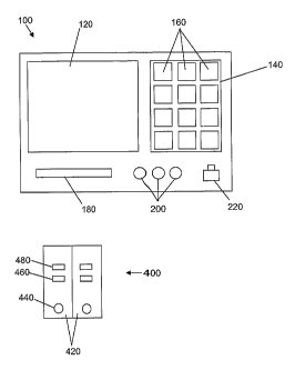
Referring to Fig. 1, diagnostic device 100 includes display 120 and input
region
140. The display 120 maybe used to display images in various formats, for
example,
joint photographic experts group (JPEG) format, tagged image file format
(TIFF),
graphics interchange format (GIF), or bitmap. Display 120 can also be used to
display
text messages, help messages, instructions, queries, test results, and various
information
to patients. In some implementations, display 120 supports the hypertext
markup
language (HTML) format such that displayed text may include hyperlinks to
additional
information, images, or formatted text. Display 120 can further provide a
mechanism for
displaying videos stored, for example in the moving picture experts group
(MPEG)
format, Apple's QuickTime format, or DVD format. Display 120 can additionally
include an audio source (e.g., a speaker) to produce audible instructions,
sounds, music,
and the like.
Input region 140 can include keys 160. In one embodiment, input region 140 can
be implemented as symbols displayed on the display 120, for example when
display 120
is a touch-sensitive screen. Patient instructions and queries are presented to
the patient on
display 120. The patient can respond to the queries via the input region.
Device 100 also includes cartridge reader 180, which accepts diagnostic test
cartridges for reading. The cartridge reader 180 measures the level of a
biomarker based
on, for example, the magnitude of a color change that occurs on a test
cartridge 400.
Device 100 also includes probe connections 200, which connect probes (e.g., a
probe of
weight, temperature, heart rate, variability of heart rate, breathing rate,
blood pressure, or
blood oxygen saturation) to the device.
Device 100 further includes a communication port 220. Communication port 220
can be, for example, a connection to a telephone line or computer network.
Device 100
can communicate the results of patient tests to a health care provider from a
remote
22
CA 02550090 2012-07-17
location. Likewise, the health care provider can communicate with the device
100 (e.g.,
to access stored test results, to adjust device parameters, or send a message
to the patient).
Cartridge 400 is shown with two testing zones 420. In general, a cartridge can
include 1, 2, 3, 4, or 5 or more testing zones. Each testing zone 420 can test
the level of a
biomarker. Each testing zone 420 includes a sample input 440, a control result
window
460 and a test result window 480. In one embodiment, the cartridge 400 is an
immunochromatographic test cartridge. Examples of immunochromatographic tests
and
test result readers can be found in, for example, U.S. Patent Nos. 5,504,013;
5,622,871;
6,235,241; and 6,399,398.
A patient can use device 100 for testing and recording the levels of various
biomarkers that provide information about the patient's health. Various
implementations
of diagnostic device 100 may access programs and/or data stored on a storage
medium
(e.g., video cassette recorder (VCR) tape or digital video disc (DVD); compact
disc (CD);
or floppy disk). Additionally, various implementations may access programs
and/or data
accessed stored on another computer system through a communication medium
including
a direct cable connection, a computer network, a wireless network, a satellite
network, or
the like.
The software controlling the diagnostic device and providing patient feedback
can
be in the form of a software application running on any processing device,
such as, a
general-purpose computing device, a personal digital assistant (PDA), a
special-purpose
computing device, a laptop computer, a handheld computer, or a network
appliance.
A diagnostic device may be implemented using a hardware configuration
including a processor, one or more input devices, one or more output devices,
a
computer-readable medium, and a computer memory device. The processor may be
implemented using any computer processing device, such as, a general-purpose
microprocessor or an application-specific integrated circuit (ASIC). The
processor can be
integrated with input/output (I/O) devices to provide a mechanism to receive
sensor data
and/or input data and to provide a mechanism to display or otherwise output
queries and
results to a service technician. Input device may include, for example, one or
more of the
following: a mouse, a keyboard, a touch-screen display, a button, a sensor,
and a counter.
The display 120 may be implemented using any output technology, including a
liquid crystal display (LCD), a television, a printer, and a light emitting
diode (LED).
The computer-readable medium provides a mechanism for storing programs and
data
either on a fixed or removable medium. The computer-readable medium may be
implemented using a conventional computer hard drive, or other removable
medium such
23
CA 02550090 2006-06-16
WO 2005/060652 PCT/US2004/042153
as those described above with reference to. Finally, the system uses a
computer memory
device, such as a random access memory (RAM), to assist in operating the
diagnostic
device.
Implementations of a diagnostic device can include software that directs the
patient in using the device, stores the result of biomarker measurements,
determines
whether a tested biomarker level requires medical attention for the patient,
instructs the
patient in adjusting or maintaining therapy, and communicates the patient's
information to
his or her caregiver. Patients suffering from, for example, heart failure or
hypertension,
or patients at risk of a myocardial infarction can use the device.
The device 100 can provide access to applications such as a medical records
database or other systems used in the care of patients. In one example, the
device
connects to a medical records database via communication port 220. Device 100
may
also have the ability to go online, integrating existing databases and linking
other
websites. Online access may also provide remote, online access by patients to
medical
information, and by caregivers to up-to-date test results reflecting the
health of patients.
The device can be used in the hospital, physician's office, clinic, and
patient's
home either by the patient or an attendant care giver. In one embodiment, the
invention is
practiced in the patient's home allowing the patient to be monitored, his or
her therapy
optimized, and adverse events that require hospitalization to be avoided.
The device can provide information on the patient's status and provide
instructions
or other actionable information to the healthcare professional and/or the
patient.
Examples, without limitation, of instructions that can be given include:
change diuretic
dose, withhold diuretic, introduce another diuretic, contact caregiver, no
change in care
plan necessary, change fluid intake, withhold potassium supplementation, and
increase
potassium supplementation. The objective is to track the patient's condition
and steer him
or her toward a stable condition through appropriate interventions made by the
patient or
the caregiver. Algorithms for treatment decisions are known. An example of a
set of
treatment algorithms can be found in: Healthcare Guideline; Congestive Heart
Failure in
Adults, Institute for Clinical Systems Improvement, Release July 2003; and
Silver M,
Pisano C, Cianci P, Outpatient management of heart failure: Program
development and
experience in clinical practice, Advocate Christ Medical Center, Oak Lawn, IL,
Post
Graduate Institute for Medicine 2003, each of which is incorporated by
reference in its
entirety.
24
CA 02550090 2006-06-16
WO 2005/060652 PCT/US2004/042153
Decision Points
The device can be configured to respond to the measured level of a biomarker,
in
particular when the level of the biomarker indicates a change in the patient's
health status.
For example, the device can be configured to store the results of tests and
determine
changes in the levels of markers over time. A change in results over time can
be an acute
change or a chronic change. An acute change can be a significant change in the
level of a
biomarker over a short period of time. The magnitude of change and period of
time can
be different for each biomarker. The device can be configured to compare each
new test
result either to a stored values of recent test results (e.g., the previous 1,
2, 3, 4, 5 or more
results), or to an aggregate measure of recent test results (such as an
average) to
determine if an acute change has occurred. In one example, an acute change is
detected
by the percentage change in a test result from the previous result.
Chronic changes can be detected as well. A chronic change can be a change in
the
level of a biomarker that occurs over a long period of time. For example, a
chronic
change can occur such that many testing intervals pass without an acute change
being
detected, yet the level of biomarker is significantly different. To detect a
chronic change,
the device can compare the results of each new test to a stored result of an
earlier test, or
to an aggregate measure of earlier tests. For detecting chronic changes, the
earlier test
can be, for example, 4-12 weeks prior to the new test result. In one example,
the
aggregate measure can be a rolling average, such as a 4-week, 8-week, or 12-
week rolling
average.
The device can also be configured to compare test results to a stored
threshold
value or range. The threshold value can be an upper or lower limit or range of
values.
Thus, the device can determine if the measured value of a marker, or group of
markers, is
a safe level, a dangerous level, or indicates an emergency. The device can
alert the
patient to the results of the test and can be configured, when appropriate to
instruct the
patient to seek medical care.
The device can also be configured to track combinations of markers, for
example,
an average value of two markers, the difference in level between two markers,
a ratio of
the levels of two markers, or whether two or more markers exceed their
respective
threshold values at the same time. The device can be configured to track one
or more
markers in combination with a patient's signs and symptoms.
The device can be personalized for a patient. The threshold values and other
parameters for each biomarker can be adjusted (for example, by a physician or
other
caregiver) based on the circumstances of the patient, such as, for example,
age, gender, or
CA 02550090 2006-06-16
WO 2005/060652 PCT/US2004/042153
disease status. The questions and responses that the device presents to the
patient can
also be adjusted.
Examples of how the device can record, detect changes, and respond to detected
changes in the level of a biomarker are presented below. The threshold values
and levels
of biomarkers referred to below are not limiting, may not be appropriate for
all patients,
and are for purposes of example only.
Marker of left ventricular volume overload and myocardial stretch
In one embodiment, the device is configured to measure the biomarker BNP in a
1o patient sample. The device can track the patient's BNP level as a function
of time and
detect changes in the BNP level. The changes can be acute or chronic. When a
change in
BNP level is detected, the device can respond with a request for additional
input for the
patient or instructions for the patient.
The device can determine a patient's baseline level of BNP, against which
future
measurements of BNP will be compared. The baseline level can be set based on
data on
the influence of the patient's gender, age, body mass, and degree of
hypertrophy. The
baseline can also be refined to set reasonable treatment targets for a patient
taking into
consideration the degree of disease comorbidities and the patient's prognosis.
A series of
BNP measurements can be used to set a baseline for a patient.
For example, the baseline can be defined as the average of the most recent two
test
results with an increase of maximum 10% (compared to the previous baseline)
out of the
last four tests. The following test results are excluded from the calculation:
o Any test result flagged with an acute symptom
o Any test of the 2 used for the calculation is older than 28 days
o The last 4 tests have been done in less than 4 days
Under certain conditions, no baseline value will be available, such as the
first use
of the device (i.e., no test results have been recorded); after the device has
been reset; or
when any of the test results used for the baseline calculation is older than
28 days. By
testing 4 times over at least 4 days, the initial baseline can be calculated.
When the
3o baseline is defined in this way, the device cannot give warnings for acute
deterioration
over this initial 4 day period. In case one value of the two used for the
calculation is older
than 28 days, one additional test can be sufficient to calculate the baseline.
The baseline
can be a variable baseline, changing as the most recent test results change in
value.
The device can detect acute changes in BNP level, and advise the patient to
take
appropriate responses. Criteria for determining an appropriate response can
include the
26
CA 02550090 2012-07-17
patient's initial BNP level, which can reflect the patient's risk profile; the
percentage
change in BNP level; the presence or absence of acute symptoms; and the
evolution of
BNP values to confirm a trend and exclude assay-to-assay, physiological, and
statistical
variations. When an acute increase in BNP level is detected by the device, the
device can
query the patient for the presence of acute symptoms. In advising a patient of
a response
to take to an increased BNP levels, the presence of one or more acute symptoms
can be a
deciding factor. Acute symptoms can include chest pain (AMI); a squeezing or
crushing
chest feel (AMI); pain radiating to neck, left arm (AMI); sweating, nausea, or
vomiting
(AMI, Stroke, pulmonary TE); loss of consciousness; acute dyspnea (AMI,
1o decompensation, pulmonary thrombo-embolism); palpitations without exercise;
dyspnea
when laying down (right heart decompensation); sudden headache (stroke); and
sudden
vision impairment (stroke). See, for example, Harrisson T.R. et al, Principles
of Internal
Medicine. McGraw Hill, Inc. 1983, 1432-34 & 1353-58 & 2038-39. When the
patient indicates that any acute symptoms are present, the device can advise
the
patient to seek medical care at once.
If there is an acute increase in BNP level, but the patient is not
experiencing any
acute symptoms, the device's response can depend on the percentage change in
BNP level
and the absolute BNP level. In general, a large percentage increase in BNP
level and a
high absolute level can indicate a deterioration in the patient's condition,
and the device
can respond by prompting the patient to seek medical care at once. A smaller
percentage
change and lower absolute level may not require immediate medical attention,
and the
change in BNP level can be confirmed by a second test. In one example, the
severity of a
patient's disease can be stratified by absolute BNP levels as follows (see,
for example,
Clerico A, et al.. Clin Chem Lab Med 2002 Apr; 40(4): 371-7; and Nomura H, et
al. JAna
Geriatr Soc 2002 Sep; 50(9): 1504-9).
o < 20 pg/mL Healthy
o 20 - 50 pg/mL 1 risk factor: hypertension or age
o 50- 100 pg/mL 2 risk factors: hypertension, age, post-AMI
o >100 pg/mL chronic hart failure patient NYHA classes 1-4
Changes in BNP level can also be grouped by severity, for example, no
increase,
an increase of less than 10%, 10-20%, 20-30%, 30-40%, or 40% or more.
A second test can exclude assay-to-assay or physiological variations and thus
confirm the increase. The second test can be given after a predetermined
interval, which
can vary depending on the severity of the increase (e.g., within 30 minutes,
60 minutes,
27
CA 02550090 2012-07-17
the same day, or within 24 hours of the first test). If the second test result
is a lower BNP
value, then a third test can be performed. The third test can confirm an
increase in this
case, or, for example, exclude a non-pathological transient rise of more than
20% due to
exercise.
For example, if the BNP level increases by 10% or less (and the patient has no
acute symptoms), the device can prompt the patient to perform a second test.
The second
test can be performed the next day (for example, if the patient's BNP level is
less than 50
pg/mL) or sooner, such as thirty minutes later (for example, if the patient's
BNP level is
50 pg/mL or greater). If the BNP level has increased by 10-20%, the device can
prompt
the patient to perform a second test, for example, within thirty minutes of
the first test. If
the BNP level has increased by more than 20%, the device can prompt the
patient to seek
medical care at once. An increase of more than 30 % can be regarded as the
strongly
indicative for ischemia and AMI or an acute heart decompensation. See, for
example,
Kyriakides ZS, et al. Clin Cardiol 2000 Apr; 23(4): 285-8; and Nakamura T, et
al. J. Ana.
Coll. Cardiol. 2002 May 15;39 (10): 1657-63. If the patient's BNP level has
not
increased, or increased by less than 5%, the device can prompt the patient to
perform a second test at a predetermined interval, such as seven days.
The device can respond to the results of the second test. If the second test
is
performed on the day after the previous test (e.g., when the patient's BNP
level is less
than 50 pg/mL), the device can respond as follows. If the second test reveals
a BNP level
more than 20% above the baseline, the patient is instructed to seek medical
care at once.
The patient can be instructed to perform a third test if the second test
reveals a BNP level
that is between 0 and 20% higher than the baseline. The third test can be
performed, for
example, on the day following the second test. If the third test indicates
that the patient's
BNP level is between 10% and 20% higher than the baseline, the patient is
instructed to
seek medical care at once. However, if the third test reveals a BNP level
between 0 and
10% higher than the baseline, the baseline can be adjusted to the average of
the previous
baseline and the result of the third test. The patient is instructed to resume
a regular test
schedule, such as once a week.
If the second test is performed within thirty minutes of the previous test
(e.g.,
when the patient's BNP level is 50 pg/mL or greater), the device can respond
as follows.
When the second test result is 20% or more above the baseline, the patient is
instructed to
seek medical care at once. The patient can be instructed to perform a third
test if the
second test reveals a BNP level that is between 0 and 20% higher than the
baseline. The
28
CA 02550090 2006-06-16
WO 2005/060652 PCT/US2004/042153
third test can be performed within thirty minutes of the second test (such as
when the
second test result was between 10% and 20% above the baseline) or within four
hours of
the second test (such as when the second test result was between 0 and 10%
above the
baseline). If the third test indicates that the patient's BNP level is between
10% and 20%
higher than the baseline, the is instructed to seek medical care at once.
However, if the
third test reveals a BNP level between 0 and 10% higher than the baseline, the
baseline
can be adjusted to the average of the previous baseline and the result of the
third test. The
patient is instructed to resume a regular test schedule, such as once a week.
Chronic chances
The device can detect chronic changes in BNP level; in other words, slow
changes
that accumulate over time to reflect a change in the patient's condition. A
chronic change
can be measured, for example, by observing changes in a rolling average of BNP
values,
such as a rolling 2-week average. To exclude increases of an acute nature, or
due to a
temporary event (such as exercise), only those chronic increases that are
manifested for at
least two weeks where the increases outnumber the decreases can be considered
as
chronic increases. A chronic increase can be small (e.g., approximately 10 %)
when
consistent over a long time (such as one month) or can be large (for example,
approximately 20%) over a relatively short term (such as two weeks). It can be
important
to exclude increases due to assay-to-assay variability, physiological rises,
etc., before
concluding that a chronic increase has occurred. In order to due so,
sufficient test results
have to be available. Therefore, upon suspicion of a chronic increase,
patients can be
instructed to perform more tests.
A rolling average can be the average of test results performed within a given
time
frame. For example, a rolling two week average can be the average of results
recorded
over the previous 15 days, a rolling 4 week average can be the average of
results recorded
over the previous 29 days, and a rolling 12 week average can be the average of
results
recorded over the previous 85 days. When calculating a rolling average, any
test result
recorded with an acute symptom flag (i.e., a test result where the patient was
suffering an
acute symptom at the time of the test) can be excluded. Under certain
circumstances, a
rolling average cannot be calculated, such as the first use of the system,
following a
system reset, or when the system has not been used over the relevant length of
time (e.g.,
2, 4 or 12 weeks). By testing once a week over at least 15 days (three test
results), an
initial 2-week rolling average can be calculated. This means that the device
cannot give
warnings for chronic deterioration over this initial 2-week period.
29
CA 02550090 2006-06-16
WO 2005/060652 PCT/US2004/042153
When a chronic increase in BNP levels is detected, the device can query the
patient for the presence of chronic symptoms. Examples of chronic symptoms
include
increasing fatigue in general (heart performance reduction); shortening of
walking
distance or step climbing (heart performance reduction); aggravating chronic
dyspnea
(right heart decompensation, multiple pulmonary thrombo-embolism);
palpitations
without exercise; aggravating dyspnea when laying down (decompensation);
aggravating
swollen feet or legs; or memory loss or paralysis or equilibrium disturbance.
When the
patient indicates that any chronic symptoms are present, the device can advise
the patient
to seek medical care at once.
If there is a chronic increase in BNP level, but the patient is not
experiencing any
chronic symptoms, the device's response can depend on the percentage change in
BNP
level and the absolute BNP level. In general, a large percentage increase in
BNP level
and a high absolute level can indicate a deterioration in the patient's
condition, and the
device can respond by prompting the patient to seek medical care at once. A
smaller
percentage change and lower absolute level may not require immediate medical
attention,
and the change in BNP level can be confirmed by a second test. In one example,
the
severity of a patient's disease can be stratified by absolute BNP levels as
follows:
o < 20 pg/mL Healthy
o 20 - 50 pg/mL 1 risk factor: hypertension or age
o 50 -100 pg/mL 2 risk factors: hypertension, age, post-AMI
o >100 pg/mL chronic hart failure patient NYHA classes 1-4
Chronic changes in BNP level can also be grouped by the duration in the
change,
for example, a change in the two-week rolling average, a change in a 4-week
rolling
average, or a change over a longer interval, such as a change in the 12-week
rolling
average. In each of these time periods, the changes in BNP level can be
grouped by
severity, such as no increase, an increase of greater or less than 7.5%, an
increase of
greater or less than 15%, an increase of less than 10%, an increase of 10-30%,
an increase
of 30-50%, or an increase of more than 50%.
For example, when the device detects a small, chronic increase in the two-week
3o rolling average (e.g., an increase of less than 10%) in the patient's BNP
level, and the
patient reports no chronic symptoms, the device can instruct the patient to
perform a
second test after a predetermined interval, such as 7 days. If there is a
moderate increase
in the patient's two-week rolling average BNP level (e.g., an increase of 10-
30%), the
device can instruct the patient to perform a second test after a predetermined
interval,
such as within 24 or 48 hours. A large increase (e.g., of 30-50%) and a small
absolute
CA 02550090 2006-06-16
WO 2005/060652 PCT/US2004/042153
BNP level (e.g., less than 50 pg/mL) can cause the device to instruct the
patient to
perform a second test after a predetermined interval, such as within 24 or 48
hours. When
the device detects a severe increase (e.g., of more than 50%) in the two-week
rolling
average, it can instruct the patient to seek medical care at once.
If the second test result is a BNP level higher than the previous two-week
rolling
average, the device can instruct the patient to seek medical care at once. If,
on the other
hand, the second test result is lower than the previous result, the device can
instruct the
patient to perform additional test (e.g., one test each day) until the BNP
level either
returns to its previous level, or the BNP level increases, which will result
in a prompt to
the patient to seek medical care at once. If the BNP level does not return to
its previous
level or increase within one week, the device can prompt the patient to seek
medical care
at once.
When there is a small increase in the 4-week rolling average (e.g., an
increase of
less than 15%), and the patient reports no chronic symptoms, the device can
instruct the
i5 patient to perform a second test after a predetermined interval, such as 7
days. When
there is a large increase in the 4-week rolling average (e.g., an increase of
15% or
greater), the device can instruct the patient to report to his or her health
care provider.
When there is a small increase in the 12-week rolling average (e.g., an
increase of
less than 7.5%), and the patient reports no chronic symptoms, the device can
instruct the
patient to perform a second test after a predetermined interval, such as 7
days. When
there is a large increase in the 4-week rolling average (e.g., an increase of
7.5% or
greater), the device can instruct the patient to report to his or her health
care provider.
The parameters used by the devices (i.e., the values of percentage change in
BNP
level, absolute BNP level, patient messages, etc.), can be altered. For
example, a
physician or other health care provider can adjust the value of acute increase
in BNP level
required to prompt the patient to seek medical care to a desired value. In
this way, the
behavior of the device can be tailored according to the preferences of a
physician or to the
needs of a particular patient or group of patients.
Markers of renal functioiz
The following uses, by example, the biomarker creatinine. The accepted method
for use in routine care is a measure of creatinine using adjustment with the
Cockroft and
Gault equation. Creatinine can provide important information on volume status
and
should be followed in patients during optimization of pharmacological agents
(e.g. an
ACE inhibitor) and ideally throughout the patient's care. Tests are performed
by the
31
CA 02550090 2012-07-17
patient or the healthcare provider every day, at a suitable testing interval,
or with the
onset of certain signs and symptoms. An increase in serum creatinine of 0.05
to 0.5
mg/dL is an indication for reassessment of volume status. Renal function
declines with
age; many elderly patients have a glomerular filtration rate below 50
mL/minute. Further,
as stated, an early increase of <30% in the concentration of creatinine is
expected when a
patient is administered an ACE inhibitor. GFR monitoring using, for example,
creatinine
is important in these patients.
Further, when using BNP to guide the optimization of pharmacological
treatment,
an estimate of GFR is essential to avoid under-hydration. An'action level'
(e.g. a level
that defines a significant reduction in renal perfusion) for GFR will require
the healthcare
professional and/or the patient to follow a predefined intervention dependent
on the rate
of change of GFR over time and the absolute level. An intervention might
include a
change in diuretic dose, withhold the diuretic, introduce another diuretic,
change fluid
intake, withhold potassium supplementation, increase potassium
supplementation, contact
the healthcare professional, refer to the Emergency Department, etc). As
described,
alternative markers of GFR can be used, such as Cystatin C.
Renal function can also be used as a prognostic marker, to provide information
on
a patient's health over a long period of time. The prognostic value of a
measure of renal
function (e.g., GFR as determined by a creatinine or Cystatin C measurement)
can be
independent of the use of renal function to monitor hydration on a short-term
basis (e.g.,
during diuretic use). An average measure of renal function determined over a
period of
time can be used for prognostic purposes. See, for example, Koenig W, et al.
Clin Chem.
10.1373/clinchem.2004.041889 2004 Nov; and Gottlieb SS, et al., J Card Fail.
2002
Jun;8(3):136-41.
Marker of myocardial apoptosis or injury
Measurement of a marker of myocardial apoptosis or injury, such as a troponin,
in
a remote setting using frequent testing is clinically useful. Tests are
performed by the
patient or the healthcare provider every day, at a suitable testing interval,
or with the
onset of certain signs and symptoms.
Marker of inflammation
Measurement of a marker of inflammation in a remote setting is clinically
useful.
The marker of inflammation can include, for example, E-selectin, P-selectin,
intracellular
adhesion molecule-1, vascular cell adhesion molecule-1, Nourin-1, interieukin-
113,
32
CA 02550090 2006-06-16
WO 2005/060652 PCT/US2004/042153
interleukin-6, interleukin-8, interleukin-10, tumor necrosis factor-alpha, hs-
CRP,
neutrophils, or white blood cell count. Tests are performed by the patient or
the
healthcare provider every day, at a suitable testing interval, or with the
onset of certain
signs and symptoms.
Marker of anemia
Measurement of a marker of anemia, such as hemoglobin or hematocrit, in a
remote setting using frequent testing is clinically useful. Tests are
performed by the
patient or the healthcare provider every day, at a suitable testing interval,
or with the
onset of certain signs and symptoms.
Markers of myocardial ischemia
Chronic myocardial ischemia is the leading cause of impaired myocardial
contractility and heart failure. Ischemia markers (e.g. ischemia modified
albumin,
oxygen-regulated peptide, and free fatty acid) have a faster release profile
than early
necrosis markers (e.g. myoglobin or fatty acid binding protein (H-FABP)).
Tests are
performed by the patient or the healthcare provider every day, at a suitable
testing
interval, or with the onset of certain signs and symptoms.
Markers of electrolyte balance and markers of sodium retention
Levels of electrolyte balance and sodium retention may be determined by
measurement of the levels of sodium and potassium ion concentrations in serum.
This
may be conveniently done using ion selective electrodes.
Examples of Use
The diagnostic kit can be used in a remote care setting (e.g. the patient's
home, a
nursing home, etc).
1. Optimization of Therapy
1.1 Normalize the patient's fluid balance using diuretics.
BNP levels will fall as the patient's fluid balance is returned to normal. If
adequate
diuresis cannot be achieved with a single diuretic (e.g., a loop diuretic), a
second diuretic
will be necessary (e.g., a thiazide diuretic). Careful monitoring of renal
function and
electrolyte balance is necessary. BNP levels cannot be used as the sole marker
of
achieving a safe fluid balance.
1.2 Introduction of an ACE inhibitor
33
CA 02550090 2006-06-16
WO 2005/060652 PCT/US2004/042153
ACE inhibitors improve prognosis mainly through their vasodilatory effect.
Patients are at a high risk of renal dysfunction and hypotension - this has
resulted in
under-dosing and under-use.
Impaired renal function can be reversed by pulling back on the diuretic dose
without needing to down-titrate the ACE inhibitor dose.
Monitoring renal function when up-titrating an ACE inhibitor will assure the
physician that renal function is not compromised.
Doses should be up-titrated over 1-4 weeks to reach target doses. If adverse
events
occur which cannot be overcome by reducing the diuretic or holding at the
current dose
for longer, then alternative drugs should be used (e.g., angiotensin II
receptor blocker
(ARB) and/or hydralazine /isosorbide dinitrates).
Effective treatment will result in a gradual fall in BNP. Measurement of a GFR
marker will identify reduced renal perfusion. Sudden increases in BNP will
indicate
volume overload.
1.3 Introduction of a Beta-Blocker
As stated, beta-blockers reduce the harmful effects of excessive and
continuous
increased adrenergic drive on the heart and lead to improvements in
ventricular structure
and function. There is an initial and transient decrease in contractility.
However, beta-blockers also reduce heart rate and blood pressure and therefore
need careful introduction and up-titration. Up-titration of a beta-blocker
should be
delayed when volume overload is present.
The initial dose needs to be up-titrated every 2-4 weeks as tolerated.
Patients must
be monitored for hypotension, fluid retention, and bradychardia. If any of
these persist,
the dose should be lowered until tolerated and then increased again, more
gradually.
Effective treatment will result in a gradual fall in BNP. Sudden increases in
BNP
will indicate volume overload.
2. Maintenance of Therapy
Assuming that target drug levels have been achieved, the patient would
normally
be responsible for tracking daily weight and be asked to comply with an
appropriate diet.
3o Some patients are allowed responsibility for their diuretics - for example,
rapid weight
gain can then be offset by up-titrating the diuretic dose for a day or so. The
diagnostic
would be used to:
i) detect the onset of decompensation
ii) steer diuretics/water/salt to maintain a 'normal fluid balance'
34
CA 02550090 2012-07-17
iii) provide the patient with a'number' to reinforce well-being and promote
compliance
Changes in renal function caused by the introduction of an ACE inhibitor are
tolerable and, in fact, an ACE inhibitor has a beneficial effect on the
kidneys. It is agreed,
however, that GFR still needs to be closely tracked to i) assess the impact of
an ACE
inhibitor on renal function and ii) to track for evidence of dehydration
through over-use of
diuretics.
A patient receiving a synthetic BNP (e.g., neseritide) as part of their
treatment can
be monitored. The synthetic BNP can be administered as an acute treatment
(i.e., in
response to an acute event) or a chronic treatment (i.e., to treat a chronic
condition). In
particular, the patient's inherent BNP production can be measured by measuring
the
patient's N-BNP (the inactive, N-terminal portion of proBNP). Synthetic BNPs
do not
include N-BNP. N-BNP is therefore a marker of the patient's BNP production,
and is not
affected by administration of a synthetic BNP.
3. Tracking exercise training
For some patients, exercise training improves cardiac health. However, the
training regimen must be calibrated to avoid overexertion. The device can
track the
patient's training regimen, for example by recording the frequency and
duration of
exercise. The records can by the patient as a log, for example, with date,
time, and a
description of the exercise, or the device can be connected to an piece of
exercise
equipment such as a treadmill, and make recordings directly from the piece of
equipment.
Tracking of exercise can be done in conjunction with measurement of actual
heart
function using biomarkersa For example, short- and long-term changes in a
biomarker
can provide information on how well the patient is responding to the exercise
training.
The device can provide instructions or reminders to the patient to exercise,
as
well as query the patient with regards to signs and symptoms during and after
exercise.
For example, if a patient's symptoms worsen after exercise, the device can
suggest a
decrease in frequency or intensity of exercise, or alert a health care
provider. If the
patient's health is improving with exercise, the device can suggest an
increase in
frequency or intensity of exercise. A health care provider can obtain records
of the
patient's exercise from the device.