Note: Descriptions are shown in the official language in which they were submitted.
CA 02550713 2014-01-22
SURGICAL INSTRUMENT HAVING
[00011 FLUID ACTUATED OPPOSING JAWS
FIELD OF THE INVENTION
100021 The present invention relates in general to surgical stapler
instruments that are
capable of applying lines of staples to tissue while cutting the tissue
between those staple
lines and, more particularly, to improvements relating to stapler instruments
and
improvements in processes for forming various components of such stapler
instruments.
BACKGROUND OF THE INVENTION
100031 Surgical instruments for minimally invasive surgery are increasingly
relied upon
to reduce the hospital stay and recovery time for various surgical procedures.
Many of
these surgical instruments include mechanisms that actuate an end effector via
an
elongate shaft that performs a surgical step that entails two opposing
surfaces being
brought into opposition to each other. For instance, pivotally opposed jaws
are used in
graspers. Pivotally attached scissor blades are incorporated into cutting
devices. Providing
an actuating control down the elongate shaft with sufficient strength is
complicated by a
design goal of minimum cross sectional area so as to pass through a small
cannula of a
trocar. In addition, the elongate shaft often has a plurality of control
functions (e.g.,
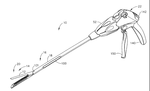
rotation, articulation, etc.) Further, it is desirable to have reduced design
complexity so as
to provide an economical device.
100041 As an illustration of a particularly challenging surgical
instrument, surgical
staplers have been used in the prior art to simultaneously make a longitudinal
incision in
tissue and apply lines of staples on opposing sides of the incision. Such
instruments
commonly include a pair of cooperating jaw members that, if the instrument is
intended
CA 02550713 2006-06-21
for endoscopic or laparoscopic applications, are capable of passing through a
cannula
passageway. One of the jaw members receives a staple cartridge having at least
two
laterally spaced rows of staples. The other jaw member defines an anvil having
staple-
forming pockets aligned with the rows of staples in the cartridge. The
instrument includes
a plurality of reciprocating wedges which, when driven distally, pass through
openings in
the staple cartridge and engage drivers supporting the staples to effect the
firing of the
staples toward the anvil.
100051 An example of a surgical stapler suitable for endoscopic
applications, described in
U.S. Pat. No. 5,465,895, advantageously provides distinct closing and firing
actions.
Thereby, a clinician is able to close the jaw members upon tissue to position
the tissue
prior to firing. Once the clinician has determined that the jaw members are
properly
gripping tissue, the clinician can then fire the surgical stapler, thereby
severing and
stapling the tissue. The simultaneous severing and stapling avoids
complications that may
arise when performing such actions sequentially with different surgical tools
that
respectively only sever or staple.
100061 These minimally invasive surgical instruments have been widely used
and have
proven to be a significant advance over traditional open surgical techniques.
It would be
desirable to incorporate yet additional features and capabilities.
BRIEF SUMMARY OF THE INVENTION
10007] The invention overcomes the above-noted and other deficiencies of
the prior art
by including a surgical instrument that is suitable for minimally invasive
surgical
procedures which has a handle that positions an end effector through a
surgical opening
via an elongate shaft. The end effector has a pair of pivoting members
opposingly
contacting tissue. A fluid actuated closure mechanism responds to a closure
action by a
fluid actuator attached to the handle by bi-directionally transferring fluid
across a fluid
conduit to a fluid reservoir positioned to urge the pair of pivoting members
closed.
Thereby, the integration of fluid conduits within an elongate shaft allows for
shafts of a
desirable small cross section which are able to perform an important surgical
operation.
100081 In one aspect of the invention, a surgical instrument has an end
effector that is
actuated by a fluid actuator to open and close upon tissue. Once closed, a
firing bar that is
2
CA 02550713 2006-06-21
received for reciprocating a longitudinal firing motion in an elongate shaft
transfers a
longitudinal firing motion from a handle to actuate a staple cartridge and to
sever the
clamped tissue in the end effector.
[0009] In yet another aspect of the invention, a surgical instrument
includes a handle that
produces closure actuation that transfers fluid through a fluid conduit in an
elongate shaft
to a fluid actuator positioned in a lever cavity to position a lever. The
lever of a first tissue
contacting member extends proximally into the lever cavity from a pivotal
connection
with a second tissue contacting member. Fluid transfer advantageously effects
pivotal
movement of the pair of tissue contacting members.
[0010] These and other objects and advantages of the present invention
shall be made
apparent from the accompanying drawings and the description thereof
BRIEF DESCRIPTION OF THE FIGURES
[0011] The accompanying drawings, which are incorporated in and constitute
a part of
this specification, illustrate embodiments of the invention, and, together
with the general
description of the invention given above, and the detailed description of the
embodiments
given below, serve to explain the principles of the present invention.
[0012] FIG. 1 is a perspective view of a surgical stapling and severing
instrument having
a fluid actuated upper jaw (anvil) in an open position and an electroactive
polymer (RAP)
medical substance dispensing shaft.
[0013] FIG. 2 is a disassembled perspective view of an implement portion of
the surgical
stapling and severing instrument of FIG. 1.
[0014] FIG. 3 is left side view in a elevation of the implement portion of
the surgical
stapling and severing instrument of FIG. 1 taken in cross section generally
through a
longitudinal axis and passing through an offset EAP syringe and receptacle
that is in fluid
communication with a dispensing groove in an E-beam firing bar.
[0015] FIG. 4 is a left side detail view in elevation of a distal portion
of the implement
portion of the surgical stapling and severing instrument of FIG. 1 taken in
cross section
generally through the longitudinal axis thereof but showing a laterally offset
fluid actuator
bladder actuator opening the anvil.
3
CA 02550713 2006-06-21
[0016] FIG. 5 is a left side detail view of an E-beam firing bar
incorporating medical
substance ducting.
[00171 FIG. 6 is a left side detail view in elevation of the distal portion
of the implement
portion of the surgical stapling and severing instrument of FIG. 4 taken in
cross section
generally through the longitudinal axis thereof with the anvil closed.
[0018] FIG. 7 is a left side detail view of the E-beam firing bar of FIG.
6.
[0019] FIG. 8 is a top detail view of a joined portion of a lower jaw
(staple channel) of
the end effector and elongate shaft taken in cross section through the lines 8-
8 depicting
guidance to the E-beam firing bar.
loom FIG. 9 is a front view of a firing bar guide of the implement portion
of the surgical
stapling and severing instrument of FIG. 2.
[0021] FIG. 10 is a left side view of the firing bar guide of FIG. 9 taken
in cross section
along lines 9-9.
[0022] FIG. 11 is a front view in elevation of the elongate shaft of the
surgical stapling
and severing instrument of FIG. 3 taken along lines 11-11 taken through a
distal end of
the EAP medical substance syringe.
[0023] FIG. 12 is a left side view of the EAP medical substance syringe of
FIG. 1 1 .
[0024] FIG. 13 is a left side view of the implement portion of the surgical
stapling and
severing instrument of FIG. 1 partially cut away to show proximal mountings
for the EAP
medical substance syringe.
[0025] FIG. 14 is a left side detail view of the EAP medical substance
syringe and
receptacle of the elongate shaft of the surgical stapling and severing
instrument of FIG.
13.
[0026] FIG. 15 is a top view of the firing bar of the surgical stapling and
severing
instrument of FIG. 2.
[0027] FIG. 16 is a left side view of a laminate firing bar showing an
internal fluid path in
phantom for the surgical stapling and severing instrument of FIG. 1.
4
CA 02550713 2006-06-21
[0028] FIG. 17 is a left side detail view of an alternate E-beam showing an
internal fluid
path in phantom showing an internal fluid path in phantom.
[0029] FIG. 18 is a front view in elevation of the laminate firing bar of
FIG. 15 taken in
cross section along line 18-18 through a proximal open groove of a fluid path.
[00301 FIG. 19 is a left side view of an alternative surgical stapling and
severing
instrument of FIG. 1 partially cut away and depicting control circuitry and
controls.
[0031] FIG. 20 is a flow diagram of a sequence of operations performed by
control
circuitry of the surgical stapling and severing instrument of FIG. 19.
DETAILED DESCRIPTION OF THE INVENTION
[0032] Turning to the Drawings, wherein like numerals denote like
components
throughout the several views, FIGS. 1-2 show a surgical stapling and severing
instrument
10{xe "0010 surgical stapling and severing instrument") that is capable of
practicing the
unique benefits of the present invention, including both fluid actuation
(e.g., opening,
closing/clamping) of an upper jaw (anvil){xe "012 upper jaw (anvil)") 12 of an
end
effector{xe "014 end effector"} 14 as well as dispensing a medical substance
onto tissue
as severed. Fluid actuation of the end effector 14 provides a range of design
options that
avoid some design limitations of traditional mechanical linkages. For example,
instances
of binding or component failure may be avoided. Further, dispensing liquids
onto severed
tissue allows for a range of advantageous therapeutic treatments to be
applied, such as the
application of anesthetics, adhesives, cauterizing substances, antibiotics,
coagulant, etc.
[0033] With particular reference to FIG. 2, the surgical stapling and
severing instrument
includes an implement portionIxe "016 implement portion") 16 formed by an
elongate
shaft{xe "018 elongate shaft") 18 and an end effector 14, depicted as a
stapling
assembly{xe "020 end effector, depicted as a stapling assembly") 20. The
surgical
stapling and severing instrument 10 also includes a handle{xe "022 handle") 22
(FIG. 1)
attached proximally to the shaft 18. The handle 22 remains external to the
patient as the
implement portion 16 is inserted through a surgical opening, or especially a
cannula of a
trocar that forms a pneumoperitoneum for performing a minimally invasive
surgical
procedure.
5
CA 02550713 2006-06-21
[0034] Left and right fluid actuator bladders (lift bags){xe "024, 026 left
and right fluid
actuator bladders (lift bags)") 24, 26 are supported within an aft portion{xe
"028 aft
portion of staple channel") 28 of a staple channel{xe "030 staple channel")
30. The anvil
12 includes a pair of inwardly directed lateral pivot pins{xe "032, 034 pair
of inwardly
directed lateral pivot pins in anvil") 32, 34 that pivotally engage outwardly
open lateral
pivot recesses{xe "036, 038 outwardly open lateral pivot recesses") 36, 38
formed in the
staple channel 30 distal to the aft portion 28. The anvil 12 includes a
proximally directed
lever tray{xe "040 proximally directed lever tray"} 40 that projects into the
aft portion 28
of the staple channel 30 overtop and in contact with the fluid actuator
bladders (lift bags)
24, 26 such that filling the fluid actuator bladders 24, 26 causes a distal
clamping
section{xe "041 distal clamping section") 41 of the anvil 12 to pivot like a
teeter-totter
toward a staple cartridge{xe "042 staple cartridge") 42 held in an distal
portion{xe "044
distal portion of the staple channel") 44 of the staple channel 30. Evacuation
and collapse
of the fluid actuator bladders 24, 26, or some other resilient feature of the
end effector 14,
causes the anvil 12 to open. Left and right fluid conduits{xe "046, 048 left
and right fluid
conduits") 46, 48 communicate respectively with the left and right fluid
actuator bladders
24, 26 to bi-directionally transfer fluid for actuation.
[0035] It will be appreciated that the terms "proximal" and "distal" are
used herein with
reference to a clinician gripping a handle of an instrument. Thus, the staple
applying
assembly 20 is distal with respect to the more proximal handle 22. It will be
further
appreciated that, for convenience and clarity, spatial terms such as
"vertical" and
"horizontal" are used herein with respect to the drawings. However, surgical
instruments
are used in many orientations and positions, and these terms are not intended
to be
limiting and absolute.
[0036] The elongate shaft 18 includes a frame{xe "050 frame") 50 (FIG. 2)
whose
proximal end is rotatably engaged to the handle 22 such that a rotation
knob{xe "052
rotation knob") 52 rotates the frame 50 along with the end effector 14. A
distal end of the
frame has lateral recesses{xe "054 lateral recesses") 54 that engage a distal
lip{xe "056
distal lip"} 56 of the staple channel 30. The frame 50 includes a laterally
centered, bottom
firing slotlxe "058 laterally centered, bottom firing slot") 58 that passes
longitudinally
through the frame 50 for receiving a two-piece firing bar{xe "060 two-piece
firing bar")
60 comprised of a firing bar{xe "062 firing bar") 62 with a distally attached
E-beam{xe
6
CA 02550713 2006-06-21
"064 E-beam") 64, the latter translating within the staple applying assembly
20 to sever
and staple tissue. A distal portion of the frame 50 includes an upper
cavity{xe "066 upper
cavity"} 66 whose distal and proximal ends communicate through distal and
proximal
apertures{xe "068, 070 distal and proximal apertures") 68, 70, defining there
between a
cross bar{xe "072 cross bar"} 72 over which a distally projecting clip 74 of a
clip spring
76 engages with a lower spring arm{xe "078 lower spring arm") 78, distally and
downwardly projecting through the upper cavity 66 to bias the firing bar 62
downwardly
into engagement with the staple channel 30, especially when the lower spring
arm 78
encounters a raised portion{xe "080 raised portion") 80 on the firing bar 62.
[0037] Medical substance dispensing is integrated into the elongate shaft
18 by including
a laterally offset cylindrical cavity{xe "090 laterally offset cylindrical
cavity") 90 formed
in the frame 50 that communicates along its longitudinal length to the outside
via a
rectangular aperture(xe "092 rectangular aperture") 92 that is slightly
shorter than an
electroactive polymer (EAP) syringe{xe "100 electroactive polymer (EAP)
syringe") 100
that is inserted through the aperture 92 into the cylindrical cavity 90. A
proximal portion
of the cylindrical cavity 90 contains a longitudinally aligned compression
spring{xe "102
longitudinally aligned compression spring") 102 that urges a distal dispensing
cone{xe
"104 distal dispensing cone") 104 of the EAP syringe 100 distally into sealing
contact
with the frame 50 and allows translation for insertion and removal of the EAP
syringe
100. An electrical conductor{xe "106 electrical conductor") 106 passes through
the frame
50 and is attached to the compression spring 102, which is also formed of an
electrically
conductive metal. An aft portion of the EAP syringe is conductive and contacts
the spring
102 to form a cathode to an EAP actuator{xe "110 EAP actuator") 110 held in a
proximal
portion of the EAP syringe 100. It will be appreciated that another conductor,
perhaps
traveling with the conductor 106, also electrically communicates to the EAP
actuator 110
to serve as the anode.
[0038] When activated, the EAP actuator 110 longitudinally expands, serving
as a
plunger to dispel a medical substance{xe "112 medical substance") 112 in a
distal portion
of the EAP syringe 100 through the distal dispensing cone 104. Insofar as the
EAP
actuator 110 laterally contracts to compensate for its longitudinal expansion,
a plunger
seal{xe "114 plunger seal"} 114 maintains a transverse seal within the EAP
syringe 100.
A vent (not shown), such as around conductor 106 allows air to refill the EAP
syringe
7
CA 02550713 2006-06-21
100 as the medical substance 112 is dispensed. The vent may rely upon the
surface
tension of the medical substance 112 to prevent leaking. Alternatively, a one-
way valve
may be used for the same purpose. As described below, the medical substance
112 is
conducted by the frame 50 to a lateral fluid groove{xe "120 lateral fluid
groove") 120 that
is formed in the firing bar 62 and the E-beam 64 to direct the medical
substance to a
cutting surface{xe "122 cutting surface"} 122 of the E-beam 64. The frame slot
58 is sized
to seal the lateral fluid groove 120. The portion of the lateral fluid groove
120 that is
positioned under the spring clip 76 is sealed by a firing bar guide{xe "124
firing bar
guide"} 124. In the illustrative version, an outer sheath{xe "130 outer
sheath"} 82
encompasses the frame 50 and proximally projecting lever tray 40 of the anvil
12. A top
distal opening{xe "131 top distal opening") 131 allows closing of the anvil
12.
100391 An outer rectangular aperture{xe "132 outer rectangular aperture")
132 of the
outer sheath 130 is sized and longitudinally positioned to correspond to the
rectangular
aperture 92 formed in the frame 50. In some applications, the outer sheath 130
may be
rotated to selectively align the rectangular aperture 92 with the outer
rectangular aperture
132 for insertion or removal of the EAP syringe 100. It should be appreciated
that in
some applications the EAP syringe 100 may be integrally assembled into an
elongate
shaft that does not allow for selecting a desired medical substance. For
instance, a
disposable implement portion with an integral staple cartridge and medical
dispensing
reservoir may be selected by the clinician as a unit. It is believed that
allowing insertion at
the time of use, though, has certain advantages including clinical flexibility
in selecting a
medical substance (e.g., anesthetics, adhesives, antibiotics, cauterizing
compound, etc.)
and extending the shelf life! simplifying storage and packaging of the
implement portion
16.
[0040] In the illustrative version, an elongate stack of many disk-shaped
EAP layers are
aligned longitudinally and configured to expand along this longitudinal axis.
Electroactive polymers (EAPs) are a set of conductive doped polymers that
change shape
when electrical voltage is applied. In essence, the conductive polymer is
paired to some
form of ionic fluid or gel and electrodes. Flow of the ions from the fluid/gel
into or out of
the conductive polymer is induced by the voltage potential applied and this
flow induces
the shape change of the polymer. The voltage potential ranges from 1V to 4kV,
depending on the polymer and ionic fluid used. Some of the EAPs contract when
voltage
8
CA 02550713 2014-01-22
is applied and some expand. The EAPs may be paired to mechanical means such as
springs or flexible plates to change the effect that is caused when the
voltage is applied.
100411 There are two basic types of EAPs and multiple configurations of
each type. The
two basic types are a fiber bundle and a laminate version. The fiber bundle
consists of
fibers around 30-50 microns. These fibers may be woven into a bundle much like
textiles
and are often called EAP yarn because of this. This type of EAP contracts when
voltage is
applied. The electrodes are usually made up of a central wire core and a
conductive outer
sheath that also serves to contain the ionic fluid that surrounds the fiber
bundles. An
example of a commercially available fiber EAP material, manufactured by Santa
Fe
Science and Technology and sold as PANIONTM fiber, is described in U.S. Pat.
No.
6,667,825.
100421 The other type is a laminate structure, which consists of a layer of
EAP polymer, a
layer of ionic gel and two flexible plates that are attached to either side of
the laminate.
When a voltage is applied, the square laminate plate expands in one direction
and
contracts in the perpendicular direction. An example of a commercially
available laminate
(plate) EAP material is manufactured by Artificial Muscle Inc, a division of
SRI
Laboratories. Plate EAP material is manufactured by EAMEX of Japan and is
referred to
as thin film EAP.
[0043] It should be noted that EAPs do not change volume when energized;
they merely
expand or contract in one direction while doing the opposite in the transverse
direction.
The laminate version may be used in its basic form by containing one side
against a rigid
structure and using the other much like a piston. The laminate version may
also be
adhered to either side of a flexible plate. When one side of the flexible
plate EAP is
energized, it expands, flexing the plate in the opposite direction. This
allows the plate to
be flexed in either direction, depending on which side is energized.
[0044] An EAP actuator usually consists of numerous layers or fibers
bundled together to
work in cooperation. The mechanical configuration of the EAP determines the
EAP
actuator and its capabilities for motion. The EAP may be formed into long
stands and
wrapped around a single central electrode. A flexible exterior outer sleeve
will form the
other electrode for the actuator as well as contain the ionic fluid necessary
for the
function of the device. In this configuration when the electrical field is
applied to the
9
CA 02550713 2014-04-30
electrodes, the strands of EAP shorten. This configuration of EAP actuator is
called a fiber EAP
actuator. Likewise, the laminate configuration may be placed in numerous
layers on either side of
a flexible plate or merely in layers on itself to increase its capabilities.
Typical fiber structures
have an effective strain of 2-4% where the typical laminate version achieves
20-30%, utilizing
much higher voltages.
10045) For instance, a laminate EAP composite may be formed from a positive
plate electrode
layer attached to an EAP layer, which in turn is attached to an ionic cell
layer, which in turn is
attached to a negative plate electrode layer. A plurality of laminate EAP
composites may be
affixed in a stack by adhesive layers there between to form an EAP plate
actuator. It should be
appreciated that opposing EAP actuators may be formed that can selectively
bend in either
direction.
100461 A contracting EAP fiber actuator may include a longitudinal platinum
cathode wire that
passes through an insulative polymer proximal end cap through an elongate
cylindrical cavity
formed within a plastic cylinder wall that is conductively doped to serve as a
positive anode. A
distal end of the platinum cathode wire is embedded into an insulative polymer
distal end cap. A
plurality of contracting polymer fibers are arranged parallel with and
surrounding the cathode
wire and have their ends embedded into respective end caps. The plastic
cylinder wall is
peripherally attached around respective end caps to enclose the cylindrical
cavity to seal in ionic
fluid or gel that fills the space between contracting polymer fibers and
cathode wire. When a
voltage is applied across the plastic cylinder wall (anode) and cathode wire,
ionic fluid enters the
contracting polymer fibers, causing their outer diameter to swell with a
corresponding contraction
in length, thereby drawing the end caps toward one another.
100471 Additional description of applications of EAP actuators in a
surgical instrument are
described in commonly-owned U.S. Pat. Application Ser. No. 11/082,495 filed on
17 March
2005, and entitled "SURGICAL INSTRUMENT INCORPORATING AN ELECTRICALLY
ACTUATED ARTICULATION MECHANISM".
100481 Returning to FIG. 1, the handle 22 controls closure of the anvil 12,
firing of the two-
piece firing bar 60, and dispensing of the medical substance. In an
illustrative version, a pistol
grip 140 may be grasped and a thumb button 142 depressed as desired to
CA 02550713 2006-06-21
control closure of the anvil 12. The thumb button 142 provides a proportional
electrical
signal to an EAP dispensing actuator (not shown) similar to the EAP syringe
100 to
transfer fluid through the conduits 46, 48 to the fluid actuator bladders 24,
26 to close the
anvil 12 (FIG. 2). When the thumb button 142 is fully depressed, a mechanical
toggle
lock (not shown) engages to hold the thumb button 142 down until a full
depression
releases the toggle lock for releasing the thumb button 142. Thus, when the
thumb button
142 is held down, the surgeon has a visual indication that the end effector 14
is closed and
clamped, and they may be maintained in this position by continued activation
of an EAP
dispensing actuator or by a locking feature. For instance, control circuitry
may sense
movement of the thumb button 142, causing a normally closed EAP shutoff valve
(not
shown) to open that communicates between the EAP dispensing actuator and the
conduits
46, 48. Once movement ceases, the EAP shutoff valve is allowed to close again,
maintaining the anvil 12 position. In addition, a manual release could be
incorporated to
defeat such a lockout to open the anvil 12.
100491 As an alternative, a closure trigger (not shown) or other actuator
may be included
that bi-directionally transfers fluid to the fluid actuator bladders 24, 26.
In the above-
referenced patent application Ser. No. 11/061,908, a number of such fluid
actuators for
articulation of a pivoting shaft are described that may be adapted for closing
the anvil 12.
To take full advantage of the differential fluid transfer described for
several of these
versions, it should be appreciated that an opposing lift bag (not shown) may
be placed
above the lever tray 40 of the anvil 12 to assert an opening force as the left
and right fluid
actuator bladders (lift bags) 24, 26 collapse.
100501 To avoid undesirable firing situations, sensing may be
advantageously
incorporated into the control circuitry. For instance, a pressure transducer
and/or position
sensing may be positioned to monitor the fluid transfer and/or anvil position.
For
instance, the proximity of the anvil to the 12 to the staple channel 30 may be
sensed and
firing locked out if not closed. Monitoring may detect a fluid pressure
exceeding a
threshold indicating that anvil 12 commanded closed with something preventing
this
closing (e.g., excessive tissue in the end effector 14). Similarly, a fluid
pressure below a
lower threshold with anvil 12 commanded open may indicate an inability for the
anvil 12
to open (e.g., abutting tissue). Colored light emitting diodes (LEDs) (not
shown) on the
handle 22 may give an indication to the surgeon by color, flashing, etc. These
indications
11
CA 02550713 2006-06-21
may include POWER ON, Self-Test GOOD, Self-Test BAD, BA FIERY LOW, ANVIL
OPEN, ANVIL CLOSED, ANVIL BLOCKED OPEN, ANVIL BLOCK CLOSED. An
indication that would warrant precluding firing may be used to disable firing.
[0051] With particular reference to FIG. 3, the handle 22 includes a firing
trigger{xe "150
firing trigger") 150 (FIG. 1) that is drawn proximally toward the pistol grip
140 to cause a
firing rod{xe "152 firing rod") 152 to move distally in a proximal portion{xe
"154
proximal portion") 154 of the elongate shaft 18. A distal bracketlxe "156
distal bracket")
156 of the firing rod 152 engages an upward proximal hook{xe "158 upward
proximal
hook") 158 of the firing bar 62. A dynamic seal 160 within the frame 50 seals
to the firing
rod 152 so that the implement portion is pneumatically sealed when inserted
into an
insufflated abdomen.
[0052] An anti-backup mechanism{xe "170 anti-backup mechanism") 170 of the
firing
rod 152 may be advantageously included for a handle 22 that includes a
multiple stroke
firing trigger 150 and a retraction biased firing mechanism coupled to the
firing rod 152
(not shown). In particular, an anti-backup locking plate{xe "172 anti-backup
locking
plate") 172 has the firing rod 152 pass through a closely fitting through hole
(not shown)
that binds when a retracting firing rod 152 tips the lock plate 172 backward
as shown with
the bottom of the locking plate held in position within the frame 50. An anti-
backup cam
sleeve{xe "174 anti-backup cam sleeve") 174 is positioned distal to the anti-
backup
locking plate 172 and urged into contact by a more distal compression
spring{xe "176
more distal compression spring") 176 through which the firing rod 152 passes
and that is
compressed within the frame 50. It should be appreciated that mechanisms in
the handle
22 may manually release the anti-backup mechanism 170 for retraction of the
firing rod
152.
[0053] In FIGS. 4-5, the end effector 14, which in the illustrative version
is a staple
applying assembly 20, is opened by having fluid actuator bladder 24 deflated,
drawing
down lever tray 40 of the anvil 12, which pivots about pin 32 raising distal
clamping
section 41 thereby allowing positioning body tissue{xe "180 body tissue") 180
between
the anvil 12 and staple cartridge 42. The E-beam 64 has an upper pin{xe "182
upper pin")
182 that resides within an anvil pocket{xe "184 anvil pocket") 184 allowing
repeated
opening and closing of the anvil 12. An anvil slot{xe "186 anvil slot") 186,
formed along
12
CA 02550713 2006-06-21
the length of the anvil 12, receives the upper pin 182 when the anvil 12 is
closed and the
two piece firing bar 60 is distally advanced. A middle pin{xe "188 middle
pin") 188
slides within the staple cartridge 42 above the staple channel 30 in
opposition to a bottom
pin or foot{xe "190 bottom pin or foot") 190 that slides along a bottom
surface of the
staple channel 30.
[0054] In FIGS. 6-7, the staple applying assembly 20 has been closed by
expanding the
fluid actuator bladder (lift bag) 24, raising the lever tray 40 of the anvil
12 until flush with
the outer sheath 130, with a proximal upwardly bent tip{xe "192 proximal
upwardly bent
tip") 192 of the lever tray 40 allowed to enter the top distal opening 131.
This bent tip
192', in combination with the opening 131, advantageously allows greater
radial travel
for the anvil 12 as well as presenting an abutting surface rather than a
piercing tip to the
underlying fluid actuator bladder 24. When the anvil 12 is closed, the upper
pin 182 is
aligned with the anvil slot 186 for firing and the tissue 180 is flattened to
a thickness
appropriate for severing and stapling.
[0055] In FIGS. 7-8, the E-beam 64 is cut away to show its bottom foot 190
riding along
a downwardly open laterally widened recess(xe "200 laterally widened recess")
200 that
communicates with a narrow longitudinal slot{xe "202 narrow longitudinal
slot") 202
through which a vertical portion{xe "204 vertical portion of the E-beam"} 204
of the E-
beam 64 passes. A proximal aperture 206 to the narrow longitudinal slot 202
allows an
assembly entrance for the lower foot 190. A bottom bump{xe "208 bottom bump"}
208 is
positioned on the firing bar 62 to drop into the proximal aperture 206 during
an initial
portion of firing travel under the urging of the clip spring 76 against the
upper portion 80
of the firing bar 62 for proper engagement and for possible interaction with
an end
effector firing lockout mechanism (not shown). Also, this position allows for
the end
effector 14 to be pinched shut to facilitate insertion through a surgical
entry point such as
a cannula of a trocar (not shown). With reference to FIGS. 8-10, the firing
bar guide 124
laterally contacts a portion of the firing bar 62 to close the corresponding
portion of the
lateral fluid groove 120. In FIG. 11, the EAP syringe 100 in the cylindrical
cavity 90 has
its distal dispensing cone 104 communicating with a radial fluid passage{xe
"220 radial
fluid passage") 220 formed in the frame 50 that communicates in turn with the
lateral
fluid groove 120. In FIG. 12, before installation in the surgical stapling and
severing
instrument 10, the EAP syringe 100 may be advantageously sealed with a
disposable
13
CA 02550713 2006-06-21
cap{xe "230 disposable cap"} 230. In FIGS. 13-14, the EAP syringe 100 is shown
without
the disposable cap 230 and urged by spring 230 distally to engage the distal
dispensing
cone 104 into communication with the radial fluid passage 220.
[0056] It should be appreciated that one or more sensor in the surgical
stapling and
severing instrument 10 may sense a firing condition (e.g., movement of firing
bar or
mechanism coupled to the firing bar, position of the firing trigger, a
separate user control
to dispense, etc.) and activate dispensing control circuitry to effect
dispensing.
[0057] In FIGS. 15-18, an alternate two-piece firing bar 300 is formed from
longitudinally laminated left half and right half firing bar portions{xe "302,
304
longitudinally laminated left half and right half firing bar portions") 302,
304 that form a
firing bar{xe "305 firing bar") 305 attached to an E-beam{xe "309 E-beam"}
309.
Thereby, fluid transfer down the firing bar 300 may be further constrained. In
particular, a
left side fluid groove 310 in the left half firing bar portion 302 transitions
distally to a pair
of aligned internal fluid grooves{xe "312, 314 aligned internal fluid
grooves") 312, 314
respectively in the left and right half firing bar portions 302, 304, defining
an internal
fluid passage{xe "316 internal fluid passage"} 316. Since the E-beam 309 is
laterally
thicker and of short longitudinal length, a drilled fluid passage 320 is
formed therein
between a cutting surface{xe "322 cutting surface"} 322 and an aft edge
aligned to
communicate with the internal fluid passage 316.
[0058] In FIG. 19, an alternate surgical stapling and severing instrument
410{xe "0410
surgical stapling and severing instrument") that is capable of practicing the
unique
benefits of the present invention, including both fluid actuation (e.g.,
opening,
closing/clamping) of an upper jaw (anvil){xe "412 upper jaw (anvil)") 412 of
an end
effector{xe "414 end effector") 414 as well as dispensing a medical substance
onto tissue
as severed. An implement portion(xe "416 implement portion") 416 is formed by
an
elongate shaft{xe "418 elongate shaft") 418 and the end effector 414, depicted
as a
stapling assembly{xe "420 end effector, depicted as a stapling assembly") 420.
The
surgical stapling and severing instrument 410 also includes a handle{xe "422
handle") 22
attached proximally to the shaft 418. The handle 422 remains external to the
patient as the
implement portion 416 is inserted through a surgical opening, or especially a
cannula of a
14
CA 02550713 2006-06-21
trocar that forms a pneumoperitoneum for performing a minimally invasive
surgical
procedure.
[0059] A fluid actuator bladders (lift bag){xe "424 fluid actuator bladder
(lift bag)"} 424
is supported within a staple channel {xe "430 staple channel") 430 beneath a
proximally
directed lever{xe "440 proximally directed lever") 440 that projects such that
filling the
fluid actuator bladder 424, 26 causes the anvil 412 to pivot like a teeter-
totter toward a
staple cartridge{xe "442 staple cartridge") 442 held in an distal portion{xe
"044 distal
portion of the staple channel") 44 of the staple channel 30. Evacuation and
collapse of the
fluid actuator bladder 424 is assisted by a resilient pressure transducer{xe
"425 resilient
pressure transducer") 425 positioned above the anvil lever 440 in opposition
to the fluid
actuator bladder 424, urging fluid to flow proximally through a fluid
conduit{xe "446
fluid conduit") 446.
[0060] Control circuitry{xe "450 control circuitry") 450 is powered when
enabled by an
ON/OFF switch 452 to electrically connect batteries 454 that are physically
accessed via
a battery cap 456 that closes a battery compartment{xe "0457 battery
compartment") 457
in a pistol grip 458 of the handle 422. A controller (e.g., microcontroller,
programmed
logic array, analog control circuit, etc.){xe "460 controller (e.g.,
microcontroller,
programmed logic array, analog control circuit, etc.)") 460 receives
electrical signals
from switches that are actuated by a user or from sensors that indicate a
state of the
instrument 410. For instance, a thumb button pressure sensor{xe " 462 thumb
button
pressure sensor") 462 contacting a thumb button{xe "464 thumb button"} 464
senses a
closure command. This closure command signal may be a discrete open / close
signal or a
more continuous value indicating intermediate degrees of closure.
Alternatively, the
controller 460 may sense a first depression of the thumb button 464 to close
and sense a
second depression of the thumb button 464 to then open.
[0061] The controller 460 responds to the closure signal by activating an
electrical fluid
control, which in the illustrative version is an EAP syringe actuator{xe "470
EAP syringe
actuator") 470 containing an EAP stack actuatorIxe "472 EAP stack actuator")
472 that
translates a plunger{xe "474 plunger"} 474 within a cylinderIxe "476
cylinder") 476 to
dispense fluid through the fluid conduit 446. The cylinder 476 may be
advantageously
CA 02550713 2006-06-21
sized to produce a desired fluid flow rate at a desired fluid pressure to
effect closure
without excessive pressure if too much tissue is grasped.
[0062] The pressure of the fluid may be advantageously sensed by a fluid
pressure
transducer{xe "478 fluid pressure transducer"} 478 attached to the cylinder
476 and/or by
sensing movement of the anvil 412 from the resilient pressure transducer 425.
Alternatively or in addition, fluid volume transferred may be advantageously
sensed, such
as by Hall effect transducers{xe "480, 482 hall effect transducers"} 480, 482
attached to
the cylinder 476 to sense a target incorporated into the plunger 474. The
controller 460
may provide indications to the surgeon via an alphanumeric display (not shown)
or via a
plurality of LEDs, such as a POWER LED 490, an ANVIL POSITION LED 492, and
FAULT LED 494. The controller 460 may also sense firing, such as a trigger
sensor 496,
and in response thereto command the EAP medical substance dispenser 100 to
dispense.
[0063] In use, as depicted in FIG. 20, an end effector closure and
dispensing control
procedure, or sequence of operations, 500 is performed by the control
circuitry 460 of
FIG. 19. In response to power being supplied (block 502), the POWER LED is
illuminated (FIG. 504). A determination is made as to whether a close command
has been
sensed (block 506). If not, a CLOSED LED is extinguished (if lit) (block 508).
The
closure actuator is deactivated (if currently activated) to allow resilient
opening of the
anvil (block 510). Firing is disabled (FIG. 512) and processing loops back to
block 504 to
continue waiting for a close command. If a close command is sensed in block
506, then
the closure actuator is activated (block 514) and a predetermined time elapses
waiting for
the anvil to respond (block 516). Then a determination is made in block 518 as
to whether
successful closing has occurred, such as by comparing a pressure profile or by
sensing a
position (e.g., anvil, anvil). If not satisfied, then the FAULT LED is
illuminated (block
520). The closure actuator is deactivated (block 522) and processing stops
(block 524).
User intervention may require cycling of power to reset the device. If in
block 518 the
anvil was successfully closed, then the CLOSED LED is illuminated (block 526).
The
closure actuator is maintained in this closed condition (block 528), which may
be
assisted by a clamping lock that allows deactivating the closure actuator.
Firing is enabled
(block 530). Then a determination is made as to whether the close command is
still
present (block 532). If not, processing loops back to block 504 to open the
end effector. If
still closed in block 532, then a further determination is made as to whether
firing of the
16
CA 02550713 2006-06-21
end effector is sensed (block 534). If so, medical substance dispensing is
activated (block
536). Else, processing loops back to block 532 to continue waiting for firing.
[0064] While the present invention has been illustrated by description of
several
embodiments and while the illustrative embodiments have been described in
considerable
detail, it is not the intention of the applicant to restrict or in any way
limit the scope of the
appended claims to such detail. Additional advantages and modifications may
readily
appear to those skilled in the art.
[00651 For example, while a non-articulating shaft is described herein for
clarity, it
should be appreciated that fluid actuated end effector and/or medical
substance
dispensing may be incorporated into an articulating shaft. In particular,
flexible fluid
conduits may be incorporated that pass through an articulation joint of a
shaft.
Alternatively, passages may be formed in a flex-neck type articulation joint
to transfer
fluid there through.
[0066] As another example, while both medical substance dispensing and
fluid actuated
anvil closing are illustrated herein, applications consistent with aspects of
the invention
may include either of these features. Further, for applications in which an
adhesive and/or
cauterizing medical substance is dispensed, it should be appreciated that
features such as
staples may be omitted.
[0067] As another example, while a staple applying assembly 20 is
illustrated herein, it
should be appreciated that other end effectors (graspers, cutting devices,
etc.) may benefit
from either or both of fluid controlled closing and medical substance
dispensing.
[0068] As yet another example, a receptacle for the EAP syringe may be
formed in the
handle rather than in the elongate shaft.
[0069] While an electroactive polymer plunger has various advantages, it
should be
appreciated that other types of electrically actuated devices may be employed
to dispense
a medical substance through the elongate shaft to the end effector.
[0070] As yet an additional example, a symmetric arrangement for a second
EAP syringe
may be formed in the elongate channel so that two medical substances may be
simultaneously dispensed during firing.
17
CA 02550713 2006-06-21
[0071] As yet a further example, while a staple applying apparatus provides
an
illustrative embodiment, it should be appreciated that other endoscopic
instruments may
benefit from the ability to dispense a liquid at or near a distal end thereof.
Examples of
instruments that may benefit include, but are not limited to, an ablation
device, a grasper,
a cauterizing tool, an anastomotic ring introduction device, a surgical
stapler, a linear
stapler, etc. As such, those instruments that do not employ a firing bar that
serves herein
as a convenient fluid passage to a cutting surface may instead incorporate
ducting or fluid
conduits to an appropriate location.
[0072] While an electroactive polymer plunger has various advantages, it
should be
appreciated that other types of electrically actuated devices may be employed
to dispense
a medical substance through the elongate shaft to the end effector.
[0073] As yet an additional example, a fluid actuator bladder that is
constrained within a
recess of the elongate shaft may be substituted with a cylinder and piston
ram.
100741 It should be appreciated that in some applications consistent with
the invention,
both pivoting members of an end effector pivot with respect to a distal end of
the end
effector in a scissor-like arrangement. Thus, a fluid actuator bladder may be
positioned to
assert a force to separate or to draw together respective levers proximally
projecting from
a pivoting connection of these pivoting members to effect closure (e.g.,
grasping, cutting)
or opening.
[0075] As an alternative, it should be appreciated that a fluid actuator
bladder may be
positioned distal to the pivotal engagement between opposing jaws to urge the
jaws open.
[0076] As another example, although a handle 22 for direct manipulation by
a surgeon is
depicted for clarity, a robotically positioned instrument consistent with
aspects of the
invention may advantageously take advantage of the electrical control and
sensing with
fluid transfer actuation as described herein.
[0077] What is claimed is:
18
CA 02550713 2006-06-21
LISTING OF COMPONENTS
0 106 electrical conductor ...... 7
0010 surgical stapling and severing 110 BAP actuator ............. 7
instrument ................... 5 112 medical substance ......... 7
012 upper jaw (anvil) ........ 5 114 plunger seal .............. 7
014 end effector ............. 5 120 lateral fluid groove ...... 7
016 implement portion ........ 5 122 cutting surface ........... 7
018 elongate shaft ........... 5 124 firing bar guide .......... 7
020 end effector, depicted as a stapling ............................ 130
outer sheath 7
assembly ..................... 5 131 top distal opening ........ 7
022 handle ................... 5 132 outer rectangular aperture .. 7
024, 026 left and right fluid actuator 150 firing trigger ........ 11
bladders (lift bags) ......... 5 152 firing rod ................ 11
028 aft portion of staple channel 5 ............................. 154
proximal portion 11
030 staple channel ........... 5 156 distal bracket ............ 11
032, 034 pair of inwardly directed lateral .......................... 158
upward proximal hook 11
pivot pins in anvil .......... 5 170 anti-backup mechanism ..... 11
036, 038 outwardly open lateral pivot 172 anti-backup locking plate .. 11
recesses .................... 5 174 anti-backup cam sleeve .... 12
040 proximally directed lever tray 6 ............................. 176 more
distal compression spring 12
041 distal clamping section .. 6 180 body tissue ............... 12
0410 surgical stapling and severing 182 upper pin ................ 12
instrument ................... 13 184 anvil pocket .............. 12
042 staple cartridge ......... 6 186 anvil slot ................ 12
044 distal portion of the staple channel ...6, ...................... 188
middle pin 12
14 190 bottom pin or foot ........ 12
0457 battery compartment ..... 14 192 proximal upwardly bent tip .. 12
046, 048 left and right fluid conduits 6 2
050 frame .................... 6 200 laterally widened recess .. 12
052 rotation knob ............ 6 202 narrow longitudinal slot .. 12
054 lateral recesses ......... 6 204 vertical portion of the E-beam 12
056 distal lip ............... 6 208 bottom bump ............... 12
058 laterally centered, bottom firing slot..6 ....................... 220
radial fluid passage 13
060 two-piece firing bar ..... 6 230 disposable cap ............ 13
062 firing bar ............... 6 3
064 E-beam ................... 6 302, 304 longitudinally laminated left
half
066 upper cavity ............. 6 and right half firing bar portions
13
068, 070 distal and proximal apertures 6 ......................... 305
firing bar 13
072 cross bar ................ 6 309 E-beam .................... 13
078 lower spring arm .......... 6 312, 314 aligned internal fluid grooves
..13
080 raised portion ........... 6 316 internal fluid passage .... 13
090 laterally offset cylindrical cavity ............................ 6 322
cutting surface 13
092 rectangular aperture ..... 7 4
1 412 upper jaw (anvil) .......... 13
100 electroactive polymer (EAP) syringe.7 ............................ 414 end
effector 13
102 longitudinally aligned compression 416 implement portion ...... 13
spring ........................ 7 418 elongate shaft ............. 13
104 distal dispensing cone .... 7
1
CA 02550713 2006-06-21
420 end effector, depicted as a stapling 460 controller (e.g.,
microcontroller,
assembly .................... 13 programmed logic array, analog control
422 handle .................. 13 circuit, etc.) .............. 14
424 fluid actuator bladder (lift bag) 14 ......................... 462
thumb button pressure sensor 14
425 resilient pressure transducer 14 ............................. 464
thumb button 14
430 staple channel .......... 14 470 EAP syringe actuator ....... 14
440 proximally directed lever .. 14 472 EAP stack actuator ....... 14
442 staple cartridge ........ 14 474 plunger .................... 14
446 fluid conduit ........... 14 476 cylinder ................... 14
450 control circuitry ....... 14 478 fluid pressure transducer .. 14
480, 482 hall effect transducers .................................... 14
2