Note: Descriptions are shown in the official language in which they were submitted.
CA 02552703 2006-07-05
WO 2005/070348 PCT/US2005/001067
1
UNIVERSAL INTERFERENCE CLEAT FOR VERTEBRAL PROSTHESIS
FIELD OF THE INVENTION
The present invention relates generally to an implant for replacement of one
or
more vertebral bodies and their adjacent discs, and more particularly, to a
vertebral
implant assembly having cleats for stabilizing the assembly.
BACKGROUND
A variety of spinal injuries and deformities can occur due to trauma, disease,
or
congenital effects. These injuries and diseases can, ultimately, result in the
destruction of
one or more vertebral bodies and lead to a vertebrectomy in which the one or
more
damaged vertebral bodies and their adjacent discs are excised. Reconstruction
of the spine
following the vertebrectomy can present a number of challenges for the
surgeon.
One surgical concern is securely interposing a vertebral implant between the
remaining rostral and caudal vertebral bodies to ensure that the implant can
resist axial,
torsional, and shear loading without causing anterior displacement ("kick-
out") or
posterior retropulsion of the implant and any associated graft material.
Existing vertebral
implants which attempt to minimize these methods of failure can often result
in other
undesirable consequences such as instrumentation pull-out, graft or implant
subsidence,
graft dislodgment, or erosion of nearby vascular and soft tissue structures
due to high
proftle design.
Therefore, a vertebral implant assembly is needed that resists kick out and
retropulsion without injuring proximate bone, vascular, or soft tissue
structures and also
without significantly lengthening or complicating the surgical procedure.
BRIEF DESCRIPTION OF THE DRAWINGS
FIG. 1 is an perspective view of a destroyed vertebral body within a vertebral
column.
CA 02552703 2006-07-05
WO 2005/070348 PCT/US2005/001067
2
FIG. 2 is an exploded perspective view of a vertebral implant assembly
according
to one embodiment of the present invention.
FIG. 3a is a perspective view of a cleat assembly according to a first
embodiment
of the present invention.
FIG. 3b is a perspective view of a cleat assembly according to a second
embodiment of the present invention.
FIG. 4 is a perspective view of a vertebral implant assembly in an unengaged
position.
FIG. 5 is a perspective view of a vertebral implant assembly disposed between
intact vertebrae.
FIG. 6 is a perspective view of a vertebral implant assembly, comprising a
biologic
strut, disposed between intact vertebrae.
DETAILED DESCRIPTION
For the purposes of promoting an understanding of the principles of the
invention,
reference will now be made to the embodiments, or examples, illustrated in the
drawings
and specific language will be used to describe the same. It will nevertheless
be understood
that no limitation of the scope of the invention is thereby intended. Any
alterations and
further modifications in the described embodiments, and any further
applications of the
principles of the invention as described herein are contemplated as would
normally occur
to one skilled in the art to which the invention relates.
Referring first to FIG. l, the reference numeral 10 refers to a vertebral
column with
a damaged vertebra 12a extending between two intact vertebrae 12b and 12c. An
intervertebral disc 14a extends between vertebrae 12a and 12b, and an
intervertebral disc
14b extends between vertebrae 12a and 12c. In a typical surgical excision, the
vertebra
12a is removed together with discs 14a and 14b creating a void between the two
intact
i
vertebra 12b and 12c. This procedure may be performed using an anterior,
anterolateral,
or other approach knomn to one skilled in the art. A vertebral implant
assembly according
to an embodiment of the present invention is then provided to fill the void
between the two
intact vertebrae 12b and 12c. Although the embodiment to be described is
premised upon
the removal of a single vertebra, it is understood that a different embodiment
of the
CA 02552703 2006-07-05
WO 2005/070348 PCT/US2005/001067
3
present invention may be inserted in an intervertebral disc space without the
removal of a
vertebrae when required by the surgical procedure. In still another
embodiment, the
present invention may be used in a vertebral column reconstruction following a
vertebrectomy removing two or more diseased or damaged vertebrae and their
adjacent
discs.
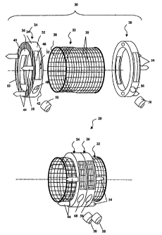
Referring now to FIG. 2, a vertebral implant assembly according to an
embodiment
of the present invention is referred to, in general, by the reference numeral
20 and includes
tubular body 22 connected between two cleat assemblies 24 and 26 in a manner
to be
described. The tubular body 22 defines a hollow bore 28 therethrough which is
configured
to receive bone osteogenetic material (not shown). To fully exploit the
osteogenetic
material and promote healing and bone restoration in the aftermath of
vertebral or disc
surgery, the tubular body 22 can be provided with a plurality of openings 30
that permit
bone and tissue ingrowth and vascularization. The body 22 may be provided in a
variety of
heights or may be trimmed to fit within the gap formed by the vertebral
ablation to avoid
damaging the weak bone of the adjacent intact vertebrae after implantation.
In one embodiment, a surgical mesh tube or "cage," which is known in the art,
can
serve as the tubular body 22. ~ne example of such a cage is disclosed in U.S.
Patent Nos.
5,897,556 and 6,149,651 to Drewry, et al. ("the Drewry patents") which are
incorporated
herein by reference. As described in the Drewry patents, a tubular body may
comprise
angled, intersecting elongate bars which form a plurality of triangular
apertures. Also as
described in the Drewry patents, the tubular body may have a non-circular
cross section
and instead be shaped to more closely match the profile of the adjacent intact
vertebrae, so
that when installed, the tubular body can be as unobtrusive as possible.
The cleat assembly 24 can include a ring-shaped member 32 having an exterior
side wall 34 and interior side wall 36 which defines a bore 38 through which
the tubular
body 22 can pass, such as by sliding. The interior side wall 36 may be smooth
to promote
the slidable passage of the tubular body 22. The member 32 can further include
an outer
end wall 40 and an inner end wall 42 extending between the exterior side wall
~34 and
interior side wall 36, the outer end wall 40 having a plurality of spikes 44
configured to
penetrate the endplate of the adjacent intact vertebrae to maintain the
position of the cleat
assembly 24 in situ.
CA 02552703 2006-07-05
WO 2005/070348 PCT/US2005/001067
4
To promote bone ingrowth and vascularization in and around cleat assembly 24,
one or more apertures 46 can be provided through the exterior side wall 34 and
the interior
side wall 36 and into communication with the bore 38. After installation,
these apertures
46 can be packed with graft material to accelerate the healing process.
Additionally, to fix
the cleat assembly 24 to the tubular body 24 after installation, one or more
threaded
apertures 48 can be provided through the side walls 34 and 36 of the member 32
in
communication with the bore 38 with each aperture 48 being adapted to receive
an
attachment mechanism 50, which can be, for example, a flat end machine type
screw.
Other examples include a pre-attached pin, a rivet, and/or a staple.
To facilitate installation, the inner end wall 42 of the cleat assembly 24 may
be
provided with a plurality of alignment positions 52 which can be configured to
mate with
corresponding pegs on an installation tool (not shown) to permit rotational
and axial
placement of the cleat assembly 24. Depending where the alignment positions 52
are
located along the inner end wall 42, the alignment positions 52 may be
configured either
as recessed areas in the inner end wall 42 or as openings that extend through
the inner end
wall 42 and into communication with the apertures 46. In another alternative,
the
alignment positions 52 may project outward from the inner, end wall 42 to mate
with
corresponding recessed areas on an installation tool (not shown). The outer
end wall 40
can comprise furrows 53 or other textures to reduce motion and promote a
secure interface
between the cleat assembly 24 after the spikes 44 of the cleat assembly have
been
embedded in the endplate of the adjacent intact vertebra.
In alternative embodiments, the configuration of the cleat assembly 24 can be
modified to accommodate a wide variety of patient anatomies and surgical
applications
while still providing a secure and stable engagement with the adjacent intact
vertebrae. To
correspond to the cervical, thoracic, or lumbar regions of the vertebral
column or to most
closely match the anatomy of a particular patient, the member 32 can be
fabricated in a
wide assortment of diameters. Further, the interior side wall 36 may be sized
to allow the
tubular body 22, having a predetermined diameter which can range for example
from 13
mm through 25 mm, to slidably pass through the member 32. Although FIG. 2
depicts the
ring-shaped member 32 as generally cylindrical, to provide an unobtrusive
alignment with
the adjacent intact vertebrae, the exterior side wall 34 and /or the interior
side wall 36 can
CA 02552703 2006-07-05
WO 2005/070348 PCT/US2005/001067
be contoured to more closely correspond to the shape of the adjacent intact
vertebrae,
resulting in a low profile installation.
Referring now to FIG. 3a, the cleat assembly 24 can have the outer end wall 40
in
substantially parallel alignment with inner end wall 42. Alternatively, as
shown in FIG.
3b, the cleat assembly 24 can have end walls 40 and 42 which may be angled
with respect
to each other to accommodate a variety of lordotic and kyphotic angles. The
angled end
walls can provide a secure and stable installation that most closely matches
the alignment
required for a particular patient. For example, the cleat assembly 24 may have
lordotic
angles of 4, 8 or 15 degrees.
Referring again to FIG. 2, the cleat assembly 26 can include one or more
spikes 54
and one or more threaded apertures 56 having a corresponding set screw 58. The
spikes
54, apertures 56, and set screw 58 can be identical to the spikes 44,
apertures 48, and set
screw 50 described above for cleat assembly 24. Other features of cleat
assembly 26 can
be the same as the cleat assembly 24 and therefore, will not be described in
detail. It
should be noted, however, that the cleat assembly 24 may not, necessarily, be
identical to
the cleat assembly 26. For example, cleat assembly 24 may comprise
substantially parallel
i
end walls as shown in FIG. 3a, whereas cleat assembly 26 may be identical to
cleat
assembly 24 shown in FIG. 3b, comprising end walls angled with respect to each
other.
The tubular body 22 and the cleat assemblies 24 and 26 maybe formed of or
iilclude a biocompatible material. The material may be strong enough to
withstand the
application of external compressive, axial, torsional, and bending loads, as
well as strong
enough to provide support for the adjacent intact vertebrae. The devices may
be formed
entirely of titanium, however other biocompatible materials may be used such
as a surgical
grade stainless steel, a porous tantalum material such as HEDROCEL~ provided
by
Implex Corporation of Allendale, New Jersey, or a radiolucent polymer
material, such as
polyether ether lcetone (PEEKTM) provided by Victrex PLC of the United
Kingdom. The
components 22, 24, and 26 of vertebral implant assembly 20 may all be formed
from the
same material or, alternatively, may be fabricated from different but
compatible materials.
Referring now to Fig. 4, the components of Fig. 2 may be preliminarily
assembled
to perniit implantation of the vertebral implant assembly 20 into the void
created in the
vertebral column by a vertebral ablation. For instance, once the tubular body
22 has been
CA 02552703 2006-07-05
WO 2005/070348 PCT/US2005/001067
selected and/or trimmed to fit within the gap between the intact vertebrae (as
discussed
with reference to Fig. 1), the cleat assembly 24 can be placed over an end of
the tubular
body 22 with spikes 44 extending toward that end of the body. The cleat
assembly 26 can
be placed over the other end of the tubular body 22 with spikes 54 extending
toward that
other end of the body and in the direction opposite the spikes 44. To permit
installation
without damaging the weak bone of the adjacent endplates, the cleat assemblies
can be
slidably positioned along the tubular body 22 such that the spikes do not
project past the
ends of the tubular body.
The tubular body 22 can then be packed with a suitable osteogenetic material
(not
shown), including autograft, allograft, xenograft, demineralized bone,
synthetic and
natural bone graft substitutes, such as bioceramics and polymers, and
osteoinductive
factors. It is understood that the osteogenetic graft material can be packed
at any time
prior to or during the installation of the vertebral implant assembly 20, and
can even be
packed after installation by inserting the graft through the openings 30 in
the tubular body
22.
Referring now to Fig. 5, the configuration of Fig. 4 can be surgically
inserted into
the void created by the surgical excision of vertebra 12a (in Fig. 1) with
spikes 44
extending toward intact vertebra 12b and spikes 54 extending toward intact
vertebra 12c.
The cleat assemblies 24 and 26 can be advanced along the tubular body 22
toward the
endplates of the intact vertebrae until the spikes are embedded into the
endplates.
Depending upon the surgical approach and the amount of surgical exposure,
embedding the cleat assemblies into the vertebral endplates may be achieved
using one or
more devices lcnown in the art. In one example, the cleat assemblies may be
installed
using an impactor having a forked or variable C-shaped head which can
accommodate a
variety of cleat assembly diameters. Pegs on the impactor head can mate with
the
alignment positions 52 in the outer end wall 40 of the cleat assembly 24 to
rotationally and
axially position the cleat assembly and to grip the cleat assembly while a
mallet is used to
strike the impactor, embedding the spikes into the adjacent vertebral
endplate. The
process may be repeated for cleat assembly 26.
Another device that can be used to install the cleat assemblies is a
distractor which
can be interposed between the two cleat assemblies to force them away from
each other
CA 02552703 2006-07-05
WO 2005/070348 PCT/US2005/001067
7
and into the adjacent vertebral endplates. After the spikes of the cleat
assemblies are
embedded using, for example the distractor or the impactor, the distractor
also may be
used create a desirable spacing between the rostral and caudal intact
vertebrae, allowing
for the surgical restoration of sagittal plane balance. Still another device
for seating the
cleat assemblies is a compressor which, when anchored to a relatively
stationary structure,
can be used to pull the spikes into the endplates of the adjacent vertebrae.
These devices
or others known in the art can be used alone or in concert to install the
cleat assemblies
and create the desired spacing between the adjacent vertebrae.
After the cleat assemblies 24 and 26 are installed and properly spaced, the
attachment mechanism 50 (a set screw in the present example) can be inserted
into the
aperture 48 of cleat assembly 24 and rotated until at least a portion emerges
through the
interior side wall 36. In some embodiments, the set screw 50 may be pre-
attached. The
set screw 50 can further pass through the mesh of the tubular body 22 to affix
the tubular
body 22 to the cleat assembly 24. Alternatively, the set screw can exert
pressure on the
surface of the tubular body 22 to affix the body 22 to the cleat assembly 24.
The cleat
assembly 26 can be affixed to the tubular body 22 in a manner identical to
that described
for assembly 24. After the vertebral body replacement assembly 20 is
installed, additional
osteogenetic material may be packed into the cleat assembly 24 through the
apertures 42
to promote healing and bone growth. The assembly 26 can be similarly packed
with
osteogenetic material.
As compared to other anterior stabilizing techniques, the installation of this
vertebral implant assembly 20 can be relatively simple and can have a
shortened procedure
duration relative to surgical procedures that require implantation of other
hardware or the
preparation of mortises. Additionally, the vertebral implant assembly 20 can
be installed
to complement and not interfere with other implanted stabilizing devices such
as screw
and plate, screw and rod, and pedicle screw systems. Once installed, the
implant 20 can
have a very low profile, reducing the risk of erosion of vascular structures.
The vertebral body replacement assembly 20 installed as described can
withstand
torsional, axial, and shear loads, reducing the risk of anterior displacement
or posterior
retropulsion and thus minimizing the development of neurologic deficits in the
patient and
the need for additional surgery. Furthermore, this installation can resist
subsidence
CA 02552703 2006-07-05
WO 2005/070348 PCT/US2005/001067
("telescoping") of the tubular body or the biologic strut into the relatively
wealc bone of
the adjacent vertebral endplates which occurs commonly with conventional
mortising
techniques. This resistance to subsidence can be due to both the embedded
spikes and the
wider surface area of the end walls which distribute loads over a greater area
of the
adjacent intact vertebrae end. Because the cleat assemblies are not positioned
within the
hollow bore of the tubular body but rather are externally fixed to the body,
the disclosed
configuration provides the further advantage of permitting increased contact
between the
osteogenetic material located within the tubular body and the endplates of the
adjacent
vertebrae to promote bone growth.
An alternative installation method may prove advantageous for some
applications,
for example, the components of the vertebral implant assembly 20 may not be
preliminarily assembled. Rather, the spikes 44 and 54 of cleat assemblies 24
and 26,
respectively, may be driven into the intact vertebrae 12b and 12c (Fig. 1)
before the
tubular body 22 is passed between the cleat assemblies 24 and 26. The tubular
body 22
may then be packed with osteogenetic material and slidably positioned into the
space
between the cleat assemblies 24 and 26. The set screws 50 & 58 can then be
installed as
described above.
Referring now to FIG. 6, in another alternative embodiment, the tubular body
22
can be replaced with a biologic strut graft 60 to form a vertebral implant
assembly 62.
The strut may be allograft or autograft material and the graft may be taken
from a fibula, a
humerus, or any other suitable source known in the art. In this embodiment,
the endplates
24 and 26 are placed over a biologic strut graft 60 which is sized to fit
between the
endplates of the adjacent intact vertebrae. The vertebral implant assembly 62
can then be
installed in a manner similar to the method described for vertebral implant
assembly 20.
In this embodiment, however, set screws 50 & 58 may be of a type that can be
threaded
into the biologic strut graft 56. For example, a pointed screw may be
appropriate.
Although only a few exemplary embodiments of this invention have been
described in detail above, those spilled in the art will readily appreciate
that many
modifications are possible in the exemplary embodiments without materially
departing
from the novel teachings and advantages of this;invention. Accordingly, all
such
modifications are intended to be included within the scope of this invention
as defined in
CA 02552703 2006-07-05
WO 2005/070348 PCT/US2005/001067
the following claims. In the claims, means-plus-function clauses are intended
to cover the
structures described herein as performing the recited function and not only
structural
equivalents, but also equivalent structures.