Note: Descriptions are shown in the official language in which they were submitted.
CA 02553542 2006-05-O1
METHODS AND FORMULATIONS FOR PROTECTING CELLS,
AND FOR TREATING DISEASES
AND CONDITIONS BY OPTIM1Z1NG THE
INTRACELLULAR CONCENTRATION OF NAD
FIELD OF THE INVENTION
(0001] The present invention relates generally to the protective and
therapeutic
uses of substances which optimise the intracellular concentration and
availability of
nicotinamide adenine dinucleotide (NAD ), and to related methods. NAD -
optimising substances include those such as pre-B cell colony-enhancing factor
(PBEF) and 5-phosphoribosyl-pyrophosphate (PRPP). The invention relates also
to
therapeutic and pharmaceutical formulations containing 1~AD-optimising
compounds
such as PBEF and PRPP, alone and in various combinations with other compounds
such as nicotinamide.
BACKGROUND OF THE INVENTION
[0002] Current techniques used to enhance cyto-protection or to inhibit
aberrant
cell functions tend to work by reducing environmental and metabolic factors
that can
harm or poison cells and tissues. Unfortunately, many of these techniques are
limited
to the dynamics of the scavenging of free radicals, and protective coatings.
[0003] Animal cells, such as human cells, experience stress many times
throughout their lifecycles often causing injury, death or irreparable DNA
damage.
The source of the stress can be environmental, such as radiation, toxic
substances, and
physical factors experienced by the cell such as mechanical injury due to
trauma, and
exposure to extreme weather. Other stresses include those caused by sunlight,
dehydration and exposure to caustic or otherwise harsh chemicals. Other
sources of
stress can occur during the natural phases of the cell cycle such as during
times of
proliferation and differentiation, and to the dynamics of carcinogenesis.
(0004] Revollo et al. postulate the regulatory interactions of Mampt, Nmnat
and
Sir2 on the intracellular dynamics of NAD. These interactions occur after the
synthesis of NAD. The NAD Biosynthetic Pathway Mediated by Nicotinamide
Phosphoribosy-ltransferase Regulates Sir2 Activity in Mammalian Cells, The
Journal
CA 02553542 2006-05-O1
2
of Biological Chemistry, Sept. 20, 2004. INSERT re Amgen/Samal PBEF; Song et
al
PBEF Enhancing Factor; and Hasmann et al, FK86.
[0005( Until the present compounds and methods, the healing, pharmaceutical,
cosmetic and metabolic arts have been lacking in effective methods and
formulations
to improve the metabolic fitness of cells. By improving cellular metabolic
fitness,
cells are best prepared to experience such commonly occurring stress without
incurring damage that would prematurely shorten the Life of the cell, cause
the cell to
function improperly or degrade the physical appearance of the cell.
[0006) This raised the possibility that PBEF was involved in the synthesis of
NAD. NAD is well known for its role in regulating the redox state of the cell.
However, recent work has identified a number of other important NAD-dependent
reactions, including histone deacetylation. Unlike the redox system, these
newly
discovered reactions deplete the pool of cellular NAD, and sometimes
contribute to
harmful imbalances in the cell.
[0007] The present inventors have discovered that Optimizing the intracellular
concentration of NAD+ facilitates a balance among the numerous intracellular
interactions of NAD~ and its related pathways such that the health of the cell
and its
resistance to stress are increased. This increased robustness attendant to the
invention
also relates to a consequent delay of apoptosis of the cell.
SUMMARY OF THE INVENTION
(0008) The present invention is based on the unexpected discovery that PBEF
and
PRPP, alone or in combination with one another, or in combination with one or
more
forms of nieotinamide, increase cell fitness, protect the cell against damage
from
stress factors, and increase the longevity of the cell.
[0009] Other aspects and features of the present invention will become
apparent
to those of ordinary skill in the art upon review of the following description
of
specific embodiments of the invention in conjunction with the accompanying
Figures
and claims. It should be understood, however, that the detailed description
and the
specific examples, while indicating preferred embodiments of the invention,
are given
CA 02553542 2006-05-O1
3
by way of illustration only, since various changes and modifications within
the spirit
and scope of the invention will become apparent to those skilled in the art
from this
detailed description.
BRIEF DESCRIPTION OF THE DRAWINGS
[0010] The figures provided herein illustrate embodiments of the present
invention, by way of example only, and not in a limiting way.
[0011] Figure 1(A) shows Hoffman-modulated contrast images of HITBS smooth
muscle cells in M199 supplemented with 10% FBS (left images) and 6 days after
culturing SMC's in serum-free M 100 (right image;
[0012] Figure 1(B) is a Northern Blot showing upregulation of the 3 major
transcripts of PBEF in HITBS SMC's following withdrawal of serum from
cultures.
[0013] Figure 1(C) is a Western Blot of cell lysates harvested from HITC6
SMC's before and after withdrawal of serum from cultures
[0014] The images of Figure 2 show the cellular elongation and aggregation of
human Smooth Muscle Cells into multilayered ridges induced by the
overexpression
of PBEF. Figure 2(A), shows Hoffman-modulated contrast images of sub-confluent
(top panel) or post-confluent (middle panel) HITBS Smooth Muscle Cells
transduced
with a retrovirus containing eDNA encoding EGFP alone (left image) or PBEF and
EGFP from a bicistronic cassette (right image). The bottom panel of Figure
2(A)
depicts fluorescence images of the post-confluent SMC cultures, showing
expression
of the transgenes as indicated by EGFP fluorescence. Bar, 50 um.
[0015] Figure 2(B) shows the quantification of length-width ratios of control
and
PBEF-overexpressing Smooth Muscle Cells cultured in the presence of serum and
3
days after serum withdrawal.
[0016] Figure 3 shows Western blots revealing expression of SMC
differentiation
markers in HITBS SMC's infected with cDNA encoding EGFP alone (left image),
and cDNA encoding both PBEF and EGFP (right image).
CA 02553542 2006-05-O1
4
[0017] The images of Figure 4 show the effect of PBEF of apoptosis. Figure
4(A) shows cell accumulation over 11 days for control and PBEF-overexpressing
HITBS SMC's, cultured in M199 with 5% FBS.
[0018] Figure 4(B) shows Thymidine incorporation into control and PBEF
overexpressing HITBS SMC's, assessed by incubating cells in log-phase growth
with
3
pCi/mL [ H]thymidine for 12 hours.
[0019] Figure 4(C) presents fluorescence images of control and PBEF-
overexpressing SMC's stained with Hoechst 33258 to identify nuclei (top
panel), and
for apoptotic nuclei by incubating with d-UTP fluorescein (bottom panel).
[0020] The images of Figure 5 show the effect on SMC viability of the
knockdown of PBEF expression and maturation induced by serum withdrawal.
Figure 5(A) shows Hoffman-modulated images of control HITC6 SMC's and HITC6-
siRNA SMC's. Western blots showing PBEF protein expression for each cell line
are
shown.
[0021] Figure 5(B) shows the length-width ratios of 50 randomly selected cells
expressing either nsIRNA I 248 or siltNA 1248.
[0022] Figure 5(C) is a.Western blot showing reduced expression of h-
Caldesmon in HITC6-siRNA 1248 Smooth Muscle Cells.
[0023] Figure 6 is a phylogenetic tree showing a tight evolutionary
relationship
between bacterial nicotinamide phosphoribosyltransferases and eukaryotic PBEF,
including human PBEF.
[0024] The depictions of Figure 7 show the effects of increasing PBEF levels
on
the levels of NAD . Figure 7(A) shows the HPLC analyses of deproteinized
nucleotide extracts obtained from HEK293 cells transfected with pQCXIP or
pQCXIP-PBEF, and HITC6 SMC's transduced with pQCXIP or pQCXIP-PBEF.
j0025] Figure 7(B) shows quantitative data from 3 separate experiments for
each
cell type of Figure 7(A), including Western blot insets depicting
representative PBEF
expression for control and for cells overexpressing PBEF.
CA 02553542 2006-05-O1
[0026] Figure 8 shows that increasing the levels of PBEF increases NAD -
dependent histone deacetylase activity in human Smooth Muscle Cells.
[0027] The images of Figure 9 show the effect of overexpression of PBEF on the
phenotype, vessel chimerism and investment of Smooth Muscle Cells. Figures
9(A)
through 9(D) show sections of matrigel implants loaded with human SMC's.
Figures
9(E) through 9(I) show sections stained with h-caldesmon and h-calponin.
Figure 9(J)
illustrates the quantification of the proportion of microvcssels invested by
at least one
EGFP-positive SMC.
[0028] Figure 10 is a schematic diagram of the structure of PPRP;
[0029) Figure 11 is a diagram of the structure of the essential layers and
components of human skin;
[0030) Figures 12 A-C are phase contrast photomicrographs at 20X of vector-
transduced or PBEF-overexpressing HITC6 SMC's in response to treatment with 5-
phosphoribosyl-pyrophosphate (PRPP), demonstrating that the PRPP increases the
health of smooth muscle cells compared to control cells;
[0031 i Figures 13 A-B are phase contrast photomicrographs at 20X
magnification of vector-transduced HITC6 SMC's in response to treatment with S-
phosphoribosyl-pyrophosphate (PRPP), demonstrating that the PRPP increases the
health of smooth muscle cells compared to control cells;
[0032) Figures 14 A-B are phase contrast photomicrographs at lOX
magnification of vector-transduced HITC6 SMC's in response to treatment with 5-
phosphoribosyl-pyrophosphate (PRPP). SMC's were cultured in M199 1% FBS for
24 hours prior to addition of 500 uM PRPP.
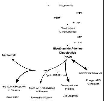
[0033] Figure 15 shows the salient biosynthetic pathways involved in the
synthesis of NAD_
[0034] Figure 16 shows the salient biosynthetic pathways involved in the
utilization and regeneration of NAD_
CA 02553542 2006-05-O1
6
[0035] It is an object of the invention to provide methods for protecting
cells and
tissues from harm by optimising the levels or concentrations of one or more
forms of
NAD in the cells and tissues.
[0036] It is a similar object of the invention to provide methods for
repairing and
healing cells and tissues from harm by optimising the levels or concentrations
of one
or more forms of NAD in the cells and tissues.
[0037] It is a further object of the invention to provide pharmaceutical
formulations efficacious in optimising the levels or concentrations of one or
more
forms ofNAD in the cells and tissues.
[0038] It is a further object of the invention to provide cosmetic
formulations
efficacious in optimising the levels or concentrations of one or more forms of
NAD in
the cells and tissues.
[0039] DETAILED DESCRIPT10N
[0040] The term "a cell" as used herein includes a single cell as well as a
plurality
or population of cells. Administering an agent to a cell includes both in
vitro and in
vivo administrations.
[0041] The term "effective amount" as used herein means an amount effective,
at
dosages and for periods of time necessary to achieve the desired result.
[0042] The term "animal" as used herein includes all members of the animal
kingdom, including humans.
[0043] Pharmaceutical Compositions may be prepared using standard techniques
known in the art.
[0044] In one embodiment, there is provided a method for treating a disease
state
characterized by cells with a non-optimal NAD cycle. The patient may be any
animal,
including a mammal, including a human.
(0045] "Treating" a condition or disease state refers to an approach for
obtaining
beneficial or desired results, including clinical results. Beneficial or
desired clinical
results can include, but are not limited to, alleviation or amelioration of
one or more
CA 02553542 2006-05-O1
7
symptoms or conditions, diminishment of extent of disease, stabilization of
the state
of disease, prevention of development of disease, prevention of spread of
disease,
delay or slowing of disease progression, delay or slowing of disease onset,
amelioration or palliation of the disease state, and remission (whether
partial or total),
whether detectable or undetectable. "Treating" can also mean prolonging
survival of
a patient beyond that expected in the absence of treatment. "Treating" can
also mean
inhibiting the progression of an aberrant condition, slowing the progression
of injury,
aging or malfunction temporarily, although more preferably, it involves
halting the
progression of the same permanently. As will be understood by a skilled
person,
results may not be beneficial or desirable if, the treatment results in
adverse effects on
the patient treated that outweigh any benefits effected by the treatment.
[0046] Through the work of the present inventors, the importance of
maintaining
an optimal concentration of intracellular NAD+ to the robustness and
completeness of
cellular function is manifest in a number of ways, particularly when viewed in
the
context of the experiments reported herein. Moreover, the discovery of the
present
invention is unexpected, particularly in view of the related work of others.
[0047] In accordance with these and other objects, the invention provides
formulations and methods for treating diseases or conditions in an animal. In
one
preferred embodiment, the method comprises the step of optimizing the
intracellular
concentration of PBEF in the cells of at least one target tissue of the
animal, wherein
the optimizing of the concentration of PBEF is effected by increasing the
intracellular,
or endogenous, concentration of the PBEF of the animal by a sufficient amount
of
PBEF.
]0048] In one preferred embodiment, optimization of the concentration of PBEF
can be effected by administering to the animal a sufficient amount of PBEF to
increase the intracellular concentration of the PBEF. The administration of
the PBEF
is preferably by at least one route, and the at least one route can be one or
more of
injection, oral administration, anal or other colonic administration,
inhalation, intra-
peritoneal administration, topical administration, intra-organ administration,
infusion
of a target tissue, transdermal and parenteral administration, including
intravenous,
intraperitoneal, subcutaneous, intramuscular, trans-epithelial, nasal,
intrapulmonary,
intrathecal, rectal and topical modes of administration or any other
efficacious means.
CA 02553542 2006-05-O1
g
For example, in accordance with other objects of the invention, the
optimization of
PBEF levels can be performed by the methods of gene therapy, including the use
of
one or more viral vectors, such as adenoviruses, lentiviruses, adeno-
associated viruses
and non viral plasmid and cosmid vectors, and any other viral or prion vector
amenable to optimizing the endogenous production and optimization of
intracellular
levels of PBEF.
[0049] Similarly, the present methods may be effected by promoting the
endogenous production of PBEF in the cells of at least one target tissue of
the animal,
or in the whole animal, such as a human. Thus, promotion of intracellular
production
of PBEF can be effected, for example, by up-regulating the nucleic acid
processes or
mechanisms which support the production of PBEF, or by up-regulating the
nucleic
acid processes which increase the endogenous production of PBEF. Moreover, the
present methods may be effected wherein the promotion of intracellular
production of
PBEF is effected by down-regulating the nucleic acid processes or mechanisms
which
repress the production of PBEF. Alternatively, the present methods may be
practiced
wherein the optimization of PBEF is effected by increasing the intracellular
concentration of at least one modulator of PBEF, for example, by administering
to the
animal, such as a human, an effective amount of the modulator.
[0050] Administration of the modulator may be by any route known, and is
preferably by at least one route, the at least one route being selected from
routes such
as injection, oral administration, anal or other colonic administration,
inhalation, intra-
peritoneal administration, topical administration, intra-organ administration,
infusion
of a target tissue, transdermal and parenteral administration, including
intravenous,
intraperitoneal, subcutaneous, intramuscular, trans-epithelial, nasal,
intrapulmonary,
intrathecal, rectal and topical modes of administration. In one preferred
embodiment
the modulator is PRPP.
[0051] In another preferred embodiment, the increase of PBEF is effected by
promoting the endogenous production of PRPP in the cells of at least one
target tissue
of the animal, or in the whole animal, such as a human. The promotion of
intracellular, or endogenous, production of PBEF may be effected, for example,
by
up-regulating the nucleic acid processes or mechanisms which increase the
production
CA 02553542 2006-05-O1
9
of PRPP, or by down-regulating the nucleic acid processes or mechanisms which
repress the production of PRPP.
[0052] In accordance with other methods of the invention, PRPP can be given in
any efficacious form, or in combination with PBEF, or in combination with at
least
one form of nicotinamidc, or in combination with PBEF and at least one form of
nicotinamide. The nicotinamide may be in any efficacious form, such as in a
substituted form, or in the form of one or more of nicotinic acid; nicotinic
acid
ribonucleotide; nicotinic acid ribonucleotide, reduced form; nicotinamide
ribonucleotide; nicotinamide ribonucleotide, reduced form; nicotinic acid
adenine
dinucleotide; nicotinic acid adenine dinucleotide, reduced form; nicotinamide
adenine
dinucleotide (NAD); nicotinamide adenine dinucleotide phosphate (NADP);
nicotinamide adenine dinucleotide, reduced form (NADH); and nicotinamide
adenine
dinucleotide phosphate, reduced form (NADPH) and pharmaceutically acceptable
salts thereof.
[0053] The present methods and formulations may be used to treat any disease
or
condition, and particularly those involving the disruption, harm or imbalance
of the
NAD pathways of cells including those diseases and conditions involving the
vascular
system including the heart, blood vessels and other portions of the
cardiovascular
system. Examples of such diseases and conditions include vascular
insufficiency,
vascular weakness, progeria, premature senescence of one or more tissues,
aging,
severe stress on one or more tissues, atherosclerosis, arteriolesclerosis and
re-
vascularization of injured or weakened tissues or organs. The present methods
and
formulations may be used to treat any disease or condition which is a result
of sever
stress wherein the severe stress on one or more tissues is due to one or more
of injury,
malnutrition, disease, toxic shock and exposure.
[0054] Also in accordance with the present methods, the optimization of PBEF
may be effected by increasing the intracellular concentration of at least one
precursor
of PBEF, for example, by administering to the animal an effective amount of
the
precursor.
[0055] Preferably, the administration of the precursor is by at least one
route, and
the at least one route is one or more of injection, oral administration, anal
or other
CA 02553542 2006-05-O1
ID
colonic administration, inhalation, intra-peritoneal administration, topical
administration, intra-organ adminis-tration, infusion of a target tissue,
transdermal and
parenteral administration, including intravenous, intraperitoneal,
subcutaneous,
intramuscular, trans-epithelial, nasal, intra-pulmonary, intrathecal, rectal
and topical
modes of administration.
[0056] In one aspect, the precursor may be at least one form of nicotinamide.
[0057]
[0058] In another aspect, the nicotinamide may be substituted or in the form
of
one or more of nicotinic acid; nicotinic acid ribonucleotide; nicotinic acid
ribonucleotide, reduced form; nicotinamide ribonucleotide; nicotinamide
ribonucleotide, reduced form; nicotinic acid adenine dinucleotide; nicotinic
acid
adenine dinucleotide, reduced form; nicotinamide adenine dinucleotide (NAD);
nicotinamide adenine dinucleotide phosphate (NADP); nicotinamide adenine
dinucleotide, reduced form (NADH); and nicotinamide adenine dinucleotide
phosphate, reduced form (NADPH) and pharmaceutically acceptable salts thereof.
[0059] In accordance with still other advantages of the present invention,
pharmaceutical and cosmetic formulations are provided. Formulations for
optimizing
the intracellular concentration of NAD of the invention include one or more of
effective amounts of PRPP, PBEF, and nicotinamide. In compositions of the
invention comprising nicotinamide, the nicotinamide may be substituted or in
the
form of one or more of nicotinic acid; nicotinic acid ribonucleotide;
nicotinic acid
ribonucleotide, reduced form; nicotinamide ribonucleotide; nicotinamide
ribonucleotide, reduced form; nicotinic acid adenine dinucleotide; nicotinic
acid
adenine dinucleotide, reduced form; nicotinamide adenine dinucleotide (NAD);
nicotinamide adenine dinucleotide phosphate (NADP); nicotinamide adenine
dinucleotide, reduced form (NADH); and nicotinamide adenine dinucleotide
phosphate, reduced form (NADPH) and pharmaceutically acceptable salts thereof.
[0060) A pharmaceutical or cosmetic composition of the invention may further
comprise one or more of an effective amount of a pharmaceutically effective
vehicle,
a pharmaceutically effective diluent, a pharmaceutically effective cream, a
pharmaceutically effective excipient, one or more pharmaceutically effective
micelles,
CA 02553542 2006-05-O1
a pharmaceutically effective carrier, pharmaceutically acceptable
concentrations of
salt, buffering agents, preservatives and various compatible carriers.
Preferably,
compositions of the invention are adaptable for administration by at least one
route,
and the at least one route is one or more of injection, oral administration,
anal or other
colonic administration, inhalation, infra-peritoneal administration, topical
administration, infra-organ administration, infusion of a target tissue,
transdermal and
parenteral administration, including intravenous, intraperitoneal,
subcutaneous,
intramuscular, trans-epithelial, nasal, intrapulmonary, intrathccal, rectal
and topical
modes of administration.
[0061] In accordance with still other objects of the invention, its
compositions
may be provided in the form of one or more of ingestible tablets, buccal
tablets,
troches, capsules, elixirs, suspensions, micelle encapsulations, syrups,
wafers and the
like, or enclosed or enclosable within hard or soft shell gelatin capsules.
Moreover,
the present compositions may further comprise one or more of an effective
amount of
a cosmetically effective vehicle, a cosmetically effective diluent, a
cosmetically
effective cream, a cosmetically effective excipient, one or more cosmetically
effective
micelles, a cosmetically effective carrier, cosmetically acceptable
concentrations of
salt, buffering agents, preservatives and various cosmetically compatible
carriers.
[0062] As one of skill in the pharmaceutical or cosmetic arts will comprehend,
numerous combinations and formulations of PBEF, PRPP and nicotinamide are
within the scope and spirit of the invention, as are numerous variations of
the present
methods.
[0063] Vascular smooth muscle cells (SMC's) can exist in an immature state
with
the capacity to proliferate and migrate ( 1 ). The switch from a
proliferative/migratory
SMC to a contractile SMC is referred to as SMC maturation and is a process
that is
central to vascular development, stability, and physiologic function (2).
Maturation of
SMC's is necessary to stabilize newly formed blood vessels and confer
vasomotor
reactivity (3). Similarly, replicating SMC's in injured arteries must
eventually mature
to a quiescent phenotype to terminate the remodeling process.
[0064] The primary function of vascular SMC's in their quiescent state is to
contract and provide vascular tone (Owens, 1995) (Sobue, 1998) (Rybalkin,
2003).
CA 02553542 2006-05-O1
12
The healthy SMC, which morphologically is similar to HITBS or HITC6 SMC's that
have been cultured in serum-free media for prolonged periods (more than 72
hours) is
characterized by an elongated appearance, increased expression of SMC
contractile
proteins, such as h-caldesmon, metavinculin, smooth muscle myosin heavy chain
and
calponin and a marked decrease in apoptosis (Li, Circ.Res, 1999). We have
found that
HITBS and HITC6 SMC's that are in this state have a significant increase in
their
transcript and protein levels of PBEF, demonstrating that PBEF is a novel
factor
involved in regulating SMC differentiation and maturation.
[0065] The molecular basis by which an immature SMC shifts to a functionally
contractile cell is incompletely defined (4). This is partly due to a paucity
of culture
systems that recapitulate this critical, late phase of the SMC developmental
program.
Recently however, we cloned three adult vascular SMC lines that, in contrast
to other
human SMC preparations, could reversibly convert between a spread, immature
state
when cultured in the presence of serum to a highly elongated, mature state
after serum
withdrawal (5, 6). Whereas cultured human SMC's often die upon serum
withdrawal,
these cells displayed decreased apoptosis, increased contractile protein
expression,
and the ability to contract in response to vasoactive agonists. This system
therefore
provided us with an opportunity to seek out factors that enable an activated
adult
SMC to return to a metabolically quiescent cell specialized to contract.
Accordingly,
we undertook differential display PCR and high-density microarray analyses to
identify genes that were differentially expressed as a homogenous population
of
human SMC's executed this key shift in phenotype.
[0066] These surveys identified pre-B cell colony-enhancing factor (PBEF) as
being consistently upregulated as SMC's switched to the mature, quiescent
state.
PBEF is a 5255 kD protein that has been proposed to be a cytokine (7).
Reported
actions in this regard include syncrgizing with other cytokines to stimulate
the
maturation of pre-B cells (7), stimulating the expression inflammatory
cytokines in
amniotic epithelial cells (8), and prolonging neutrophil survival (9).
However, the
contention that PBEF is a secreted cytokine is controversial. PBEF does not
have a
signal sequence for secretion and the presence of PBEF in culture media has
been
suggested to be a consequence of activation-induced cell death, rather than
secretion
by either a classical or alternative pathway (10, l l). Moreover, PBEF has
sequence
CA 02553542 2006-05-O1
13
similarity with bacterial nadY, a protein that confers bacteria with the
ability to grow
in nicotinamide adenine dinucleotide (NAD )-deficient conditions (l2, 13). In
keeping with this, Rongvaux and co-workers have shown that mouse PBEF
functions
within the cell as a nicotinamide phosphoribosyltransferase ( 10). This enzyme
catalyzes the rate-limiting step in the salvage pathway for NAD biosynthesis,
whereby nicotinamide that is generated during NAD~-consuming reactions is
utilized
+
to regenerate NAD ( 14, I S).
[0067j The experimental data reported here indicates that phenotype switching
of
human vascular SMC's is dependent on PBEF. Whereas SMC's deficient in PBEF
were compromised in their ability to elongate and express SMC differentiation
markers, genetic augmentation of PBEF expression promoted SMC survival and
conversion to a mature phenotype. 'These actions were associated with an
increase in
steady state NAD levels and increased NAD -dependent histone deacetylase
activity.
SMC's with augmented expression of PBEF manifested enhanced ability to
associate
with endothelial cells and wrap around nascent blood vessels in a human-mouse
chimeric vascular development model. These findings establish PBEF as a novel,
intracellular regulator of vascular SMC phenotype and implicate PBEF-mediated
NAD flux as a driver of human SMC maturation.
[0068] The mechanism by which PBEF exerts its pleiotropic effects is
controversial. It has been suggested that PBEF functions as a secreted
cytokine
(Samal, 1994)(Jia, 2004), while it has also been reported to be involved in
the salvage
pathway of NAD+ biosynthesis as a nicotinamide phosphoribosyltransferase
(Rongvaux, 2002)(Martin, 2001 ). From the results shoen herein, we believe
that
PBEF is an intracellular protein that catalyzes the rate-limiting reaction of
the salvage
pathway. This pathway involves the conversion of nicotinamide (IVAm) and 5-
phosphoribosyl-pyrophosphate (PRPP) to nicontinamide mononucleotide (NMN)
(Pilz, JBC, 1984)(Willis, Adv. Enzyme Reg., 1989). In resting human
lymphocytes,
the PRPP pools are very low (Snyder, JCI, 1976), and these cells were
characterized
by an impaired ability to generate ATP, inability to respond to mitogenic
stimuli,
while activation of these cells results in expansion of NAD+ and PRPP pools
concomitant with the repair of DNA single-strand breaks (Johnstone, Eur. J.
Biochem,
CA 02553542 2006-05-O1
14
1984)(Berger, Exp. Cell Res., 1982)(Williams, Exp. Cell Res., 1985). Carson,
D.A.,
Seto, S., and Wasson, D.B. Pyridine Nucleotide Cycling and Poly(ADP-Ribose)
Synthesis in Resting Human Lymphocytes. Journal of Immunology. 138: 1904-1907,
1987. Willis, R.C., Nord, L.D., Fujitaki, J.M., and Robins, R.K. Potent and
Specific
Inhibitors of Mammalian Phosphoribosylpyrophosphate (PRPP) Synthetase.
Advances in Enzyme Regulation. 28: 167-180, 1989.
[0069] PPRP and Nicotinamide are molecules critical for the regeneration of
NAD in animal cells. NAD+ is a critical coenzyme involved in oxidation-
reduction
reactions in the cell. Cellular maintainance of NAD+ levels for redox
reactions is
likely tightly regulated due to its critical importance in energy generation.
However,
recent work has identified a number of other NAD+-dependent reactions that,
unlike
the redox system, deplete the free pool of intracellular NAD+, As such,
regeneration
of NAD+ can be achieved by de novo biosynthesis in primarily the liver or via
the
salvage pathway in the peripheral tissues (Magni, CMLS, 2004)(Bender, Brit.
Jour.
Nutr, 1988). The catabolism of NAD(P)+ in the liver results in the release of
nicotinamide into the blood, which is utilized by peripheral tissues for the
regeneration of NAD+ (Bender, 1988). Conversion of nicotinamide to nicotinic
acid
by nicotinamide deamidase is an important aspect of NAD+ generation in
bacteria and
yeast (Anderson, Nature, 2003)(Schuette, Am. .1. Physiol, 1983). However, it
is
believed that nicotinamide deamidase is not expressed in mammals (Schuette,
Am. J.
Physiol, 1983)(Rongvaux, Bioessays, 2003). Therefore, conversion of
nicotinamide
and 5-phosphoribosyl pyrophosphate to NMN by PBEF is one critical step for the
regeneration of NAD+ in peripheral tissues (Rongvaux, Eur. J. Biochem, 2002),
such
as SMC's. The marked upregulation of PBEF in differentiating SMC's
demonstrates
that the differentiation of SMC's is an NAD+ consuming process.
[0070 The following experimental methods were employed to elucidate the data
provided herein. Unnecessary details of the employed methods have been omitted
for
the sake of efficiency. Nonetheless, one of skill in the art will comprehend
with
certainty the significance and scope of the results reported herein.
[0071 [ Smooth Muscle Cell Culture lines were employed to assess the role of
PBEF. Experiments were performed using the maturation-competent human vascular
SMC lines, HITBS and HITC6, generated from the human internal thoracic artery,
as
CA 02553542 2006-05-O1
' described previously (S, 6). SMCs were maintained in M 199 (GibcoBItL,
Burlington,
ON) supplemented with the designated concentration of FBS (Hyclone). HEK293
cells were grown in DMEM with 10% FBS
j0072] Overexpression of PBEF in Human Smooth Muscle Cells was evaluated
by use of a viral vector. A retroviral gene delivery system was used to
generate
human SMCs stably overexpressing PBEF. Full-length cDNA encoding PBEF was
amplified from HITBS SMC mRNA by RT-PCR and subcloned into the pIRES-EGFP
vector (Clontech). The PBEF-IRES-EGFP bicistronic fragment was then excised
using X~aoI and NotI and inserted into the retroviral expression vector pLNCX2
(Clontech). A second retroviral expression construct was generated by
inserting
PBEF cDNA into pQCXIP-IRES-PURO (Clontech). Retrovirus containing the cDNA
of interest was obtained by calcium phosphate-mediated transfection of the
Phoenix-
amphotropic retrovirus packaging cell line (kindly provided by Dr. G Nolan,
Stanford
University Medical School, CA, distributed by ATCC, Manassas, VA) as described
previously (29). Virus-containing supernatant was added to proliferating SMCs
and
stable transductants were selected with 500 pg/ml 6418 for 14 days, for pLNCX2-
based constructs, and with 3 yg/ml puromycin for 48 hours, for pQCXIP-based
constructs. Overexpression of PBEF was confirmed before each experiment by
Western blot analysis.
[0073] Western Blot Analyses were employed to assay marker expression.
Expression of PBEF and SMC differentiation markers was assessed by Western
blot
analysis with chemiluminescence detection, as described (29). Equal amounts of
protein were resolved on 12% (for PBEF and a-tubulin), 9% (for smoothelin A
and
smoothelin B) and 6% (for caldesmon and vinculin-metavinculin) SDS-
polyacrylamide gels and transferred PVDF membranes (Immobilon, Millipore).
PBEF was detected using a polyclonal rabbit antibody against human PBEF (1857,
1:5000, kindly provided by Amgen). Monoclonal antibodies were used to detect
heavy (h)-caldesmon (clone hHCD, 1:1000, Sigma), smoothelin isoforms A and B
(clone MAB3242, 1:500, Chemicon), vinculin-metavinculin (clone VIN-11-5,
1:2000,
Sigma), and a-tubulin (clone B-5-I-2; 1:16000, Sigma).
[0074] Cell Proliferation, DNA Synthesis, and Apoptosis were also evaluated.
To
2
assess SMC proliferation, cells were plated at a density of 3000 cells per cm
and
CA 02553542 2006-05-O1
16
cultured in M199 containing 5% FBS. Triplicate wells were harvested at the
designated times and counted using a hemacytometer. To quantify DNA synthesis,
3
cells in log-phase growth were incubated for 12 hours with [ H]thymidine ( 10
pCi/mL) and TCA-precipitable counts determined as described (5). Thymidine
incorporation was expressed relative to DNA content, quantified by
spectrofluorimetry of an aliquot of cell lysate incubated with 500 uglml
Hoechst
33258. Apoptosis was assessed in SMCs seeded on glass coverslips by in situ
end-
labeling of DNA fragments using terminal deoxynucleotide transferase and
fluorescein 12-dUTP (Promega) (5). Cells were fixed with 4% paraformaldehyde
and
counterstained with Hoechst 33258.
[0075) Knockdown of PBEF by RNA Interference was evaluated with a viral
vector. PBEF knockdown was accomplished by infecting human SMC's with
retrovirus containing sequences encoding hairpin siRI~TA fragments.
Complementary
oligodeoxynucleotides were synthesized, annealed, and inserted between the
BamH 1
and EcoRl sites of the retroviral expression vector pSIREN-RetroQ. Three
different
targeting sequences were used, each consisting of 19 nucleotides starting at
nucleotides 147, 384, and 1248 of the PBEF coding sequence (siRNA147 S'-
GGAAGGTGAAATATGAGGA-3'; siRNA384 5'-ATGTTCTCTTCACGGTGGA-
3'; siRNA1278 5'-AGGGCCGATTATCTTTACA-3'). Each sequence was separated
by a 9-nucleotide noncomplementary spacer from the reverse complement of the
same
l9nucleotide sequence. Blast search confirmed that only the PBEF gene was
targeted.
Control inserts contained the gene-specific 19-nucleotide sequence and hairpin
loop
sequence but not the antisense component. Infection of SMCs was performed as
described above and cells were selected using 3 gg/ml puromycin for 48 hours.
[0076] Real-time RT-PCR was used to analyze nucleic acid evidence. RNA was
harvested using RNeasy mini-column and reagents (Qiagen) and subjected to
DNaseI
treatment. Probe (5'-CAGTTGCTGATCCCA-3') and flanking primers (5'-primer
5'-TGCAGCTATGTTGTAACCAATGG-3'; 3'-primer 5'-
ACAAAAGGTCGAAAAAGGGCC-3') for Taqman real-time RT-PCR for PBEF
were designed using Primer Express software (Applied Biosystems). Reactions
were
performed using an ABI-Prism 7900 Sequence Detection System (Applied
Biosystems). Optimum signal was obtained with concentrations of 200nM, 300nM
CA 02553542 2006-05-O1
17
and SOnM for the probe, S' primer, and 3' primer, respectively. Standard
curves were
generated using RNA derived from human aortic SMCs, enabling correlation of
the
determined threshold cycle to transcript abundance. GAPDH transcript abundance
was used as an endogenous RNA control (Assays on Demand Hs9999905, Applied
Biosystems) to which PHEF transcript abundance was normalized.
+
[0077] NAD analysis was determined by HPLC. Cellular nucleotides were
extracted using perchloric acid, neutralized with KOH, and stored at -
80°C (30). The
deproteinized cell lysate residues were then analyzed by HPLC using a mobile
phase
of IOmM KHZP04, 0.12% di-n-butylamine, pH, 3Ø Sample was injected onto a
Prodigy C8 column ( I50 X 3.2 mm, Sp,m) (Phenomenex, Torrance, CA, USA) by a
Hewlett Packard 1090 chromatograph. The column temperature and flow rate were
maintained at 40°C and 0.5 ml/min, respectively. The effluent was
monitored at 260
nm by a Hewlett Packard 1050 UV-V1S detector and NAD retention time,
determined from an NAD standard, was 10 minutes.
[0078] Histone Deacetylase (HDAC) Assays were employed. Histone H4 peptide
3
(Upstate) was labeled with [ H)-acetyl Coenzyme A (ICN) using PCAF histone
acetylase. Cell lysates were harvested using Passive Lysis Buffer (Promega)
and 25
3
~g of protein was incubated at 37°C for 6 hours with 50,000 epm of [ H]-
acetyl
3
histone and 1 mM PMSF in HDAC Assay Buffer (Upstate). Released [ H)-acetate
was extracted with ethyl acetate (31 ) and counted on a Beckman LS3801
scintillation
counter.
(0079] Human-Mouse Chimeric Angiogenesis in vivo was measured. HITC6
SMCs in M199 with 10% FBS and fibroblast growth factor-2 (FGF-2) were mixed
with an equal volume of growth factor-reduced matrigel (BD Discovery Labware)
s
yielding final concentrations of 5 x 10 cells/ml and 250 ng/ml FGF-2. The cell-
matrigel suspension (SOOgI) was subcutaneously injected into the abdomen of
mice
with severe combined immunodefiiency syndrome (SCID). After 8 days, implants
were harvested, fixed for 8h in Tris-buffered zinc (32), and paraffin-embedded
tissues
were sectioned at Spm. After deparaffinization, endogenous peroxidases were
quenched with 0.3% H202, nonspecific binding was blocked with S% goat serum
and
CA 02553542 2006-05-O1
18
sections were immunostained for h-caldesmon, calponin (clone hCP, Sigma), CD31
(BD Pharmingen, Bedford, MA), and GFP (BD Clontech, Palo Alto, CA). To
simultaneously visualize mouse endothelial cells and human SMCs, sections were
°
incubated overnight at 4 C with biotinylated rat anti-mouse CD31 and bound
antibody
reacted with ABC reagent (Vector labs, Inc., Burlingame, CA) and
diaminobenzidine
(DAB, Vector). Tissue was then incubated with rabbit anti-GFP ( 1:200, BD
Clontech, Palo Alto, CA) and bound primary antibody detected with alkaline
phosphatase-conjugated goat anti-rabbit secondary antibody and visualized with
red
alkaline phosphatase substrate (Vector). Single and double immunolabeled
sections
were counterstained with Harris' hematoxylin.
[0080] Microscopy and Image Analysis were used to measure cell robustness and
function. Cell images were collected using a Zeiss Axiovert S100 microscope
(Carl
Zeiss Microimaging Inc.) equipped with Hoffman Modulation Contrast Plan
objectives and TCT condenser (Modulation Optics Inc.), cooled QICAM 12-bit
Mono
Fast 1394 camera (QImaging Inc.) and Northern Eclipse image analysis software
(Empix Imaging Inc.). Histology images were acquired with an Olympus BX50
microscope with a BX-FLA illuminator, UPlanF1 objective lenses (Olympus
Optical
Co. Ltd.), cooled Retiga EXi Mono Fast 1394 camera (QImaging Inc.) and
Northern
Eclipse image analysis software. Linear image processing was done using
Photoshop
CS (Adobe Systems Inc.).
[0081 With respect to Figure 1, we show that PBEF is upregulated during
maturation of HITBS and HITC6 human vascular SMC's. Figure 1(A} shows
Hoffman-modulated contrast images of HITBS smooth muscle cells in M199
supplemented with 10% FBS (left images) and 6 days after culturing SMC's in
serum-free M 100 (right image);
[0082] Figure 1(B) is a Northern Blot showing up-regulation of the 3 major
transcripts of PBEF in HITBS SMC's following withdrawal of serum from
cultures.
Figure 1(C) is a Western blot of cell lysates harvested from HITC6 SMC's
before and
after withdrawal of serum from cultures. Expression of PBEF protein increases
as
does expression of the SMC differentiation markers, h-caldesmon and smoothelin
A.
CA 02553542 2006-05-O1
19
[0083] With respect to Figure 2, we show that overexpression of PBEF in human
SMC's induces cellular elongation and aggregation of cells into multilayered
ridges.
The images of Figure 2 show the cellular elongation and aggregation of human
Smooth Muscle Cells into multilayered ridges induced by the overexpression of
PBEF. Figure 2(A), shows Hoffman-modulated contrast images of sub-confluent
(top panel) or post-confluent (middle panel) HITBS Smooth Muscle Cells
transduced
with a retrovirus containing cDNA encoding EGFP alone (left image) or PBEF and
EGFP from a bicistronic cassette (right image). The bottom panel of Figure
2(A)
depicts fluorescence images of the post-confluent SMC cultures, showing
expression
of the transgenes as indicated by EGFP fluorescence. Bar, 50 pm. Figure 2(B)
shows
the quantification of length-width ratios of control and PBEF-overexpressing
Smooth
Muscle Cells cultured in the presence of serum and 3 days after serum
withdrawal.
The dimensions of 50 randomly selected SMC's were determined using Northern
Eclipse software (*p<0.01 vs H1TB5-EGFP SMC's).
[0084] With respect to Figure 3, we show that overexpression of PBEF in human
SMC's stimulates expression of SMC differentiation proteins. Figure 3 shows
Western blots revealing expression of SMC differentiation markers in HITBS
SMC's
infected with cDNA encoding EGFP alone (left image), and cDNA encoding both
PBEF and EGFP (right image). Transductants were selected with 6418 and lysates
harvested before and on the designated days after serum withdrawal. Blots for
control
and PBEF-overexpressing SMC's were probed with a given antibody and exposed
simultaneously.
(0085] With respect to Figure 4, we demonstrate that PBEF reduces SMC
apoptosis. Figure 4(A) shows cell accumulation over 11 days for control and
PBEF-
overexpressing HITBS SMC's, cultured in M199 with 5% FBS. Cell numbers from
quadruplicate wells were quantified using a hemacytometer and the result shown
is
representative of 2 separate experiments (*p<0.01 ). Figure 4(B) shows
Thymidine
incorporation into control and PBEF-overexpressing HITBS SMC's, assessed by
3
incubating cells in log-phase growth with 10 ~CCi/mL [ H]thymidine for 12
hours.
Thymidine incorporation is expressed relative to cellular DNA content,
determined by
fluorescence spectrometry of Hoechst 33258-stained lysates. Figure 4(C) shows
fluorescence images of control and PBEF-overexpressing SMC's stained with
CA 02553542 2006-05-O1
Hoechst 33258 to identify nuclei (top panel) and for apoptotic nuclei by
incubating
with d-UTP fluorescein (bottom panel). SMC's were plated on glass slides,
cultured
in M 199 with 10% FBS, and fixed with 4% paraformaldehyde. *p<0.01 vs control
HITBS SMC's.
[0086] With respect to Figure 5, we demonstrate that knock-down of PBEF
expression reduces the viability of Smooth Muscle Cells, and prevents serum
withdrawal-induced maturation. The images of Figure S show the effect on SMC
viability of the knockdown of PBEF expression and maturation induced by serum
withdrawal. Figure 5(A) shows Hoffman-modulated images of control HITC6 SMC's
and HITC6-siRNA SMC's. Western blots showing PBEF protein expression for each
cell line are shown. HITC6 SMC's were transduced with the pSIREN-RetroQ vector
containing a non-silencing oligodeoxynucleotide (HITC6nsRNA) or an
oligodeoxynucleotide encoding a hairpin siRNA fragment (HITC6siRNA). Fragments
were 19 nucleotides in length beginning at nucleotides 147, 384, and 1248 from
the
start of the coding sequence. Transductants were selected with puromycin.
Figure
5(B) shows the length-width ratios of 50 randomly selected cells expressing
either
nsRI~lA 1248 or siRNA 1248. B. Length-width ratios were determined for cells
in
M199 with l0% FBS and 3 days after serum withdrawal. *p<0.05 vs control SMC's
expressing the non-silencing RNA. Figure 5(C) is a Western blot showing
reduced
expression of hcaldesmon in HITC6-siRNA 1248 SMC's.
(0087] Figure 6 shows that human PBE,F is a phylogenetically conserved
nicotinamide phosphoribosyltransferase. Figure 6 depicts a phylogenetic tree
showing a tight evolutionary relationship between bacterial nicotinamide
phosphoribosyltransferases and eukaryotic PBEF, including human PBEF.
Sequences bearing similarity to the full length H. Sapiens PBEF protein were
used to
generate a phylogenetic tree which establishes a tight evolutionary
relationship
between bacterial nicotinamide phosphoribosyltransferases and eukaryotic PBEF,
including human PBEF. A representative selection of organisms were chosen for
illustration and expect (E) values for the designated sequence alignments are
shown.
The point accepted mutation matrix (PAM) distances between the leaves and
nodes of
the phylogenetic tree are also shown. Sequences depicted are: H. Sapiens
CA 02553542 2006-05-O1
21
(gi~1172027); M. musculus (gi~113278525); R. norvegicus (gi~29293R13); X.
laevis
(gi~28278775); S. domuncula (gi~6689202); H. ducreyi (gi~33152518).
[0088] The depictions of Figure 7 show the effects of increasing PBEF levels
on
the levels of NAD . Figure 7(A) shows the HPLC analyses of deproteinized
nucleotide extracts obtained from HEK293 cells transfected with pQCXIP or
pQCXIP-PBEF, and HITC6 SMC's transduced with pQCXIP or pQCXIP-PBEF.
NAD , eluted from the column after approximately 10 minutes, is indicated by
the
arrow on the chromatograms. Quantitative data from 3 separate experiments for
each
cell type are shown in Figure 7(B). Representative PBEF expression for control
and
PBEF-overexpressing cells is shown in the Western blot insets. The left lane
of each
blot depicts the control SMC's. *p<0.U1 vs control cells.
[0089) Figure 8 shows that increasing the levels of PBEF, increases NAD -
dependent histone deacetylase activity in human Smooth Muscle Cells. HITC6
SMC's were stably transduced with pQXCIP-PURO or pQXCIP-PBEF-PURO and
3
HDAC activity determined in cell lysates (25 pg of protein), using [ H]histone
H4
peptide as the substrate. The reactions were quenched by acid hydrolysis and
3
catalytically released [ H]-acetate was extracted in ethyl acetate.
Deacetylation
reactions were performed in the presence of vehicle, 50 ~M sirtinol, or 40 nM
trichostatin A. *p<0.01 vs vector-infected SMC's under the same assay
conditions.
[0090[ With respect to Figure 9, Smooth Muscle Cells overexpressing PBEF
maintain a mature phenotype in vivo and promote vessel chimerism and SMC
investment. Figures 9(A) through 9(D) show sections of malrigel implants
loaded
with human SMC's. Figures 9(E) through 9(I) show sections stained with h-
caldesmon and h-calponin. Figure 9(J) illustrates the quantification of the
proportion
of microvessels invested by at least one EGFP-positive SMC.HITC6 SMC's were
stably transdueed with pLNCX2-EGFP or pLNCX2-PBEF-EGFP, mixed with
matrigel and 250 nglml FGF-2 and transplanted beneath the skin of SCID mice.
The
matrigel implants were harvested 8 days later and paraffin-embedded sections
studied
by immunohistochemistry. Figures 9(A-D), show sections of matrigel implants
that
had been loaded with either control or PBFF-overexpressing human SMC's,
immunostained for human hcaldesmon (A, B) or calponin (C, D). Implants are
CA 02553542 2006-05-O1
22
' populated by newly formed microvessels and xenotransplanted human SMC's.
Staining of h-caldesmon and hcalponin is more prominent in implants containing
PBEF-overexpressing SMC's (arrows). In Figure 9(E-I), sections of zinc-fixed
matrigel implants double-immunolabeted for endothelial cells (anti-mouse CD31)
and
human SMC's (anti-GFP). Bound anti-CD31 antibody was identified using DAB
chromogen (brown color) and bound anti-E('~FP antibody was visualized using
red
alkaline phosphatase substrate (red color). A proportion of newly formed blood
vessels are invested by exogenously added human SMC's and this is especially
prominent for implants containing PBEF-overexpressing SMC's (arrows). Figures
9(G-I) show high-magnification images showing intimate apposition of EGFP-
positive, PBEFoverexpressing SMC's with mouse endothelial cells. In Figure
9(G), a
human SMC is shown aligned parallel to an endothelial cell-lined vessel
containing
red blood cells and leukocytes. Figure 9(H) shows the partial contact of an
elongated
human SMC with an endothelial cell, possibly reflecting active investment of
the
microvessel by the transplanted SMC. In Figure 9(I), which corresponds to the
box in
F, a human SMC is circumferentially wrapped around the mouse microvessel. All
sections were counterstained with Harris' hematoxylin. Bar, 50 pm. J.
Quantification
of the proportion of microvessels invested by at least one EGFP-positive SMC.
(*p<0.05 vs HITC6-EGFP-loaded gels).
[0091] Figures 12 A-C are phase contrast photomicrographs, at 20X
magnification, of vector-transduced or PBEF-overexpressing HITC6 SMC's in
response to treatment with 5-phosphoribosyl-pyrophosphate (PRPP),
demonstrating
that the PRPP increases the health of smooth muscle cells compared to control
cells.
Figures 12(A-C) show cells infected with vector HITC6 with vehicle that have
been
cultured in M199 containing 1% FBS for 24 hours prior to addition of 250 uM
PRPP.
In Figure 12(A), HITC6-infected SMC's with vehicle have been cultured in 1%
FBS
for 72 hours. In Figure 12(B), the SMC's have been cultured with PBEF plus
PRPP.
In Figure 12(C), the SMC's have been cultured with PBEF plus PRPP plus 1mM
Nam.
[0092] Figures 13 A-B are phase contrast photomicrographs at 20X
magnification of vector-transduced HITC6 SMC's in response to treatment with S-
phosphoribosyl-pyrophosphate (PRPP), demonstrating that the PRPP increases the
CA 02553542 2006-05-O1
23
health of smooth muscle cells compared to control cells. The cells were
cultured were
cultured in M 199 containing 1 % FBS for 24 hours prior to addition of 500 uM
PRPP.
Figure 13(A) shows the results of HITC6-Vector with vehicle at 24 hours.
Figure
13(B) shows the results of HITC6-Vector plus PRPP at 24 hours.
[0093] Figures 14 A-B show the protective effects of providing Smooth Muscle
Cells with PRPP. After 24 hours in a stressing low serum environment, cells
given
PRPP alone are protected from cell death and are generally more robust.
Figures 14
A-B are phase contrast photomicrographs at lOX magnification of vector-
transduced
HITC6 SMC's in response to treatment with 5-phosphoribosyl-pyrophosphate
(PRPP). SMC's were cultured in M199 1% FBS for 24 hours prior to addition of
500
uM PRPP. Figure 14(A) shows the results of HITC6-Vector with vehicle at 24
hours.
Figure 14(B) shows the results of HITC6-Vector plus PRPP at 24 hours.
[0094] Pre-B Cell Colony Enhancing Factor is Upregulated During SMC
Maturation. SMC maturation entails the final stages of the SMC developmental
program and confers cells with the capacity to contract. The generation of
clonal
populations of SMC's from the human internal thoracic artery, designated HITBS
and
HITC6, which can convert from a proliferative state to a contractile SMC (5,
6),
enabled us to screen for endogenous factor involved in this phenotype
conversion.
[0095] The Smooth Muscle Cell lines designated HITBS and HITC6, in contrast
to other vascular preparations, are capable of reversibly converting their
phenotype
between a spread, noncontractile state in the presence of serum and an
elongated,
contractile state after serum withdrawal (Li, Circ. Res, 1999). The withdrawal
of
serum induces in these cells a stress response, one that requires the cells to
adapt to
ensure survival. Primary cultures of SMC's cultured from explants are
generally
incapable of acquiring a truly, differentiated state in such stress
conditions, and
display an increased apoptotic rate. One of the hallmarks of HITBS and HITC6
SMC's is their ability to adapt to media that is serum-free, suggesting that
they
possess the capability to activate a stress response that is sufficient to
deal with the
decreased abundance of nutrients and mitogens. Li, S., Sims, S., Jiao, Y.,
Chow, L.H.,
and Pickering, J.G. Evidence From a Novel Human Cell Clone That Adult Vascular
Smooth Muscle Cells Can Convert Reversibly Between Noncontractile and
Contractile Phenotypes. Circulation Research. 85(4): 338-348, 1999.
CA 02553542 2006-05-O1
_ 24
[0096] These screens suggested that PBEF was substantially upregulated as
SMC's adopted a mature, contractile state. To verify this finding, HITBS SMC's
were analyzed for PBEF mRNA and protein expression by Northern and Western
blot
analysis. As shown in Figure 1, six days after serum withdrawal, HITBS SMC's
converted from spread cells variably oriented on the dish to highly elongated
cells that
had crawled in a directed fashion into ntultilayered cell aggregates.
Concurrently, the
three major transcripts of PBEF (4.8, 2.9, and 2.2 kb) were substantially
upregulated
(Fig. lb). Intracellular PBEF protein abundance also increased in maturing
HITBS
and HITC6 SMC's, as did the expression of the contractile apparatus proteins,
h-
caldesmon and smoothelin A (Fig. lc), confirming the relationship between PBEF
expression and SMC maturation. PBEF was not detected in concentrated culture
media at any stage of the maturation program (data not shown).
[0097] Overexpression of PBEF stimulates the maturation of smooth muscle
cells.
To determine if PBEF was functionally linked to SMC phenotype, we
overexpressed
PBEF in immature human SMC's. HITBS SMC's were infected with retrovirus
containing either pLNCX2-IRES-EGFP (HITBS-EGFP) or pLNCX2-PBEF-IRES-
EGFP (HITBS-PBEF) and stable transductants were selected with neomycin. Under
baseline, serum-supplemented conditions, SMC's overexpressing PBEF were longer
and thinner than vector-infected SMC's. Elongation in response to PBEF was
also
observed with 1-iITC6 SMC's and primary SMC's (data not shown). Furthermore,
elongation of PBEF-overexpressing SMC's relative to control SMC's persisted as
the
cells further elongated in response to serum withdrawal (Figure 2). 'To
determine if
the spatial organization of maturing SMC's was impacted by PBEF, cells were
plated
at higher densities (12,000 cells/cm-) and subjected to serum withdrawal.
After 3
days, control SMC's had begun to aggregate although the extent of patterning
was
modest. In contrast, HITBS-PBEF-EGFP SMC's rapidly aggregated and by 3 days
had already assembled into discrete, multi-layered ridges and nodules (Fig.
2).
[0098] To assess the effect of PBEF on SMC contractile protein expression,
cells
were studied by Western blot analysis. As shown in Figure 3, HITBS-EGFP SMC's
displayed the characteristic upregulation of PBEF following serum withdrawal,
together with increased expression of h-caldesmon and smoothelin A.
Interestingly,
PBEF-overexpressing HITBS SMC's displayed increased levels of h-caldesmon and
CA 02553542 2006-05-O1
smoothelin A under baseline, serum-supplemented conditions, assessed from cell
lysates probed and exposed simultaneously with that of vector-infected SMC's.
As
well, smoothelin B, which was not detected in control SMC's, was expressed in
PBEF-overexpressing SMC's. Withdrawal of serum from cultures of HITBS-PBEF
SMC's lead to further upregulation of smoothelin A. Moreover, metavinculin
expression was induced following serum withdrawal from PBEF-overexpressing
SMC's but remained undetected in HITBS-EGFP SMC's. Thus, augmented
expression of PBEF shifts the morphological and biochemical phenotype of SMC's
closer to that of mature SMC's in the adult vessel wall. This similarity to
contractile
SMC's in vivo was especially strong when serum was removed from the culture
environment.
[0099] Overexpression of PBEF reduces the degree and extent of SMC apoptosis.
To determine whether SMC growth was impacted by PBEF expression, SMC's in 5%
serum were tracked over an 11-day period. As shown in Figure 4(A), HITBS-PBEF
SMC's accumulated faster than HITBS-EGFP SMC's, with a doubling time of 5.4
days versus
7.3 days, respectively (p<0.01). Similar results were seen with primary
cultures of
SMC's overexpressing PBEF (data not shown). To determine if this increase in
cell
accumulation was due to increased DNA synthesis, SMC's were incubated with
3
H]thymidine and thymidine incorporation, relative to total DNA content, was
assessed.
[00100] As shown in Fig. 4b, there was no detectable difference in thymidine
incorporation between control and PBEF-overexpressing cells. To assess if SMC
survival was affected by PBEF, apoptosis was assessed using TU1VEL. This
revealed
that the proportion of apoptotic HITBS-PBEF SMC's was approximately half that
of
HITBS-EGFP SMC's (p<0.01, Fig. 4c). This improved survival was consistent with
the appearance of PBEFoverexpressing SMC's, including their smoothly contoured
cell surface and the paucity of culture debris.
[00101] siRNA-mediated PBEF knockdown impairs the survival and maturation of
SMC's. We next determined if endogenous PBEF was required for SMC maturation.
For this, HITC6 SMC's were infected with retrovirus containing cDNA encoding a
hairpin-forming siRNA fragment. To ensure the siRNA responses reflected PBEF
CA 02553542 2006-05-O1
26
knockdown, 3 different targeting fragments were studied and both PBEF mRNA and
protein were quantified. Two of the 3 siRNA constructs (siRNA147, siRNAl248)
yielded a significant decrease in PBEF mRNA, quantified by real-time RT-PCR,
compared to control SMC's infected with retrovirus containing eDNA encoding
the
corresponding non-silencing, RNA fragment (nsRNA147, nsRNA1248). SMC's
expressing siRNA147 or siRNA1248 also showed significant suppression of PBEF
protein. These SMC's had a short, truncated morphology (Figure Sa) and they
survived poorly, precluding serial passages. As well, the small fraction of
PBEF-
knockdown SMC's that remained adhered to the culture dish did not elongate
following serum withdrawal (Fig. Sb). Heavycaldesmon expression was also
significantly lower in PBEF-knockdown SMC's than control SMC's (Fig. Sc). The
limited cell viability precluded assessing h-caldesmon expression in response
to
serum withdrawal. Overall however, the poor cell survival, the perturbed
morphology
and inability to elongate, and the low expression of h-caldesmon in surviving
SMC's
indicate an inability of PBEF-knockdown SMC's to mature in culture. In
contrast,
SMC's expressing the siRNA construct (siRNA384) that did not manifest a
reduction
in PBEF mRNA or protein maintained an elongated morphology (Fig. Sa) and
responded to serum withdrawal normally.
[00102] PBEF Increases Intracellular NAD-. In view of the controversy over
human PBFF function and whether it acts intracellular or extracellularly, we
generated a phylogenetic tree using protein sequences similar to human PBEF,
derived from a Blast search of the NCBI sequence database. Multiple sequence
alignment generated using ClustalW revealed that sequences from diverse
species,
bacterial and eukaryotic, were similar in length, contained strongly conserved
regions
with respect to helix propensity and hydrophobicity, and a conserved
phosphoribosyltransferase domain. As shown in Figure 6, the phylogenetic tree
using selected sequences revealed very low point-assisted mutation scores,
reflecting
the short distances amongst all nodes and leaves of the tree. Thus, the PBEF
protein
has been well conserved throughout evolution which suggests a fundamental and
invariant role. As this role has been shown in bacteria and rodent cells to
involve
NAD biosynthesis ( I 0, 12), we next determined if the Level of NAD in human
cells
was affected by PBEF. Analysis of cellular nucleotides by HPLC revealed that
NAD
CA 02553542 2006-05-O1
27
content in HEK293 cells stably expressing the PBEF transgene was significantly
6
higher than control cells (1.6010.15 vs 0.850.08 pmol/10 cells, p<0.01).
Likewise,
t
NAD content in HITC6-PBEF SMC's was greater than that in HITC6-Vector SMC's
6
( 1.9010.02 vs 1.31 f0.04 pmol/ l 0 cells, p<0.01 ) (Figure 7).
[00103] NAD -Dependent HDAC Activity is Increased in SMC's Overexpressing
PBEF. Having established that PBEF increases intracellular NAD content in
+
SMC's, we considered how this might impact SMC performance. NAD -consuming
reactions that depend on NAD regeneration include the deacetylation of certain
histones and other proteins, postranslational modifications critical to gene
silencing
and cell survival ( 16, 17). To determine if PBEF influenced histone-
deacetylase
(HDAC) activity in SMC's, lysates from control and PBEF-overexpressing SMC's
s
were incubated with [ H]-acetylated histone H4 peptide and HDAC activity
quantified. As shown in Fig, 8, total HDAC activity was significantly greater
in
HITC6-PBEF SMC's than HITC6-Vector SMC's. To determine the relative amount
of NAD -dependent HDAC activity, deacetylase reactions were performed in the
+
presence of 50 pM sirtinol, a noncompetitive inhibitor of NAD -dependent
(Class III)
HDACs. Sirtinol significantly inhibited IIDAC activity in PBEF-overexpressing
SMC's, with a more modest inhibition of HDAC activity in control SMC's such
that
there was no longer a difference in HDAC activity between control and PBEF-
overexpressing cells. We also examined the effect of 40 nM trichostatin A
(TSA), an
+
inhibitor of Class I and II HDACs, but not the NAD -dependent HDACs. This
substantially inhibited 11DAC activity in both control and PBEF-overexpressing
SMC's. However, the residual TSA-independent HDAC activity remained
significantly greater in PBEF-overexpressing SMC's than in control SMC's.
Taken
together, the findings indicate that PBEF increases HDAC activity in human
vascular
SMC's and most, if not all, of this increase can be attributed to NAD -
dependent
HDAC activity.
[00104) SMC's overexpressing PBFF Invest Newly Formed Blood Vessels In
Yiva. In order to determine if the survival and maturation profile observed in
PBEF-
CA 02553542 2006-05-O1
28
overexpressing SMC's in vitro could translate into enhanced SMC performance in
vivo, we studied SMC-based remodeling of newly formed blood vessels. During
angiogenesis, SMC's wrap around the nascent vessel and assume the specialized
phenotype. This investment process both stabilizes the microvessel and
provides the
machinery for vasomotor control (3, 18). To assess the integration of SMC's
into the
nascent vasculature, we developed a human-mouse chimeric model of
angiogenesis.
Growth factor-reduced matrigel mixed with FGF-2 and either PBEF-overexpressing
(HITC6-PBEF-EGFP) or vector-transduced HITC6 SMC's (HITC6-EGFP) was
injected subcutaneously into the abdominal regions of SCID mice. After eight
days,
mice were sacrificed and zinc-fixed, paraffin-embedded sections were studied
histologically. By design, most of the interstitial cells in the implants were
xenotransplanted human SMC's. Compared to control human SMC's, PBEF-
overexpressing SMC's displayed greater immunoreactivity for the SMC maturation
markers, h-caldesmon and calponin (Figure 9(A) - (D)). Vascular integration of
transplanted SMC's was determined by doubleimmunolabeling for mouse
endothelial
cells (anti-CD31 ) and human SMC's (anti-EGFP). Immunostaining for EGFP, as
opposed to assessing EGFP fluorescence, proved to be a more sensitive
detection
system for SMC's in paraffin-embedded tissue. Paraffin processing, in turn,
was
critical to maintaining tissue architecture so that investment of endothelial-
lined
channels by SMC's could be unequivocally determined. As illustrated in Fig. 9e-
i, a
small proportion (approximately 2-5%) of human SMC's associated with
endothelial
cells. SMC's that invested microvessels assumed an elongated morphology with a
more compact and elongated nucleus compared to SMC's in the interstitium. The
proportion of microvessels that were invested by one or more human SMC's was
significantly higher in matrigel implants containing PBEF-overexpressing SMC's
(17.8 t2.5%) than in implants loaded with vector-infected SMC's (10.7 t2.2%,
p<0.05) (Fig. 9j). PBEF-overexpressing SMC's could be found aligned with the
long
axis of microvessels, partially apposed to the endothelial cell as if actively
extending
to form the wall, or wrapped circumferentially around the microvessel (Fig.
9). Thus,
SMC's with augmented capacity for NAD biosynthesis responded to the angiogenic
environment in a specialized manner and integrated into the vasculature more
efficiently than control SMC's.
CA 02553542 2006-05-O1
29
[00105] SMC's in developing arteries, and a proportion of SMC's in diseased
adult
arteries, exist in an immature, non-contractile state. To mature into a
phenotype
found in the normal adult artery wall, these cells must exit the cell cycle,
elongate,
and acquire the capacity to contract in response to vasoactive stimuli. We
have
shown that this switch in SMC phenotype depends on the actions of PBEF. PBEF
was substantially upregulated as human SMC's converted to the contractile
state and
was essential for the survival of SMC's in an environment that no longer
supported
SMC proliferation. Furthermore, increased expression of PBEF stimulated
cellular
elongation, increased the expression of multiple SMC marker genes, and
promoted
vascular maturation in vivo. This augmentation in SMC maturation by PBEF was
associated with increased intracellular levels of intracellular NAD and
increased
NAD -dependent histone deacetylase activity. Collectively, these findings
implicate
PBEF-mediated NAD flux as a regulatory system for SMC phenotype.
(00106] The SMC differentiation program is characterized by the ordered
expression of genes that encode proteins of the SMC contractile apparatus (2).
The
HITBS and HITC6 SMC's used in this study express a number of these proteins,
even
in the proliferative, noncontractile phenotype, including smooth muscle a-
actin, h-
caldesmon, and smoothelin A (5). Thus, the shift to a contractile state for
these cells,
induced by serum withdrawal, represents a late phase of the SMC developmental
program. Primary human SMC cultures typically are not capable of this shift
and also
do not survive in the absence of serum or exogenous survival factors. In
addition,
expression of PBEF is lower in primary human SMC cultures than in the
maturation-
competent clones used in this study (data not shown). In this context, our
finding that
SMC viability was compromised by PBEF knockdown implies that PBEF is, as a
minimum, permissive for SMC maturation. That is, PBEF may ensure the cell's
survival in an environment that does not support cell growth. In addition,
overexpression of PBEF yielded elongated SMC's with elevated levels of h-
caldesmon, smoothelin A, and smoothelin B, even in the presence of scrum,
suggesting that PBEF also participates more directly in the maturation
program.
Following serum withdrawal, SMC maturation was even more robust, evidenced by
expression of metavinculin and the striking multicellular patterning. Thus,
PBEF may
influence SMC differentiation by both facilitative and stimulatory mechanisms.
CA 02553542 2006-05-O1
These actions may also be relevant to other cell types because upregulation of
PBEF
expression has been observed during maturation of dendritic cells and B-
lymphocytes
(7, 19, 20).
[00107] The molecular pathways by which PBEF acts have been controversial.
PBEF was initially identified during a screen for cytokines and proposed to be
a
cytokine based on sequences in the 3' untranslated region and on effects of
recombinant PBEF on the formation of pre-B-lymphocyte colonies. In these
studies,
recombinant PBEF by itself did not influence colony formation and the effect
was
observed only after addition of stem cell factor and IL-7 (7). Support for the
notion of
PBEF as a cytokine has come from studies wherein recombinant PBEF, generated
by
bacteria, stimulated the expression of inflammatory genes (8, 21 ) and
inhibited
neutrophil apoptosis (9). Whether these effects were mediated by a cytokine
receptor
is unclear. Moreover, the assignment of PBEF as a cytokine has been challenged
because PBEF has no coding sequence homology to cytokines, no signal sequence
for
secretion, and does not appear to be secreted from cells by either a classical
or
alternative pathway (10, 11). We were unable to detect PBEF protein in the
concentrated conditioned media from PBEF-overexpressing cell lines despite
substantial upregulation of intracellular protein.
[00108] In the alternative, the inventors theorize that PBEF is a nicotinamide
phosphoribosyltransferase, the rate-limiting, intracellular enzyme for
generating
+
NAD from nicotinamide (10). The sequence analyses and phylogenetie tree
described herein are entirely consistent with the identity of human PBEF as a
nicotinamide phosphoribosyltransferase. This assignment of PBEF is further
strengthened by our finding that expression of PBEF increases the
intracellular level
F
of NAD in both HEK293 cells and human vascular SMC's. We propose that the
sequence data, the close evolutionary relationship with prokaryotic orthologs,
the fact
that PBEF upregulates NAD , and previous complementation and enzyme analyses
of
mouse PBEF ( 10) bring the weight of evidence to the conclusion that human
PBEF is
a nicotinamide phosphoribosyltransferase and not a cytokinc.
[00109] A number of vital enzymatic reactions, including deacetylation of
histones,
utilize and consume NAD (22). Degradation of NAD in these non-redox reactions
CA 02553542 2006-05-O1
31
+
liberates nicotinamide, from which NAD can be regenerated via a two-step
salvage
pathway. Nicotinamide phosphoribosyltransferase/PBEF catalyzes the conversion
of
nicotinamide to nicotinamide mononucleotide (NMN). NMN is then converted to
NAD by nicotinamide mononucleotide adenylyl-transferase-1 (Nmnatl) (14, 15).
Interestingly, Nmnatl has recently been found to increase NAD synthesis and
prevent axonal degeneration in explanted mouse neurons (23). Furthermore, NAD
salvage pathway genes in yeast and mammals have been found to activate the NAD
-
dependent HDAC, Sir2 (or the mammalian ortholog, SIRT1). Sir2 is a longevity
enzyme that deacetylates H3 and H4 histones, certain transcription factors,
and p53
(22, 24-26). The current findings with PBEF thus strengthen an emerging
paradigm
that NADt salvage impacts mammalian cell survival and they specifically link
PBEF
+
with NAD -dependent protein deacetylation. Moreover, the data establish that
the
+
functional consequences of NAD salvage are not limited to cell survival but
also
strongly impact the SMC maturation program.
[OOllU[ In order for newly formed vascular networks to survive, they must be
ensheathed by SMC's for support and stabilization (3, 27). Investment by SMC's
also
enables the vessels to respond to vasoactive stimuli and thereby appropriately
distribute blood. We capitalized on this in vivo, integrative response as a
functional
readout for SMC maturation. Human SMC's overexpressing PBEF retained their
biochemical attributes of maturity when transplanted into mice and they
productively
responded to the angiogenic environment by integrating into the microvessel
wall.
This physiologic response not only confirms the positive impact of PBEF on SMC
specialization but opens new possibilities for improving angiogenesis and
arteriogenesis. SMC investment is very important for neovessels in ischemic
tissues
which are prone to regression. It has also been established that tumor
vasculature is
inadequately invested by SMC's, which can result in leaky vessels that are
predisposed to tumor shedding (28). The current findings suggest that NAD
biosynthesis pathways may be relevant to these clinical problems.
(OO111J In summary, PBEF is not a cytokine but an intracellular protein that
regulates NAD biosynthesis, NAD-dependent histone deacetylation, and vascular
CA 02553542 2006-05-O1
32
SMC maturation. Augmentation of NAD flux, via PBEF, could have therapeutic
potential for vascular disease, angiogenesis, and tumor metastases.
[00112] Similar conclusions can be reached with respect to PRPP, and with
respect
to the effect of optimizing the concentration of intracellular NAD on
protecting alld
rejuvenating skin.
[00113[ The Skin, the body's largest organ, is a mufti purpose organ that
plays
many important roles. Surprisingly, until the last 10 years, little has been
known about
the way the skin functions and its components. The surface of the skin is made
of a
conglomeration of dead cells. Underneath the surface, there are very thin and
distinct
layers, which are called the epidermis, the dermis and the hypodermis.
[00114] The Epidermis, which thickness varies from 0-04mm to l.6mm, is an
important layer. The Langerhans cells, responsible for the immunology of the
skin,
the melanocytes and tyrosinase enzyme, responsible for the production of
melanin and
pigment, are located in the epidermis. Thus, the epidermis is the skin layer
that is
responsible for both the look and the health of the skin. It protects the skin
from
moisture loss and the penetration of chemical products and bacteria and acts
as the
initial barrier to oxidant assault. Since it houses essential free radical
scavengers such
as vitamins E and C and super oxide dismutase many treatments for skin
disorders
include anti-oxidants to protect the skin cells from damage. We are disclosing
the first
treatment that enhances the cells metabolic fitness and increases cell
longevity by
allowing the cells to perform in an optimal manner.
[00115] This method of using PRPP alone or in combination with nicotinamide
also serves as a cyto-protective purpose by reducing the amount of damage that
a cell
undergoes in times of stress. Maintaining a the health of the epidermal layer
is vastly
important, especially since they house Langerhans cells which are ultraviolet
radiation
(UVR) sensitive and even minor UVR exposure will damage the langerhans cells
enough to reduce the skin's immune capacities. With age, these cells also
decrease in
number, one reason why the elderly have higher potential rates of skin
disease. By
increasing the longevity of cells by application of PPRP we can reduce the
rates of
skin disease by maintaining healthy cells.
CA 02553542 2006-05-O1
33
[00116[ The Dermis is the second layer of the skin and is 5 to 7 times thicker
than
the epidermis, it lies below the epidermis and is connected to it by the
basement
membrane.
The Dermis consists of a thick connective membrane criss-crossed by blood
vessels,
lymphatic vessels, nerve fibers and many sensory nerve endings. The Dermis
provides
nutrients to the epidermis though its vast network of capillaries and blood
vessels and
forms a supporting framework, composed of collagen and elastin protein fibers.
It is
primarily responsible for the skin's elasticity and acts as a water storage
site. The
dermis protects the body from mechanical injury and plays an important role in
sensory perception and as an internal regulator and would benefit from the
therapeutic
effects of PRPP applications.
[00117] The inventors have also discovered that PRPP can be used to treat such
diseases and conditions, including cancer, with low toxicity for normal
healthy cells
and can also be used to treat degeneration and inflammation.
[00118) In one embodiment, the disease state is cancer. Types of cancer that
may
be treated according to the present invention include, but are not limited to,
hematopoietic cell cancers including leukemias and lymphomas, colon cancer,
lung
cancer, kidney cancer, pancreas cancer, endometrial cancer, thyroid cancer,
oral
cancer, ovarian cancer, laryngeal cancer, hepatocellular cancer, bile duct
cancer,
squamous cell carcinoma, prostate cancer, breast cancer, cervical cancer,
colorectal
cancer, melanomas and any other tumours. Solid tumours such as sarcomas and
carcinomas include but are not limited to fibrosarcoma, myxosarcoma,
liposarcoma,
chondrosarcoma, osteogenic sarcoma, and other sarcomas, synovioma,
mesothelioma,
Ewing's tumor, leiomyosarcoma, rhabdomyosarcoma, colon carcinoma, lymphoid
malignancy, pancreatic cancer, breast cancer, lung cancers, ovarian cancer,
prostate
cancer, hepatocellular carcinoma, squamous cell carcinoma, basal cell
carcinoma,
adenocarcinoma, sweat gland carcinoma, sebaceous gland carcinoma, papillary
carcinoma, papillary adenocarcinomas, medullary carcinoma, bronchogenic
carcinoma, renal cell carcinoma, hepatoma, bile duct carcinoma,
chorioearcinoma,
Wilms' tumor, cervical cancer, testicular tumor, bladder carcinoma, and CNS
tumors
(such as a glioma, astrocytoma, medulloblastoma, craniopharyogioma,
ependymoma,
CA 02553542 2006-05-O1
34
pinealoma, hemangioblastoma, acoustic neuroma, oligodendroglioma, menangioma,
melanoma, neuroblastoma and retinoblastoma).
[00119] The PPRP and or the nicotinamide molecules may be administered to the
patient using standard methods of administration. In one embodiment, the
molecule is
administered systemically. In another embodiment, the molecule is administered
by
injection at the disease site. In a particular embodiment, the disease state
is a solid
tumour and the molecule is administered by injection at the tumour site. In
various
embodiments, the molecule may be administered orally or parenterally, or by
any
standard method known in the art.
[00120] When administered to a patient, an effective amount of PRPP is the
amount required, at the dosages and for sufficient time period, to alleviate,
improve,
mitigate, ameliorate, stabilize, prevent the spread of, slow or delay the
progression of
or cure the disease.
[00121] Effective amounts of the molecule can be given repeatedly, depending
upon the effect of the initial treatment regimen. Administrations are
typically given
periodically, while monitoring any response. It will be recognized by a
skilled person
that lower or higher dosages than those indicated above may be given,
according to
the administration schedules and routes selected.
[00122] The molecule may be administered alone or in combination with other
therapies, including nicotinamide, PBEF, anti-oxidants, herbal and non-
medicinal oils
and substances.
[00123] To aid in administration, the molecules) may be formulated as an
ingredient in a pharmaceutical composition. Therefore, in a further
embodiment,
there is provided a pharmaceutical composition comprising the PRPP and a
pharmaceutically acceptable diluent. The compositions may routinely contain
pharmaceutically acceptable concentrations of salt, buffering agents,
preservatives
and various compatible carriers. Generally, the pharmaceutical composition
will be
formulated with components that will not significantly impair the biological
properties of the PRPP molecule.
CA 02553542 2006-05-O1
[00124] The pharmaceutical composition can be prepared by known methods for
the preparation of pharmaceutically acceptable compositions suitable for
administration to patients, such that an effective quantity of the active
substance is
combined in a mixture with a pharmaceutically acceptable vehicle. Suitable
vehicles
are described, for example, in Remington's Pharmaceutical Sciences
(Remington's
Pharmaceutical Sciences, Mack Publishing Company, Easton, Pa., USA 1985).
[00125] The pharmaceutical composition may be administered to a patient in a
variety of forms depending on the selected route of administration, as will be
understood by those skilled in the art. The composition of the invention may
be
administered topically, orally or parenterally. Parenteral administration
includes
intravenous, intraperitoneal, subcutaneous, intramuscular, transepitheliai,
nasal,
intrapulmonary, intrathecal, rectal and topical modes of administration.
Parenteral
administration may be by continuous infusion over a selected period of time.
[00126] The pharmaceutical composition may be administered orally, for
example,
with an inert diluent or with an assimilable earner, or it may be enclosed in
hard or
soft shell gelatin capsules, or it may be compressed into tablets. For oral
therapeutic
administration, the PRPP molecules) may be incorporated with an excipient and
be
used in the form of ingestible tablets, buccal tablets, troches, capsules,
elixirs,
suspensions, syrups, wafers and the like.
[00127] In different embodiments, the composition is administered topically,
by
injection (subcutaneously, intravenously, intramuscularly, etc.) directly at
the disease
site, such as a tumour site, or by oral administration, alternatively by
transdermal
administration.
[00128] The dose of the pharmaceutical composition that is to be used depends
on
the particular condition being treated, the severity of the condition, the
individual
patient parameters including age, physical condition, size and weight, the
duration of
the treatment, the nature of concurrent therapy (if any), the specific route
of
administration and other similar factors that are within the knowledge and
expertise of
the health practioner. These factors are known to those of skill in the art
and can be
addressed with minimal routine experimentation.
CA 02553542 2006-05-O1
36
EXAMPLES
Cell Fitness
(00129] The primary function of vascular SMCs in their quiescent state is to
contract and provide vascular tone (Owens, 1995) (Sobue, 1998) (Rybalkin,
2003).
The healthy SMC, which morphologically is similar to HITBS or HITC6 SMCs that
have been cultured in serum-free media for prolonged periods (more than 72
hours) is
characterized by an elongated appearance, increased expression of SMC
contractile
proteins, such as h-caldesmon, metavinculin, smooth muscle myosin heavy chain
and
calponin and a marked decrease in apoptosis (Li, Circ.Res, 1999). We have
found that
HITBS and HITC6 SMCs that are in this state have a significant increase in
their
transcript and protein levels of PBEF, demonstrating that PBEF is a novel
factor
involved in regulating SMC differentiation and maturation.
[00130] The Smooth Muscle Cell lines designated HITBS and HITC6, in contrast
to other vascular preparations, are capable of reversibly converting their
phenotype
between a spread, noncontractile state in the presence of serum and an
elongated,
contractile state after serum withdrawal (Li, Circ. Res, 1999). The withdrawal
of
serum induces in these cells a stress response, one that requires the cells to
adapt to
ensure survival. Primary cultures of SMCs cultured from explants are generally
incapable of acquiring a truly, differentiated state in such stress
conditions, and
display an increased apoptotic rate. One of the hallmarks of HITBS and HITC6
SMCs
is their ability to adapt to media that is serum-free, suggesting that they
possess the
capability to activate a stress response that is sufficient to deal with the
decreased
abundance of nutrients and mitogens. Li, S., Sims, S., Jiao, Y., Chow, L.H.,
and
Pickering, J.G. Evidence From a Novel Human Cell Clone That Adult Vascular
Smooth Muscle Cells Can Convert Reversibly Between Noncontractile and
Contractile Phenotypes. Circulation Research. 85(4): 338-348, 1999.
[00131] We show that vector-transduced or PBEF-overexpressing HITC6 SMCs in
response to treatment with 5-phosphoribosyl-pyrophosphate (PRPP),
demonstrating
that the PRPP increases the health of cells compared to control cells;
[00132] The mechanism by which PBEF exerts its pleiotropic effects is
controversial. It has been suggested that PBEF functions as a secreted
cytokine
(Samal, 1994)(Jia, 2004), while it has also been reported to be involved in
the salvage
CA 02553542 2006-05-O1
' 37
pathway of NAD+ biosynthesis as a nicotinamide phosphoribosyltransferase
(Rongvaux, 2002)(Martin, 2001). We believe that PBEF is an intracellular
protein that
catalyzes the rate-limiting reaction of the salvage pathway. This involves the
conversion of nicotinamide (NAm) and 5-phosphoribosyl-pyrophosphate (PRPP) to
nicontinamide mononucleotide (NMN) (Pilz, JBC, 1984)(Willis, Adv. Enzyme Reg.,
1989). In resting human lymphocytes, the PRPP pools are very low (Snyder, JCI,
1976), and these cells were characterized by an impaired ability to generate
ATP,
inability to respond to mitogenic stimuli, while activation of these cells
results in
expansion of NAD+ and PRPP pools concomitant with the repair of DNA single-
strand breaks (Johnstone, Eur. J. Biochem, 1984)(Berger, Exp. Celi Res.,
1982)(Williams, Exp. Cell Res., 1985). Carson, D.A., Seto, S., and Wasson,
D.B.
Pyridine Nucleotide Cycling and Poly(ADP-Ribose) Synthesis in Resting Human
Lymphocytes. Journal of Immunology. 138: 1904-1907, 1987. Willis, R.C., Nord,
L.D., Fujitaki, J.M., and Robins, R.K. Potent and Specific Inhibitors of
Mammalian
Phosphoribosylpyrophosphate (PRPP) Synthetase. Advances in Enzyme Regulation.
28: 167-180, 1989.
[00133] PPRP and Nicotinamide are molecules critical for the regeneration of
NAD in animal cells. NAD+ is a critical coenzyme involved in oxidation-
reduction
reactions in the cell. Cellular maintainance of NAD+ levels for redox
reactions is
likely tightly regulated due to its critical importance in energy generation.
However,
recent work has identified a number of other NAD+-dependent reactions that,
unlike
the redox system, deplete the free pool of intracellular NAD+. As such,
regeneration
of NAD+ can be achieved by de novo biosynthesis in primarily the liver or via
the
salvage pathway in the peripheral tissues (Magni, CMLS, 2004)(Bender, Brit.
Jour.
Nutr, 1988). The catabolism of NAD(P)+ in the liver results in the release of
nicotinamide into the blood, which is utilized by peripheral tissues for the
regeneration of NAD+ (Bender, 1988). Conversion of nicotinamide to nicotinic
acid
by nicotinamide deamidase is an important aspect of NAD+ generation in
bacteria and
yeast (Anderson, Nature, 2003)(Schuette, Am. J. Physiol, 1983). However, it is
believed that nicotinamide deamidase is not expressed in mammals (Schuette,
Am. J.
Physiol, 1983)(Rongvaux, Bioessays, 2003). Therefore, conversion of
nicotinamide
and 5-phosphoribosyl pyrophosphate to NMN by PBEF is likely critical for the
regeneration of NAD+ in peripheral tissues (Rongvaux, Eur. J. Biochem, 2002),
such
CA 02553542 2006-05-O1
38
as SMCs. The marked upregulation of PBEF in differentiating SMCs demostrating
that the SMC differentiation is a NAD+ consuming process.
[00134] The present methods and formulations are adaptable in a myriad of ways
to provide cyto-protection and healing to cells and tissues. As those of skill
in the
pharmaceutical or cosmetic arts will comprehend, numerous combinations and
formulations of PBEF, PRPP and nicotinamide are within the scope and spirit of
the
invention, as are numerous variations of the present methods.