Note: Descriptions are shown in the official language in which they were submitted.
CA 02554257 2006-07-20
WO 2005/070490 PCT/US2005/002603
Composite Ophthalmic Microcannula
Field of the Invention
The present invention relates to microcannulae that are constructed with
multiple components in
a composite design. The composite design allows the microcannula to have
varying mechanical
and delivery properties that will enable ophthalmic treatments by minimally
invasive means.
Background of Invention:
A variety of catheters and cannulae are used in ophthalmic surgery to deliver
fluid, gas, suction
and energy to select regions of the eye. Existing cannulae are typically
straight or curved
segments of rigid plastic or metal tubing attached to a connector. In the
development of
advanced surgical methods to treat the eye, it is desired to have cannulae
that can access and be
advanced into very small structures or channels in the eye to perform
minimally invasive
procedures. Such microcannulae that access curved or tortuous spaces such as
Schlemm's Canal
or small blood vessels require a combination of flexibility and "pushability",
while maintaining a
diameter in the range of 50 to 350 microns. The present invention describes
microcannulae that
are constructed with multiple components in a composite design. The composite
design allows
the microcannula to have varying mechanical and delivery properties that will
enable ophthalmic
treatments by minimally invasive means.
Prior Art:
United States Patent 6,524,275
Lynch, et al February 25, 2003
Inflatable device and method for treating glaucoma
United States Patent 6,355,027
Le, et al. March 12, 2002
Flexible microcatheter
United States Patent 6,142,990
Burls November 7, 2000
Medical apparatus, especially for reducing intraocular pressure
CA 02554257 2006-07-20
WO 2005/070490 PCT/US2005/002603
United States Patent 6,036,670
Wij eratne, et al. March 14, 2000
Coiled transition balloon catheter, assembly and procedure
United States Patent 5,911, 715
Berg, et al. June 15, 1999
Guide catheter having selected flexural modulus segments
United States Patent 5,791,036
Goodin, et al. August 11, 1998
Catheter transition system
United States Patent 5,569,218
Berg October 29, 1996
Elastic guide catheter transition element
United States Patent 5,486,165
Stegmann January 23, 1996
Method and appliance for maintaining the natural intraocular pressure
United States Patent 5,308,342
Sepetka, et al. May 3, 1994
Variable stiffness catheter
Patent Number: EP1114627 Al
Inventor(s): Grieshaber Hans R (Ch); Stegmann Robert Prof M D (Za)
Method and apparatus to improve the outflow of the aqueous humor of an eye
Patent Number: W00064389
Inventor(s): Brown Reay H (Us); Lynch Mary G (Us); King Spencer B Iii (Us)
Trabeculotomy device and method for treating glaucoma
2
CA 02554257 2006-07-20
WO 2005/070490 PCT/US2005/002603
Patent Number: W002074052
Inventor(s): Smedley Gregory T; Gharib Morteza; Tu Hosheng
Applicator and methods for placing a trabecular shunt for glaucoma treatment
Patent Number: W003/045290
Inventor(s): Conston S, Yamamoto R
Ophthalmic Microsurgical System
Patent Number W02004/093761
Inventor(s): Conston S, Kupiecki D, McKenzie J, Yamamoto R
Ophthalmic Microsurgical Instruments
Summary of the Invention
A composite microcannula for access and advancement into a tissue space of the
eye comprising
at least one flexible, tubular communicating element with an outer diameter of
350 microns or
less, with proximal and distal ends, and sized to fit within the tissue space;
a proximal connector
for introduction of materials, energy and tools; and a reinforcing member in
conjunction with the
communicating element.
A microcannula having a reinforcing member that provides for greater axial and
flexural
stiffness at the proximal end of the microcannula and lower axial and flexural
stiffness to the
distal end.
A microcannula having a reinforcing element formed of metal.
A microcannula having a communicating element fornled of a flexible polymer
and a reinforcing
member formed of metal.
A microcannula having two or more communicating elements.
A microcannula having communicating elements in concentric alignment.
A microcannula having communicating elements in parallel alignment
3
CA 02554257 2006-07-20
WO 2005/070490 PCT/US2005/002603
A microcannula comprising two communicating elements where the second
communicating
element is located within the lumen of the first communicating element.
A microcannula having two or more reinforcing elements.
A microcannula having a reinforcing element in the form of a coil.
A microcannula having a reinforcing element that is tapered toward the distal
end of the
microcannula.
A microcannula having a communicating element formed of a segment of tubing,
optical fiber or
an electrical conductor.
A microcannula designed to fit within a tissue space such as Schlemm's Canal,
an aqueous
collector channel, aqueous vein, suprachoroidal space or retinal blood vessel
of the eye.
A microcannula having a distal tip with a rounded leading edge.
A microcannula having a communicating element and a reinforcing element that
are joined by an
outer sheath.
A microcannula having an outer sheath formed of heat shrink tubing.
A microcannula having an outer sheath that is thermally fused to the
communicating element(s).
A microcannula having a communicating element and a reinforcing element that
are joined with
an adhesive.
A microcannula having a communicating element and a reinforcing element that
are bonded
through non-adhesive means such as thermal or ultrasonic welding.
4
CA 02554257 2006-07-20
WO 2005/070490 PCT/US2005/002603
A composite microcannula for access and advancement into a tissue space of the
eye comprising
at least one flexible, tubular communicating element with an outer diameter of
350 microns or
less, with proximal and distal ends, to fit within the tissue space; and a
coiled metal reinforcing
member attached to the communicating element; wherein the communicating
element is formed
of a flexible polymer or a superelastic metal alloy.
A composite microcannula for access and advancement into a tissue space of the
eye comprising
at least one flexible, tubular communicatiyg element with an outer diameter of
350 microns or
less, with proximal and distal ends, and a fluid communicating lumen sized to
fit within the
tissue space; a proximal connector for introduction of fluid and a second
communicating element
comprising an optical fiber, where the microcannula provides means for the
delivery of both
fluid and a visible light signal to the distal tip of the microcannula
simultaneously.
A composite microcannula for access and advancement into a tissue space of the
eye comprising
at least one flexible, tubular communicating element with an outer diameter of
350 microns or
less, with proximal and distal ends, and a fluid communicating lumen sized to
fit within the
tissue space; a proximal connector for introduction of fluid and a second
communicating element
comprising an optical fiber, where the microcannula has a rounded distal tip
and provides means
for the delivery of both fluid and a visible light signal to the distal tip of
the microcannula
simultaneously.
A composite microcannula for access and advancement into a tissue space of the
eye comprising
at least one flexible, tubular communicating element with an outer diameter of
350 microns or
less, with proximal and distal ends, and a fluid communicating lumen sized to
fit within the
tissue space; a proximal connector for introduction of fluid, a second
communicating element
comprising an optical fiber, and a reinforcing member, where the microcannula
provides means
for the delivery of both fluid and a visible light signal at the distal tip of
the microcannula
simultaneously.
Brief Description of the Drawings
Figure 1 is a cross-sectional view of a composite microcannula having a
tapered reinforcing
element.
CA 02554257 2006-07-20
WO 2005/070490 PCT/US2005/002603
Figure 2 is a cross-sectional view of a composite microcannula having two
reinforcing elements,
one full length and one partial length.
Figure 3 is a part cross-sectional view of a composite microcannula having a
spiral wound
reinforcing element in the form of a round wire.
Figure 4 is a part cross-sectional view of a composite microcannula having a
spiral wound
reinforcing element in the form of a flat ribbon.
Figure 5 is a side view and close up view of a curved composite microcannula
having a signaling
beacon tip extending beyond the distal tip of outer sheath.
Figure 6 is a cross-sectional view of a composite microcannula having a
tapered reinforcing
element and a rounded distal tip.
Figure 7 is a cross-sectional view of a composite microcannula having a ball-
end distal tip
formed separately from the communicating element and an optical fiber to
provide for a beacon
with light dispersed at the tip.
Description of Invention:
The invention comprises a microcannula designed to be advanced into very small
tissue spaces
during surgery. In particular for ophthalmic surgery, the microcannula may be
used to cannulate
Schlemm's Canal, aqueous humor collector channels, aqueous veins, retinal
veins and the
suprachoroidal space. Such structures range from 50 to 250 microns in
diameter, thereby
restricting the outer diameter of the microcannula to similar dimensions. The
microcannula
comprises a flexible elongated element with a connector at the proximal end 3,
a distal tip, and a
communicating channel 1 therebetween, as seen in Figure 1. The communicating
channel 1 of
the microcannula may be used to deliver fluids, materials, energy, gases,
suction, surgical tools
and implants to a distal surgical site for a variety of surgical tasks. The
communicating channel
1 may be the lumen of a tube-like elongated element to transport materials, an
optical fiber to
transpout light energy, or a wire to transport electrical signals. The
flexible elongated element
with a communicating channel 1 is referred to as the communicating element. A
single
communicating element may have more than one communicating channel.
6
CA 02554257 2006-07-20
WO 2005/070490 PCT/US2005/002603
The microcannula of the present invention incorporates specific design
features that enable it to
be placed into very small tissue spaces. A key feature is the use of a
composite microcarmula
design that has the appropriate combination of axial stiffness and compliance.
The microcammla
is desired to be flexible to allow it to be advanced along a curved or
tortuous tissue space with
minimal tissue trauma, but with sufficient axial stiffness or "pushability" to
allow transfer of
force to advance the microcannula. For a fixed outer dimension, the mechanical
properties of the
microcannula may be tailored by the selection of materials of construction and
cross-sectional
dimensions. In one embodiment, a reinforcing element 2 is attached to the
outside of a
communicating element. Typically, the reinforcing element 2 comprises a
material with higher
flexural modulus than the communicating element. The communicating element may
be a thin
wall polymer or metallic tube. The reinforcing element 2 may be formed of.any
high modulus
material such as, but not limited to, metals including stainless steel and
nickel titanium alloys,
ceramic fibers and high modulus polymers, filled or reinforced polymers, and
polymer-polymer
composites.
For optimal use in small tissue spaces, the microcannula is desired to be
flexible at the distal tip,
but transitioning to more rigid mechanical properties toward the proximal end.
The transition
may comprise one or more steps in mechanical compliance, or a gradient of
compliance along
the length of the microcannula. The transition in mechanical properties may be
accomplished by
a change in the cross-sectional area or material properties of the
microcannula along its length,
the incorporation of one or more stiffening members, or a combination thereof.
In one
embodiment of the invention, the microcannula incorporates a communicating
element 1 forming
the communicating channel 1 fabricated from a flexible polymer with two
reinforcing members
4, 5 attached along the length, as seen in Figure 2. One of the reinforcing
members 5 extends
along the communicating element but not completely to the distal tip, while
the other reinforcing
member 4 extends completely to the distal tip to provide a transition in
flexural compliance. The
reinforcing members 4, 5 may be formed of a high modulus polymer or metal. In
a similar
embodiment, a single reinforcing member with a transition in flexural
stiffness, such as a tapered
wire 2, may be used to reinforce the communicating element. Alternatively, a
reinforcing
member may be formed of sequential segments of varying modulus or cross-
sectional
dimensions. The reinforcing elements may be held in place by an outer sheath 6
which may
comprise a tight fitting polymer tube or polymer shrink tubing. Alternatively,
the reinforcing
7
CA 02554257 2006-07-20
WO 2005/070490 PCT/US2005/002603
elements may be adhered or bonded to the communicating element, or may be
fully or partially
contained within the communicating element.
The reinforcing element may also provide kink resistance to the communicating
element. This;is
especially advantageous for use with communicating elements fabricated from
high modulus
polymers, such as polyimide, polysulfone, ultra-high molecular weight
polyethylene and fiber
reinforced polymer composites, which kink or deform under high loads, forming
a permanent
mechanical defect. The reinforcing element may also comprise a malleable
material to allow the
shape of the microcannula to be adjusted manually to better accommodate a
curved shape of the
tissue space. Possible malleable materials for the reinforcing element include
but are not limited
to steel, silver and platinum alloys.
The reinforcement of the communicating element may also be accomplished by the
incorporation
of coil-lilce members to provide high flexural compliance but also high axial
stiffness for
pushability, as seen in Figures 3 & 4. A reinforcing member 7, 8 attached to
an outer sheath may
be a coiled or wound element on or formed into the exterior surface of the
sheath. The
reinforcing member 7, 8 may be any suitable high modulus material including
metals such as,
but not limited to, stainless steel, titanium and superelastic alloys,
ceramics such as ceramic
fibers, and high modulus polymers or composite polymer structures such as
carbon fiber
reinforced epoxy. The members may have any suitable cross-section such as
round or semi-
circular 7 or rectangular 8, as in the case of a flat wire winding. The
winding pitch of the
reinforcing members may be constant, or it may be varied to achieve
differential flexural
properties along the length of the microcannula. Multiple wound elements may
be incorporated,
with the elements being formed of like or different materials. The reinforcing
element or
multiple reinforcing elements may also be configured to provide a preferred
deflection
orientation of the microcannula.
The composite microcannula of the present invention may also include multiple
communicating
elements. In one embodiment, the microcannula may include two or more
elongated
communicating elements with a reinforcing member to form a composite
structure. The
components may be adhered together, placed within an outer sheath, such as
heat shrink tubing
or an outer communicating element may contain one or more other communicating
elements.
One of the communicating elements may be used for transport of materials,
another for transport
CA 02554257 2006-07-20
WO 2005/070490 PCT/US2005/002603
of light or energy, thus providing a multifunctional surgical tool. The
communicating elements
may be aligned side-by-side or arranged around one or more reinforcing
elements. In one
embodiment, one communicating element with an ammlar cross-section forming a
lumen may be
fitted with a second communicating element within the lumen. Such concentric
alignment of
communicating elements may also be used in combination with other
communicating elements
that are not in concentric alignment.
In one particular embodiment, the composite microcannula may be used only to
transfer
mechanical energy. For example, the microcannula may be used to advance into a
tissue space
and used to snare a foreign object or area of tissue. In such cases, the
elongated communicating
element may be a material such as a wire, polymer, or fiber composite of
appropriate mechanical
properties. An inner member, which fits and slides within the communicating
element, may also
be incorporated, the inner member having at least a proximal end and a distal
tip. Advancement
or withdrawal of the inner member may be used to change the shape of the
distal tip of the
microcannula, or alternatively to effect a mechanical action at the distal
tip.
In one embodiment, the microcannula also comprises a proximal connecter for
the
communicating element. The connector may serve to connect a supply of material
or energy,
such as an infusion syringe or light source to the communicating channel 1 of
the communicating
element. Additionally, the microcannula may contain a central section
comprising a single or
multiple side connectors to allow the attachment of ancillary equipment such
as syringes,
vacuum or pressure sources, sensing means and the like. The attachment
connectors may use
standard designs such as Luer fittings or may be designed to only accept
connection with specific
components. In another embodiment, the composite microcannula may incorporate
fenestrations
or windows along the length. The fenestrations may be used to deliver
materials from the sides
of the microcannula, for instance the delivery of therapeutic agents to the
tissues of Schlemrn's
Canal. Alternately, with the connection of a vacuum generating device to the
proximal
connector of the communicating element, the fenestrations may be used to
provide suction
against soft tissues. The suction may be used for the removal of tissue or may
be used to anchor
the microcannula in place while another element is advanced through the
microcannula. For
example, a composite suction microcannula may be used to strip the
juxtacanicular tissues from
the inner wall of Schlemm's Canal.
9
CA 02554257 2006-07-20
WO 2005/070490 PCT/US2005/002603
The communicating element may be formed of a thin walled polymer or metallic
tube of
sufficient stiffness to allow it to be advanced into tissues or along a tissue
space such as
Schlermn's Canal, and of sufficient flexibility to follow the circular tract
of Schlemm's Canal.
Due to the small size of the target tissue spaces, the microcannula must be
appropriately sized.
Typically, the microcannula is sized in the range of 50 to 350 microns outer
diameter with a wall
thickness from 10-100 microns. The cross-section of the microcannula may be
round or oval or
other bound shape to approximate the shape of a tissue space such as Schlemm's
Canal. In some
embodiments, a predetermined curvature may be applied to the device during
fabrication.
Suitable materials for the communicating element include metals,
polyetheretherketone (PEEK),
polyethylene, polypropylene, polyimide, polyamide, polysulfone, polyether
block amide
(PEBAX), fluoropolymers or similar materials. The outer sheath may also have
surface
treatments such as lubricious coatings to assist in tissue penetration and
ultrasound or light
interactive coatings to aid in location and guidance. The microcannula may
also have marlcings
on the exterior for assessment of depth in the tissue space. For example, the
markings may take
the form of rings around the outer shaft located at regular intervals along
the length of the
microcannula. The external markings allow user assessment of the length of the
tissue space or
channel accessed by the microcannula, and the approximate location of the
microcannula tip.
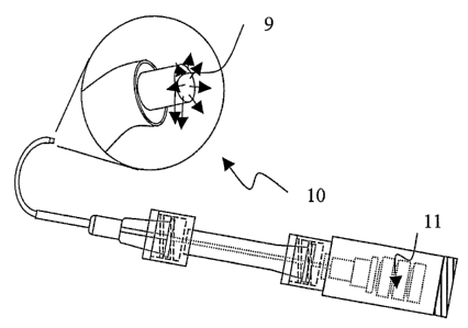
In an embodiment of the invention, a first communicating element used for
initial placement of
the microcannula has a signaling beacon to identify the location of the
microcannula distal tip
relative to the target tissues, as seen in Figure 5. The signaling means may
comprise an
echogenic material for ultrasound guidance, an optically active material for
optical guidance or a
light source for visual guidance placed at the microcannula tip or placed to
indicate the position
of the microcannula tip. In one embodiment, a plastic optical fiber (POF) 9 is
used as a
communicating element to provide a bright visual light source at the distal
tip 10. The distal tip
of the POF 9 is positioned proximal to, near or slightly beyond the distal end
of the
microcannula sheath and the emitted signal may be detected through tissues
visually or using
sensing means such as infrared imaging. The POF 9 may also have a tip that is
beveled,
mirrored or otherwise configured to provide for a directional beacon. The
beacon may be
illuminated by a laser, laser diode, light-emitting diode, or an incandescent
source such as a
mercury halogen lamp. In an alternate embodiment, the signaling means may
comprise
visualization aids along the length of the microcannula, for example a side
emitting optical fiber
CA 02554257 2006-07-20
WO 2005/070490 PCT/US2005/002603
of discrete length leading up to the distal end or at a known point along the
microcannula may be
used to indicate the position of the microcannula and the distal tip. Upon
placement of the
microcannula at the target tissues, the beacon assembly 11 and POF 9 may be
removed. The
connection point may be sealed with a cap or with a self sealing mechanism
such as a one-way
valve or an elastomer seal. Alternatively, the POF may be placed co-linear to
or within the
lumen of a delivery communicating channel, allowing for delivery of fluids or
gases through the
delivery communicating channel without requiring removal of the beacon
assembly.
Alternate embodiments of the microcannula may use other imaging technologies
to locate the
signal beacon. Other possible imaging technologies include but are not limited
to magnetic
resonance imaging, fluoroscopy and ultrasound. In these embodiments, the
beacon signal may
take other forms to match the imaging technology such as a radiopaque marker
attached to or
embedded at or near the distal tip of the microcannula. Alternatively or in
addition, an echogenic
material or coating may be added to the distal tip, etc.
It is also preferred for the microcannula to have a rounded distal tip 12 to
minimize tissue trauma
and aid the ability of the microcannula to be advanced into small tissue
spaces, as seen in Figures
6 and 7. The rounded tip 12 may be the same outer diameter as the microcannula
or larger,
depending on the specific properties desired. The rounded tip 12 may be formed
and attached to
the microcannula during assembly or alternatively, the microcannula tip may be
processed by a
secondary operation to form a rounded contour. When the rounded tip 12 is used
in conjunction
with a light emitting signaling beacon 9 such that the light is delivered
proximal to the rounded
tip, the tip acts to disperse the light 13. The dispersed light aids
visualization when viewing the
microcannula off axis, for example when advancing the microcannula in
Schlemm's Canal.
Another lcey feature of the invention is the use of a communicating element to
deliver fluid to the
distal tip during advancement of the microcannula within the tissue space. The
injection of small
amounts of fluid may serve to open the tissue space ahead of the microcannula
tip and lubricate
the channel to greatly increase the ability to advance the microcannula
atraumatically. Delivery
of surgical viscoelastic materials such as hyaluronic acid solutions and gels
are especially
efficacious in aiding advancement and placement of the microcannula. Delivery
of fluids,
especially gel-like viscoelastic materials, allows for the dilation of the
tissue space in the
circumstance that a constriction or partial blockage is reached during
advancement of the
11
CA 02554257 2006-07-20
WO 2005/070490 PCT/US2005/002603
microcannula. A particularly effective embodiment comprises a microcannula
with a
communicating element such as an optical fiber to provide a signaling beacon
at the
microcannula tip and a second communicating element to deliver a fluid such as
a solution of
hyaluronic acid to the microcannula tip while the signaling beacon is active.
Such a
microcannula may be manually manipulated and used to deliver fluids to aid
microcannula
advancement while simultaneously observing the microcannula tip location along
the tissue
space. The combination of fluid delivery in the path of the microcannula and
the observation of
the microcannula tip when advanced, retracted and torsioned allows precisely
controlled
manipulation and advancement in tight tissue spaces. The ease of manipulation
is further aided
with the addition of a reinforcing member to the communicating element of the
microcannula.
Examples:
Example l:
In the following example, a composite microcannula with two communicating
elements was
fabricated. A communicating element with a lumen (Polyimide Tubing 0.003 inch
TD x 0.004
inch OD), a second communicating element comprising a plastic optical fiber
(85-100 microns,
0.0034-0.0039 inch OD), a reinforcement element (304SS wire ground to 0.001
inches in the
distal 2.5 inches tapering up over a 1.0 inch length to a diameter of 0.003
inches for the
remaining length of the microcannula), and an outer sheath comprising
polyethylene
teraphthalate (PET) shrink tubing (0.008 inch ID and 0.00025 inch wall
thickness), were all cut
to lengths appropriate for setting the final overall length of the
microcannula. The distal ends of
the inner components were then aligned flush and joined with an adhesive. The
reinforcing
element was tapered and aligned to provide more flexibility distally and
stiffer reinforcement
more proximal in the microcannula. The three elements were aligned in a
triangular pattern
rather than an in-line pattern to create an assembled profile with the
smallest major-axis
dimension. The assembly of multiple components was then inserted into the heat
shrinlc tubing
outer sheath so that the inner elements were aligned for capture in the heat
shrink tubing. At the
proximal end of the microcannula assembly, the two communicating elements were
extended
outside of the heat shrink tubing and separated.
The assembly was placed in a hot air stream at 220-240 degrees F, so the heat
shrink recovered
and the inner elements were captured to form a multi-component shaft of the
microcannula. The
composite microcannula demonstrated a final outer dimension of 200 to 230
microns with a
12
CA 02554257 2006-07-20
WO 2005/070490 PCT/US2005/002603
lumen of 75 microns. To finish the assembly, extension communicating elements
were bonded
to the proximal end of the two communicating elements respectively. The
extensions were
finished by adding a Luer infusion connector and an optical connector to serve
as interfaces to
the communicating elements. Testing of the completed microcannula was
performed,
demonstrating simultaneous fluid delivery from the Luer connector and light
delivery from the
optical connector to the microcannula tip.
Example 2:
The microcannula fabricated in Example 1 was tested in accessing Schlemm's
Canal of an
enucleated human eye. The first communicating element, the infusion lumen, was
attached to a
syringe filled with fluid at the proximal Luer connection. The second
communicating element,
the optical fiber, was attached to a light emitting source at the proximal
connection. Operating at
the temporal-superior segment of the anterior portion of the eye, two radial
incisions were made
to a depth of Schlemm's Canal and extending from the clear cornea
approximately 3 mm
posterior. A third incision was made across the posterior end of the radial
incisions to define a
surgical flap. The flap was then excised up toward the limbus, exposing
Schlemm's Canal. The
distal tip of the composite microcannula was inserted into Schlemm's Canal.
The light source for
the second communicating element was activated and the microcannula was
advanced along
Schlemm's Canal. The light emitting from the microcannula tip was seen through
the sclera and
used to help guide the microcannula. The microcannula was advanced along
Schlemm's Canal
until the tip was seen reaching an appropriate location. The syringe connected
to the first
communicating element extension was used to inject fluid (Healon GV, Advanced
Medical
Optics, Inc.) into Schlemm's Canal as needed to aid microcannula advancement.
After the
desired microcannula positioning was completed, the microcannula was
repositioned for
additional fluid injections and subsequently completely retracted from
Schlemm's Canal.
Example 3:
In the following example, an atraumatic rounded distal tip component was
fabricated for
placement over a composite microcannula. Polyethylene teraphthalate (PET)
shrink tubing
(Advanced Polymers, Nashua NH) O.OOS inch ID and 0.00025 inch wall thickness
was obtained.
A length of shrink tubing approximately 2 cm long was placed over a mandrel
comprised of a
section of hypodermic tubing 0.003 inch x 0.007 inch diameter. Teflon coated
steel wire, 0.0025
inch diameter was held inside the hypodermic tubing and extending beyond the
end of the shrine
13
CA 02554257 2006-07-20
WO 2005/070490 PCT/US2005/002603
tubing. Under stereomicroscope visualization, a point heat source (adjustable
soldering iron) set
to 500 degrees C was brought into close proximity to the end of the heat
shrink tubing. The heat
was allowed to melt the end of the tube without touching the heat source to
the polymer. The
surface tension of the polymer melt created a rounded "ball-end" tip with a
0.0025 inch diameter
lumen. The polymer was allowed to cool and then stripped off of the mandrel
and wire. The
length of PET shrink tubing held beyond the end of the mandrel determined the
final diameter of
the rounded tip. Approximately 0.08 inches of extension yielded tips
approximately 0.008 inch
or 200 micron outer diameter.
The finished component was then drawn over the distal end of a composite
microcannula similar
to Example 1, which was 0.0075 inches or 190 microns in largest diameter. The
tip component
was butted up to the end of the composite elements and then shrunk in place
with a hot air stream
at 240 degrees F to attach the tip.
Example 4:
In the following example, the body of a composite microcannula was formed out
of a wire coil
and polymer heat shrink tubing. The coil was fabricated by progressively
winding a 0.003 inch
by 0.001 inch stainless steel ribbon under 20 grams tension around a 0.0055
inch diameter
stainless steel mandrel. Following removal from the mandrel, the resulting
wire ribbon coil had
an outside diameter of 0.008 inches or 200 microns, an inside diameter of
0.006 inches or 150
microns, and overall length of approximately 5 inches. A 6 inches long piece
of 0.010 inch or
250 micron ID PET heat shrink with a preformed rounded tip at one end was
slipped over the
coil and recovered using hot air over the entire length of the coil. A 0.004
inch diameter optical
fiber was then loaded into the lumen of the microcannula and advanced to the
distal end. The
proximal ends were terminated into a fluid infusion lumen and O.Smm diameter
optical fiber
respectively. The distal portion of the assembly was found to have desirable
mechanical
characteristics of flexibility and resistance to kinking.
Example 5:
An experiment was performed to test the coil-wound microcannula design as
described in
Example 3. Whole globe human eyes were obtained from a tissue bank. The
enucleated eyes
were prepared by first injecting the vitreous chamber with phosphate buffered
saline to replace
fluid lost post-mortem and bring the globes to a natural tone. Operating at
the temporal-superior
14
CA 02554257 2006-07-20
WO 2005/070490 PCT/US2005/002603
segment of the anterior portion of the eye, two radial incisions were made to
a depth of
Schlemm's Canal and extending from the clear cornea approximately 3 rnm
posterior. A third
incision was made across the posterior end of the radial incisions to define a
surgical flap. The
flap was then excised up toward the limbus, exposing Schlemm's Canal. The
microcannula was
inserted into Schlemm's Canal and advanced to approximately 90 degrees around
from the access
site. The metal coil was able to be seen through the scleral wall allowing the
amount of
microcannula advancement to be determined.
Example 6:
In the following example, a composite microcannula with several communicating
elements in
parallel alignment forming a distal segment with a maximum outer diameter of
250 microns was
fabricated. The outer member comprised a tubular structure and the two
internal communicating
elements comprised elongated linear elements. At the distal end of the outer
structure, an
atraumatic spherical-shaped distal tip was formed. A communicating lumen was
formed in the
annular space between the outer tube and the inner members. The inner members
comprised an
optical fiber and a reinforcement element. The outer member was a tubular
structure comprised
of three sizes of PEBAX (polyamide/polyether copolymer), 63 durometer tubing:
1) Proximal Section 0.016 inch ID x 0.026 inch OD, 24 inch length
2) Mid Section 0.010 inch ID x 0.014 inch OD, 4 inch length
3) Distal Section 0.006 inch ID x 0.008 inch OD, 1.8 inch length
The outer tubular element was constructed by first cutting the individual
shaft segments to
lengths appropriate for setting the final overall length of the microcannula.
The mid section was
inserted into the proximal section with appropriate length for an overlapping
bond. The tubular
elements were then bonded together with an adhesive or by melt-fusing the
polymeric tubes
together with a controlled heat process. The distal section was bonded to the
mid shaft similarly.
These tubes were bonded together to form a decreasing outer diameter toward
the distal tip.
The reinforcement element comprised 304 Stainless Steel wire size 0.0010 +/-
0.0005 inch OD,
and the optical fiber comprised a plastic optical fiber fabricated from
polystyrene and
polymethylmethacrylate with an 85 to 100 micron OD. The reinforcement element
and the
optical fiber were cut to lengths appropriate for setting the final overall
length of the
CA 02554257 2006-07-20
WO 2005/070490 PCT/US2005/002603
microcannula. The reinforcement element and optical fiber were inserted into
the outer member
assembly. The inner elements were aligned with the distal tip of the distal
shaft.
An atraumatic rounded tip was formed at the end of the distal section. A quick
drying UV
curable adhesive (Loctite Brand 4305) was applied to the outer section of the
distal tip. An
adhesive of medium to high viscosity was chosen so that the adhesive
application formed a
bulbous structure approximately 0.001 inch thickness. A small, approximately
0.03 microliter
amount of adhesive was used to create the tip. The adhesive was cured to form
the spherically
shaped atraumatic tip with a diameter of 0.010 inches or 250 microns.
The free end of the infusion lumen was terminated with a female Luer port. The
proximal end of
the optical fiber was connected to a Plastic Optical Fiber (POF) that
terminated in an optical
SMA connector.
The area of the microcannula assembly where the optical fiber and
reinforcement enter the inside
of the outer member was sheathed in a protective plastic housing forming a
hub. The hub also
provided a means for manipulation of the microcannula.
The optical SMA termination was connected to a light source and light was
conducted to the tip
of the microcannula to provide a signal beacon: The Luer termination was
connected to a
fluid-filled syringe and activation of the syringe resulted in fluid delivery
through the
microcannula exiting from the distal tip. Delivery of the signal beacon light
and fluid could be
activated individually or simultaneously.
Example 7:
In the following example, a composite microcannula with several communicating
elements in
parallel alignment forming a distal segment with a maximum outer diameter of
350 microns was
fabricated similarly to Example 6. In this embodiment the outer member was
constructed with
three sizes of PEBAX tubing with slightly larger dimensions:
1) Proximal Section 0.016 inch ID x 0.026 inch OD, 24 inch length
2) Mid Section 0.0130 inch ID x 0.015 inch OD, 4 inch length
3) Distal Section 0.008 inch ID x 0.012 inch OD, 1.8 inch length
16
CA 02554257 2006-07-20
WO 2005/070490 PCT/US2005/002603
A spherically shaped atraumatic tip was fabricated on the microcannula by the
method described
in Example 6, forming a distal tip with a diameter of 0.014 inches or 350
microns. In this
embodiment, no reinforcing element was placed into this cannula construction,
however a plastic
optical fiber was incorporated similar to Example 6.
The optical SMA termination was connected to a light source and light was
conducted to the tip
of the microcannula. The Luer termination was connected to a fluid-filled
syringe and activation
of the syringe resulted in fluid delivery through the microcannula exiting
from the distal tip.
Example 8:
The composite microcannulae of Example 6 and Example 7 were tested in human
eyes similarly
to the method of Example 2. The distal tip and distal segments of the
microcannulae could be
advanced along the entire circumference of Schlermn's Canal for 360 degrees
while observing
the beacon signal at the microcammla tip through the sclera. Injection of
small amounts of
hyaluronic acid-based surgical viscoelastic fluid (Healon GV, Advanced Medical
Optics Inc.)
delivered during advancement of the microcannulae decreased the force required
for
advancement and provided for more progressive advancement.
Example 9:
A composite microcannula with several collinear elements was fabricated
similar to Example 6.
In this embodiment, the outer structure had no mid section in that the
proximal section was
connected directly to the distal section.
Example 10:
In order to determine the optimal flexural properties of a composite
microcannula for
introduction into small tissue spaces, a family of microcannulae were
fabricated with the same
outer dimensions and material characteristics but with varying flexural
rigidity. Flexural rigidity
of a body is equal to the product of the flexural modulus, E, and the moment
of inertia of the
cross-section, I, and is typically called EI. The outer sheath comprised PEBAX
tubing with
0.008 inch (200 micron) OD and 0.006 inch (150 micron) ID. The sample set
comprised the
tubing alone without reinforcing element(s), the tubing with a 100 micron
outer diameter plastic
optical fiber placed within the lumen and the tubing with stainless steel
reinforcing wires of
varying size in the lumen. The ends of the components were secured with
adhesive, while
17
CA 02554257 2006-07-20
WO 2005/070490 PCT/US2005/002603
forming an atraumatic spherically shaped tip, as described in Example 6. The
lumen allowed
fluid delivery to the tip of the microcannula from a proximally attached Luer
connector.
The flexural rigidity of the microcannulae were evaluated by mechanical
testing. The
microcannulae cantilever force-displacement characteristics were tested on a
mechanical testing
apparatus with a high sensitivity load cell (Instron model 5542, SN Load
Cell). The linear region
of the resultant data was used to calculate the measured flexural rigidity of
the test samples.
Microcannula Description Measured Flexural
Rigidity
(EI) [kN~'m2~
PEBAX Outer Sheath 3.09 E-11
PEBAX Outer Sheath with 0.001 in diameter SS 3.76 E-11
wire
PEBAX Outer Sheath with 100 micron diameter 6.33 E-11
plastic optical
fiber
PEBAX Outer Sheath with 0.002 in diameter SS 9.69 E-11
wire
PEBAX Outer Sheath with 0.003 zn diameter SS 2.86 E-10
wire
PEBAX Outer Sheath with 0.004 in diameter SS 7.5 E-10
wire
Example 11:
The microcannulae fabricated in Example 10 were tested for the ability to
access Schlemm's
Canal of a human eye similar to the methods described in Example 2. In a first
trial, the distal tip
of the microcannulae were inserted into the Canal and advanced without
delivery of fluid from
the microcannula tip. The number of degrees of advancement around the eye was
recorded for
each microcannula. In the next trial, the test was repeated with the delivery
of a small amount of
viscoelastic fluid (Healon GV, Advanced Medical Optics Inc.) from the
microcannula tip during
advancement. One property of Healon GV, a hyaluronic acid based viscoelastic
fluid, is very
high lubricity. Three eyes were used for the evaluation, with cannulations
performed both
clockwise and counterclockwise from the surgical access site.
When tested for the degree of advancement within Schlemm's Canal, the
microcannulae with
low flexural rigidity could be slowly advanced along Canal until further
advancement was no
longer possible due to lack of force transfer. These lower flexural rigidity
devices tended to
bend or kink when reaching the limit of travel. The microcannulae with very
high flexural
18
CA 02554257 2006-07-20
WO 2005/070490 PCT/US2005/002603
rigidity could be advanced a short distance until fi~.rther advancement was no
longer possible due
to the inability of the microcannula to bend with the curve of Schlemm's
Canal. If advanced
further, the microcannula with very high flexural rigidity in some cases
punctured through the
outer wall of the Canal, an undesirable result. The testing was performed by
advancing each
device manually, attempting to use a comparable maximum force for each test
run, so as to
maintain an adequate comparison. In cases where the cammla did not traverse
the full extent of
the Canal, the force required to advance the cannula increased with increased
extent of
cannulation, which was attributed to interaction of the compliance properties
of the device and
the frictional forces between the device and the tissues of the Canal.
Microcannula Degrees Degrees Degrees Degrees
Flexural RigidityCannulation Cannulation Cannulation Cannulation
(EI) [ kN*m2]Achieved - Achieved - Achieved - Achieved -
No No Fluid Fluid
Fluid DeliveryFluid DeliveryDelivery Delivery
AVG Std Dev AVG Std Dev
3.09 E-11 183 64 360 0
3.76 E-11 242 35 360 0
6.33 E-11 265 78 360 0
9.69 E-11 203 23 360 0
2.86 E-10 177 25 360 0
7.5 E-10 80 20 89 26
The results of advancing the microcannulae into Schlemm's Canal without fluid
delivery
demonstrated an optimal flexural rigidity of approximately 6.33 E-11 kN*m2.
Flexural rigidity
in the range of 3.09 E-11 to 2.86 E-10 provided a microcannula that was able
to access
approximately 180 degrees of the eye. Such properties would allow the entire
eye to be accessed
from a single surgical site by advancing the microcannula in both directions.
The results of advancing the microcannula into Schlemm's Canal with fluid
delivery
demonstrated improved performance except for the microcannula with the highest
flexural
rigidity. Flexural rigidity in the range of 3.09 E-11 to 2.86 E-10 kN*m2
coupled with the
delivery of a lubricious material (Healon GV) allowed the entire circumference
of Schlemm's
Canal (360 degrees) to be accessed by the test microcannulae. It was noted
that the amount of
19
CA 02554257 2006-07-20
WO 2005/070490 PCT/US2005/002603
force required to advance each device was significantly decreased by the
presence of the
lubricious fluid being delivered from the distal tip of the microcannula
during the cannulation.
In addition, a number of attempts to advance a microcannula into Schlemm's
Canal without fluid
delivery were made by depositing a small amount of the viscoelastic fluid at
the surgical site and
then passing the cannula through the gel. These did not result in any
significant decrease in force
or increase in advancement of the test devices, indicating the advantage of
delivering fluid at the
microcannula tip during manipulation and advancement.
Many features have been listed with particular configurations, options, and
embodiments. Any
one or more of the features described may be added to or combined with any of
the other
embodiments or other standard devices to create alternate combinations and
embodiments.
The preferred embodiments described herein are illustrative only, and although
the examples
given include many specifics, they are illustrative of only a few possible
embodiments of the
invention. Other embodiments and modifications will no doubt occur to those
skilled in the art.
The examples given should only be interpreted as illustrations of some of the
preferred
embodiments of the invention.