Note: Descriptions are shown in the official language in which they were submitted.
CA 02554303 2014-05-01
73612-74
INJECTABLE INTRAOCULAR IMPLANT WITH FLEXIBLE HAPTICS
FIELD OF THE INVENTION
The present invention relates to ocular implants generally and more
particularly to intraocular implants.
BACKGROUND OF THE INVENTION
The following patent publications are believed to represent the current
state of the art:
U.S. Patents 5,814,103; 5,876,442; 5,928,283; 6,007,579; 6,066,171;
5,653,751; 6,596,026; 6,464,725; 5,391,202; 5,384,606; 4,074,368; 4,994,082;
5,628,798; 5,222,981; 4,172,297; 5,769,890; 4,892,543; 4,373,218; 4,968,127;
=
4,759,761; 4,976,732 and 5,769,889;
Published U.S. Application 2001/018,612;
Published PCT Applications WO 94/07,435; WO 00/38593 and WO
83/01566;
Foreign Patent Publications DE 4,403,326; EP 1,092,402; EP 0,419,740;
GB 2,181,355; EP 0,897,702; EP 0,212,616; DE 3,428,895 and DE 19,501,444.
1
CA 02554303 2006-07-27
SUMMARY OF THE INVENTION
The present invention seeks to provide an improved intraocular implant.
There is thus provided in accordance with a preferred embodiment of the
present invention an injectable intraocular implant including an optics
portion and a
resilient, flexible haptics portion mounted coaxially with the optics portion.
In accordance with a preferred embodiment of the present invention the
optics portion and the haptics portion are arranged for mutual snap fit
engagement.
Preferably, the optics portion includes a telescope. Additionally or
alternatively, the
haptics portion is formed of biocompatible plastic.
In accordance with another preferred embodiment of the present
invention the haptics portion includes a cylindrical portion having integrally
formed
therewith a plurality of outwardly extending haptics wings. Alternatively, the
haptics
portion includes an outwardly extending helical portion including at least one
helical
section. Preferably, each of the at least one helical section includes a
haptics spiral
portion, a residual spiral portion and a frangible portion connecting the
haptics spiral
portion and the residual spiral portion.
In accordance with yet another preferred embodiment of the present
invention the haptics portion includes a hollow, generally cylindrical
structure defining
a generally circular inward facing wall portion and a generally circular
outward facing
wall portion. Preferably, the inward facing wall portion defines a generally
circular
optics engagement aperture therein. Additionally or alternatively, the inward
facing wall
portion and the outward facing wall portion each define a generally circular
optics
engagement aperture therein. Additionally or alternatively, each of the
haptics wings is
tinted so as to block passage of parasitic light therethrough.
There is also provided in accordance with another preferred embodiment
of the present invention a method for inserting an intraocular implant into an
eye
including providing an injectable intraocular implant including an optics
portion and a
resilient, flexible haptics portion mounted coaxially with the optics portion,
locating the
injectable intraocular implant in a delivery tube of a syringe, inserting the
delivery tube
2
CA 02554303 2006-07-27
into a lens capsule of an eye and injecting the injectable intraocular implant
into the lens
capsule.
In accordance with a preferred embodiment of the present invention the
injecting includes utilizing fluid pressure to force the implant into the lens
capsule.
Preferably, the fluid is biocompatible fluid. Additionally or alternatively,
the haptics
portion includes haptics wings and the locating includes folding the haptics
wings over
the optics portion.
In accordance with another preferred embodiment of the present
invention the locating includes arranging the implant with an outward facing
end of the
optics portion arranged rearward in the delivery tube. Preferably, the haptics
portion
includes an outwardly extending helical portion and the locating includes
winding the
helical portion into a coil. Additionally, the injecting includes rotating the
syringe.
In accordance with yet another preferred embodiment of the present
invention the outwardly extending helical portion includes a haptics spiral
portion, a
residual spiral portion and a frangible portion connecting the haptics spiral
portion and
the residual spiral portion and the rotating causes the frangible portion to
break, thereby
separating the haptics spiral portion from the residual spiral portion.
In accordance with still another preferred embodiment of the present
invention the haptics portion includes an outward facing wall portion and the
locating
includes pulling the outward facing wall portion over an outward facing end of
the
optics portion. Preferably, the locating includes sealing a fluid flow
passageway in the
syringe. Additionally, the injecting includes unsealing the fluid flow
passageway.
In accordance with a further preferred embodiment of the present
invention the locating includes drawing the haptics portion into a delivery
syringe.
Preferably, the method also includes puncturing the haptics portion.
There is further provided in accordance with a further preferred
embodiment of the present invention an intraocular implant injection assembly
including an intraocular implant positioning subassembly, an intraocular
implant
including haptics and having an optical axis, the intraocular implant being
mounted in
the intraocular implant positioning subassembly in one of at least one first
predetermined azimuthal orientation with respect to the subassembly, a housing
retaining the intraocular implant positioning subassembly, in one of at least
one second
3
CA 02554303 2006-07-27
,
,
predetermined azimuthal orientation with respect to the housing and a syringe
having an
angled forward edge, the syringe being retained in the housing, such that the
angled
forward edge is in one of at least one third predetermined azimuthal
orientation with
respect to the housing.
There is yet further provided in accordance with yet a further preferred
embodiment of the present invention an intraocular implant injection assembly
including an intraocular implant positioning subassembly, an intraocular
implant
including haptics and having an optical axis, the intraocular implant being
mounted in
the intraocular implant positioning subassembly in one of at least one first
predetermined azimuthal orientation with respect to the subassembly, a housing
retaining the intraocular implant positioning subassembly, in one of at least
one second
predetermined azimuthal orientation with respect to the housing and a syringe
having a
user-sensible azimuthal orientation indicator, the syringe being retained in
the housing,
in one of at least one third predetermined azimuthal orientation with respect
to the
housing.
In accordance with a preferred embodiment of the present invention, the
syringe has an angled forward edge and is retained in the housing, such that
the angled
forward edge is in one of at least one fourth predetermined azimuthal
orientation with
respect to the housing.
In accordance with another preferred embodiment of the present
invention, the intraocular implant positioning subassembly includes a rearward
positioning element including an azimuthal registration aperture and a forward
positioning element including an azimuthal registration pin operative to
engage the
azimuthal registration aperture and to prevent relative rotation between the
rearward
positioning element and the forward positioning element. Preferably, the
forward
positioning element includes a circumferential flange and the rearward
positioning
element includes a plurality of snap-fit engagement elements, operative to
engage the
circumferential flange by snap-fit engagement.
In accordance with yet another preferred embodiment of the present
invention a slanted portion of the haptics lies between a forward, inwardly
facing,
tapered portion of the rearward positioning element and a rearward, outwardly
facing,
tapered portion of the forward positioning element. Preferably, the tapered
portion of
4
CA 02554303 2006-07-27
the rearward positioning element and the tapered portion of the forward
positioning
element are configured to guide the haptics while the intraocular implant is
loaded into
the syringe, thereby maintaining mechanical integrity of the haptics.
In accordance with still another preferred embodiment of the present
invention the intraocular implant positioning subassembly includes a plurality
of
intraocular implant positioning protrusions operative to retain the
intraocular implant in
the one of at least one first predetermined azimuthal orientation with respect
to the
subassembly. Preferably, the intraocular implant positioning subassembly
includes an
angled shoulder having an angular orientation which is generally identical to
that of the
forward angled edge and which is adapted to engage the forward angled edge,
thereby
to provide at least one predetermined azimuthal orientation of the syringe
with respect
to the intraocular implant positioning subassembly.
In accordance with a further preferred embodiment of the present
invention the forward angled edge of the syringe is configured to allow the
haptics to
unfold sequentially as the intraocular implant is injected into an eye.
Preferably, the
housing includes at least one longitudinal slot, arranged to slidably
accommodate the
user-sensible azimuthal orientation indicator. Additionally or alternatively,
the housing
is formed of a resilient material and is at least partially bifurcated to
permit insertion
thereinto of the syringe.
In accordance with yet a further preferred embodiment of the present
invention the intraocular implant injection assembly also includes an
intraocular implant
displacer adapted to engage the housing. Preferably, the intraocular implant
displacer
includes a shaft portion operative to push the intraocular implant from the
positioning
subassembly into the syringe. Additionally or alternatively the haptics are
adapted to
fold over a body portion of the intraocular implant and symmetrically about
the optical
axis, and to extend forwardly of the body portion when the intraocular implant
is
located within the syringe.
In accordance with still a further preferred embodiment of the present
invention the intraocular implant includes a capsular body portion and the
haptics are
adapted to urge the capsular body portion away from the cornea when the
intraocular
implant is implanted in the eye. Preferably, the haptics include a plurality
of haptics
wings, each being tinted so as to block passage of parasitic light
therethrough.
5
CA 02554303 2006-07-27
There is also provided in accordance with another preferred embodiment
of the present invention an intraocular implant injection assembly including
an
intraocular implant including haptics, a syringe having an angled forward edge
and an
intraocular implant azimuthal positioner operative to retain the intraocular
implant in
one of at least one predetermined azimuthal orientation with respect to the
angled
forward edge of the syringe.
There is additionally provided in accordance with yet another preferred
embodiment of the present invention an intraocular implant injection assembly
including an intraocular implant including haptics, a syringe having a user-
sensible
azimuthal orientation indicator and an intraocular implant azimuthal
positioner
operative to retain the intraocular implant in one of at least one
predetermined azimuthal
orientation with respect to the syringe. Preferably, the syringe has an angled
forward
edge and the intraocular implant azimuthal positioner is operative to retain
the
intraocular implant in the one of at least one predetermined azimuthal
orientation with
respect to the angled forward edge of the syringe.
In accordance with a preferred embodiment of the present invention the
intraocular implant azimuthal positioner includes a rearward positioning
element
including an azimuthal registration aperture and a forward positioning element
including an azimuthal registration pin operative to engage the azimuthal
registration
aperture and to prevent relative rotation between the rearward positioning
element and
the forward positioning element. Preferably, the forward positioning element
includes a
circumferential flange and the rearward positioning element includes a
plurality of snap-
fit engagement elements, operative to engage the circumferential flange by
snap-fit
engagement.
In accordance with another preferred embodiment of the present
invention a slanted portion of the haptics lies between a forward, inwardly
facing,
tapered portion of the rearward positioning element and a rearward, outwardly
facing,
tapered portion of the forward positioning element. Preferably, the tapered
portion of
the rearward positioning element and the tapered portion of the forward
positioning
element are configured to guide the haptics while the intraocular implant is
loaded into
the syringe, thereby maintaining mechanical integrity of the haptics.
6
CA 02554303 2006-07-27
,
In accordance with yet another preferred embodiment of the present
invention the intraocular implant azimuthal positioner includes a plurality of
intraocular
implant positioning protrusions operative to retain the intraocular implant in
the one of
at least one predetermined azimuthal orientation with respect to the syringe.
Preferably,
the intraocular implant azimuthal positioner includes an angled shoulder
having an
angular orientation which is generally identical to that of the angled forward
edge and
which is adapted to engage the angled forward edge, thereby to provide the one
of at
least one predetermined azimuthal orientation between the intraocular implant
and the
syringe.
In accordance with a further preferred embodiment of the present
invention the intraocular implant injection assembly also includes an
intraocular implant
displacer including a shaft portion operative to push the intraocular implant
from the
intraocular implant azimuthal positioner into the syringe. Preferably, the
haptics are
adapted to fold over a body portion of the intraocular implant and
symmetrically about
the optical axis, and to extend forwardly of the body portion when the
intraocular
implant is located within the syringe. Additionally or alternatively, the
angled forward
edge of the syringe is configured to allow the haptics to sequentially unfold
when the
intraocular implant is injected into an eye.
In accordance with yet a further preferred embodiment of the present
invention the intraocular implant includes a capsular body portion and the
haptics are
adapted to urge the capsular body portion away from the cornea when the
intraocular
implant is implanted in the eye. Preferably, the haptics include a plurality
of haptics
wings, each being tinted so as to block passage of parasitic light
therethrough.
There is further provided in accordance with a further preferred
embodiment of the present invention a method for injecting an intraocular
implant into
an eye, the method including providing an intraocular implant including
haptics and
having an optical axis, arranging the intraocular implant inside an injector
having an
injector axis, such that the optical axis is parallel to the injector axis and
injecting the
intraocular implant into the eye by displacing the intraocular implant along
an injection
axis which is coaxial with the optical axis. Preferably, the arranging
includes arranging
the intraocular implant inside the injector such that the optical axis is
coaxial with the
injector axis.
7
CA 02554303 2013-08-23
73612-74
There is yet further provided in accordance with yet a further preferred
embodiment of the present invention a system for injecting an intraocular
implant into an eye,
including an intraocular implant including haptics and having an optical axis,
an injector
having an injector axis, adapted to have the intraocular implant arranged
therein such that the
optical axis is parallel to the injector axis and an intraocular implant
displacer operative to
inject the intraocular implant into the eye by displacing the intraocular
implant along an
injection axis which is coaxial with the optical axis. Preferably, the
intraocular implant
arranged in the injector such that the injector axis is coaxial with the
optical axis.
In accordance with another preferred embodiment of the present invention
there is provided an injectable intraocular implant comprising: an optics
portion arranged
along an optical axis; and a resilient, flexible haptics portion mounted
coaxially with said
optics portion, said haptics portion including a cylindrical portion having
integrally formed
therewith a plurality of haptics wings, each of said plurality of haptics
wings including a pair
of side peripheral edges joined by an outer peripheral edge, said peripheral
edges
circumscribing a haptics wing area, said plurality of haptics wings having a
post-implantation
operative orientation, wherein said haptics wing areas of said plurality of
haptics wings lie
generally transverse to said optical axis, and a pre-implantation operative
orientation, wherein
said haptics wing areas of said plurality of haptics wings lie generally
parallel to said optical
axis, said plurality of haptics wings being adapted to automatically unfold
from said pre-
implantation operative orientation to assume said post-implantation operative
orientation by
pivoting of said side peripheral edges about pivot axes which are tangential
to the
circumference of said cylindrical portion.
8
CA 02554303 2006-07-27
,
BRIEF DESCRIPTION OF THE DRAWINGS
The present invention will be understood and appreciated more fully
from the following detailed description, taken in conjunction with the
drawings in
which:
Figs. 1A and 1B are simplified respective exploded and assembled
pictorial illustrations of an injectable intraocular implant constructed and
operative in
accordance with a preferred embodiment of the present invention;
Figs. IC and 1D are simplified respective exploded and assembled side
view illustrations of the injectable intraocular implant of Figs. IA and 1B;
Figs. 2 and 3 are simplified pictorial and sectional illustrations of the
injectable intraocular implant of Figs. IA ¨ 113 located in a fluid-filled
hypodermic
delivery syringe;
Figs. 4A, 4B, 4C and 4D are simplified sectional illustrations of four
stages of the injection of the intraocular implant of Figs. 1A - 1D in the
delivery syringe
arrangement of Figs. 2 and 3 into the eye of a patient;
Figs. 5 and 6 are simplified pictorial and sectional illustrations of the
injectable intraocular implant of Figs. 1A - 1D located in a specially
designed, non
fluid-filled hypodermic delivery syringe;
Figs. 7A, 7B, 7C and 7D are simplified sectional illustrations of four
stages of the injection of the intraocular implant of Figs. lA - 1D in the
delivery syringe
arrangement of Figs. 5 and 6 into the eye of a patient;
Figs. 8A and 8B are simplified respective exploded and assembled
pictorial illustrations of an injectable intraocular implant constructed and
operative in
accordance with another preferred embodiment of the present invention;
Figs. 8C and 8D are simplified respective exploded and assembled side
view illustrations of the injectable intraocular implant of Figs. 8A and 8B;
Figs. 9 and 10 are simplified pictorial and sectional illustrations of the
injectable intraocular implant of Figs. 8A-8D located in a hypodermic delivery
syringe;
Figs. 11A, 11B, 11C and 11D are simplified sectional illustrations of four
stages of the injection of the intraocular implant of Figs. 8A-8D in the
delivery syringe
arrangement of Figs. 9 and 10 into the eye of a patient;
9
CA 02554303 2006-07-27
Figs. 12A, 12B, 12C and 12D are simplified sectional illustrations of four
stages of the injection of the intraocular implant of Figs. 8A-8D in the
delivery syringe
arrangement of Figs. 9 and 10 into the eye of a patient in accordance with
another
preferred embodiment of the present invention;
Figs. 13A and 13B are simplified respective exploded and assembled
pictorial illustrations of an injectable intraocular implant constructed and
operative in
accordance with yet another preferred embodiment of the present invention;
Fig. 13C is a simplified assembled side view illustration of the injectable
intraocular implant of Figs. 13A and 13B;
Figs. 14A and 14B are simplified pictorial and sectional illustrations of
the injectable intraocular implant of Figs. 13A - 13C located in a delivery
syringe;
Figs. 15A, 15B, 15C and 15D are simplified sectional illustrations of four
stages of the injection of the intraocular implant of Figs. 13A-13C in the
delivery
syringe arrangement of Figs. 14A and 14B into the eye of a patient;
Figs. 16A and 16B are simplified respective exploded and assembled
pictorial illustrations of an injectable intraocular implant constructed and
operative in
accordance with still another preferred embodiment of the present invention;
Fig. 16C is a simplified assembled side view illustration of the injectable
intraocular implant of Figs. 16A and 16B;
Figs. 17A and 17B are simplified pictorial and sectional illustrations of
the injectable intraocular implant of Figs. 16A - 16C located in a delivery
syringe;
Figs. 18A, 18B, 18C and 18D are simplified sectional illustrations of four
stages of the injection of the intraocular implant of Figs. 16A-16C in the
delivery
syringe arrangement of Figs. 17A and 17B into the eye of a patient;
Figs. 19A and 19B are simplified pictorial and sectional illustrations of
the injectable intraocular implant of Figs. 16A-16C located in a delivery
syringe;
Figs. 20A, 20B, 20C and 20D are simplified sectional illustrations of four
stages of the injection of the intraocular implant of Figs. 16A-16C in the
delivery
syringe arrangement of Figs. 19A and 19B into the eye of a patient;
Figs. 21A and 21B are, respectively, a simplified exploded view
illustration and a simplified pictorial illustration of an injection assembly
for an
CA 02554303 2006-07-27
A
intraocular implant, constructed and operative in accordance with a further
preferred
embodiment of the present invention;
Fig. 22 is a simplified pictorial illustration of a plunger forming part of
the injection assembly of Figs. 21A and 21B;
Fig. 23 is a simplified sectional illustration of the plunger of Fig. 22,
taken along section lines XXIII ¨ XXIII in Fig. 22;
Fig. 24 is a simplified pictorial illustration of a syringe forming part of
the injection assembly of Figs. 21A and 21B;
Fig. 25 is a simplified sectional illustration of the syringe of Fig. 24,
taken along section lines XXV ¨ XXV in Fig. 24;
Fig. 26 is a simplified pictorial illustration of a housing element forming
part of the injection assembly of Figs. 21A and 21B;
Fig. 27 is a simplified sectional illustration of the housing element of Fig.
26, taken along section lines XXVII ¨ XXVII in Fig. 26;
Figs. 28A and 28B are simplified pictorial illustrations of a rearward
positioning element forming part of the injection assembly of Figs. 21A and
21B;
Fig. 29 is a simplified sectional illustration of the rearward positioning
element of Figs. 28A and 28B, taken along section lines XXIX ¨ XXIX in Fig.
28A;
Figs. 30A and 30B are simplified pictorial illustrations of a forward
positioning element forming part of the injection assembly of Figs. 21A and
21B;
Fig. 31 is a simplified sectional illustration of the forward positioning
element of Figs. 30A and 30B, taken along section lines XXXI ¨ XXXI in Fig.
30A;
Figs. 32A and 32B are simplified pictorial illustrations of an injectable
intraocular implant forming part of the injection assembly of Figs. 21A and
21B;
Fig. 33 is a simplified sectional illustration of the injectable intraocular
implant of Figs. 32A and 32B, taken along section lines XXXIII ¨ XXXIII in
Fig. 32A;
Figs. 34A and 34B are simplified pictorial illustrations of an intraocular
implant displacer element forming part of the injection assembly of Figs. 21A
and 21B;
Fig. 35 is a simplified sectional illustration of the intraocular implant
displacer element of Figs. 34A and 34B, taken along section lines XXXV ¨ XXXV
in
Fig. 34A;
11
CA 02554303 2006-07-27
. 1
Fig. 36 is a simplified pictorial illustration of the injection assembly of
Figs. 21A and 21B in a storage orientation;
Fig. 37 is a simplified sectional illustration of the injection assembly of
Fig. 36, taken along section lines )(XXVII ¨ XXXVII in Fig. 36;
Fig. 38 is a simplified pictorial illustration of the injection assembly of
Figs. 36 and 37 in an intraocular implant pre-loading orientation;
Fig. 39 is a simplified sectional illustration of the injection assembly of
Fig. 38, taken along section lines XXXIX ¨ XXXIX in Fig. 38;
Fig. 40 is a simplified pictorial illustration of the injection assembly of
Figs. 38 and 39 in an intraocular implant loading orientation;
Fig. 41 is a simplified sectional illustration of the injection assembly of
Fig. 40, taken along section lines XLI ¨ XLI in Fig. 40;
Fig. 42 is a simplified pictorial illustration of the injection assembly of
Figs. 40 and 41 in a partial syringe retraction orientation;
Fig. 43 is a simplified sectional illustration of the injection assembly of
Fig. 42, taken along section lines XLIII ¨ XLIII in Fig. 42;
Fig. 44 is a simplified pictorial illustration of the injection assembly of
Figs. 42 and 43 in a full syringe retraction orientation;
Fig. 45 is a simplified sectional illustration of the injection assembly of
Fig. 44, taken along section lines XLV ¨ XLV in Fig. 44;
Fig. 46 is a simplified pictorial illustration of the syringe of Figs. 21A
and 21B in a ready to use orientation;
Fig. 47 is a simplified sectional illustration of the syringe of Fig. 46,
taken along section lines XLVII ¨ XLVII in Fig. 46; and
Figs. 48A, 48B, 48C and 48D are simplified sectional illustrations of four
stages of injection of the injectable intraocular implant of Figs. 32A, 32B
and 33 into
the eye of a patient.
12
CA 02554303 2013-08-23
73612-74
DETAILED DESCRIPTION OF PREFERRED EMBODIMENTS
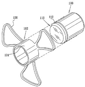
Reference is now made to Figs. 1A - 1D, which illustrate an injectable
intraocular implant constructed and operative in accordance with a preferred
embodiment of the present invention. It is seen that the implant preferably
includes an
optics portion 100 and a haptics portion 102 which is preferably snap-fitted
onto the
optics portion 100.
The optics portion 100 may be any suitable optics portion and is
preferably, but not necessarily, a telescope. Preferred intraocular implants
are described
in applicants/assignee's published patent documents listed hereinbelow :
U.S. Patents 5,391,202; 5,354,135; 5,814,103; 5,876,442; 5,928,283;
6,007,579; 6,066,171; 6,569,199 and 6,596,026, and U.S. Patent Publication
Nos.
2004/0138746 and 2004/0117011.
The optics portion 100 may incorporate any one or more of the features
described in the abovementioned patent documents in any suitable combination
and is
preferably in the form of a cylinder having adjacent one end thereof,
hereinafter referred
to as the outward facing end 110, a peripheral groove 112. The haptics portion
102 is
preferably formed of a resilient, flexible material, such as biocompatible
plastic, and
includes a cylindrical portion 124 having integrally formed therewith a
plurality of
outwardly extending haptics wings 126. Cylindrical portion 124 is preferably
formed
with an inwardly directed peripheral protrusion 128 adjacent one end thereof,
hereinafter referred to as the outward facing end 130. Protrusion 128 is
arranged for
normally non-removable snap-fit engagement with groove 112 on optics portion
100,
when cylindrical portion 124 is in coaxial surrounding relationship with
optics portion
100 as shown.
It is appreciated that the haptics wings 126 are preferably at least
partially opaque, so as to block passage of parasite light therethrough.
It is appreciated that the peripheral groove 112 of the optics portion 100
may be located at any suitable location therealong and the inwardly directed
peripheral
protrusion 128 of cylindrical portion 124 of haptics portion 102 may be
located at any
13
CA 02554303 2006-07-27
suitable location therealong to provide normally non-removable snap-fit
engagement of
optics portion 100 and haptics portion 102.
Reference is now made to Figs. 2 and 3, which are simplified pictorial
and sectional illustrations of the injectable intraocular implant of Figs. lA
¨ 1D located
in a fluid-filled hypodermic delivery syringe 140. It is seen that haptics
wings 126 are
folded over the optics portion 100 and that the implant is arranged with
outward facing
ends 110 and 130 arranged rearward in a delivery tube 142 of delivery syringe
140.
Fluid, such as biocompatible fluid 144, is located forward of a piston 146 of
delivery
syringe 140 and rearward of the implant.
Reference is now made to Figs. 4A, 4B, 4C and 4D, which are simplified
sectional illustrations of four stages of the injection of the intraocular
implant of Figs.
lA - 1D in the delivery syringe arrangement of Figs. 2 and 3 into the eye of a
patient.
Fig. 4A shows initial insertion of the tip of the delivery tube 142 of
delivery syringe 140
into the lens capsule of the eye. Fig. 4B shows the implant being forced out
of the
delivery syringe 140 into the lens capsule. Fig. 4C shows unfolding of haptics
wings
126 inside the lens capsule and Fig. 4D shows proper orientation of the
implant,
including fully deployed haptics wings 126, within the lens capsule.
Reference is now made to Figs. 5 and 6, which are simplified pictorial
and sectional illustrations of the injectable intraocular implant of Figs. 1 A
- 1D located
in a specially designed, non fluid-filled hypodermic delivery syringe 160. It
is seen that
haptics wings 126 are folded over the optics portion 100 and that the implant
is arranged
with outward facing ends 110 and 130 arranged rearward in a delivery tube 162
of
delivery syringe 160. A piston 166 of delivery syringe 160 engages outward
facing end
110 of the optics portion 100 of the implant.
Reference is now made to Figs. 7A, 7B, 7C and 7D, which are simplified
sectional illustrations of four stages of the injection of the intraocular
implant of Figs.
IA - 1D in the delivery syringe arrangement of Figs. 5 and 6 into the eye of a
patient.
Fig. 7A shows initial insertion of the tip of the delivery tube 162 of
delivery syringe 160
into the lens capsule of the eye. Fig. 7B shows the implant being forced out
of the
delivery syringe 160 into the lens capsule. Fig. 7C shows unfolding of haptics
wings
126 inside the lens capsule and Fig. 7D shows proper orientation of the
implant,
including fully deployed haptics wings 126, within the lens capsule.
14
CA 02554303 2013-08-23
73612-74
Reference is now made to Figs. 8A - 8D, which illustrate an injectable
intraocular implant constructed and operative in accordance with another
preferred
embodiment of the present invention. It is seen that the implant preferably
includes an
optics portion 200 and a haptics portion 202 which is preferably snap-fitted
onto the
optics portion 200.
The optics portion 200 may be any suitable optics portion and is
preferably, but not necessarily, a telescope. Preferred intraocular implants
are described
in applicants/assignee's published patent documents listed hereinbelow, :
U.S. Patents 5, 391,202; 5,354,335; 5,814,103; 5,876,442; 5,928,283;
6,007,579; 6,066,171; 6,569,199 and 6,596,026, and U.S. Patent Publication
Nos.
2004/0138746 and 2004/0117011.
The optics portion 200 may incorporate any one or more of the features
described in the abovementioned patent documents in any suitable combination
and is
preferably in the form of a cylinder having adjacent one end thereof,
hereinafter referred
to as the inward facing end 210, a peripheral groove 212. The haptics portion
202 is
preferably formed of a resilient, flexible material, such as biocompatible
plastic, and
includes a generally cylindrical optics engagement portion 220 integrally
formed with
an outwardly extending helical portion 222 and a generally cylindrical end
portion 224.
Cylindrical optics engagement portion 220 is preferably formed with an
inwardly directed peripheral protrusion 228 adjacent one end thereof,
hereinafter
referred to as the inward facing end 230. Protrusion 228 is arranged for
normally non-
removable snap-fit engagement with groove 212 on optics portion 200, when
cylindrical
optics engagement portion 220 is in coaxial surrounding relationship with
optics portion
200 as shown.
It is appreciated that the peripheral groove 212 of the optics portion 200
may be located at any suitable location therealong and the inwardly directed
peripheral
protrusion 228 of cylindrical optics engagement portion 220 of haptics portion
202 may
be located at any suitable location therealong to provide normally non-
removable snap-
fit engagement of optics portion 200 and haptics portion 202.
As seen in Fig. 8A, outwardly extending helical portion 222 preferably
includes at least one, and preferably two or more, helical section 234 joined
at one end
CA 02554303 2006-07-27
,
to generally cylindrical optics engagement portion 220 and at an opposite end
thereof to
generally cylindrical end portion 224. Each of the at least one helical
sections 234
preferably include a haptics spiral portion 236 connected to residual spiral
portion 238
at a notched frangible portion 240. Notched frangible portions 240 provide for
separation of haptics spiral portions 236 from residual spiral portion 238, as
described
hereinbelow with reference to Fig. 11B.
Alternatively, notched frangible portion 240 and residual spiral portion
238 may be obviated and haptics spiral portion 236 may be joined directly to
generally
cylindrical end portion 224. In this embodiment, end portion 224 is also
injected into
the lens capsule of an eye, as described hereinbelow with reference to Figs.
12A-12D.
As seen further in Figs. 8A, 8B and 8C, prior to injection of the
intraocular implant of Figs. 8A-8D into an eye, generally cylindrical end
portion 224 is
placed into a delivery syringe 250 and outwardly extending helical portion 222
is
wound, as indicated by arrows 252 (Fig. 8A), into a tightly coiled position
inside
delivery syringe 250. As seen in Fig. 8D, cylindrical optics engagement
portion 220 is
then placed into snap fit engagement with optics portion 200.
Reference is now made to Figs. 9 and 10, which are simplified pictorial
and sectional illustrations of the injectable intraocular implant of Figs. 8A -
8D located
in delivery syringe 250. It is seen that outwardly extending helical portion
222 of
haptics portion 202 is coiled over the optics portion 200 and that the implant
is arranged
with inward facing ends 210 and 230 arranged forwardly in a delivery tube 254
of
delivery syringe 250. A piston 256 of delivery syringe 250 engages generally
cylindrical
end portion 224 of haptics portion 202.
Reference is now made to Figs. 11A, 11B, 11C and 11D, which are
simplified sectional illustrations of four stages of the injection of the
intraocular implant
of Figs. 8A - 8D in the delivery syringe arrangement of Figs. 9 and 10 into
the eye of a
patient.
Fig. 11A shows initial insertion of the tip of the delivery tube 254 of
delivery syringe 250 into the lens capsule of the eye.
Fig. 11B shows the implant being forced out of the delivery syringe 250
into the lens capsule. As seen in Fig. 11B, haptics spiral portion 236 uncoils
and extends
outwardly into the lens capsule upon exiting delivery syringe 250. Following
the exiting
16
CA 02554303 2006-07-27
of notched frangible portion 240 from delivery syringe 250, delivery syringe
250 is
rotated, as designated by arrow 260, causing frangible portion 240 to break
and
separating haptics spiral portion 236 and residual spiral portion 238.
As seen in Fig. 11C, following the separation of haptics spiral portion
236 and residual spiral portion 238, haptics spiral portion 236 remains in the
lens
capsule together with optics portion 200, while residual spiral portion 238
remains
within delivery syringe 250, which is then removed from the eye.
Fig. 11D shows proper orientation of the implant, including fully
deployed haptics spiral portion 236 of haptics portion 202 and optics portion
200,
within the lens capsule.
Reference is now made to Figs. 12A, 12B, 12C and 12D, which are
simplified sectional illustrations of four stages of the injection of the
intraocular implant
of Figs. 8A - 8D in the delivery syringe arrangement of Figs. 9 and 10 into
the eye of a
patient in accordance with another preferred embodiment of the present
invention. In
the embodiment of Figs. 12A-12D, haptics spiral portion 236 of the at least
one helical
section 234 is preferably joined at one end to generally cylindrical optics
engagement
portion 220 and at an opposite end thereof to generally cylindrical end
portion 224, and
helical section 234 preferably does not include a notched frangible portion
240 and
residual spiral portion 238. Alternatively, helical section 234 may also
include a notched
frangible portion 240 and a residual spiral portion 238 joined to end portion
224.
Fig. 12A shows initial insertion of the tip of the delivery tube 254 of
delivery syringe 250 into the lens capsule of the eye.
Fig. 12B shows the implant being forced out of the delivery syringe 250
into the lens capsule. As seen in Fig. 12B, haptics spiral portion 236 uncoils
and extends
outwardly into the lens capsule upon exiting delivery syringe 250. The
embodiment of
Fig. 12B differs from the embodiment of Fig. 11B in that delivery syringe 250
is not
rotated to break notched frangible portion 240 and the entire haptics portion
202 is
injected into the lens capsule.
As seen in Figs. 12C-12D, following the injection of haptics portion 202
and optics portion 200, delivery syringe 250 is removed from the eye and
outwardly
extending helical portion 222 uncoils to its original form providing proper
orientation of
17
CA 02554303 2013-08-23
73612-74
the implant, including fully deployed haptics spiral portion 236 of haptics
portion 202
and optics portion 200, within the lens capsule.
Reference is now made to Figs. 13A ¨ 13C, which illustrate an injectable
intraocular implant constructed and operative in accordance with yet another
preferred
embodiment of the present invention. It is seen that the implant preferably
includes an
optics portion 300 and a haptics portion 302 which is preferably snap-fitted
onto the
optics portion 300.
The optics portion 300 may be any suitable optics portion and is
preferably, but not necessarily, a telescope. Preferred intraocular implants
are described
in applicants/assignee's published patent documents listed hereinbelow :
U.S. Patents 5, 391,202; 5,354,335; 5,814,103; 5,876,442; 5,928,283;
6,007,579; 6,066,171; 6,569,199 and 6,596,026, and U.S. Patent Publication
Nos.
2004/0138746 and 2004/0117011.
The optics portion 300 may incorporate any one or more of the features
described in the abovementioned patent documents in any suitable combination
and is
preferably in the form of a cylinder having adjacent one end thereof,
hereinafter referred
to as the inward facing end 310, a peripheral groove 312.
The haptics portion 302 is preferably formed of a resilient, flexible
material, with a hollow, generally cylindrical structure defining a generally
circular
inward facing wall portion 314 and a generally circular outward facing wall
portion 316.
Each of wall portions 314 and 316 preferably define a generally circular
optics
engagement aperture 320 therein. Each of wall portions 314 and 316 is
preferably
formed with an inwardly directed peripheral protrusion 328 adjacent optics
engagement
aperture 320 thereof. Protrusion 328 of wall portion 314 is arranged for
normally non-
removable snap-fit engagement with groove 312 of optics portion 300, when
haptics
portion 302 is in coaxial surrounding relationship with optics portion 300 as
shown.
Protrusion 328 of wall portion 316 is arranged for engagement of wall
portion 316 with a delivery syringe as described further hereinbelow.
It is appreciated that while haptics portion 302 is preferably formed of a
transparent material, it may be formed with non-transparent portions as
suitable to
eliminate and/or reduce glare.
18
CA 02554303 2006-07-27
Reference is now made to Figs. 14A and 14B, which are simplified
pictorial and sectional illustrations of the injectable intraocular implant of
Figs. 13A-
13C located in a delivery syringe and to Figs. 15A, 15B, 15C and 15D, which
are
simplified sectional illustrations of four stages of the injection of the
intraocular implant
of Figs. 13A-13C in the delivery syringe arrangement of Figs. 14A and 14B into
the eye
of a patient.
As seen in Figs. 14A-14B, prior to injection of the intraocular implant of
Figs. 13A-13C into an eye, outward facing wall portion 316 of haptics portion
302 is
pulled over an outward facing end 330 of optics portion 300 and drawn into
engagement
with an outer wall 340 of a delivery syringe 350. As seen particularly in Fig.
14B, outer
wall 340 preferably includes an inwardly directed peripheral protrusion 352
which
engages protrusion 328 of wall portion 316 and thereby secures wall portion
316 of
haptics portion 302 to delivery syringe 350. As seen further in Figs. 14A-14B,
the
pulling of wall portion 316 causes haptics portion 302 to be temporarily
deformed into
an elongated generally cylindrical orientation overlying optics portion 300.
Delivery syringe 350 preferably also includes a fluid flow passageway
354 in fluid communication with a rearward facing wall 356 of syringe 350 at a
location
358. Fluid flow passageway 354 is also preferably in fluid communication with
a
vacuum pump (not shown) via a tube 360.
As seen in Fig. 15A, prior to insertion of the injectable intraocular
implant into the lens capsule, fluid flow passageway 354 is temporarily sealed
by
covering location 358, and the vacuum pump is operated to remove air contained
within
haptics portion 302. Fig. 15A shows initial insertion of delivery syringe 350
into the
lens capsule of the eye. It is appreciated that the operation of the vacuum
pump allows
haptics portion 302 to closely overly optics portion 300.
As seen further in Figs. 15B-15C, following placement of the implant
into the lens capsule, haptics portion 302 of the implant expands outwardly
from the
optics portion 300 as wall portions 314 and 316 of haptics portion 302 return
to their
original generally circular orientation. As seen in Fig. 15B, the unsealing of
fluid flow
passageway 354 allows air to flow therethrough and into the space formed
between
haptics portion 302 and optics portion 300, causing expansion of haptics
portion 302
19
CA 02554303 2013-08-23
73612-74
and causing outer wall 340 of delivery syringe 350 to disengage from
protrusion 328 of
wall portion 316 of haptics portion 302.
As seen in Fig. 15D, following the injection of haptics portion 302 and
optics portion 300, delivery syringe 350 is removed from the eye and haptics
portion
302 returns to its original form providing proper orientation of the implant,
including
fully deployed haptics portion 302 and optics portion 300, within the lens
capsule.
Reference is now made to Figs. 16A ¨ 16C, which illustrate an injectable
intraocular implant constructed and operative in accordance with yet another
preferred
embodiment of the present invention. It is seen that the implant preferably
includes an
optics portion 400 and a haptics portion 402 which is preferably snap-fitted
or shrink-
fitted onto the optics portion 400.
The optics portion 400 may be any suitable optics portion and is
preferably, but not necessarily, a telescope. Preferred intraocular implants
are described
in applicants/assignee's published patent documents listed hereinbelow :
U.S. Patents 5, 391,202; 5,354,335; 5,814,103; 5,876,442; 5,928,283;
6,007,579; 6,066,171; 6,569,199 and 6,596,026, and U.S. Patent Publication
Nos.
2004/0138746 and 2004/0117011.
The optics portion 400 may incorporate any one or more of the features
described in the abovementioned patent documents in any suitable combination
and is
preferably in the form of a cylinder having adjacent one end thereof,
hereinafter referred
to as the inward facing end 410, a peripheral groove 412.
The haptics portion 402 is preferably formed of a resilient, flexible
material, with a hollow, generally cylindrical structure defining a generally
circular
inward facing wall portion 414 and a generally circular outward facing wall
portion 416.
Wall portion 414 preferably defines a generally circular optics engagement
aperture 420
therein. Wall portion 414 is preferably formed with an inwardly directed
peripheral
protrusion 428 adjacent optics engagement aperture 420. Protrusion 428 of wall
portion
414 is arranged for normally non-removable snap-fit or tension-fit engagement
with
groove 412 of optics portion 400, when haptics portion 402 is in coaxial
surrounding
relationship with optics portion 400 as shown.
CA 02554303 2006-07-27
It is appreciated that while haptics portion 402 is preferably formed of a
transparent material, it may be formed with non-transparent portions as
suitable to
eliminate and/or reduce glare.
Reference is now made to Figs. 17A and 17B, which are simplified
pictorial and sectional illustrations of the injectable intraocular implant of
Figs. 16A-
16C located in a delivery syringe and to Figs. 18A, 18B, 18C and 18D, which
are
simplified sectional illustrations of four stages of the injection of the
intraocular implant
of Figs. 16A-16C in the delivery syringe arrangement of Figs. 17A and 17B into
the eye
of a patient.
As seen in Figs. 17A-17B, prior to injection of the intraocular implant of
Figs. 16A-16C into an eye, outward facing wall portion 416 of haptics portion
402 is
drawn into a vacuum delivery syringe 450 by extending a plunger 456 in the
direction
of arrow 458. As seen in Figs. 17A-17B, the drawing of wall portion 416 into
delivery
syringe 450 causes haptics portion 402 to be temporarily deformed into an
elongated
generally cylindrical orientation overlying optics portion 400.
Fig. 18A shows initial insertion of delivery syringe 450 into the lens
capsule of the eye, in the direction of arrow 460. It is appreciated that
maintaining
plunger 456 in the position of Figs. 17A-17B maintains the vacuum inside
delivery
syringe 450 and allows haptics portion 402 to closely overly optics portion
400 during
initial insertion of syringe 450 into the lens capsule.
As seen further in Fig. 18B, following placement of the implant into the
lens capsule, plunger 456 is pushed in the direction of arrow 464 and haptics
portion
402 of the implant is released from syringe 450. Following release of the
implant from
delivery syringe 450, haptics portion 402 expands outwardly from the optics
portion
400 as wall portions 414 and 416 of haptics portion 402 return to their
original generally
circular orientation.
As seen further in Fig. 18C, following release of the implant and removal
of delivery syringe 450, an aperture is made in wall portion 416, preferably
through
puncturing wall portion 416 with a hook 468, to allow aqueous fluid from the
eye to
flow into the area between optics portion 400 and haptics portion 402. As seen
further in
Fig. 18D, following the removal of hook 468, haptics portion 402 fills with
aqueous
fluid 470 and returns to its original form providing proper orientation of the
implant,
21
CA 02554303 2006-07-27
including fully deployed haptics portion 402 and optics portion 400, within
the lens
capsule.
Reference is now made to Figs. 19A and 19B, which are simplified
pictorial and sectional illustrations of the injectable intraocular implant of
Figs. 16A-
16C located in a fluid-filled delivery syringe and to Figs. 20A, 20B, 20C and
20D,
which are simplified sectional illustrations of four stages of the injection
of the
intraocular implant of Figs. 16A-16C in the delivery syringe arrangement of
Figs. 19A
and 19B into the eye of a patient.
As seen in Figs. 19A-19B, prior to injection of the intraocular implant of
Figs. 16A-16C into an eye, outward facing wall portion 416 of haptics portion
402 is
pulled over an outward facing end 490 of optics portion 400 and the
intraocular implant
is inserted into a fluid-filled delivery syringe 500. As seen in Figs. 19A-
19B, the pulling
of wall portion 416 over end 490 causes haptics portion 402 to be temporarily
deformed
into an elongated generally cylindrical orientation overlying optics portion
400. Fluid,
such as biocompatible fluid 504, is located forward of a piston 506 of
delivery syringe
500 and rearward of the implant.
It is appreciated that haptics portion 402 may be filled, using any suitable
method, with any suitable biocompatible fluid 510, such as saline or air,
prior to
insertion into delivery syringe 500. It is further appreciated that insertion
of fluid 510
into haptics portion 402 provides cushioning for the intraocular implant
during the
injection thereof into an eye.
Fig. 20A shows initial insertion of the tip of delivery syringe 500 into the
lens capsule of the eye. It is appreciated that the temporary deforming of
haptics portion
402 allows haptics portion 402 to closely overly optics portion 400 inside
syringe 500.
As seen further in Fig. 20B, following placement of the delivery syringe
500 into the lens capsule, the implant is forced out of the syringe 500 into
the lens
capsule. As seen in Fig. 20B, haptics portion 402 expands outwardly from the
optics
portion 400 as wall portions 414 and 416 of haptics portion 402 return to
their generally
circular orientation. Syringe 500 is subsequently removed from the lens
capsule.
As seen further in Fig. 20C, an aperture is made in wall portion 416,
preferably through puncturing wall portion 416 with a hook 518, to allow
aqueous fluid
from the eye to flow into the area between optics portion 400 and haptics
portion 402
22
CA 02554303 2006-07-27
,
and mix with biocompatible fluid 510. Alternatively, haptics portion 402 is
not filled
with biocompatible fluid 510 prior to insertion into delivery syringe 500 and
the
puncturing of wall portion 416 allows aqueous fluid from the eye to flow into
the area
between optics portion 400 and haptics portion 402.
As seen further in Fig. 20D, following the removal of hook 518, haptics
portion 402 returns to its original form providing proper orientation of the
implant,
including fully deployed haptics portion 402 and optics portion 400, within
the lens
capsule.
Reference is now made to Figs. 21A and 21B, which are a simplified
exploded view illustration and a simplified pictorial illustration of an
injection assembly
for an intraocular implant, constructed and operative in accordance with a
further
preferred embodiment of the present invention.
As seen in Figs. 21A and 21B, the injection assembly preferably
comprises a plunger 600 which cooperates with a syringe 610. Syringe 610 is
movably
located in a housing element 620, which is arranged along a longitudinal axis
622. An
intraocular implant 630 is pre-positioned for injection by a positioning
assembly
including a rearward positioning element 640, which cooperates with syringe
610, and a
forward positioning element 650. A compression spring 660 is seated between
forward
positioning element 650 and an intraocular implant displacer element 670 which
engages a forward end of housing 620.
Reference is now made to Figs. 22 and 23, which are, respectively, a
simplified pictorial illustration and a simplified sectional illustration of
plunger 600,
forming part of the injection assembly of Figs. 21A and 21B. Plunger 600 is
preferably
formed of a single piece of plastic, as by injection molding and includes a
rearward
axial portion 672, arranged along axis 622, which terminates in a generally
disc-like
engagement surface 674. Forward of rearward axial portion 672 there is
provided a first
disc-like plunger portion 676. Forward of plunger portion 676 and coaxial with
rearward axial portion 672 is a forward axial portion 678, which terminates in
a second,
cylindrical plunger portion 680.
Reference is now made to Figs. 24 and 25, which are, respectively, a
simplified pictorial illustration and a simplified sectional illustration of
syringe 610.
Syringe 610 preferably includes a generally cylindrical rear portion 682,
having an open
23
CA 02554303 2006-07-27
,
rearward end 684 and opposite side apertures 686. Forward of cylindrical rear
portion
682 is a tapered portion 688 which terminates in a generally cylindrical
forward portion
690 having a sharpened and angled forward edge 692.
Forward portion 690 includes a rearward part 694, having a wall
thickness similar to that of cylindrical rear portion 682, and a forward part
696, having a
wall thickness substantially less than that of cylindrical rear portion 682
and being
formed with opposite side apertures 698. Intermediate rear part 694 and
forward part
696 there is provided a tapered ring part 700. Rearwardly of tapered ring part
700 finger
engagement protrusions 702 extend from rear part 694.
As seen in Fig. 25, along an outer surface of forward part 696 there is
preferably formed a shoulder 704, at which the outer diameter of the outer
surface
changes. The outer diameter of an outer surface forward of shoulder 704 is
less than the
outer diameter of the outer surface rearward of shoulder 704.
Reference is now made to Figs. 26 and 27, which are, respectively, a
simplified pictorial illustration and a simplified sectional illustration of
housing element
_
620. Housing element 620 preferably includes a generally cylindrical rear
portion 710,
having an open rearward end 712 and two pairs of opposite axially elongated
side slots,
respectively designated by reference numerals 714 and 716. Side slots 714 are
preferably angularly offset by 90 degrees with respect to side slots 716. Side
slots 716
are configured to slidably accommodate finger engagement protrusions 702
(Figs. 24
and 25).
It is noted that generally cylindrical rear portion 710 is preferably formed
of a resilient material, and is partially bifurcated along lines 718 to permit
insertion
thereinto of syringe 610.
Forward of cylindrical rear portion 710 and integrally joined thereto by a
ring portion 720, defining a shoulder 722, is a generally cylindrical forward
portion 724,
having an open forward end 726 and being formed with multiple axially
elongated slots
728.
Reference is now made to Figs. 28A, 28B and 29, which are,
respectively, simplified pictorial illustrations and a simplified sectional
illustration of
rearward positioning element 640. The rearward positioning element 640
preferably
comprises an inner generally cylindrical portion 742 and an outer generally
cylindrical
24
CA 02554303 2006-07-27
portion 744 which are joined by generally triangular-shaped portion 746, which
engages
the outer generally cylindrical portion 744 at three engagement locations 748
thereon.
The inner surface of inner generally cylindrical portion 742 includes a
rearward facing tapered portion 750 which terminates forwardly in a generally
cylindrical portion 752 having an angled forward edge 754 defining an angled
shoulder
756. The angular orientation of angled shoulder 756 preferably is identical to
that of
edge 692 of syringe 610 (Figs. 24 and 25).
Forward of angled shoulder 756 is a generally cylindrical portion 758
having a diameter which is less than the diameter of generally cylindrical
portion 752.
Disposed forwardly of generally cylindrical portion 758 is a tapered portion
760, which
protrudes slightly forwardly from generally triangular-shaped portion 746. A
lubrication
bore 761 extends diagonally through cylindrical portion 758 and tapered
portion 760.
An azimuthal registration aperture 762 is preferably formed in generally
triangular-shaped portion 746 adjacent one of engagement locations 748.
Arranged at each of engagement locations 748 there are formed a
plurality of snap-fit engagement elements 764, typically three in number,
which extend
parallel to axis 622. Each of snap-fit engagement elements 764 preferably has
a smooth
radially outwardly facing surface 766 which as seen in Fig. 29 is slightly
recessed
inwardly with respect to the outer surface of outer generally cylindrical
portion 744.
Each of snap-fit engagement elements 764 terminates in a radially inwardly
directed
protrusion 768 which has a radially inward facing tapered surface 770.
Rearwardly of
protrusion 768 and axially spaced therefrom is a radially inwardly directed
protrusion
772, which extends radially inwardly to an extent greater than protrusion 768
and
defines, together with protrusion 768, an undercut recess 774. Alternatively,
protrusions
768 and 772 of snap-fit engagement elements may be radially outwardly
directed.
Reference is now made to Figs. 30A, 30B and 31, which are,
respectively, simplified pictorial illustrations and a simplified sectional
illustration of
forward positioning element 650. The forward positioning element 650
preferably
comprises an inner generally cylindrical portion 782 and an outer generally
cylindrical
portion 784 which are joined by a generally ring-shaped portion 786, which
extends
radially outwardly beyond outer generally cylindrical portion 784 to define a
snap-fit
engagement flange 788 which is configured for snap fit engagement with
recesses 774
CA 02554303 2013-08-23
73612-74
of rearward positioning element 640 (Figs. 28A, 28B and 29). A spring seat for
compression spring 660 is defined between inner generally cylindrical portion
782 and
outer generally cylindrical portion 784 by a forwardly-facing surface 789 of
generally
ring-shaped portion 786.
The inner surface of inner generally cylindrical portion 782 includes a
forward facing tapered portion 790 which terminates rearwardly in a generally
cylindrical portion 792. A rearward edge 794 of generally cylindrical portion
792 has a
tapered outer surface 796. An azimuthal registration pin 798 extends
rearwardly of ring-
shaped portion 786 and is configured to engage azimuthal registration aperture
762 of
rearward positioning element 640 (Figs. 28A, 28B and 29).
Rearwardly facing haptics positioning protrusions 800 also extend
rearwardly from ring-shaped portion 786.
Reference is now made to Figs. 32A, 32B and 33, which are,
respectively, simplified pictorial illustrations and a simplified sectional
illustration of
injectable intraocular implant 630. The intraocular implant 630 may be
constructed and
operative in accordance with any suitable optical design and preferably
incorporates the
teachings of one or more of applicant/assignee's intraocular implants as
described in
U.S. Patents 5, 391,202; 5,354,335; 5,814,103; 5,876,442; 5,928,283;
6,007,579;
6,066,171; 6,569,199 and 6,596,026, and U.S. Patent Publication Nos.
2004/0138746 and
2004/0117011.
The injectable intraocular implant 630 preferably includes a generally
cylindrical sealed capsule 810 containing one or more lenses 812 having an
optical axis
814. Haptics 816 extend generally radially outwardly from capsule 810 and
preferably
include a generally resilient rearwardly slanted portion 818, terminating in a
rearwardly-
and inwardly- directed tapered portion 820.
It is appreciated that the haptics 816 are preferably formed of a material
which allows passage of light through the haptics 816 at a level which is
similar to the
level of passage of light through the capsule 810.
Reference is now made to Figs. 34A, 34B and 35, which are,
respectively, simplified pictorial illustrations and a simplified sectional
illustration of
intraocular implant displacer element 670. The intraocular implant displacer
element
670 preferably comprises an inner generally cylindrical portion 822 and an
outer
26
CA 02554303 2006-07-27
generally cylindrical portion 824 which are joined by a generally disc-shaped
portion
826. A central shaft 828 extends rearwardly from generally disc-shaped portion
826
along axis 622. A spring seat for compression spring 660 is defined between
shaft 828
and inner generally cylindrical portion 822 by a rearwardly-facing surface 830
of
generally disc-shaped portion 826.
Reference is now made to Figs. 36 and 37, which are simplified pictorial
and sectional illustrations of the injection assembly of Figs. 21A and 21B in
a storage
orientation. Disposed in forward portion 724 of housing 620 is an intraocular
implant
mounting subassembly 900, including rearward positioning element 640 and
forward
positioning element 650 which together retain intraocular implant 630 in a
desired
orientation for injection into the eye.
As seen in the enlarged portion of Fig. 37, snap fit engagement elements
764 of rearward positioning element 640 lockingly engage flange 788 of forward
positioning element 650. Flange 788 is lockingly engaged in undercut recess
774 of
rearward positioning element 640, by protrusions 768 and 772 of rearward
positioning
element 640.
Relative rotation of rearward positioning element 640 and forward
positioning element 650 is prevented by engagement of azimuthal registration
pin 798
with azimuthal registration aperture 762.
Haptics 816 of intraocular implant 630 are retained between respective
forward and rearward positioning elements 650 and 640. As seen, a radially
inward end
902 of slanted portion 818 lies axially rearward of rearward edge 794 of
forward
positioning element 650 and a rearward facing intermediate part of slanted
portion 818
lies against a forward edge 904 of rearward positioning element 640.
Capsule 810 of intraocular implant 630 lies within cylindrical portion
792 of forward positioning element 650 and extends rearwardly of tapered
portion 760
of rearward positioning element 640. Desired azimuthal orientation of the
intraocular
implant 630 is maintained by location of the haptics 816 between haptics
positioning
protrusions 800.
A rearward facing edge 908 of outer generally cylindrical portion 744 of
rearward positioning element 640 is urged against shoulder 722 of housing
element 620
by compression spring 660, a rearward facing end of which is seated against
forwardly
27
CA 02554303 2006-07-27
facing surface 789 of ring shaped portion 786 of forward positioning element
650. A
forward facing end of spring 660 is seated against rearwardly facing surface
830 of disc-
shaped portion 826 of intraocular implant displacer element 670, which is
preferably
threaded onto cylindrical portion 724 of housing element 620 by engagement of
outer
generally cylindrical portion 824 of intraocular implant displacer element 670
with an
outer surface of cylindrical portion 724 of housing element 620.
Syringe 610 is disposed within generally cylindrical rear portion 710 of
housing element 620 and is arranged such that finger engagement protrusions
702 are
slidably accommodated in side slots 716. In the storage orientation shown in
Figs. 36
and 37, the finger engagement protrusions 702 are fully retracted and thus lie
against a
rearward edge of side slots 716. As seen, engagement surface 674 of plunger
600
engages rearward end 712 of housing element 620.
It is a particular feature of the present invention that angled forward edge
754 of rearward positioning element 640, together with sharpened and angled
edge 692
of syringe 610, azimuthal registration aperture 762 and azimuthal registration
pin 798,
are operative to provide predetermined azimuthal alignment between the
intraocular
implant 630, rearward positioning element 640, forward positioning element 650
and
syringe 610, such that when intraocular implant 630 is injected from syringe
610 into
the eye, one of haptics 816 will be aligned with the shorter portion of
sharpened and
angled edge 692 of syringe 610, as described hereinbelow with reference to
Figs. 48A ¨
48D.
At this stage, a user preferably provides a viscoelastic lubricant into the
rearward positioning element 640 via lubrication bore 761. The viscoelastic
lubricant
preferably lubricates external surfaces of intraocular implant 630 during the
injection
process described hereinbelow with reference to Figs. 38 ¨ 48D.
Reference is now made to Figs. 38 and 39, which are simplified pictorial
and sectional illustrations of the injection assembly of Figs. 36 and 37 in an
intraocular
implant pre-loading orientation. The injection assembly in its storage
orientation as seen
in Figs. 36 and 37 is brought to the pre-loading orientation of Figs. 38 and
39 by action
of a user, pushing the finger engagement protrusions 702 of syringe 610
forwardly
along axis 622, thereby forwardly displacing the syringe 610 with respect to
housing
element 620 and the plunger 600.
28
CA 02554303 2006-07-27
The forward displacement of syringe 610 along axis 622 preferably is
stopped by engagement of sharpened and angled forward edge 692 of syringe 610
with
angled shoulder 756 of rearward positioning element 640.
Reference is now made to Figs. 40 and 41, which are simplified pictorial
and sectional illustrations of the injection assembly of Figs. 38 and 39 in an
intraocular
implant loading orientation. The injection assembly in its intraocular implant
pre-
loading orientation, as seen in Figs. 38 and 39, is brought to the intraocular
implant
loading orientation of Figs. 40 and 41 by continuing action of the user,
pushing the
finger engagement protrusions 702 of syringe 610 forwardly, thereby further
forwardly
displacing the syringe 610 along axis 622 with respect to housing element 620
and
plunger 600. As shown, the finger engagement protrusions 702 preferably lie
against a
forward edge of side slots 716.
Due to the engagement between sharpened and angled forward edge 692
of syringe 610 and angled shoulder 756 of rearward positioning element 640,
forward
displacement of syringe 610 results in forward displacement of rearward
positioning
element 640 with respect to housing element 620. The locking engagement
between
rearward positioning element 640 and forward positioning element 650, which
together
support intraocular implant 630 in a predetermined orientation, causes forward
positioning element 650 and intraocular implant 630 to be forwardly displaced
along
axis 622 by rearward positioning element 640 with respect to housing element
620
against the urging of compression spring 660.
Forward displacement along axis 622 of rearward positioning element
640, together with forward positioning element 650 and intraocular implant 630
causes
shaft 828 of intraocular implant displacer element 670 to engage a forward
facing edge
910 of capsule 810 of intraocular implant 630. Shaft 828 is preferably covered
by a cap
portion which does not harm the capsule 810 when engaged therewith.
Continued forward displacement along axis 622 of forward and rearward
positioning elements 650 and 640 and intraocular implant 630 with respect to
intraocular implant displacer element 670 results in intraocular implant 630
being
displaced rearwardly by shaft 828, with respect to forward and rearward
positioning
elements 650 and 640, into the interior of forward part 696 of forward portion
690 of
syringe 610.
29
CA 02554303 2006-07-27
As seen with particular clarity in the enlarged portion of Fig. 41, the
intraocular implant 630 is oriented within the interior of forward part 696 of
forward
portion 690 such that haptics 816 are folded over capsule 810 and extend
forwardly
thereof.
It is a particular feature of the present invention that the mechanical
integrity of haptics 816 of intraocular implant 630 is maintained during
loading thereof
into syringe 610 by the particular configuration of the haptics 816 within
subassembly
900. When capsule 810 is pushed rearwardly along axis 622 with respect to
forward and
rearward positioning elements 650 and 640 by shaft 828, slanted portions 818
(Figs.
32A ¨ 33) of haptics 816 slide along and between tapered outer surface 796 of
forward
positioning element 650 and tapered portion 760 of rearward positioning
element 640,
and thus extend forwardly alongside capsule 810.
Reference is now made to Figs. 42 and 43, which are simplified pictorial
and sectional illustrations of the injection assembly of Figs. 40 and 41 in a
partial
syringe retraction orientation. The injection assembly in its intraocular
implant loading
orientation, as seen in Figs. 40 and 41, is brought to the partial syringe
retraction
orientation of Figs. 42 and 43 by the user releasing finger engagement
protrusions 702
of syringe 610. Upon release of the finger engagement protrusions 702, the
syringe 610,
together with plunger 600, and rearward and forward positioning elements 640
and 650
are rearwardly displaced along axis 622 with respect to housing element 620 by
compression spring 660, which preferably returns to a fully relaxed
orientation.
Engagement of compression spring 660 with forward facing surface 789
of ring shaped portion 786 of forward positioning element 650, produces
rearward
displacement thereof with respect to housing element 620, and rearwardly
displaces
rearward positioning element 640. Preferably, forward and rearward positioning
elements 650 and 640 return to their respective storage orientation positions
shown in
Figs. 36 and 37. Due to the engagement between angled shoulder 756 of rearward
positioning element 640 and sharpened and angled forward edge 692 of syringe
610,
rearward displacement of rearward positioning element 640 results in rearward
displacement of syringe 610, together with plunger 600 and intraocular implant
630
which is located at the interior of forward part 696 of forward portion 690 of
syringe
610.
CA 02554303 2006-07-27
Reference is now made to Figs. 44 and 45, which are simplified pictorial
and sectional illustrations of the injection assembly of Figs. 42 and 43 in a
full syringe
retraction orientation. The injection assembly in its partial syringe
retraction orientation,
as seen in Figs. 42 and 43, is brought to the full syringe retraction
orientation of Figs. 44
and 45 by the user pushing finger engagement protrusions 702 of syringe 610
rearwardly along axis 622 with respect to housing element 620, thereby
disengaging
sharpened and angled forward edge 692 of syringe 610 from angled shoulder 756
of
rearward positioning element 640.
In the orientation shown in Figs. 44 and 45, finger engagement
protrusions 702 of syringe 610 are fully retracted and thus lie against a
rearward edge of
side slots 716.
At this stage, the user typically continues to push finger engagement
protrusions 702, thereby spreading apart bifurcated parts of cylindrical
portion 710 of
housing 620 along lines 718, and removes therefrom the syringe 610, which
accommodates the plunger 600 and the intraocular implant 630.
Reference is now made to Figs. 46 and 47, which are simplified pictorial
and sectional illustrations of the syringe of Figs. 21A and 21B in a ready to
use
orientation. As seen in Figs. 46 and 47, the intraocular implant 630 remains
within the
interior of the forward part 696 of forward portion 690 of syringe 610. A
forward
surface 912 of second plunger portion 680 engages a rear surface of capsule
810.
Reference is now made to Figs. 48A, 48B, 48C and 48D, which are
simplified sectional illustrations of four stages of the injection of the
intraocular implant
630 located within forward part 696 of syringe 610 into the eye of a patient.
Fig. 48A
shows initial insertion of the sharpened and angled edge 692 of syringe 610
into the lens
capsule of the eye.
Fig. 48B shows the implant being forced out of syringe 610 into the lens
capsule, such that one of haptics 816, which is located adjacent the shorter
side of
angled and sharpened edge 692 of syringe 610, unfolds inside the lens capsule.
Fig. 48C
shows unfolding of a second one of haptics 816 inside the lens capsule and
Fig. 48D
shows proper orientation of the implant, including fully deployed haptics 816,
within
the lens capsule.
31
CA 02554303 2006-07-27
. ,
As seen with particular clarity in Figs. 48A ¨ 48C, the plunger 600
pushes the intraocular implant 630 along the axis 814 thereof.
It is a particular feature of the present invention that the haptics 816 are
configured and arranged with respect to the lens capsule so as to urge the
capsule 810
rearwardly away from the cornea in order to minimize, insofar as possible,
engagement
between the intraocular implant 630 and the endothelium.
It will be appreciated by persons skilled in the art that the present
invention is not limited to what has been particularly shown and described
hereinabove.
Rather the scope of the present invention includes both combinations and
subcombinations of features described hereinabove as well as variations and
modifications thereof which would occur to a person skilled in the art upon
reading the
foregoing description, taken together with the drawings, and which are not in
the prior
art.
32