Note: Descriptions are shown in the official language in which they were submitted.
CA 02555370 2014-07-17
IMPROVED MODALITIES FOR THE TREATMENT OF
DEGENERATIVE DISEASES OF THE RETINA
10
FIELD OF '111E INVENTION
This invention relates generally to methods for improved cell-based therapies
for retinal degeneration and other visual disorders as well as treatment of
Parkinson's disease and for differentiating mammalian embryonic stem cells and
mammalian embryo-derived cells into retinal pigment epithelium (RPE) cells and
other eye tissue including, but not limited to) rods, cones, bipolar, corneal,
neural,
iris epithelium, and progenitor cells.
BACKGROUND OF TH __________________ V, INVENTION
Many parts of the central nervous system (CNS) exhibit laminar
organization, and neuropathological processes generally involve more than one
of
these multiple cellular layers. Diseases of the CNS frequently include
neuronal cell
loss, and, because of the absence of endogenous repopulation, effective
recovery of
function following CNS-related disease is either extremely limited or absent.
In
. particular, the common retinal condition known as age-related macular
degeneration
(AMD) results from the loss of photoreceptors together with the retinal
pigment
epithelium (RPE), with additional variable involvement of intemuncial
("relay")
neurons of the inner nuclear layer (INTL). Restoration of moderate-to-high
acuity
vision, therefore, requires the functional replacement of some or all of the
damaged
cellular layers.
Anatomically, retinitis pigmentosa (RP), a family of inherited retinal
degenerations, is a continuing decrease in the number of photocreceptor cell
nuclei
which leads to loss of vision. Although the phenotype is similar across most
forms
of RP, the underlying cellular mechanisms are diverse and can result from
various
mutations in many genes. Most involve mutations that alter the expression of
photoreceptor-cell¨specific genes, with mutations in the rhodopsin gene
accounting
for approximately 10% of these. In other forms of the disease, the regulatory
genes
of apoptosis are altered (for example, Bax and Pax2). AMD is a clinical
diagnosis
encompassing a range of degenerative conditions that likely differ in etiology
at the
1
CA 02555370 2006-07-24
WO 2005/070011 PCT/US2005/002273
molecular level. All cases of AMD share the feature of photoreceptor cell loss
within the central retina. However, this common endpoint appears to be a
secondary
consequence of earlier abnormalities at the level of the RPE,
neovascularization, and
underlying Brach's membrane. The latter may relate to difficulties with
photoreceptor membrane turnover, which are as yet poorly understood.
Additionally, the retinal pigment epithelium is one of the most important cell
types
in the eye, as it is crucial to the support of the photoreceptor function. It
performs
several complex tasks, including phagocytosis of shed outer segments of rods
and
cones, vitamin A metabolism, synthesis of mucoploysacharides involved in the
metabolite exchange in the subretinal space, transport of metabolites,
regulation of
angiogenesis, absorption of light, enhancement of resolution of images, and
the
regulation of many other functions in the retina through secreted proteins
such as
proteases and protease inhibitors..
An additional feature present in some cases of AMD is the presence of
aberrant blood vessels, which result in a condition known as choroidal
neovascularization (CNV). This neovascular ("wet") form of AMD is particularly
destructive and seems to result from a loss of proper regulation of
angiogenesis.
Breaks in Brach's membrane as a result of RPE dysfunction allows new vessels
from
the choroidal circulation access to the subretinal space, where they can
physically
disrupt outer-segment organization and cause vascular leakage or hemorrhage
leading to additional photoreceptor loss.
CNV can be targeted by laser treatment. Thus, laser treatment for the "wet"
form of AMD is in general use in the United States. There are often
undesirable side
effects, however, and therefore patient dissatisfaction with treatment
outcome. This
is due to the fact that laser bums, if they occur, are associated with
photoreceptor
death and with absolute, irreparable blindness within the corresponding part
of the
visual field. In addition, laser treatment does not fix the underlying
predisposition
towards developing CNV. Indeed, laser burns have been used as a convenient
method for induction of CNV in monkeys (Archer and Gardinerõ 1981). Macular
laser treatments for CNV are used much more sparingly in other countries such
as
the U.K. There is no generally recognized treatment for the more common "dry"
foul' of AMD, in which there is photoreceptor loss overlying irregular patches
of
RPE atrophy in the macula and associated extracellular material called drusen.
Since RPE plays an important role in photoreceptor maintenance, and
regulation of angiogenesis, various RIDE malfunctions in vivo are associated
with
vision-altering ailments, such as retinitis pigrnentosa, RPE detachment,
displasia,
athrophy, retinopathy, macular dystrophy or degeneration, including age-
related
macular degeneration, which can result in photoreceptor damage and blindness.
2
CA 02555370 2006-07-24
WO 2005/070011 PCT/US2005/002273
Specifically and in addition to AMD, the variety of other degenerative
conditions
affecting the macula include, but are not limited to, cone dystrophy, cone-rod
dystrophy, malattia leventinese, Doyne honeycomb dystrophy, Sorsby's
dystrophy,
Stargardt disease, pattern/butterfly dystrophies, Best vitelliform dystrophy,
North
Carolina dystrophy, central areolar choroidal dystrophy, angioid streaks, and
toxic
maculopathies.
General retinal diseases that can secondarily effect the macula include
retinal
detachment, pathologic myopia, retinitis pigmentosa, diabetic retinopathy, CMV
retinitis, occlusive retinal vascular disease, retinopathy of prematurity
(ROP),
choroidal rupture, ocular histoplasmosis syndrome (POHS), toxoplasmosis, and
Leber's congenital amaurosis. None of the above lists is exhaustive.
All of the above conditions involve loss of photoreceptors and, therefore,
treatment options are few and insufficient.
Because of its wound healing abilities, RPE has been extensively studied in
application to transplantation therapy. In 2002, one year into the trial,
patients were
showing a 30-50% improvement. It has been shown in several animal models and
in
humans (Gouras et. al., 2002, Stanga et. al., 2002, Binder et. al., 2002,
Schraermeyer
et. al., 2001, reviewed by Lund et. al., 2001) that RPE transplantation has a
good
potential of vision restoration. However, even in an immune-privileged site
such as
the eye, there is a problem with graft rejection, hindering the progress of
this
approach if allogenic transplantation is used. Although new photoreceptors
(PRCs)
have been introduced experimentally by transplantation, grafted PRCs show a
marked reluctance to link up with surviving neurons of the host retina.
Reliance on
RPE cells derived from fetal tissue is another problem, as these cells have
shown a
very low proliferative potential. Emory University researchers performed a
trial
where they cultured RPE cells from a human eye donor in vitro and transplanted
them into six patients with advanced Parkinson's Disease. Although a 30-50%
decrease in symptoms was found one year after transplantation, there is a
shortage of
eye donors, this is not yet FDA approved, and there would still exist a need
beyond
what could be met by donated eye tissue.
Thus far, therapies using ectopic RPE cells have been shown to behave like
fibroblasts and have been associated with a number of destructive retinal
complications including axonal loss (Villegas-Perez, et. al., 1998) and
proliferative
vitreoretinopathy (PVR) with retinal detachment (Cleary and Ryan, 1979). RPE
delivered as a loose sheet tends to scroll up. This results in poor effective
coverage
of photoreceptors as well as a multilayered RPE with incorrect polarity,
possibly
resulting in cyst formation or macular edema.
3
CA 02555370 2006-07-24
WO 2005/070011 PCT/US2005/002273
Delivery of neural retinal grafts to the subretinal (submacular) space of the
diseased human eye has been described in Kaplan et. al. (1997), Humayun et.
al.
(2000), and del Cerro et. al. (2000). A serious problem exists in that the
neural
retinal grafts typically do not functionally integrate with the host retina.
In addition,
the absence of an intact RPE monolayer means that RPE dysfunction or
disruption
of Brach's membrane has not been rectified. Both are fundamental antecedents
of
visual loss.
Thus, there exists no effective means for reconstituting RPE in any of the
current therapies and there remain deficiencies in each, particularly the
essential
problem of a functional disconnection between the graft and the host retina.
Therefore there exists the need for an improved retinal therapy.
SUMMARY OF THE INVENTION
The purpose of the present invention is to provide improved methods for the
derivation of eye cells including, but not limited to, neural cells, including
horizontal
cells and amacrine cells, retinal cells such as rods and cones, corneal cells,
vascular
cells, and RPE and RPE-like cells from stem cells and to provide improved
methods
and therapies for the treatment of retinal degeneration. In particular, these
methods
involve the use of RPE and RPE-like cells derived from human embryonic stem
cells.
One embodiment of the present invention provides an improved method of
generating cells for therapy for retinal degeneration using RPE cells, RPE-
like cells,
the progenitors of these cells or a combination of two or three of any of the
preceding derived from mammalian embryonic stem cells in order to treat
various
conditions including but not limited to retinitis pigmentosa and macular
degeneration and associated conditions. The cell types which can be produced
using
this invention include, but are not limited to, RPE, RPE-like cells, and RPE
progenitors. Cells which may also be produced include iris pigmented
epithelial
(IPE) cells. Vision associated neural cells including internuncial neurons
(e.g.
"relay" neurons of the inner nuclear layer (INL)) and amacrine cells
(interneurons
that interact at the second synaptic level of the vertically direct pathways
consisting
of the photoreceptor-bipolar-ganglion cell chain - they are synaptically
active in the
inner plexiform layer (TPL) and serve to integrate, modulate and interpose a
temporal domain to the visual message presented to the ganglion cell) can also
be
produced using this invention. Additionally, retinal cells, rods, cones, and
corneal
cells can be produced. hi a further embodiment of the present invention; cells
providing the vasculature of the eye can also be produced. The cells of the
present
invention may be transplanted into the subretinal space by using vitrectomy
surgery.
4
CA 02555370 2006-07-24
WO 2005/070011 PCT/US2005/002273
Non-limiting examples include the transplantation of these cells in a
suspension,
matrix, or substrate. Animal models of retinitis pigmentosa that may be
treated
include rodents (rd mouse, RPE-65 knockout mouse, tubby-like mouse, RCS rat,
cats (Abyssinian cat), and dogs (cone degeneration "cd" dog, progressive rod-
cone
degeneration "prod" dog, early retinal degeneration "erd" dog, rod-cone
dysplasia 1,
2 & 3 "redl, rcd2 & rcd3" dogs, photoreceptor dysplasia "pd" dog, and Briard
"RPE-65" (dog). Evaluation is performed using behavioral tests, fluorescent
angiography, histology, or functional testing such as measuring the ability of
the
cells to perform phagocytosis (photoreceptor fragments), vitamin A metabolism,
tight junctions conductivity, or evaluation using electron microscopy. One of
the
many advantages to the methods presented here is the ability to produce and
treat
many more patients than it would be possible to treat if one were limited to
using
eye donor tissue.
A further embodiment of the present invention provides methods for the
spontaneous differentiation of hES cells into cells with numerous
characteristics of
RPE. These RPE preparations are capable of phenotypic changes in culture and
maintaining RPE characteristics through multiple passages. The present
invention
also provides for methods of differentiation of established RPE cell lines
into
alternate neuronal lineages, corneal cells, retinal cells as a non-limiting
example
through the use of bFGF or FGF.
Another embodiment of the present invention is a method for the derivation
of new RPE lines and progenitor cells from existing and new ES cell lines.
There
can be variations in the properties, such as growth rate, expression of
pigment, or de-
differentiation and re-differentiation in culture, of RPE-like cells when they
are
derived from different ES cell lines. There can be certain variations in their
functionality and karyotypic stability, so it is desirable to provide methods
for the
derivation of new RPE lines and new ES cell lines which would allow choosing
the
lines with desired properties that can be clonally selected to produce a pure
population of high quality RPE-like cells.
Cells which may also be derived from existing and new ES cell lines include
iris pigmented epithelial (IPE) cells. In an additional embodiment, vision
associated
neural cells including intemuncial neurons (e.g. "relay" neurons of the inner
nuclear
layer (INL)) and amacrine cells can also be produced using this invention.
Additionally, retinal cells, rods, cones, and corneal cells can be produced.
In a
further embodiment of the present invention, cells providing the vasculature
of the
eye can also be produced.
Another embodiment of the present invention is a method for the derivation
of RPE lines or precursors to RPE cells that have an increased ability to
prevent
5
CA 02555370 2006-07-24
WO 2005/070011
PCT/US2005/002273
neovascularization. Such cells can be produced by aging a somatic cell from a
patient such that telomerase is shortened where at least 10% of the normal
replicative lifespan of the cell has been passed, then the use of said somatic
cell as a
nuclear transfer donor cell to create cells that overexpress angiogenesis
inhibitors
such as Pigment Epithelium Derived Factor (PEDF/EPC-1). Alternatively such
cells
may be genetically modified with exogenous genes that inhibit
neovascularization.
Another embodiment of the present invention utilized a bank of ES or
embryo-derived cells with homozygosity in the HLA region such that said cells
have
reduced complexity of their HLA antigens.
Therefore, an additional embodiment of the present invention includes the
characterization of ES-derived RPE-like cells. Although the ES-derived
pigmented
epithelial cells strongly resemble RPE by their morphology, behavior and
molecular
markers, their therapeutic value will depend on their ability to perform RPE
functions and to remain non-carcinogenic. Therefore, the ES-derived RPE cells
are
characterized using one or more of the following techniques: (i) assessment of
their
functionality, i.e. phagocytosis of the photoreceptor fragments, vitamin A
metabolism, wound healing potential; (ii) evaluation of the pluripotency of
RPE-like
ES cells derivatives through animal model transplantatiOns, (as a non-limiting
example this can include SOD mice); (iii) phenoytping and karyotyping of RPE-
like
cells; (iv) evaluation of ES cells-derived RPE-like cells and RPE tissue by
gene
expression profiling, (v) evaluation of the expression of molecular markers of
RPE
at the protein level, including bestrophin, CRALBP, RPE-65, PEDF. The cells
can
also be evaluated based on their expression of transcriptional activators
normally
required for the eye development, including rx/rax, chx10/vsx-2/alx, ots-1,
otx-2,
six3/optx, six6/optx2, mitf, pax6/mitf, and pax6/pax2 (Fischer and Reh, 2001,
Batuner et. al., 2003).
An additional embodiment of the present invention is a method for the
characterization of ES-derived RPE-like cells using at least one of the
techniques
selected from the group consisting of (i) assessment of the ES-derived RPE-
like
cells functionality; (ii) evaluation of the pluripotency of RPE-like ES cell
derivatives
through animal model transplantations; (iii) phenoytping and karyotyping of
RPE-
like cells; (iv) evaluation of gene expression profiling, (v) evaluation of
the
expression of molecular markers of RPE at the protein level; and (vi) the
expression
of transcriptional activators normally required for the eye development. In a
further
embodiment these techniques may be used for the assessment of multiple hES
cell-
derived cell types.
6
CA 02555370 2014-07-17
Another embodiment of the present invention is a method for the derivation
of RPE cells and RPE precursor cells directly from human and non-human animal
morula or blastocyst-staged embryos (EDCs) without the generation of ES cell
lines.
Embryonic stem cells (ES) can be indefinitely maintained in vitro in an
undifferentiated state and yet are capable of differentiating into virtually
any cell
type. Thus human embryonic stem (hES) cells are useful for studies on the
differentiation of human cells and can be considered as a potential source for
transplantation therapies. To date, the differentiation of human and mouse ES
cells
into numerous cell types have been reported (reviewed by Smith, 2001)
including
cardiomyocytes [Kehat et. al. 2001, Mummery et. al. 2003,, Xu et al. Circ Rs.
2002]
neurons and neural precursors (Reubinoff et. a1,2000, Carpenter et. al.2001,
Schuldiner et. al., 2001), adipocytes (Bost et. al., 2002, Aubert et. al.,
1999),
hepatocyte-like cells (Rambhatla et. al., 2003), hematopoetic cells (Chadwick
et. al.,
2003). oocytes (Hubner et. al., 2003), thymocyte-like cells (Lin RY et, al.,
2003),
pancreatic islet cells (Kahan, 2003), and osteoblasts (Zur Nieden et. al.,
2003).
Another embodiment of the present invention is a method of identifying cells
such
as RPE cells, hematopoietic cells, muscle cells, liver cells, pancreatic beta
cells,
neurons, endothelium, progenitor cells or other cells useful in cell therapy
or
research, derived from embryos, embryonic stem cell lines, or other embryonic
cells
with the capacity to differentiate into useful cell types by comparing the
messenger
RNA transcripts of such cells with cells derived in-vivo. This method
facilitates the
identification of cells with a normal phenotype and for deriving cells
optimized for
cell therapy for research.
The present invention provides for the differentiation of human ES cells into
a specialized cell in the neuronal lineage, the retinal pigment epithelium
(RPE). RPE
is a densely pigmented epithelial monolayer between the choroid and neural
retina.
It serves as a part of a barrier between the bloodstream and retina, and it's
functions
include phagocytosis of sheF1 rod and cone outer segments, absorption of stray
light,
vitamin A metabolism, regeneration of retinoids, and tissue repair. (Grierson
et. al.,
1994, Fisher and Reh, 2001, Marmorstein et. al., 1998). The RPE is easily
recogni ,ed by its cobblestone cellular morphology of black pigmented cells.
In
addition, there are several known markers of the RPE, including cellular
retinaldehyde-binding protein (CRALBP), a cytoplasmic protein that is also
found in
apical microvilli (Bunt-Milam and Saari, 1983); RPE65, a cytoplasmic protein
involved in retinoid metabolism (Ma et. al., 2001, Redmond et. at., 1998);
bestrophin, the product of the Best vitelliform macular dystrophy gene (VMD2,
Marmorstein et. al., 2000), and pigment epithelium derived factor (PEDF) a
48kl)
7
CA 02555370 2014-07-17
secreted protein with angiostatic properties (Karakousis et. al., 2001,
Jablonski et.
al., 2000).
An unusual feature of the RPE is its apparent plasticity. RPE cells are
normally mitotically quiescent, but can begin to divide in response to injury
or
photocoagulation. RPE cells adjacent to the injury flatten and proliferate
forming a
new monolayer (Zhao et. al, 1997). Several studies have indicated that the RPE
monolayer can produce cells of fibroblast appearance that can later revert to
their
original RPE morphology (Grierson et. al., 1994, ICirchhof et. al., 1988, Lee
et. al.,
2001). It is unclear whether the dividing cells and pigmented epithelial layer
are
from the same lineage as two populations of RPE cells have been isolated:
epithelial
and fusiform,s. (McKay and Burke, 1994). In vitro, depending on the
combination of
growth factors and substratum, RPE can be maintained as an epithelium or
rapidly
dedifferentiate and become proliferative (Zhao 1997, Opas and Dziak, 1994).
Interestingly, the epithelial phenotype can be reestablished in long-term
quiescent
cultures (Griersion et. al., 1994).
In mammalian development, RPE shares the same progenitor with neural
retina, the neuroepithelium of the optic vesicle. Under certain conditions, it
has been
suggested that RPE can transdifferentiate into neuronal progenitors (Opas and
Dziak, 1994), neurons (Chen et. al., 2003, Vinores e.t al., 1995), and lens
epithelium
(Eguchi, 1986). One of the factors which can stimulate the change of RPE into
neurons is bFGF (Opas and Dziak, 1994, a process aRsociated with the
expression of
transcriptional activators normally required for the eye development,
including
rx/rax, chx10/vsx-2/alx, ots-1, otx-2, six3/optx, six6/optx2, mitf, and
pax6/pax2
(Fischer and Reh, 2001, Baumer et. al., 2003). Recently, it has been shown
that the
margins of the chick retina contain neural stem cells (Fischer and Reh, 2000)
and
that the pigmented cells in that area, which express pax.6/mitf, can form
neuronal
cells in response to FGF (Fisher and Reh, 2001).
The present invention provides for the derivation of trabecular meshwork
cells from hES and also far genetically modified trabecular meshwork cells for
the
treatment of glaucoma.
The present invention also provides for the derivation of trabecular
meshwork cells from RPE progenitors and RPE-like cells and also for
genetically
modified trabecular meshwork cells for the treatment of glaucoma.
The present invention includes methods for the derivation of RPE cells and
RPE precursor cells directly from human and non-human animal morula or
blastocyst-staged embryos (EDCs) without the generation of ES cell lines,
comprising a) maintaining ES cells in vitro in an undifferentiated state; b)
differentiating the ES cells into RPE and RPE precursor cells; and, c)
identifying
8
cells the RPE cells by comparing the messenger RNA transcripts of such cells
with cells derived
in-vivo.
Further provided by the present invention are methods for the derivation of
RPE lines or
precursors to RPE cells that have an increased ability to prevent
neovascularization, said
methods comprising: a) aging a somatic cell from an animal such that
telomerase is shortened
wherein at least 10% of the nonnal replicative lifespan of the cell has been
passed; and, b) using
the somatic cell as a nuclear transfer donor cell to create cells that
overexpress angiogenesis
inhibitors, wherein the angiogenesis inhibitors can be Pigment Epithelium
Derived Factor
(PEDF/EPC-1).
The present invention provides methods for the treatment of Parkinson's
disease with hES
cell-derived RPE, RPE-like and/or RPE progenitor cells. These may be delivered
by stereotaxic
intrastriatal implantation with or microcarriers. Alternately, they may be
delivered without the
use of microcarriers. The cells may also be expanded in culture and used in
the treatment of
Parkinson's disease by any method known to those skilled in the art.
Other features and advantages of the invention will be apparent from the
following
detailed description.
Various embodiments of the present invention relate to a method for the
differentiation of
human stem cells into human retinal pigment epithelium (RPE) cells,
comprising: a) culturing a
human embryoid body made from human stem cells under conditions that do not
maintain the
undifferentiated state of the human stem cells, until the appearance of
pigmented cells; and b)
isolating from the culture of step (a), the pigmented cells based on their
pigmented appearance
and culturing the isolated pigmented cells to form a monolayer comprising
cells having a
cobblestone, polygonal, epithelial-like appearance and brown pigment dispersed
in their
cytoplasm, thereby obtaining a cell population comprising human RPE cells,
wherein the human
stem cells are Oct-4+, alkaline phosphatase+, SSEA-3+, SSEA-4+, TRA-1-60+ and
TRA-1-
81+.Various embodiments of the present invention relate to a composition
comprising human
RPE cells, produced by the method, with a matrix or substrate. Various
embodiments of the
present invention relate to a suspension of human RPE cells that are produced
by the method.
The cells, composition or suspension may be used for treatment or prevention,
or for
manufacturing a treatment or prevention, of retinal degeneration.
9
Date Recue/Date Received 2022-09-12
Various embodiments of the present invention relate to a composition
comprising human
RPE cells, produced by a method comprising differentiating human stem cells in
vitro through an
embryoid body under conditions that do not maintain the undifferentiated state
of the human
stem cells to form RPE cells, and isolating the RPE cells based on their
pigmented appearance,
the human RPE cells having an epithelial-like appearance and brown pigment
dispersed in their
cytoplasm, with a matrix or substrate, wherein the human stem cells are Oct-
4+, alkaline
phosphatase+, SSEA-3+, SSEA-4+, TRA-1-60+ and TRA-1-81+.
Various embodiments of the present invention relate to a method for producing
human
RPE cells for transplant therapy, the method comprising: a) allowing human
stem cells that
express Oct-4, alkaline phosphatase, SSEA-3, SSEA-4, 1RA-1-60 and TRA-1-81 to
form human
embryoid bodies; b) culturing the human embryoid bodies until pigmented cells
appear; c)
isolating from the culture of step (b), the pigmented cells based on their
pigmented appearance
and culturing the isolated pigmented cells to form a monolayer comprising
cells having a
cobblestone, polygonal, epithelial-like appearance and brown pigment dispersed
in their
cytoplasm, thereby obtaining a cell population comprising human RPE cells; and
d) formulating
the human RPE cells for transplant therapy. Various embodiments of the present
invention relate
to a composition comprising human RPE cells, produced by the method, with a
matrix or
substrate. Various embodiments of the present invention relate to a suspension
of human RPE
cells that are produced by the method. The cells, composition or suspension
may be used for
treatment or prevention, or for manufacturing a treatment or prevention, of
retinal degeneration.
Various embodiments of the present invention relate to a composition
comprising human
RPE cells, produced by a method comprising differentiating human stem cells in
vitro through an
embryoid body under conditions that do not maintain the undifferentiated state
of the human
stem cells to form RPE cells, and isolating the RPE cells based on their
pigmented appearance,
the human RPE cells having an epithelial-like appearance and brown pigment
dispersed in their
cytoplasm, with a matrix or substrate, wherein the human stem cells are Oct-
4+, alkaline
phosphatase+, SSEA-3+, SSEA-4+, TRA-1-60+ and TRA-1-81+. The composition may
be used
for treatment or prevention, or for manufacturing a treatment or prevention,
of retinal
degeneration.
Various embodiments of the present invention relate to a suspension of human
RPE cells,
produced by a method comprising differentiating human stem cells in vitro
through an embryoid
9a
Date Recue/Date Received 2022-09-12
body under conditions that do not maintain the undifferentiated state of the
human stem cells to
form RPE cells, and isolating the RPE cells based on their pigmented
appearance, the human
RPE cells having brown pigment dispersed in their cytoplasm, wherein the human
stem cells are
Oct-4+, alkaline phosphatase+, SSEA-3+, SSEA-4+, TRA-1-60+ and TRA-1-81+. The
suspension may be used for treatment or prevention, or for manufacturing a
treatment or
prevention, of retinal degeneration.
Various embodiments of the present invention relate to use of an embryoid body
for the
manufacture of a medicament for treating or preventing retinal degeneration in
a subject, wherein
the manufacture comprises differentiating RPE cells from the embryoid body in
vitro as
pigmented cells that, when in a monolayer, have an epithelial-like appearance,
and isolating the
RPE cells based on their pigmented appearance, and using the RPE cells in the
medicament.
Various embodiments of the present invention relate to use of human embryonic
stem
cells for the in vitro differentiation in culture to RPE cells through an
embryoid body under
conditions that do not maintain the undifferentiated state of the human stem
cells, wherein the
RPE cells are isolated based on their pigmented appearance and have brown
pigment dispersed
in their cytoplasm and, when in a monolayer, an epithelial-like appearance,
wherein the human
stem cells are Oct-4+, alkaline phosphatase+, SSEA-3+, SSEA-4+, TRA-1-60+ and
TRA-1-81+.
Various embodiments of the present invention relate to a method for the
differentiation of
human pluripotent cells into human RPE cells, said method comprising: a)
culturing the human
pluripotent cells to form a multilayer cell population, under conditions that
do not maintain the
undifferentiated state of the human pluripotent cells, and until the
appearance of pigmented cells;
and b) isolating from the culture of step (a) the pigmented cells based on
their pigmented
appearance, and culturing the isolated pigmented cells to form a monolayer
comprising cells
having a cobblestone, polygonal, epithelial-like appearance and brown pigment
dispersed in their
cytoplasm, thereby obtaining a cell population comprising human RPE cells;
wherein the human
pluripotent cells are human stem cells that are Oct-4+, alkaline phosphatase+,
SSEA-3+, SSEA-
4+, 1RA-1-60+ and TRA-1-81+. Various embodiments of the present invention
relate to a
composition comprising human RPE cells, produced by the method, with a matrix
or substrate.
Various embodiments of the present invention relate to a suspension of human
RPE cells that are
produced by the method. The cells, composition or suspension may be used for
treatment or
prevention, or for manufacturing a treatment or prevention, of retinal
degeneration.
9b
Date Recue/Date Received 2022-09-12
Various embodiments of the present invention relate to a composition
comprising human
RPE cells, produced by a method comprising differentiating RPE cells from a
multilayer
population of human stem cells in vitro under conditions that do not maintain
the
undifferentiated state of the human stem cells, and isolating the RPE cells
based on their
pigmented appearance, the human RPE cells having an epithelial-like appearance
and brown
pigment dispersed in their cytoplasm, with a matrix or substrate, wherein the
human stem cells
are Oct-4+, alkaline phosphatase+, SSEA-3+, SSEA-4+, 1RA-1-60+ and TRA-1-81+.
The
composition may be used for treatment or prevention, or for manufacturing a
treatment or
prevention, of retinal degeneration.
Various embodiments of the present invention relate to a suspension of human
RPE cells,
produced by a method comprising differentiating RPE cells from a multilayer
population of
human stem cells in vitro under conditions that do not maintain the
undifferentiated state of the
human stem cells, and isolating the RPE cells based on their pigmented
appearance, the human
RPE cells having brown pigment dispersed in their cytoplasm, wherein the human
stem cells are
Oct-4+, alkaline phosphatase+, SSEA-3+, SSEA-4+, TRA-1-60+ and TRA-1-81+. The
suspension may be used for treatment or prevention, or for manufacturing a
treatment or
prevention, of retinal degeneration.
Various embodiments of the present invention relate to the use of human
embryonic stem
cells for the in vitro differentiation in culture to RPE cells through a
multilayer human
embryonic stem cell population under conditions that do not maintain the
undifferentiated state
of the human stem cells, wherein the RPE cells are isolated based on their
pigmented appearance
and have brown pigment dispersed in their cytoplasm and, when in a monolayer,
an epithelial-
like appearance, wherein the human stem cells are Oct-4+, alkaline
phosphatase+, SSEA-3+,
SSEA-4+, TRA-1-60+ and '1RA-1-81+.
BRIEF DESCRIPTION OF THE DRAWINGS
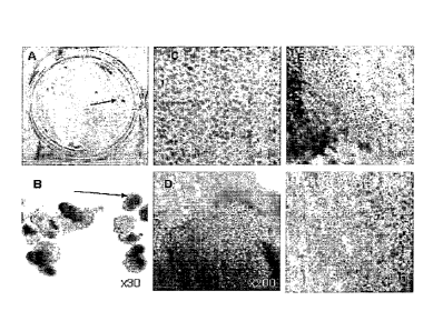
Figure 1A-F. is a series of photographs showing the appearance of pigmented
areas
(characteristic of RPE cells) in spontaneously differentiating hES cells.
Figure 1A is a
photograph of pigmented regions in a 2.5 month old adherent culture, a well of
a 6-well plate,
scanned; Figure 1B is a photograph of pigmented regions in a 2.5 month old
cultured grown in
EB, at 45x magnification; Figure 1C is a photograph of a pigmented area of an
adherent culture;
9c
Date Recue/Date Received 2022-09-12
Figure 1D is a photograph of a pigmented region of an EB grown culture; Figure
1E is a
photograph of the boundary between pigmented region and the rest of the
culture, x200; Figure
F same as Figure E but at x400 magnification. Arrows in A and B point to
pigmented regions.
Figure 2A-F. is a series of photographs which show the loss and regain of
pigmentation
and epithelial morphology in culture. Figure 2A is a photograph showing
primary EB
outgrowth, 1 week; Figure 2B is a photograph showing the primary culture of
cells, isolated by
trypsin, 1 week; Figure 2C is a photograph showing epithelial islet surrounded
by proliferating
cells; Figure 2D is a photograph showing the regain of pigmentation and
epithelial morphology
in 1 month old culture; Figure 2E is a photograph showing the culture after 3
passages, x200
magnification; Figure 2F shows the same culture as in E, x400 magnification,
9d
Date Recue/Date Received 2022-09-12
CA 02555370 2006-07-24
WO 2005/070011 PCT/US2005/002273
Hoffman microscopy. Black arrows point to pigmented cells, white arrows show
outgrowing cells with no pigment.
Figure 3 Left Panel (A-D) and Right Panel is a series of photographs and
one graph - these show markers of RPE in hES cells-derived pigmented
epithelial
cells. Figures 3A and 3B are photographs showing imm' unolocalization of RPE
marker, bestrophin and corresponding phase microscopy field, x200
magnification;
Figures 3C and 3D are photographs showing CRALBP and corresponding phase
contrast microscopy field, x400 magnification. Arrows show the colocalization
of
bestrophin (A) and CRALBP (C) to pigmented cells (C,D); arrowheads point to
the
absence of staining for these proteins (A,B) in non-pigmented regions (C,D)
Figure 3, Right Panel shows a photograph and graph of western blot of cell
lysates (line hES #36) with antibodies to bestrophin (a) and CRALBP (b); c,d ¨
undifferentiated hES cells, c--control to anti-CRALBP antibody, d __ control
to anti-
bestrophin antibody
Figure 4 shows photographs which demonstrate the expression of markers of
Pax6 (Figure 4A), Pax2 (Figure 4E) and mitf (Figure 4B, Figure 4F) in RPE-like
cells in long-term quiescent cultures. Figure 4C, Figure 4G ¨ phase contrast,
Figure 4D, Figure 411¨ merged images of Pax6/mitf/phase contrast (Figure 4A,
Figure 4B, Figure 4C) and Pax2/mitf/phase contrast (Figure 4E, Figure 4F,
Figure 4G).
Figure 5A-B show photographs of RPE differentiation in the culture of
human embryo-derived cells: bypassing the stage of derivation of ES cell
lines.
Figure 6 shows the transcriptional comparison of RPE preparations. Figure
6A-F - Based on the Ontological annotation, this table represents the
expression
patterns of RPE related genes for hES cell-derived retinal pigment epithelium
(hES-
RPE), hES cell derived transdifferentiated (hES-RPE-TD), ARPE-19 and D407, and
freshly isolated human RPE (fe-RPE). Figure 6G - Further data mining revealed
known RPE specific ontologies, such as melanin biosynthesis, vision, retinol-
binding, only in fetal RPE and ES-RPE but not ARPE-19.
DETAILED DESCRIPTION OF THE INVENTION
Various embodiments of the invention are described in detail and may be
further illustrated by the provided examples. As used in the description
herein and
throughout the claims that follow, the meaning of "a," "an," and "the"
includes
plural reference unless the context clearly dictates otherwise. Also, as used
in the
description herein, the meaning of "in" includes "in" and "on" unless the
context
clearly dictates otherwise.
CA 02555370 2014-07-17
=
The terms used in this specification generally have their ordinary meanings
in the art, within the context of the invention, and in the specific context
where each
term is used. Certain terms that are used to describe the invention are
discussed
below, or elsewhere in the specification, to provide additional guidance to
the
practitioner in describing the compositions and methods of the invention and
how to
make and use them. For convenience, certain terms may be highlighted, for
example using italics and/or quotation marks. The use of highlighting has no
influence on the scope and meaning of a term; the scope and meaning of a term
is
the same, in the same context, whether or not it is highlighted. It will be
appreciated
that the same thing can be said in more than one way. Consequently,
alternative
language and synonyms may be used for any one or more of the terms discussed
herein, nor is any special significance to be placed upon whether or not a
term is
elaborated or discussed herein. Synonyms for certain terms are provided. A
recital
of one or more synonyms does not exclude the use of other synonyms. The scope
of
the claims should not be limited by the preferred embodiments set forth in the
examples. but should be given the broadest interpretation consistent with the
description as a whole.
Definitions
By "embryo" or "embryonic" is meant a developing cell mass that has not
implanted into the uterine membrane of a maternal host. An "embryonic cell" is
a
cell isolated from or contained in an embryo. This also includes blastomeres,
obtained as early as the two-cell stage, and aggregated blastomeres.
The term "embryonic stem cells" refers to embryo-derived cells. More
specifically it refers to cells isolated from the inner cell mass of
blastocysts or =
morulae and that have been serially passaged as cell lines.
The term "human embryonic stem cells" (hES cells) refers human embryo-
derived cells. More specifically hES refers to cells isolated from the inner
cell mass
of human blastocysts or morulae and that have been serially passaged as cell
lines
and can also include blastomeres and aggregated blastomeres.
The term "human embryo-derived cells" (hEDC) refers to morula-derived
cells, blastocyst-derived cells including those of the inner cell mass,
embryonic
shield, or epiblast, or other totipotent or pluripotent stem cells of the
early embryo,
including primitive endoderm, ectoderm, and mesoderm and their derivatives,
also
including blastomeres and cell masses from aggregated single blastomeres or
embryos from varying stages of development, but excluding human embryonic stem
cells that have been passaged as cell lines.
11
CA 02555370 2006-07-24
WO 2005/070011 PCT/US2005/002273
Embryonic stem (ES) cells which have the ability to differentiate into
virtually any tissue of a human body can provide a limitless supply of
rejuvenated
and histocompatible cells for transplantation therapy, as the problem of
immune
rejection can be overcome with nuclear transfer and parthenogenetic
technology.
The recent findings of Hirano et. al. (2003) have shown that mouse ES cells
can
produce eye-like structures in differentiation experiments in vitro. Among
those,
pigmented epithelial cells were described, resembling retinal pigment
epithelium.
Preliminary experiments carried out at Advanced Cell Technology with
primate and human ES cell lines show that a in a specialized culture system
these
cells differentiate into RPE-like cells that can be isolated and passaged.
Human and
mouse NT, Cyno parthenote ES cell derivatives have multiple features of RPE:
these
pigmented epithelial cells express four molecular markers of RPE ¨ bestrophin,
CRALBP, PEDF, and RPE65; like RPE, their proliferation in culture is
accompanied by dedifferentiation ¨ loss of pigment and epithelial morphology,
both
of which are restored after the cells form a monolayer and become quiescent.
Such
RPE-like cells can be easily passaged, frozen and thawed, thus allowing their
expansion.
The inventors have further shown that human ES cells also produce multiple
eye (vitreous body)-like structures in differentiation experiments in vitro.
Histological analysis of these structures show a pattern of cells consistent
with early
retinal development, including aggregates of cells similar to rods and cones.
RPE Transplantation
At present, chronic, slow rejection of the RPE allografis prevents scientists
from deteimining the therapeutic efficacy of this RPE transplantation. Several
methods are being considered to overcome this obstacle. The easiest way is to
use
systemic immunosuppression, which is associated with serious side-effects such
as
cancer and infection. A second approach is to transplant the patient's own
RPE, i.e.
homografts, but this has the drawback of using old, diseased RPE to replace
even
more diseased RPE. Yet, a third approach is to use iris epithelium (IPE) from
the
same patient but this has the drawback that IPE may not perform all the vision
related functions of RPE. Ultimately a method will need to be found to
eliminate
rejection and then scientists can determine the true efficacy of RPE
transplantation
in AMD and ARMD. Nuclear transfer and parthenogenesis facilitate
histo compatibility of grated RPE cells and progenitors.
RPE defects in Retinitis Pigmentosa
Retinitis pigmentosa is a hereditary condition in which the vision receptors
are gradually destroyed through abnormal genetic programming. Some fowls cause
total blindness at relatively young ages, where other forms demonstrate
12
CA 02555370 2014-07-17
characteristic "bone spicule" retinal changes with little vision destruction.
This
disease affects some 1.5 million people worldwide. Two gene defects that cause
autosomal recessive RP have been found in genes expressed exclusively in RPE:
one
is due to an RPE protein involved in vitamin A metabolism (cis retinaldehyde
binding protein), a second involves another protein unique to RPE, RPE65. Once
rejection is conquered, both of these forms of RP should be treatable
immediately by
RPE transplantation. This treatment was inconceivable a few years ago when RP
was a hopelessly untreatable and a poorly understood form of blindness.
New research in RPE transplantation suggests there is promise for the
treatment of retinal degeneration, including macular degeneration. In
addition, a
number of patients with advanced RP have regained some useful vision following
fetal retinal cell transplant. One of the patients, for instance, improved
from barely
seeing light to being able to count fingers held at a distance of about six
feet from
the patient's face. In a second case, vision improved to ability to see
letters through
tunnel vision. The transplants in these studies were performed by injection,
introducing the new retinal cells underneath the existing neural retina. Not
all of the
cells survived since the transplanted fetal cells were allogeneic (i.e. not
genetically-
matched), although those that did survive formed connections with other
neurons
and begin to function like the photoreceptors around them. Approximately a
year
after the first eight people received the transplants, four have recovered
some visual
function and a fifth shows signs of doing so.
Three newly derived human embryonic stem cell lines are similar in
properties to those described earlier (Thomson et. al. 1998, Reubinoff et.
al., 2000,
Richards et al. Nat Biotechnol. 2002, Lanzendorf et. al., 2001): they maintain
undifferentiated
phenotype and express known markers of undifferentiated hES cells, Oct-4,
alkaline
phosphatase, SSEA-3, SSEA-4, TRA-I-60, TRA-1-81 through 45 passages in culture
or over 130 population doublings. All hES cell lines differentiate into
derivatives of
three germ layers in ED or long term adherent cultures and in teratomas. One
of the
differentiation derivatives of hES cells is similar to retinal pigment
epithelium by the
following criteria: morphologically, they have a typical epithelial
cobblestone
monolayer appearance and contain dark brown pigment in their cytoplasm, which
is
known to be present in the human body only in melanocytes, keratinocytes,
retinal
and iris pigment epithelium (2E). Melanocytes, however, are non-epithelial
cells,
and lceratynocytes don't secrete but only accumulate melanin. The set of RPE-
specific proteins -- bestrophin, CRALBP, PEDF ¨ present in these cells
indicates
that they are likely to be similar to RPE and not IPE. Another similarity is
the
behavior of isolated pigmented cells in culture, when little or no pigment was
seen in
proliferating cells but was retained in tightly packed epithelial islands or
re-
13
CA 02555370 2014-07-17
expressed in newly established cobblestone monolayer after the cells became
quiescent. Such behavior was described for RPE cells in culture (reviewed by
Zhao
et. al., 1997), and it was previously reported (Vinores et. al., 1995) that a
neuronal
marker tubulin beta III was specifically localized in dedifferentiating RPE
cells in
vitro and not in the cells with the typical RPE morphology suggesting that it
reflects
the plasticity of RPE and its ability to dedifferentiate to a neural lineage.
The
inventors have observed the same pattern of tubulin beta III localization in
primary
and passaged cultures of RPE and RPE-like cells which can reflect a
dedifferentiation of such cells in culture or indicate a separate population
of cells
committed to a neuronal fate, that were originally located next to pigmented
cells
through differentiation of hES cells in long-term cultures and could have been
co-
isolated with RPE-like cells.
In the growing optic vesicle RPE and the neural retina share the same
bipotential neuroepithelial progenitor, and their fate was shown to be
determined by
Pax2, Pax6, and Mitf (Baumer et. al., 2003), the latter being a target of the
first two.
Pax6 at earlier stages acts as an activator of proneural genes and is
downregulated in
the RPE in further development, remaining in amacrine and ganglion cells in
mature
retina (reviewed by Ashery -Padan and Grass, 2001). In goldfish, it is, also
found in
mitotically active progenitors of regenerating neurons (Hitchcock et. al.,
1996). The
inventors have found that many of the RPE-like cells expressed mitf and Pax6
in a
pattern similar to tubulin beta III and were found only in non-pigmented cells
of
non-epithelial morphology that surround pigmented epithelial islands in long
term
cultures or in cells with a "partial" RPE phenotype (lightly pigmented and
loosely
packed). In proliferating cells in recently passaged cultures all these
markers were
found nearly in every cell suggesting either a reversal of RPE-like cells to
progenitor
stage at the onset of proliferation or massive proliferation of retinal
progenitors.
Interestingly, in teratomas where islands of pigmented cells of epithelial
morphology
were also found, Pax6 was expressed in non-pigmented cells adjacent to
pigmented
regions (data not shown). Multiple studies have previously shown
dedifferentiation
of RPE in culture and their transdifferentiation into cells of neuronal
phenotype (Reh
and Gretton, 1987, sakaguchi et. al., 1997, Vinores et. al., 1995, Chen et.
al., 2003),
neuronal, amacrine and photoreceptor cells (Zhao et. al., 1995), glia (
Sakaguchi et.
al., 1997), neural retina (Galy et. al., 2002), and to neuronal progenitors (0
pas and
Dziak,1994 ). Such progenitors can in turn coexist with mature RPE-like cells
in
culture or appear as a result of dedifferentiation of RPE-like cells. At the
same time,
cells of neural retina can transdifferentiate into RPE in vitro (Opas et. al.,
2001), so
alternatively, tubulin beta III and Pax6 positive cells could represent a
transient stage
14
CA 02555370 2006-07-24
WO 2005/070011 PCT/US2005/002273
of such transdifferentiation of co-isolated neural cells or neural progenitors
into
RPE-like cells.
Differentiation of hES cells into RPE-like cells happened spontaneously
when using methods described in the Examples below, and the inventors noticed
that
pigmented epithelial cells reliably appeared in cultures older than 6-8 weeks
and
their number progressed overtime -- in 3-5 months cultures nearly every EB had
a
large pigmented region. In addition to the described fiRS lines, six more
newly
derived hES lines turned into RPE-like cells, which suggests that since neural
fate is
usually chosen by ES cells spontaneously, RPE-like cells can arise by default
as an
advanced stage of such pathway. It is also possible that in such long term
cultures,
where differentiating hES cells foini a multi-layered environment, permissive
and/or
instructive differentiation signals come from extracellular matrix and growth
factors
produced by differentiating derivatives of hES cells. The model of
differentiation of
hES cells into RPE-like cells could be a useful tool to study how such
microenvironment orchestrates RPE differentiation and transdifferentiation.
RPE plays an important role in photoreceptor maintenance, and various RPE
malfunctions in vivo are associated with a number of vision-altering ailments,
such
as RPE detachment, displasia, athrophy, retinopathy, retinitis pigmentosa,
macular
dystrophy or degeneration, including age-related macular degeneration, which
can
result in photoreceptor damage and blindness. Because of its wound healing
abilities, RPE has been extensively studied in application to transplantation
therapy.
It has been shown in several animal models and in humans (Gouras et. al.,
2002,
Stanga et. al., 2002, Binder et. al., 2002, Schraermeyer et. al., 2001,
reviewed by
Lund et. al., 2001) that RPE transplantation has a good potential of vision
restoration. Recently another prospective niche for FtPE transplantation was
proposed and even reached the phase of clinical trials: since these cells
secrete
dopamine, they could be used for treatment of Parkinson disease (Subramanian,
2001). However, even in an immune-privileged eye, there is a problem of graft
rejection, hindering the progress of this approach if allogenic transplant is
used. The
other problem is the reliance on fetal tissue, as adult RPE has a very low
proliferative potential.
As a source of immune compatible tissues, hES cells hold a promise for
transplantation therapy, as the problem of immune rejection can be overcome
with
nuclear transfer technology. The new differentiation derivative of human ES
cells,
retinal pigment epithelium-like cells and the reliability and simplicity of
such
differentiation system may offer an attractive potential supply of RPE cells
for
transplantation.
CA 02555370 2012-06-05
EXAMPLES
Example 1
Spontaneous differentiation into pigmented epithelial cells in long term
cultures
When hES cell cultures are allowed to overgrow on MEF in the absence of
LIF, FGF and Plasmanate, they form a thick multilayer of cells. About 6 weeks
later, dark islands of cells appear within the larger clusters (Fig 1). These
dark cells
are easily seen with the naked eye and looked like "freckles" in a plate of
cells as
shown in Fig 1A. At higher magnification these islands appear as tightly
packed
polygonal cells in a cobblestone monolayer, typical of epithelial cells, with
brown
pigment in the cytoplasm (Fig. IC). There are differences in the amount of
pigment
in the cells with cells in the central part of the islands having the most
pigment and
those near the edges the least. (Fig 1, E,F).
When hES cells form embryoid bodies (EB) - pigmented epithelial cells
appear in about 1-2% of EBs in the first 6-8 weeks (fig 1B) . Over time more
and
more EBs develop pigmented cells, and by 3 months nearly every EB had a
pigmented epithelial region (fig 1D). Morphology of the cells in the pigmented
regions of EBs was very similar to that of adherent cultures (fig 1D).
Example 2
Isolation and culture of pigmented epithelial cells
The inventors isolated pigmented epithelial cells from both adherent liES cell
cultures and from EBs. Pigmented polygonal cells were digested with enzymes
(trypsin, and/or collagenase, and/or dispase), and the cells from these
pigmented
islands were selectively picked with a glass capillary. Although care was
taken to
pick only pigmented cells, the population of isolated cells invariably
contained some
non-pigmented cells. After plating cells on gelatin or laminin for 1-2 days,
the cells
were considered to be primary cultures (PO).
Primary cultures contained islands of pigmented polygonal cells as well as
some
single pigmented cells. After 3-4 days in culture, non-pigmented cells that
seemed to
have lost epithelial morphology (flatter and cells with larnellipodia)
appeared at the
periphery of some islands (fig.2). The number of such peripheral cells
increased
over time, suggesting that these cells were proliferating, and after 2 weeks
most cells
in the newly formed monolayer contained very little or no pigment. After
continued
culture, for another 2-3 weeks, pigmented epithelial cells began to reappear,
visibly
indistinguishable from those in the original cultures (fig 2).
16
CA 02555370 2006-07-24
WO 2005/070011 PCT/US2005/002273
Example 3
Detection of RPE markers
The preliminary characterization of these differentiated human cells as RPE
is based on their similarity to RPE cultures previously described;
principally, their
epithelial morphology and possession of pigment. There are three types of
pigmented epithelial cells in human body: retinal and iris pigmented
epithelium and
keratinocytes, but the latter don't secrete pigment. The epithelial structure
and
cobblestone morphology are not shared by other pigmented cells, e.g.
melanocytes.
It is also noteworthy that RPE cells have been shown to lose and regain their
pigment and epithelial morphology when gown in culture (Zhao 1997, Opas and
Dziak, 1994), and the pigmented cells behaved in a similar manner, so to test
the
hypothesis that the ES derived cells may be RPE, they were stained with
antibodies
to known markers for RPE: bestrophin and CRALBP. Figure 4 (left panel) shows
membrane localization of bestrophin (A) and CRALBP (C), both are found in
pigmented epithelial islands. Not all of the cells stain with these antibodies
and
intensity of staining correlated with pigment expression and "tightness" of
colonies
¨ the borders of each pigmented island where cells were larger and more
loosely
packed showed lower expression of both proteins.
To further characterize presumably RPE cells, analysis was performed on the
expression of bestrophin, CRALBP by Western blotting. Figure 4 (right panel)
shows the bands, corresponding to bestrophin, 68 lcD (a), CRALBP, 361W (b) in
cell lysates. All these proteins were found in both primaty cultures and
subsequent
passages.
Another known PRE marker, RPE65, was found in the RPE-like cells by
real-time RT-PCR (Figure 4, right panel, bottom), the
PEDF ELISA assay showed the presence of PEDF in cell lysates of all
presumed RPE cultures, and Western blot showed a band of approximately 48 kD
(not shown).
Detection of markers of neuronal and retinal progenitors in RPE-like cultures
Figure 4 shows localization of PAX-6, Pax2, mitf, and tubulin beta III in
recently passaged and old cultures of hES cells-derived RPE. In proliferating
cultures (day 3 after trypsinization, not shown) where RPE-like morphology of
the
proliferating cells is lost, nearly every cell showed the presence of mitf,
Pax6,
tubulin beta III and nestin (not shown). Pax2 was found only a small subset of
cells
which appeared mitf-negative, while there was a strong degree of co-
localization of
Pax6/mitf, mitf/tubulin beta III, and Pax6/tubulin beta III. In 21 days old
quiescent
cultures after pigmented epithelial islands were reestablished, groups of PAX-
6 and
mitf were found mostly in non-pigmented cells of non-epithelial morphology
17
CA 02555370 2012-06-05
between pigmented epithelial islands (Figure 4, A-C). and tubulin beta III had
a
similar pattern of distribution (not shown). However, there were populations
of mitf-
positive and Pax6-negative cells, located close to the periphery of pigmented
islands
(figure 4, A-C). Pax2 was found only in a very small subset of mitf-negative
cells
(Figure 4, E-H). No presence of either of these proteins was ever detected in
the
cells of "mature" pigmented epithelial islands. However, these markers in
cells that
only had some RPE features were often visible, i.e. either looked epithelial
but had
no pigment or in certain single pigmented cells away from pigmented epithelial
islands.
Example 4
Characterization of RPE-like cells derived from hES cell lines H9 and ACT J-I
from
Cyno-I ES cells and derivation of RPE-like cells from existing hES cell lines
111 and
H7.
An RPE-like cell line is expanded, tested for freezing and recovery, and
characterized using the following methods and molecular markers of RPE cells:
bestrophin and CRALBP by Western blot and immunofluorescence, PEDF by
ELISA and Western blot, and REP65 by RT-PCR. The cells are injected in SOD
mice with undifferentiated bES or Cyno-1 cells as a control to evaluate
tumorigenicity. Karyotyping of RPE-like cells will be done by a clinical
laboratory
on a commercial basis. Characterization of the functional properties of RPE-
like
cells and studies of their transplantation potential are then carried out as
otherwise
described in this application and also using those techniques known to those
skilled
in the art.
Gene expression profiling experiments are done using Affymetrix human
genome arrays. Gene expression is compared in RPE-like cells derived from ES
cells and in retinal samples from autopsies. Several animal models can be used
to
verify the effectiveness of the transplanted RPE-like cells, including but not
limited
to, rhesus monkey, rat, and rabbit.
Example 5
Optimization of the differentiation culture system ensuring high yields
of RPE-like cells.
ES cells are cultured on feeder cells or as embryoid bodies (EB) in the
presence of bFGF, insulin, TGF-beta, IBMX, bmp-2, bmp-4 or their combinations,
including stepwise addition. Alternatively, ES cells are grown on various
extra.cellular matrix-coated plates (laminin, fibronectin, collagen I,
collagen IV,
TM
Matrigel, etc.) in evaluating the role of ECM in RPE formation. Expression of
18
CA 02555370 2006-07-24
WO 2005/070011 PCT/US2005/002273
molecular markers of early RPE progenitors (Pax6, Pax2, mitf) and of RPE cells
(CRALBP, bestrophin, PEDF, REP65) are evaluated at various time intervals by
real-time RT-PCR to verify and determine successful combinations of the above
mentioned agents and stepwise procedure that produces enrichment in RPE-like
cells or their progenitors. This approach can also be used to produce common
progenitors of RPE and other eye tissues, such as photoreceptor or neural
retina
which can be isolated and further characterized for their differentiation
potential and
used in transplantation studies.
Example 6
Derivation of RPE and other eye tisSue progenitors from existing
and new ES cell lines.
Using the data from the gene expression profiling, expression of the RPE
progenitor markers will be correlated with the expression of the surface
proteins in
order to find a unique combination of surface markers for RPE progenitor
cells. If
such markers are found, antibodies to surface proteins can be used to isolate
a pure
population of RPE progenitors that can be then cultured and further
differentiated in
culture or used in transplantation studies to allow their differentiation
after grafting.
If the data from the gene expression profiling experiments is insufficient, to
isolate the RPE progenitors the following approach will be used. ES cells and
RPE-
like cells will be transfected with GFP under the control of a Pax6 promoter,
and
stable transfectants will be selected. From a culture of transfected
differentiating ES
cells or proliferating (dedifferentiated) RPE cells, GFP/Pax6-positive cells
will be
isolated by FACS and used as an antigen source for mouse injection to raise
monoclonal antibodies to the surface molecules of Pax6 positive cells. Because
Pax6
is present not only in RPE progenitors, screening will be done (by FACS) using
several strategies: a) against proliferating RPE-like cells, b) against Pax2-
positive
RPE cells, c) against mitf-positive RPE cells. For b) and c) RPE cells will be
transfected with GFP under the corresponding promoter; as a negative control,
RPE
or ES cells negative by these antigens will be used. After expansion of
positive
clones selected by all three strategies, antibodies will be tested against all
types of
cells used in screening and further analyzed: since this strategy can produce
antibodies that recognize cell surface antigens specific and non-specific for
RPE
progenitors, the cells from differentiating total population of ES cells or of
RPE cells
selected with these antibodies will be assessed for molecular markers of RPE
progenitors and for their ability to produce RPE.
Using the optimized defined stepwise procedures to produce RPE or other
early progenitors of eye tissues and the antibodies to their unique surface
markers,
19
CA 02555370 2006-07-24
WO 2005/070011 PCT/US2005/002273
such progenitors will be isolated from differentiated ES cells and cultured in
vitro.
Their ability to differentiate into various tissues of the eye will be
investigated using
the strategy described in Aim 2.
Three ES cell lines that already produced RPE-like cells (119, ACT J-1,
Cyno-1), RPE-like cells will be used to continue to derive RPE-like cells and
their
progenitors as described in Aims 1 and 2, and H1 and H7 hES cell lines will be
used
to produce new RPE-like cell lines. After expansion and characterization for
molecular markers of RPE, these lines will be single-cloned, and the resulting
lines
will be characterized as described in Aim 1. The lines meeting criteria for
RPE cells
will be used for transplantation studies. New human ES cell lines will be
derived
from unused IVP embryos, from donated oocytes, stimulated to develop without
fertilization (parthenote), and from generated developing blastocysts obtained
from
donated oocytes with the application of nuclear transfer technology. RPE-like
cells
and common eye progenitors will be derived from these lines using the approach
in
Aim 2, and the resulting lines will be characterized as in Aim 1. [Optional]
new
human ES cell lines will be derived in a virus-free system, characterized and
submitted for clinical trials.
Example 7
Therapeutic potential of RPE-like cells and progenitors in various animal
models of retinitis pigmentosa & macular degeneration.
Primate ES cells are tested in cynomologus monkeys (Macaques). Initially,
vitrectomy surgery is performed and the cells are transplanted into the
subretinal
space of the animals. The first step is the transplantation of the cells in
the
suspension format after which a substrate or matrix is used to produce a
monolayer
transplantation. This can also be performed in immunosuppressed rabbits using
cells derived from human ES-cells and also in various other animal models of
retinitis pigmentosa, including rodents (rd mouse, RPE-65 knockout mouse,
tubby-
like mouse, RCS rat, cats (Abyssinian cat), and dogs (cone degeneration
"cd"
dog, progressive rod-cone degeneration "prcd" dog, early retinal degeneration
"erd"
dog, rod-cone dysplasia 1, 2 & a "rcd1, rcd2 & rcd3" dogs, photoreceptor
dysplasia "pd" dog, and Briard "RPE-65) dog). Evaluation is performed using
fluorescent angiography, histology (whether or not there is photoreceptor
restoration
and possibly ERG. Functional testing will also be carried out, including
phagocytosis (photoreceptor fragments), vitamin A metabolism, tight junctions
conductivity, and electron microscopy.
CA 02555370 2014-07-17
Example 8
Direct differentiation of RPE cells from human embryo-derived cells.
Human blastocyst-staged embryos are plated in the presence of murine or
chick embryo fibroblasts with or without immunosurgery to remove the
trophectoderrn or directly plates on extracellular matrix protein-coated
tissue
cultureware. Instead of culturing and passaging the cells to produce a human
ES cell
line, the cells are directly differentiated.
When hEDC cell cultures are allowed to overgrow on MEF in the absence of
LIF, FGF and Plasmanate, they will form a thick multilayer of cells.
(Alternate
growth factors, media, and PBS can be used to alternate direct differentiation
as is
known to those skilled in the art.) About 6 weeks later, dark islands of cells
will
appear within the larger clusters. These dark cells are easily seen with the
naked eye
and looked like "freckles" in a plate of cells as shown in Fig 5B. At higher
magnification these islands appear as tightly packed polygonal cells in a
cobblestone
monolayer, typical of epithelial cells, with brown pigment in the cytoplasm
(Fig.
5A). There are differences in the amount of pigment in the cells with cells in
the
central part of the islands having the most pigment and those near the edges
the
least. (Fig. 5B).
When hEDC cells are directly differentiated they may, though typically have
not, formed embryoid bodies (EB). Pigmented epithelial cells appear in about 1-
2%
of these differentiated cells and/or EBs in the first 6-8 weeks. Over time
more and
more EBs develop pigmented cells, and by 3 months nearly every EB had a
pigmented epithelial region. Morphology of the cells in the pigmented regions
of
EBs was very similar to that of adherent cultures.
Materials and methods:
MEF medium: high glucose DMEM, supplemented with 2mM GlutaMAX I,
and 500 u/ml Penicillin, 500 pg/m1 streptomycin (all from Invitrogen) and 16%
FCS
(HyCLone). hES Cells Growth medium: knockout high glucose DMEM
supplemented with 500 Ps/En I Penicillin, 500 ug/mlstreptomycin, 1 % non-
essential
amino acids solution, 2mM GlutaMAX Iõ 0.1 mM beta-mercaptoethanol, 4 ng/ml
bFGF (Invitrogen), 1-ng/m1 human LIF (Chemicon, Temecula, CA), 8.4% of Serum
Replacement (SR, Invitrogen) and 8.4% Plasmanate (Bayer). Derivation medium
contained the same components as growth medium except that it had lower
concentration of SR and Plasmanate (4.2% each) and 8.4 % FCS and 2x
concentration of human LIF and bFGF, as compared to growth medium. EB
medium: same as growth medium except bFGF, LIF, and Plasmanate; the SR
concentration was 13%. RPE medium: 50% EB medium and 50% MEF medium.
21
CA 02555370 2012-06-05
hES cell lines
The cell lines, hES 35, 36, 45, used for these studies were derived with
modifications of previously reported procedures (Thomson et. al., 1998,
Reubinoff
et. al., 2000, Lanzendorf et. al., 2001). Human frozen blastocysts (line
hES35) or
cleaved embryos (lines hES36 and hES45) were donated to the study, approved by
two institutional review board, by couples who had completed their fertility
treatment.
Differentiation experiments were performed with adherent hES cells or with
embryoid bodies (EBs). For adherent differentiation, hES cells were allowed to
overgrow on MEFs until the hES colonies lost their tight borders at which time
the
culture media was replaced with EB medium (usually, 8-10 days after
passaging).
The medium was changed every 1-2 days. For EB formation, hES cells were
trypsinized and cultured in EB medium on low adherent plates (Costar).
Immunostaining
Cells were fixed with 2% paraformaldehyde, permeabilized with 0.1% NP-
40 for localization of intracellular antigens, and blocked with 10% goat
serum, 10%
donkey serum (Jackson Immunoresearch Laboratories, West Grove, PA) in PBS
(Invitrogen) for at least one hour. Incubation with primary antibodies was
carried out
overnight at 4oC, the secondary antibodies (Jackson Immunoreseareh
Laboratories,
West Grove, PA) were added for one hour. Between all incubations specimens
were
washed with 0.1% Tween-20 (Sigma) in PBS 3-5 times, 10-15 minutes each wash.
Specimens were mounted using Vectashield with DAPI (Vector Laboratories,
Burlingame, CA) and observed under fluorescent microscope (Nikon).
Localization
of alkaline phosphatase was done either by Vector Red (Vector Laboratories,
Burlingame, CA) to live cells or after the second wash during immunostaining
according to manufacturer's instructions. Antibodies used: bestrophin (Novus
Biologicals, Littleton, CO), anti-CRALBP antibody was a generous gift from Dr.
Saari, University of Washington. Secondary antibodies were from Jackson
Immunoresearch Laboratories, and Streptavidin-FITC was purchased from
Amersham.
Isolation and passaging of RPE-like cells
Adherent cultures of hES cells or EBs were rinsed with PBS twice and
incubated in 0.25% Trypsin/1 inM EDTA (Invitrogen) at 37oC until the monolayer
loosened. Cells from the pigmented regions were scraped off with a glass
capillary,
transferred to MEF medium, centrifuged at 200X g, and plated onto gelatin-
coated
22
CA 02555370 2006-07-24
WO 2005/070011 PCT/US2005/002273
plates in RPE medium. The medium was changed after the cells attached (usually
in
1-2 days) and every 5-7 days after that; the cells were passaged every 2-4
weeks
with 0.05% Trypsin/0.53mM EDTA (Invitrogen).
Western blot and ELISA
Samples were prepared in Laemmli buffer (Laemmli, 1970), supplemented with 5%
Mercaptoethanol and Protease Inhibitor Cocktail (Roche), boiled for 5 minutes
and
loaded onto a 8-16% gradient gel (Bio-Rad, Hercules, CA) using a Mini-Protean
apparatus; the gels were run at 25-30 mA per gel; proteins were transferred to
a 0.2
Nitrocellulose membrane (Schleicher and Shull, Keene, NH) at 20 volt
overnight.
Blots were briefly stained with Ponceau Red (Sigma) to visualize the bands,
washed
with Milli-Q water, and blocked for 1 hour with 5% non-fat dry milk in 0.1%
TBST
(Bio-Rad). Primary antibodies to bestrophin, CRALBP or PEDF (Chemicon) were
added for 2 hours followed by three 15-minute washes with TBST; peroxidase-
conjugated secondary antibodies were added for 1 hour, and the washes were
repeated. Blots were detected using ECL system with Super-Signal reagent
(Pierce).
PEDF ELISA was performed on cell lysates using PEDF ELISA kit (Chemicon)
according to manufacturer's protocol.
Real-time RT-PCR
Total RNA was purified from differentiating ES cultures by a two-step
procedure Crude RNA was isolated using Trizol reagent (Invitrogen) and further
purified on RNeazy minicolunms (Qiagen). The levels of RPE65 transcripts were
monitored by real-time PCR using a commercial primer set for RPE65 detection
(Assay on Demand # 11s00165642_ml, Applied Biosystems) and Quantitect Probe
RT-PCR reagents (Qiagen), according to the manufacturer's (Qiagen) protocol.
Derivation and characterization of undifferentiated hES cell lines
Two female one male hES cell lines were used in these studies. Details on the
derivation of these hES lines are reported elsewhere. All lines have been
passaged
more than 50 times during which time they maintain an undifferentiated colony
morphology, high alkaline phosphatase activity, and express Oct-4, SSEA-3,
SSEA-
4, TRA 1-60, and TRA 1-81 (data not shown). Two lines have normal karyotype
(hES36, hES35), while there were both normal and aneuploid subpopulations in
hES45. Upon spontaneous differentiation both in vitro and in teratomas all
lines
expressed the markers of three germ layers ¨ muscle actin, alpha-fetoprotein,
and
tubulin beta III.
23
CA 02555370 2006-07-24
WO 2005/070011
PCT/US2005/002273
Example 9
Use of transcript genomics to identi& normal differentiated
cells differentiated ex vivo.
Transcriptomics ¨ hES-cell derivatives are likely to play an important role in
the future of regenerative medicine. Qualitative assessment of these and other
stem
cell derivatives remains a challenge that could be approached using functional
genomics. We compared the transcriptional profile of hES-RPE vs. its in vivo
counterpart, fetal RPE cells, which have been extensively researched for its
transplantation value. Both profiles were then compared with previously
published
(Rogojina et. al., 2003) transcriptomics data on human RPE cell lines.
The gene expression profile of our data set was compared to two human RPE
cell lines (non-transformed ARPE-19 and transformed D407, Rogojina et. al.,
2003)
to determine whether hES-RPE have similar global transcriptional profiles. To
account for common housekeeping genes expressed in all cells, we used publicly
available Affymetrix data sets from undifferentiated hES cells (111 line, hl-
hES, --
sato et. al., 2003) and bronchial epithelial cells (BE, Wright et. al., 2004)
as a control
based on its common epithelial origin that would allow to exclude common
housekeeping and epithelial genes and identify RPE-specific genes.
There were similarities and differences between hES-RPE, hES-RPE-TD,
ARPE-19, D407. The similarities were further demonstrated by analyzing the
exclusive intersection between those genes present in hFS-RPE/ARPE-19 but not
in
BE (1026 genes). To account for background, we compared this to the exclusive
intersection of genes present in BE/hES-RPE, but not ARPE-19 (186 genes),
which
results in a five- to six-fold greater similarity in hES-RPE and ARPE-19 when
compared to BE. D407/ARPE19 appear to lose RPE specific genes such as RPE65,
Bestrophin, CRALBP, PEDF, which is typical of long-term passaged cells (figure
6). Further data mining revealed known RPE specific ontologies such as melanin
biosynthesis, vision, retinol-binding, only in fetal RPE and ES-RPE but not
ARPE19.
Comparison of hES-RPE, ARPE-19 and D407 to their in vivo counterpart,
freshly isolated human fetal RPE (feRPE), was in concordance with our previous
data, demonstrating that the transcriptional identity of hES-RPE to human
feRPE is
significantly greater than D407 to fe RPE (2.3 fold difference- 849 genes/373
genes)
and ARPE-19 to feRPE (1.6 fold difference ¨ 588 genes/364 genes (Figure
5c/5d).
The RPE specific markers identified above, which were only present in hES-RPE
and not in ARPE-19 or D407 were also present in feRPE, demonstrating a higher
similarity of hES-RPE to its in vivo counterpart than of the cultured RPE
lines.
24
CA 02555370 2006-07-24
WO 2005/070011 PCT/US2005/002273
Seven-hundred-and-eighty-four genes present in hES-RPE were absent in
feRPE and ARPE-19 data sets. Since the retention of "sternness" genes could
potentially cause transformation of hES derivatives into malignant teratomas
if
transplanted into patients, we created a conservative potential "sternness"
genes data
using currently available Affymettix microarray data sets Abeyta et. al. 2004
Sato
2003). This resulted in a list of 3806 genes present in all 12 data sets
(including
common housekeeping genes). j Only 36 of the 784 genes present in the hES-RPE
dats et but not feRPE-ARPE-19 were common to the 3806 potential sternness
genes.
None of these were known sternness genes such as 0ct4, Sox2, IDGF1.
Example 10
Use of RPE cells for treatment of Parkinson's Disease.
hRPE can be used as an alternative source of cells for cell therapy of
Parkinson's Disease because they secrete L-DOPA. Studies have showed that such
cells attached to gelatin-coated microcathers can be, successfully
transplanted in
hemiparkinsonian monkeys and produced notable improvements (10-50) thousand
cells per target), and in FDA-approved trial started in 2000 the patients
received
hRPE intrastriatial transplants without adverse effects. One of the many
advantages
to the use of hES cell-derived RPE is that it circumvents the shortage of
donor eye
tissue. It also facilitates the use of gene therapy.
Other Embodiments
From the foregoing description, it will be apparent that variations and
modifications may be made to the invention described herein to adopt it to
various
usages and conditions.
25
CA 02555370 2012-06-05
Reference List
Archer DB, Gardiner TA. "Morphologic fluorescein angiographic, and light
microscopic
features of experimental choroidal neovascularisation"; Am J Ophthalmol. 1981
Mar;91(3):297-
311.
Ashery-Padan R, Gruss P. "Pax6 lights-up the way for eye development"; Curr
Opin Cell Biol.
2001 Dec;13(6):706-14.
Aubert J, Dessolin S, Belmonte N, Li M, McKenzie FR, Staccini L, Villageois P,
Barhanin
B, VernaIlls A, Smith AG, Ailhaud G, Dani C. "Leukemia inhibitory factor and
its receptor
promote adipocyte differentiation via the mitogen-activated protein kinase
cascade"; J Biol
Chem. 1999 Aug 27;274(35):24965-72.
Baumer N, Marquardt T, Stoykova A, Spieler D, Treichel D, Ashery-Padan R,
Gruss P.
"Retinal pigmented epithelium determination requires the redundant activities
of Pax2 and
Pax6"; Development. 2003 Jul;130(13):2903-15.
Binder S, Stolba U, Krebs I, Kellner L, Jahn C, Feichtinger H, Povelka M,
Frohner U,
Kruger A, Hilgers RD, KrugInger W. "Transplantation of autologous retinal
pigment
epithelium in eyes with foveal neovascularization resulting from age-related
macular
degeneration: a pilot study"; Am J Ophthalmol. 2002 Feb;133(2):215-25.
Bost F, Caron L, Marchetti I, Dani C, Le Marchand-Brustel Y, Binetruy B.
"Retinoic acid
activation of the ERK pathway is required for embryonic stem cell commitment
into the
adipocyte lineage"; Biochem J. 2002 Feb 1;361(Pt 3):621-7.
Bunt-Milam AH, Saari JC. "Immunocytochemical localization of two retinoid-
binding proteins
in vertebrate retina"; J Cell Biol. 1983 Sep;97(3):703-12.
Carpenter MK, Inokuma MS, Denham J, Mujtaba T, Chiu CP, Rao MS. "Enrichment of
neurons and neural precursors from human embryonic stem cells"; Exp Neurol.
2001
Dee;172(2):383-97.
Carpenter MK, Rosier E, Rao MS. "Characterization and differentiation of human
embryonic
stem cells"; Cloning Stern Cells. 2003;5(1):79-88.
Chadwick K, Wang L, Li L, Menendez P, Murdoch B, Rouleau A, Bhatia M.
"Cytokines
and BMP-4 promote hematopoietic differentiation of human embryonic stem
cells"; Blood. 2003
Aug 1;102(3):906-15. Epub 2003 Apr 17.
Chen S, Samuel W, Fariss RN, Duncan T, Kutty RK, Wiggert B. "Differentiation
of human
retinal pigment epithelial cells into neuronal phenotype by N-(4-
hydroxyphenyl)retinamide"; J
Neurochem. 2003 Mar;84(5):972-81.
Cleary PE, Ryan SJ. "Histology of wound, vitreous, and retina in experimental
posterior
penetrating eye injury in the rhesus monkey"; Am J Ophthalmol. 1979
Aug;88(2):221-31.
25A
CA 02555370 2012-06-05
del Cerro M, Humayun MS, Sadda SR, Cao J, Ilayashi N, Green WR, del Cerro C,
de
Juan E Jr. "Histologic correlation of human neural retinal transplantation";
Invest Ophthalmol
Vis Sci. 2000 Sep;41(10):3142-8.
Eguchi G. "Instability in cell commitment of vertebrate pigmented epithelial
cells and their
transdifferentiation into lens cells"; Curr Top Dev Biol. 1986;20:21-37
Fischer AJ, Reh TA. "Identification of a proliferating marginal zone of
retinal progenitors in
postnatal chickens"; Dev Biol. 2000 Apr 15;220(2):197-210.
Fischer AJ, Reh TA. "Transdifferentiation of pigmented epithelial cells: a
source of retinal stem
cells?"; Dev Neurosci. 2001;23(4-5):268-76.
Galy A, Neron B, Planque N, Saute S, Eychene A. "Activated MAPK/ERK kinase
(MEK-1)
induces transdifferentiation of pigmented epithelium into neural retina"; Dev
Biol. 2002 Aug
15;248(2):251-64.
Gouras P, Kong J, Tsang SH. "Retinal degeneration and RPE transplantation in
Rpe65(-/-)
mice"; Invest Ophthalmol Vis Sci. 2002 Oct;43(10):3307-11.
Grierson I, Hiscott P, Hogg P, Robey H, Mazure A, Larkin G. "Development,
repair and
regeneration of the retinal pigment epithelium"; Eye (Lond). 1994;8 (Pt 2):255-
62.
Hitchcock PF, Macdonald RE, VanDeRyt JT, Wilson SW. "Antibodies against Pax6
immunostain amacrine and ganglion cells and neuronal progenitors, but not rod
precursors, in the
normal and regenerating retina of the goldfish"; J Neurobiol. 1996
Mar;29(3):399-413.
Hiibner K, Fuhrmann G, Christenson LK, Kehler J, Reinhold R, De La Fuente R,
Wood J,
Strauss JF 3rd, Boiani M, Schdler HR. "Derivation of oocytes from mouse
embryonic stem
cells"; Science. 2003 May 23;300(5623):1251-6. Epub 2003 May 1.
Humayun MS, de Juan E Jr, del Cerro M, Dagnelie G, Radner W, Sadda SR, del
Cerro C.
"Human neural retinal transplantation"; Invest Ophthalmol Vis Sci. 2000
Sep;41(10):3100-6.
Jablonski MM, Tombran-Tink J, Mrazek DA, Iannaccone A. "Pigment epithelium-
derived
factor supports normal development of photoreceptor neurons and opsin
expression after retinal
pigment epithelium removal"; J Neurosci. 2000 Oct 1;20(19):7149-57.
Kahan BW, Jacobson LM, Hullett DA, Ochoada JM, Oberley TD, Lang KM, Odorico
JS.
"Pancreatic precursors and differentiated islet cell types from murine
embryonic stem cells: an in
vitro model to study islet differentiation"; Diabetes. 2003 Aug;52(8):2016-24.
Kaplan HJ, Tezel TH, Berger AS, Wolf ML, Del Priore LV. "Human photoreceptor
transplantation in retinitis pigmentosa. A safety study"; Arch Ophthalmol.
1997
Sep;115(9):1168-72.
2513
CA 02555370 2012-06-05
Karakousis PC, John SK, Behling KC, Surace EM, Smith JE, Hendrickson A, Tang
WX,
Bennett J, Milam AH. "Localization of pigment epithelium derived factor (PEDF)
in
developing and adult human ocular tissues-; Mol Vis. 2001 Jun 30;7:154-63.
Kehat I, Kenyagin-Karsenti D, Snir M, Segev H, Amit M, Gepstein A, Livne E,
Binah 0,
Itskovitz-Eldor J, Gepstein L. "Human embryonic stern cells can differentiate
into myocytes
with structural and functional properties of cardiomyocytes"; J Clin Invest.
2001
Aug;108(3):407-14.
Kirchhof B, Kirchhof E, Ryan SJ, Sorgente N. "Human retinal pigment epithelial
cell
cultures: phenotypic modulation by vitreous and macrophages"; Exp Eye Res.
1988
Sep;47(3):457-63.
Laemmli UK. "Cleavage of structural proteins during the assembly of the head
of bacteriophage
T4"; Nature. 1970 Aug 15;227(5259):680-5.
Lanzendorf SE, Boyd CA, Wright DL, Muasher S, Oehninger S, Hodgen GD. "Use of
human gametes obtained from anonymous donors for the production of human
embryonic stem
cell lines"; Feral Steril. 2001 Jul;76(1):132-7.
Lee SC, Kwon OW, Seong GJ, Kim SH, Ahn JE, Kay ED. "Epitheliomesenchymal
transdifferentiation of cultured RPE cells"; Ophthalmic Res. 2001 Mar-
Apr;33(2):80-6.
Lin RY, Kubo A, Keller GM, Davies TF. "Committing embryonic stem cells to
differentiate
into thyrocyte-like cells in vitro"; Endocrinology. 2003 Jun;144(6):2644-9.
Lund RD, Kwan AS, Keegan DJ, Sauve Y, Coffey PJ, Lawrence JM. "Cell
transplantation as
a treatment for retinal disease"; Prog Retin Eye Res. 2001 Jul;20(4):415-49.
Ma J, Zhang J, Othersen KL, Moiseyev G, Ablonczy Z, Redmond TM, Chen Y, Crouch
RK. "Expression, purification, and MALDI analysis of RPE65"; Invest Ophthalmol
Vis Sci.
2001 Jun;42(7):1429-35.
Marmorstein AD, Finnemann SC, Bonilha VL, Rodriguez-Boulan E. "Morphogenesis
of the
retinal pigment epithelium: toward understanding retinal degenerative
diseases"; Ann N Y Acad
Sci. 1998 Oct 23;857:1-12.
Marmorstein AD, Marmorstein LY, Rayborn M, Wang X, Hollyfield JG, Petrukhin K.
"Bestrophin, the product of the Best vitelliform macular dystrophy gene
(VMD2), localizes to
the basolateral plasma membrane of the retinal pigment epithelium"; Proc Natl
Acad Sci U S A.
2000 Nov 7;97(23):12758-63.
McKay BS, Burke JM. "Separation of phenotypically distinct subpopulations of
cultured
human retinal pigment epithelial cells"; Exp Cell Res. 1994 Jul;213(1):85-92.
25C
CA 02555370 2012-06-05
Mummery C, Ward-van Oostwaard D, Doevendans P, Spijker R, van den Brink S,
Hassink
R, van der Heyden M, Opthof T, Pera M, de la Riviere AB, Passier R, Tertoolen
L.
"Differentiation of human embryonic stem cells to cardiomyoeytes: role of
coculture with
visceral endoderm-like cells"; Circulation. 2003 Jun 3;107(20:2733-40. Epub
2003 May 12.
Opas M, Dziak E. "bFGF-induced transdifferentiation of RPE to neuronal
progenitors is
regulated by the mechanical properties of the substratum"; Dev Biol. 1994
Feb;161(2):440-54.
Opas M, Davies JR, Zhou Y, Dziak E. "Formation of retinal pigment epithelium
in vitro by
transdifferentiation of neural retina cells"; Int J Dev Biol. 2001
Jun;45(4):633-42,
Rambhatla L, Chiu CP, Kundu P, Peng Y, Carpenter MK. "Generation of hepatocyte-
like
cells from human embryonic stem cells"; Cell Transplant. 2003;12(1):1-11.
Redmond TM, Yu S, Lee E, Bok D, Hamasaki D, Chen N, Goletz P, Ma JX, Crouch
RK,
Pfeifer K. "Rpe65 is necessary for production of 11-cis-vitamin A in the
retinal visual cycle";
Nat Genet. 1998 Dec;20(4):344-51.
Reh TA, Nagy T, Gretton H. "Retinal pigmented epithelial cells induced to
transdifferentiate to
neurons by laminin"; Nature. 1987 Nov 5-11;330(6143):68-71.
Reubinoff BE, Pera MF, Fong CV, Trounson A, Bongso A. "Embryonic stern cell
lines from
human blastocysts: somatic differentiation in vitro"; Nat Biotechnol. 2000
Apr;18(4):399-404.
Richards M, Fong CY, Chan WK, Wong PC, Bongs A. "Human feeders support
prolonged
undifferentiated growth of human inner cell masses and embryonic stem cells";
Nat Biotechnol.
2002 Sep;20(9):933-6. Epub 2002 Aug 5.
Rogojina AT, Orr WE, Song BK, Geisert EE Jr. "Comparing the use of Affymetrix
to spotted
oligonucleotide microarrays using two retinal pigment epithelium cell lines";
Mol Vis. 2003 Oct
6;9:482-96.
Sakaguchi DS, Janick LM, Reh TA. "Basic fibroblast growth factor (FGF-2)
induced
transdifferentiation of retinal pigment epithelium: generation of retinal
neurons and glia"; Dev
Dyn. 1997 Aug;209(4):387-98.
Sato N, Sanjuan IM, Heke M, Uchida M, Naef F, Brivanlou AH. "Molecular
signature of
human embryonic stem cells and its comparison with the mouse."; Dev Biol. 2003
Aug
15;260(2):404-13.
Schraermeyer U, Thumann G, Luther T, Kociok N, Armhold S, Kruttwig K,
Andressen C,
Addicks K, Bartz-Schmidt KU. "Subretinally transplanted embryonic stem cells
rescue
photoreceptor cells from degeneration in the RCS rats"; Cell Transplant.
2001;10(8):673-80.
251)
CA 02555370 2012-06-05
Schuldiner M, Eiges R, Eden A, Yanuka 0, Itskovitz-Eldor J, Goldstein RS,
Benvenisty N.
"Induced neuronal differentiation of human embryonic stem cells"; Brain Res.
2001 Sep
21;913(2):201-5.
Smith AG. "Embryo-derived stem cells: of mice and men"; Annu Rev Cell Dev
Biol.
2001;17:435-62.
Stanga PE, Kychenthal A, Fitzke FW, Halfyard AS, Chan R, Bird AC, Aylward GW.
"Retinal pigment epithelium translocation after choroidal neovascular membrane
removal in age-
related macular degeneration"; Ophthalmology. 2002 Aug;109(8):1492-8.
Subramanian T. "Cell transplantation far the treatment of Parkinson's
disease"; Semin Neurol.
2001;21(1):103-15.
25E
CA 02555370 2014-07-17
Thomson JA, Itskovitz-Eldor J, Shapiro SS, Waknitz MA, Swiergiel JJ, Marshall
VS,
Jones JM.
"Embryonic stem cell lines derived from human blastocysts"; Science. 1998 Nov
6;282(5391):1145-7.
Villegas-Perez MP, Lawrence JM, Vidal-Sanz M, Lavail MM, Lund RD.
"Ganglion cell loss in RCS rat retina: a result of compression of axons by
contracting intraretinal
vessels linked to the pigment epithelium"; J Comp Neurol. 1998 Mar 2;392(1):58-
77.
Vinores SA, Derevjanik NL, Mahlow J, Hackett SF, Haller JA, deJuan E,
Frankfurter A,
Campochiaro PA.
"Class III beta-tubulin in human retinal pigment epithelial cells in culture
and in epiretinal
membranes"; Exp Eye Res. 1995 Apr;60(4):385-400.
Wright JM, Zeitlin PL, Cebotaru L, Guggino SE, Guggino WB.
"Gene expression profile analysis of 4-phenylbutyrate treatment of IB3-1
bronchial epithelial
cell line demonstrates a major influence on heat-shock proteins"; Physiol
Genomics. 2004 Jan
15;16(2):204-11.
Zhao S, Rizzolo LJ, Barnstable CJ.
"Differentiation and transdifferentiation of the retinal pigment epithelium";
Int Rev Cytol.
1997;171:225-66.
Zhao S, Thornquist SC, Barnstable CJ.
"In vitro transdifferentiation of embryonic rat retinal pigment epithelium to
neural retina"; Brain
Res. 1995 Apr 24;677(2):300-10.
zur Nieden NI, Kempka G, Ahr HJ.
"In vitro differentiation of embryonic stem cells into mineralized
osteoblasts"; Differentiation.
2003 Jan;71(1):18-27. '
Xu C, Police S, Rao N, Carpenter MK.
"Characterization and enrichment of cardiomyocytes derived from human
embryonic stem cells";
Circ Res. 2002; 91(6): 501-508.
Hirano M, Yamamoto A, Yoshimura N, Tokunaga T, Motohashi T, Ishizaki K,
Yoshida H,
Okazaki K, Yamazaki H, Hayashi SI, Kunisada T.
"Generation of structures formed by lens and retinal cells differentiating
from embryonic stem
cells"; Dev. Dynam. 2003, 228(4): 664-671.
25F