Note: Descriptions are shown in the official language in which they were submitted.
CA 02555411 2006-08-04
WO 2005/075629 PCT/NZ2005/000002
A METHOD AND APPARATUS FOR ORIENTING ASPHERICAL CELLS
FIELD OF INVENTION
The present invention generally relates to a method and apparatus for
orienting desired cells, or parts
of cells, preferably, desired sperm cells and, more particularly, the
invention relates to a method and
apparatus for orienting, selecting and retaining viable desired sperm cells.
BACKGROUND OF THE INVENTON
There has been a long felt need for a reliable, qualitative, quantitative and
cost-effective method for
selecting sperm, which may be used to produce animals of a desired sex
In particular, in the livestock industry farmers or breeders require cows,
pigs, sheep, goats, deer,
buffalo, horses, etc which are of a preferred sex. For example, bulls are of
limited use to a dairy
farmer, whereas pig farmers have long been aware that female pigs grow at a
faster rate than their
male counterparts.
Similarly, cattle and sheep farmers understand only too well that the males of
these species produce
meat at a faster rate than females.
In mammals the egg carries only the X chromosome whereas the sperm carries
either an X or a Y
chromosome. The sex of progeny is therefore determined by the sperm cell. When
a sperm and an
egg are combined and the sperm carries the X chromosome the offspring is
female (XX). However, if
the sperm carries the Y chromosome, once combined with the X chromosome
carried by the female
the resultant offspring will be male (XY).
In sperm there is a known difference in DNA content between the X (larger) and
the Y (smaller) sperm
of for example 3.4% in pigs, 3.9% in cattle and 4.2% in sheep. This measurable
difference can be
used to determine the sex of the sperm, that is, if it is an X chromosome
(female) or if it is a Y
chromosome (male) bearing sperm.
The prior art discusses and provides for methods for sorting mammalian sperm
into X and Y
populations. However, the only reliable methods that maintain sperm viability
post-analysis describe
the measurement of the DNA mass of individual sperm. These methods essentially
use a modified
flow cytometer utilising fluorescence measurement to detect what are
essentially small differences
between the X and Y sperm, wherein the sperm pass single file through a system
that measures the
DNA content of individual sperm. ,
Some techniques have been expanded to use a bevelled sample injection tip and
a second
fluorescence detector in a forward position. This second fluorescence detector
is adapted to
CA 02555411 2006-08-04
WO 2005/075629 PCT/NZ2005/000002
determine the orientation of flat oval shaped sperm heads with respect to the
first detector as they
pass through the system.
In both cases it is the magnitude of fluorescence that is being measured. This
requires two separate
fluorescence detectors, or at the very least two discrete fluorescence
readings.
Further adaptations allow for those unwanted sperm to be gated and pass
through as waste and
discarded.
The prior art therefore describes a flow cytometric system, which requires two
separate measurements
of the magnitude of fluorescence of the sperm cell, one to determine the sex
of the sperm, the other to
determine the sperm's orientation. Those skilled in the art would recognise
that due to the morphology
of sperm cells (flat ovoid shape) and extremely high refractive indices, it is
not possible to accurately
measure the DNA content of sperm unless said sperm are correctly oriented to
the DNA fluorescence
detector.
The prior art methods have proven to be expensive - and do not always provide
for routine
efficiencies much in excess of 80%, although 95% efficiencies have been
reported. Furthermore,
previously used methods can sometimes overload the photomultiplier tube
resulting in a relatively high
background noise to signal ratio and an unacceptably high number of
incorrectly sexed sperm.
Johnson and Pinkel teach in Cytometry 7: 268-273 (1986), of the provision of
two fluorescence
detectors, one at 90 degrees and a second at 0 degrees. These detectors
simultaneously collect
fluorescence signals from the edge and flat side of the sperm nucleus. The
fluorescent detector at 90
degrees is used to determine the orientation of an individual spermatozoon
orthogonal to a second
fluorescence detector, which measures the magnitude of fluorescence and hence
total DNA content
(and thereby sex) of the spermatozoon.
The prior art disclosed by Lawrence Johnson in US 5,135,759, and assigned to
XY Inc., teaches of a
method, which measures the magnitude of fluorescence from both detectors to
provide relevant data.
That is, fluorescence is used to determine both orientation and the DNA
content (sex) of any given
sperm cell. This Johnson Patent does not visit the novel concept of
determining orientation using
refracted non-fluorescent light emission.
The Johnson method/apparatus is based solely around a modified flow cytometer.
The flow cytometer
is a commonly used laboratory instrument for the analysis of individual cells
and separates the cells
into three populations. Essentially the flow cytometer injects cells into a
sheath fluid system that
teases cells out into single file and orients them within an optical/focal
plane. Dependent upon internal
2
CA 02555411 2006-08-04
WO 2005/075629 PCT/NZ2005/000002
geometry, the nozzle may also orient cells radially within the sheath fluid
flow.
The cells are then ejected under pressure from the nozzle in a stream of
droplets, each droplet ideally
containing a single spermatozoon (some droplets contain multiple spermatozoa
and some none).
Individual spermatozoa are typically optically analysed within the droplets
and by means of applying a
positive, negative or zero charge to individual droplets, according to
analysis, and then passing said
droplets between electrically charged deflection plates, sorting into separate
populations may be
accomplished. Without going into detail the process can be problematic,
particularly at high speed.
Nevertheless XY Inc. claim sort purities of 90-98% dependent on processing
speeds, i.e. the higher
the speed the lower the accuracy of sorting.
A significant disadvantage of the Johnson/XY, Inc. process is the viability of
the sorted spermatozoa.
Droplets exit the nozzle at high speed and dependent upon sorting speeds,
droplet velocity may reach
speeds of 20 metres per second resulting in huge stresses upon the spermatozoa
impacting upon
fluids in, or the walls of, the collection vessel. Streaming of tiny droplets
into air also exposes
spermatozoa to oxidative stress and it is thought that such stresses may
affect sperm viability and
result in relatively lower yields of viable sperm.
Various nozzle systems are disclosed in US 6,263,745, US 6,357,307 & US
6,604,435. These
documents form differing aspects of the same invention. They all relate to an
improved nozzle system
for a flow cytometer and generally describe a means to accelerate the delivery
of sperms cells,
hopefully at the correct orientation, to be sorted and analysed. US 5,985,216.
This document
describes a tapered sorting nozzle. It is reported to be able to both orient
and allow for sorting of
desired and viable sperms types from a sample. None of the above documents
disclose the novel
aspects of the present invention.
WO 98/34094 teaches of an epillumination system adapted to a flow cytometer
that does not require
sperms to be aligned or oriented. In effect it organises and directs the
collection of fluorescent light
from an illuminated sample stream in a flow cytometer by using a paraboloid or
ellipsoid shaped
collector. The '094 method gives comparatively slow passage flows and may
compromise cell
viability.
WO 01/85913 describes a method of analysing the DNA volume of X and Y carrying
sperm. The
document discusses the use of electromagnetic radiation (or simply light which
is electromagnetic
radiation) and modified differential interterence contrast optics to measure a
sex differentiation
characteristic such as volume of sperm cell heads. The electromagnetic
radiation can be a laser,
microwave or UV light. The thrust of the '913 document attacks the problem of
orientation, distorted
readings and background signals caused by fluorescence measurement. The
document states that
this "can allow small differences in photoemissive light to be differentiated
even when total light
emitted from each photoemissive event is high, or even when there are a high
number of bright serial
CA 02555411 2006-08-04
WO 2005/075629 PCT/NZ2005/000002
events per secondu. The '913 patent measures minute changes in phase shift, ie
the difference
between the waveform characteristics of light prior to and after penetration
of the sperm. The
document teaches of the use of complicated, modified interference optics and
polarised light to
determine sample orientation. The use of phase contrast or Dark field optics
to measure refracted
non-fluorescent light to determine sample orientation is not contemplated.
There is a need therefore for a simple and effective method and apparatus,
which enables individual
cells to be sorted accurately and quickly from a population of cells and
wherein the cells remain viable.
OBJECT OF THE INVENTION
It is an object of the present invention to provide an improved method and/or
apparatus for selecting
desired cells, or parts of cells, or is one which will obviate or minimise the
foregoing disadvantages or
will at least provide the public with a useful choice.
STATEMENT OF THE INVENTION
Accordingly, a first aspect of the invention provides for a method of
determining the orientation of a
cell in a process wherein said orientation is used to allow for the
determination of cell differences due
to size, mass, volume or density and whereby the orientation of the cell is
determined by measuring
non-fluorescent light.
Preferably, the orientation is determined by measuring light using a band pass
filter to exclude all light
other than from a phase contrast optical system or a system utilising Dark
field optical techniques.
Preferably, the cell is an aspherical cell.
Preferably, the cell is a sperm cell.
Preferably, the method for determining the orientation of the cell does not
require the cell to be
encapsulated within a droplet
Preferably, the method for determining the orientation of the cell is used in
tandem with a method for
measuring the DNA content of the cell.
Preferably, the method for determining the orientation of the cell is used
simultaneously with a method
for measuring the DNA content of the cell.
Preferably, the method for determining the orientation of the cell is used in
a method for selecting
sperm of a desired chromosome complement.
Preferably, the method for determining the orientation of the cell is further
used in a method for
4
CA 02555411 2006-08-04
WO 2005/075629 PCT/NZ2005/000002
differentiating X chromosome bearing sperm from Y chromosome bearing sperm
and/or selecting a
population of cells having a desired sex.
Accordingly, a second aspect of the invention provides for a method of
selecting a desired cell, or
parts of a cell, the method having the following steps:
(i) passing suitably maintained cells from a sample of cells of interest into
a testing zone,
(ii) exposing said cell sample of interest to a first light source having a
first wavelength,
(iii) exposing said cell sample of interest to a second different light source
of a second different
wavelength,
(iv) collecting light energy emitted at (ii) and (iii) above,
(v) analysing the light collected at (iv) to determine whether the desired
predetermined
parameters are met,
(vi) selecting those cells, or parts of cells, which meet said desired
parameters,
(vii) collecting the selected cells in a suitable viability maintenance
medium, and/or
(viii) eliminating those unwanted cells, or parts of cells, as waste.
Preferably, the cells are sperm cells.
Preferably, the sperm cells are stained with a suitable DNA-specific binding
fluorochrome.
Preferably, the first light source is of a suitable wavelengths) to excite
fluorescence in said DNA
specific binding fluorochrome(s).
Preferably, the first light source develops one or more wavelengths of emitted
fluorescent light to
enable analysis of the DNA content of a sperm cell.
Preferably, the fluorochrome is selected from SYBR green I, SYBR green II,
SYBR gold, and
Bisbenzimide H33342
Preferably, the second light source is used to determine the orientation of
the cell.
Preferably, the second different light source uses a light source derived from
a phase contrast optical
system or one using Dark field optical techniques.
Preferably, the cell is simultaneously exposed to said first light energy and
second different light
energy.
Preferably, the cell is passed through an orientation device wherein the
orientation of the cell is
hydrodynamically oriented to achieve a uniform radial geometry with respect to
the detectors)
Preferably, the testing zone is a rectangular receiving area adapted to
maintain the orientation of
CA 02555411 2006-08-04
WO 2005/075629 PCT/NZ2005/000002
single cells, most preferably sperm cells during analysis.
Preferably, the cells to be tested are delivered to the rectangular testing
zone at a flow rate, sufficient
to maintain and retain cell viability, preferably at above 1,000 cells per
second, and most preferably
between 1,000 to 100,000 cells per second.
Preferably, the cells to be tested are delivered to the rectangular testing
zone at a flow rate of from
5,000 to 40,000 cells per second.
A third aspect of the invention provides for an apparatus for selecting a
desired cell, or parts of a cell,
the apparatus comprising:
(i) a means for passing suitably maintained cells from a sample of cells of
interest into a testing
zone,
(ii) a means of exposing said cell sample of interest to a first light source
having a first
wavelength,
(iii) a means of exposing said cell sample of interest to a second different
light source having a
different wavelength,
(iv) separate means for collecting and, if necessary, amplifying light emitted
by said sample at (ii)
and (iii)
(v) a means for analysing the data collected by separate means (iv) to
determine whether desired
predetermined parameters are met,
(vi) a means for selecting, collecting and maintaining cells in viable
condition meeting said desired
predetermined parameters, and/or
(vii) a means for eliminating, those unwanted cells, or parts of cells, as
waste.
Preferably, said first light source is a source of electromagnetic radiation,
such as a laser.
Preferably, said first light source is adapted to allow for the analysis of
the DNA content of a cell.
Preferably, said second light source is derived from a phase contrast optical
system or a system
utilising Dark field optical techniques.
Preferably, said second light source is adapted to determine the orientation
of a cell.
Preferably, said means for collecting light emitted from said sample after
exposure to said first light
source comprises one or more microscope objective(s), or similar.
Preferably, said means for collecting light emitted by said sample after
exposure to said second light
source is an optical detection system adapted to collect light energy of a non-
fluorescent wavelength.
6
CA 02555411 2006-08-04
WO 2005/075629 PCT/NZ2005/000002
Preferably, said analysis and identification means is a multi-channel analyser
or computer
programmed with suitably developed computer software.
A fourth aspect of the invention provides for a delivery device for delivering
in a laminar flow suitably
maintained sperm cells from a sample injection tube via a hydrodynamic
radially orienting nozzle and
thereafter to a testing zone, the delivery device comprising:
an elongated tube having a first end portion and a second end portion,
the first end portion comprising a nozzle,
the second end portion comprising a pre-collection or deceleration zone, and
wherein,
the first and second end portions are spaced apart either side of a
substantially rectangular cross-
sectioned testing zone and wherein,
said first end portion comprising said nozzle has a first end and a second
end, said first end adapted
to communicate with a sample injection tube to receive said sample and said
second end being
contiguous with said testing zone, the nozzle being of a size and shape
sufficient to maintain said
sperm cells in~a laminar flow at a hydrodynamic radial orientation, and
said second end portion comprising said pre-collection or deceleration zone is
configured to convey
sperm cells to a collection means such that said cells after exiting the
testing zone are maintained in a
viable condition suitable for use in an in-vitro or in-vivo fertilisation
procedure.
Preferably, the pre-collection or deceleration zone is flared outwards from
the substantially rectangular
cross-sectioned testing zone.
Preferably, in use, as the cells pass from the injection tube and into the
delivery device the orientation
nozzle orients and maintains individual cells into a position which allows for
each individual cell to
pass through a first light source having a first wavelength and light emitted
by said cell to be detected
and analysed for DNA mass, and which simultaneously allows for said cell to
pass through a second
different light source having a second different wavelength to be detected and
analysed for correct
orientation.
Preferably, individual cells are exposed to said first and second light
sources simultaneously.
A fifth aspect of the invention further provides for a method of selecting a
desired sperm cell, or part of
a sperm cell, the method having the following steps:
(i) staining intact, viable sperm collected from a male mammal with a suitable
fluorescent
dye, such that the DNA takes up the fluorescent dye uniformly,
(ii) maintaining the stained sperm in a suitable maintenance medium sufficient
to maintain the
sperm and/or contained DNA within the cell in a viable condition,
(iii) passing the maintenance medium containing the sperm before a suitable
excitation light
source causing the stained DNA to fluoresce,
7
CA 02555411 2006-08-04
WO 2005/075629 PCT/NZ2005/000002
(iv) passing the maintenance medium containing the sperm through both a means
for
measuring the fluorescence of the stained DNA and a means for detecting the
orientation
of the sperm,
(v) collecting light energy emitted by said sperm cell, converting the light
energy into electrical
signals and analysing the electrical signals via a multi-channel analyser or
suitably
programmed CPU,
(vi) selecting those sperm cells, or parts of sperm cells meeting desired
predetermined
criteria, and
(vii) a means for eliminating those cells, or parts of cells, which fail to
meet the desired
predetermined criteria.
SUMMARY OF THE INVENTION
The prior art disclosed by Lawrence Johnson in US 5,135,759, and assigned to
XY Inc., teaches of a
method, which uses both a 90 degree and 0 degree optical detector to collect
and measure the
magnitude of fluorescence to determine both orientation and the DNA content
(sex) of any given
sperm cell.
By contrast, the present invention provides for a novel method, which uses a
first detector to measure
the magnitude of fluorescence for DNA measurement from the flat surface of the
spermatozoon), and
a second detector to measure the magnitude of refracted non-fluorescent light
derived from a separate
light source. The separate light source is derived from part of a phase
contrast or Dark field optical
system to provide orientation data. Importantly, all excitation and
fluorescent light and any unwanted
or aberrant light from any other sources is excluded from the second detection
system by appropriate
band-pass optical filters thereby providing for a cleaner signal from the
concave edge (ie any
fluorescence signal emitted from the flat surfaces of the spermatozoon is
excluded and not
measured). The use of phase contrast or Dark field optics to measure said non-
fluorescent light
achieves a significant lesser loading of the PMT. This reduction in PMT
loading therefore allows for
higher processing speeds, economies in processing costs and significantly
higher sperm viability
retention due to shorter individual sample processing time. The Johnson method
is speed limited as
higher processing speeds can result in an undesirable high background noise to
signal ratios created
by signal bounce.
Surprisingly, the present inventor has found that a process wherein the
orientation of a sperm cell is
determined by passing light using optical phase contrast or Dark field
techniques through a sperm cell
of interest provides for improved efficiencies and increased reliability in
the results obtained. In other
words orientation of sperm cells - the correct orientation defining whether a
result should be accepted
for further analysis - can be determined by measuring non-fluorescent light
emitted by a sperm cell.
The use of phase contrast optics or Dark field optical techniques as a means
to measure refracted
non-fluorescent light has never before been considered as a means to determine
the orientation of
8
CA 02555411 2006-08-04
WO 2005/075629 PCT/NZ2005/000002
aspherical cells.
The inventor has also found unlike the prior art that there is no need for the
cells of interest to be
encapsulated or confined within an electrically charged medium during the
analysis and collection
phase of the process. Previously, once the DNA content of a sperm cell had
been determined, the
cells were encapsulated in a droplet to which is appended an electric charge,
the charge being
dependent on a cell's X or Y sex chromosome content. The droplets were then
separated based
upon the charge they received. The present invention simply selects those
cells having a desired sex
chromosome based upon predetermined parameters programmed in the analyser. If
the criteria are
met the cell passes untreated and is retained in a viable condition suitable
for either in-vivo or in-vitro
fertilisation uses. If the criteria are not met, the cell may be permanently
immobilised through
permeabilisation of the plasma membrane by heat transference generally, but
not necessarily, by
exposure to a laser or, partially or completely destroyed by a process of
ablative photodecomposition,
generally by exposure to a laser.
This invention also teaches the use of a rectangular testing zone located
downstream of an orienting
nozzle. A cell emerging from the orienting nozzle can be maintained at the
correct radial orientation to
allow for accurate DNA analysis. The substantially rectangular configuration
of the testing zone of the
invention has been found to provide for superior accuracy and more reliability
in the results obtained.
Previous testing processes maintain the cell being measured in a circular
cross sectional fluid stream
or liquid droplet, which is of an essentially elliptical or circular
configuration as the cell emerges from
the nozzle. This configuration, although allowing for commercially acceptable
cell flow rates of a
desired orientation, also allows for inaccuracies due to light being refracted
from the curved surfaces
of the fluid stream or droplets being measured.
Importantly, this rectangular testing zone provides for four flat surfaces.
The improvement results in a
significant reduction in unwanted refracted light over systems where curved
surfaces are used thus
eliminating false readings. As such the provision of four flat surtaces
provides for a much-improved
reliability over previously disclosed systems.
The present invention therefore comprises at least four novel components, the
aspects of which will be
outlined later in greater depth. Firstly, the invention uses phase contrast or
Dark field optics to
determine a desired cells orientation with respect to a DNA measurement
detector. Secondly, the
invention makes no requirement for the cells of interest to be encapsulated in
droplets or otherwise to
enable desired cells to be physically separated from those that are not
wanted. Thirdly, the use of a
substantially rectangular testing zone reduces the effects measurement of
unwanted light has on the
process. Fourthly, the invention teaches of a laser actuated means for
temporarily or permanently
immobilising or even destroying unwanted cells.
The above features therefore provide significant, surprising and novel
advantages over existing cell
CA 02555411 2006-08-04
WO 2005/075629 PCT/NZ2005/000002
selection/sorting processes and in particular over those processes directed to
the sexing of sperm
cells.
DETAILED DESCRIPTION OF THE INVENTION
The following examples are illustrative only and, where specific integers are
mentioned which have
known equivalents, such equivalents are deemed to be incorporated herein as if
individually set forth.
The examples describe preferred embodiments only and are non-limiting.
The present application has particular relevance in the selection of sperm
cells carrying a desired sex
chromosome. The ability to provide for populations having viable X chromosome
bearing sperm or Y
chromosome bearing sperm at purity of 95% or even 98% or more is now
achievable.
BRIEF DESCRIPTION OF THE DRAWINGS
Various other objects and features and attendant advantages of the invention
will become more fully
appreciated as the same becomes understood in conjunction with the
accompanying drawings, in
which like reference characters designate the same or similar parts throughout
several views, and
wherein
Figure 1 is a schematic showing the system methodology.
Figure 2 is a flow chart of the general process.
Figure 3 illustrates the delivery device.
Figure 4 illustrates the injection tube, delivery device and collection point
relationship.
Figure 5 shows the cross-sectional relationship of components comprising the
apparatus as seen
through line 'A - A' of figure 4.
Figure 6 provides an overview of the components comprising the apparatus.
Example 1
Turning now to figures 1 to 6, the various apparatus used and method steps
involved in the process
are described in detail.
Live sperm to be differentiated according to their sex characteristic are
collected by standard collection
techniques and maintained in a suitable medium such as a Tris buffer medium.
The DNA within the
cells is stained with a non-toxic fluorochrome, preferably SYBR green I, SYBR
green II, SYBR gold or
Bisbenzimide H 33342. Intact stained sperm are then subjected to a
fluorescence excitation energy
source provided by an optical fibre or hollow glass fibre (26). The preferred
excitation wavelength is
CA 02555411 2006-08-04
WO 2005/075629 PCT/NZ2005/000002
about 488-497mm and is dependent on the particular fluorochome being used.
Signals emitted are
collected via a fluorescence collection objective (11) or similar, measured by
a photo multiplier tube
(PMT) (18A) and processed/analysed by a suitably programmed CPU/analyser (19)
to determine
orientation of individual sperm. If after analysis the sperm cell under
investigation meets desired
criteria the sperm cell is selected, collected and maintained in an
appropriate maintenance fluid - for
later use in in-vivo or in-vitro fertilisation procedures.
Those cells that do not meet the predetermined criteria are either permanently
immobilised by a
process of heat transference (generally but not necessarily by exposure to a
laser) or are destroyed by
a process of ablative photodecomposition, generally by exposure to a laser
(20).
Referring now to Figures 1, & 3-6, individual sperm cells are allowed to pass
in single file through a
nozzle (8) (See figure 3) and into a testing zone (10). The testing zone (10)
(see figure 5) is generally
rectangular in shape and is of a dimension, which allows for individual sperm
cells to be
accommodated and their orientation maintained for DNA analysis. The flow of
sperm cells is
continuous throughout the process. Processing flow rates of about 10,000 to
35,000 sperm per
second are contemplated, although flow rates of between 1,000-100,000 per
second are thought to be
possible. The flow rate will be such that the sperm remain viable and will
depend on system factors.
Factors include the pressure at which the system is run, which is likely to be
between 30psi and 70psi
and the intensity/PMT loadings and laser repetition rate of about 3 to 300
KHz.
The DNA analysis and selection of desired sperm comprises two phases. The
phases are preferably
conducted simultaneously, but not necessarily. There may be occasions when the
phases are
concomitant, for example when individual sperm queue after analysis before
unwanted sperm are
immobilised. There may also be instances when sperm leaving the testing zone
undergo further tests
before being retained or discarded depending on predetermined criteria.
In phase A, an individual sperm cell (1) has previously been stained with a
fluorochrome. The
fluorochrome binds to the DNA. The amount of fluorochrome that binds to the
DNA is dependent on
the amount of DNA present. Given that an X chromosome contains more DNA than a
Y chromosome,
a female sperm (X) will take up a greater measurable amount of fluorochrome
than does a male sperm
m.
The more fluorochrome taken up, the more fluorescence is emitted, and the
differences between
individually fluorescing cells can be measured.
Individual sperm cells pass through the rectangular testing zone (10) and are
exposed to a
fluorochrome excitation light source (27) delivered via a hollow, rectangular
glass fibre (26). The
fluorochrome bound to the DNA is excited and fluoresces. The fluorescence is
collected through an
objective (11), filtered by an appropriate band pass filters (24,25) to filter
all non-fluorescent light and
11
CA 02555411 2006-08-04
WO 2005/075629 PCT/NZ2005/000002
collected by a PMT (18A), and forwarded to a suitably programmed CPU/analyser
(19) for analysis
Referring to Figure 1, Phase B operates simultaneously with Phase A. Here,
individual sperm (1)
arriving at the rectangular testing zone (10) are subjected to a phase
contrast or Dark field optical
condenser (22) and whereby refracted non-fluorescent light emitted from the
sperm being tested is
collected. Bandpass filter (24) are used to ensure that any residual
fluorescent light or any other
unwanted light occurring in bandwidths (450nm - 550 nm) is excluded. The
refracted light is
optionally filtered through a further filter (24) to exclude electromagnetic
energy emitted from the heat
transference or ablative photo decomposition laser, collected by a PMT (18),
and transported to a
suitably programmed CPU/analyser (19) for analysis. Utilisation of the above
phase contrast or Dark
field orientation determination method essentially requires that all
measurable electromagnetic energy
other than that derived from the phase contrast system (16,22,22A) be excluded
from measurement
by the 90 degree PMT system, through the provision of appropriate optical
filters (23,24).
Once analysis is completed those cells not meeting predetermined criteria are
permanently
immobilised by a process of heat transference or destroyed by an ablative
photodecomposition device
(laser). The laser/immobiliser input (21) is located downstream of the
analysis/measurement
processing point and is controlled by the CPU/analyser (19).
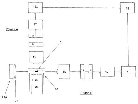
Figure 1 is described by the following:
1. cell
10. testing zone (rectangularly configured to provide four substantially flat
surfaces)
11. Fluorescence collection objective
16. Phase contrast or Dark field objective
17. Pre-amplifier (optional)
18. PMT
18A. PMT
19. CPU/Analyser
20. Immobilising external triggered Q-switched laser or Ablative external
triggered O-Switched laser
21. Optical fibre or hollow rectangular glass fibre to deliver immobilising or
ablative energy
22. Phase contrast or Dark field condensers
22A. Second Light Source for the Phase Contrast or Dark field optical system
23. Band pass filter to exclude wavelengths from 450nm-550nm
24. Band pass filter to exclude aberrant unwanted light from 20.
25. Band pass filter to exclude all non-fluorescent wavelengths
26. Optical fibre or Hollow rectangular glass fibre to provide Fluorescent
excitation light.
27. Fluorescence excitation light source.
A means comprising a second geometric axial motion system allowing gentle
deceleration of desired
cells to be collected via a pipette or the like and maintained in a suitable
medium for later use is also
12
CA 02555411 2006-08-04
WO 2005/075629 PCT/NZ2005/000002
contemplated. The collection means is located downstream of the delivery
device and the testing
zone and will act much like a groyne in a river system. See Example 4.
Example 2
The device shown in figures 3 to 6 illustrates the mechanism by which suitably
stained and intact
sperm are provided for testing as described previously.
In one embodiment, the delivery device is defined by an elongate tube having
five functional zones.
In the first zone, the orientation zone, a majority of sperm (1) exiting a
sample injection tube (110),
(preferably having a bevelled injection tip to minimise the effects to the
laminar flow of the sheath fluid
entering the nozzle via entry points (18)), are oriented into the desired
position for analysis at testing
zone (10). The unique internal geometry of the nozzle combined with the
laminar flow of the sheath
fluid create special hydrodynamic forces which provide a stream of sperm, a
significant proportion of
which are at the correct orientation for testing. The maintenance of a sperm's
orientation is achieved
via a substantially rectangular cross-sectional tube (5), which is contiguous
with a nozzle (8).
Downstream of the orientation maintenance zone is a second zone, the testing
zone (10). After
analysis unwanted sperm are immobilised or eliminated at a third zone. The
fourth zone is a
deceleration pre-collection area ('3-3') before selected viable sperm of a
desired sex are collected in a
fifth zone, the collection zone. The collected cells are maintained in a
suitable environment (4,41) for
post-selection use.
The testing zone (10) is substantially a cavity, tube or aperture that is of a
size and shape sufficient to
accommodate and maintain the orientation of individual cells and which allows
for testing, analysis
and consequent selection of those cells meeting desired criteria. In one
embodiment the short axis of
the rectangular testing zone when looked at in cross section is approximately
32 Nm, the long axis 70
Nm.
In particular, the rectangular cross-sectioned tube (5) maintains the
orientation created by the nozzle
(8) of delicate cells and allows the cells to pass in single file into the
testing zone (10) through a first
light source. The first light source is preferably derived from a laser. Sperm
cells stained with an
appropriate fluorochrome, such as SYBR I, SYBR II, SYBR gold bisbenzimide H
33342, are excited,
fluoresce and the magnitude of fluorescence is measured. SYBR I I, for example
has a peak excitation
at 488nm and peak emission at 525nm.
Simultaneous to the above, the individual sperm cells are exposed to a second
and different light
source, such as light derived from an optical phase contrast or Dark field
system. This light source is
projected orthogonal to the first light source. The sperm being tested emits
light. The light is
captured, amplified and analysed by a multi-channel analyser or appropriate
computer analysis tool.
The fluorescence emitted as the sperm passes through the first light source is
used to identify whether
13
CA 02555411 2006-08-04
WO 2005/075629 PCT/NZ2005/000002
the sperm carries an X or Y chromosome. The non-fluorescent light refracted by
the sperm cell from
the second different light source provides for an improved determination of
its orientation.
Example 3
Having regard to Figures 3-6 the inventor uses the novel hydrodynamic radial
orienting nozzle (8)
described in Example 2 to radially orient individual sperm into a rectangular
cross-sectioned capillary
tube located at the nozzle "exit" ('A - A'). The exit of the orientation
nozzle (8) is contiguous with the
testing zone (10). This enables spermatozoa emitted from the sample injection
tube (110) to develop
the ideal radial and focal plane orientation whilst passing through the nozzle
as required for optical
analysis. As a consequence, a much higher proportion of individual sperm
entering the testing zone
(10) will be correctly aligned to the fluorescence objective (11) to
facilitate the identification of the
chromosome complement of individual spermatozoa within the testing zone (10).
Downstream of the
testing zone a high-speed laser (20) permanently immobilises or destroys
spermatozoa of
indeterminate sex i.e. not correctly oriented and also spermatozoa of the non-
desired chromosome
complement.
Upon completion of processing, the spermatozoa are gently decelerated through
a gently tapered
deceleration zone or pre-collection area ('3 - 3') into a collection vessel
(not shown).
This unique pre-collection/ deceleration zone is a gently flared continuation
of the capillary tube. It has
been observed that the degree of divergence and length of flare directly
influence deceleration speed.
The pre-collection zone in one embodiment takes the form of a "P" trap (4) and
is situated at the end
of the deceleration zone. The "P" trap (4) is pre-filled with spermatozoa
diluent to the level shown (41 )
prior to commencing processing to stop jetting from the analysis/processing
zone.
For In-Vitro fertilisation purposes the immotile/dead spermatozoa may be
removed through percoll
density gradient centrifugation or swim-up techniques as is pro forma for IVF.
The skilled reader will
understand that any non-viable, immotile or dead spermatozoa are of no concern
in In-Vivo
insemination applications.
Examples 1 to 3 illustrate a key difference between the existing art and the
present invention, namely
that the use of a phase contrast or Dark field optical system, or similar, is
used to determine
orientation of individual spermatozoons (90 degree detector) with respect to
the DNA detector (0
degree detector). This provides for surprising system efficiency gains, and
also provides for higher
processing speeds and increased accuracy of analysis, through non-overloading
of the Photo
Multiplier Tube (PMT).
Example 4
A significantly high proportion of sperm that pass from the sample injection
tube (110) and through the
hydrodynamic orientation nozzle (8) are correctly aligned before entering the
testing zone (10) as
described above. On entering the testing zone individual sperm are
simultaneously analysed for
14
CA 02555411 2006-08-04
WO 2005/075629 PCT/NZ2005/000002
orientation using phase contrast or Dark field optics (16,22,22A) at 90
degrees and for DNA content
utilising a fluorescence detector at 0 degrees (11) as shown in Figure 6. Data
is collected and
processed via a CPU/analyser (19) and sperm of a desired sex selected
utilising an immobilising
external triggered Q-switched laser (20), preferably emitting at the 2.69Nm
wavelength, although other
wavelengths may be used or, an ablative external triggered Q-switched laser
utilising other
wavelengths may be used.
Needless to say, the selection/immobilisation stage takes place downstream of
the testing zone and
before entering the deceleration/pre-collection area. It has been found that
the 2.69 um laser system
is well suited to the sperm sexing method of the invention as it delivers the
required power,
penetration and absorption characteristics.
The Specifications relevant to the above immobilising Laser (although some
specifications may be
modified within overall operating requirements) are:
~ Wavelength 2,690nm (or 2,620nm providing for higher penetration but lower
absorption,
calculations for this wavelength have not been made)
~ Solid State. Chromium, Thulium, Erbium doped YAG crystal (CTE:YAG) (Although
diode lasers
may be used provided sufficient power can be generated at required repetition
rates and pulse
duration levels.
~ External triggered Q switched
~ Pulse duration approximately 500ns
~ Repetition rate - variable up to 300kHz
~ Split beam, pulse delivery through two dehydrated (low OH) silica optical
fibres or hollow
rectangular low OH glass fibres positioned directly opposite each other.
Surface
measurements of internal core or hollow centre delivery fibres at sample
interface =
70x10um rectangle with rounded ends, shaped from a flattened optical fibre of
30um
(inner core) diameter, or the electromagnetic energy may be delivered by two
essentially
rectangular cross-sectioned hollow cored, low OH glass fibres, the hollow
section being
approximately 70Nm x 32 Nm.
~ External triggered Q switched Laser will revert to very low power CW
Alignment Mode
between pulses to maintain the correct internal thermal condition of the
resonator.
The reader will be aware that only those cells oriented correctly can be used
to predict with accuracy
the DNA content and therefore the sex characteristics of the sperm.
All of the features disclosed in this specification (including any
accompanying claims, abstract and
drawings), and/or all of the steps of any method or process so disclosed, may
be combined in any
combination, except combinations where at least some of such features and/or
steps are mutually
exclusive.
Alternative features serving the same, equivalent or similar purpose may
replace each feature
CA 02555411 2006-08-04
WO 2005/075629 PCT/NZ2005/000002
disclosed in this specification (including any accompanying claims, abstract
and drawings), unless
expressly stated otherwise. Thus, unless expressly stated otherwise, each
feature disclosed is one
example only of a generic series of equivalent or similar features.
The invention is not restricted to the details of the foregoing embodiment(s).
The invention extends to
any novel one, or any novel combination, of the features disclosed in this
specification (including any
accompanying claims, abstract and drawings), or to any novel one, or any novel
combination, of the
steps of any method or process so disclosed.
ADVANTAGES
The present invention has one or more of the following advantages:
o comparatively inexpensive
o allows for impressive sample flow rates
o provides increased purity of collected sample
o improved viability of selected samples
o provides increased efficiencies
o easier mechanical operation
o improved reliability
a increased sample orientation dependability
VARIATIONS
Some preferred aspects of the invention have been described and illustrated by
way of example, but it
will be appreciated that other variations of and modifications to the
invention can take place Without
departing therefrom.
For example, it is envisaged that although the specification is predominantly
directed to the selection
of X and Y chromosome-bearing sperm cells the possibility of selecting red or
white blood cells from a
blood sample or, gram negative bacteria from a suitably prepared sample is
also contemplated.
The use of such a method to isolate and select for viruses of interest is also
an option.
The skilled reader will also instantly realise that the use of filters to
exclude light energy of unwanted
wavelength might also vary depending on the sample under investigation.
Similarly, although the use
of an optical/hollow fibre arrangement at a wavelength of 2.69pm is preferred
for the immobilising or
ablative laser referred to in the Examples, less suitable but perfectly
adequate wavelengths can be
delivered through air. In fact, some potential wavelengths are not suited to
fibre delivery.
Correspondingly, although fluorescence can be delivered through air, it is
preferred that the
fluorescence excitation wavelength is delivered via fibre optics,
It will also be understood that any reference to a cell will also be directed
to parts of a cell and in
16
CA 02555411 2006-08-04
WO 2005/075629 PCT/NZ2005/000002
particular to components of a cell such as nuclear DNA, mitochondrial DNA,
RNA, or to organisms or
viruses that have invaded or are not normally found within or associated with
said cells or parts of said
cells.
This document describes the use of the invention with respect to selecting
sperm having a desired sex
derived from agriculturally important animals, but a skilled reader will
instantly realise that above
described methods and apparatus will have application in selecting sperm of a
desired sex for all
placental mammals.
Throughout the description and claims of this specification the word
"comprise" and variations of that
word, such as "comprises" and "comprising", are not intended to exclude other
additives, components,
integers or steps.
REFERENCES
1. M. Montag, K. Rink, G. Delacretaz & H. van der Ven, 2,000. Laser induced
immobilisation and
plasma membrane permeabilization in human sperm. Human Reproduction, Vol 15,
No. 4, 846-852.
2. V. Kachell, et al. 1977. Uniform Lateral Orientation caused by Flow Forces,
of Flat Particles in Flow
through Systems, Journal of Histochemistry and Cytochemistry, Vol. 25, No. 7,
pp. 774-780.
3. XY, Inc. PCT Patent Application 15 Nov 2001 No. WO 01/85913 A2
4. XY Inc. US Patent 12 August 2003, US 6,604,435 B2
5. G. M. Hale and M. R. Querry, Optical constants of water in the 200nm to
200um wavelength region,
Appl. Opt., 12, 555-563, (1973). Web page -
http://omic.oQi.edu/soectra/water/data/hale73.dat
6. US 6,263,745, US 6,357,307 & US 5,985,216 to nozzle systems.
7. WO 98/34094.
8. L.A. Johnson and D Pinkel, "Modification of a Laser-based Flow Cytometer
for High Resolution DNA
Analysis of Mammalian Spermatozoa", Cytometry 7:268-273 (1986).
17