Note: Descriptions are shown in the official language in which they were submitted.
CA 02555580 2006-08-09
WO 2005/078424 PCT/US2005/002821
ANALYTE SENSOR, AND ASSOCIATED
SYSTEM AND METHOD EMPLOYING A
CATALYTIC AGENT
CROSS-REFERENCE TO RELATED APPLICATIONS
[0001] This application is a continuation-in-part of U.S. Patent Application
No.
10/819,498 of Feldman et al., filed on April 6, 2004, which is a continuation-
in-part
of U.S. Patent Application No. 10/775,604 of Feldman et al., filed on February
9,
2004. This application is additionally related to U.S. Patent Application No.
10/146,518 of Mao et al., filed on May 14, 2002, the corresponding U.S. Patent
Application Publication No. US 2003/0042137A1 of Mao et al., published on
March
6, 2003, and U.S. Provisional Patent Application No. 60/291,215 of Mao, filed
on
May 15, 2001. Each of the aforementioned applications, publication, and
provisional
application, is incorporated in its entirety herein by this reference.
FIELD OF THE INVENTION
[0002] This invention generally relates to the provision of catalytic agents
within
the locale of an interface between a biofluid and derivatives of the biofluid,
where the
derivative of the biofluid contacts the sensing mechanism of an analyte
sensor. The
invention additionally relates to analyte sensors that make use of any of a
variety of
transducing mechanisms, and which may be placed internally, transcutaneously,
or
externally, relative to a body.
BACKGROUND OF THE INVENTION
[0003] Various analyte sensors, such as glucose biosensors, have been
developed
that provide continuous information from the body with regard to analyte
concentrations. These sensors thus can be described as operating ifz vivo,
i.e., partially
or wholly within a living body. Such irz vivo sensors are thus exposed, in
varying
degree, to the biological environment, and they differ fundamentally in the
way in
which they are used from ex vivo sensors, such as glucose strip readers, in
which a
biofluid sample is talcen from a subject and conveyed away to an external
device for a
discrete sample reading. Various methodologies or mechanisms have been applied
to
CA 02555580 2006-08-09
WO 2005/078424 PCT/US2005/002821
the taslc of transducing the concentration of an analyte of interest into an
informative
signal (Pearson et al., Afzal~tical Aspects of BioseyasoYS, Ann. Clin.
Biochem, 37: 119-
145, 2000). Such transducing methodologies include electrochemical methods,
such
as amperometric, potentiometric, and coulometric methods, by way of example.
Other
transducing methodologies include optical methods, such as luminescence-, and
fluorescence-, and refractive index-based methodologies, by way of example.
There
are still other methodologies, such as thermal transduction, piezoelectric
transduction,
and viscosimetric transduction, merely by way of example.
[0004] Clinical use of biosensors that provide continuous data has been a
significant step toward helping diabetic patients achieve tight control over
their blood
glucose levels, a goal considered desirable ever since the report of the
Diabetes
Control and Complications Trial Research Group Study (N.E.J.M. 329: 977 - 986,
1993). Sensors designed for ih vivo operation can be described variously in
terms of
the particular technologies they employ, the site of their placement in or on
a body,
and the degree of their invasiveness into the body. Some transcutaneous sensor
systems, such as the Freestyle~ NavigatorTM Continuous Glucose Monitoring
System
(Abbott Diabetes Care, formerly known as TheraSense, Inc., Alameda, CA), are
designed for the placement of a sensor portion into a subcutaneous area of the
body,
while a base portion remains external to the body. The sensor portion includes
a
membrane that covers its sensing surface, provides a level physical protection
of the
sensing surface, and also limits the rate of analyte flux to the sensing
surface in a way
that is advantageous to the electrochemical kinetics of the sensor.
[0005] Some transcutaneous continuous sensor systems include a microdialysis
loop placed into a subcutaneous area of the body, while a sensor portion
remains
external to the body. The microdialysis loop provides for the circulation of a
solution
into and out of the subcutaneous space where it contacts the transducing
apparatus of
a sensor placed externally, on the skin. The microdialysate fluid emerging
from the
transit through the subcutaneous space is in equilibrium with the interstitial
fluid
respect to the concentration of the analyte, and thus is a useful analyte-
sensing
medium. Examples of microdialysis-based analyte sensing systems suitable for
glucose sensing have been described in U.S. Patent Nos. 5,640,954 of Pfeiffer
et al.,
filed on May 5, 1995, 6,091,976 of Pfeiffer et al., filed on October 28, 1998,
and
2
CA 02555580 2006-08-09
WO 2005/078424 PCT/US2005/002821
6,591,126 of Reoper et al., filed on July 20, 2001; U.S. Patent Application
Publication
No. 2001/0041830 A1 of Varalli et al., filed on May 7, 2001; and European
Patent
Application No. EP 1153571 A1 of Varalli et al., filed on May 3, 2001.
[0006] Still other sensor systems are associated with means or methods that
are
used to create a disruption, or a wound, or an opening in the skin, or in more
functional terms, a cutaneous port out of which fluid exudes. A sensor placed
externally, on the skin, is used to sense the analyte concentration in the
exuded fluid.
This exuded fluid can differ from the interstitial fluid from which it is
derived in
terms of composition, but with respect to the analyte, is reflective of, or a
function of
the analyte concentration in the interstitial fluid. The exuded fluid may also
differ
from its "parent" biofluid according to the process or injury that gave rise
to the
cutaneous port, which may encompass any of various technologies or
methodologies,
such as laser burning, ultrasonic disruption, particle propulsion, and reverse
iontophoresis, merely by way of example.
[0007] An example of an iya vivo continuous analyte sensing system that makes
use of a cutaneous port is one in which the port is photothennally-induced by
a laser
technology device as described in U.S. Patent Nos. 6,508,785 of Eppstein,
issued on
January 21, 2003, U.S. 6,530,915 of Eppstein et al., issued on March 11, 2003,
U.S.
6,679,841 of Bojan et al., issued on January 20, 2004, and U.S. 6,685,699 of
Eppstein
et al., issued on February 3, 2004. Further by way of example, another way to
create a
cutaneous port is through the use of focused ultrasonic waves to disrupt the
ordered
lipid bilayer of the stratum corneum. This disruption creates pores through
which an
interstitial fluid-derived wound fluid exudes, whereupon the exuded fluid is
used as a
sample fluid for a sensor external to the skin. Patents that describe this
system include
U.S. Patent Nos. 6,620,123 of Mitragotri et al., issued on September 16, 2003,
U.S.
6,190,315 of Lost et al., issued on February 20, 2001, U.S. 6,234,990 of Rowe
et al.,
issued on May 22, 2001, and U.S. 6,491,657 of Rowe et al., issued on December
10,
2002.
[0008] A further example of an approach to continuous i~ vivo analyte sensing
has
involves reverse iontophoresis, whereby weak electrical current is applied to
a site on
the skin to drive compounds outwardly through the skin. Patents describing a
reverse
iontophoretic sensing system include U.S. Patent Nos. 6,023,629 of Tamada,
issued
3
CA 02555580 2006-08-09
WO 2005/078424 PCT/US2005/002821
on February 8, 2000, U.S. 6,393,318 of Corn et al., issued on May 21, 2002,
U.S.
6,438,414 of Conn et al., issued on August 20, 2002, U.S. 5,771,890 of Tamada,
issued on June 30, 1998, and U.S. 6,298,254 of Tamada, issued on October 2,
2001.
As with other cutaneous port systems, internal from the iontophoretic site or
wound
surface is interstitial fluid in its native form, with its native immune cell
population,
albeit disturbed in varying degree by local reaction to the iontophoretic
process, and
external to the iontophoretic site or wound surface on the skin is an exuded,
iontophoretically-driven fluid that comes into contact with the sensing
surface.
[0009] In vivo or continuous sensing systems have had technical challenges to
overcome in order to be able to compare favorably with the high standards of
accuracy and dependability established by ex vivo strip-reading glucose
sensors. For
example, the operation and performance of an in vivo enzyme-based biosensor
may be
complicated by high rates of analyte flux, such that the relationship between
the
concentration of glucose in a sample fluid and the response from the biosensor
becomes non-linear. This kinetic problem has been solved by the interposition
of an
analyte-flux-limiting membrane between the sample fluid and the sensing layer
of the
biosensor, as described in the above-mentioned U.S. Patent Application
Publication
No. US 2003/0042137A1 of Mao et al. Still other challenges, such as usage
limitations, have become evident. For example, data from studies of rthe
recently
available, transcutaneous CGMS system of Medtronic MiniMed, indicate spurious,
low-glucose-reading incidents, particularly during periods of stillness, such
as when a
subject is asleep. (See Metzger et al., Reproducibility of Glucose
Measure3nents Using
t7~e Glucose Sensor, Diabetes Care, July 2002, Vol. 25, 1185-1191; McGowan et
al.,
Spurious Reporting of Nocturnal H,~poglycemia by CGMS in Patients -with
TiglZtly
Controlled Type 1 Diabetes, Diabetes Care, September 2002, Vol. 25, 1499-1503;
authored by The Diabetic Research in Children Network (DirecNet) Study Group,
Accuracy of the GZucoWatcla G2 Biograplaer and the Continuous Glucose
Mofaitoring
System During Hypoglycemia, Diabetes Care vol. 27, no. 3, 722-726, March 2004;
and Mauras et al., Lack of Accuracy of Continuous Glucose Sensors in Healthy,
Nondiabetic Children, Results of the Diabetes Research in ClaildYefa Network
(DirecNet) Accuracy Study, J. Pediatrics 144 (6), 770-775, June 2004.) While
nocturnal hypoglycemic events are indeed a clinical reality, especially in
patients
being aggressively treated with insulin, it has become recognized that false
indications
4
CA 02555580 2006-08-09
WO 2005/078424 PCT/US2005/002821
of such events are particular fallibilities of the CGMS system that complicate
the
interpretation of the data obtained using this system. (See Monsod et al., Do
Sensor
Glucose Levels Accurately Predict Plasma Glucose Coracentrations .During
Hypoglyceyraia and Hyperinsulinenaia?, Diabetes Care, May 2002; and Kaufman et
al.,
Nocturnal Hypoglycemia Detected with tlae Continuous Glucose Monitoring
Sys~ena in
Pediatric Patients with Type IDiabetes, J. Pediatrics 2002; vol. 14.1, 625-
630).
Spurious low-glucose-reading incidents are very problematic in the monitoring
and
treatment of a diabetic subject, as such incidents wrongly indicate that a
euglycemic
subject is hypoglycemic. As an example, when a spurious, low-glucose readW g
is
used as a signal to control insulin dosage, a subject may receive an improper
or a
reduced dose of insulin and thus be put at risk for becoming hyperglycemic.
Spurious
low glucose readings can be further problematic as they may lead to
incorrectly
calibrated sensors, resulting in subsequent false, high glucose readings,
which may
reduce the credibility and usefulness of the alarm function, by way of
example.
Further development of biosensor components and biosensors for continuous isz
vivo
monitoring of analyte levels, such as glucose levels, is desirable.
SUMMARY OF THE INVENTION
[0010] This invention generally relates to the provision of biocompatibility-
promoting catalytic agents to in vivo analyte sensors within the locale of an
interface
between a biofluid and a derivative of the biofluid, where the derivative of
the
biofluid is the fluid that contacts the transducing mechanism sensor. The
locale of the
interface includes locations that may be within the interface or chemically
incorporated into it, immediately adj acent to or in contact with the
interface, or at a
distance near enough to the interface that the effect of the catalytic agents
is such that
it alters the composition or population of chemical species that comprise the
chemical
environment surrounding the interface. These catalytic agents include both
organic,
proteinaceous compounds, such as enzymes, as well as non-proteinaceous organic-
metal compounds that degrade reactive oxygen species or reactive nitrogen
species in
the locale of the sensor. In this catalytic degradation process, such a
reactive species
moves through a metabolic pathway in which it is a reactant. In this manner,
the
concentration of such a reactive species in solution may be reduced. AccordW g
to
some aspects of the invention, catalytic agents engage reactive oxygen and
nitrogen
species of biological origin within the biofluid. Further, according to some
aspects of
CA 02555580 2006-08-09
WO 2005/078424 PCT/US2005/002821
the invention, that catalytic activity enhances the biocompatibility of
sensors, more
particularly, one or more aspects of biocompatibility that may manifest in the
form of
improvements in sensor performance. Improvement or enhancement of sensor
performance may coincide or be associated with higher quality data, as
determined by
various statistical methods that evaluate internal consistency or agreement
with data
from other sources. Higher quality data may include, for example, data that
are more
accurate with respect to reference data from standard strip-reading sensors,
data more
reflective of actual systemic levels of analyte, or data that are more
internally
consistent and as such contain less noise. Enhanced sensor perforniance may
also
include a lengthening of the effective lifetime of a sensor, the effective
lifetime being
reflected in an extended period of the delivery of accurate data.
[0011] Embodiments of the invention include analyte sensors that may sample
any of several bodily fluids or their derivatives, and may be placed in
positions
variously internal within the body, transcutaneously across the skin, or
external to the
body. The types of analyte sensor systems include transcutaneous sensing
systems,
microdialysis systems, cutaneous-port systems and fully implanted systems,
merely
by way of example. Functionally open cutaneous ports in the skin may be
provided by
various methods, such as propelled particles, laser photothermal burning,
sonic
disruption of stratum corneum, and reverse iontophoresis, merely by way of
example.
[0012] Embodiments of the invention further include analyte sensors that
detect
the concentration of the analyte through any available transducing method,
including
electrochemical and viscosimetric mechanisms, merely by way of example.
According to some aspects of the invention, sensing systems are generally
applied to
the continuous sensing of an analyte by virtue of their izz vivo relation to
the body, but
are not limited to any particular biofluid to be sampled, by any particular
position of
the sensing mechanisms with respect to their position internal,
transcutaneous, or
external relative to the body, or by any particular transducing mechanism. A
feature
common to all embodiments of the invention, however, is a structural interface
between a biofluid (a first fluid) being sampled, and a second fluid that
actually
engages or comes in contact with the transducing mechanism of the sensor. The
second fluid is one that has passed through the interface, and as such is a
derivative of
the first fluid, whose composition, at least in part, is determined by the
permeability
6
CA 02555580 2006-08-09
WO 2005/078424 PCT/US2005/002821
features of the interface. All embodiments of the present invention include a
biocompatibility-promoting catalytic agent in the locale of this structural
interface.
[0013] This interface may be synthetic, such as a membrane or gel, or
biological,
as exemplified by a cutaneous site or wound, or my suitable combination
thereof. In
the case of a transcutaneous sensor, the interface is emb odied in a synthetic
membrane
that covers the sensing surface. In the case of a microdialysis system, the
interface is
embodied in the microdialysis membrane of the syste~rn. In the case of a
cutaneous
port system, whether created by propelled particles, a laser, or ultrasound,
or through
reverse iontophoresis, the interface is the cutaneous site or wound through
which fluid
has moved from the interstitium to the post-biological space outside the body.
In the
case of an iontophoretic system, the interface is the site on the skin that is
exposed to
the iontophoretic current, and through which fluid and solute then pass. In
some cases
a combination of biological and synthetic elements rnay constitute the
operational
interface. For example, in the case of a transcutaneous sensor, the full
extent of the
interface between (1) the biofluid, the undisturbed interstitial fluid and (2)
the biofluid
derivative that actually contacts the sensing surface can be considered to
include not
only the synthetic protective membrane over the sensing surface, but also the
wound
site within the skin that develops in the immediate vicinity of tissue into
which the
sensor has been inserted.
[0014] The first fluid can be any definable biofluid, such as blood or
interstitial
fluid. The second fluid or biofluid derivative varies in composition according
to
specifics of the sensing technology and the interface. Ln the case of a
transcutaneous
system, the biofluid is interstitial fluid, the interface ~.s the membrane
covering the
subcutaneously-located sensor surface, and biofluid derivative is the filtrate
that
penetrates the membrane to contact the sensing surface _ In the case of a
microdialysis
system, the biofluid is interstitial fluid, the interface is the
subcutaneously-located
dialysis membrane, and the biofluid derivative is the dialysate that contacts
the
sensing surface of an external sensor. In the case o~ cutaneous port systems,
the
biofluid is interstitial fluid, the interface is the cutaneous surface or the
cutaneous
wound, and the biofluid derivative is the wound fluid exuded out of the body,
which
then ultimately contacts the sensing surface of a sensor placed on the skin.
In the case
of reverse iontophoretic systems, the biofluid is intersrtitial fluid, the
interface is the
7
CA 02555580 2006-08-09
WO 2005/078424 PCT/US2005/002821
site on the skin that is subj ected to current, and across which solute-
containing fluid is
driven, and the biofluid derivative is the solute-containing fluid that
contacts the
sensing surface on a sensor attached to the skin.
[0015] The catalytic agents, or more particularly, the organic-metal catalysts
of
the present invention, catalyze the degradation of reactive oxygen species and
reactive
nitrogen species, such as superoxide, hydrogen peroxide, and peroxynitrite, by
way of
example. Examples of such catalysts include superoxide dismutase/catalase
catalysts,
including catalytic enzymes and non-proteinaceous mimics of such enzymes. One
particular example of a superoxide/dismutase catalyst is manganese 5,10,15,20-
tetra(4-pyridyl)-21H,23H-porphine chloride (MnTPyP). Such a catalyst may be
incorporated into a membrane that covers the sensing surface of a
transcutaneous
electrochemical sensor, or incorporated into the dialysis membrane of
microdialysis-
based sensing systems. As a result of its presence in the locale of the
interface
between the biological fluid and the sensing mechanism, the catalyst reduces
the local
concentration of reactive oxygen species, such as those mentioned above. While
this
invention is not bound by any proposed theory, it is thought that reactive
oxygen
species are present in the locale of the interface by virtue of metabolic
activity of cells
of the immune system, such as neutrophils, which are generally engaged in the
initial
phases of a foreign body response to the presence of the sensor. The reactive
oxygen
species in the locale of sensors may have effects that are deleterious to the
sensor and
may also further accelerate the recruitment of immune cells to the sensor
site. The
reactive oxygen species may further have effects on the metabolism of other
cells in
the locale, which may create local areas that are depleted of glucose, which,
in turn,
would disconnect local glucose values from systemic glucose values. Through
the
action of the superoxide dismutase/catalase catalysts and the consequent
reduction of
local concentrations of reactive oxygen species, the sensor may be rendered
more
biocompatible and its performance may be improved. Examples of enhanced sensor
performance include a decrease in failure rate, an increase in operating
lifetime, a
decrease in the level of signal-interfering noise, and the prevention or
decrease in
incidence of spurious hypoglycemic incident reporting, by way of example.
[0016] These and various other aspects, features and embodiments of the
present
invention are further described herein.
8
CA 02555580 2006-08-09
WO 2005/078424 PCT/US2005/002821
BRIEF DESCRIPTION OF THE DRAWINGS
[0017] A detailed description of various aspects, features and embodiments of
the
present invention is provided herein with reference to the accompanying
drawings,
which are briefly described below. The drawings are illustrative and are not
necessarily drawn to scale. The drawings illustrate various aspects or
features of the
present invention and may illustrate one or more embodiments) or examples) of
the
present invention in whole or in part. A reference numeral, letter, and/or
symbol that
is used in one drawing to refer to a particular element or feature may be used
in
another drawing to refer to a like element or feature.
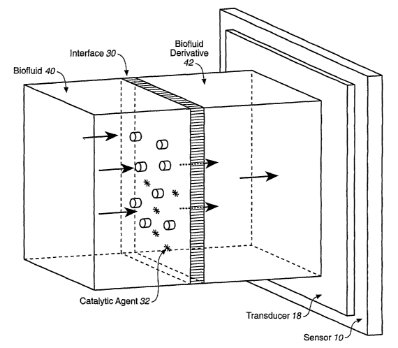
[0018] Figure 1 depicts a sensing system where an upstream biofluid, and a
downstream derivative of the biofluid are separated by a porous or partially-
permeable interface with a catalytic agent disposed in the locale thereof, and
the
biofluid derivative comes into contact with the transducing apparatus of an
analyte
sensor.
(0019] Figure 2A is a schematic, side-view illustration of a portion of a two-
electrode glucose sensor having a working electrode, a combined
counter/reference
electrode, and a dip-coated membrane that encapsulates both electrodes.
Figures 2B
and 2C are schematic top- and bottom-view illustrations, respectively, of the
portion
of the glucose sensor of Figure 2A. Herein, Figures 2A, 2B and 2C may be
collectively referred to as Figure 2.
[0020] Figure 3A depicts a typical structure of a section of an analyte-
diffusion-
limiting membrane with a catalytic agent incorporated therein. Figure 3B is an
illustration of a membrane similar to that shown in Figure 3A, except that a
specific
superoxide dismutase catalyst, manganese 5,10,15,20-tetra(4-pyridyl)-21H,23H-
porphine chloride, is shown covalently incorporated therein.
[0021] Figures 4A and 4B, together, depict a transcutaneous electrochemical
sensor. Figure 4A is a perspective view of a fully fabricated sensor as it
would be
seen partially implanted into the skin, and Figure 4B is an expanded and
cutaway
view of a sensor insertion tip, showing a membrane, enhanced with a catalytic
agent,
covering a sensing layer.
9
CA 02555580 2006-08-09
WO 2005/078424 PCT/US2005/002821
[0022] Figure 5 is a graph of electrical current versus glucose concentration
for
electrochemical sensors having glucose-diffusion-limiting membranes that are
enhanced with a catalytic agent.
[0023] Figure 6 is a graph of glucose concentration versus time for a human
subject over three days, as reported by two continuously operating,
transcutaneous
sensors. One sensor has a conventional membrane; the other has a membrane
containing a superoxide-dismutase/catalase catalyst, MnTPyP. Intermittent
readings
obtained manually from a strip reading glucose meter are also shown.
[0024] Figure 7 depicts a microdialysis-based sensing system with a catalytic
agent associated with the membrane.
[0025] Figures 8A and 8B, together, schematically illustrate a cutaneous port-
based sampling system in which a catalytic agent is disposed between a
biofluid and a
biofluid derivative. Figure 8A illustrates a method of creating a cutaneous
port in the
shin and an associated system. Figure 8B schematically illustrates a method of
sampling wound fluid from such a cutaneous port and an associated system.
[0026] Figure 9 depicts a cutaneous-port-based sampling system for an external
sensor, in which the port comprises an iontophoretic site, and in which a
catalytic
agent is disposed between the biofluid and the biofluid derivative.
[0027] Figure l0A depicts the head of a fully iniplantable analyte sensing
system
in which a catalytic agent is disposed in the locale of an interfacing
membrane
between biofluid and the biofluid derivative. Figure lOB is schematic cross
sectional
view of a sensing region of the head. Figures l0A and lOB may be collectively
referred to as Figure 10.
DESCRIPTTON OF THE INVENTION
1. Various Conventions and Terms
[0028] In the description of the invention herein, it will be understood that
a word
appearing in the singular encompasses its plural counterpart, and a word
appearing in
the plural encompasses its singular counterpart, unless implicitly or
explicitly
understood or stated otherwise. Further, it will be understood that for any
given
component described herein, any of the possible candidates or alternatives
listed for
CA 02555580 2006-08-09
WO 2005/078424 PCT/US2005/002821
that component, may generally be used individually or in combination with one
another, unless implicitly or explicitly understood or stated otherwise.
Additionally, it
will be understood that any list of such candidates or alternatives, is merely
illustrative, not limiting, unless implicitly or explicitly understood or
stated otherwise.
[0029] Various terms shown in quotation marks below are described to
facilitate
an understanding of the invention. It will be understood that a corresponding
description of these various terms applies to corresponding linguistic or
grammatical
variations or forms of these various terms. It will also be understood that
the invention
is not limited to the terminology used herein, or the descriptions thereof,
for the
description of particular embodiments. Merely by way of example, the invention
is
not limited to particular analytes, bodily fluids, or sensor designs or
usages, unless
implicitly or explicitly understood or stated otherwise, as such may vary.
[0030] A "biofluid", or biological fluid, is any bodily fluid that exists
physiologically within the bounds of the living body, such as, for example,
whole
blood (arterial, venous, or capillary), interstitial fluid, or cerebrospinal
fluid. A
"derivative" of a biofluid, or a "biofluid derivative" is a fluid derived from
a biofluid
by, for example, passage through an interface, such as dialysis membrane to
yield a
dialysate, or by passage through a protective membrane to yield a filtrate.
The
exudation of fluid through a biological interface, such as a cutaneous port at
the site of
a disruption in slcin, is a fluid of biofluid origin that has left the bounds
of a living
body, is no longer a part of the biology of the subject body, is changed in
some way
during and as a result of its passage out of the body, and as such is also a
derivative of
a biofluid.
[0031] "Biocompatibility" is a property of a material that allows it to be
compatible with the host biological environment with which it is in contact.
Biocompatible material does not provoke a substantial or apparent foreign body
response (which involves the immune system's recognition of non-self), or a
substantial or apparent wound-healing response (which may not involve the
immune
system), the two of which together can contribute to the full biological
response to the
intrusion of a foreign body. This description of biocompatibility is not
absolute, as the
term can serve as a comparative descriptor, or can serve as a functional
descriptor,
such that it describes a compatibility sufficient to allow a material or
device to
11
CA 02555580 2006-08-09
WO 2005/078424 PCT/US2005/002821
perform its intended function within the host, for example. To some extent
biocompatibility is described in terms of the negative, i.e., by the
substantial or
apparent absence of biological incompatibility. "Biological incompatibility"
is used
commonly even without an understanding of the specifics of the physical or
chemical
features responsible for the incompatibility and/or an understanding of the
details of
the biological response to the foreign body. Accordingly, the meanng of the
biocompatibility is flexible, and becomes more specific according to the
specifics of
the context.
[0032] "Catalase" (systematic name: hydrogen-peroxide:hydrogen-peroxide
oxidoreductase) is an enzyme that catalyzes the decomposition or "dismutation"
of 2
molecules of hydrogen peroxide to yield water and molecular oxygen. The
hydrogen
peroxide substrate of this reaction is a product of a superoxide dismutase
reaction, as
described below under "superoxide dismutase".
[0033] An "organic-metal catalytic agent" describes a compound that
facilitates a
chemical reaction or reactions. Catalytic agents increase the flux of
compounds
through metabolic pathways, are themselves, unchanged by the reaction they
facilitate, and are thus available for further activity. A single catalytic
agent may
affect the flux through metabolic pathways at a single step, or at multiple
steps, and it
may affect flux through multiple pathways. Organic-metal catalytic agents
described
herein include metal-containing enzymes, which are proteinaceous catalysts, as
well
as non-proteinaceous, organic-metal compounds that are catalytic and mimic the
action of particular enzymes. Catalytic agents that improve the
biocompatibility (see
above) of a device may be referred to as biocompatibility-promoting catalytic
agents.
[0034] A "counter electrode" refers to (a) a counter electrode or (b) a
counter
electrode that also functions as a reference electrode (i.e., a
counter/reference
electrode).
[0035] A "crosslinker" is a molecule that contains at least two reactive
groups
capable of linking at least two molecules together, or linking at least two
portions of
the same molecule together. Linking of at least two molecules is called
intermolecular
crosslinlcing; linking of at least two portions of the same molecule is called
intramolecular crosslinking. A crosslinlcer having more than two reactive
groups may
12
CA 02555580 2006-08-09
WO 2005/078424 PCT/US2005/002821
be capable of coincidental intermolecular and intramolecular crosslinkings.
[0036] An "electrochemical sensor" is a device configured to detect the
presence
of or measure the concentration or amount of an analyte in a sample via an
electrochemical oxidation or reduction reaction. Typically, the reaction is
transduced
to an electrical signal that is correlated to an amount or concentration of
analyte.
"Electrochemical" describes a method of transducing a concentration of analyte
to an
informative signal. Other methods of transduction include, for example,
optical and
viscosimetric methods. While the description herein includes an exemplary
embodiment that is electrochemical in nature, the invention is not limited in
terms of
the transduction method employed.
[0037] A "heterocyclic nitrogen group" refers to a generally carbon-based
cyclic
structure containing an sp2-hybridized nitrogen integrated within a ring of
the
structure.
[0038] "Iontophoresis" refers to the application of an electric current to
cause the
electro-osmotic transport of fluid aald solutes contained therein across the
skin.
"Reverse iontophoresis" refers to the process when it is being operated so as
to extract
an analyte-containing fluid of biological origin outwardly from the skin. In
the
context of this invention, such iontophoretic biofluid derivative is then
provided as a
sample to a sensor on the surface of the skin.
[0039] An "i~ vivo analyte sensor" is a sensor that is designed for placement
in a
body or on a body, with varying degrees of invasiveness, but having in common
a
continuous exposure to biofluid. Ih viv~ sensors do not require, as is the
case with ex
vivo sensors, a separate step or steps by which a biofluid sample is taken
from the
body and conveyed to an external device for a discrete sample-specific sensing
event.
An in vivo sensor, for example, may be fully or partially implanted in the
body and
exposed to a biofluid, inserted across the skin such that a subcutaneous
biofluid
contacts the sensor, or placed on the surface of the skin such that an exuded
biofluid
contacts the sensor. Irz vivo sensors transduce the presence of analyte into
an
informative signal by any of a variety of methods, including electrochemical,
optical,
piezoelectric, and viscosimetric methods.
[0040] "Interface" describes an intervening structure between (1) a space in a
13
CA 02555580 2006-08-09
WO 2005/078424 PCT/US2005/002821
body wherein a biofluid exists in its native form, and (2) a space in which a
derivative
of the biofluid (derivative by virtue of having passed through the interface)
comes
into contact with a transducing element or mechanism of the sensor, such as a
sensing
surface of an electrochemical sensor. The interface may be embodied in various
forms, including such forms as a membrane over the sensing surface of a
sensor, a
microdialysis membrane, or the surface of a cutaneous wound which functions as
a
transcutaneous port, allowing the egress of a wound fluid.
[0041] "Interstitial fluid," also known as "extracellular fluid," refers to
the fluid in
the body that occupies the space between cells. This fluid is distinct from
intracellular
fluid, as well as from the fluid or plasma portion of blood contained within
the vessels
of the circulatory system. A transcutaneously-placed glucose sensor is exposed
to
interstitial fluid.
[0042] A "low-glucose-reading incident" describes an occurrence of a glucose
reading by a sensor that is lower than a value that would be reasonably
expected by a
qualified observer exercising judgment based on a view of the overall medical
context. Such a glucose reading is considered spurious in that it may not
accurately
reflect the systemic blood glucose level.
[0043] A "membrane solution" is a solution that comprises components for
crosslinking and forming the. membrane, such as a modified polymer containing
heterocyclic nitrogen groups, a crosslinker, and a buffer or an alcohol-buffer
mixed
solvent. A "catalyst-enhanced membrane solution" is a membrane solution that
includes a catalytic agent, such as an enzyme or a mimic thereof.
[0044] "Microdialysis" refers to a sampling technology used in a biosensor
system wherein a catheter incorporating a thin dialysis tube section is
inserted
subcutaneously into a body. The tube is constructed of a membrane partially
permeable to solutes, through which an isotonic solution is circulated. During
the
circulation cycle, the concentration of glucose within the isotonic solution
equilibrates
with the glucose concentration within the surrounding interstitial fluid. This
solution
or dialysate becomes the sample fluid that is contacted to a sensor on the
skin. The
"micro" of microdialysis simply refers to the size of the tubing, in terms of
diameter
or volume/length, which is small compared to that of standard research or
preparative
14
CA 02555580 2006-08-09
WO 2005/078424 PCT/US2005/002821
dialysis tubing. Simply as an example of dimensions, the dialysis membrane of
the
CMA 60 microdialysis catheter of CMA Microdialysis AB (Soma, Sweden) has a
length of 30 mm and a diameter of 0.6 ruin, with a molecular weight cut-off
(i.e., pore
size) of approximately 20,000 Daltons.
[0045] A "mimic" or "non-proteinaceous mimic" refers to a non-proteinaceous
compound that has a catalytic activity like that of a known enzyme, and thus
is a
"mimic" of that enzyme. The non-proteinaceous compound may comprise a metallic
component and an organic component, wherein a metal ion or atom of the
metallic
component and a nonmetallic ion, molecule, portion, or ligand of the organic
component form a union. Such a non-proteinaceous compound may be referred to
as a
metal-nonmetallic or nonmetallic-metal compound, a metal-organic or organic-
metal
compound, and/or the like, and is sometimes referred to as an organometallic
compound, as that term is often loosely used or as that term is strictly used.
When the
union is coordinative or complexing in nature, such a non-proteinaceous
compound
may be referred to as a coordination compound, a complex compound, a metal-
nonmetallic or nonmetallic-metal complex or coordination compound, a metal-
organic or organic-metal complex or coordination compound, and/or the like.
When
the union is in the form of a direct metal-to-carbon attachment, whether of a
coordinative, complexing, or other nature, the non-proteinaceous compound may
be
referred to as an organometallic compound, as that term is strictly used. The
non-
proteinaceous compound may comprise any suitable metal, such as any suitable
metal
in any of Groups 3 through 12 (new notation) or IB through VIIB and VIII (CAS
notation) of the Periodic Table of the Elements or any suitable metal in the
family of
transition metals, such as manganese, iron, copper, or zinc, merely by way of
example.
[0046] "Peroxidase" (systematic name: donor:hydrogen-peroxide reductase) is an
enzyme that catalyzes the reduction of hydrogen peroxide to yield water. The
reaction
catalyzed by peroxidase may be expressed as follows: donor + H202 = oxidized
donor
+ 2 H20. The hydrogen peroxide substrate of the reaction is a product of a
superoxide
dismutase reaction, as described below under "superoxide dismutase."
[0047] "Polyvinylimidazole" refers to any of poly(1-vinylimidazole), poly(2-
vinylimidazole), or poly(4-vinylimidazole).
CA 02555580 2006-08-09
WO 2005/078424 PCT/US2005/002821
[0048] "Polyvinylpyridine" refers to any of poly(4-vinylpyridine), poly(3-
vinyl-
pyridine), or poly(2-vinylpyridine), as well as any copolymer of vinylpyridine
and a
second or a third copolymer component.
[0049] A "reactive group" is a functional group of a molecule that is capable
of
reacting with another compound to couple or covalently bind at least a portion
of that
other compound to the molecule. Reactive groups include carboxy, activated
ester,
sulfonyl halide, sulfonate ester, isocyanate, isothiocyanate, epoxide,
aziridine, halide,
aldehyde, ketone, amine, acrylamide, thiol, acyl azide, acyl halide,
hydrazine,
hydroxylamine, alkyl halide, imidazole, pyridine, phenol, alkyl sulfonate,
halotriazine, imido ester, maleimide, hydrazide, hydroxy, and photo-reactive
azido
aryl groups. Activated esters, as understood in the art, generally include
esters of
succinimidyl, benzo-triazolyl, or aryl substituted by electron-withdrawing
groups,
such as sulfo, nitro, cyano, or halo groups; or carboxylic acids activated by
carbodiimides.
[0050] "Reactive species" describes a free radical or other molecule that is
easily
converted to a free radical or is a powerful oxidizing agent. "Reactive oxygen
species" (or ROS) refers to at least one of a superoxide (02 }, hydrogen
peroxide
(HZOZ), hypochlorous acid (HOCI), and hydroxyl radical (OH~). Synonymous terms
include "reactive oxygen metabolite" (ROM) and "reactive oxygen intermediate"
(ROI). "Reactive nitrogen species" (or RNS) refers to nitric oxide (NO) of
various
redox states and related species including at least one of nitric oxide
radical (NO~),
nitric oxide nitrosonium cation (NO+), and nitroxyl anion (NO~), and
peroxynitrite
(ONOO-). Synonymous terms include "reactive nitrogen metabolite" (RNM) and
"reactive nitrogen intermediate" (RNI). For a review of these reactive oxygen
and
nitrogen species in the context of neutrophil biology, see J. Paul Robinson
and George
F. Babcoclc (eds), Plaag~ocyte Function: A Guide fof° Reseay~ch and
Clinical
Evaluation, ISBN 0471123641, John Wiley (1998).
[0051] A "redox mediator" is an electron-transfer agent for carrying electrons
between an analyte, an analyte-reduced or analyte-oxidized enzyme, and an
electrode,
either directly, or via one or more additional electron-transfer agents. A
redox
mediator that includes a polymeric backbone may also be referred to as a
"redox
polymer."
16
CA 02555580 2006-08-09
WO 2005/078424 PCT/US2005/002821
[0052] A "reference electrode" is (a) a reference electrode or (b) a reference
electrode that also functions as a counter electrode (i.e., a
counter/reference
electrode), unless otherwise indicated.
[0053] A "transducing mechanism", a "transducing apparatus", or a "transducer"
describes at least one element of a sensor that is directly involved in
identifying an
analyte and its concentration, and from that information, generating a signal
informative of this information. Transducing mechanisms vary according to the
physical and/or chemical method and apparatus by which the analyte is
recognized
and by which the concentration of the analyte is determined.
[0054] A "substituted" functional group (e.g., substituted alkyl, alkenyl, or
alkoxy
group) includes at least one substituent selected from the following: halogen,
alkoxy,
mercapto, aryl, alkoxycarbonyl, alkylaminocarbonyl, dialkylaminocarbonyl, --
OH, --
NHZ, allcylamino, dialkyl-amino, trialkylammonium, alkanoylamino,
arylcarboxamido, hydrazino, alkylthio, alkenyl, and reactive groups.
[0055] "Superoxide dismutase" (SOD) refers to an enzyme that catalyzes the
dismutation of superoxide to yield oxygen and hydrogen peroxide. The reaction
catalyzed by superoxide dismutase may be expressed as follows: 2 OZ~- + 2 H+ =
02 +
H~02. The hydrogen peroxide product of the reaction is a substrate for
catalase and/or
peroxidase.
[0056] A "superoxide-dismutase/catalase catalyst" refers to a catalyst,
whether an
enzyme or an enzyme mimic, that possesses the catalytic activity of either
superoxide
dismutase or catalase, to any degree, or the catalytic activities of both
superoxide
dismutase and catalase, to any degree. The term, superoxide-dismutase/catalase
catalyst, encompasses a preferred embodiment in which a catalytic agent that
catalyzes the dismutation of superoxide also catalyzes the decomposition of
hydrogen
peroxide, and vice ve~~sa. The term, superoxide-dismutase/catalase catalyst,
also
encompasses embodiments in which an agent catalyzes the dismutation of
superoxide,
but not the decomposition of hydrogen peroxide, and embodiments in which an
agent
catalyzes the decomposition of hydrogen peroxide, but not the dismutation of
superoxide.
[0057] A "superoxide-dismutase/catalase mimic" refers to an enzyme mimic that
17
CA 02555580 2006-08-09
WO 2005/078424 PCT/US2005/002821
possesses the catalytic activity of one or more of the superoxide dismutase
and
catalase enzymes, to any degree, or the catalytic activities of both
superoxide
dismutase and catalase, to any degree. The term, superoxide-dismutase/catalase
mimic, encompasses a preferred embodiment in which a catalytic agent that
catalyzes
the dismutation of superoxide also catalyzes the decomposition of hydrogen
peroxide,
and vice versa. The term, superoxide-dismutase/catalase mimic, also
encompasses
embodiments in which a catalytic agent catalyzes the dismutation of
superoxide, but
not the decomposition of hydrogen peroxide, and embodiments in which a
catalytic
agent catalyzes the decomposition of hydrogen peroxide, but not the
dismutation of
superoxide. Mimics that catalase the superoxide dismutase and catalase
reactions may
catalyze other reactions as well, such as peroxynitrite decomposition.
[0058] "Transcutaneous" refers to the site or location or nature of a
biosensor
when a portion of the biosensor is placed across the cutaneous layer, such
that one
portion of the biosensor remains external to the skin, and another portion of
the
biosensor is inserted into the subcutaneous space, and in contact with
interstitial fluid.
"Transcutaneous" is also a descriptive term that may be applied to sensors of
this
type. Transcutaneous sensors are considered to be partially implanted in the
body, in
contrast to sensors that are fully implanted within the body.
2. Sensing of derivatives of biofluid samples from the iiz vivo environment
2a. Reco~Tnition that aspects of the ih vivo environment, such as the cellular
immune
system and its metabolism, may be pertinent to the operation of an analyte
sensor
[0059] Various types of biosensors have been designed to operate partially or
wholly in a living body. As these biosensors are exposed to the chemistry and
biology
of the body, it is now theorized that various chemical and biological factors
may
complicate specific aspects of their operation or performance that may
manifest in
subtle ways. For example, an implanted biosensor is typically completely
enclosed
within a body and remains within the body for a period varying from weeks to
many
months. Such an implanted sensor may have longer-term effects in the region
surrounding the implantation site, such as the effects of the immunologic
reaction to
the sensor as a foreign body, including the related vascular processes,
biofouling, and
fibrotic sequelae at the site of foreign presence. In cases where such clear
and
apparent biological response to the presence of a sensor occurs, the
performance of
18
CA 02555580 2006-08-09
WO 2005/078424 PCT/US2005/002821
the sensor might be expected to be compromised. However, as theorized above,
the
highly sensitive sensor processes could be compromised before such obvious
manifestation of biological response, or even in its apparent absence.
Similarly, a
transcutaneous sensor may be subject to subtle biological or biochemical
interference
in ways that do not coincide with the lengthy timeline marked by vascular
processes,
biofouling, and fibrosis. A transcutaneous biosensor, in contrast to a fully
implanted
sensor, is much less invasive, as only a portion of the sensor intrudes into
the
subcutaneous space, and that portion resides there only for a period on the
order of
about three to about five days. Even within this relatively short period,
however, the
early phases of the immune system response to the inserted portion of the
sensor are
activated; as neutrophils, the main phagocytic leukocytes in the blood, are
quickly
recruited to the site, whereupon, it is now theorized, they may have
significant effects
on sensor performance.
[0060] At the site of foreign intrusion, neutrophils release destructive
enzymes
and oxidants to damage the intruder, while at the same time, they attempt to
physically engulf and devour it. The released oxidants are derived from
hydrogen
peroxide, superoxide radicals, nitric oxide and chloride, the former two of
which, at
least, may act to attract further neutrophils and thereby accelerate their own
respective
accumulation. The released oxidants include hydroxyl radicals, formed through
the
reaction of hydrogen peroxide with reduced transition metal cations or their
complexes; peroxy-nitrous acid, formed of nitric oxide and superoxide
radicals; and
hypochlorite, formed of hydrogen peroxide and chloride. The resulting oxidant
cocktail is strong, able to oxidize most organic chemicals and to provide a
local
antiseptic effect that is generally beneficial at a site of a potentially
infectious
intrusion. A broad review of this subject appears in eds. J.P. Robinson and
G.F.
Babcock, Phagocyte Fuhctio~c: A Cuide fog Research and Clinical Evaluatio~t,
John
Wiley, ISBN 0471123641 (1998), and particularly in Chapter 9 thereof, J.P.
Robinson, Oxygen arad Nitrogef~ Reactive Metabolites and Phagocytic Cells,
p.217.
[0061] As noted in the background, ih vivo sensors have been observed to
report
glucose values that are considered to be spuriously low. In view of these
observations,
and in view of what is known about the biology of neutrophils, it is possible
to
formulate theories (without being bound by such theories) that hold
neutrophils at
19
CA 02555580 2006-08-09
WO 2005/078424 PCT/US2005/002821
least partially responsible for affecting the performance of a glucose sensor
and
creating spurious results by several mechanisms. For example, newly recruited
neutrophils axe known to be in the midst of an "oxidative burst" that is
characterized
by high rates of internal metabolic activity, as well as extensive release of
superoxide
as part of their anti-infective effort. Metabolically active cells of the
immune system,
in high concentration, may deplete the local environment of the glucose they
consume
for energy. Another possible explanation for low glucose readings focuses on
the
presence of neutrophil-originating reactive oxygen species. Superoxide gives
rise to
hydrogen peroxide, which in addition to playing an antiseptic role, may have
further
effects on the internal metabolism of local tissue. Hydrogen peroxide may, for
example, increase the consumption of glucose by local cells via the pentose
phosphate
pathway (also l~nown as the HMP shunt), an effect mediated by intracellular
glutathione levels. Briefly, according to this proposition, hydrogen peroxide
oxidizes
glutathione to its oxidized dimer form, oxidized glutathione oxidizes NADPH to
NADP+, and finally NADP+ oxidizes glucose-6-phosphate to ribulose-S-phosphate
in
the first step of the pentose phosphate pathway. The result of this
glutathione-
mediated effect would be to accelerate the intracellular glucose metabolism,
and such
affected cells would then consequently draw upon and deplete the local
extracellular
concentration of glucose. Local glucose depletion, by either of these
processes, could
compromise the value of glucose sensing data, as however accurate the data may
be in
a very local sense, the data may not be reflective of the clinically relevant
level of
glucose in the bloodstream.
[0062] It is now also theorized that accumulated neutrophils, in their attempt
to
engulf the sensing surface, may physically cover it to the extent that the
sensor no
longer has effective contact with the surrounding interstitial fluid. This
latter theory,
particularly, is consistent with the observation that low glucose readings
often occur
during periods of stillness, such as sleep, and the recovery of those glucose
readings
to normal upon body movement that may either disturb the accumulated
neutrophils,
or more generally, stir the stagnant interstitial fluid surrounding the
sensor. Further, in
terms of the panoply of effects that neutrophils may have on glucose sensor
data, it is
now theorized that the oxidants released by the neutrophils in an immune
system
response may have direct disrupting effects on the electrochemistry of the
sensor.
CA 02555580 2006-08-09
WO 2005/078424 PCT/US2005/002821
[0063] An immune system response, such as that described above, typically
results in inflammation. One particular approach to controlling inflammation
associated with the presence of long-term device implants, such as cardiac
stems,
replacement joints and the lilce, involves the use of superoxide dismutase
(SOD) to
consume accumulated superoxide. Superoxide, a product of neutrophil
metabolism, as
well as an attractor of neutrophils and other cells of the inunune system, is
a highly
reactive species that gives rise to other oxygen metabolites. Superoxide
generation
often occurs under conditions which nitric oxide is also being generated. The
two
species can then combine to form peroxynitrite, a species that can be
classified as
either a reactive oxygen species or a reactive nitrogen species, which then
has further
inflammatory consequences. The reaction catalyzed by the SOD enzyme, known as
"dismutation" of superoxide, consumes two superoxide ions and two hydrogen
ions to
yield molecular oxygen and hydrogen peroxide, per the following reaction: 02 +
Oa-
+ 2H+ -~ 02 + H2O2. As such, the SOD enzyme would appear to be capable of
catalyzing the removal of at least some of the superoxide that is present at a
site of
neutrophil metabolism.
[0064] The SOD enzyme has been shown to be effective in reducing
inflammation in the context of the vascular system (J.M. McCord, Superoxide
Dismutase: Rcztiorzale for Use irz Reperfusiora Injury arid Inflammation, Free
Radical
Biol. Med. 2: 307-310, 1986; and V.R. Muzykantov, Targetirzg of Superoxide
Dismzstase arid Catalase to T~ascular~ Endotlzelium, J. Control Release, 71: 1-
21,
2001), leukocyte biology (J.F. McCord, Superoxide Produeti~rz irz Humarz
Disease, in
ed. A. Jesaitis and E. Dratz, Molecular Basis of Oxidative Damage by
Leukocytes,
CRC Press 1992, ISBN: 0849363632, pp. 225-239), and in the brain (Chan et al.,
Protective Effects of Liposonze-ErztYapped Superoxide Disrnutase orz
Posttr~aurnatic
Brain Ederna, Ann. Neurol. 1987; 21, 540-547; Chan et al., Free Fatty Acids,
Oxygerz
Free Radicals, and Membrane Alterations in Brain Ischemia and Injury, in ed.
Plum
et al., Cerebrovczscular Diseases, Raven Press, New York 1985, 161-171). The
ubiquity of this enzyme suggests the broad significance that controlling the
local
concentrations of superoxide has in regulating physiological homeostasis as
well as
the role superoxide plays in inflammatory processes. Various forms of the SOD
enzyme are known; each includes a transitional metal component that is
important in
the enzyme's catalytic activity. For example, a manganese-containing form of
the
21
CA 02555580 2006-08-09
WO 2005/078424 PCT/US2005/002821
enzyme is found in mitochondria, a copper- or zinc-containing form of the
enzyme is
found in plasma and in extracellular fluid, and an iron-containing form of the
enzyme
is found in anaerobic prokaryotes (D.P. Riley, Functional Mimics of
Supef~oxide
Disnzutase Enzymes as Tlzez°apeutic Agents, Chem. Rev. 1999, 99, 2573-
2587).
[0065] Non-proteinaceous, mimics of superoxide dismutase (SOD mimics) have
also been shown to reduce inflammation (Weiss et al., Manganese-Based Supez-
oxide
Diszzzutase Mimetics Izzhibit Neutz°ophil Infiltration In-Vivo, J.
Biol. Chem. 1996; 271,
26149-26156). For example, a class of manganese- or iron-complexes of nitrogen-
containing, fifteen-membered, macrocyclic ligands has recently been shown to
have
the catalytic activity of SOD, and to be effective, when attached to the
surface of
small plastic subcutaneous implants, in reducing the inflammation caused by
implantation (U.S. Patent No. 6,525,041 of Neumann et al., filed on March 14,
1996;
Published PCT Application, International Publication No. WO 00/72893 A2 of
Ornberg et al., filed on May 26, 2000; U.S. Patent Application No.
2004/0110722A1
of Ornberg et al., filed November 5, 2003, and U.S. Patent Application No.
2004/0116332A1 of Ornberg et al., filed November 5, 2003, and Udipi et al., J.
Biomed. Mater. Res. 2000, 51(4), 549-560). Articles providing an overview of
SOD
mimics include those of Riley, Functiozzal Mimics of Supez°oxide
Dismutase Enzymes
as The>~apeutic Agents, Chemical Reviews 1999, 99, 2573-2587, and Salvemini et
al.,
Supez°oxide Diszzzutase Minzetics, Pulmonary Pharmacology and
Therapeutics 2002,
15, 439-447, and patents disclosing such mimics include U.S. Patent Nos.
5,610,293
and 6,084,093 of Riley et al., filed on May 16, 1995, and 6,214,817 of Riley
et al.,
filed on September 16, 1999.
[0066] Yet another enzyme, catalase, and non-proteinaceous mimics of catalase,
may have ameliorative effects on inflammation. Like superoxide, hydrogen
peroxide
is a reactive oxygen species, and one that may further attract neutrophils.
The reaction
catalyzed by catalase, namely, the decomposition of hydrogen peroxide,
consumes
two molecules of hydrogen peroxide to produce two molecules of water and one
molecule of oxygen gas. As such, the catalase enzyme, and mimics thereof,
would
appear to be capable of catalyzing the removal of at least some of the
biologically-
derived hydrogen peroxide at the site of the intrusion of a sensor, or a
portion of a
sensor. More broadly, enzymes that catalyze the decomposition of hydrogen
peroxide
22
CA 02555580 2006-08-09
WO 2005/078424 PCT/US2005/002821
make up a large class and come from a variety of sources, such as microbial,
plant,
and animal cells. For example, according to the International Union of
Biochemistry,
a large group of oxidoreductase enzymes includes a subgroup (EC 1.11) of
peroxidases that act on hydrogen peroxide as electron acceptors. These
peroxidases
generate water and an activated donor molecule when acting on hydrogen
peroxide.
Catalase (hydrogen peroxide oxidoreductase, EC 1.11.1.6) is but one of these
peroxidases that more specifically generates water and oxygen when acting on
hydrogen peroxide. Further, some peroxidases (sometimes referred to as
catalase-
peroxidase) from various microorganisms, such as Peraicillium
simplicissifnuna,
exhibit both peroxidase and catalase activity. Superoxide-dismutase/catalase
catalysts
encompass any of the foregoing peroxidases, and any non-proteinaceous mimic
thereof. According to the present invention, these superoxide
dismutaselcatalase
catalysts act to deplete concentrations of the metabolite, hydrogen peroxide,
in useful
ways, such as in biosensor applications, as further described herein.
[0067] Some non-proteinaceous, organic-metal compounds have been shown to
catalyze both superoxide dismutation and hydrogen peroxide decomposition.
These
compounds may be referred to as "superoxide dismutase/catalase mimics."
Eukarion,
Inc. of Bedford, Massachusetts has developed such mimics, termed "synthetic
catalytic scavengers," and has provided references to publications concerning
same
(such as S.R. Doctrow et al., "Salen Manganese Complexes" Combined Super~xide
DismutaselCatalase Mimics with Broad Pharmacological Efficacy, Advances in
Pharmacology 1996, 38, 247-269) on its website (http:J/www.eukarion.coxn/).
Patents
and a patent application that disclose compounds having such dual catalytic
activity
include U.S. Patent Nos. 5,202,317 and 5,217,966 of Bruice, filed on September
13,
1990 and January 17, 1992, respectively; U.S. Patent Nos. 6,403,788 of Meunier
et
al., filed on July 11, 2000, 6,541,490 of Campbell et al., filed on November
27, 2000,
and 6,573,257 and 6,589,948 of Malfroy-Camine et al., filed on April 4, 2000
and
November 28, 2000, respectively; and U.S. Patent Application Publication No.
US
2003/0118577A1 of Weill et al., filed on February 3, 2003.
[0068] According to the present invention, various catalytic agents are used
in
connection with biosensors that are used to measure analyte concentration,
such as
glucose concentration, in biofluid derivatives with which the biosensor is in
contact.
23
CA 02555580 2006-08-09
WO 2005/078424 PCT/US2005/002821
The catalytic agents catalyze the removal of at least some of the harmful
reactive
metabolites, such as reactive oxygen species or reactive nitrogen species, or
more
particularly, such as superoxide or hydrogen peroxide, that may be present in
the
vicinity of the biosensor. Hydrogen peroxide, in particular, could have a
biological
source, or be locally present through its production by the chemical processes
of an
electrochemical sensor. However, while not being bound by theory, in general,
such
reactive species are theorized to have a biological source, such as various
cells of the
immune system, and more particularly, phagocytic cells of myeloid lineage,
such as
neutrophils. As demonstrated herein, biosensors equipped with such catalytic
agents
axe better able to handle the complex and variable biological environment that
is
associated with in vivo biosensing. Wlule such biosensors are for the most
part
described in relation to transcutaneous, amperometric glucose sensors herein,
it will
be understood that the present invention encompasses the use of catalysts in
connection with other analyte sensors.
2b. Recognition of the commonality of the presence of an interface between the
biofluid and the transducin~ mechanism of ih vivo analyte sensors that
provides a site
for the disposition of a biocompatibility_promoting catal, is agent
(0069] It has been observed that various types of i~r vivo analyte sensors,
regardless of the specifics of their placement in or on a body, and regardless
of their
method of transducing an analyte concentration into an informative signal,
commonly
have an interface that is breached by a biofluid before an analyte-containing
fluid
gains access to the transducing mechanism, as for example, the sensing surface
of an
enzyme-based electrochemical sensor. The interface, in this context, refers to
a
demarcating structure or set of structures between (1) a space in which a
physiological
fluid or biofluid exists in its native form within the living body, and (2) a
space in
which a second fluid, a derivative of the biofluid, comes into contact with
the
transducing apparatus of the sensor. Such a structural interface can either be
synthetic,
biological, or a combination or intermingling of the two. Regardless of its
physical
composition, the interface is, in its entirety, partially porous or
selectively permeable,
such that it bars the free flow of the native biofluid, permits a limited flow
of the
biofluid and a subset of its solutes, and generally excludes the passage of
particulates,
such as whole cells, platelets, or suspended components of the biofluid, such
as
lipoproteins. In the context of an irz viv~ sensor, benefits of the interface
may include
24
CA 02555580 2006-08-09
WO 2005/078424 PCT/US2005/002821
(1) an exclusion of interferents from the sensing surface that would otherwise
compromise the intended specificity of the sensor to the analyte of interest,
(2) a
lowering of analyte concentrations to levels that can improve linearity of the
sensor
response, and (3) an improvement in some aspect of the biocompatibility of the
sensor, as it is configured irT. situ.
[0070] The form of the interface varies with the type of the continuous irr
vivo
sensor. For example, in the case of transcutaneous electrochemical sensors,
the
interface can be a membrane covering the sensing surface, as described in U.S.
Patent
Application No. 10/146,518 of Mao et al., filed on May 14, 2002, the
corresponding
U.S. Patent Application Publication No. US 2003/0042137A1 of Mao et al.,
published
on March 6, 2003, and U.S. Provisional Patent Application No. 60/291,215 of
Mao,
filed on May 15, 2001. In the case of microdialysis-based transcutaneous
sensors, the
dialysis membrane, itself, is an interface between the native biofluid and the
derivative fluid, the dialysate, which comes in contact with the transducing
apparatus
of the externally-attached sensor.
[0071] In the case of cutaneous, port-type systems, there may be one or more
structures that collectively constitute the interface between the native
biofluid,
generally interstitial fluid, and the fluid that ultimately contacts the
transducing
mechanism of the sensor. The surface of the skin, whether it is, for example,
a wound,
or an iontophoretic site, is an interface of biological form. Additionally,
there may be
synthetic membranes, for example, that cover the sensing surface of an
electrochemical sensor, and function as another interface. The interface may
be
appropriately viewed as the totality of structures (biological and synthetic)
that are
interposed between the native biofluid and the biofluid derivative that
finally makes
contact with the transducing system of the sensor. Returning to the concept of
the
surface of the skin functioning as an interface, a cutaneous port or wound is
not
simply a stable, free-flowing conduit for the escape of interstitial fluid
from the body,
but rather a site physiologically and structurally distinct from the
surrounding tissue,
that effects a selection on the fluid components that exude therefrom. The
biological
structures of wounds vary according to the nature and magnitude of the wound,
and
also are dynamic, as form and composition change over time. Wound structures
may
include extracellular components, such as extracellular matrix protein and
clotting
CA 02555580 2006-08-09
WO 2005/078424 PCT/US2005/002821
proteins such as fibrin, and wound structures further may contain cellular
components
drawn from the local population of dermal, epidermal, and cutaneous cells, and
fibroblasts from local underlying connective tissue. References concerning
cutaneous
wound healing include The Phases of Cutarzeous Wound Healing, Accession
Information, vol. 5 (5), March 21, 2003; Cytokines in Wound Healing, R & D
Systems Catalog 2002 (Minneapolis MN, also available at
http://www.rndsystems.com/, under "Reviews and Tech Notes); A.J. Singer and R.
Clark, Cutaneous Wourzd Healing, NEJM 341 (10) 73~-746, September 2, 1999; S.
Cockbill, Wou~rds: The Healing Process, Hospital Pharmacist, vol. 9, 255-260,
October 2002; Anderson, Biological Responses to Materials, Ann. Rev. Mater.
Res.
31, ~1-110, 2001; and V. Falanga, Cutaneous Wound Healing, ISBN 1853172049,
published by Taylor & Francis Group, October 2001..
[0072] It has been recognized herein of the commonality of the presence of
physical structures or sites, albeit of various form, in a disparate variety
of
continuously-sensing irz vivo sensors that function as an interface between
the
sampled biofluid and the derivative fluid that actually contacts the
transducing
apparatus of the sensor. Further, it has been recognized herein that such an
interface
provides a site for the disposition of catalytic agents that may enhance the
biocompatibility and consequent performance of such sensors, as described
further
below.
2c. Schematic depiction of the interface and its utilization as a site for the
disposition
of catalytic agents
[0073] The sensing of biological fluids by continuous irz vivo sensing systems
may be schematically depicted, as in Figure 1, as involving two fluids 40 and
42 that
are separated by an interface 30 that allows passage of a portion, as
indicated by
directional arrows, of the first fluid to create or contribute to the second
fluid.
Embedded within the interface 30 is an amount of catalytic agent 32 (indicated
by
stars). The first fluid or upstream fluid 40 is contiguous with a native
biological fluid,
whether it is arterial blood, capillary blood, venous blood, interstitial
fluid,
cerebrospinal fluid, or any other biological fluid within a living organism,
and will be
referred to simply as a biofluid. The second fluid or downstream fluid 42 is a
fluid
that has passed through the interface 30, and such fluid, accordingly, can be
referred
26
CA 02555580 2006-08-09
WO 2005/078424 PCT/US2005/002821
to as a biofluid derivative. This biofluid derivative 42 is the fluid that
actually
contacts the transducing apparatus 18 of an analyte sensor 10. In the case an
enzyme-
based electrochemical sensor, merely by way of example, the transducing
apparatus
comprises the sensing surface with an enzyme that recognizes and
quantitatively
responds to the presence of an analyte by generating an informative signal.
This
interface is generally porous with respect to the movement of water, and
partially or
selectively permeable with respect to solutes contained in the fluids, thereby
creating
a flow-through fluid that differs from the first fluid. W asmuch as the fluid
that passes
through the interface constitutes the second fluid 42 that differs from the
source
biofluid, this second fluid can be referred to as a biofluid derivative. In
the
embodiments of the invention that follow, the volume of the second or
derivative fluid
may vary to considerable degree. For example, in the case of a microdialysis-
based
sensor, the biofluid derivative can be relatively large, comprising the
microdialysate
flow-through fluid admixed with the original bulk dialysis fluid. W other
embodiments, as is the case with a transcutaneous electrochemical sensor with
a
membrane covering the sensing layer, the volume of the membrane filtrate is
quite
small, consisting of only of the fluid that has transited to the far side of a
protective
membrane, creating but a thin layer of fluid on the inner side of a protective
membrane, against the sensing surface. Thus, even though the volume of the
solution
that penetrates through a protective membrane in the case of a transcutaneous
electrochemical sensor is small, it still is a fluid, and a fluid whose
solutes are derived
from a native biofluid, and as such, analogous to the dialysate fluid that
contacts the
transducer of a microdialysis-based sensor system.
[0074] Embodiments of the present invention include a catalytic agent 32
associated with, or disposed in the locale of the interface, i.e., near enough
to the
interface that the catalytic agent alters the composition or population of
chemical
species that comprise the chemical environment surrounding the interface. A
superoxide dismutase/catalase catalyst is an example of a suitable catalytic
agent.
Catalytic agents of this invention more generally include catalysts that
degrade locally
present reactive oxygen species or reactive nitrogen species, and as a result
of such
catalytic activity, may improve the biocompatibility and performance of the
analyte
sensor. The substrate of such catalytic agent or agents is the population of
local
reactants in the biofluid 40, and the ultimate result in terms of the changes
in
27
CA 02555580 2006-08-09
WO 2005/078424 PCT/US2005/002821
concentrations of reactants and products is reflected in the composition of
the biofluid
derivative 42, the whole of which has passed through the interface.
[0075] The interface 30, in its totality as a structure or combination of
structures
that separate the biofluid 40 from the biofluid derivative 42, may comprise
not only
the hardware of the manufactured sensor but also the biological elements or
structures
that serve and enable the operation of the sensor as it is implanted, or
partially
implanted in a living body. The operable interface that serves the implanted
sensor,
thus may be either synthetic and an integral part of the sensor itself, or it
may be
biological, or it may include both synthetic and biological elements. What
follows
now is a brief description of various types of analyte sensors, and how they
relate in
particular detail to the highly schematic representation of Figure 1.
[0076] Sensors of a transcutaneous type are those that are inserted across the
skin,
with one portion penetrating into the subcutaneous space, and another portion
remaining exposed on the surface of the skin, and as such are also considered
to be
partially implantable, or semi-invasive sensors. In the case of a
traalscutaneous
electrochemical type of sensor, the biofluid 40 is interstitial fluid, the
interface 30
may be a synthetic biocompatible membrane of at least one layer covering the
transducer, in this case, the sensing surface, and the biofluid derivative 42
is a filtered
subset of interstitial fluid that crosses the membrane to contact the sensing
surface. In
this case, the "subset" generally refers to a solute or solutes within the
fluid and more
specifically, can refer to a number of specific solutes) and also to the
concentrations
of such specific solute(s). As shown in Figure 1, the transducing apparatus 18
of the
sensor is in contact with the biofluid derivative 42.
[0077]- In the case of a microdialysis type of sensor (also a transcutaneous
sensor),
as it is configured iu situ, the biofluid 40 is interstitial fluid, the
interface 30 is the
microdialysis membrane, and the biofluid derivative 42 is the microdialysate
fluid,
which ultimately contacts the transducing apparatus 18 of an external sensor.
(0078] In the case of a cutaneous port type of sensor, as it is configured in
situ, the
biofluid 40 is interstitial fluid, the surface of the cutaneous wound that
defines the
port functions as an interface 30, and biofluid derivative 42 is the wound
fluid that
exudes from the wound and is available to contact the sensing surface, or more
28
CA 02555580 2006-08-09
WO 2005/078424 PCT/US2005/002821
generally the transducing apparatus 12. The sensing surface, or more generally
the
transducing apparatus 18 of the externally-placed sensor is in contact with
the biofluid
derivative 42.
[0079] In the case of an iontophoretic type of sensor, as it is configured ira
situ, the
biofluid 40 is interstitial fluid, the operable interface 30 may be the
surface of the
cutaneous site that is defined by area through which weak current passes, and
the
biofluid derivative 42 is that subset of interstitial fluid and solute that
crosses the skin
and is available to contact the transducing apparatus 18, of a sensor
configured on the
surface of the skin. In this case, the "subset" generally refers to a solute
or solutes
within the fluid, and further can refer to a number of specific solutes) and
also to the
concentration of such specific solute(s). The transducing apparatus 18 of the
skin
surface-placed sensor is in contact with the biofluid derivative 42.
[0080] In the case of a fully implanted ifa vivo sensor, the biofluid 40 may
either
be interstitial fluid, whole blood depending on the site of implantation, the
interface
30 may be a biocompatible membrane covering the sensing surface, and the
biofluid
derivative 42 is a filtrate of the interstitial fluid or blood that crosses
the membrane to
contact the transducing apparatus 18 of the sensor.
(0081] The foregoing discussion of various examples of types of sensors, as
they
are configured, ih situ, examples of their respective relevant fluids 40 and
42, and
examples of their respective fluid-separating interfaces 30, as depicted in
Figure 1,
are set forth in Table 1, below. In the embodiments described in sections to
follow,
and in the associated figures, parts are numbered in a manner that is generic
and
consistent with analogous parts having the same numeral, but with variations
or
component sections being denoted by a letter following the numeral. Sensors,
for
example, are identified by numeral 10, with a letter following the numeral to
identify
distinct sensors, or some major portion of a sensor, such as a head or a body.
Similarly, all electrodes, regardless of their type (working, reference, or
counter),
polarity, or chemical composition are identified by numeral 29, transducers of
any
type are identified by numeral 18, and a transducing sensing layer of an
electrochemical sensor is identified by numeral 18a. Similarly, interfaces are
identified by the numeral 30, whether the interface is a synthetic membrane
overlaying a sensing surface, a dialysis membrane, or a biological structure
that is
29
CA 02555580 2006-08-09
WO 2005/078424 PCT/US2005/002821
functioning as an interface. The catalytic agent associated with an interface
is
identified by number 32, and is generally depicted by x's within the interface
or in the
locale of the interface.
Table 1: Sensing
systems and
associated
biofluid,
interface,
and
biofluid derivative
Sensing SystemBiofluid Interface Biofluid
Derivative
Transcutaneousinterstitial membranes) post-membrane
fluid
electrochemical filtrate
Microdialysis interstitial dialysis dialysate
fluid
membrane
Cutaneous Portinterstitial wound surface wound fluid
fluid
Iontophoretic interstitial iontophoretic iontophoretically
type fluid site
of cutaneous driven fluid
ort
Fully Implantedinterstitial membranes) post-membrane
fluid
or whole blood filtrate
3. Provision of biocompatibility-promoting catalytic agents relative to an
interfacing membrane in a transcutaneous electrochemical analyte sensor
3a. Utility of catalytic agents active in the degradation of biolo i'g calls-
on i~ hating
reactive oxygen species and reactive nitrogen species
[0082] The functioning and performance of a transcutaneous type of analyte
sensor may be complicated by the biological response to the intrusion of a
foreign
body, which generally is associated with the biological generation of reactive
oxygen
and nitrogen species, and manifests as inflammation. It may be possible to
intervene
in such a biological response in a variety of ways, such as, for example, by
providing
a bioactive agent or biological response modifier (BRM), such as a drug, a
steroid, a
protein hormone, an antibody, a cytokine, or any suitable combination thereof,
that
has a direct effect on cells of the immune system which may ultimately reduce
inflammation at the site of a sensor insertion or implantation. (See, for
example, U.S.
Patent No. 6,497,729 B1, filed on November 19, 1999, and U.S. Patent
Application
Publication No. 2003/0099682A1, filed on January 31, 2002, each of Moussy et
al.,
and U.S. Patent Application Publication No. 2003/0199837 Al of Vachon, filed
on
April 22, 2002, and U.S. Patent No. 6,770,72982 of Van Antwerp, filed on
September
30, 2002.) It may also be possible to approach the problem of the presence of
oxidants
and inflammation by providing stoichiometrically oxidant scavenging agents
such as
CA 02555580 2006-08-09
WO 2005/078424 PCT/US2005/002821
vitamins C and E. It may be further possible to provide a metal that can
affect the
concentration of a metabolite in the extracellular fluid surrounding such a
site and
thereby mediate the immune response and its effect. For example, it is
theorized that
certain metals such as titanium, zirconium, palladium, gold, and platinum, or
certain
metal oxides, such as titanium dioxide and zirconium oxide, may inhibit the
production or net accumulation of reactive oxygen species that are associated
with
inflammation at an implant site. (See, for example, Published PCT Application,
International Publication No. WO 03/063925A1 of Bjursten et al., filed on
January
31, 2003.)
[0083] Embodiments of the present invention make use of proteinaceous and non-
proteinaceous organic-metal catalytic agents in order to ameliorate aspects of
a
biological response to the intrusion of a foreign body, which can ultimately
degrade
the performance of an implanted or partially-implanted device such as an
analyte
sensor. Such catalytic agents include proteinaceous catalysts such as enzymes,
and
catalytic organic-metal compounds, further described below, which are termed
enzyme mimics. By way of example, superoxide-dismutase/catalase catalysts, can
metabolically inactivate biologically originating reactive oxygen species,
while
remaining unchanged by the reaction and thus available for further catalytic
activity.
The effect of catalytic agents in the body is one that relies not on directly
modifying
cellular behavior, but rather their effect is mediated by the changes they
create in the
composition of chemical species in solution.
[0084] Yet further catalytic agents that act on local concentrations of one or
more
metabolite(s), such as reactive nitrogen species, may be usefully employed
according
to the present invention. Merely by way of example, catalysts that act to
decompose
peroxynitrite may be so employed. (See, for example, U.S. Patent Nos.
6,245,758 of
Stern et al., filed on September 9, 1996, and 6,448,239 of Groves et al.,
filed on June
1, 2000; U.S. Patent Application Publication No. US 2003/0055032 A1 of Groves
et
al., filed on July 29, 2002; and Published PCT Applications, International
Publication
Nos. WO 95/31197 A1 of Stern et al., filed on May 9, 1995, WO 98/43637 Al of
Riley et al., filed on March 26, 1998, and WO 00/75144 A2 of Groves et al.,
filed on
June 2, 2000.) Such catalysts include metalloporphyrin peroxynitrite
catalysts, for
example. (See Szabo et al., Part L~ Pathogenetic Role of Per-oxyfaitf-ite i~z
the
31
CA 02555580 2006-08-09
WO 2005/078424 PCT/US2005/002821
Development of Diabetes and Diabetic Tlasculan Complications: Studies with
FPIS, a
Novel Poterat Penoxynitf°ite Decomposition Catalyst, Mol. Med. 2002,
8(10), 571-580;
Mabley et al., Pant Il.~ Beneficial Effects of the Penoxyraitnite
Decomposition Catalyst
FPI S in Munine Models ofAnthnitis and Colitis, Mol. Med. 2002, 8(10), 581-
590; and
Pacher et al., Potent Metallopofphynir~ Penoxynitnite Decomposition Catalyst
Pnotects
Against the Development of DoxonubiciTZ-Ifaduced Candiac Dysfunction,
Circulation,
Feb. 18, 2003; 107(6), 896-904.)
[0085] Thus, according to an embodiment of the present invention, at least one
catalytic agent is provided in proximity to a sensor such that the catalytic
agent
changes the concentration of at least one biologically-derived reactive oxygen
species
or reactive nitrogen species in the biofluid environment surrounding and in
contact
with the sensor. The provision of such a catalytic agent in this manner may be
used to
influence various families of oxygen and nitrogen metabolite and associated
biological pathways, such as oxygen radicals, superoxide, hydrogen peroxide,
and any
associated oxidant or metabolic pathway; nitric acid, peroxynitrite, and any
associated
nitric acid or metabolic pathway; nitric oxide, nitric chloride, and any
associated
metabolic pathway; and any catabolic pathway of intermediary metabolism. The
provision of superoxide-dismutase/catalase catalysts is further described
herein, in
detail and by way of example, in the context of various continuously-sensing
sensor
systems.
3b. A Transcutaneous Electrochemical Sensor and Its Fabrication
[0086] Synthesis of inventive membranes suitable for covering the sensing
surface of a transcutaneous glucose sensor have been detailed in the related
applications (U.S. Patent Application No. 10/819,498 of Feldman et al., filed
on April
6, 2004, U.S. Patent Application No. 10/775,604 of Feldman et al., filed on
February
9, 2004, U.S. Patent Application No. 10/146,518 of Mao et al., filed on May
14, 2002,
the corresponding U.S. Patent Application Publication No. US 2003/0042137 A1
of
Mao et al., published on March 6, 2003, and U.S. Provisional Patent
Application No.
60/291,215 of Mao, filed on May 15, 2001) all of which have been incorporated
by
reference. An example of fabricating a sensor with such a membrane that
demonstrates the effects on sensor performance of such a membrane, and of such
a
membrane with a superoxide dismutase/catalase catalyst incorporated therein,
is now
32
CA 02555580 2006-08-09
WO 2005/078424 PCT/US2005/002821
provided. Sensor fabrication typically consists of depositing an enzyme-
containing
sensing layer laid over a working electrode, and casting the diffusion-
limiting
membrane layer over the sensing layer, as well as (optionally and preferably)
over the
counter and reference electrodes. The procedure below concerns the fabrication
of a
two-electrode sensor, such as that depicted in Figures 2A-2C, described in
detail
below. Sensors having other configurations such as a three-electrode design
can be
prepared using similar methods.
[0087] A particular example of sensor fabrication, wherein all numerical
designations are approximate, is now provided. A sensing layer solution was
prepared
from a 7.5 mM HEPES solution (0.5 ~,L, pH 8), containing 1.7 ~,g of the
polymeric
osmium mediator compound L, as disclosed in the Published Patent Cooperation
Treaty (PCT) Application, International Publication No. WO 01/36660 A2 of Mao
et
al., filed on November 14, 2000; 2.1 ~.g of glucose oxidase (Toyobo); and 13
~,g of
polyethylene glycol) diglycidyl ether (molecular weight 400). Compound L is
shown below.
~n ~ ~n~ ~ /nJ
IN
w
( +
H
4C1-
~N~'~ +++
N~o--~~ CHa
w
~N-Os-N
eN ~ ;;' ' \ N.CH3
H3C ~=N N=
H C~N~ ~N~CH
3 3
33
CA 02555580 2006-08-09
WO 2005/078424 PCT/US2005/002821
Compound L
[0088] The sensing layer solution was deposited over carbon-ink working
electrodes and cured at room temperature for two days to produce a number of
sensors. A membrane solution was prepared by mixing 4 volumes of a polymer of
Formula 1 below, dissolved at 64 mg/mL in 80% EtOH / 20% HEPES buffer (10
mM, pH 8), and one volume of polyethylene glycol) diglycidyl ether (molecular
weight 200), dissolved at 4 mg/mL in 80% EtOH / 20% HEPES buffer (10 mM, pH
8). The above-described sensors were dipped three times into the membrane
solution:
about 5 seconds per dipping, with intervals of about 10 minutes between dips.
The
sensors were then cured at room temperature and normal humidity for 24 hours.
Formula 1
[0089] Such a membrane may be employed in a variety of sensors, such as the
two- or three-electrode sensors described previously in detail in U.S. Patent
Application Publication No. US 2003/0042137A1 of Mao et al., published on
March
6, 2003, which is incorporated in its entirety herein by this reference. By
way of
example, the membrane may be used in a two-electrode amperometric glucose
sensor,
as shown in Figures 2A - 2C (collectively Figure 2). The amperometric glucose
sensor l0a of Figure 2 comprises a substrate 13 disposed between a working
electrode 29a that is typically carbon-based, and an Ag/AgCl counter/
reference
electrode 29b. A sensor or sensing layer 18a is disposed on the working
electrode. A
membrane or membrane layer 30a encapsulates the entire glucose sensor 10a,
including the Ag/AgCI counter/reference electrode. The sensing layer 18a of
the
glucose sensor l0a consists of crosslinked glucose oxidase and a low potential
polymeric osmium complex mediator, as disclosed in the above-mentioned
Published
34
CA 02555580 2006-08-09
WO 2005/078424 PCT/US2005/002821
PCT Application, International Publication No. WO 01/36660 A2. The enzyme- and
mediator-containing formulation that can be used in the sensing layer, and
methods
for applying them to an electrode system, are known in the art, for example,
from the
above-mentioned U.S. Patent No. 6,134,461 of Say ~t ezl. According to the
present
invention, the membrane overcoat was formed by thrice dipping the sensor into
a
membrane solution comprising 4 mg/mL polyethylene glycol) diglycidyl ether
(molecular weight of about 200) and 64 mg/mL of a polymer of Formula l above,
wherein [n/(n+1+p)] x 100% ~ 10%; [I/(n+1+p)] x 100% ~ 80%; and [p/(n+1+p)] x
100% ~ 10%, and curing the thrice-dipped sensor at ambient temperature and
normal
humidity for at least 24 hours, such as for about one to about two days. The q
value
for such a membrane overcoat may be greater than or equal to about 950, where
n is
l, l is 8, and p is 1.
3c. The Incorporation of Compounds with Superoxide Dismutase and/or Catalase
Activity into the Protective Membrane of a Transcutaneous Electrochemical
Sensor
[0090] According to the present invention, polymers utilized in the synthesis
of a
membrane that may be disposed over the sensing surface of an analyte sensor
have a
large number of heterocyclic nitrogen groups, such as pyridine groups, only a
few
percent of which are used in crosslinlcing during membrane formation. The
membrane
thus has an excess of these groups present both within the membrane matrix and
on
the membrane surface. More specifically, incorporation of superoxide-
dismutase/catalase catalysts, such as an enzyme or an enzyme mimic, is
accomplished
by using the biosensor membrane chemistry. In the case of a glucose biosensor
membrane, the membrane chemistry relies on crosslinks formed between glycidyl
ethers (as supplied by a bifunctional crosslinker such as polyethylene glycol)
diglycidyl ether (as described above, in connection with the description of
polymer of
Formula 1) or by a trifunctional crosslinker such as triglycidyl glycerol (as
described
below, in the context of Example 1), and either amino groups (from enzymes,
such as
glucose oxidase) or pyridyl groups (from the poly(vinylpyridine)-based
membrane
polymer)). Figure 3A depicts a typical structure of a section of an analyte-
diffusion-
limiting membrane with a catalytic agent incorporated therein with a bi-
functional
cross-linker. Figure 3B depicts a membrane structure similar to that in the
Figure 3A,
except that it shows a specific a SOD/catalase mimic, MnTPyP, covalently
incorporated in the membrane, and in this case, with a tri-functional cross-
linker.
CA 02555580 2006-08-09
WO 2005/078424 PCT/US2005/002821
Since SOD contains amino groups and SOD mimics can be prepared that contain
amino or pyridyl groups, the SOD enzyme or mimic thereof can be incorporated
throughout the bulk of the membrane material. The bulk loading procedure of
the
present invention can readily yield membranes with at least about a 10 weight
percent
loading of an SOD mimic, and possibly higher levels are also achievable. A
higher
loading efficiency offers the potential for greater anti-inflammatory
activity, greater
robustness, and/or an increased shelf life. Superoxide-dismutase/catalase
catalysts can
be incorporated into a glucose-flux-reducing membrane in a variety of ways,
some of
which can result in the catalyst being irreversibly bound to the membrane, by
covalent
bonds. Weaker types of chemical association between the polymers and the
catalyst
include ion-exchange interactions. Finally, functionality of the superoxide-
dismutase/catalase catalysts could be supported as well by highly constraining
polymer structures that effect a containment or adsorption of the catalyst,
and allow it
to leach out over the lifetime of the sensor.
[0091] The appropriate weight percent level of the SOD catalyst or mimic may
be
determined by empirical observation of the performance and the effectiveness
of
membrane-covered sensors in human subjects. For example, as described relation
to
Example 3 in the following section, sensors covered with membranes having a
weight loading of about 5% of a mimic (manganese 5,10,15,20-tetra(4-pyridyl)-
21H,23H-porphine chloride (hereafter MnTPyP) showed a lower incidence of, or
complete absence of, low-glucose-reading incidents, such that this weight
loading of
mimic was considered appropriate for these sensors_ The effective weight
percent
loading may vary with the effectiveness of the catalyst. In separate assays,
the
catalytic effectiveness (k~at) of various superoxide disrnutase mimics has
been shown
to vary over several orders of magnitude (see Figure 1 of Batinic-Haberle,
Manganese
POf~yfyY'dYIS C132d Rehlted Cofy2pOZtf2ds Cls Mimics of Superoxide Dismutase,
Methods
Enzymol. 2002, 349, 223-33). Further, the effective weight percent loading may
vary
somewhat as a function of the relative weights of the specific mimics) and
specific
polyrner(s) that are used as membrane components. Lower limits of weight %
loading
are contemplated herein, as may be evident from empirical measures of sensor
performance and/or the defining of a useful threshold level of performance,
such as
performance in human subjects, particularly upon the accumulation of a
sufficient
amount of data. Upper limits of weight % loading are also contemplated herein,
and
36
CA 02555580 2006-08-09
WO 2005/078424 PCT/US2005/002821
may be founded on constraints in the synthetic process and/or on evidence of
negative
consequences of an excess amount of mimic on the physical characteristics or
the
performance of the membrane. These considerations notwithstanding, it is
contemplated that the weight percent of a mimic (MnTPyP) relative to the
membrane
is preferably from about 0.0001 to about 30 weight %, more preferably from
about
0.001 to about 20 weight %, and most preferably from about 0.01 to about 10
weight
%. Further, it is contemplated that these weight percent ranges are applicable
to other
catalysts and mimics, particularly when such amounts axe expressed in terms of
comparable weight relative to a sensor, or comparable weight relative to a
sensing
surface area, as described below.
[0092] As described above, some embodiments of the present invention include a
superoxide-dismutase/catalase catalyst that is not covalently incorporated
into a
polymeric membrane, but is otherwise associated with a polymeric membrane. By
way of example, a superoxide-dismutase/catalase catalyst or mimic may be held
within the membrane by ionic interactions. In such cases, the catalyst or
mimic may
be allowed to leach out from the polymer. In other embodiments, the catalyst
or
mimic can be adsorbed onto the membrane, or held within it by the polymeric
matrix.
In still other embodiments, the superoxide-dismutase/catalase catalyst or
mimic may
not be strictly associated with a polymeric membrane covering a sensor surface
pey
se, but rather may be disposed in proximity with respect to a polymeric
membrane
that is sufficient to have a beneficial effect on membrane or sensor
performance. In
these various embodiments, it may be more appropriate to express the amount of
catalyst or mimic present in terms other than weight % relative to the
membrane, such
as weight relative to the sensing surface area of the sensor. For example, as
described
in Examples 1 and 2 of the following section, MnTPyP in an amount of about 5
weight % relative to a membrane has a clear beneficial effect on sensor
performance.
For a sensor having a sensing surface area of about 7 mmz, this value may be
expressed as a total mimic amount of about 20 micrograms/sensor, or about 3
micrograms/mma of the sensing surface area. Such a value may be used as an
initial
benchmark for estimating an effective amount of a superoxide-
dismutase/catalase
catalyst or mimic when such is disposed within the locale of the sensor, but
not
necessarily on the flux-limiting membrane per se, as is the case in some
embodiments
described below.
37
CA 02555580 2006-08-09
WO 2005/078424 PCT/US2005/002821
[0093] According to the present invention, a catalytic agent, such as a
superoxide-
dismutase/catalase mimic, may be associated with a polymeric matrix of a
sensor. For
example, a catalyst or a mimic may be closely held in association with a flux-
limiting
membrane of a sensor by way of covalent bonds, as previously described. As
metabolites diffuse in the extracellular fluid environment surrounding a
sensor, even a
closely-held catalyst or mimic that affects the local concentration of
metabolites, such
as superoxide and hydrogen peroxide, affects not only the environment in
immediate
contact with the sensor, but also a more extended environment that surrounds
the
sensor. Thus, according to the present invention, the catalyst or mimic need
not be
associated or closely associated with a flux-limiting membrane, peg se, but
need only
be sufficiently local relative to the sensor to affect the concentration of
one or more
metabolite(s), such as superoxide and/or hydrogen peroxide, in the environment
surrounding the sensor. Thus, in some embodiments of the invention, a
catalytic agent
is not associated with a flux-limiting membrane, peg se, but is instead
associated with
any membrane, surface or reservoir that is present in a location sufficiently
near the
sensing surface, such that composition of the fluid surrounding the membrane
is
altered by the presence of the catalytic agent in terms of metabolites and
their
respective concentrations. For example, according to an embodiment of the
invention,
a catalyst or mimic may be disposed on an inner surface of a protective
covering of a
transcutaneous sensor.
[0094] Superoxide-dismutase/catalase catalysts may be incorporated into the
existing membrane formulation in various ways. For example, a preparation of
one or
more enzyrne(s), such as superoxide dismutase and/or catalase, may be
incorporated
into a membrane covering a sensing surface, or into a matrix or matrices, or a
reservoir or reservoirs, in a vicinity or locale of the sensing surface. Such
enzymes
can be derived from various natural sources (including plant, animal,
bacteria, or
yeast), or through genetic engineering and production of improved versions of
the
proteins by known methods. These enzymes may contain suitable metal elements
or
transition metal elements, such as manganese, iron, copper, zinc, or any
combination
thereof, merely by way of example. For example, superoxide dismutase may
comprise
a metal such as manganese, iron, copper, or zinc; catalase may comprise iron,
and
thus, be referred to as a "heme" enzyme; and a superoxide-dismutase/catalase
catalyst
may comprise any suitable metal.
38
CA 02555580 2006-08-09
WO 2005/078424 PCT/US2005/002821
[0095] According to embodiments of the invention, one or more compounds)
from a broad class of non-proteinaceous compounds that mimic the catalytic
action of
superoxide dismutase and/or catalase may be used in place of, or in addition
to,
superoxide dismutase and/or catalase. Examples of such compounds, include, but
are
not limited to the following: (1) manganese 5,10,15,20-tetra(4-pyridyl)-
21H,23H-
porphine chloride (MnTPyP); (2) MnTPyP quaternized at one to three of the
pyridyl
sites; (3) MnTPyP quaternized at all four pyridyl sites; (4) MnTPyP
quaternized at at
least one pyridyl site by a quaternizing moiety and having a free pyridyl or
an amino
functional group attached to at least one quaternizing moiety; (5) a compound
comprising manganese coordinated in a macrocyclic, penta-amine ring, and also
comprising a reactive amino or pyridyl moiety, such as M40403 or M40470, from
Metaphore Pharmaceuticals, Inc. (St. Louis, Missouri); (6) a compound, other
than
that of item (5) above, having SOD activity, such as any such compound
described by
Metaphore Pharmaceuticals, Inc. or in the above-mentioned Published PCT
Application, International Publication No. WO 00/72983 A2, or in U.S. Patent
No.
5,696,109 to Malfroy-Camine et al., filed on June 7, 1995, such as a
transition metal
chelate of pentaaza-cyclopentadecane compound or a salen compound (for
example, a
manganese or an iron chelate of any such corizpound), derivatized with a
reactive
amino or pyridyl group; (7) a bipyridine manganese complex or a cyclic salen-
transition-metal complex, such as any disclosed by Eukarion, Inc. (Bedford,
Massachusetts) or in above-referenced U.S. Patent Nos. 6,403,788, 6,541,490,
6,573,257 and 6,589,948; (8) any suitable manganese porphyrin, iron porphyrin,
manganese polyamine, iron polyamine, manganese salen, and iron salen complex,
such as those described by Batinic-Haberle (Manganese Porphynins and Related
Compounds as Mimics of Supe~oxide Dismutase, Methods Enzymol. 2002, 349, 223-
33), and in published patents or patent applications (U.S. Patent Nos.
5,227,405 of
Fridovich et al., filed on September 28, 1988, 5,994,339 of Crapo et al.,
filed on June
7, 1995, 6,103,714 of Fridovich et al., filed on July 24, 1996, 6,127,356 of
Crapo et
al., filed on June 7, 1996, 6,479,477 of Crapo et al., filed on April 23,
1999, and
6,544,975 of Crapo et al., filed on January 25, 2000, and U.S. Patent
Application
Publication Nos. 2002/0082490 Al of Roeper et al., filed on July 20, 2001, and
2003/0069281 A1 of Fridovich et al., filed on June 14, 2001); (9) any of the
biporphyrin superoxide-dismutase/catalase mimics of Bruice (above-mentioned
U.S.
Patent Nos. 5,202,317 and 5,217,966); and (10) the compound manganese (III)
39
CA 02555580 2006-08-09
WO 2005/078424 PCT/US2005/002821
tetrakis (4-benzoic acid) porphyrin (MnTBAP), marketed by Alexis Biochemicals
(Paris, France), whose use as a superoxide dismutase mimic is described by
Weill et
al., in the above-mentioned U.S. Patent Application Publication No. US
2003/0118577 A1.
[0096] Figure 4A and 4B, together, illustrate a fully fabricated sensor, with
a
catalytic agent incorporated into a protective membrane, as the sensor would
be seen
placed on the skin, with a portion of the sensor transcutaneously inserted
into the
subcutaneous space. Figure 4A provides a perspective view of a sensor 10a, the
major portion of which is above the surface of the skin 50, with an insertion
tip 11
penetrating through the skin and into the subcutaneous space 52, where it is
bathed in
biofluid 40. Contact portions of a working electrode 29aa, a reference
electrode 29bb,
and a counter electrode 29cc can be seen on the portion of the sensor l0a
situated
above the skin surface. Worlcing electrode 29a, a reference electrode 29b, and
a
counter electrode 29c can be seen at the end of the insertion tip 11. Figure
4S
provides an expanded and cutaway view of sensor insertion tip 11. The working
electrode 29a is shown resting on top of a plastic substrate 13, a wired
enzyme
sensing layer 18a rests on top of a portion of the working electrode 29a.
Overlaying
the sensing layer and a portion of the electrode, depicted transparently, is
an
interfacing membrane 30a, and associated with and dispersed throughout the
membrane is a catalytic agent 32, the membrane covering the sensing layer 18a
of the
enzyme-based electrochemical sensor. The tip 11 is in the subcutaneous space
52 (as
seen in Figure 4A) and is consequently bathed in the surrounding biofluid 40.
The
catalytic agent is dispersed in the membrane by admixing into the membrane
solution
used in the synthesis of the membrane, a bulk loading procedure, as described
in U.S.
Patent Application No. 10/819,498 of Feldman et al., filed on April 6, 2004,
which is
a continuation-in-part of U.S. Patent Application No. 10/775,604 of Feldman et
al.,
filed on February 9, 2004. This procedure is a modification of a membrane
synthesis
procedure described earlier in the U.S. Patent Application Publication No. US
2003/0042137A1 of Mao et al., published on March 6, 2003.
4. Examples of the performance of transcutaneously-placed electrochemical
sensors with a membrane that includes a biocompatibility-promoting catalytic
agent
CA 02555580 2006-08-09
WO 2005/078424 PCT/US2005/002821
Example 1: Performance of Sensors With Catalyst-Enhanced Membranes in In
Vitf°o
Tests
[0097] A catalytic membrane solution that included a buffer solution and a
membrane polymer preparation was prepared. The buffer solution comprised 4
parts
of ethanol to 1 part of 10 mM HEPES, for a final concentration of 2 rnM HEPES.
The
membrane polymer preparation comprised 116 mg/ml of a formulation called lOQS,
as depicted below (wherein x = 0.85, y = 0.1, z = 0.05, n = 9, m = 1, and p =
about
10), 8 mg/ml triglycidyl glycerol (the crosslinker), and 7.5 mg/ml manganese
5,10,15,20-tetra(4-pyridyl)-21H,23H-porphine chloride (MnTPyP), a compound
possessing both superoxide dismutase and catalase activity.
Formulation lOQS
[0098] A batch of sensors was prepared by dipping membrane-less sensors (which
contained previously deposited, wired-enzyme sensing layers) three times, in
succession, into the catalytic membrane solution. Each resulting sensor
membrane
contained approximately 13 micrograms of the catalyst, MnTPyP, or a load with
respect to the membrane of about 5.7 weight %. Incorporation of the catalyst
was
broadly verified by the visual observation of an intense dark color imparted
to the
membrane.
[0099] Figure 5 depicts the in vity~o performance of a group of individually
fabricated sensors in terms of the current output in nanoAmps (nA) as a
function of
the glucose concentration (from 0 to 30 mM) to which the sensors were exposed
in a
bench-top experiment. Two features of the graphed results are of interest.
First, the
slopes of the performance of each of these separately prepared sensors are
very close
to each other, indicating substantial consistency in the fabrication process.
Second, the
graph is substantially identical to the results observed for a control
sensors, i.e., those
fabricated with conventional membranes, containing no MnTPyP (data are shown
in
41
CA 02555580 2006-08-09
WO 2005/078424 PCT/US2005/002821
U.S. Patent Application No. 10/819,49 of Feldman et al., filed on April 6,
2004,
which is a continuation-in-part of U.S. Patent Application No. 10/775,604 of
Feldman
et al., filed on February 9, 2004.) For example a concentration of 30 rnM
glucose
elicits a current output of approximately 30 nA in sensors regardless of the
presence
or absence of the catalytic agent MnTPyP. These data, collectively, taken from
tests
run with separate preparations, and at different times, offer strong support
for the
robustness of the method of preparing membrane-covered sensors, for the
consistency
of performance, and for the absence of any negative effects of an incorporated
superoxide-dismutase/catalase mimic on the linearity of sensor performance in
this
bench-top context.
Example 2: Comparison of Performance of Sensors With a Conventional Membrane
and Sensors With a Catalyst-Enhanced Membrane in Human Sub'lects
[0100] The performance of sensors with catalyst-enhanced membranes was tested
in 22 volunteer, non-diabetic human subjects, and compared to the simultaneous
performance of sensors with conventional membranes that have no catalyst
enhancement. The human-subj ect study was approved by the Institutional Review
Board of TheraSense, Inc. (now Abbott Diabetes Care, Alameda, CA). Subjects
were
informed of risks and consented to participate in view of possible risks, such
as
bruising, edema, erythema, and excessive bleeding. Subjects were free to
discontinue
the study at any time, and were limited to three sensor-attachment attempts
over the
course of the three-day study. Following the study and sensor removal,
subjects were
examined for any manifestation of the identified risks.
[0101] lii this experiment, each volunteer subject was fitted simultaneously
with
two transcutaneous glucose sensors, a control sensor and an experimental
sensor. The
control sensor and the experimental sensor were prototypes of a Freestyle~
NavigatorTM continuous glucose-monitoring system manufactured by Abbot
Diabetes
Care. Each of the sensors had a protective membrane as described previously,
but the
membrane of the control sensor was not catalytically enhanced, while the
experimental sensor was enhanced with a MnTPyP catalyst in an amount of about
5%
by weight with respect to the weight of the polymer membrane. Each of the
sensors
transmitted its data by radio frequency transmission to an external hand-held
display
unit that stored and processed the data.
42
CA 02555580 2006-08-09
WO 2005/078424 PCT/US2005/002821
[0102] For each human subject, glucose values were automatically and
continuously collected from the control sensor and the experimental sensor
over a
three-day period to obtain a stream of control sensor data and a stream of
experimental sensor data, respectively. Additionally, each human subject
manually
collected glucose values from his or her capillary blood using a Freestyle~
glucose
strip-reading meter manufactured by Abbott Diabetes Care at irregular
intervals, but
at a rate of about 10 to about 15 samples per day. The human-subject data and
associated statistical data, shown in Table 2 below, were then compared to
evaluate
the effect of the superoxide-dismutase/catalase catalyst associated with the
experimental sensor.
Table 2. Comparison
of Performance
for C antrol
and Experimental
(Inventive)
Sensors
Sensor Type Control Experimental Comment
(not enhanced)[enhanced with
catalytic agent)
Number of Subjects22 22 Identical, by
design
Number of Data 1046 1037 Comparable, by
Points ~ design
Clarke Statistics
%A (accurate zone)71.6% 82.8% 16% improvement
%B (indifferent 27.9% 16.8% 66% improvement
zone)
%D (inaccurate 0.5% 0.4% 25% improvement
zone)
Average Error 14.7% 11.4% 29% improvement
Noise Parameter 0.050 0.037 26% improvement
Total % of time 1.78% 0.43% 76% improvement
reporting glucose
<40
mg/dL
[0103] In a first comparison, the accuracy of data from the control sensors
(control sensor data) and data from the experimental sensors (experimental
sensor
data), relative to the reference data from coincidentally obtained, manual
capillary
blood measurements, were compared. This involved analyzing the control sensor
data
and the experimental sensor data using Clarke statistics (Clarke et al.,
Evaluating
Clinical Accuracy of Systems for Self MonitoYing of Blood Glucose, Diabetes
Care,
vol. 10, issue 5, 622-628, 1987) to characterize the error relative to the
reference data
and to determine an average error for each of the two data streams.
43
CA 02555580 2006-08-09
WO 2005/078424 PCT/US2005/002821
[0104] In the characterization and determination of error associated with the
experimental sensors, the percentage of the data associated with the accurate
zone was
about 82.8%; the percentage of the data associated with the inaccurate zone
(D) was
about 0.4% and with the indifferent zone (B) was about 16.8%; and the overall
error
was about 11.4%. These values compare favorably with those associated with the
control sensor (Table 2). More particularly, the accuracy of experimental
sensors was
about 16% higher than that of the control sensors; the inaccuracy of the
experimental
sensors was about 25% less than that of the control sensors; and the overall
average
error of the experimental sensors was about 29% less than that of the control
sensors.
These data demonstrate that the experimental sensors are capable of providing
data of
greater accuracy than the data provided by the control sensors.
[0105] In a second comparison, data from the control sensors and the
experimental sensors with respect to noise within the data stream were
compared.
This involved calculating a "noise parameter" for the control sensors and a
noise
parameter for the experimental sensors to compare the level of noise
associated with
each of the two data streams. Each noise parameter was calculated by
determining the
percentage difference between (a) the average rate of change in glucose
concentration
(in mg/dL per minute) for a complete stream of continuous data from each
sensor and
(b) the average rate of change of glucose concentration (in mg/dL per minute)
for the
same data stream, after it has been smoothed by the application of a 10-minute
boxcar
filter. As data-smoothing inherently reduces the mean rate of change, the
value
associated with the latter, smoothed average rate (b, above) is at least some
degree
less than the value for the former, raw average rate (a, above). If the raw
data are
relatively smooth to begin with, these two average rates will be very similar,
such that
the noise parameter will be relatively small. If the raw data are noisy, the
two average
rates will differ more greatly, such that the noise parameter will be
relatively large.
Thus, the noise parameter is relatively small for smooth data and relatively
large for
noisy data.
(0106] In this noise comparison, the noise parameter associated with the
experimental sensors was about 0.037, while that associated with the control
sensors
was about 0.050, as shown in Table 2. The experimental sensors thus
outperformed
the control sensors by reducing noise by about 26%. These data demonstrate
that
44
CA 02555580 2006-08-09
WO 2005/078424 PCT/US2005/002821
experimental sensors are capable of providing glucose readings with much less
noise
than are control sensors. The foregoing comparisons demonstrate that a
superoxide-
dismutase/catalase catalyst can be used according to the present invention to
enhance
or improve sensor performance.
[0107] In the course of this experiment, the experimental sensors and the
control
sensors were also evaluated as to the total fraction of data-reporting time in
which
values of less than 40 mg/dL were reported (bottom line, Table 2), and of the
occurrence or non-occurrence of low-glucose-reading incidents. The
significance of
glucose values of less than 40 mg/dL is that experienced health care
professionals
tend to regard them as spurious in apparently normal controls. Thus, the 76%
decrease
in the reporting of these values by sensors in the "experimental" group is
regarded as
aai improvement in performance. In this evaluation it was further determined
that no
low-glucose-reading incidents occurred when catalyst-enhanced, experimental
sensors
were used. By contrast, it was determined that several low-glucose-reading
incidents
occurred when non-enhanced, control sensors were used. One such incident is
described below in relation to Figure 6, after the following general
discussion of such
incidents.
[0108] When a transcutaneous sensor is used by either a diabetic or a non-
diabetic
subject, it may provide, on occasion, a glucose reading that a health care
professional
or an experienced observer would judge to be spurious, i.e., as not being
reflective of
the subject's systemic glucose level. These low-glucose-reading incidents
generally
occur in the first 24 hours following transcutaneous placement of the sensor,
especially when the subject is sleeping. A non-diabetic human subject is an
appropriate model for the study of these incidents, as even though diabetic
and non-
diabetic human subjects have different glucose values in absolute terms, both
groups
exhibit low-glucose-reading incidents that are broadly similar. A low-glucose-
reading
incident may be recognized in a healthy, non-diabetic human subject when a
glucose
reading is below about 60 mg/dL for a period of time longer than but a few
minutes,
as such a person rarely has a blood glucose value that is actually that low.
Such an
incident may also be inferred as spurious when the subject's physical movement
causes the glucose reading to quickly return to a normal level. A low-glucose-
reading
incident might also be more directly shown to be spurious by comparing the
glucose
CA 02555580 2006-08-09
WO 2005/078424 PCT/US2005/002821
reading from the transcutaneous sensor with a glucose reading obtained
simultaneously from a conventional blood sample, and noting a significant
discrepancy. This latter comparison method, however, is generally impractical
in an
experiment of the design described herein, as these low-glucose-reading
incidents
generally occur when a person is sleeping and thus not able to obtain a
conventional
blood sample (i.e., a capillary blood sample obtained via a blood-lancing
device),
manipulate the sample (i.e., apply it to a conventional test-strip), and
obtain a reading
from a glucose meter (i.e., a conventional glucose meter that is used in
connection
with a conventional test-strip).
[0109] By any of the approaches or scenarios described above, experienced or
informed observers may recognize low-glucose-reading incidents as being
spurious.
Nevertheless, these incidents remain highly problematic, as falsely indicating
hypoglycemia. Further, if such an incident occurs soon after the sensor is
inserted,
calibration of the sensor may be compromised such that the problem is
amplified. The
problem of low-glucose-reading incidents has been observed to result from
variability
inherent in human subjects rather than from variable quality in transcutaneous
sensors. That is, data from a known group of human subjects suggests that
these
incidents occur more often with some subjects than with others. Thus, it
appears that
these incidents might be better understood in terms of the variability of the
biology
and biochemistry of the subcutaneous space in human subjects, as well as other
subjects. In this regard, it is contemplated that the presence of cells, such
as
neutrophils, from the immune system, and/or the metabolic activity of those
cells,
such as the consumption of glucose and the generation of highly reactive
oxidative
species, such as superoxide ion and hydrogen peroxide, may play a role in
these
incidents.
[0110] In this evaluation of low-glucose-reading incidents, experimental data
obtained from one non-diabetic human subj ect in the manner described above
was
charted over a three-day period, as shown in Figure 6. These data included the
continuous readings of glucose concentration (mg/dL) from the control sensor,
as
represented by the darkly shaded "curve;" the continuous readings of glucose
concentration (mg/dL) from the experimental sensor, as represented by the
lightly
shaded "curve;" and the intermittent glucose readings that were manually
obtained
46
CA 02555580 2006-08-09
WO 2005/078424 PCT/US2005/002821
from capillary blood, as represented by shaded triangles. It should be noted
that, for
reasons mentioned above, capillary blood reference data were not obtained
during
typical periods of sleep. As low-glucose-reading incidents typically occur
during
sleep, the capillary reference data, while shown for certain times, were not
relevant to
this evaluation.
[0111] A portion of the curve associated with the control sensor (i.e., the
sensor
having a membrane, but no catalyst enhancement) is circled in Figure 6 to
highlight a
particular low-glucose-reading incident. This incident appears to be typical
of those
associated with conventional sensors in that it occurred within the first 24
hours of the
transcutaneous use of the control sensor; it occurred from late in the night
to early in
the morning, a typical sleep period; and it was associated with apparent
glucose
concentrations that are below 60 mg/dL and fell as low as about 22 mg/dL. If
true, a
glucose concentration as low as 22 mg/dL would indicate a threateningly
dangerous
level of hypoglycemia. In contrast, a corresponding portion of the curve
associated
with the experimental sensor (i.e., the sensor having a membrane, as well as
MnTPyP
enhancement) that lies directly above the circle in Figure 6, shows no such
low-
glucose-reading incident. That is, this portion of the curve corresponds to
normal
glucose readings from about 65 mg/dL to about 85 mg/dL that were obtained from
the
same person during the same period. The data from this experiment support the
conclusion that during the period associated with the low-glucose-reading
incident,
the control sensor data were false and the experimental sensor data were
accurate with
respect to systemic blood glucose levels.
[0112] The foregoing results demonstrate that under conditions in which a
conventional sensor produces a low-glucose-reading incident, a catalyst-
enhanced
experimental sensor according to the present invention produces no such
incident.
These results suggest that the catalyst acts to reduce, mitigate, or prevent
low-glucose-
reading incidents. The results of the experiment described above also
demonstrate that
relative to a non-enhanced sensor, a catalyst-enhanced sensor according to the
present
invention provides data of higher accuracy and less noise. It is believed,
without being
so bound, that the catalyst reduces the local concentration of metabolites,
such as
superoxide and hydrogen peroxide, in the area surrounding the sensor, and
thereby
enhances the performance of the transcutaneous sensor. Inasmuch as these
47
CA 02555580 2006-08-09
WO 2005/078424 PCT/US2005/002821
improvements in performance are rectifying problems associated with the in
vivo
environment and various biological reactions to the presence of the sensor, it
may be
concluded that the enhancement of the sensor with a catalyst, as described
herein,
significantly improves the biocompatibility of such an enhanced sensor. It may
further
be concluded that the improved biocompatibility of the sensor is coincident
with or
associated with the delivery of higher quality signal" and consequently higher
quality
data from the sensor, as exemplified by the decreased level of noise,
described earlier,
the improvement in the Clarke statistics, and the diminished or eliminated
number of
spurious low-glucose incidents. From these observations, a catalytic agent
provided in
connection with an analyte sensor may be described variously as having the
activity
of an agent of biocompatibility or an agent of signal quality, preferably
both. Further,
as the effect of the catalytic agent is to mitigate the effect that
neutrophils have in
increasing local concentrations of such reactive species as superoxide and
hydrogen
peroxide, as well as to mitigate or diminish the rate of recruitment of
neutrophils to
the sensor site, such a catalytic agent may be described as an anti-
neutrophilic agent.
[0113] According to the invention, a membrane may be applied to a sensor or a
portion of a sensor in any useful way. That is, a membrane need not be applied
directly on the sensing surface and need not fully cover the sensing surface,
but may
be applied less immediately and less completely relative to the sensor. Any
such
membrane may host one or more catalytic agents, such a superoxide-
dismutase/catalase catalyst, either in the form of an enzyme or a non-
proteinaceous
mimic, or any combination thereof. Further, a surface other than a membrane
surface,
or a reservoir, such as any of plastic or metallic composition, may host an
enzyme or a
mimic or any combination of such catalytic agents. The present discussion
makes use
of electrochemical sensors, with a membrane overlaying the sensing layer by
way of
example. In other embodiments described in this application, the membrane can
be
understood as an example of an interface that may include other forms, and the
sensing layer can be understood as one example of a transducer, of which there
may
be other types. Returning to the electrochemical sensor with a sensing layer
as an
example, a transcutaneous sensor may have a protective medium that covers its
sensing surface, but at some distance therefrom rather than immediately
thereon. A
surface of such a medium may host a superoxide-dismutase/catalase catalyst or
mimic
or any combination of such catalytic agents. Thus, various embodiments of the
48
CA 02555580 2006-08-09
WO 2005/078424 PCT/US2005/002821
invention include those in which a superoxide-dismutase/catalase catalyst or
mimic or
any combination of same is incorporated into a membrane, such as an analyte-
flux-
limiting membrane, immediately overlaying the surface of a sensor, as well as
those
in which such a catalyst or a mimic or any combination of same is in the
general
locale of the surface of a sensor, though at a distance therefrom and in an
amount that
is sufficient to enhance the performance of the sensor. A catalyst or mimic or
a
combination of same, however incorporated or hosted, may act to reduce a local
concentration of one or more metabolite(s), such as superoxide and hydrogen
peroxide. Any such reduced local concentration of metabolite may act to slow
the
influx of cells from the immune system that might otherwise be recruited by
any such
metabolite.
5. Biocompatibility-promoting catalytic agents incorporated into sensors that
analyze a microdialysate of interstitial fluid
Sa. A microdialysis probe and microdialysis membranes as an interface
[0114] According to an embodiment of the invention, a superoxide-
dismutase/catalase catalyst or mimic may be incorporated into a microdialysis
membrane and thence into an analyte sensing system, such as a glucose sensing
system, that employs such a microdialysis membrane. This type of sensing
system is
of a partially-implantable type, inserted transcutaneously. The implantation
is partial
in that a portion of the system (a microdialysis probe) is inserted through
the skin,
penetrating in to the subcutaneous space, while another portion, including the
transducer, remains above the surface of the skin. Examples of microdialysis-
based
analyte sensing systems, such as those suitable for glucose sensing, include
those
developed by companies such as Roche (Basel, Switzerland), Disetronic Medical
Systems (Bergdorf, Switzerland) and Menarini Diagnostics (Florence, Italy).
Such
systems are also described in various patents and patent applications,
including U.S.
Patent Nos. 5,640,954 of Pfeiffer et al., filed on May 5, 1995, 6,091,976 of
Pfeiffer et
al., filed on October 28, 1998, and 6,591,126 of Reoper et al., filed on July
20, 2001;
U.S. Patent Application Publication No. 2001/0041830 Al of Varalli et al.,
filed on
May 7, 2001; and European Patent Application No. EP 1153571 A1 of Varalli et
al.,
filed on May 3, 2001. In brief, such a microdialysis-based glucose-sensing
system and
its functioning can be described as follows: an analyte sensor is located on
the surface
49
CA 02555580 2006-08-09
WO 2005/078424 PCT/US2005/002821
of the skin of a subject being monitored, and the analyte sample to be sensed
comprises a volume of buffer that has been pumped into and out of a
subcutaneous
space via a tube that is made of a semi-permeable, microdialysis membrane.
During
its transit through the subcutaneous space, the buffered fluid within the
microdialysis
tube and the interstitial fluid outside the tube equilibrate in terms of
glucose
concentration, such that the buffer fluid or dialysate exiting the body is
representative
of the body's interstitial fluid glucose concentration. A useful reading of
glucose
concentration is then obtained from the exiting buffer fluid via the external
sensor.
[0115] In the embodiments of the presently described invention in which a
microdialysis process is included with the sensing system, as well as in other
embodiments described below, in which a biofluid derivative is obtained from a
transcutaneous pore, exemplary sensor systems described in other patents are
referenced merely by way of illustration of extant systems. According to the
present
invention, a catalytic agent is disposed or applied in the locale of an
interface between
a native analyte-containing biofluid and an analyte-containing derivative of
the
biofluid that contacts the transducing apparatus of the sensor. In a
microdialysis
system, such as that described in further detail below, this interface is the
dialysis
membrane itself. The benefit offered by such a catalytic agent to these
sensing
systems is generally one of improved biocompatibility and/or improved sensor
performance. Schemes that detail the synthesis of dialysis membranes that
incorporate
a catalytic agent on to their surface are described in the sections that
immediately
follow this one.
[0116] An exemplary microdialysis probe shown in Figure 7, where it is
inserted
through the surface of the skin 50, and extends into the subcutaneous space
52, where
it is surrounded by interstitial biofluid 40. As will be discussed further
below, in this
embodiment of the invention, the dialysis tubes embody an interface between
the
interstitial fluid and the derivative dialysate, and accordingly they are
labeled with a
variant of part number 30. The probe includes a supply dialysis membrane tube
30s,
for supplying a fresh dialyzing fluid, and a discharge dialysis membrane tube
30d for
removing post-dialysis fluid, each tube comprising a dialysis membrane, the
microdialysis membrane as a whole functioiung as an interface between the
biofluid
and a derivative of the biofluid, the dialysate. The dialysis membrane
inventively has
CA 02555580 2006-08-09
WO 2005/078424 PCT/US2005/002821
a catalytic agent 32 incorporated onto its surface, which promotes the
biocompatibility of the membrane. In this microdialysis probe, fresh dialysis
fluid
flows in the membrane tubes 30s and 30d in the direction indicated
respectively by
the arrows shown within the tubes. The fluid flow is diverted at its lower end
directly
from tube 30s into tube 30d, the tubes being contiguous or connected to each
other.
While flowing through these tubes, the dialysis fluid picks up dissolved
constituents,
such as glucose or other analytes, from the surrounding interstitial biofluid
40, as
shown by the directional arrows.
[0117] The head portion of microdialysis probe, indicated as a whole by the
reference numeral 16 lies outside the body; at its base is a support plate 15,
which
provides conduits for two tubes 30a and 30b that pass through into the
interior of the
body. Plate 15 rests on the cutaneous surface of the body 50, generally held
thereon
by an adhesive. The inlet 27 for the dialysis probe, which is formed as a hose
or a pipe
and feeds into the supply tube 25, and the outlet 28, which can likewise be a
hose or a
tube and into which the discharge tube 26 feeds from the interior of the body,
are
situated above the supporting plate 15. Dialysis thus takes place in such a
way that
fresh dialysis fluid is introduced into the supply tube 30s in the interior of
the body
via the inlet 27, as indicated by directional arrows. While flowing through
the tubes
30s and 30d, which comprise a dialysis membrane, the dialysis fluid draws in
solutes) such as an analyte from the surrounding tissue, and the thus-modified
fluid
then leaves the dialysis probe through the outlet 28. Osmotic equilibration of
solute
concentrations between the fluids internal and external to the dialysis tube
as a whole
(30s and 30d) occurs across all exposed portions of the tube. Dialysate from
the outlet
is then brought into contact with the transducing apparatus 18 of an analyte
sensor
(not shown). The transducing apparatus 18 may be either of an electrochemical
type
or a viscosimetric type, as described further below.
[0118] It can be understood by reference to the Figure 1 that the dialysis
membrane is an interface between a biofluid and a derivative of that fluid,
the
dialysate. The biofluid specifically referenced here is the interstitial
fluid, which
occupies the subcutaneous space. The dialysate, once it concludes its circuit
through
the dialysis membrane tubes can be understood as a derivative of the biofluid
that
actually contacts the transducing mechanism of the sensor. The
biocompatibility-
51
CA 02555580 2006-08-09
WO 2005/078424 PCT/US2005/002821
promoting catalytic agent 32 that is associated with the dialysis membrane is
in a
location such that it is in contact with the biofluid, and can thereby
catalytically
engage appropriate reactants in the biofluid. More specifically, a catalytic
agent that
catalyzes the degradation of reactive oxygen species (ROS) or reactive
nitrogen
species (RNS) may decrease the concentration of such species in the local
biofluid
surrounding the dialysis membrane.
[0119] Once an analyte-containing microdialysate sample has exited fiom its
course through the subcutaneous space, it is available for engagement by any
of
various types of transduction mechanisms within an external sensor attached to
the
skin. Viscosimetry is one method, where the interaction of an analyte with
another
compound creates a measurable increase in the viscosity of the solution that
reflects
the analyte concentration. This transduction method, known more specifically
as
affinity viscosimetry relies on sensitive liquids with analyte-dependent
viscosity
which are localized within a perfusable dialysis chamber and contain colloidal
constituents that are cross-linked by affinity bonds. A viscosimetric affinity
sensor
includes a spatial or temporal separation of analyte diffusion within the body
from the
measurement of the flow resistance for such sensitive liquid flowing through a
narrow
tube outside the body. A feature of this approach is the small volume-
displacement
and thus minimal structural change within the subcutaneous site where the
dialysis is
taking place. Affinity viscosimetry is described in detail in U.S. Pat. No.
6,267,002 of
Ehwald, filed October 12, 1999, and U.S. Pat. No. 6,477,891 of Ehwald, filed
July 2,
2001). Another method is that of enzyme-based electrochemical transduction, as
described in U.S. Pat. No. 6,434,409 of Pfeiffer and Hoss, filed June 6, 2000.
[0120] Various modifications of the arrangement and configuration of the above-
described embodiment of a microdialysis-based sensor system will be readily
apparent to those of ordinary skill in the art to which the present invention
is directed,
upon review of the specification. The above-described embodiment, as depicted
in
Figure 7, is a simply an example of a system that relies on movement of a
dialyzing
fluid through a subcutaneous space, the bulk fluid being contained within a
tube that
comprises a partially permeable dialysis membrane that allows ingress of
solute(s),
including analyte(s), from the interstitial fluid. The rate of movement of the
dialysis
fluid and the amount of exposure to the dialysis membrane surface is such that
by the
52
CA 02555580 2006-08-09
WO 2005/078424 PCT/US2005/002821
time the bulk dialysate emerges from its transit through the tube, the
concentration of
the analyte is in equilibrium with the concentration of the analyte in the
interstitial
fluid. The analyte-containing dialysate drawn from the internal subcutaneous
dialyzing space is then contacted to an analyte sensor placed on the skin, and
the
sensor reading is thus reflective of the ifa vivo analyte concentration.
[0121] Dialysis membranes are used in various types of medical and research
devices. An example is that of clinical hemodialysis units that allow osmotic
exchange of solutes between blood and standard physiological solutions for the
purpose of clearing blood of waste products. Another example is that of
microdialysis
probes, as described above, that allow osmotic exchange of solutes between
interstitial fluid and balanced salt solutions for the purpose of extracting
physiological
solutes for analysis, such as for research and clinical purposes. In both of
these
examples of the application of dialysis membranes, the membrane acts as an
interface
between a fluid within a biological space and a fluid within a post-biological
space.
This membranous interface is a place where either biocompatibility or
biological
incompatibility is manifested.
[0122] Based on hemodialysis experience primarily, the materials used to make
dialysis membranes are understood to have varying properties that relate to
dialysis
performance and to biocompatibility, however broadly defined. Cellulose-based
materials, such as cellulose acetate, cellulose diacetate, and a Cuprophane~
cellulose
material, a so-called "regenerated" cellulose, were the first materials used
for dialysis
membranes and are still widely used. More recently, synthetic materials, such
as
polysulfone, polymethylmethacrylate, polyamide, polyacrylonitrile,
ethylenevinylalcohol, and various derivatives thereof, have been used for
dialysis
membranes. These synthetic ~ membranes have been commonly called
"biocompatible," thus encouraging the perception of the cellulose-based
membranes
as being biologically "incompatible" or in some way less biologically
compatible than
the synthetic membranes. This characterization of cellulose-based materials as
being
more or less "biologically incompatible" and of synthetic materials as being
"biocompatible" may well be a marketing language-based oversimplification, as
the
evaluation of biocompatibility is rather more involved.
53
CA 02555580 2006-08-09
WO 2005/078424 PCT/US2005/002821
[0123] By way of example, a determination of biocompatibility typically rests
on
the evaluation of a variety of parameters, such as complement activation,
expression
of surface and adhesion molecules, leukocyte adhesion to dialysis membranes,
neutrophil degranulation, etc. (See Unattributed editor, Bioclzenzical
Reactions
Subsequezzt to Complemezzt arzd Leukocyte Activation, Nephrol Dial. Transplant
2002,
vol. 17 [Supp. 7]: 32-34; Gasparovic et al., Do Bioconzpatible Membranes Mahe
a
Difference in the Treatment of Acute Rezzal Failuf°e?, Dialysis &
Transplantation
(1998), vol. 27, no. 10: 621-627; Hoffmann et al., Induction of Gytokines and
Adhesion Molecules ifz Stable Hemodialysis Patients: Is There an Effect of
Menzbrane
Material?, Am J. Nephrol. (2003, vol. 23 (6): 442-447); Gorbet and Sefton,
Biomaterial-Associated Tlzronzbosis: Roles of Coagulation Factors,
Complemetzt,
Platelets and Leukocytes, Biomaterials (2004) vol. 25 (26): 5681-5703; and
Gorbet et
al., Flow Cytometric Study of In hitro Neutrophil Activation by Bionzaterials,
J.
Biomed. Mater Res, vol 44: 289-297.)
[0124] While such parameters, listed above, may reflect on biological
compatibility, the caution included in the general description of the teen
"biocompatibility" (see "Various Conventions and Terms" section above) is apt
in
that the term is highly dependent on specifics of circumstance. Further,
although the
labeling of commercial materials in this area is broadly accurate, there are
also
examples of inaccuracy and confusion. For example, in practice, membrane
materials
can be subject to considerable batch-to-batch variation in terms of their
apparent
properties (Heineman, Continuous Glucose Monitoring by Means of the
Microdialysis
Technique: Underlying Fundamental Aspects, Diabetes Technology & Therapeutics,
2003, vol 5: 545-561). Further by way of example, membrane materials may be
confusingly described, labeled, or named, such as in a case detailed by Gores
et al.
(hez°ification of the Chemical Compositiozz and Specifications of
Haemodialysis
Membranes by NMR and GPGFTIR-Coupled Spectroscopy, Biomaterials 23 (2002)
3131-3140), in which a commercial Polyamide STM product of Gambro
Medizintechnik (Hechingen, Germany) contains no polyamide at all and comprises
solely polyarylethersulfone, also known as polyethersulfone. Finally, it is
should be
noted that, according to the manufacturer, the CMA/Microdialysis Co. (Soma,
Sweden), the "CMA 60" dialysis membranes of the microdialysis probes that are
used
in clinical and research markets are composed of polyamide. Roche Diagnostics
54
CA 02555580 2006-08-09
WO 2005/078424 PCT/US2005/002821
GMBH (Mannheim, Germany) reports that this CMA 60 material is the material
that
is used in its microdialysis probes that are currently in clinical testing
(U.S. Patent No.
6,591,126 of Reoper, filed July 20, 2001; and Shoemaker et al., The SCGMI
System:
Subcutaneous Continuous Glucose Moraito~irag Based on Microdialysis Technique,
Diabetes Technology & Therapeutics 2003, vol. 5 no. 4: 599-608).
[0125] It is contemplated herein that the enhancement of a microdialysis
membrane with a catalytic agent may improve the biocompatibility or
performance of
the membrane within a subcutaneous space in much the same way that catalytic
enhancement of a polymeric membrane covering the sensing surface of a
transcutaneous sensor (described above) enhances the biocompatibility and
performance of such a membrane. It is contemplated, more specifically, that a
microdialysis membrane enhanced with a catalytic agent may demonstrate more
accurate and less noisy data relative to a non-enhanced microdialysis
membrane. A
catalytic agent, such as superoxide-dismutaselcatalase catalyst, may be used,
in any
suitable manner, such as any previously described herein, in pursuit of any
such
enhancement, improvement, or benefit. Further, as previously described with
regard
to a sensor-covering membrane, a superoxide-dismutase/catalase catalytic agent
may
be associated with a microdialysis membrane in various ways, such as via
covalent
bonds, ionic interactions, and/or adsorption. In some embodiments, the
catalytic agent
may diffuse away from the polymer; in some embodiments, the catalytic agent
may
remain closely bound to the polymer; and in some embodiments, some portion of
the
catalytic agent may remain bound to the polymer while another portion may
diffuse
away from the polymer. In any case, regardless of the degree of binding and/or
diffusion, the anticipated effect of the association of the catalytic agent
with the
membrane is a reduction in the concentration of one or more reactive
metabolite(s),
such as reactive oxygen species or reactive nitrogen species, in the locale of
the
dialysis membrane. The following two sections provide examples of the
synthesis of
various membranes that comprise MnTPyP as an exemplary catalytic agent, such
membranes being appropriate for use as microdialysis membranes in
transcutaneous
analyte sensing systems.
CA 02555580 2006-08-09
WO 2005/078424 PCT/US2005/002821
Sb. Dialysis Membrane Example 1: Immobilization of MnTPyP onto the surface of
a
poyamide or a polyacrylonitrile dia~sis membrane
[0126] Described now is an example of a chemical method for associating an
exemplary catalytic agent, such as a superoxide dismutase/catalase catalyst,
with a
surface of a dialysis or microdialysis membrane. The method comprises a
covalent
immobilization procedure that leaves the pore size and other functional
properties of
the dialysis membrane substantially unchanged.
[0127] In this example, the dialysis membrane may be either polyamide (PA)-
based or polyacrylonitride (PAN)-based and the catalyst is MnTPyP. The method
of
associating the MnTPyP with the membrane is described below in terms of two
converging pathways, as shown separated by the dotted line in Scheme I, which
may
be followed in any order or contemporaneously. In a first pathway, shown in
Scheme
I to the left and below the dotted line, a carboxyl group is introduced at the
surface of
a membrane via hydrolysis. In the case of a PA-based membrane (a single amide
group is shown), the amide bond is hydrolyzed, while in the case of a PAN-
based
membrane (a single acrylonitride group is shown), the -CN group is hydrolyzed.
The
carboxyl group is then activated with TSTU (O-(N-succininidyl)-N, N, N', N'-
tetramethyluronium tetrafluoroborate)) in the presence of DIPEA (N, N-
diisopropyl-
ethylamine) to form a N-hydroxysuccinimide (NHS) ester. The NHS ester is then
coupled with one of the two amino groups of propylenediamine, which is present
in
excess. In a second pathway, depicted above and to the right side of the
dotted line in
Scheme I, the starting compound, meso-tetrapyridyl porphyrin, is quaternized
at its
pyridyl nitrogen(s) to introduce one carboxylic acid group (as shown) or two
carboxylic acid groups (not shown). This is followed by acidification and
metallation,
via Mna+ in the presence of tetrafluoracetic acid (TFA), resulting in MnTPyP
with
one carboxylic acid group (as shown) or two carboxylic groups (not shown).
This is
farther followed by activation of the carboxylic acid groups) to form a NHS
ester of
the MnTPyP. Finally, as the first and second pathways converge, the amino
groups
associated with the membrane obtained via the first pathway axe coupled with
the
NHS ester of MnTPyP obtained via the second pathway to provide the desired
dialysis membrane associated with a catalytic agent.
56
CA 02555580 2006-08-09
WO 2005/078424 PCT/US2005/002821
0
(1) Br~
0
(2) TFA
(3) Mn2+
N
__.__.. _________________________
H~ '
N '
\\(~ OI ~ ;
n n
CN p ; O \N/
PAN pA ~ N-p~ (TSTU)
OH- ; o SNO+~
BF4
~! / /;
COON '
~ N ~ (DIPEA)
(1) TSTU, DIPEA
HZN~NH2 ~ N
O
O-N
IIi
O
\ O O / ~/ N
-N~OO _
H~NH~N, \ ~ N Mn/N / \ sN
iWJ
Scheme I
57
CA 02555580 2006-08-09
WO 2005/078424 PCT/US2005/002821
Sc. Dialysis Membrane Example 2: Immobilization of MnTPyP onto the surface of
polysulfone dialysis membranes
[0128] Described now is a second example of a method for associating an
exemplary catalytic agent, such superoxide dismutase/catalase catalyst, with a
surface
of a dialysis or microdialysis membrane. Here, the dialysis membrane is a
polysulfone
(PS)-based dialysis membrane and the catalyst is MnTPyP. The method of
associating the MnTPyP with the membrane is described below in terms of two
converging pathways, as shown separated by the dotted line in Scheme II, which
may
be followed in any order or contemporaneously. In the first pathway, as shown
in
Scheme II below and to the left of the dotted line, an amino group is
introduced at the
surface of a membrane via a Lewis acid (A1C13)-catalyzed Friedel-Crafts
reaction,
which may be described as an electrophilic substitution of aromatic rings in
the
polysulfone molecule(s). The second pathway, as shown in Scheme II above and
to
the right side of the dotted line, results in an NHS ester of MnTPyP, as
described in
the immediately preceding example. Finally, as the first and second pathways
converge, the amino groups associated with the membrane obtained via the first
pathway are coupled with the NHS ester of MnTPyP obtained via the second
pathway to provide the desired dialysis membrane associated with a catalyst.
58
CA 02555580 2006-08-09
WO 2005/078424 PCT/US2005/002821
0
(1) Br~
0 0
(2) TFA \ ~
2+ v 'OH
(3) Mn
______________________________________.,
TSTU
/ ° \ / s w °'~ ~ DIPEA
0
Br~NHa ;
i
A1C13 ,
0
I - _ o
/ O \ / S \ / O' ; -N
O / '
' O
HzN N
-C-
O
i ~
NH~
O
i
O=S=O
i ~
O
Scheme II
59
CA 02555580 2006-08-09
WO 2005/078424 PCT/US2005/002821
Sd. Dialysis System Example 3: Performance of sensors iu situ: operation with
a
conventional dialysis membrane vs. operation with a dialysis membrane
comprising
superoxide dismutase/catalase cataly-tic activity
[0129] As has been described in the two immediately preceding examples, a
superoxide dismutase/catalase catalyst may be associated with, or incorporated
onto,
dialysis or microdialysis membranes of various composition, such as polyamide-
,
polyacrylonitride-, or polysulfone-based membranes. In an analyte sensor that
utilizes
a catalyst-associated membrane, the membrane is a structural interface that
separates
the native interstitial fluid from the dialysate, which having crossed the
membrane is
in a post-biological space and constitutes the fluid to which the sensor has
access.
There is an approximate order of magnitude difference in total area between a
microdialysis membrane and a membrane covering the surface of an
electrochemical
sensing surface; as an example, a microdialysis tube of 0.6 mm diameter x 30
mm in
length has a total area of about 56 mm2, while a membrane covering the surface
of the
transcutaneous electrochemical sensor described above has a total area of
about 7
mm2. However, aside from such difference in dimension, a microdialysis
membrane is
highly analogous to a protective membrane that covers a sensing layer of a
transcutaneous sensor, both in terms of physical location, within the
subcutaneous
space, and in terms of function, as an interface between native interstitial
fluid and a
derivative fluid that engages a transducing apparatus of the sensor.
[0130] Just as the catalyst-enhanced membrane of the transcutaneous sensor may
improve sensor performance, as described above, it is contemplated that a
superoxide
dismutase/catalase-associated microdialysis membrane may improve the
performance
of a microdialysis-based analyte sensor. For example, it is anticipated that a
comparison of microdialysis-based analyte sensors with superoxide
dismutase/catalase catalyst-associated membranes and such sensors with
conventional
membranes, would show the former to be better or superior in terms of
performance.
An indication of better performance may be an increase in the correlation
coefficient
between the data delivered by the sensor and the data delivered by an in vitr
o sensor,
merely by way of example, where in vitro sensors include conventional glucose
strip
readers, such as those marketed by Abbott Diabetes Care (Alameda, CA),
MediSense
Products (Bedford, MA), or Medtronic MiniMed (Northridge, CA), and bench-top
CA 02555580 2006-08-09
WO 2005/078424 PCT/US2005/002821
clinical glucose analyzers, such as those marketed by YSI (Yellow Springs,
OH),
merely by way of example.
[0131] Another indication of better performance may be found by way of a
Clarlce
grid analysis of glucose data from an experimental microdialysis-based sensor
system
(i.e., with a superoxide dismutase/catalase-associated microdialysis membrane)
and
such data from a reference microdialysis-based sensor system (i.e., with a
conventional microdialysis membrane) (see the data from the transcutaneous
electrochemical system, detailed above in Table 2). The Clarke system
classifies the
relationship of experimental data points to reference data points into five
zones. Zone
A includes experimental data points that are within 20% of reference values,
and are
considered to be in agreement with the reference values. Zone B includes
experimental data points that are greater than reference data by more than
20%, but
are considered benign errors. Zone C includes experimental data that deviate
from
reference values by more than 20%, and if reacted to by insulin treatment,
would
cause an overcorrecting blood glucose response. Zone D includes experimental
data
that deviate from reference values by more than 20% and would lead to a
potentially
dangerous failure to detect, and correct via insulin treatment, glucose levels
that are
outside of the normal range. Finally, Zone E errors would result in insulin
treatment
errors that would move the glucose concentration in the direction opposite
that which
would be appropriate. It is anticipated that a comparison of glucose data from
an
experimental microdialysis-based sensor system and from a reference
microdialysis-
based sensor system via a Clarke grid analysis, would associate the former
with a
greater percentage of data points falling into Zone A (the accurate zone), and
a lesser
percentage of data points falling into Zone B (benign error), Zone C (error
leading to
an overcorrecting error), Zone D (error leading to a dangerous failure to
detect and
correct), and/or Zone E (error leading to a dangerous error in the direction
of
correction).
[0132] Still another indication of better performance may be found by way of
noise-related metrics, such as the noise parameter described above in the
example of
the performance of a superoxide dismutase/catalase mimic-enhanced
electrochemical
sensor embodiment. That is, it is anticipated that a level of noise in a
signal coming
61
CA 02555580 2006-08-09
WO 2005/078424 PCT/US2005/002821
from a superoxide dismutase/catalase-associated microdialysis sensor system
would
be less than that coming from a convention microdialysis sensor system.
[0133] Yet another indication of better performance may be found by way of
determining the operating lifetime of a superoxide dismutase/catalase-
associated,
microdialysis-based sensor system. In general, microdialysis-based sensors
have a
limited time during which they reliably deliver signals that accurately report
glucose
concentration in bodily fluid. It is anticipated that various statistical
measures of
operating lifetime, such as a median effective operating lifetime, of a
superoxide
dismutase/catalase-associated, microdialysis-based sensor system and a
conventional
microdialysis-based sensor system, would show the operating lifetime of the
former to
be greater, perhaps considerably so, than to the latter.
[0134] A further indication of better performance may be found by observing
failure rates. A certain percentage of microdialysis-based sensors fail after
insertion in
that they do not begin generating reliable signals within a few hours of
subcutaneous
implantation. It is anticipated that a comparison of failure rates associated
with
superoxide dismutase/catalase-associated, microdialysis-based sensor systems
and
those associated with a conventional microdialysis-based sensor systems, would
show
the former to be less, perhaps considerably so, than the latter.
6. Use of biocompatibility-promoting catalytic agents in connection with
sensors
placed on the skin that sample transcutaneously-drawn fluid
6a. Wound Fluid Sampling from a Cutaneous Port
[0135] An approach to obtaining an analyte-sample-containing fluid is to
create a
disruption in the skin or a cutaneous wound that then serves as a port from
which a
wound fluid exudes, the exudate or wound fluid being derived from interstitial
fluid,
and thus a derivative portion of a biofluid. The exuded or extracted fluid is
then
provided to an external analyte sensor. In a system that makes use of a
cutaneous port,
the port itself is a biological, structural interface that separates the
native interstitial
fluid from the post-biological exudate. A small cutaneous wound can be created
by
any of several methods, including photothermally disrupting or burning via
laser,
disrupting via ultrasonic waves, and wounding via propelled particles (as
noted
below, with regard to needle-less injection technology), merely by way of
example.
62
CA 02555580 2006-08-09
WO 2005/078424 PCT/US2005/002821
Each of these methods provides wounds, or cutaneous ports that vary in their
specifics, but, for the purpose of understanding this invention, may be
appreciated as
providing the biological structure that comprises a cutaneous port, and as a
whole
constitutes an interface of biological composition that separates the native
interstitial
fluid from the wound exudate. Such exudate, having crossed through the
biological
interface is in an external post-biological space, is different from and
derived from the
interstitial fluid, and is a biofluid derivative that may be provided to the
sensor placed
on the skin. The present invention provides a biocompatibility-promoting
catalytic
agent or agents disposed in the locale of such an interface.
[0136] Thus, embodiments of the present invention provide an analyte-sensing
kit
that comprises a sensor, for example, one that is placed on the skin over a
cutaneous
port, and a catalytic agent in a form or formulation, or in appropriate
vehicle, that is
capable of being applied to the locale of an interface between a biofluid and
the
transducing apparatus of the sensor, such interface being exemplified by a
cutaneous
port. The kit may further comprise an applicator with which to apply the
catalytic
agent to the locale of the interface. The sensor can be any of the types of
analyte
sensors described herein, which are applied to continuous or near-continuous
analyte
sensing such that the sensor, or a portion of the sensor, is in contact with a
bodily
fluid. Such a sensor, as described elsewhere herein, may make use of any form
of
transduction, whereby the presence of an analyte and its concentration are
transduced
into an informative signal generated by the sensor. The catalytic agent, as
described
elsewhere herein, may promote the biocompatibility of the sensor, and in so
doing,
may improve the performance of the sensor. Appropriate formulations for the
catalytic agent, and applicators are described further below. Embodiments of
the
analyte-sensing kit are appropriate for any type of sensor where the provision
of the
catalytic agent to the locale of the described interface is not feasible or
easily
accomplished by incorporation into the sensor itself. A sensor placed on the
body that
is contacted by fluid from a cutaneous port is an example of such an
embodiment. A
sensor placed over a cutaneous port may also have an interface other than that
represented by the cutaneous port itself, such as a synthetic membrane that
intervenes
between the wound exudate, itself a biofluid derivative, and the sensing
surface or
transducing mechanism of the sensor. Any such intervening structure is also an
63
CA 02555580 2006-08-09
WO 2005/078424 PCT/US2005/002821
interface, and embodiments of the present invention include a catalytic agent
disposed
in the locale of any such interface.
[0137] An exemplary cutaneous port-based sensing system is depicted in Figures
8A and 8B. In this exemplary type of cutaneous port-based sensing, a cutaneous
port
30c is created by a laser beam, however this embodiment of the invention
serves more
generally as a representative of a variety of cutaneous port-based analyte
sensing
systems. By way of further explanation, in the context of the present
invention, the
cutaneous port 30c serves as an interface between native interstitial fluid
and a
derivative fluid that actually contacts the transducing apparatus of the
sensor, and
accordingly it is labeled with a variant of part number 30, as is the case
with all types
of interface as are present in the various embodiments of the invention
described
herein. As depicted in Figure 8A, a cutaneous port is created by a laser beam
23
supplied by a laser device 24, and as depicted in Figure 8B the exuded fluid
is
captured by a sampling patch portion lOc of a sensor that is then applied to
the skin,
over the site of the transcutaneous port 30c. The mechanical and operational
details of
appropriate laser devices are known (IJ.S. Patent No. 6,679,841 to Bojan) and
include
the use of electromagnetic radiation preferably from the infrared range
(wavelengths
of about 380 rim to about 780 nm) and visible range (wavelengths of about 780
nm to
about 300,000 nm); photosensitizing materials may include dyes and pigments;
and
the period of exposure time required to burn an operable cutaneous port is
between
about 10 millisecond to about 1 second. According to the present invention, a
catalytic agent 32 is applied in the locale of the interface, or cutaneous
port, 30c.
Behind the transcutaneous port, or interface 30c, lies the biofluid 40, in
this case the
interstitial fluid that occupies the subcutaneous space broadly beneath the
surface of
the slcin 50. As depicted in Figure 8B, following the initial step that
involves creation
of the transcutaneous port, for continuous sensing, an exudate capturing
portion lOc
of a sensor (not further shown) is then attached for a period of time to the
surface of
the skin 50. The exudate capturing device lOc collects fluid that exudes
outwardly (as
shown by a directional arrow) through the transcutaneous port, conveys such
fluid to
the transducing apparatus 18 for generation of an informative signal, which
signal is
then further conveyed to a data storage system.
64
CA 02555580 2006-08-09
WO 2005/078424 PCT/US2005/002821
[0138] Topically applied to the skin 50, and more specifically in the locale
of the
cutaneous port or interface 30c is a catalytic agent 32, which is sufficient
to catalyze
the degradation of local biologically generated reactive oxygen species or
reactive
nitrogen species. Such a catalyst, MnTPyP, for example, can be provided to the
cutaneous port site, or the surface of the wound associated with the port, in
any
suitable formulation or vehicle or topical form, such as a liquid or
dispersion, a gel, a
lotion, an ointment, or a dry powder, by way of example. Liquid formulations
may be
dispensed by spraying with a manual pump, or by pressurizing into an aerosol
form
and releasing as a spray. The appropriately formulated catalyst may be applied
to the
surface of the skin prior to creating the cutaneous wound, or immediately
after wound
creation, or any combination of applications such that the catalytic agent is
effective
in its capacity of degrading local reactive metabolites. The catalyst
formulation may
comprise one or more excipient cutaneous penetration enhancer(s), such as
water, an
alcohol, such as methanol, ethanol, and 2-propanol, an alkyl methyl sulfoxide,
a
pyrrolidone, laurocapram, a solvent, such as acetone, dimethyl acetamide,
dimethyl
formamide, and tetrahydrofuryl alcohol, an amphiphile, such as an L-amino
acid, an
anionic surfactant, a cationic surfactant, an amphoteric surfactant, a
nonionic
surfactant, and a fatty acid, and any suitable combination thereof. Other
penetration
enhancers may be used, alone or in any suitable combination, such as those
disclosed
in Remington, Tlae Science and Practice of Pha~~macy, 19th Ed., p. 1583
(1995).
Applicators appropriate to the formulation or vehicle that carries the
catalytic agent
are well known in the art, and are included within the kit that are suitable
for
embodiments of the invention that make use of a cutaneous port. In the case of
aerosol
embodiments, for example, an aerosol sprayer is included. In the case of
liquid,
cream, or ointment kit embodiments, the applicator is generally an
appropriately
designed opening of the container holding the catalytic agent-containing
vehicle. In
still other embodiments, the agent in an appropriate vehicle is embedded or
otherwise
included on a portion of the sensor that makes contact with the skin, when the
sensor
is positioned correctly on the skin. The disposing of a catalytic agents) 32
in such
topical form means that the catalytic agents would have a broad locale of
catalytic
action. The site of catalytic action would include reactants in fluid on the
outer
surface of the skin, as for example between the skin and a sensor mounted on
the skin,
as well as below the surface of the skin, where the catalytic agent would
engage
reactants within a native biofluid such as interstitial fluid.
CA 02555580 2006-08-09
WO 2005/078424 PCT/US2005/002821
[0139] Another example of a potential continuous, in vivo, analyte sensing
system
based on drawing fluid from a cutaneous port takes advantage of a so-called
needle-
less injection technology, which in addition to effecting a needle-less
injection, also
creates a transcutaneous port. Needle-less injection involves high-pressure
blasting
fine particles into the skin to create a wound from which small volumes of
fluid
exude. Various patents describe this technology, including U.S. Pat. No.
6,372,045 to
McCabe (filed May 12, 1999), U.S. Pat. No. 6,475,181 to Potter (filed May 22,
2000).
U.S. Pat. No. 6,602,678 to Kwon (filed December 17, 2001) describes the
application
of the technology to non-invasive or minimally invasive analyte moiutoring
methods.
[0140] As discussed above, in the context of the laser-based system for
creating a
cutaneous port (as depicted in Figure 8), a catalytic agent can be topically
disposed
onto the skin, where it is thus localized at the interface between native
biofluid
(interstitial fluid) and the biofluid derivative or wound fluid, that exudes
and actually
contacts the transducing system of an analyte sensor. Further, and more
particularly in
the case where the cutaneous wound is created by the driving force of
propelled
particles, as in the needle-less inj ection approach to cutaneous pon
creation,
embodiments of the present invention provide for the inclusion of a catalytic
agent,
such as a superoxide dismutase/catalase catalyst, in the particulate material
that is
propelled into the skin. The catalytic agent may be associated with particles
of
appropriate composition; it may, for example be incorporated within particles,
coated
onto the surface of particles. The catalytic agent may also be configured into
a
particulate form itself, where the catalytic agent comprises a substantially
major
fraction of the particle. Finally, propellant, port-creating particles may
comprise a
heterogeneous population that includes a catalytic agent in at least one of
these
particulate forms. Such a method of application of a catalytic agent
consolidates
wound creation and provision of a biocompatibility-promoting catalytic agent
into a
single methodology.
[0141] It is anticipated that performance of cutaneous-port based sensors in
which
a catalytic agent, such as a superoxide dismutase/catalase catalyst, is
disposed in the
locale of the wound, will improve compared to the performance of sensors
without
such a catalyst disposed in the locale of the wound. Superior performance will
manifest as higher quality data. Sensor data can be improved in a variety of
ways, as
66
CA 02555580 2006-08-09
WO 2005/078424 PCT/US2005/002821
has been detailed above in the examples of enhanced performance described
above, in
the context of experimental data coming from a transcutaneous electrochemical
glucose sensor, as well as in the prophetic example of the performance of a
catalyst-
associated, microdialysis-based sensor system.
6b. Iontophoretically-Driven Fluid Sampling
[0142] As previously described, another type of cutaneous port that can be
utilized in connection with an is~ vivo analyte sensing system can be provided
via
reverse iontophoresis, such as that the devices and methods described in
various U.S.
patents, including U.S. Patent Nos. 5,771,890 of Tamada, issued on June 30,
1998;
6,023,629 of Tamada, issued on February 8, 2000; 6,144,869 of Berner et al.,
issued
on November 7, 2000; 6,298,254 of Tamada, issued on October 2, 2001; 6,393,318
of
Conn et al., issued on May 21, 2002; and 6,438,414 of Corn et al., issued on
August
20, 2002. The application of reverse iontophoresis toward the creation of a
cutaneous
port involves a weak current being applied to a cutaneous site, such that
charged
compounds move outwardly from the subcutaneous space through the iontophoretic
site on the skin. As an example of reverse iontophoretic conditions, the
applied
current density may be in the range of about 0.01 to 0.5 mA/cm2. Two
electrodes are
involved, each corresponding to a cutaneous port through which
iontophoretically
driven fluid is driven. The polarity of the two electrodes involved is
alternated at a
rate of that can vary at a rate of about one switch per 10 seconds to one
switch per
hour. The movement of charged solutes is accompanied by the movement of water
through the skin, the osmotic force of this process, in turn, brings uncharged
solutes
from the subcutaneous space through the iontophoretic site as well. In the
context of a
glucose sensor, for example, glucose is thus moved out of the body, the
concentration
of glucose in the iontophoretic fluid being a function of the concentration in
the
interstitial fluid. When the iontophoretic fluid contacts a sensor on the
skin, such as an
electrochemical glucose sensor, the sensor transduces the concentration of an
analyte
of interest, such as glucose, into an informative signal.
[0143] A schematic illustration of such an iontophoretic system is provided in
Figure 9. The sensor lOd rests on the surface of the shin 50, and comprises an
electrode assembly 15, which itself, includes a negative electrode 29n and a
positive
electrode 29p. The electrodes overlay iontophoretic sites or cutaneous ports
30a and
67
CA 02555580 2006-08-09
WO 2005/078424 PCT/US2005/002821
30b on the surface of the skin 50. Biofluid 40 moves (indicated by arrows)
from
within the subcutaneous space 52 outwardly across the surface of the skin 50,
specifically through the interface sites 30a and 30b, where it emerges onto
the skin as
a biofluid derivative 42, or iontophoretic fluid, which is then drawn into the
reservoir
assembly 17 of the sensor lOd, where it is conveyed further to the transducing
apparatus of the (sensor not shown). A catalytic agent 32 is disposed
generally on the
skin beneath the sensor, but more specifically at the interfacing cutaneous
ports 30a
and 30b.
[0144] In the context of the iontophoretic embodiments of the presently
described
invention, a sufficient amount of a superoxide dismutase/catalase catalyst,
such as
MnTPyP, is disposed in the locale of the interface, the iontophoretic site on
the skin.
In that locale, the catalyst affects the local concentrations of reactive
oxygen species,
for example, reduces these local concentrations, and affects the biology of
the site, for
example, by slowing the recruitment of neutrophils to the site. A superoxide
dismutase/catalase catalyst, such as MnTPyP, may be provided to the
iontophoretic
site in any suitable formulation or vehicle, such as an aerosol, a liquid or
dispersion, a
powder, and any combination thereof. An aerosol preparation with minimal
ingredients, including primarily propellant and alcohol is the preferred mode
of
application over ointments and creams, as the latter tend to interfere with
adhesion of
the sensor to the skin. The appropriately formulated catalytic agent may be
applied to
the surface of the skin prior to placing the iontophoretic sensor on the
surface of the
skin.
[0145] It is anticipated that data from iontophoretic-port based sensors in
which a
catalytic agent, such as a superoxide dismutase/catalase catalyst, is provided
to the
iontophoretic site will provide superior quality data compared to the data
coming from
such sensors without such a catalytic agent disposed in the locale of the
iontophoretic
site. Sensor data may be improved in a variety of ways, as has been detailed
above in
the examples of enhanced performance described above, in the context of
experimental data coming from a transcutaneous electrochemical sensor, or in
the
prophetic example of the performance of a microdialysis-based sensor system
with a
catalytic agent incorporated into the membrane.
68
CA 02555580 2006-08-09
WO 2005/078424 PCT/US2005/002821
7. Biocompatibility-promoting catalytic agents incorporated into long-term
implanted sensors
[0146] Some analyte sensors are designed to be fully implanted within the
body,
in contrast to the sensors in previous sections, all of which are designed for
partial
implantation, or transcutaneous insertion. Fully implantable sensors are more
invasive
in nature, and their insertion is more medically intensive than the various
methods
described above, but the advantage offered by fully implanted sensors is that
of a
longer-term solution, with stable sensor operation and minimal maintenance
over a
period of months. Communication of sensor data to external devices is
accomplished
through wire leads that exit the body for external connections, or by radio
frequency
transmission of data to external receivers. Implanted analyte sensors can be
located in
many sites in the body, so long as they are exposed to a biofluid that is in
osmotic
communication with the vascular circulatory system. Accordingly, sensors can
be
implanted subcutaneously, intramuscularly, intraperitoneally, neurally (e.g.,
within
the brain or spinal cord), or vascularly (e.g., in veins or arteries), merely
by way of
example. The biofluid to which such sensors are exposed, thus includes
subcutaneous
fluid, interstitial fluid, cerebral fluid, such as cerebrovascular fluid,
cerebrospinal
fluid, and the like, and vascular fluid, such as arterial fluid, venous fluid,
capillary
fluid, blood, and the like, merely by way of example. In general, long-term
implantable sensors make use of enzyme-based electrochemical transduction, but
other transduction methods have been utilized, such as the optical
transduction
approach described in U.S. Patent No. 6,122,536 to Sun (filed June 23, 1998)
and
U.S. Patent No. 6,049,727 to Crothall, filed April 3, 1998, and U.S. Patent
Application No. US2004028612A1 to Bakthan and Wessling, filed June 5, 2003.
[0147] Just as a transcutaneous electrochemical sensor includes a membrane
that
serves various purposes, including slowing the rate of exposure to high
analyte
concentrations and protecting against interfering species, so too, does a
fully
implanted electrochemical sensor generally have a protective interface between
the
native biofluid and the fluid that actually engages the sensing mechanism.
See, for
example, U.S. Patent No. 6,466,810 of Ward, filed November 2, 2000, U.S.
Patent
No. 6,702,857 of Brauker et al., filed July 27, 2001, U.S. Patent No.
6,741,877 of
Shults et al., filed January 21, 2000, U.S. Patent Application Publication
2003/0217966 Al of Tapsak, filed May 22, 2002, and U.S. Patent No. 6,400,974
of
69
CA 02555580 2006-08-09
WO 2005/078424 PCT/US2005/002821
Lesho, filed June 29, 2000. These various fully implantable sensors within the
prior
art all have a structural interface, such as a protective membrane, between
the original
biofluid that provides an analyte sample, and the actual and final fluid that
engages
the transducing mechanism of the sensor, even though they differ significantly
in
some their features, for example, in the type of transducing system they use
to
recognize an analyte and quantify its concentration. Such protective membranes
in
fully implanted sensors are, in the context of the presently described
invention, an
interface between a native biofluid and a second fluid, a filtrate of the
native biofluid,
which actually engages the sensing surface of an electrochemical sensor.
According to
the present invention, a catalytic agent is disposed in the locale of such an
interface or
protective membrane. The catalytic agent may be one capable of catalyzing the
degradation of reactive species of oxygen and/or nitrogen, capable of being
located to
engage such species within the biofluid to which the sensor is exposed, and
present in
sufficient quantity to decrease local concentrations of such reactive species
in the
biofluid. The presence and activity of such a catalytic agent may promote,
enhance, or
improve the biocompatibility of the sensor and thereby, the performance of the
sensor.
[0148] Figures l0A and lOB provide an illustration of a portion of an
implantable sensor, with a protective membrane between the biofluid and the
transducing apparatus, and with a catalytic agent associated with the
membrane.
Figure l0A is a cutaway perspective view of a portion of an electrochemical
sensor
10e; in particular a head portion which is attached to a body portion (not
shown) that
includes a housing that encloses other parts that contribute to the sensor
function, such
as a circuit board and microprocessor for processing electrochemical input
into an
informative signal, a battery for power, and an antenna for sending signal to
an
external device. Three electrodes, a working electrode 29a, a reference
electrode 29b,
and a counter electrode 29c axe partially exposed in the figure, and can be
seen each
to be surrounded for part of their length within the ceramic head portion of
the sensor
10e, with one end of each penetrating the surface of the head portion, and the
other
end of each extending into the interior of the body of the sensor. The most
distal
portion of the head portion of the sensor 10e, where the electrodes terminate
is the
sensing region 17, and is shown in an enlarged side view in Figure 10B. There,
the
end portions of electrodes 29a, 29b, and 29c can be seen terminating in such a
way as
to be contiguous with the surface of the head of sensor 10e. Covering the
sensing
CA 02555580 2006-08-09
WO 2005/078424 PCT/US2005/002821
region 17 is a transducer, including a sensing membrane 18 that includes
glucose
oxidase, which recognizes glucose and initiates the first step in transduction
of the
glucose concentration into an informative signal. The extenlal portion of the
sensing
region 17 is exposed to the surrounding biofluid 50. The sensing membrane 18
and its
function is analogous to the sensing membrane 18 of the transcutaneous sensor
shown
in Figure 4B. Covering this sensing layer 18a is a second layer, an
interfacing
membrane 30, which includes a catalytic agent 32, such as a superoxide
dismutase/catalase catalyst, or other organic or organic-metal compound that
catalyzes the degradation of reactive species of oxygen or nitrogen. In other
embodiments (not shown), the catalytic agent 32 may be incorporated directly
into the
sensing layer 18a.
[0149] In fully implantable sensors, such as those described above, and as in
the
example illustrated in Figure 10, it can be appreciated that the sensing
surface
represents but a small fraction of the total surface of the implanted sensor.
A
biological response to the sensor, such as foreign body response that would be
mounted by the immune system, could thus be directed to the sensor as a whole,
and
such response to the entirety or a portion of the surface could contribute to
diminishing biocompatibility or performance of the sensor through its sensing
surface.
Accordingly, biocompatibility-promoting catalytic agents could be disposed
over or
associated with portions of the sensor other than immediately over the sensing
region,
such as the outer surface of the head or body portions of a sensor, in its
entirety. The
surface of the sensor may comprise metallic or polymeric compounds, for
example,
with which a catalytic agent may be associated, by covalent linkage for
example.
Such association of biocompatibility agents with portions of a fully
implantable
sensor other than those immediately in the locale of an interface between the
biofluid
and the transducing mechanism may improve the biocompatibility of the complete
sensor.
(0150] It is anticipated that data from fully implanted sensors in which a
superoxide dismutase/catalase catalyst is provided in the locale of such an
interface
will provide superior quality data compared to the data coming from such
sensors
without such a catalyst disposed in the locale of the interface. Analyte
sensor data can
be improved in a variety of ways, as has been detailed above in the examples
of
71
CA 02555580 2006-08-09
WO 2005/078424 PCT/US2005/002821
enhanced performance of transcutaneous electrochemical sensors, and in the
prophetic
example of the performance of a catalyst-enhanced rnicrodialysis-based sensor
system. Finally, fully implanted sensors stay in the body for periods of many
months,
in contrast to the periods of several days that transcutaneous sensors are
applied to
body surfaces. Accordingly, the fully implanted sensors are more subject to
the
longer-term processes of immune rejection or the foreign body response, and
the
attendant biofouling and fibrous encapsulation. It is anticipated that the
inventive
sensors, with catalytic agents disposed at the interface between the biofluid
and the
transducing surface of the sensor will experience a lower rate of the
occurrence of
such long-term complications.
[0151] It will be apparent from the description of the invention, that the
invention
encompasses a membrane for use in an analyte sensor that comprises at least
one
polymer and at least one superoxide-dismutase/catalase catalyst. The membrane
may
be or is sufficient for transcutaneous use (for example, in a transcutaneous
sensor),
sufficient for amperometric use (for example, in an amperometric sensor),
and/or
sufficient for use in any suitable analyte sensor, such as a glucose sensor.
As to the
polymer, it may be or is selected from a group consisting of
polyvinylpyridine, a
derivative of polyvinylpyridine, polyvinylimidazole, a derivative of
polyvinylimidazole, and any combination thereof. The polymer may or does
comprise at least one functional group selected from a nitrogen group, a
pyridine
group, a reactive group, and any combination thereof. As to the catalyst,
there are
many options for the catalyst, as further set forth below.
[0152] For example, the catalyst may or does comprise at least one of
superoxide
dismutase and catalase; and/or a mimic of at least one of superoxide dismutase
and
catalase, by way of example. The catalyst may or does comprise a metal
selected
from a group consisting of manganese, iron, copper and zinc. Merely by way of
example, the catalyst may or does comprise MnTPyP; MnTPyP quaternized at at
least
one pyridyl site; MnTPyP quaternized at at least one pyridyl site by a
quaternizing
moiety and a pyridyl or an amino functional group attached to at least one
quaternizing moiety; and/or manganese coordinated in a macrocyclic, penta-
amine
ring. Further by way of example, the catalyst may or does comprise a reactive
amino
or pyridyl group; and/or an agent having superoxide-dismutase activity and a
reactive
72
CA 02555580 2006-08-09
WO 2005/078424 PCT/US2005/002821
amino or pyridyl group. Still further, the catalyst may or does comprise a
transition
metal chelate of pentaazacyclopentadecane; a transition metal chelate of
salen; a
bypyridine manganese complex; and/or a cyclic salen-transition-metal complex,
for
example. In further examples, the catalyst may or does comprise an agent
selected
from a group consisting of a manganese porphyrin complex, an iron porphyrin
complex, a manganese polyamine complex, an iron polyamine complex, a manganese
salen complex, an iron salen complex, and any combination thereof. In other
examples, the catalyst may or does comprise a biporphyrin superoxide-
dismutase/catalase mimic. Still further, the catalyst may or does comprise
MnTBAP,
for example.
[0153] The invention encompasses an analyte sensor, as indicated above, that
comprises a working electrode comprising a conductive material and a sensing
layer
in contact with the conductive material; a membrane disposed on the sensing
layer,
wherein the membrane comprises at least one polymer and a superoxide-
dismutase/catalase catalyst; and a counter electrode in operable communication
with
the working electrode. The analyte sensor may comprise an analyte-flux-
reducing
membrane and a superoxide-dismutatse/catalase catalyst, wherein the catalyst
is
incorporated into the membrane, such as via covalent bonds (for example,
between a
polymer of the membrane and the catalyst), via ion-exchange interactions (for
example, ion-exchange interactions between a polymer of the membrane and the
catalyst), via a structure of the membrane (for example, a polymer structure
of the
membrane, a structure sufficient to confine the catalyst for a period relative
to a
lifetime of the sensor, and/or a structure that allows the catalyst to leach
therefrom
over a lifetime of the sensor), and/or via adsorption. The catalyst may be
present in
varying amounts, such a from 0.0001 to about 30 weight percent relative the
membrane, from 0.001 to about 20 weight percent relative the membrane, or from
0.01 to about 10 weight percent relative the membrane.
[0154] Still further, the invention encompasses an analyte sensor that
comprises a
working electrode comprising a conductive material and a sensing layer in
contact
with the conductive material; a counter electrode in operable communication
with the
working electrode; and a superoxide-dismutase/catalase catalyst disposed in a
locale
of the sensing layer in an amount sufficient to reduce a concentration of at
least one of
73
CA 02555580 2006-08-09
WO 2005/078424 PCT/US2005/002821
superoxide and hydrogen peroxide within the locale of the sensing layer. Such
a
sensor may be a transcutaneou_s glucose sensor, for example. The invention
also
encompasses an analyte sensor that comprises a working electrode comprising a
conductive material and a sensing layer in contact with the conductive
material; a
counter electrode in operable communication with the working electrode; and a
catalyst disposed in a locale of the sensing layer in an amount sufficient to
reduce a
concentration of at least one metabolite within the locale of the sensing
layer, wherein
the catalyst is selected from a group consisting of a proteinaceous catalyst,
a non-
proteinaceous catalyst comprising a metallic component and an organc component
wherein a metal atom of the metallic component and a nonmetallic lzgand of the
organic component form a union, and any combination thereof. As mentioned
above,
the catalyst may be a superoxide-dismutase/ catalase catalyst.
[0155] Methods of the invention encompass a method of making an analyte
sensor comprise applying a solution to an analyte sensor, wherein the solution
comprises at least one polymer and at least one superoxide-dismutase/catalase
catalyst. Such a method may further comprise curing the solution after the
abovementioned applying of the solution to the sensor. Methods of the
invention also
encompass a method that comprises providing a superoxide-dismutase/catalase
catalyst in a locale of a sensing layer of an analyte sensor. For example,
providing
such a catalyst may comprise providing the catalyst in an amount sufficient to
decrease noise associated with data from the sensor, and/or providing the
catalyst in
an amount sufficient to decrease a number of low-glucose-reading incidents
associated with the sensor.
[0156] The foregoing description of this invention, including the examples and
embodiments therein, demonstrates various advantages of the inclusion of
superoxide-
dismutase/catalase catalysts in sensors that sample analytes in bodily fluids
or their
derivatives. Various embodiments of these inventive sensors include systems
that
make use of transcutaneously-placed sensors, cutaneous-port systems with
sensors
placed on the skin, transcutaneous microdialysis systems with sensors placed
on the
skin, and sensors fully implanted in the body. Embodiments of this invention
include
examples of various forms of transduction by which analyte concentrations
generate
informative signals, including electrochemical and viscosimetric technologies.
In each
74
CA 02555580 2006-08-09
WO 2005/078424 PCT/US2005/002821
system, an interface has been described between the native physiological fluid
and the
sample fluid that actually contacts or otherwise engages the sensor. The
invention
provides for the disposition of superoxide-dismutase/catalase catalysts) in
the locale
of such interface. Various modifications of the inventive sensors and methods
of
using them will be readily apparent to those of skill in the art to which the
present
invention is directed upon review of the specification.
[0157] Various references, publications, provisional and/or non-provisional
U.S.
patent applications, U.S. patents, non-U.S. patent applications, and/or non-
U.S.
patents, have been identified herein, each of which is incorporated herein in
its
entirety by this reference. Various aspects and features of the present
invention may
have been explained or described in relation to understandings, beliefs,
theories,
underlying assumptions, and/or working or prophetic examples, although it will
be
understood that the invention is not bound to any particular understanding,
belief,
theory, underlying assumption, and/or working or prophetic example. Although
various aspects and features of the present invention may have been described
largely
with respect to applications, or more specifically, medical applications,
involving
diabetic humans, it will be understood that such aspects and features also
relate to any
of a variety of applications involving non-diabetic humans and any and all
other
animals. Further, although various aspects and features of the present
invention may
have been described largely with respect to applications involving
transcutaneous
sensors, it will be understood that such aspects and features also relate to
any of a
variety of sensors that are suitable for use in connection with the body of an
animal or
a human, such as those suitable for implantation within the body of an animal
or a
human. Finally, although the various aspects and features of the present
invention
have been described with respect to various embodiments and specific examples
herein, all of which may be made or carried out conventionally, it will be
understood
that the invention is entitled to protection within the full scope of the
appended
claims.