Note: Descriptions are shown in the official language in which they were submitted.
CA 02556007 2006-08-10
METHOD FOR MAKING A NEEDLE-FREE JET INJECTION DRUG DELIVERY
DEVICE
FIELD AND BACKGROUND OF THE INVENTION
The present invention relates, in general, to drug delivery and, in
particular, to a new and
useful device and method for the needle-free delivery of drugs with minimal
trauma to tissue
and that are suitable for delivering drugs in sensitive areas of the body such
as the eye, nasal
passageways, mouth and other areas of the body.
Despite the continual advances in medical technology, particularly in the
treatment of
various diseases such as heart disease, vascular disease, ophthalmic disease,
cancer, pain,
allergies, orthopedic repair and many other diseases and conditions, there are
a significant
number of patients for whom conventional surgical and interventional therapies
are not
feasible or are insufficient to treat the disease or condition. For many
patients, medical
treatment with drugs and the like is the only feasible treatment available.
There have been many recent advances in drug therapies, particularly with
regard to cell
or site-specific therapeutics also known as "local" drug delivery. Unlike the
systemic
administration of therapeutics, typically taken orally or given intravenously,
much of the
effectiveness of local drug delivery or cell or site-specific therapeutics is
based on the ability
to accurately and precisely deliver the therapeutics to the targeted site
within the body.
Needle injection devices are the most commonly used means for the local
delivery or site-
specific administration of agents or solutions. Although there have been
advances in needle-
based drug delivery/injection systems, these systems have significant
shortcomings and
disadvantages. One such disadvantage is that the use of a needle or other
penetrating means
to inject the targeted tissue area unavoidably involves making a hole into the
target site
thereby causing trauma and tissue injury at the local tissue site.
Another disadvantage with this needle penetrating and injection approach is
that it is very
common for a substantial amount of the injectate to leak back out or exude
from the hole
created by the needle or penetrating member. Often, this leaked injectate is
released
1
CA 02556007 2006-08-10
systemically throughout the body or is wasted depriving the patient of the
prescribed therapy
or dosing amounts of the drug. This also results in increased treatment costs
and requires
more injections, time and agent in order to achieve the desired affect.
Furthermore, it is known that needle injections or penetration into the tissue
can
traumatize or destroy tissue cells and, as a result, increase a patient's risk
of post-operative
trauma, pain and discomfort at the local site and surrounding area. This is
particularly due to
the difficulty in precisely controlling the penetration of the needle during
injection. The more
injections or penetrations, the greater the cell destruction and tissue trauma
that is likely
experienced. Still another disadvantage of needle-based injections, especially
where multiple
injections are required, is the inability to carefully track the location of
each injection site so
as to prevent the accidental delivery of drug to non-diseased tissue or repeat
delivery of the
drug to the same injection hole.
Other known drug delivery devices and methods do not involve needle-based drug
delivery. Instead, devices such as indwelling catheters are used for releasing
the therapeutic
agent in a steady, controlled-release fashion. These types of devices could
present a greater
risk of releasing the agent systemically. Additionally, with these types of
devices, it is more
difficult to assess the actual dosing of the target area that takes place.
Thus, these types of
devices have the disadvantages of being less effective, possibly not as safe,
and definitely
more costly than the commonly known needle injection approaches and
technology.
Another condition in which site-specific or local drug delivery is commonly
employed is
in the treatment of peripheral vascular disease (such as deep vein thrombosis
and embolisms).
One such treatment is venous lytic therapy, the dissolving of blood clots
(thrombus) in the
peripheral vasculature (e.g., femoral and iliac arteries and veins). Lytic
therapy involves
systemically infusing thrombolytics, such as urokinase, streptokinase,
reteplase and tPA.
Other more recently developed procedures involve directly delivering the
thrombolytics into
the thrombus site through the use of indwelling infusion catheters. In order
to effectively lyse
the thrombus, the thrombolytics are typically infused for many hours, even as
much as a day
or more, increasing the necessary length of hospital stay and the overall cost
of the procedure.
2
CA 02556007 2006-08-10
One common approach for eliminating a needle in local drug delivery is to use
conventional needle-free jet injectors. Needle-free jet injection technology
was introduced
nearly 40 years ago for use in mass immunization campaigns. Today, more than
fifteen
companies develop and manufacture jet injectors for the intradermal and
transdermal
(subcutaneous and intramuscular) delivery of drugs. And while these modern
designs offer
tremendous improvements in size, cost and convenience over their predecessors,
the
fundamental functionality has remained unchanged. Principally, compressed gas
is used to
drive a medicament (either liquid or dry powder) through a single orifice at
moderately high
speed, allowing the medicament to be deposited in or beneath the skin by
piercing through it.
One example of a known needle-free jet injector is disclosed in WO 00/35520
and US Patent
6,406,455 B1 (Willis et al. - assigned to BioValve Technologies, Inc.).
Further, needle-free jet injection has long been touted as a painless
procedure, but clinical
studies comparing jet injecting devices to a conventional needle and syringe
have shown pain
scores to be equivalent to that of a 25 ga. needle. In great part, this is due
to the size of the
injection stream and, thus, the size of the nozzle orifice. Existing devices
all use a nozzle
orifice of about .006" to .008" in diameter. These conventional needle-free
jet injectors are
known to incorporate only a single injection chamber and inject the entire
drug content
through a single plastic nozzle having a typical orifice diameter of 0.006" ¨
0.008" or 150 ¨
200 microns (0.15 mm ¨0.2 mm). These jet injectors typically deliver volumes
ranging from
0.100 cc (100 micro liters) to 0.500 cc (500 micro liters), and even as much
as 1 cc (1,000
micro liters). There are several significant limitations with current jet
injection technology.
First, injection times associated with these conventional needle-free jet
injectors are typically
several seconds in length, which puts the patient at risk of laceration if
they should move
(e.g., flinch) or if the injector should be jarred from the injection site
during an injection.
Second, the perceived pain is equivalent to a conventional needle and syringe.
This has
perhaps been the greatest single reason why jet injection has not been more
widely accepted.
Third, jet injectors are prone to deliver so-called "wet injections" where
medicine leaks back
out through the site of injection, a result that has given rise to concerns
about accuracy of the
delivered dose.
The first two items, pain and wet injections, are the result of the nozzle
orifice size
(approximately .006" in current jet injectors). This size resulted more from
the practical
3
CA 02556007 2006-08-10
limitations of plastic injection molding for high volume commercial
manufacturing than from
any effort at optimizing the size for user comfort and minimization or
elimination of any
"leaking" of the injected medicament. This trade-off of sub-optimal
performance for
manufacturability has resulted in a marginalized product that has not enjoyed
the market
acceptance it otherwise might have.
One particular type of conventional needle free jet is described in U.S.
Patent No.
6,716,190 B1 (Glines et al.) which teaches a device and methods for the
delivery and
injection of therapeutic and diagnostic agents to a target site within a body.
This device and
method uses a complex system comprising a nozzle assembly having an ampule
body and
channels milled or machined within the distal surface of the ampule body.
These channels
operate as a manifold and are arranged orthogonal to a reservoir orifice. The
reservoir orifice
ejects or expels the contents contained within the ampule body to the
orthogonally arranged
channels which channel the contents to a plurality of dispersion orifices
orthogonally
arranged to the channels. The dispersion orifices are orthogonal to the
channels and located
within the generally planar distal target-facing surface. Not only is this
particular
arrangement complex, but it requires high delivery pressures for the contents
in the ampule in
a range from about 1800 to 5000 psi, with some applications in a range from
about 1800 to
2300 psi. Additionally, the dispersion orifices have a diameter of from about
0.1 mm to about
0.3 mm (100 to 300 microns). Even though such a device does not use a needle,
the negative
outcome involved with using such a device and arrangement is that it is likely
to cause
excessive trauma to the tissue at the delivery site as well as cause unwanted
and unnecessary
pain and/or discomfort to the end user or patient due to the required high
delivery pressures
as well as the relatively large size of the dispersion orifices. Accordingly,
the Glines et al.
device and method are not suitable for microjet delivery of drugs especially
in sensitive areas
of the body such as the eye, nasal passageways and mouth or other sensitive
areas of the body
especially those areas that are easily prone to trauma, pain and discomfort.
Accordingly, there are a number of sensitive areas in the body and disease
states that are
extremely difficult to treat using local drug delivery. For example, there are
a myriad of
ophthalmic diseases that are difficult to treat and delivery of the drug to
the site of disease,
i.e. the eye, is often painful or psychologically uncomfortable for the
patient. Relevant
examples of these diseases that are extremely difficult to treat include age-
related macular
4
CA 02556007 2006-08-10
degeneration (AMD), diabetic retinopathy, choroidal neovascularization (CNV),
macular
edema, uveitis, and the like.
For these types of disease, systemic administration of drug commonly yields
subtherapeutic drug concentrations in the eye and may have significant adverse
effects.
Consequently, current treatment for diseases of the eye often involves direct
injection of the
medicament into the eye via a conventional needle and syringe ¨ a painful and
undesirable
means of delivery for the patient. Further, chronic treatment requires
repeated injections that
can result in plaque formations and scarring in the eye, retinal detachment,
and
endophthalmitis.
As a result of these complications, alternative means of drug delivery to the
eye are being
developed. Research areas for delivery include iontophoresis, drug-eluding
ocular implants,
photodynamic therapy, "sticky" eye drops, and the like. And, it is well
established that each
of these approaches has its own limitations.
For instance, iontophoresis has a practical limit to the size of the drug
molecule being
delivered. It could not, for instance, be expected to deliver molecules with a
molecular
weight above 20,000 Daltons. Yet, many new compounds, especially some
promising
proteins, are well above this size, ranging to as large as 150,000 Daltons.
Additionally, ocular implants require a surgical procedure for implantation
and
explantation ¨ procedures that are costly, painful, and can result in scarring
to the eye.
Implants have the further limitation of physical size and the amount of drug
that can be
loaded or put on board the implant.
It is also known that photodynamic therapy is an unproven technology whose
long-term
effects are not understood and may well be harmful to the retina.
Alternatively, eye drops
have long been considered the most convenient (and therefore perceived to be
more
acceptable) means of delivery of drugs to the eye. Eye drops, however, are
very quickly
washed out of the eye and afford only minimal delivery of the contained drug.
5
CA 02556007 2006-08-10
As a result, "sticky" eye drops, that is eye drops which provide mucosal
adhesion, have
been developed to prevent the "wash-out" effect. But, the rapidity of the
cellular turnover at
the surface of the eye is believed to be limiting in the effectiveness of this
means of delivery.
Further, the mechanism of delivery from eye drops is passive diffusion across
the sclera.
And, passive diffusion cannot deliver drugs with a molecular weight greater
than about 500
Daltons. Still further, the delivery is systemic rather than targeted to the
eye itself.
Consequently, there are currently no truly acceptable means of delivering
active
therapeutic agents to the eye and other sensitive areas of the body,
especially the emerging
macromolecules that are showing promise in the treatment of a variety of
ophthalmic diseases
and diseases associated with these other sensitive areas of the body.
To date, there have been no known devices or methods that provide for true
needle-free
delivery of drugs regardless of size of the drug molecules involved as well as
provide for true
needle-free delivery of drugs with minimal trauma to tissue and that are
suitable for
delivering drugs in sensitive areas of the body such as the eye, nasal
passageways or mouth.
To date, there have also been no known devices that provide for the true
needle-free
delivery of drugs wherein the devices are microjet delivery devices that are
simple and
efficient in design and construction, low cost and easy to manufacture.
6
CA 02556007 2013-04-26
SUMMARY OF THE INVENTION
The present invention is directed to new and useful devices and methods for
the
needle-free delivery of drugs with minimal trauma to tissue and that are
suitable for
delivering drugs in sensitive areas of the body such as the eye, nasal
passageways, mouth
and other areas of the body.
In one aspect, there is described a device for delivering a drug comprising:
a housing;
at least one nozzle at a portion of the housing;
a source of drug in the housing;
an energy source for providing a driving pressure of from about 800 to about
2,000 psi
for driving the drug through the at least one nozzle and out of the housing.
Additionally, the drug is driven through the at least one nozzle within a time
ranging
from about 10 msec to about 200 msec upon activation of the energy source.
Moreover,
the at least one injection nozzle has a diameter ranging between about 10 pm
to about 50
t_tm.
Furthermore, in another aspect there is described a device for delivering a
drug
comprising:
a delivery tube, the delivery tube having a pressure chamber therein;
at least one nozzle at a distal end of the delivery tube and in fluid
communication
with the pressure chamber;
a source of drug adjacent the at least one nozzle;
a handle at a proximal end of the delivery tube; and
an energy source in the handle for providing a driving pressure from about 800
to
about 2,000 psi for driving the drug through the at least one nozzle and out
of the
delivery tube.
DOCSTOR 2692946\1
7
CA 02556007 2014-08-11
In one aspect, there is provided a method for making a jet injection drug
delivery device,
wherein the drug delivery device has at least one drug reservoir and at least
one injection
nozzle as components, the method comprising the steps of:
a) identifying a drug desired to be delivered;
b) identifying a volume of the drug desired to be delivered;
c) establishing a reservoir diameter for the at least one drug reservoir;
d) establishing a nozzle diameter for the at least one injection nozzle;
e) identifying an in vitro tissue model for delivery of the drug;
0 identifying a penetration depth in the in vitro tissue model for the
delivery of the
drug;
g) fabricating drug delivery device components based on steps a) ¨ e),
h) assembling the drug delivery device from said components,
i) injecting the drug into the in vitro tissue model under variable
pressure until the
desired penetration depth is achieved; and
j) identifying an optimal pressure range for the drug delivery device that
achieves the
desired penetration depth.
In another aspect, there is provided a method for making a jet injection drug
delivery
device, wherein the drug delivery device has at least one drug reservoir and
at least one
injection nozzle as components, the method comprising the steps of:
a) identifying a drug desired to be delivered;
b) identifying a volume of the drug desired to be delivered;
c) establishing a reservoir diameter for the at least one drug reservoir;
d) establishing a nozzle diameter for the at least one injection nozzle;
e) identifying a synthetic tissue model for delivery of the drug;
0 identifying a penetration depth in the synthetic tissue model for
the delivery of the
drug;
g) fabricating drug delivery device components based on steps a) ¨ e),
h) assembling the drug delivery device from said components,
8
CA 02556007 2014-08-11
i) injecting the drug into the synthetic tissue model under variable pressure
until the
desired penetration depth is achieved; and
j) identifying an optimal pressure range for the drug delivery device that
achieves the
desired penetration depth.
Moreover, the method further comprises identifying an optimal pressure range
for the
drug delivery device that achieves the desired penetration depth. An optimal
pressure range
for the device according to the present invention is from about 800 to about
2,000 psi and an
optimal pressure range at a tip of the at least one injection nozzle for the
device of the present
invention is from about 4,000 to about 25,000 psi.
In another aspect, there is also described a method for delivering a drug into
tissue
comprising the steps of:
providing a drug delivery device having at least one nozzle and a drug
contained in a
portion of the device;
identifying a site for delivery of the drug in or on tissue;
placing a portion of the device on or near the site; and
delivering the drug into the tissue at the site through at least one nozzle of
the device
under microjet propulsion at a driving pressure from about 800 to about 2,000
psi.
The method further comprises delivering the drug into the tissue at the site
with a
pressure at a tip of the at least one nozzle ranging up to about 4,000 to
about 25,000 psi.
8a
CA 02556007 2006-08-10
BRIEF DESCRIPTION OF THE DRAWINGS
The novel features of the invention are set forth with particularity in the
appended
claims. The invention itself, however, both as to organization and methods of
operation,
together with further objects and advantages thereof, may be understood by
reference to the
following description, taken in conjunction with the accompanying drawings in
which:
FIG. 1 is a perspective view of one embodiment of a microjet drug delivery
device in
accordance with the present invention;
FIG. 2 is an exploded view of the device of FIG. 1 in accordance with the
present
invention;
FIG. 3 is a view in cross-section of the device of FIG. 1 in a pre-fired
configuration in
accordance with the present invention;
FIG. 4 is a view in cross-section of the device of FIG. 1 in a fired
configuration in
accordance with the present invention;
FIG. 5 is a proximal, perspective view of another embodiment of a microjet
drug delivery
device particularly useful for applications such as ocular use in accordance
with the
present invention;
FIG. 6 is distal, perspective view of the device of FIG. 5 in accordance with
the present
invention;
FIG. 7A is a view in cross-section of the device of FIG. 5 in accordance with
the present
invention;
FIG. 7B is a view in cross-section of an alternative embodiment of the device
of FIG. 7A
having an LED focusing light in accordance with the present invention;
9
CA 02556007 2006-08-10
FIG. 8 is side view in partial cross-section of another embodiment of a
microjet drug
delivery device particularly useful for applications such as nasal use in
accordance with
the present invention;
FIG. 9 is a partial, enlarged side view of the distal end of the device of
FIG. 8 in
accordance with the present invention;
FIG. 10 is an illustration of the device in FIG. 8 in use for a nasal
application in
accordance with the present invention; and
FIG. 11 is a graph depicting depth of penetration versus pressure study for
the microjet
drug delivery device having nozzle diameter of 50 pm and volume of drug
delivered of
100 p.1 in accordance with the present invention.
10
CA 02556007 2006-08-10
DESCRIPTION OF THE PREFERRED EMBODIMENTS
The present invention is directed to novel drug delivery devices, their
methods of
manufacture and their methods of use. As best shown in FIGS. 1 ¨ 10, the
present
invention is a needle-free (needle-less) microjet drug delivery device 20, 20a
and 20b,
their methods of manufacture and their methods of use which are all elaborated
in greater
detail below. The drug delivery device 20, 20a and 20b, in accordance with the
present
invention is a needle-free jet injection device that delivers drugs, such as
liquid drug
formulations, to a patient by injecting very fine streams of the drug
formulations at high
velocity. Drug delivery device 20, 20a and 20b provides for a less painful
means of
administering drugs than a conventional needle and syringe devices as well as
known
needle-less injection devices. Drug delivery device 20, 20a and 20b, in
accordance with
the present invention, can be used in a variety of medical applications,
including
transdermal, dermal, intra-ocular, intranasal, oral, and, generally,
transmucosal drug
delivery.
The terms "drug delivery device", "delivery device", "needle-free drug
delivery
device", "needle-free microjet drug delivery device", "microjet drug delivery
device",
"needle-less drug delivery device", "needle-less microjet drug delivery
device", "needle-
free jet injection device", "needle-less jet injection device", "jet injection
device",
"microjet device" and "microjet" including various combinations of any parts
of these
terms, are all intended to have the same meaning and are used interchangeably
herein.
The terms "active agent formulation" and "drug formulation" and "formulation"
intends the drug or active agent optionally in combination with
pharmaceutically
acceptable carriers and additional inert ingredients. The formulation can be
either in
solid, liquid or semi-solid or semi-liquid or combinations thereof.
The terms "drug", "agent", active agent" and "pharmaceutical composition" are
used
interchangeably herein and refer to an agent, drug, compound, composition of
matter or
mixture thereof, including its formulation, which provides some therapeutic,
often
beneficial, effect. This includes pesticides, herbicides, germicides,
biocides, algicides,
rodenticides, fungicides, insecticides, antioxidants, plant growth promoters,
plant growth
11
CA 02556007 2006-08-10
inhibitors, preservatives, antipreservatives, disinfectants, sterilization
agents, catalysts,
chemical reactants, fermentation agents, foods, food supplements, nutrients,
cosmetics,
drugs, vitamins, sex sterilants, fertility inhibitors, fertility promoters,
microorganism
attenuators and other agents that benefit the environment of use. As used
herein, the terms
further include any physiologically or pharmacologically active substance that
produces a
localized or systemic effect or effects in animals, including warm blooded
mammals,
humans and primates; avians; domestic household or farm animals such as cats,
dogs,
sheep, goats, cattle, horses and pigs; laboratory animals such as mice, rats
and guinea
pigs; fish; reptiles; zoo and wild animals; and the like. The active drug that
can be
delivered includes inorganic and organic compounds, including, without
limitation, drugs
which act on the peripheral nerves, adrenergic receptors, cholinergic
receptors, the
skeletal muscles, the cardiovascular system, smooth muscles, the blood
circulatory
system, synoptic sites, neuroeffector junctional sites, endocrine and hormone
systems, the
immunological system, the reproductive system, the skeletal system, autacoid
systems,
the alimentary and excretory systems, the histamine system and the central
nervous
system. Suitable agents may be selected from, for example, proteins, enzymes,
hormones,
polynucleotides, nucleoproteins, polysaccharides, glycoproteins, lipoproteins,
polypeptides, steroids, hypnotics and sedatives, psychic energizers,
tranquilizers,
anticonvulsants, muscle relaxants, antiparkinson agents, analgesics, anti-
inflammatories,
local anesthetics, muscle contractants, antimicrobials, antimalarials,
hormonal agents
including contraceptives, sympathomimetics, polypeptides and proteins capable
of
eliciting physiological effects, diuretics, lipid regulating agents,
antiandrogenic agents,
antiparasitics, neoplastics, antineoplastics, hypoglycemics, nutritional
agents and
supplements, growth supplements, fats, ophthalmics, antienteritis agents,
electrolytes and
diagnostic agents.
Examples of drugs or agents useful in this invention include prochlorperazine
edisylate, ferrous sulfate, aminocaproic acid, mecaxylamine hydrochloride,
procainamide
hydrochloride, amphetamine sulfate, methamphetamine hydrochloride,
benzphetamine
hydrochloride, isoproteronol sulfate, phenmetrazine hydrochloride, bethanechol
chloride,
methacholine chloride, pilocarpine hydrochloride, atropine sulfate,
scopolamine bromide,
isopropamide iodide, tridihexethyl chloride, phenform in hydrochloride,
methylphenidate
hydrochloride, theophylline cholinate, cephalexin hydrochloride, diphenidol,
meclizine
12
CA 02556007 2006-08-10
hydrochloride, prochlorperazine maleate, phenoxybenzamine, thiethylperazine
maleate,
anisindione, diphenadione, erythrityl tetranitrate, digoxin, isoflurophate,
acetazolamide,
methazolamide, bendroflumethiazide, chlorpropamide, tolazamide, chlormadinone
acetate,
phenaglycodol, allopurinol, aluminum aspirin, methotrexate, acetyl
sulfisoxazole,
hydrocortisone, hydrocorticosterone acetate, cortisone acetate, dexamethasone
and its
derivatives such as betamethasone, triamcinolone, methyltestosterone, 17-
.beta.-estradiol,
ethinyl estradiol, ethinyl estradiol 3-methyl ether, prednisolone, 17-.beta.-
hydroxyprogesterone acetate, 19-nor-progesterone, norgestrel, norethindrone,
norethisterone,
norethiederone, progesterone, norgesterone, norethynodrel, indomethacin,
naproxen,
fenoprofen, sulindac, indoprofen, nitroglycerin, isosorbide dinitrate,
propranolol, timolol,
atenolol, alprenolol, cimetidine, clonidine, imipramine, levodopa,
chlorpromazine,
methyldopa, dihydroxyphenylalanine, theophylline, calcium gluconate,
ketoprofen,
ibuprofen, cephalexin, erythromycin, haloperidol, zomepirac, ferrous lactate,
vincamine,
phenoxybenzamine, diltiazem, milrinone, captropril, mandol, quanbenz,
hydrochlorothiazide,
ranitidine, flurbiprofen, fenbufen, fluprofen, tolmetin, alclofenac,
mefenamic, flufenamic,
difuninal, nimodipine, nitrendipine, nisoldipine, nicardipine, felodipine,
lidoflazine, tiapamil,
gallopamil, amlodipine, mioflazine, lisinopril, enalapril, captopril,
ramipril, enalaprilat,
famotidine, nizatidine, sucralfate, etintidine, tetratolol, minoxidil,
chlordiazepoxide,
diazepam, amitriptylin, and imipramine. Further examples are proteins and
peptides which
include, but are not limited to, insulin, colchicine, glucagon, thyroid
stimulating hormone,
parathyroid and pituitary hormones, calcitonin, renin, prolactin,
corticotrophin, thyrotropic
hormone, follicle stimulating hormone, chorionic gonadotropin, gonadotropin
releasing
hormone, bovine somatotropin, porcine somatropin, oxytocin, vasopressin,
prolactin,
somatostatin, lypressin, pancreozymin, luteinizing hormone, LHRH, interferons,
interleukins,
growth hormones such as human growth hormone, bovine growth hormone and
porcine
growth hormone, fertility inhibitors such as the prostaglandins, fertility
promoters, growth
factors, human pancreas hormone releasing factor,
antiproliferative/antimitotic agents
including natural products such as vinca alkaloids (i.e. vinblastine,
vincristine, and
vinorelbine), paclitaxel, epidipodophyllotoxins (i.e. etoposide, teniposide),
antibiotics
(dactinomycin (actinomycin D) daunorubicin, doxorubicin and idarubicin),
anthracyclines,
mitoxantrone, bleomyc ins, plicamyc in (mithramycin) and mitomycin, enzymes (L-
asparaginase which systemically metabolizes L-asparagine and deprives cells
which do not
have the capacity to synthesize their own asparagine); antiplatelet agents
such as G(GP)IIbIlla
13
CA 02556007 2006-08-10
inhibitors and vitronectin receptor antagonists; antiproliferative/antimitotic
alkylating agents
such as nitrogen mustards (mechlorethamine, cyclophosphamide and analogs,
melphalan,
chlorambucil), ethylenimines and methylmelamines (hexamethylmelamine and
thiotepa),
alkyl sulfonates-busulfan, nirtosoureas (carmustine (BCNU) and analogs,
streptozocin),
trazenes ¨ dacarbazinine (DTIC); antiproliferative/antimitotic antimetabolites
such as folic
acid analogs (methotrexate), pyrimidine analogs (fluorouracil, floxuridine,
and cytarabine),
purine analogs and related inhibitors (mercaptopurine, thioguanine,
pentostatin and 2-
chlorodeoxyadenosine {cladribine}); platinum coordination complexes
(cisplatin,
carboplatin), procarbazine, hydroxyurea, mitotane, aminoglutethimide; hormones
(i.e.
estrogen); anticoagulants (heparin, synthetic heparin salts and other
inhibitors of thrombin);
fibrinolytic agents (such as tissue plasminogen activator, streptokinase and
urokinase),
aspirin, dipyridamole, ticlopidine, clopidogrel, abciximab; antimigratory;
antisecretory
(breveld in); anti inflammatory: such as adrenocortical steroids (cortisol,
cortisone,
fludrocortisone, prednisone, prednisolone, 6a-methylprednisolone,
triamcinolone,
betamethasone, and dexamethasone), non-steroidal agents (salicylic acid
derivatives i.e.
aspirin; para-aminophenol derivatives i.e. acetominophen; indole and indene
acetic acids
(indomethacin, sulindac, and etodalac), heteroaryl acetic acids (tolmetin,
diclofenac, and
ketorolac), arylpropionic acids (ibuprofen and derivatives), anthranilic acids
(mefenamic
acid, and meclofenamic acid), enolic acids (piroxicam, tenoxicam,
phenylbutazone, and
oxyphenthatrazone), nabumetone, gold compounds (auranofin, aurothioglucose,
gold sodium
thiomalate); immunosuppressives: (cyclosporine, tacrolimus (FK-506), sirolimus
(rapamycin), azathioprine, mycophenolate mofetil); angiogenic agents: vascular
endothelial
growth factor (VEGF), fibroblast growth factor (FGF) platelet derived growth
factor (PDGF),
erythropoetin,; angiotensin receptor blocker; nitric oxide donors; anti-sense
oligionucleotides
and combinations thereof; cell cycle inhibitors, mTOR inhibitors, growth
factor signal
transduction kinase inhibitors, chemical compound, biological molecule,
nucleic acids such as
DNA and RNA, amino acids, peptide, protein or combinations thereof.
It is to be understood that more than one drug or agent may be combined or
mixed
together and incorporated into or used by the present invention, and that the
use of the term
"drug", "agent" or "drug" or "pharmaceutical composition" in no way excludes
the use of
two or more such drugs, agents, active agents and or pharmaceutical
compositions.
14
CA 02556007 2006-08-10
One embodiment of the drug delivery device 20 in accordance with the present
invention
is illustrated in FIGS. 1 ¨4. The drug delivery device 20 is a needle-free jet
injection device
especially useful for injecting drug delivered under microjet propulsion in
very fine streams
at high velocity into various types of body tissue, to include organs. By way
of example, the
drug delivery device 20 in accordance with the present invention is
particularly useful for the
dermal or transdermal delivery of drugs to a patient, i.e. as a dermal or
transdermal drug
delivery device for delivering drugs without a needle to the various layers of
skin or through
the layers of skin and into the patient's blood stream and circulatory system.
Although, the
drug delivery device 20, in accordance with the present invention, is not
limited to dermal
and transdermal applications, but rather, is intended to be used for other
types of tissue and
other medical, therapeutic and diagnostic applications.
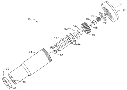
The drug delivery device 20 has a housing 24 and a cap 28 at a proximal end of
the
housing 24 and a nozzle plate 30 at the distal end of the housing 24. One or
more nozzles 34
or a plurality of nozzles 34, which are jet injection nozzles (also referred
to as "micro-
nozzles"), are arranged in the nozzle plate 30. As shown in FIGS. 1 ¨ 4,
injection nozzles 34
terminate as small outward protrusions from the outer surface of nozzle plate
30 thereby
providing the user with tactile feedback for the proper positioning and
alignment of the
injection nozzles 34 on the surface of the user's body tissue. As best
illustrated in FIGS. 2, 3
and 4, housing 24 further includes one or more reservoirs 38 aligned with and
in fluid
communication with the one or more nozzles 34. Each reservoir 38 is
longitudinally arranged
in the housing 24 and serves as a drug reservoir or storage space for drug 40.
Each reservoir is shaped to receive a pushrod 48 and a reservoir seal 54
attached or fixed
to the distal end of each pushrod 48. Pushrod 48 and reservoir seal 54 are in
direct
longitudinal alignment with each reservoir 38 and pushrod 48 and reservoir
seal are movably
located (longitudinally movable) within each drug reservoir 38. Each reservoir
seal 54 is
designed to prevent drug 40 from leaching or leaking from the drug reservoir
38. Thus,
reservoir seal 54 is in movable sealable contact with the inner wall of the
drug reservoir 38.
The pushrod 48 and reservoir seal 54 are slidably movable longitudinally in
each
reservoir 38. Piston 44 is integral to or fixed to the proximal end of each
pushrod 48 and
serves as a driving platform for accumulating and exerting a driving force to
the pushrods 48.
CA 02556007 2006-08-10
Piston 44 can be fixed as a single unit to the proximal end of all pushrods 48
in order to
operate and move each pushrod 48 simultaneously within each reservoir 38 or
piston 44 can
be fixed to the proximal end of each pushrod 48 individually in order to
selectively and
individually operate and move each pushrod 48 within reservoir 38.
In this example, piston 44 has a cylindrical shape shaped to fit securely
within and in
moveable engagement with inner wall of housing 24 that is also of a
cylindrical shape. Piston
44 has a circumferential space shaped to receive an 0-ring seal 52 that is
also shaped to fit
securely within and in moveable engagement with inner wall of housing 24 along
with piston
44. Seal 52 can be any type of seal so long as it prevents gas, discharge
contents, or other
matter from leaking or penetrating past piston 44.
As best shown in FIG. 3 (drug delivery device 20 loaded with drug 40 and in
its pre-fired
configuration), an energy source for discharging a driving force to the piston
44 is located
proximal or superior to the piston 44 within housing 24, for instance, in one
embodiment
according to the present invention, a charge housing 60 located in the
proximal or superior
portion of the housing 24. Pyrotechnic charge 64 is contained within charge
housing 60. A
primer 68 is located adjacent pyrotechnic charge 64 for holding a small
explosive charge that
delivers pyrotechnic energy or ignition energy to the pyrotechnic charge 64
for igniting the
pyrotechnic charge 64 upon activation of primer 68.
A striker pin 70 is located in cap 28 and moveably engages or moveably
contacts primer
68 for activating primer 68 and initiating the explosive charge contained in
primer 68. Striker
pin 70 is moveably connected to an activation element such as an activation
button 74 that is
movably biased by spring 72. Thus, activation button is movably biased to
striker pin 70
within cap 28 for driving striker pin 70 into the primer 70 upon a sufficient
downward force
pressed upon activation button 74, for instance, by the thumb of the user or
patient.
As best shown in FIG. 4 (drug delivery device 20 in its fired configuration
after having
injected drug 40 under microjet propulsion), upon depressing the activation
button 74, striker
pin 70 strikes primer 68 thereby activating primer 68, which, in turn, cause
the extremely
rapid combustion of a pyrotechnic charge 64. This controlled explosion
provides the driving
force necessary to slidably advance the piston 44 and the affixed pushrods 48
through the
16
CA 02556007 2006-08-10
reservoirs 48 causing the pushrods 48 to expel by microjet propulsion the drug
40 out through
the injection nozzles 34.
The energy source, such as pyrotechnic charge 64 or compressed gas 36 (FIGS. 8
and 10)
delivers sufficient energy and driving pressure to main drive piston 44 and
associated
pushrods 48 that ranges from about 800 to about 2,000 psi. In turn, the energy
and pressure at
the tips of microzzles 34 ranges from about 4,000 to about 25,000 psi at each
microzzle tip,
and preferably at a range from about 8,000 to about 12,000 psi at each
microzzle tip, and
more preferably at about 10,000 psi at each microzzle tip.
For all embodiments of the present invention, the same reference numerals are
used to
designate the same or similar features and parts. Accordingly, FIGS. 5, 6, 7A
and 7B,
illustrate another embodiment of the present invention that is particularly
useful for
ophthalmic and ocular applications such as delivering drug 40 a patient eye
100. Thus, nozzle
plate 30a at distal end of housing 24 has a contoured distal end 31 that is a
concave ring
having an opening in a center portion thereof In this example, contoured
distal end 31 has a
plurality of injection nozzles 34 circumferentially arranged within the
contour (concave
region) defined by the contoured distal end 31 and spaced proximally a
distance away from
the outer surface edge of the outer circumference (periphery or outer edge) of
contoured
distal end 31. Accordingly, in this example, nozzle plate 30a having contoured
distal end 31
is shaped to receive a patient's eye 100 wherein the pupil of the eye 100 can
be situated
within the center portion (open space) of the circumferential ring of the
contoured distal end
31. Thus, if desired, drug 40 can be delivered under microjet propulsion to
areas of the eye
100 outside the pupil, such as the vitreous or sclera, as best shown in FIG.
5.
FIG. 7B depicts an alternative embodiment of drug delivery device 20a wherein
a light
emitting diode (LED) cavity 76 is provided at the center portion (open space)
of the
circumferential ring of the contoured distal end 31 of nozzle plate 30a. An
LED 80 is
positioned in the LED cavity 76 for dispersing a focusing light (focusing LED
light) 88 under
operational control from switch 86 movably positioned at an exterior portion
of the housing
24 (in this example near the proximal end of housing 24). Switch 86 serves as
a power switch
for activating LED 80 to project focusing light 88, i.e. switch 86 serves as
an "On", "Off"
switch for the LED 80 and light 88. For sake of brevity, the contacts, leads
and wires
17
CA 02556007 2006-08-10
operatively connecting the LED 80 to the switch 86 are not shown, but are well
understood
and can be well appreciated by one having a level of ordinary skill in this
field.
Focusing light 88 is used to attract the direct attention of the patient,
align and focus the
pupil of eye 100 and serves as a focal point of patient's attention in order
to get the patient to
mentally relax (basically distract the patient) while drug 40 is delivered to
the eye 100 under
microjet propulsion. Thus, LED 80 and focusing light 88 serves as a means for
lowering the
patient's stress levels and anxiety normally associated with receiving a drug
injection,
particularly, in such a sensitive area as the eye 100.
Alternatively, in lieu of an LED 80, an element or feature that is luminescent
(including
self-luminescent) or an element or feature having a luminescent coating, such
as a dot having
self-luminescent coating that is used as a focal point and can be used to
attract the direct
attention of the patient and focus of the pupil of eye 100 for serving as a
focal point of
patient's attention in order to get the patient to mentally relax in
anticipation of and while
receiving the injected drug 40 under microjet propulsion. A tritium-coated dot
is one of these
suitable substitutes as an example.
FIGS. 8, 9 and 10 illustrate another embodiment of the present invention
wherein the drug
delivery device 20b uses an elongated, cylindrical tube as a delivery tube 25
having a
pressure chamber 27 therein. A handle 23 is connected to the delivery tube 25
at a proximal
portion of the delivery tube 25. A valve 33 is connected to the proximal end
of the delivery
tube 25 and pressure chamber 27 and a source of compressed gas 36, such as
compressed
CO2 gas contained in a cartridge 36 and is connected at another end of the
valve 33 and
contained within the handle 23. Cartridge 36 is a miniature compressed gas
cylinder
containing a compressed gas such as CO2 with ability to achieve and delivery
pressures as
high as 2,000 psi. Valve 33 regulates the release of compressed gas from the
cartridge 23 into
the pressure chamber 27 of delivery tube 25 by activation button 74a located
at a convenient
location on the handle 23, for instance, easily accessible with the pad of the
fore finger of
patient or user's hand.
If desired, a detachably connected cover (not shown) can be used with handle
23 in order
to provide direct access to the gas cartridge 36 for exchanging the cartridge
36 after
18
CA 02556007 2006-08-10
expenditure of its contents (when empty) with a freshly charged (full) gas
cartridge 36
thereby making the drug delivery device 20b a multiple use device or reusable
device.
As shown in FIG. 9, nozzle plate 30 and nozzles 34 are located at the distal
end of the
delivery tube 25 and pressure chamber 27 and are arranged as outwardly
extending
protrusions from the outer surface of nozzle plate 30 for providing the user
90 with tactile
feedback for the proper positioning and alignment of the injection nozzles 34
on a surface of
the user's body tissue, for instance, on the tissue located within a nostril
of the nose 110 (as
shown in FIG. 10) or tissue located within the patient's mouth (bucal
application), such as the
gums or roof of the mouth, or a location within a patient's ear, etc. Thus,
drug delivery device
20b is appropriate for delivering drug 40 to difficult areas to access of a
patient's body due to
the elongated and low profile design.
Drug reservoirs 38, drug 40, reservoir seals 54, pushrods 48, piston 44 and 0-
ring 52 are
arranged and function in the same manner or similar fashion as described for
the
embodiments of FIGS 1 ¨ 7B, except that these features are located within the
delivery tube
and pressure chamber 27 at the distal end of the delivery tube 25 and pressure
chamber 27.
Pressure chamber 27 allows compressed gas to be released from the cartridge 36
and
20 channels the gas from the handle 23 to the piston 44 along the entire
length of the delivery
tube 25 which provides the driving force necessary to slidably advance the
piston 44 and the
affixed pushrods 48 through the reservoirs 48 causing the pushrods 48 to expel
by microjet
propulsion the drug 40 out through the injection nozzles 34.
25 Drug delivery device 20 (FIGS. 1 ¨ 4), 20a (FIGS. 5, 6, 7A and 7B) and
20b (FIGS. 8 ¨
10) are intended to be compact in design, for example, having outer surface
dimensions
measuring about 2.00" in length and 0.600" in diameter (for the embodiments of
FIGS. 1 ¨ 4
and FIGS. 5, 6, 7A and 7B respectively), and very light in weight, for example
only weighing
several ounces. Ergonomically, it may be desirable to increase the size or
significantly
change the geometry, but the underlying functionality remains exactly the same
as that
presented in these figures.
19
CA 02556007 2006-08-10
=
Alternatively, the energy source for discharging a driving force to the piston
44 is
compressed gas, such as CO2 as one example, releasably housed in a gas
cartridge 36 (FIG.
8). Moreover, the energy source for discharging a driving force to the piston
44 can be any
type of energy force so long as it is capable of delivering drug under
microjet propulsion
according to the requirements set forth below and later in this disclosure.
For example, the
energy source must discharge ample energy sufficient enough in order to drive
main drive
piston 44 and associated pushrods 48 at a driving pressure that ranges from
about 800 to
about 2,000 psi. In turn, the energy and force at the tips of microzzles 34
ranges from about
4,000 to about 25,000 psi at each microzzle tip, and preferably at a range
from about 8,000 to
about 12,000 psi at each microzzle tip, and more preferably at about 10,000
psi at each
microzzle tip.
The volume of drug 40 delivered under microjet propulsion by drug delivery
device 20
(FIGS. 1 ¨4), 20a (FIGS. 5, 6, 7A and 7B) and 20b (FIGS. 8 ¨ 10), in
accordance with
present invention, is customizable, adjustable and variable in order to
accommodate delivery
of any type of drug, any tissue type, and any type of medical application.
Total delivered
drug volumes may be adjusted according a volume range that is from about 10
micro liters
(21) or less to about 1 milliliter (m1) or greater depending upon the
configuration or design of
the drug delivery device 20, 20a and 20b.
Further, the diameter of the injection nozzle(s) 34 are variable and range
from about 10
(p,m) to about 50 ( m) or greater, yielding exceptionally fine injection
streams of drug 40 and
minimizing the number of nerve receptors impacted by an injection thereby
reducing trauma,
pain and discomfort for the patient. One aspect of the novelty and uniqueness
of drug
delivery device 20 (FIGS. 1 ¨4), 20a (FIGS. 5, 6, 7A and 7B) and 20b (FIGS. 8
¨ 10) in
accordance with the present invention is its use of one or more discrete drug
reservoirs 38
which serve as injection chambers wherein each reservoir contains drug 40 as a
portion of the
overall injection volume of total dosage for drug 40 as best shown in FIG. 3
(drug delivery
device 20 shown in its pre-fired configuration prior to delivering drug 40).
And, each
reservoir 38 has its own dedicated injection nozzle 34 of extremely small
diameter. For
instance, the diameter of each nozzle 34 ranges from about 10 gm to about 50
microns pm or
from about 0.0004" to about 0.002". Thus, drug delivery device 20 (FIGS. 1 ¨
4), 20a (FIGS.
5, 6, 7A and 7B) and 20b (FIGS. 8 ¨ 10) in accordance with the present
invention divides the
CA 02556007 2006-08-10
total delivery volume for drug 40 into and across multiple, discrete
reservoirs 38 (for those
embodiments according to the present invention having more than one injection
reservoir 38),
and delivers each drug volume contained therein into the patient's tissue at
higher velocities
as best shown in FIG. 4 (drug delivery device 20 shown in fired configuration
after delivering
drug 40 under microjet propulsion) than those injection velocities achieved
with the
conventional jet injectors such as those jet injectors outlined previously.
Accordingly, one advantage associated with drug delivery device 20 (FIGS. 1 ¨
4), 20a
(FIGS. 5, 6, 7A and 7B) and 20b (FIGS. 8¨ 10) in accordance with the present
invention is a
dramatic decrease in the time required to inject drug 40 wherein this time can
be as short as
40 milliseconds (msec.). Even for a requirement for the delivery of 0.5 cc (or
0.5 ml)
injection of drug 40, the injection time achieved by drug delivery device 20
(FIGS. 1 ¨4),
20a (FIGS. 5, 6, 7A and 7B) and 20b (FIGS. 8 ¨ 10) ranges from about 10 msec.
to about 200
msec. (and, in one example, ranges from about 40 msec. to about 100 msec. for
about 0.5 ml
of certain types of drugs). A further aspect of the present invention is that
since the area of
the jet stream decreases with the square of the diameter, there is nearly a
100-fold reduction
in the area of the skin or tissue affected by injection with drug delivery
device 20 (FIGS. 1 ¨
4), 20a (FIGS. 5, 6, 7A and 7B) and 20b (FIGS. 8 ¨ 10) as compared to the
known thinnest
conventional hypodermic needle (ultra-fine insulin needle having a 31-gauge
cannula with a
diameter of 0.010").
In one embodiment according to the present invention, drug delivery device 20
(FIGS.
1 ¨4), 20a (FIGS. 5, 6, 7A and 7B) and 20b (FIGS. 8 ¨ 10) is a single-use pre-
filled drug
delivery device (designed for one time use as a disposable unit, i.e. one
time, single patient
use only) that requires no advance preparation or adjustment by the healthcare
provider or the
patient. Thus, drug delivery device 20 (FIGS. 1 ¨ 4), 20a (FIGS. 5, 6, 7A and
7B) and 20b
(FIGS. 8¨ 10) is ready-to-use as manufactured and provided.
Alternatively, drug delivery device 20 (FIGS. 1 ¨ 4), 20a (FIGS. 5, 6, 7A and
7B) and
20b (FIGS. 8 ¨ 10) is also intended to be a re-usable unit (for example, the
main housing 24,
cap 28 with activation button 74 and delivery tube 25 and handle 23 with
activation button
74a would be re-used and re-sterilized if required) with a single-use,
disposable inner
assembly that is either pre-filled or reloaded by the patient or healthcare
provider prior to
21
CA 02556007 2006-08-10
administration, inserted into the housing 24 or handle 23 and delivery tube 25
(for the drug
delivery device 20b) and then removed and discarded after use. In this case,
the disposable
inner assembly comprises primer 68, pyrotechnic charge 64 (or compressed gas
cylinder 36),
drug reservoir pushrods 48, drug reservoirs 38, injection nozzles 34. The re-
usable housing
24 and delivery tube 25 and handle 23 and other components such as the cap 28
and
activation buttons 74 and 74a are made of an appropriate material such as
metal or metal
alloy capable of withstanding re-use and re-sterilization if needed.
Additionally, in all embodiments of the present invention, the injection
nozzles 34 can
be in the form of array of injection nozzles 34 (in any desired pattern on the
nozzle plate 30
and 30a) that are configured out-of-plane or at different angles of
trajectory, for example, in
order to provide targeted convergence of the drug 40 to either a particular
target point in
tissue, i.e. a single target point in the tissue for receipt of the entire
injected volume of drug
40 or a plurality of desired target points in tissue.
Optimization of Microjet Propulsion Drug Delivery and Method of Manufacture
There are two mechanisms that are used to characterize and measure the
performance
of the drug delivery device 20 (FIGS. 1 ¨ 4), 20a (FIGS. 5, 6, 7A and 7B) and
20b (FIGS. 8 ¨
10) according to the present invention. The first mechanism is a predictive
model based on
the so-called Hagen-Pouiselle equation. This equation was used to estimate the
affects of
differing designs in the major elements and components of the drug delivery
device 20
(FIGS. 1 ¨4), 20a (FIGS. 5, 6, 7A and 7B) and 20b (FIGS. 8 ¨ 10) and their
methods of use
and the resulting driving forces that are required to operate the drug
delivery device in
accordance with the performance criteria of the present invention.
Additionally, the actual
forces required to deliver requisite amounts of drug 40 under microjet
propulsion were
determined empirically through both in vitro and in vivo testing. For example,
FIG. 11 is a
graph representing the findings of one of these relevant in vitro studies used
to determine
depth of penetration versus pressure for the microjet drug delivery device
(20, 20a and 20b)
having nozzle diameter of 50 m and volume of drug delivered of 100 I in
accordance with
the present invention.
22
CA 02556007 2006-08-10
In development and manufacturing of the drug delivery device 20, 20a and 20b
in
accordance with the present invention, there is a force/volume/length trade-
off based on the
diameter of the individual drug reservoirs 38, as well as the diameters of the
injection nozzles
34 and the desired injection velocity or mass flow rate of the expelled drug
40 or drug
formulation 40. Further, the design of these components has implications for
the duration of
injection, the number of drug reservoirs 38 and injection nozzles 34 that are
used, the size of
the main piston 44 and even the physical properties needed by the materials of
construction
for many of the key elements of the drug delivery device 20, 20a and 20b.
This relationship is modeled by the Hagen-Pouiselle equation as follows:
F = 8Q L(R2/r4) where: F = Injection force
Q = Flow rate of drug formulation or
injectate
= viscosity of drug formulation or
injectate
L = Length of injection nozzle
R = Radius of drug reservoir
r = radius of injection nozzle
To demonstrate the usefulness of this equation, let's assume that it is
desirable to
deliver 500 micro liters (1/2 cc) of an aqueous drug formulation 40 (a drug
solution 40 with
viscosity = 1 cps) to the subcutaneous layer of tissue at a flow rate Q of 5
cc/second.
Further, let's assume that we are using an injection microjet or nozzle
diameter of 50 microns
(0.002"), or r = 25 microns (0.001"). While we want to minimize the drug
reservoir length,
we also want to minimize the injection force. Thus, while shorter length is
better, smaller
diameter also means less force but a longer length. Thus, a convenient size is
selected with
respect to a reservoir length suitable to a hand-held microjet drug delivery
device (20, 20a
and 20b) while also attempting to minimize injection force. Consequently,
0.072" diameter
drug reservoirs, or R = 0.036" (0.914 mm) were selected. The length L of the
injection
nozzle 34 is determined by manufacturing constraints (a very small hole can
only be made in
23
CA 02556007 2006-08-10
a given material for a limited length). Accordingly, it is assumed that a
suitable length L is
0.050" (1.27 mm). Thus, the Hagen-Pouiselle equation can estimate the
injection force
required for any given injection nozzle as follows:
With: Q = 5cc/s
= 1 cps
L = 0.050" = 0.127 cm
R = 0.036" = 0.091 cm
r = 25 pm =0.0025 cm
F = 8Q L (R2/r4) = 10,218,121 dynes or about 23 lbf.
The number of drug reservoirs 38 is determined by the total force the main
drive
piston 44 can exert divided by the force required to propel each of the drug
reservoir
pushrods 48 which act as individual pistons simultaneously in this example
(expressed as a
whole integer). The practical pressure achieved by either the pyrotechnic
charge 64 or a
compressed gas cylinder 36 is limited to about 2,000 psi. Consequently, given
a main piston
44 diameter of 0.500" and the resulting area of (0.250)2 times pi = 0.196
square inches, the
maximum driving force available is 2,000 psi x 0.196 square inches or 392
pounds of force.
With 23 pounds of force required to drive each drug reservoir pushrod 48 and
392 pounds of
force available, the maximum number of drug reservoirs 38 that can be
accommodated (as a
whole integer) is 392 divided by 23 or a total of seventeen (17) reservoirs
38.
The length of each drug reservoir 38 is calculated as a result of the volume
requirement for each. For purpose of example, assume that five (5) reservoirs
are used.
Thus, given that a total of 500 micro liters is required to be delivered
through the five (5)
reservoirs 38, each reservoir 38 will deliver 100 micro liters of drug 40.
Given a reservoir
diameter of 0.072" (1.83 mm), each reservoir length will be 100 micro liters
divided by the
reservoir area (pi x (.914mm)2) or 38.1 mm long (1.50").
And, the injection flow rate Q has already been defined as 5 4. cc/s (as
outlined
above). Consequently, the total injection time is determined by the time
required to inject the
volume of drug 40 contained within each individual reservoir 38, which we have
found to be
24
CA 02556007 2006-08-10
100 micro liters or 1/10th of a cc. Thus, the injection time is 0.10cc times
the reciprocal of the
flow rate Q or 20 milliseconds.
As a predictive model, the Hagen-Pouiselle equation is a useful tool for
preliminary
analysis and prediction of necessary design parameters for the elements of the
drug delivery
device 20, 20a and 20b, but as would be expected the empirical findings did
differ from the
predictive analysis. Both in vitro testing which included using a 2mm thick
ballistics gelatin
over a saturated Pluronic (F127) solution and in vivo testing which including
testing the drug
delivery device in accordance with the present invention on the hairless
guinea pig model had
demonstrated that the drug formulation 40 is required to be pressurized to
approximately
8,000 psi in order to achieve microjet propulsion, i.e. the velocities
necessary for the drug
formulation 40 to be delivered through the injection nozzles 34 to a depth of
penetration in
tissue, such as the skin, needed for therapeutic administration, i.e. in this
case, subcutaneous
administration.
Given, for example, that the drug reservoirs 38 have a diameter of 0.072", the
cross-
sectional area of each drug reservoir 38 is (.036)2 times pi or 0.004 square
inches. With force
F equal to pressure P times area A, the force needed to drive the pushrods 48
to achieve an
8,000 psi pressure in the drug formulation 40 is 8,000 times 0.004 or thirty-
two (32) pounds
of force. This was a modest increase over the 23 pounds of force predicted by
Hagen-
Pouiselle, but certainly along the same order of magnitude. Much of the
increase is explained
by the friction of the sliding reservoir seals 54 and 0-ring 52.
Continuing with the values used in the example for the Hagen-Pouiselle
equation,
assuming that 500 micro liters of drug formulation 40 is required for the
total administration
and five (5) drug reservoirs 38 are being used for the design, then each
reservoir 38 contains
500/5 or 100 micro liters of drug formulation 40. With thirty-two (32) pounds
of force
needed for each drug reservoir 38 and the five drug reservoirs total, it was
calculated that 32
x 5 or 160 pounds of total force is needed to drive all of the drug reservoir
pushrods 48.
Thus, the main drive piston 44 must exert a force of 160 pounds.
Given a diameter of 0.500" for the main drive piston 44 (note that this
dimension may
be higher or lower depending on the application and the practical ergonomic
limitations of
CA 02556007 2006-08-10
physical size), the area of the piston 44 is (0.250)2 times pi or 0.196".
Thus, the energy
source must apply a pressure of F/A (160/0.196) or 816 psi to the main drive
piston 44. This
pressure requirement is well within the performance specifications of either a
pyrotechnic
charge 64 or a miniature compressed gas source 36. The lengths of the drug
reservoirs 38
and duration of injection will remain the same as those given in the Hagen-
Pouiselle
example.
The main drive piston assembly 44 acts as an accumulator for the pressure
generated
by the pyrotechnic charge 64 as shown in FIGS. 2, 3, 7A and 7B (or,
alternatively, a
compressed gas source 36 as shown in FIGS. 8 and 10), distributing the
pressure and
translating it as a driving force to the individual pushrods 48. The pushrods
48 are integral to
the main drive piston 44, so the total load applied to the piston 44 is
transferred
proportionally to each of the pushrods 48. In the event that a larger size
main piston diameter
is required, this will translate to a larger exerted force for any given
engine pressure. For
example, if the main piston diameter is increased in our previous examples
from 0.500" to
0.600", then the resulting force from a maximum engine pressure of 2,000 psi
will increase
from 2,000 psi x 0.196 sq. in. = 392 pounds of force to 2,000 psi x 0.283 sq.
in. = 565 pounds
of force. This increase in effective driving force permits the use of
additional injection
nozzles 34, which, in turn, reduces the volume in each nozzle 34, which, in
turn, reduces the
duration of the injection time, etc.
Finally, the nozzle geometry is determined by the desired diameter of the drug
stream, the
tensile / yield strength of the materials of construction, and the practical
limitations of
manufacturing a very small orifice at a cost effective economy of scale. While
one goal of
achieving a lightweight, compact hand-held drug delivery device 20, 20a and
20b with
respect to nozzle geometry is "smaller is better", there are practical limits
to constructing
such nozzles 34.
In the known and conventional needle-free drug injectors, these known devices
have a
relatively large orifice (approximately 0.006" ¨ 0.008") because these are the
practical limits
of high volume injection molding in suitable thermoplastics (i.e., core pins
smaller than this
diameter are not practical at the high pressures and high shear required by
injection molding
in high volume production).
26
CA 02556007 2006-08-10
As noted for the drug delivery device 20, 20a and 20b in accordance with the
present
invention, the drug delivery device 20, 20a and 20b uses nozzles 34 in the 10
to 50 micron
size and at significantly higher operating pressure than found with the known,
conventional
needle free jet injectors, such as those described previously above.
Consequently, drug delivery device 20, 20a and 20b in accordance with the
present
invention takes advantage of materials having high tensile and burst strength
properties for
the components of the drug delivery device 20, 20a and 20b. Such materials
include
ceramics, various metals and metal alloys, high strength engineering
thermoplastics (such as
PEEKTM, TorlonTm, UlternTM, etc.), and others. Thus, the present invention is
also directed to
using the most cost effective combination of such materials and to minimize
part count, i.e.
minimize the number of components and parts required.
Since the material used will need to withstand a given injection pressure in
excess of
8,000 psi immediately at the nozzle tip, it is desirable to use discrete
nozzles 34 fabricated in
metal, metal alloy or ceramic (for instance, alumina or zirconia) and assemble
to the housing
24 (FIGS. 1 ¨ 7B) or delivery tube 25 (FIG. 8) by bonding or ultrasonic
welding, for
example. All of these materials can be formed by injection molding, although
the final
nozzle orifice would be secondarily formed using laser drilling, ultrasonic
drilling, wire EDM
machining, or the like. While not currently believed to be practical,
developments in micro-
injection molding may make molding of integral, fully finished injection
nozzles entirely
feasible and more cost effective than current approaches involving secondary
finishing
operations. Nonetheless, injection molding in high strength materials coupled
with laser
drilling to produce precise, repeatable injection nozzles 34 should satisfy
engineering and
cost requirements associated with the present invention.
In another example in accordance with the present invention, FIGS. 1 ¨ 4
depict various
views of the drug delivery device 20 that can be used to accelerate a
multiplicity of small
drug volumes 40 to a suitable velocity for delivery into tissue, for example,
across the skin as
part of a transdermal drug delivery procedure. Using this example to
illustrate the function of
the drug delivery device 20 under the assumption that the design of the drug
delivery device
20 will require a total of thirty (30) injection nozzles 34 with each nozzle
34 having a
27
CA 02556007 2006-08-10
diameter of 40 microns and a calculated drug volume of 3.3p1 per drug
reservoir 38, or a total
drug volume of 30 x 3.3 = 100 1. Further, given a required velocity of 200 m/s
for delivery
of the drug 40, the force needed for each injection nozzle 34 can be
calculated from the
Hagen-Poiseuille equation yielding a value of approximately 10 lbs. per
injection nozzle 34.
Given thirty (30) injection nozzles 34, the total required loading force is 30
x 10 = 300 lbf.
Assuming the main piston 44 surface area is 1 square inch, then 300 psi of
pressure is needed
to achieve the requisite performance parameters. Again, this performance
criteria is
achievable using the miniature compressed gas cylinder 36 (FIGS. 8 and 10) or
the
pyrotechnic charge 64 (FIGS. 2, 3, 7A and 7B). The advantage of the
pyrotechnic charge is
that the pressure profile can be controlled throughout the entire dispensing
cycle, providing
varying pressures at different times to optimize the drug dispensing.
Moreover, as one can
readily appreciate, a number of suitable energy sources may exist that can be
used for the
purpose of accelerating the drug 40 to the required velocities in order to
achieve microjet
propulsion criteria according to the present invention and the examples
provided herein are in
no way meant to limit the kind of energy source that may be used in the
present invention.
As best illustrated in the graph depicted in FIG. 11, an in vitro study was
conducted for
the microjet drug delivery device (20, 20a and 20b) in accordance with the
present invention
in order to determine an optimal range for the depth of penetration (in cm)
versus an optimal
range of pressure (in psi). The nozzle 34 diameter was approximately 50 micron
diameter
wherein the volume of drug 40 delivered was approximately 100 pl. As clearly
illustrated in
FIG. 11, the delivery pressures for the microjet drug delivery device (20, 20a
and 20b) can
readily be adjusted to target any selected tissues. Thus, the microjet drug
delivery device (20,
20a and 20b) is customizable in a manner that ensures that any particular drug
can be
delivered to a particular depth of penetration in a particular tissue type
based on a particular
delivery pressure according to the graph of FIG. 11. Accordingly, this
customizable approach
even allows for particular layers of tissue to be targeted for drug delivery.
For example, the
submucousal layer of tissue can be targeted exactly according to the algorithm
depicted in
FIG. 11.
Additionally, any number of drug reservoirs 38 and injection nozzles 38 can be
utilized
for the present invention (within practical limits). As demonstrated above,
this can be
28
CA 02556007 2006-08-10
anywhere from a single reservoir 38 and a single nozzle 34 to as many as fifty
(50) or more
reservoirs 38 and nozzles 34 respectively.
Standard semiconductor processes can readily fabricate the injection nozzles
34 similar to
the fabrication of nozzles used in inkjet printing. Thus, injection nozzles 34
may be mass-
produced silicon devices having an orifice diameter of between 3 and 10
microns as one
example. The injection nozzles 34 can be fabricated as dense arrays on a
silicon wafer and
subsequently cut to the desired geometry. Wafer patterns, and therefore the
array geometry,
can be fabricated in any desired design. Consequently, the micronozzle array
can be
fabricated in any desired pattern such as a circular, elliptical, or semi-
circular pattern, for
example, and with any practical density of injection nozzles 34 that is
required. Typically,
every effort would be made to reduce the size of injection nozzles 34 and to
maximize the
number of injection nozzles 34 that such a wafer can yield.
Micro-molding of thermoplastics is an emerging technology that may also be
useful for
manufacturing the drug delivery device 20, 20a and 20b in accordance with the
present
invention. The advantages would be significant. While silicon wafers are
planar structures,
injection molded plastics are not. Thus, the array of injection nozzles 34 can
be configured
out-of-plane, for example, which would provide tremendous benefit in creating
an array that
is intended to be positioned with a targeted convergence. A further
significant advantage is
cost. A micronozzle array molded in a thermoplastic would cost pennies, in
comparison to a
silicon device that could easily range into dollars.
Other methods that may be used to construct the micronozzles 34, include micro-
machining the orifices in place as part of the nozzle plate 30 or nozzle plate
30a having
contoured distal end 31 (annular cup), machining or forming the orifices in
glass, metal,
ceramic, plastic, or other suitable material and then assembling (e.g., press
fitting) into the
contoured distal end 31 (annular cup), etc. Like the other major components of
the drug
delivery device 20, 20a and 20b in accordance with the present invention, the
design or
fabrication of the micronozzles 34 is not intended to be limited to a specific
embodiment.
Thus, in general, the present invention is directed to a method for making or
manufacturing a drug delivery device 20, 20a, and 20b in accordance with the
present
29
CA 02556007 2006-08-10
invention. Accordingly, this method comprises several key steps such as
identifying a drug
desired to be delivered (can be based on any desired treatment or diseases
state or condition
that is being targeted for treatment). Additionally, a volume of the drug
desired to be
delivered is also identified. Moreover, key parameters for features of the
device 20, 20a and
20b are determined. This includes parameters such as the diameter for the one
or more drug
reservoirs 38 and diameter for the one or more injection nozzles 34 which are
established in
advance. Furthermore, a tissue model for the tissue type or disease to be
treated is identified.
For example, the tissue model is any appropriate in vitro or in vivo model
acceptable for this
purpose. Thus, the tissue model can be based on material, for example, tissue
model that is
synthetic, natural, mammal (to include any animal or human tissue), living
tissue, preserved
tissue, etc.
Additionally, other key steps include identifying a penetration depth in the
tissue
model for the delivery of the drug. This includes targeting any desired or
particular layer of
the tissue that is considered appropriate for microjet injection of the drug
40. And, the drug
40 is tested in the tissue model by injecting the drug 40 into the tissue
model using the drug
delivery device 20, 20a and 20b in accordance with the present invention under
variable
pressure until the desired penetration depth or desired tissue layer is
achieved.
In using the method according to the present invention, an optimal pressure
range is
identified for the drug delivery device 20, 20a and 20b that achieves the
desired penetration
depth or desired tissue layer. As outlined previously above, an optimal
pressure range has
been identified to be <2,000 psi at main piston 44 and an optimal pressure
range of < 8,000
psi has been identified for the area at a tip of the injection nozzle 34.
The method according to the present invention also includes using predictive
modeling
for predicting the optimal pressure range required by determining the required
injection force
(F). Determining the injection force (F) is accomplished according to the
formula: F =
8Qp.L(R2/r4); where Q = flow rate of drug; p. = viscosity of drug; L = length
of injection
nozzle; R = radius of drug reservoir; and r = radius of injection nozzle.
Methods of Use
CA 02556007 2006-08-10
For transdermal or dermal delivery, the drug delivery device 20 (FIGS. 1 ¨ 4)
is in its
pre-fired configuration and loaded with the total volume of drug 40 to be
delivered wherein
drug delivery device 20 is placed firmly against and perpendicular to any
desired site of
injection (typically the back of the arm, the stomach or the thigh) with the
skin pinched in a
conventional manner. Since the injection nozzles 34 terminate as small outward
protrusions
from the outer surface of nozzle plate 30, the user is provided with instant
tactile feedback for
the proper positioning and alignment of the injection nozzles 34 on the
surface of the user's
body tissue at the desired site of injection.
As best shown in FIG. 4, upon depressing the activation button 74, striker pin
70 strikes
primer 68 thereby activating primer 68, which, in turn, cause the extremely
rapid combustion
of pyrotechnic charge 64. This controlled explosion provides the driving force
necessary to
slidably advance the piston 44 and the affixed pushrods 48 through the
reservoirs 38 causing
the pushrods 48 to expel by microjet propulsion the drug 40 out through the
injection nozzles
34.
Although this example described immediately above is directed to subcutaneous
or
cutaneous delivery, there are other examples for the drug delivery device 20a
and 20b that are
used in applications such as intra-ocular (drug delivery device 20a), intra-
oral (drug delivery
device 20b), intra-nasal (drug delivery device 20b), intra-aural (drug
delivery device 20b),
and, more broadly, intra-mucosal delivery in general (drug delivery devices
20, 20a and 20b).
It should be also noted that "transdermal" delivery is intended to mean all
forms of delivery
such as: intradermal, subcutaneous, and intramuscular.
In another embodiment according to the present invention, the drug delivery
device 20a
(FIGS. 5, 6, 7A and 7B) is particularly well suited for ocular use and can
deliver any drug 40
needed for intra-ocular micro-injection (especially intra-scleral or intra-
vitreal injections).
Such drugs known for these particular applications include VEGF antagonists,
corticosteroids, and anti-angiogenic drugs in general. Indications treated by
the drug delivery
device 20a (FIGS. 5, 6, 7A and 7B) in accordance with the present invention
include, for
example, diabetic retinopathy, macular degeneration and other diseases
involving
neovascularization in the eye.
31
CA 02556007 2006-08-10
=
In this embodiment, contoured distal end or cup 31 is placed over or on the
surface of the
eye 100 with the open center portion of cup 31 overlaying the cornea. The
micronozzles or
injection nozzles 34 are spaced and configured about the concentric ring of
contoured distal
end 31 such that they are in contact with the sclera. In one embodiment in
accordance with
the present invention, the injection nozzles 31 are configured in a circular
or elliptical pattern.
However, it is contemplated by the present invention that the injection
nozzles 34 be arranged
or configured in any desired configuration or pattern.
Upon depression of activation button 74, the injection stream of drug 40, as
shown in
FIG. 5, penetrates deep into the eye 100 through the sclera, and into the
aqueous humor or the
vitreous or any other desired tissue layer of portion of eye 100. Preferably
the injected drug
40 under microjet propulsion is targeted toward the back of the eye 100 as
presently depicted.
As mentioned previously, currently, many of the drugs of interest are
administered by
injecting directly into the eye with a conventional needle and syringe. As can
be greatly
appreciated, this is a somewhat risky procedure and requires that the
injection be
administered by a trained ophthalmologist. There are significant risks to the
patient
associated with these conventional techniques and include retinal detachment,
scarring after
repeated injections, and even blindness. Further, the injection itself is
dismaying to the
patient and requires that the patient be very still during the several seconds
of the injection
itself.
In the present invention, injection of the drug 40 into the eye 100 is
extremely rapid. For
example, given a stream velocity of the injected drug 40 under microjet
propulsion of 100
m/s for a drug reservoir 38 having a volume of 20 micro liters, the entire
injection only
requires about 10 milliseconds using drug delivery device 20a in accordance
with the present
invention. Assuming that the patient were to intentionally move his or her
eyes 100 from one
side to the other during the injection, and assuming eye movement occurs at a
rate of about 1
cm/s, the eye could only travel about 1/10th of a millimeter in this period of
time, a distance
of no consequence when using drug delivery device 20a in accordance with the
present
invention. Consequently, this invention also represents a safer, more
comfortable means of
administering drugs to the eye 100 for both the physician and patient.
32
CA 02556007 2006-08-10
=
As contemplated by the present invention, drug delivery device 20a (FIGS. 5,
6, 7A and
7B) in accordance with the present invention offers a number of advantages
over the
conventional technology and techniques. For instance, the injection nozzles 34
can be
designed to "aim" the injection stream at specific areas in the eye 100 (e.g.,
the back of the
eye 100). Additionally, the depth of penetration of the drug 40 can be
controlled without
relying on the skill of the caregiver. Moreover, the risk of injury to the eye
100 is minimized
with drug delivery device 20a (FIGS. 5, 6, 7A and 7B) in accordance with the
present
invention by minimizing the energy and tearing (trauma) to which the eye 100
is subjected
due to the extremely rapid nature of the microjet propulsion of the drug into
the tissue of the
eye 100 (estimated to be as fast as about 10 milliseconds for injection of
small doses of drug
40).
Moreover, the drug delivery device 20a has the ability to modulate the jet
injection
energy and injection stream geometry as a means to control the depth of
delivery of the drug
into the eye. Also, the design of the micronozzle geometry allows for the
control the stream
diameter, trajectory, cohesion, and focus. Additionally, the flexibility in
the design of the
micronozzle array allows for optimization of the drug delivery profile for any
given drug,
disease, or site of disease within the eye. Furthermore, the drug delivery
device 20a provides
an extremely rapid means of administering drug 40 to the eye 100 such that eye
movement
does not present an element of risk.
Additionally, many drugs 40 currently under pre-clinical and/or clinical
investigation are
potent drugs and require only periodic administration of small doses to the
eye 100. Drug
delivery device 20a (FIGS. 5, 6, 7A and 7B) in accordance with the present
invention offers a
more controlled, repeatable, safe, and comfortable means of delivering these
drugs 40 to the
eye 100 over any known devices and techniques available to date.
Another embodiment in accordance with the present invention is an intra-nasal
application depicted in FIG. 10. Accordingly, the drug delivery device 20b
(FIGS. 8 ¨ 10) has
particularly useful application in administering CNS (central nervous system)
drugs 40 via
microjet injection to the olfactory bulb of the nose 110 of the patient 90.
33
CA 02556007 2006-08-10
=
In this embodiment, drug delivery device 20b (FIGS. 8 ¨ 10) is used to provide
direct
injection of drug 40 under microjet propulsion into the submucousal space of
the nose 110 to
the CSF of the olfactory lobe. For this purpose, doses of drug 40 of 20 mg or
greater can be
injected extremely rapidly (<50 milliseconds) into the submucousal space and
the depth of
injection can be precisely controlled such that the drug 40 is delivered
precisely to this area
without any harm of penetrating to an undesired location.
In another embodiment according to the present invention, drug delivery device
20b is
also used for the intra-oral delivery of drug 40 wherein the drug can be
microinjected into any
desired area in the mouth such as intra-mucosal for such applications as
treating tumors, i.e.
targeted delivery of drug 40 under microjet propulsion aimed at treating a
tumor, for
example.
In yet another embodiment according to the present invention, drug delivery
device 20b is
used for the intra-aural delivery of drug 40 such that drug 40 can be
microinjected into any
desired portion of the ear or auditory canal for treating various diseases and
conditions of the
ear or those conditions that affect hearing, for example.
Additionally, in other embodiments according to the present invention, drug
delivery
device 20b is also useful for areas of the body that are difficult to access
such as various
canals, passageways, cavities or difficult surfaces to reach. Extended
delivery tube 25
facilitates easy access to these injection sites for the injection of drug 40
under microjet
propulsion to these difficult areas.
Thus, as described above the drug delivery device 20, 20a and 20b in
accordance with the
present invention has many novel features and advantages. Some of these novel
features and
advantages are summarized here for convenience such as extremely small
injection nozzles
(0.002" or smaller); multiple injection reservoirs and injection nozzles
minimizing each
volume of injection and injection time resulting in less pain; customizable,
variable pressure
injections to include high pressure injection to reach deep tissues and lower
pressure to target
more shallow tissues; ability to concentrate drug dose into a confined area or
spread it out
over a larger surface area; high volume injections divided into a small
volume, discrete
injectors (can achieve injection volumes equivalent to or larger than
conventional jet injectors
34
CA 02556007 2006-08-10
at more rapid delivery times; multiple medical applications (i.e.,
transdermal, intra-ocular,
intranasal, intrabucal, etc.); efficient operation to include total energy
requirements
equivalent to those total energy requirements available with the prior art
devices, but with the
present invention being much faster in administering the drug and much less
painful injection
for the patient; ability to deliver multiple drugs (i.e., different drugs can
be housed in
different drug reservoirs which is something not possible with the known drug
delivery
devices currently available); and ability to separate excipients during
storage until time of
injection which improves long term stability of the drug 40.
There is no known or existing technology that provides the advantages afforded
by
the present invention, including safety, ease of use, precision in both dose
and depth of
penetration, patient comfort and acceptance. Other advantages associated with
the present
invention is that it can provide for the precise, targeted delivery of small
molecules and
large molecules alike to include macromolecules such as large proteins, cells
or other
biological molecules and drugs. And, another advantage is that the microjet
drug delivery
device in accordance with the present invention is extremely rapid in its
delivery of the
drug, i.e. about <10ms delivery resulting in nearly pain free injection.
The present invention contemplates that a significant reduction in the nozzle
orifice
size will result in reduced pain to the patient. Further, the present
invention enables
practical new uses of jet injection technology such as transmucosal delivery.
It is an advantage of the present invention that a plurality of nozzles may be
employed, arranged in an array and having space between each adjacent nozzle,
defining
a two-dimensional planar structure that can lie flat on the skin and, thus,
ensure
perpendicularity.
Moreover, the present invention provides for true needle-free delivery of
drugs
regardless of size of the drug molecules involved as well as provide for true
needle-free
delivery of drugs with minimal trauma to tissue and that are suitable for
delivering drugs
in sensitive areas of the body such as the eye, nasal passageways, mouth, etc.
CA 02556007 2013-04-26
And, the drug delivery device 20, 20a and 20b are simple and efficient in
design
and construction, low cost and easy to manufacture. Accordingly, the microjet
drug
delivery device in accordance with the present invention has an appropriate
design that
is extremely suitable for a single patient use only disposable device if
desired.
Inasmuch as the foregoing specification comprises preferred embodiments of the
invention, it is understood that variations and modifications may be made
herein, in
accordance with the inventive principles disclosed, without departing from the
scope of the
invention.
While preferred embodiments of the present invention have been shown and
described herein, it will be obvious to those skilled in the art that such
embodiments are
provided by way of example only. Numerous variations, changes and
substitutions will
now occur to those skilled in the art. The scope of the claims should be given
the broadest
interpretation consistent with the description as a whole.
DOCSTOR 2692986\1 36