Note: Descriptions are shown in the official language in which they were submitted.
CA 02556098 2006-08-11
WO 2005/079822
PCT/GB2005/000523
Wound Healing Composition
The present invention relates to compositions and methods for tissue
regeneration, particularly for treating skin lesions such as wounds. The
compositions and methods are useful especially for assisting the process of
wound healing, particularly chronic open lesions that are slow to heal or
resistant to healing.
Healing of open wounds extending through the germinal epithelium in
otherwise healthy tissue takes place by the process classically described as
"second intention", which, following initial haemostasis, involves a well-
ordered sequence of inflammation, cellular infiltration, angiogenesis,
granulation and re-epithelialisation. As part of the normal healing response,
resident fibroblasts are required to undergo a series of phenotypic changes,
migrating to the wound site, then proliferating, then synthesising and
secreting
extracellular matrix molecules. In vivo, a least a proportion of fibroblasts
then
switch to a myofibroblastic phenotype in order to facilitate wound
contraction.
In vitro, a series of phenotypically distinguishable mitotic and post-mitotic
fibroblast populations have been described (Bayreuther et al., 1988, Proc Nat!
Acad Sci USA 85: 5112-5116). The pathway of differentiation appears to be
controlled, at least in part, by interactions between fibroblasts and
extracellular
matrix (ECM) proteins present at the wound site. Growth factors and cytokines
undoubtedly also exert an important influence, although their effects too,
appear
to be modulated by fibroblast exposure to particular ECM proteins. Among the
ECM proteins that appear to have an important role in fibroblast
differentiation
are fibrinogen and fibrin. Fibroblasts specifically interact with fibrin and
fibrinogen "RGD" motifs through ce,03 integrin receptors although the cellular
response is complex and modulated by other factors. In vitro studies of the
effect of fibrin glue on human periodontal ligament fibroblasts have suggested
that fibrin appeared to slightly inhibit fibroblast proliferation. The
presence of a
fibrin matrix has also been reported to increase the synthesis of collagen by
CA 02556098 2006-08-11
WO 2005/079822
PCT/GB2005/000523
entrapped fibroblasts (Neidert et al, 2001, Proceedings of the ASME
Bioengineering Conference, Kamm et al. [Eds], Vol 50: 215-216).
Fibroblasts are also known to have a role in the remodelling of fibrin clots.
As
new extracellular matrix proteins such as collagen type I and III, fibronectin
and
vitronectin are laid down, the fibrin matrix is broken down, predominantly by
the activation of the plasma-derived enzyme plasmin. This is regulated by the
activation (or inhibition) of its proenzyme, plasminogen, by a variety of
plasminogen activators and inhibitors. In vivo, a number of infiltrating
cells,
such as neutrophils and macrophages, secrete urokinase-type plasminogen
activator (uPA), whilst endothelial cells are largely responsible for
producing
tissue plasminogen activator (tPA). Fibroblasts also secrete both uPA and
plasminogen activator inhibitors, such as plasminogen activator inhibitor-1
(PA-
1). The balance between these antagonistic mediators is crucial in
controlling.
fibrin remodelling and scar formation. The expression of the antagonistic
mediators is developmentally regulated, as well as being controlled by
extracellular matrix components and local growth factors.
To facilitate movement through a cross-linked fibrin clot and a tight meshwork
of extracellular matrix, a variety of fibroblast- and serum-derived enzymes
cleave a path for migration. These include interstitial collagenase (matrix
metalloproteinase-1, MMP-1), gelatinase (matrix metalloproteinase-2, MMP-2),
stromelysin (matrix metalloproteinase-3, MMP-3) and the plasminogen
activators. Chemotactic factors such as TGF-I3 and PDGF may upregulate the
production and secretion of these enzymes.
Once migrating fibroblasts reach a wound, they gradually become secretory and
protein synthesis is increased. The previously retracted endoplasmic reticulum
and Golgi apparatus becomes dispersed throughout the cytoplasm and a loose
matrix is produced, which is mainly composed of fibronectin and type III
collagen. Ultimately, this profibrotic phenotype takes over, which is
characterised by an abundance of rough endoplasmic reticulum and Golgi
2
CA 02556098 2006-08-11
WO 2005/079822
PCT/GB2005/000523
apparatus, secreting newly synthesised collagen in response to highly
expressed
TGF-(3. Notwithstanding, TGF-0 fails to upregulate further collagen
deposition,
once a matrix has been deposited. It is also thought that IL-4 released by
mast
cells induces a modest increase in types I and III collagen together with
fibronectin. Mast cells furthermore produce tryptase (a serine esterase) in
abundance, which has been shown to upregulate fibroblast proliferation.
Stimuli such as TGF-a, TGF-f3 and PDGF responsible for fibroblast
proliferation and matrix synthesis have been extensively investigated in vitro
(Derynck, 1988, Cell 54: 593-595; Ross & Raines, 1990, In: Growth Factors:
From genes to clinical applications, Sara et al. [Eds], pp. 193-199, Raven
Press,
New York; Sporn & Roberts, 1992, J Cell Biol 119: 1017-1021) and by in vivo
manipulation of wounds (Sprugel et al., 1987, Am J Pathol 129: 601-613;
Pierce et al., 1991, J Cell Biochem 45: 319-326). 7-interferon on the other
hand
was demonstrated to have a negative effect on the mitogenic and synthetic
potential of fibroblasts in vitro and in vivo (Duncan & Berman, 1985, J Exp
Med 162: 516-527; Granstein et al. , 1987, J Clin Invest 79: 1254-1258). In
addition, the collagen matrix itself can suppress these activities (Grinnell,
1994,
J Cell Biol 124: 401-404; Clark et al., 1995, J Cell Sci 108: 1251-1261),
whilst
fibrin or fibronectin matrix have little or no suppressive effect (Clark et
al.,
1995, supra). Many fibroblasts undergo apoptosis (programmed cell death) in
day-10 healing wounds, thereby marking the transition from a fibroblast-rich
granulation tissue to a scar tissue with reduced cell density.
Where a wound has destroyed the germinal layer of epithelium, collagen
deposition by infiltrating fibroblasts and re-epithelialisation results in a
degree
of scarring, with incomplete restoration of function in terms of the
flexibility
and elasticity of the original dermis and failure to regenerate auxiliary
structures
such as hair follicles and sweat glands.
A number of factors may adversely affect the rate and extent of such wound
healing, in particular, poor blood supply. Poorly perfused tissue, often
3
CA 02556098 2006-08-11
WO 2005/079822
PCT/GB2005/000523
associated with impaired venous return and varicose veins, peripheral vascular
disease or diabetes, often fails to heal satisfactorily, resulting in chronic
ulcers,
although the details of the pathogenesis are still unclear. Chronic leg ulcers
in
particular are a significant and growing problem world-wide.
Various approaches have been tried for the treatment of wounds. Autologous
skin-grafting has been used to close open wounds, minimise the risk of
opportunistic infection, accelerate healing and minimise scarring. Skin
grafting
has significant limitations, not least the requirement for a suitable donor
site
from which grafts can be taken which is a particular problem where wounds are
extensive (for example, with burns). In addition, grafts have a low success
rate
where wound healing is compromised.
With respect to chronic leg ulcers in particular, the introduction of
compression
therapy in combination with moist wound dressings has been the standard
therapeutic management.
More recently, tissue-engineering solutions have become available. Research
into regenerative medicine has shown that human cells have substantial
potential to heal and regenerate damaged tissue especially when primed by an
environment that closely mimics the natural physiological condition being
treated. Much of this research has focused on the production of so-called
"tissue
equivalents", which aim to provide a temporary functional replacement for
missing tissue and accelerate healing. Tissue equivalents may be dermal
equivalents or total skin equivalents, with the aim being to provide effective
coverage of the wound as quickly as possible. The development and production
of tissue equivalents usually involves the isolation of replacement skin
cells,
which are expanded and seeded onto or into a supporting structure such as a
three-dimensional bio-resorbable matrix, or within a gel-based scaffold.
A variety of materials have been used as acellular protein matrices for wound
healing applications. These include synthetic polyesters (polyglycolic acid
4
CA 02556098 2006-08-11
WO 2005/079822
PCT/GB2005/000523
(PGA), polylactic acid (PLA), polyglactide (Dermagraft [RTM], Smith &
Nephew, described below), polydioxanone, polyhydroxyalkonoates and
hyaluronic acid derivatives), hydrophilic polyurethanes (polyetherpolyester,
polyethylene oxide and carboxymethylcellulose ethylene), and collagen-based
scaffolds (cross-linked elastin collagen material (Matriderm [RTM]),
cross-linked collagens manufactured from acid-soluble type I bovine collagen
material (such as Vitaphore [RTM]). An alternative approach is to use an
acellular derivative of allogeneic human dermis, a natural dermal matrix from
which cells have been removed (such as Alloderm [RTM], LifeCell
Corporation). Some preparations use an organised, layered structure in order
to
more closely mimic the structure and function of the dermis. For instance, a
preparation comprising an underlying layer of bovine collagen and shark
glycosaminoglycans with an overlying layer of silicone is known (Integra
[RTM], Integra LifeSciences Corporation).
Other approaches to wound healing have involved the use of fibrin sealants,
for
example Tisseel [RTM] (Baxter), Beriplast [RTM] (Aventis), Quixil [RTM]
(Omrix Biopharmaceuticals), Haemaseel [RTM] (Haemacure) and Crosseal
[RTM] (Omrix). These commercially available fibrin sealants are derived from
cryoprecipitate of pooled plasma from virally-screened allogeneic donors.
Fibrin products rely on the natural polymerisation process that occurs during
the
physiological blood clotting cascade, in which a monomeric fibrin precursor,
fibrinogen, is acted on by activated thrombin with the resultant production of
polymeric fibrin. Fibrin forms the protein scaffold component of blood clots,
to
which platelets adhere.
Fibrin has been recognised as a convenient and clinically acceptable cell
carrier
to be used in tissue engineering applications. Commercially available products
that utilise fibrin sealants for cell delivery include Bioseed [RTM]
(Biotissue
Technologies). The use of fibrin sealants for cell delivery purposes for the
treatment burns has been suggested by several groups (see Brown et al., 1993,
5
CA 02556098 2006-08-11
WO 2005/079822
PCT/GB2005/000523
Am J Pathol 142: 273-283; Neidert et al., 2001, supra; Tuan et al., 1996, Exp
Cell Res 223: 127-134; and US Patent App!. No. 2003/01654482).
Exogenously applied dermal cells have been shown to have beneficial effects on
wound healing including shorter time to complete healing (Falanga &
Sabolinski, 1999, Wound Repair Regen 7: 210-207), delivery of active growth
factors to the wound (Naughton et al., 1997, Artif Organs 21: 1203-1210),
reduced potential for lesion recurrence (Gentzkow et al., 1996, Diabetes Care
19: 350-354), and reduced pain (Muhart et al., 1999, Arch Dermatol 135: 913-
918).
Known combinations of protein matrices and dermal cells for wound healing
applications include a preparation called Dermagraft [RTM] (Smith & Nephew)
comprising cryo-preserved primary human foreskin fibroblasts seeded onto a
bioabsorbable glycolic-lactic acid polyester (polyglactide) scaffold (Naughton
et al, 1997, supra; U54,963,489). The fibroblasts are allowed to proliferate
in
the scaffold, secreting extracellular matrix proteins and growth factors and
cytokines. The mature preparation is packaged in 10% dimethylsulphoxide and
bovine serum as a cryoprotectant to allow storage of the product by freezing
prior to use. Disadvantages of this approach include difficulty in
manipulating
the product during application to the wound (such as ulcers), and the
necessity
of storing and transporting the product at very low temperatures (-70 C) and
use
of careful thawing procedures in order to ensure viability of the cells (see
WO
87/06120).
Various combinations of collagen-based matrices and living cells are known.
Apligraf [RTM] (Organogenesis, Inc.) is a bilayered structure comprising a
lower ('dermal') layer of a bovine collagen scaffold supporting living human
fibroblasts and an upper ('epidermal') layer comprising human keratinocytes on
a collagen scaffold (Falanga & Sabolinski, 1999, supra; WO 99/63051). The
preparation is supplied as a circular disk approximately 75 mm in diameter and
0.75 mm thick on an inert polycarbonate membrane. Apligraf [RTM] is
6
CA 02556098 2006-08-11
WO 2005/079822
PCT/GB2005/000523
packaged individually for use and has a 5-day shelf life. It is maintained in
an
agarose-rich nutrient with a 10% CO2/air atmosphere and is shipped and stored
at room temperature (20 C to 31 C; 68 F to 88 F). The removal of the product
form the storage dish and polycarbonate membrane involves teasing away the
edge of the Apligraf [RTM] using sterile forceps. Problems associated with
this
method include excessive folding which can make accurate, close application of
the preparation to the wound difficult and time-consuming.
A similar product (Orcel [RTM]; Ortec International Inc) is described in
US6,039,760. Orcel [RTM] is a bilayered structure of bovine collagen with
fibroblasts and keratinocytes. The preparation is packaged between 2 non-
adherent pieces of mesh, which are differently coloured to distinguish between
sides. The device is then packaged in a plastic tray containing media to
maintain
cell viability during storage and shipping, which is further packaged into
pouches with chill packs to maintain a temperature of 11 C to 19 C for 72
hours.
Another example of a tissue equivalent that attempts to reproduce a dermis-
like
arrangement of fibroblasts in a protein matrix supporting an overlying layer
of
keratinocytes is described in Meana et al. (1998, Burns 24: 621-630). Rama et
al. (2001, Transplantation 72: 1478-1485) describe a method of culturing
autologous limbal stem cells on a fibrin gel substrate for grafting to the
contralateral cornea.
US Patent Appl. No. 20030165482 discloses a wound healing preparation
(Allox [RTM], Modex Therapeutiques SA) comprising growth-arrested
allogeneic human fibroblasts and keratinocytes applied to a wound in a viscous
paste of fibrinogen (Tisseel [RTM]) to which thrombin has been added, so that
fibrinogen cleavage and fibrin polymerisation occur in situ. Alternatively,
the
separate liquid components are sprayed onto the wound, to set in situ, on
mixing.
7
CA 02556098 2006-08-11
WO 2005/079822
PCT/GB2005/000523
The present invention provides an alternative wound healing preparation and
associated products and methods which address problems associated with prior
art products and methods.
According to a first aspect of the present invention there is provided a wound
healing composition comprising living cells within a support matrix, in which
the cells have a wound healing phenotype, and in which the composition is
single-layered and has been incubated for up to about 8 days to allow
development of the wound healing phenotype.
The invention provides an approach to treatment of chronic wounds based, not
on providing an immediately functional tissue-equivalent, but on delivering
cells, in a support matrix (which could also be referred to as a maturation
matrix
or a development matrix; for example a biocompatible matrix), with the
potential to promote and accelerate the healing process. Although developing a
viable skin equivalent (for example, a cultured dermal tissue equivalent
comprising fibroblasts, extracellular matrix and overlying keratinocytes
organised into functional and anatomically relevant structures) remains a
worthwhile goal, so far this has proven elusive. However, for many situations,
the present invention shows that such an approach may be unnecessarily
complex and that a simpler solution, that of providing a single layer of cells
at
the appropriate stage of development and exhibiting a particular phenotype in
a
wound-healing composition for rapid, convenient and accurate application to
wounds, is remarkably effective. The cells used in the present invention
develop
surprising rapidly to have a "wound-healing phenotype" to encourage
immediate wound healing. It is believed that the wound healing phenotype
represents the optimal phenotype for accelerating or assisting wound healing.
The invention allows delivery of such cells (in the composition) to a wound,
preferably in a manner which is consistent with the maintenance of the wound-
healing phenotype.
Whether or not cells in a composition have a wound healing phenotype may be
8
CA 02556098 2006-08-11
WO 2005/079822
PCT/GB2005/000523
tested by applying the composition to a wound (as defined herein) and
observing whether or not healing of the wound is accelerated or assisted.
Wounds to which the wound healing composition may be applied include
wounds such as an ulcer such as a venous ulcer or a diabetic ulcer, a pressure
sore, a burn or an iatrogenic grating wound. The composition is particularly
useful for treating recalcitrant wounds, i.e. wounds which have not healed
within three months using standard treatment.
The term "single-layered" indicates that the composition has only one layer
containing cells within a support matrix, i.e. it is not a multi-layered "skin
equivalent" with multiple layers of (different) cells. However, the invention
also
encompasses compositions having additional non-cellular layers as well as
compositions having stacked layers comprising substantially uniform single
layers.
The composition may be incubated for up to about 96 h, for example up to 72 h,
48 h, 25 h, or 24 h, preferably for 16 h to 24 h. Incubation is preferably in
vitro,
but may also be in situ (for example, with the composition applied to a
wound).
In one embodiment, it has been found by the present inventors that taking
cells
such as passaged human dermal fibroblasts, casting (or seeding) the cells in a
matrix such as a protein-based matrix and then incubating this mixture for up
to
96 h, for example, results in a wound healing phenotype that is particularly
beneficial for use in wound healing applications. It has been observed that
such
cells are predominantly in a proliferative phase in culture (encouraged by low
density seeding, avoiding contact inhibition).
The present inventors have found that under normal culture conditions, for
example, a liquid culture of human dermal fibroblasts incubated in a standard
culture medium at 37 C, development of a wound-healing phenotype may
typically take 2 to 3 days. However, incubation of such fibroblasts in a
suitable
9
CA 02556098 2006-08-11
WO 2005/079822
PCT/GB2005/000523
environment such as in a support matrix and/or a wound shortens the
development process, so that before 24 hours the cells may have entered or
reached the wound-healing phenotype. Thus, incubation of cells in a suitable
support matrix and/or wound results in a shorter development time to reach a
wound healing phenotype than standard (for example, liquid) culture
conditions.
The composition is preferably incubated at a temperature of about 37 C. If
incubation takes place at a lower temperature, the living cells will develop
at a
slower rate and incubation time may need to be extended.
Preferably, the composition excludes mitotically inactivated cells (for
example
cells mitotically inactivated by administration of mitomycin C or other
chemically-based mitotic inhibitors, irradiation with 7-rays, irradiation with
X-
rays, or irradiation with UV light, as described for example in
US2003/0165482).
The composition may be stored after incubation (i.e. have a shelf-life) for up
to
about 40 days, preferably up to 28 days or 21 days or 19 days, and more
preferably about 7 to 14 days or about 7 to 11 days at a temperature of 2 C to
8 C, for example 3 C to 5 C, preferably about 4 C, while retaining an ability
to
heal wounds. The composition therefore does not require freezing, as do
certain
prior art wound healing compositions. The present composition preferably does
not contain a substance added as a cryopreservant or cryoprotectant (such as
glycerol and/or human serum albumin).
Once the cells of the composition have been incubated to reach or approach a
wound healing phenotype phase, the composition can preferably conveniently
be stored at approximately 4 C for up to 40 days, and certainly 7 to 14 days,
before use without significant loss of viability or change of phenotype. This
has
significant practical advantages in that it provides not only an efficacious
product comprising cells with a wound healing phenotype (for example cells
that are optimally suited for secretion of extracellular matrix with minimal
CA 02556098 2006-08-11
WO 2005/079822
PCT/GB2005/000523
inappropriate fibrinolysis), but also gives a relatively long shelf-life under
commonly available standard refrigeration conditions. The ability to ship such
products at approximately 4 C also considerably simplifies transportation.
Maintaining a cold chain at 2 to 8 C is considerably simpler and cheaper than
shipping at -70 C, as is commonly required for live cells.
The cells are preferably mammalian, for example human.
Cells of the present invention may include fibroblasts, keratinocytes, stratum
germinativum cells, and combinations or admixtures of such cells. However, in
a preferred embodiment, the cells of the composition may substantially exclude
keratinocytes. The cells may be isolated from any suitable mammalian source,
and preferably are human. The cells are preferably allogeneic, although
autologous and/or xenogeneic cells may be used. The cells may be substantially
of one type only, for example 90% to 100%, preferably 95% to 99.5%, and
more preferably 97.5% to 99% of one type. In a preferred embodiment, the cells
are substantially fibroblasts, for example 90% to 100%, preferably 95% to
99.5%, and more preferably 97.5% to 99% fibroblasts. The fibroblasts may be
dermal fibroblasts, preferably human dermal fibroblasts. A preferred
embodiment comprises allogeneic human foreskin-derived fibroblasts.
As required for manufacture, cells may be thawed, recovered, expanded in
culture (for example, for about a week) or until they reach confluence, and
resuspended in appropriate volumes and densities as required. Although early
passage cells are preferred, later passage cells may also be used. Preferably
the
cells have undergone less than 20 passages, more preferably less than 15
passages, most preferably less than 10 passages, for example 7 passages. Once
defrosted for use in the present invention, the cells may be incubated further
as
described.
For the purposes of the present invention, day 0 is the day on which the cells
are
incubated and begin development and they will reach a wound healing
11
CA 02556098 2006-08-11
WO 2005/079822
PCT/GB2005/000523
phenotype within the time-frame described above (for example, up to 4 days, or
96 hours, after day 0).
The cells may be actively synthetic or able to become actively synthetic
rapidly
(for example, following storage).
The cells are preferably not proliferating and/or not senescent. Optimally the
cells must be in a synthetic phase of development (or maturity), rather than a
proliferative or senescent phase. Proliferation may be useful to increase cell
numbers, but delays the important synthesis of extracellular matrix proteins
such as collagen types I and III, fibronectin and vitronectin. Cells that have
become senescent do not contribute to wound healing and so serve little
purpose
as such a therapeutic.
The cells may be suspended within the matrix, preferably substantially
uniformly within the matrix.
The matrix may be protein-based, for example having a protein concentration in
the range of about 3 to 12 mg.m1-1. For example, the matrix is preferably a
clottable or gelling substance such as fibrin, collagen, fibronectin,
vitronectin,
alginate, agar, collagen, PVA, hyaluronic acid, modified starches,
carrageenans,
carob, gelatine, pectin or gelling agent.
The matrix is preferably non-pyrogenic and/or sterile.
In a preferred embodiment, the matrix is a fibrin matrix. The fibrin may have
a
concentration in the composition in the range of 3 to 12 mg.m1-1, for example
7
to 12 mg.m1-1 or 3 to 5 mg.m1-1. The fibrin matrix is preferably formed by
thrombin-mediated polymerisation of fibrinogen.
The matrix is preferably solid or semi-solid. The matrix of the composition is
"pre-cast" in the sense that it is provided as a solid or semi-solid form
(such as a
12
CA 02556098 2006-08-11
WO 2005/079822
PCT/GB2005/000523
gel). The matrix may be insoluble. Most preferably, the cells are cast in the
matrix prior to development of a wound healing phenotype.
The rate of fibrinolysis occurring within the composition may be a factor
taken
into account with a fibrin matrix-based composition. As described above,
fibrinolysis is a normal part of the wound healing process, by which the
fibrin
matrix is gradually replaced by other extracellular matrix proteins. If,
however,
fibrinolysis occurs too early or too rapidly, the wound healing gel is broken
down before useful collagen deposition has occurred. Fibroblast expression of
pro-fibrinolytic factors such as urokinase-type plasminogen activator is
developmentally regulated and so the phenotype of fibroblasts where included
in the composition is relevant if premature fibrinolysis is to be avoided.
The wound healing composition may further comprise a protease inhibitor
suitable for preventing breakdown of the matrix. The inhibitor may be a serine
protease inhibitor, most preferably one or more selected from the list
consisting
of aprotinin, e-aminocaproic acid and tranexamic acid. Preferably, especially
where the concentration of protein is in the range 7 to 12 mg.m1-1, the
protease
inhibitor is aprotinin. Alternatively, especially where the concentration of
protein is in the range 3 to 5 mg.m1-1, the protease inhibitor may be
tranexamic
acid.
The composition may be incubated in a protein-rich environment.
Where the composition is sufficiently solid, it may be provided in any
suitable
shape and size, to suit the wounds it is design to be used with. Preferably,
the
composition is substantially disk-shaped. The composition may have a
thickness of approximately 8 mm or less, preferably 5 mm or less. The
thickness of the matrix will normally determine the thickness of the
composition.
The wound healing composition may comprise about 450 to 2500 cells per
13
CA 02556098 2006-08-11
WO 2005/079822
PCT/GB2005/000523
mm2, about 500 to 1500 cells per mm2, about 750 to 2000 cells per mm2, or
about 900 to 1700 cells per mm2 such as about 1450 to 1550 cells per mm2 and
preferably about 1500 cells per mm2, or for example about 450 to 550 cells per
mm2 and preferably about 500 cells per mm2, as measured per unit area. Lower
cell densities than those indicated may result in poor cell viability. Higher
cell
densities may result in inhibition of extracellular matrix protein synthesis
and
progression to a senescent cell phenotype. Within the range of cell densities
provided above, specific embodiments of the invention have been developed
using approximately 500 cells per mm2 and approximately 1500 cells per mm2.
In a preferred embodiment, the wound healing composition comprises cells
which are human dermal fibroblasts within a sterile, non-pyrogenic fibrin
support matrix formed by thrombin-mediated polymerisation of fibrinogen, and
in which the composition has been incubated for 16 to 24 h at about 37 C.
The composition may be packaged in a container suitable for transporting the
composition (for example, while storing the composition) and/or topically
applying the composition to a skin surface. The container may comprise a
flexible pouch consisting of two sheets of impermeable flexible material
peripherally sealed to provide a means of containment for the composition, the
pouch comprising a first internal surface to which the composition is adherent
at
a level of adhesion more than that between the composition and a second
internal surface of the pouch but less than that between the composition and
the
skin surface, such that in use the pouch may be opened by parting the sheets
and
the composition conveniently manipulated and directly applied to the skin
surface without further requirement for the composition to be touched directly
by any other means prior to application. For example, the container may be an
Oliver (RTM) Products Company "Solvent Resistant Peelable Pouching
Material" (Product number Q15/48BF1).
In a further aspect of the invention there is provided a wound healing
composition as described herein for use as a medicament. For example, the
14
CA 02556098 2006-08-11
WO 2005/079822
PCT/GB2005/000523
composition may be for use as a medicament in the treatment of a skin lesion.
The composition as a medicament may be used for topical application to a skin
lesion or wound as described herein.
In a further aspect of the invention there is provided a method of
manufacturing
a wound healing composition as defined herein, comprising the steps of:
suspending living cells in a solution comprising a polymerisation agent and/or
a
monomer capable of being polymerised by the polymerisation agent into a
matrix;
forming a single-layered support matrix comprising the cells by polymerisation
of the monomer with the polymerisation agent; and
incubating the matrix under conditions (for example, conditions as defined
herein, such as temperature and time conditions) which allow development of a
wound healing phenotype in the cells, thereby forming a wound healing
composition.
Where for example the composition comprises monomer without
polymerisation agent or polymerisation agent without monomer, the matrix may
be formed by adding monomer or polymerisation agent as required to the
solution such that both monomer and polymerisation agent are present in
sufficient concentrations to effect polymerisation.
In another aspect of the invention there is provided a method of manufacturing
a
wound healing composition as defined herein, comprising the steps of forming a
single-layered support matrix by polymerising a polymerisable monomer with a
polymerisation agent, casting living cells into the support matrix, and
incubating the matrix under conditions (for example, conditions as defined
herein, such as temperature and time conditions) which allow development of a
wound healing phenotype in the cells, thereby forming a wound healing
composition.
Also provided is a method of manufacturing a wound healing composition,
CA 02556098 2006-08-11
WO 2005/079822
PCT/GB2005/000523
preferably a wound healing composition as described herein, comprising the
steps of suspending living mammalian cells in a solution comprising a protein
monomer capable of polymerisation into an insoluble matrix, adding an agent
capable of promoting such polymerisation (i.e. a polymerisation agent) and
allowing polymerisation to occur in a mould such that the solid polymerised
composition may be removed from the mould and packaged ready for topical
administration to a patient. The cells are preferably as described herein.
The monomer may be fibrinogen and the polymerisation agent may be
thrombin. Alternatively, the polymerisation agent may be vitamin K-dependent
clotting factors, venom serine proteases (for example, Crotalax, Batroxobin,
Gabonase, Okinaxobin, Reptilase, Calobin and Fibrozyne) or other agents with
thombin¨like fibrinogen cleaving activity.
The cells may have a wound healing phenotype as described herein prior to
being suspended in the monomer, or may adopt or develop into such a
phenotype during incubation within the time-frames described herein (for
example, within 0 h to 96 h after suspension).
Polymerisation may occur in a mould.
The methods may include steps adding additional components as described
herein to the composition.
The method of the invention may comprise the further step of packaging the
wound healing composition into a container for storing the composition and/or
for transporting the composition and/or for topically applying the composition
to a skin surface of a patient.
In a further aspect of the invention there is provided the use of living cells
as
defined herein in the manufacture of a wound healing composition as defined
herein for the treatment of a skin lesion.
16
CA 02556098 2006-08-11
WO 2005/079822
PCT/GB2005/000523
The invention also provides a method of treating a patient suffering from a
skin
lesion comprising topically applying of a wound healing composition as defined
herein to the skin lesion.
In a further aspect, the invention provides a container (or package) for a
solid or
semi-solid, sterile, topical composition (preferably a wound healing
composition as described herein) comprising a flexible pouch consisting of two
sheets of impermeable flexible material peripherally sealed to provide a means
of containment for the composition, the pouch comprising a first internal
surface to which the composition is adherent at a level of adhesion more than
that between the composition and a second internal surface of the pouch but
less
than that between the composition and a bodily surface to be treated, such
that
in use the pouch may opened by parting said sheets and the composition
conveniently manipulated and directly applied to the bodily surface without
any
requirement for the medicament to be directly touched by any other means
before application. The container per se aspect of the invention may exclude
the
Oliver (RTM) Products Company "Solvent Resistant Peelable Pouching
Material" (Product number Q15/48BF1).
In a further aspect, there is provided use of a container as described herein
for
storing, transporting and/or applying a -solid or semi-solid, sterile, topical
composition (preferably a wound healing composition as described herein).
The container provides a convenient means of storage, delivery and application
of any form of solid or, especially, semi-solid, materials, especially those
intended for topical application to bodily surfaces. Preferably such materials
are
of a semi-solid or gel nature, such that physical manipulation would without
the
container be difficult. The preferential adherence of the material to an
element
of the container, with the ease of transfer thereafter to the skin or other
bodily
surface, provides a considerable advantage. In particular, such materials may
be cut to the required size before application to the intended area. In the
case of
17
CA 02556098 2006-08-11
WO 2005/079822
PCT/GB2005/000523
wound healing compositions as herein described, this is a particular
advantage.
In a preferred embodiment, the container comprises metal foil, laminated or
metalised plastic. In one preferred embodiment it comprises a transparent area
allowing visual inspection of its contents.
Preferably, the internal surfaces of the container and its contents are
sterile.
In a preferred embodiment, the first internal surface of the pouch is modified
to
increase the adherence of the composition thereto. In one embodiment this
comprises application of a coating to the first internal surface. Preferably
the
coating is selected from the list consisting of: a polymer, a thermoplastic, a
thermo-setting plastic, a protein, an amino acid, a carbohydrate.
Alternatively, the first internal surface is modified by roughening to
increase the
adherence of the composition thereto. As used herein, the term "roughening"
includes any physical modification of the surface intended to improve
adherence, such as embossing, scratching, abrading or scuffing, or chemical
roughening by means of etching, erosion, acid or alkali treatment. Other means
of modifying the surface energy properties of the surface in order to improve
or
modulate the degree of adherence of the solid or semi-solid product are
disclosed. Such means include coating the first internal surface of the pouch.
Preferably such a coating is selected from the list consisting of a polymer,
thermoplastic, thermo-setting plastic, protein, amino acid or carbohydrate.
In one particularly preferred embodiment, the first internal surface is
modified
by means of a discontinuous coating, in the form of raised areas or dots,
having
the effect of providing a roughened surface.
Also provided according to the present invention is a method of packaging a
sterile, solid or semi-solid topical composition as described herein
comprising
the step of placing the composition in a container pouch as described herein.
18
CA 02556098 2006-08-11
WO 2005/079822
PCT/GB2005/000523
Specific examples of the invention will now be described with reference to the
accompanying figures, in which:
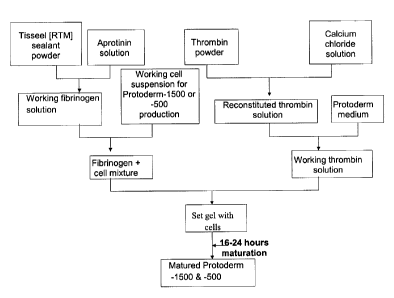
Figure 1 is a flow chart summarising a process of manufacturing a wound
healing composition according to preferred embodiments of the invention; and
Figure 2 shows the packaging, manipulation and application of a preferred
wound healing composition produced according to a process shown in Fig. 1.
A: shows a matrix (or set gel) preferentially adhering to a modified internal
surface of one of two metalised plastic sheets of a container pouch. B: shows
the use of one of the sheets of the container to apply the gel of the wound
healing composition to skin. Note that the sheet may used to support the gel
while both are cut to the appropriate shape and size. C: shows the wound
healing composition in place.
The process of manufacturing preferred compositions of the invention is
summarised in Fig. 1. Alternative components or methods as described above
may be used in place of those described here.
In principle, the composition comprises two components, which are cast
together. The first component comprises a solution of fibrinogen together with
one or more protease inhibitors to prevent unwanted proteolysis by protease
contaminants and premature matrix breakdown by cells during storage. In
particular, contaminants may include the naturally fibrinolytic enzyme
plasmin,
or its precursor plasminogen. Serine protease inhibitors such as aprotinin, e-
aminocaproic acid, or its analogue tranexamic acid, are frequently used in
order
to inhibit plasmin or prevent its activation. Added to this fibrinogen
solution is a
suspension of living cells in a suitable medium or buffer solution (a "working
cell suspension").
The second component comprises a solution of thrombin (an enzyme that
19
CA 02556098 2006-08-11
WO 2005/079822
PCT/GB2005/000523
naturally acts upon fibrinogen), calcium ions (a required cofactor), and a
medium suitable for the culture of living cells. A further clotting factor,
Factor
XIII, is also activated by thrombin in the presence of calcium ions. Activated
Factor XIII promotes polymerisation of monomeric fibrin (cleaved from
fibrinogen by thrombin) into a three-dimensional protein insoluble scaffold.
In order to cast a gel (i.e. a matrix in the form of a gel), these two
components
are combined and, whilst still liquid, poured into a pre-coated suitable
mould.
Although commonly circular, the gels may be cast into any desired shape. For
some applications, other shapes may be more suitable. In particular,
essentially
or substantially rectangular or elliptical gels may be more convenient for
larger
wounds.
Enzymatic cleavage of fibrinogen into fibrin monomers and polymerisation of
these monomers results in setting of the liquid into a semi-solid gel in which
living cells are suspended. For many applications, this gel is then maintained
for a period of about 24 hours under suitable conditions for cell growth,
division
and secretion of extracellular matrix proteins, and other proteins such as
growth
factors. Following development (or maturation), the cast gel is removed from
the casting mould and placed directly into a sterile package (which term is
taken
herein to have the same meaning as "container"). A small amount of medium,
for example a buffer medium, is added to each package to maintain the product
during storage and shipping, and the packages are sealed. During storage and
shipping the packages are maintained at a temperature of 2 C to 8 C.
In two preferred embodiments, called Protoderm 500 and Protoderm 1500, the
composition comprises cells at a density of about 500 cells per mm2 and about
1500 cells per mm2, respectively.
Advantages of such a product over the currently available alternatives include
the following. The use of a protein sealant as a scaffold or support matrix
allows
convenient topical delivery of cells to the wound. The pre-cast gel allows
CA 02556098 2006-08-11
WO 2005/079822
PCT/GB2005/000523
convenient and accurate application of regenerative cells to the wound surface
with control of the distribution and density of cells applied. Manufacture and
shipping of other tissue equivalents may take approximately 3 weeks for the
matrix alone, whereas the product of the present invention may be
manufactured within 10 days, or even as little as 2 days if sufficient growing
cells are available. These factors combine to give cost advantages, so
manufacture and production is more cost effective than many other
commercially available products.
As described below, the product of the invention when packaged also features a
unique flat pack system (adhesive backing) ensuring maintenance of product
during shipping and "ease of use" of final product. The precast gels can be
shipped and stored for up to 28 days at 2 to 8 C, whereas other available
products must either be frozen or shipped at room temperature.
Example 1: High protein concentration product ('Protoderm 500' and
Protoderm 1500')
A first embodiment of the invention is designed to optimise both rapid
manufacturing of the wound healing product and rapid wound healing by
containing cells and protein components at relatively high concentrations.
Matrix
In the first embodiment, the matrix protein is fibrin, derived from a
commercial
fibrinogen product, Tisseel [RTM] (Baxter). When reconstituted, this provides
a
convenient two component system to which cells may be added. Components of
the matrix are summarised in Table 1. It should be noted that Tisseel [RTM]
also contains Factor XIII, as well as plasmafibronectin and plasminogen.
Table 1 Primary components of Tisseel [RTM]
21
CA 02556098 2006-08-11
WO 2005/079822
PCT/GB2005/000523
Component Final concentration in
cellularised scaffolds
Matrix protein (fibrinogen) 7.5 - 11.5 mg/ml
Aprotinin 300 K IU/ml
Thrombin 25 IU/m1
Calcium chloride 4 mM
As will be apparent to one of appropriate skill in the art, the concentrations
of
these components can be varied as required. For example, fibrinogen may be
used in concentrations of the approximate range 7-20mg.m1-1 for this
application, thrombin in the range 5-50 IU/ml (in fact, trace levels of
contaminating thrombin may lead eventually to fibrin formation and gel setting
without additional thrombin, but this is inconvenient and unpredictable), and
calcium chloride in the range 2-20mM. Aprotinin is used to prevent unwanted
fibrinolysis but, again, the exact concentration may be varied.
Cells
Human dermal fibroblasts were obtained by culture of cells derived from
neonatal foreskin tissue. Under GMP (Good Manufacturing Practice)
conditions, fibroblastic cells were isolated by collagenase digestion and
expanded by culture and serial passage according to routine laboratory
practice
to establish a master cell bank (MCB). The MCB was screened against a panel
of human and animal-derived viruses, bacteria, mycoplasma and fungi, and for
tumorigenicity by a GLP (Good Laboratory Practice)-accredited facility and
determined to be free of contamination. Several working cell banks (WCB)
were then established for manufacture of the product, rescreened and stocks of
cells frozen according to standard procedures.
It is also envisaged that for various patient-specific applications,
autologous
22
=
CA 02556098 2006-08-11
WO 2005/079822
PCT/GB2005/000523
fibroblasts or other cells obtained from biopsies may be cultured and expanded
for use.
The cells were suspended in the quantities shown below (P-500 refers to
Protoderm-500; P-1500 refers to Protoderm-1500) in Liebowitz L-15 cell
culture medium buffered and supplemented as shown in Table 2 before addition
to the fibrinogen component. As will be apparent to one of skill in the art,
medium not intended for use in a CO2-enriched atmosphere (commonly used in
tissue culture incubators or sealed flasks) must be appropriately buffered by
some other system. Such media, supplemented with, for instance, HEPES, are
well-known in the art. Liebowitz L-15 medium relies on a phosphate buffering
system. The medium was supplemented with sodium bicarbonate and dextrose,
as shown.
For convenience and consistency, a standard 'working cell suspension' of
1.5x10⁶ cells.m1-1 was generally prepared.
Preparation of fibrin sealant
As outlined in Fig. 1 and summarised below, Tisseel [RTM] thrombin powder
was reconstituted in a calcium chloride solution according to the
manufacturer's
directions.
Once dissolved, the Thrombin/CaCl2 solution was further diluted with
supplemented L-15 medium to obtain a 'Working Thrombin Solution' and
refrigerated until further use for a minimum of 15 minutes. (Gels may also be
manufactured with 'Working Thrombin Solution' at room temperature.) Freeze-
dried fibrinogen was reconstituted with an aprotinin solution before being
added
to the working cell suspension in supplemented L-15 medium. Once
reconstituted, the fibrinogen should be used within 4 h, ideally within 1 to 2
h.
Working thrombin solution (6.75 ml) contains:
Thrombin: 5011J/m1 (or 337.5IU total)
23
CA 02556098 2006-08-11
WO 2005/079822
PCT/GB2005/000523
Calcium chloride: 8 moles/m1 (or 54 moles total)
In supplemented L-15
(Total refers to the amount in 6.75m1s)
Working fibrinogen and cell suspension mix (total volume 6.75m1):
Tisseel: 19mg/m1 (or 128.25mg total)
Aprotinin: 600KIU/m1 (or 4050KIU total)
Cells: 1.2x10⁶ cell/ml (8.1x10⁶ cells total for P-1500); or
0.4x10⁶ cell/ml (2.7x10⁶ cells total for P-500)
in supplemented L-15
(Total refers to the amount in 6.75m1s)
Table 2 - Details of Medium Used for Example 1
Components Function Concentration per ml
(Supplier shown in
parentheses)
L-15 medium Nutrient delivery to the N/A (base medium)
(Cambrex) cellular component of the
product. Maintains cell
viability and structure of
the gel.
Sodium Bicarbonate Required for cell 202.5 jig
(Mallinckrodt viability
Chemical)
Dextrose Nutrient 4.5mg
(J.T. Baker)
Adenine Base required for cell 24.4ps
(ABCR) viability
L-Glutamine Amino acid for cell 0.29mg
(Molekula) viability
24
CA 02556098 2006-08-11
WO 2005/079822
PCT/GB2005/000523
Ethanolamine Phospholipid for cell 6.2ptg
(Molekula) metabolism
0-phosphoryl- Phospholipid for cell 14.12 g
ethanolamine metabolism
(Merck)
Hydrocortisone Steroid required for cell 0.4mg
(Spectrum Laboratory metabolism
Products, Inc.)
Human Recombinant Essential hormone 5t.tg
Insulin
(Serologicals)
Selenious acid Trace substrate for 6.78ng
(Molekula) metabolism
3,3',5-Triiodo-L- Hormone 1.35ng
thyronine
(ABCR)
apo-Transferrin, Cofactor for iron 5 ,g
bovine metabolism
(Serologicals)
Gamma Irradiated Nutrients 2%v/v
Foetal Bovine serum
Or
New Born calf serum
(JRH or Hyclone)
Note: As will be apparent to one of ordinary skill in the art, sources of
ingredients used to producing the wound healing composition may differ
depending on the grade or purity required for different applications. For
example, for clinical applications of the product, pharmaceutical grade
materials
may be required.
CA 02556098 2006-08-11
WO 2005/079822
PCT/GB2005/000523
Casting the gels
The working thrombin solution (6.75m1) and Tisseel [RTM] fibrinogen/cell
suspension mixture (6.75m1) were combined by means of a Duplojet mixer unit
and loaded into a suitable pre-coated casting container (conveniently a
sterile
Petri dish or similar) via a 16G needle or equivalent. It is useful to pre-
coat the
casting dish with serum containing media or albumin to prevent the gel from
adhering. The gel set within a few minutes. The gel was then bathed in 20m1 of
medium (Table 2) and the casting dish covered with a lid. The set gel was
incubated at 37 C for 16-24 hours to allow development (or maturation) of the
cells.
Packing and Storage
After development (or maturation), the set gels were removed from their
casting
containers and placed into pre-irradiated, sterile foil pouches, stored within
a
sterile roto-seal bag. 10m1 serum-free medium (as per Table 2, without the
foetal bovine serum) was added to each pouch before sealing. The shelf life of
the sealed units is up to 28 days at 4 C.
Example 2: Low protein concentration product
For certain applications, it is possible to use lower protein concentrations.
The
chief advantage of this is reduction of production costs, since serum-derived
proteins and many protease inhibitors, such as aprotinin, are expensive. In a
preferred embodiment, the concentration of fibrin in the set product is
reduced
to less than 7 mg.m11. In practice, 3.0-4.0 mg.m1-1 is found to be effective.
One important consideration is the effectiveness (as well as the cost) of
using
aprotinin as protease inhibitor in such low protein' products. In particular,
pro
rata dilution of commercial products results in aprotinin concentrations that
are
too low to be effective. A preferable solution is to use an alternative
inhibitor,
such as tranexamic acid. Not only is this a highly effective inhibitor of
26
CA 02556098 2006-08-11
WO 2005/079822
PCT/GB2005/000523
fibrinolysis, but it has significant cost advantages.
Matrix
In this embodiment the matrix protein is fibrin, sourced from a commercial
fibrin sealant, Tisseel [RTM], using tranexamic acid instead of aprotinin. The
key components of the matrix are summarised in Table 3. It should be noted
that the same matrix composition could also be achieved using another
commercially available fibrin sealant, Quixil. However the addition of
exogenous tranexamic acid should be reduced as it already contains this
inhibitor.
Table 3 - Components of the Fibrinogen Matrix in Example 2
Component Final concentration in
cellularised scaffolds
Matrix protein (fibrinogen) 3.5mg/m1
Tranexamic acid 10mg/m1
Thrombin 25 IU/m1
Calcium chloride 4mM
Freeze-dried Tisseel [RTM] fibrinogen is reconstituted with supplemented L-15
medium solution before being added to the working cell suspension in
supplemented L-15 medium. Once reconstituted, Tisseel [RTM] fibrinogen
should be used within 4 hours, ideally within 1-2 hours.
Tisseel [RTM] thrombin powder is reconstituted in a calcium chloride solution
according to the manufacturer's directions. Once dissolved, the thrombin/CaCl2
solution is further diluted with supplemented L-15 medium containing
tranexamic acid to obtain a working thrombin solution.
The cell density used is again in the range 450 to 2500 cells mm-2. In order
to
minimise costs, it may be desirable to use a cell density of approximately 450
to
27
CA 02556098 2006-08-11
WO 2005/079822
PCT/GB2005/000523
550 cells mm-2. It should be noted, however, that protein concentration and
cells density are independent variables. Lowering protein concentration is the
major cost determinant, rather than cell density. However, being able to use
fewer cells may have implications for the speed of production. In any case,
high cell density/low protein concentration and low cell density/high protein
concentration embodiments are envisaged and may be preferred in specific
circumstances.
Example 3: Packaging, storage and delivery
A major factor contributing to the success of topical wound healing
compositions is the ease of accurately applying them to the wound surface so
that a close contact is established, without air bubbles or creases, under
sterile
operating conditions. Wound healing compositions may be fragile, and handling
should be kept to a minimum. The composition of the invention is preferably
packaged in such a way as to significantly assist and facilitate application.
In
addition, the composition is shipped and stored chilled, rather than frozen,
so
that detailed thawing procedures are not required prior to use.
After setting and the 16-24 hour culture and development (or maturation)
period, the individual gel discs are packaged by insertion into a flexible
foil or
metalised plastic pouch comprising two rectangular sheets, sealed along a
substantial portion of three of their sides so as to form an open pocket. The
inner surface of one of these sheets is modified so as to increase its
adherence to
the gel product. In a preferred embodiment as shown in Fig. 2, the packaging
used is an Oliver [RTM] Products Company (Grand Rapids, Michigan USA)
peelable foil pouch comprising one foil sheet and one sheet of laminated
polyester/foil sheet with Q15 Adhesive dot pattern coating. Q15/48BF1 is a
laminated lidding and pouching material for medical devices. The purpose of
this dot pattern adhesive coating is to improve the efficiency of the heat
sealing
process which is used to seal the edges of the sheets together. However, the
adhesive and raised dot pattern prove highly effective in providing a surface
to
28
CA 02556098 2006-08-11
WO 2005/079822
PCT/GB2005/000523
which composition preferentially adheres, as compared with the smooth,
uncoated inner surface of the opposing sheet. Other forms of coating and/or
roughening of the surface of one of the internal surfaces of the pouch could
be
used to achieve the same effect. Similarly, any suitably durable, flexible,
water
and gas-impermeable sheet material might be used to manufacture such a
pouch. All or part of the packaging might be transparent to allow visual
inspection, for example, of the integrity of the composition or of the colour
of a
pH indicator dye in the cell culture medium, a small volume of which is
inserted
in the pouch, along with the composition, before the pouch is sealed along its
remaining open edge.
Thus sealed, the composition has a shelf-life of at least 7-11 days, and
preferably up to 28 days, more preferably 21 days, at 2 to 8 C.
For application, as shown in Fig. 2, the pouch is peeled apart, under sterile
conditions, leaving the composition adhering to the treated inner surface of
one
of the sheets comprising the pouch. Using the sheet as a backing or means of
support the composition is then applied to the surface of the wound, to which,
in
the absence of excessive exudation, it will preferentially adhere allowing it
to be
peeled away from the sheet. This means of application allows the composition
to be applied without wrinkling or incorporation of air bubbles, and with the
minimum of manipulation. The edges of the composition may be easily
trimmed to fit the limits of the wound. Another advantage of delivering the
composition in a format that is reversibly adherent to the packaging, as
described, is that it allows the easy identification of the orientation of the
product and facilitates oriented application, should this be required. In the
case
of a homogenous wound-healing product, orientation of the product on the
wound is not important. However, where a multilayered composition is
involved, such as one with a fibroblast layer that is intended to be applied
in
contact with the wound surface and a keratinocyte layer that is intended to be
oriented away from the wound surface, it may be difficult or impossible to
establish the orientation visually. In this case, the ability to deliver the
product
29
CA 02556098 2014-04-29
in such a way as makes incorrect application impossible without first removing
the composition from the packaging offers a significant advantage.
The foregoing examples are meant to illustrate the invention and do not limit
it in
any way. The scope of the claims should not be limited by the preferred
embodiments and examples, but should be given the broadest interpretation
consistent with the description as a whole.