Note: Descriptions are shown in the official language in which they were submitted.
CA 02556790 2006-08-17
WO 2005/084713 PCT/GB2005/000789
ENZYME-PRODRUG THERAPY FOR PROSTHETIC JOINT
REPAIR
Field of the invention
The invention relates to the use of gene therapy in the treatment of aseptic
loosening
of orthopaedic prostheses. In particular, it discloses methods of refixing
such
prostheses without open revision surgery.
Background to the Invention
Approximately 1 million total hip replacement (total hip arthroplasty)
operations are
carried out world-wide annually, with more than 120,000 of these undertaken in
the
USA, and about 35,000 in England alone (NIH Consensus Statement, 1994; NHS
Review 1996). This is likely to increase to approximately 3 million worldwide
per
annum within the next decade. Hip replacements are very often performed in
elderly
patients and, amongst this group, loosening of one or both components of the
prosthesis, resulting in severe mobility restriction, occurs within 15 years
in about a
third of patients. Where prosthetic loosening occurs, patients' experience
increased
pain and walking difficulty and have a higher risk of dislocations and
pathological
fractures. Within 10 years, approximately 10% of all patients require revision
surgery, which has a high rate of complications and failure (Hellman et al
,1999).
The most common cause of implant failure is aseptic loosening as a result of
particulate-induced osteolysis. Wear particles, such as particles of
polyethylene,
polymethylmethacrylate, titanium, cobalt chrome or ceramic debris, depending
on the
type of prosthesis, stimulate an inflammatory response termed periprosthetic
osteolysis (Goldring et al, 1986). The phagocytosis of wear particles by
macrophages activates them, leading to secretion of the' inflammatory
cytokines IL-1,
TNF-cc, and IL-6. The resulting chronic inflammatory response eventually
produces a
pseudomembrane of granulomatous 'interface tissue' including activated
macrophages, fibroblasts, giant cells and osteoclasts, similar to the pannus
characteristic of arthritic joints. The end result of this complex
inflammatory and
proliferative foreign body response is osteoclast-mediated resorption of bone,
leading
to loosening of one or both components of the prosthetic implant. Prostheses
for
total hip arthroplasty consist of two components. An artificial socket, or
acetabular
component, is located in a prepared cavity in the acetabulum of the pelvis.
This
CA 02556790 2006-08-17
WO 2005/084713 PCT/GB2005/000789
articulates with a femoral component comprising a ball attached to a process,
which
is introduced into a prepared cavity in the medulla of the femur. Many
variations of
both components exist, and they may be retained with or without cements.
Aseptic loosening eventually leads to an unacceptable degree of pain,
immobility or
walking difficulties and instability, with a higher risk of dislocations and
pathological
fractures. In some patients revision surgery may be undertaken to remove the
inflammatory tissue and replace the prosthesis. However, revision surgery is
very
expensive and has a high morbidity and mortality rate, especially in elderly
patients
(who are in the majority). In patients with cardiac insufficiency revision
surgery often
has major complications such as myocardial failure or coronary artery disease
(Strehle et al, 2000). Many patients are not eligible for revision surgery
because the
risk of mortality is considered to be too high. There is no alternative
treatment for
such patients, who are then wheelchair-bound. The clinical need for a less
traumatic
alternative to revision surgery for treatment of loosened prostheses is
therefore clear.
At present experimental approaches to this problem are preventative rather
than
therapeutic. One such preventative approach to controlling aseptic loosening
involves the use of bisphosphonate compounds, especially alendronate, as
either a
systemic medication or as a component of a cement used to fix such prostheses
(US
Patent 5,972,913, WO 96/39107, Shanbhag et al, 1997, Leung et al, 1999).
However, although bisphosphonates are known to produce an increase in skeletal
bone density, they have not been shown to have a significant effect in
treating
rheumatoid arthritis, which shares many similar pathological features with
periprosthetic osteolysis, nor on periprosthetic osteolysis itself (Ralston et
al, 1989;
Eggelmeijer et al, 1996; Ulrich-Vinther, 2002). It thus remains to be seen
whether
bisphosphonates have a useful role to play in the prevention of aseptic
loosening.
In an attempt to prevent osteoclast-mediated periprosthetic bone resorption
directly,
an alternative preventative approach involves gene therapy (reviewed in Wooley
and
Schwarz, 2004), using an osteoclast inhibitory protein, osteoprotegerin,
delivered by
means of adeno-associated virus vector has been described (Ulrich-Vinther,
2002).
Osteoprotegerin is a competitive inhibitor of an osteoclast differentiation
factor,
receptor activator of nuclear factor KB ligand (RANKL), which binds to a
receptor
expressed on the surface of macrophage-derived osteoclast precursor cells,
known
as receptor activator of nuclear factor KB (RANK). RANKL is secreted by
osteoblasts, stroriial cells and activated T cells at an early stage of the
inflammatory
CA 02556790 2006-08-17
WO 2005/084713 PCT/GB2005/000789
response initiated by macrophage phagocytosis of wear particles (Teitelbaum,
2000).
Binding of RANKL to RANK leads to activation of osteoclast precursor cells,
differentiation, and stimulation of bone resorption. Binding of RANK by
osteoprotegerin fails to activate the osteoclast precursor cells with the
result that
osteoprotegerin competitively inhibits RANKL.
Ulrich-Vinther et al used a recombinant adeno-associated virus (rAAV) vector
to
express osteoprotegerin and inhibit titanium particle-induced resorption in a
mouse
calvarial resorption model. Titanium particles were implanted on the calvaria
(bones
of the vault of the skull) and the vector administered by intramuscular
injection into
the quadriceps. The inhibitory effect of the osteoprotegerin was therefore
systemic,
with detectable increases in serum levels, and this appeared to be successful
in
inhibiting the experimental titanium-induced osteoclastogenesis and bone
resorption
seen in the untreated controls. Although interesting, it remains to be seen
whether
this model will form the basis of a viable preventative for clinical
periprosthetic
osteolysis. Even if effective, it is unclear what long-term systemic effects
prolonged
elevations in serum osteoprotegerin levels might have. For example, such a
strategy
would need to demonstrate a lack of deleterious effects on normal osteoclast
function
in bone remodelling.
There remains a need for effective treatments for the common and debilitating
condition of periprosthetic osteolysis and its resultant aseptic loosening.
One approach to preferentially killing pathological cells, most widely used
for treating
cancer, is to introduce a gene into the target cells that encodes an enzyme
capable
of converting a prodrug of relatively low toxicity into a potent cytotoxic
drug.
Systemic administration of the prodrug is then tolerated since it is only
converted into
the toxic derivative locally, for example in a tumour, by cells expressing the
prodrug-
converting enzyme. This approach is known as gene-directed enzyme prodrug
therapy (GDEPT), or when the gene is delivered by means of a recombinant viral
vector, virus-directed prodrug therapy (VDEPT) (McNeish et al, 1997).
An example of an enzyme/prodrug system is nitroreductase and the aziridinyl
prodrug CB1954 (5-(aziridin-1-yl)-2,4-dinitrobenzamide) (Knox et al 1988).
Following
the observation that the Walker rat carcinoma cell line was particularly
sensitive to
CB1954, it was shown that this was due to the expression of the rat
nitroreductase
DT diaphorase. However, since CB 1954 is a poor substrate for the human form
of
CA 02556790 2006-08-17
WO 2005/084713 PCT/GB2005/000789
this enzyme, human tumour cells are far less sensitive to CB1954. GDEPT was
conceived as a way of introducing a suitable nitroreductase, preferably with
greater
activity against CB1954, in order to sensitise targeted cells. The Escherichia
coli
nitroreductase (EC1.6.99.7, alternatively known as the oxygen-insensitive
NAD(P)H
nitroreductase or dihydropteridine reductase, and often abbreviated to NTR)
encoded
by the NFSB gene (alternatively known as NFNB, NFSI, or DPRA) has been widely
used for this purpose (Reviewed in Grove et al, 1999). The NFSB-encoded
nitroreductase (NTR) is a homodimer that binds two flavin mononucleotide (FMN)
cofactor molecules. Using NADH or NADPH as an electron donor, and bound FMN
as a reduced intermediate, NTR reduces one or other of the two nitro-groups of
CB
1954 to give either the highly toxic 4-hydroxylamine derivative or the
relatively non-
toxic 2-hydroxylamine. Within cells, 5-(aziridin-1-yl)-4-hydroxylamino-2-
nitrobenzamide, probably via a further toxic metabolite, becomes very
genotoxic
(Knox et al, 1991). The exact nature of the lesion caused is unclear, but is
unlike that
caused by other agents. A particularly high rate of inter-strand cross-linking
occurs
and the lesions seem to be poorly repaired, with the result that CB 1954 is an
exceptionally affective anti-tumour agent (Friedlos et al, 1992).
The aim of GDEPT is to obtain efficient conversion of a prodrug such as CB1954
in
target cells in order to kill not only NTR-expressing cells but also bystander
tumour
cells that may not have been successfully transfected or transduced.
Another enzyme-prodrug system used in this way is that of a cytochrome P450 as
a
prodrug-converting enzyme and acetaminophen as the prodrug, as described in
international application WO 00/40271 (incorporated herein in its entirety). A
number
of cytochrome P450 enzymes, naturally expressed in the liver (for example
CYP1A2,
CYP 2E1 and CYP3A4) are capable of converting acetaminophen into a highly
cytotoxic metabolite, N-acetylbenzoquinoneimine (NABQI). This system has been
proposed for a variety of clinical applications, especially in the field of
cancer therapy.
Cytochrome P450 enzymes are also capable of activating several conventional
cytotoxic prodrugs, for example cyclophosphamide and ifosfamide (Chen and
Waxman, 2002).
A number of other enzyme-prodrug systems are widely used, including HSV
thymidine kinase and ganciclovir (Moolten, 1986), cytosine deaminase and 5-
fluorocytosine (Mullen et al, 1992).
CA 02556790 2006-08-17
WO 2005/084713 PCT/GB2005/000789
Goossens et al (1999) describe a viral gene therapy approach to infect and
kill
isolated cultured synovial cells in vitro, and to kill pannus tissue in a
monkey
collagen-induced arthritis model in which inflamed joints are induced by
collagen
injections. Inflamed joints in such animals contain a hyperplastic tissue
resulting from
the chronic inflammation termed pannus.
Summary of the Invention
As used herein:
"Cell-type selective" means; facilitating expression preferentially in a
limited range of
tissues. Preferably, such expression is substantially limited to a single
tissue or cell
type.
An "operably-linked promoter" is one in a substantially adjacent cis-
relationship,
wherein said promoter directs expression of the operably-linked element.
"Periprosthetic" relates to the space surrounding any,part of an implanted
prosthesis
"Periprosthetic osteolysis" is synonymous with "aseptic loosening" and relates
to any
progressive loosening of an implanted prosthesis not associated with frank
infection
or trauma.
"Interface tissue" is synonymous with "osteolytic membrane" and means
inflammatory tissue in the periprosthetic space round an implanted prosthesis,
implicated in periprosthetic osteolysis.
"Prosthesis" or "Orthopaedic implant" as herein used means any material or
device
surgically implanted into a bony structure of an animal or human.
An aim of the invention is to provide a non-surgical alternative to revision
surgery for
treatment of loosened prostheses that destroys interFace tissue (and the cells
within it
that are involved in the inflammatory processes and bone resorption) and
allows the
implant to be recemented.
The invention seeks to achieve this by using an enzyme-prodrug therapy
strategy
using a gene therapy vector to deliver a prodrug-converting enzyme to cells in
the
CA 02556790 2006-08-17
WO 2005/084713 PCT/GB2005/000789
interface tissue, thus sensitising them to a particular prodrug.
Administration of the
prodrug leads to ifs conversion to an active cytotoxic drug in the target
cells, killing
the interface tissue. Release of active cytotoxic drug from lysed interface
cells may
also kill neighbouring interface or inflammatory cells ('bystander' killing),
which is
advantageous in that cells that have escaped direct vector delivery (by
transduction,
for viral vectors, or transfection for non-viral vectors) are also eliminated.
In one strategy, a viral vector carrying nucleic acid encoding the enzyme is
injected
into the intra-articular space, and the prodrug subsequently administered
through a
small drill hole, which can also be used to inject cement to refix the
prosthesis in situ.
Alternatively, the prodrug may be administered by intra-articular injection.
Arthrography has shown that the interface tissue forms a continuous closed
compartment around the loosened prosthesis, which allows a high local
concentration of both vector and prodrug to be achieved with very low risk of
systemic escape. The concept thus offers more favourable circumstances in
terms
of both efficacy and safety than intra-tumoral injection in cancer patients, a
procedure
with which there is considerable clinical experience. In the case at least of
adenoviral vectors, it may be preferable to remove existing fluid in the intra-
articular /
periprosthetic space before introducing the vectors, to reduce the possibility
of
neutralising antibodies in the fluid inactivating the vector and preventing
satisfactory
levels of transduction.
Preferably, following introduction of the prodrug and consequent killing of
cells of the
interface tissue, said tissue is removed. This may be aided by the
introduction of,
either simultaneous with, or subsequent to, introduction of the prodrug, one
or more
enzymes capable of digesting extracellular components of the interface tissue,
such
as collagenase, elastase or hyaluronidase, matrix metalloproteases or
cathepsins.
Other compounds useful for this purpose include the chelating agents EDTA
(Ethylenediamine-N,N,N',N'-tetra-acetic acid) and EGTA (Ethylene glycol-bis-(2-
aminoethyl)-N,N,N', N'-tetraacetic acid). Such treatment digests and loosens
the
interface tissue, such that it may be flushed out through a suitable drill
hole or via a
wide bore needle introduced into the intra-articular space.
The fully loosened and debrided implant is then recemented, to solidly
reattach all
loosened components and restore a fully functional prosthetic joint.
CA 02556790 2006-08-17
WO 2005/084713 PCT/GB2005/000789
Alternatively, especially with prodrugs such as acetaminophen with very low
systemic
toxicity, the vector encoding the prodrug converting enzyme (such as
cytochrome
P450) may be injected locally, so that only cells within the interface tissue
l joint
compartment are transduced, whilst the prodrug is subsequently administered
systemically.
In one aspect of the invention, the approach is to kill cells resident in the
interface
tissue, irrespective of their type. In practice, the predominant cells are
fibroblasts
responsible for producing the extracellular matrix proteins of which much of
the tissue
is comprised, and cells of the monocyte/macrophage lineage responsible for
inflammatory effects. In this case, the expression of the enzyme encoded by th
a
vector is controlled by a strong non-cell type specific promoter, providing
high level
expression in a variety of cell and tissue types, such as the cytomegalovirus
early/immediate promoter and the cytotoxic effect is limited to cells of the
interface
tissue by the physical constraints of the space into which the vector and/or
prodrug
are injected. The normal cells of most concern from the safety viewpoint are
the
osteoblasts responsible for bone regeneration. In most instances, and with
most
gene delivery vectors, these cells are inaccessible to vector injected into
the
periprosthetic space, hence are not transduced or transfected, do not express
the
prodrug converting enzyme even with a non-cell type specific promoter, and ara
therefore not killed upon subsequent administration of the prodrug.
Examples of such non-cell specific promoters include: cytomegalovirus
immediate/early promoter, Rous sarcoma virus long terminal repeat (RSV LTR),
murine leukaemia virus LTR, simian virus 40 (SV40) early or late promoters,
herpes
simplex virus (HSV) thymidine kinase (tk) promoter, actin or ubiquitin
promoters.
In some circumstances it may be advantageous to achieve more selective cell
killing,
in which case the enzyme encoded by the vector may be expressed under the
control of a tissue- or cell type-selective promoter. Use of such a promoter
permits
selective killing of cells of particular lineages, such as fibroblasts, cells
of the
monocyte/ macrophage lineage or, more specifically, cells of a particular
phenotype,
such as osteoclast precursor cells, or fully differentiated osteoclasts.
Examples of promoters suitable for preferentially expressing a gene, such as a
gene
encoding a prodrug-converting enzyme, in cells of the monocyte / macrophage
lineage include, c-fes and CD68. Promoters characterised by containing one or
more
CA 02556790 2006-08-17
WO 2005/084713 PCT/GB2005/000789
binding sites for the transcription factor PU.1 are generally suitable
(Greaves and
Cordon, 2002).
Promoters suitable for expressing a gene preferentially in osteoclasts or
osteoclast
precursors include the tartrate-resistant acid phosphatase (TRAP) promoter,
the
RANK promoter and the cathepsin K promoter. Promoters characterised by
containing one or more binding sites (E-boxes, containing the consensus
binding
sequences 5'-CA(T/~)GTG) for microphthalrnia transcription factor family
(MITF,
TFE3, TFEB and TFEC), optionally also containing binding sites for the
transcription
factor PU.1 are generally suitable (Motyckova et al, 2001; Mansky et al, 2002,
Greaves and Cordon, 2002).
By the use of such specific promoters, expression of the enzyme may be
restricted to
particular target cells, such as those responsible for laying down of
extracellular
matrix proteins such as collagen (fibroblasts), those responsible for
secreting
inflammatory cytokines (such as macrophages) or those responsible directly for
bone
resorption (osteoclasts), whilst protecting other cell types (such as
osteoblasts,
responsible for depositing new bone).
The various possible combinations of local administration of vector and/ or
prodrug
with or without tissue-selective expression allow non-surgical treatment of
loosened
prostheses and recementation of the implant, overcoming limitations in the
prior art
methods aimed at preventing periprosthetic loosening by systemic
administration of
compounds such as bisphosphonates, or of systemic expression of highly
bioactive
molecules such as osteoprotegerin.
Accordingly, the invention provides an isolated polynucleotide encoding an
enzyme
capable of converting a prodrug into an active cytotoxic compound, expression
of the
enzyme being controlled by an operably-linked promoter that gives
substantially ce(I
type-selective expression. Preferably expression is restricted to cells of the
monocyte / macrophage lineage. Preferred examples such promoters include the
promoters of such genes as c-fes, and CD68. Promoters characterised by
containing
one or more binding sites for the transcription factor PU.1 are generally
suitable.
Alternatively, expression is restricted to fibroblasts.
CA 02556790 2006-08-17
WO 2005/084713 PCT/GB2005/000789
More preferably expression is restricted to osteoclasts or osteoclast
precursors.
Amongst suitable promoters providing such expression are those naturally
functionally linked to genes such as tartrate-resistant acid phosphatase
(TRAP),
receptor activator of nuclear factor xB (RANK) and cathepsin K.
Promoters characterised by containing one or more binding sites (E-boxes,
containing the consensus binding sequences 5'-CA(T/c)GTG) for microphthalmia
transcription factor family (MITF, TFE3, TFEB and TFEC), optionally also
containing
binding sites for the transcription factor PU.1 are generally suitable.
Preferably, the enzyme encoded is a nitroreductase, preferably a
nitroreductase
suitable for the activation of the prodrug CB1954 (5-(aziridin-1-yl)-2,4-
dinitrobenzamide). Alternatively, it a cytochrome P450. Other suitable
enzyme/prodrug systems include HSV thymidine kinase and ganciclovir (Moolten,
1986), cytosine deaminase and 5-fluorocytosine (Mullen et al, 1992).
In another aspect, the invention provides a vector comprising said
polynucleotide.
The vector may be any vector capable of transferring DNA to a cell.
Preferably, the
vector is an integrating vector or an episomal vector.
Preferred integrating vectors include recombinant retroviral vectors. A
recombinant
retroviral vector will include DNA of at least a portion of a retroviral
genome which
portion is capable of infecting the target cells. The term "infection" is used
to mean
the process by which a virus transfers genetic material to its host or target
cell.
Preferably, the retrovirus used in the construction of a vector of the
invention is also
rendered replication-defective to remove the effect of viral replication on
the target
cells. In such cases, the replication-defective viral genome can be packaged
by a
helper virus in accordance with conventional techniques. Generally, any
retrovirus
meeting the above criteria of infectivity and capability of functional gene
transfer can
be employed in the practice of the invention. Lentiviral vectors are
especially
preferred.
Suitable retroviral vectors include but are not limited to pLJ, pZip, pWe and
pEM, well
known to those of skill in the art. Suitable packaging virus lines for
replication-
defective retroviruses include, for example, ~'Crip, ~I'Cre, ~I'2 and ~I'Am.
CA 02556790 2006-08-17
WO 2005/084713 PCT/GB2005/000789
Other vectors useful in the present invention include aderiovirus, adeno-
associated
virus, SV40 virus, vaccinia virus, HSV and poxvirus vectors. A preferred
episomal
vector is the adenovirus. Adenovirus vectors are well known to those skilled
in the
art and have been used to deliver genes to numerous cell types, including
airway
epithelium, skeletal muscle, liver, brain and skin (Hitt et a/, 1997;
Anderson, 1998).
A further preferred vector is the adeno-associated (AAV) vector. AAV vectors
are well
known to those skilled in the art and have been used to stably transduce human
T-
lymphocytes, fibroblasts, nasal polyp, skeletal muscle, brain, erythroid and
haematopoietic stem cells for gene therapy applications Philip et al., 1994;
Russell
et al., 1994; Flotte et al., 1993; Walsh et al., 1994; Miller et al., 1994;
Emerson,
1996). International Patent Application WO 91/18088 describes specific AAV
based
vectors.
Other preferred episomal vectors include transient non-replicating episomal
vectors
and self-replicating episomal vectors with functions derived from viral
origins of
replication such as those from EBV, human papovavirus (BK) and BPV-1. Such
integrating and episomal vectors are well known to those skilled in the art
and are
fully described in the body of literature well known to those skilled in the
art. In
particular, suitable episomal vectors are described in W0~98/07876.
Mammalian artificial chromosomes can also be used as vectors in the present
invention. The use of mammalian artificial chromosomes is discussed by Calos
(1996).
In a further preferred embodiment, the vector of the present invention is a
plasmid.
The plasmid may be a non-replicating, non-integrating plasmid.
The term "plasmid" as used herein refers to any nucleic acid encoding an
expressible
gene and includes linear or circular nucleic acids and double or single
stranded
nucleic acids. The nucleic acid can be DNA or RNA and may comprise modified
nucleotides or ribonucleotides, and may be chemically modified by such means
as
methylation or the inclusion of protecting groups or cap- or tail structures.
A non-replicating, non-integrating plasmid is a nucleic acEd which when
transfected
into a host cell does not replicate and does not specifically integrate into
the host
to
CA 02556790 2006-08-17
WO 2005/084713 PCT/GB2005/000789
cell's genome (i.e. does not integrate at high frequencies and does not
integrate at
specific sites).
Replicating plasmids can be identified using standard assays including the
standard
replication assay of Ustav and Stenlund (1991).
The present invention also provides a host cell transfected with the isolated
polynucleotide or vector comprising such a polynucleotide of the present
invention.
The host cell may be any eukaryotic cell. Preferably it is a mammalian cell.
More
preferably, it is a human cell and, most preferably, it is an autologous cell
derived
from the patient and transfected or transduced either in vivo or ex vivo.
Numerous techniques are known and are useful according to the invention for
delivering the vectors described herein to cells, including the use of nucleic
acid
condensing agents, electroporation, complexing with asbestos, polybrene, DEAF
cellulose, Dextran, liposomes, cationic liposomes, lipopolyamines,
polyornithine,
particle bombardment and direct microinjection (reviewed by Kucherlapati and
Skoultchi,1984; Keown et al., 1990; Weir, 1999; Nishikawa and Huang, 2001).
A vector of the invention may be delivered to a host cell non-specifically or
specifically (i.e., to a designated subset of host cells) via a viral or non-
viral means of
delivery. Preferred delivery methods of viral origin include viral particle-
producing
packaging cell lines as transfection recipients for the vector of the present
invention
into which viral packaging signals have been engineered, such as those of
adenovirus, herpes viruses and papovaviruses. Preferred non-viral based gene
delivery means and methods may also be used in the invention and include
direct
naked nucleic acid injection, nucleic acid condensing peptides and non-
peptides,
cationic liposomes and encapsulation in liposomes.
The direct delivery of vector into tissue has been described and some, mostly
short-
term, gene expression has been achieved. Direct delivery of vector into
thyroid
(Sikes et al., 1994) melanoma (Vile et al., 1993), skin (Hengge et al., 1995),
liver
(Hickman et al., 1994) and after exposure of airway epithelium (Meyer et al.,
1995) is
clearly described in the prior art. Direct DNA injection into muscle has been
shown to
give longer-term expression (Wolff et al., 1990).
11
CA 02556790 2006-08-17
WO 2005/084713 PCT/GB2005/000789
Various peptides derived from the amino acid sequences of viral envelope
proteins
have been used in gene transfer when co-administered with polylysine DNA
complexes (Plank et al.,1994; Trubetskoy et al.,1992; WO 91 /17773; WO
92/19287)
and Mack et al., (1994) suggest that co-condensation of polylysine conjugates
with
cationic lipids can lead to improvement in gene transfer efficiency.
International
Patent Application WO. 95/02698 discloses the use of viral components to
attempt to
increase the efficiency of cationic lipid gene transfer.
Nucleic acid condensing agents useful in the invention include spermine,
spermine
derivatives, histones, cationic peptides, cationic non-peptides such as
polyethyleneimine (PEI) and polylysine. 'Spermine derivatives' refers to
analogues
and derivatives of spermine and include compounds as set forth in
International
Patent Application WO 93/18759 (published September 30, 1993).
Disulphide bonds have been used to link the peptidic components of a delivery
vehicle (Gotten et al., 1992); see also Trubetskoy et al. (supra).
Delivery vehicles for delivery of DNA constructs to cells are known in the art
and
include DNA/poly-ration complexes which are specific for a cell surface
receptor, as
described in, for example, Wu and Wu, 1988; Wilson et al., 1992; and U.S.
Patent
No. 5,166, 320.
Delivery of a vector according to the invention is contemplated using nucleic
acid
condensing peptides. Nucleic acid condensing peptides, which are particularly
useful
for condensing the vector and delivering the vector to a cell, are described
in
International Patent Application WO 96/41606. Functional groups may be bound
to
peptides useful for delivery of a vector according to the invention, as
described in
WO 96/41606. These functional groups may include a ligand that targets a
specific
cell-type such as a monoclonal antibody, insulin, transferrin,
asialoglycoprotein, or a
sugar. The ligand thus may target cells in a non-specific manner or in a
specific
manner that is restricted with respect to cell type.
The functional groups also may comprise a lipid, such as palmitoyl, oleyl, or
stearoyl;
a neutral hydrophilic polymer such as polyethylene glycol (PEG), or
polyvinylpyrrolidine (PVP); a fusogenic peptide such as the HA peptide of
influenza
virus; or a recombinase or an integrase. The functional group also may
comprise an
intracellular trafficking protein such as a nuclear localisation sequence
(NLS), an
12
CA 02556790 2006-08-17
WO 2005/084713 PCT/GB2005/000789
endosome escape signal such as a membrane disruptive peptide, or a signal
directing a protein directly to the cytoplasm.
The invention provides a pharmaceutical composition comprising the isolated
polynucleotide, vector or host cell of the invention as described, and a
pharmaceutically acceptable excipient, carrier, diluent or buffer.
In another aspect, the invention provides a product comprising a combination
of the
isolated polynucleotide, vector or host cell of the invention as described,
and a
prodrug capable of being converted into an active cytotoxic compound by the
enzyme encoded by said nucleotide or vector, or expressed by the host cell, as
a
combined medicament for simultaneous, separate or sequential use in the
treatment
of aseptic loosening of orthopaedic implants, such as prostheses used for
total hip
arthroplasty. The loosening may be of the acetabular component or the femoral
component, or both. The invention is not restricted to prostheses of the hip,
but may
be applied to any intraosseous implant where aseptic loosening may occur.
Accordingly its use for prostheses used in arthroplasty of the knee, elbow,
shoulder,
or any other joint of the skeleton is specifically envisaged.
Such use need not be restricted to human use. The method is equally applicable
to
loosening of prostheses of animal joints, in particular horses and dogs.
Preferably, the enzyme of such a product is a nitroreductase, more preferably
a
nitroreductase suitable for activation of CB1954. Most preferably, the prodrug
is
CB1954.
Alternatively, the enzyme is a cytochrome P450 of a type herein described.
Most
preferably the prodrug is acetaminophen.
In a further aspect of the invention, the use of a product comprising a
combination of
at least one vector, which comprises an isolated polynucleotide encoding an
enzyme
capable of converting a prodrug into an active cytotoxic compound, expression
of the
enzyme being controlled by an operably-linked promoter; and a prodrug capable
of
being converted into an active cytotoxic compound by said enzyme, for the
manufacture of a combined medicament for simultaneous, separate or sequential
use in the treatment of aseptic loosening of orthopaedic implants is provided.
13
CA 02556790 2006-08-17
WO 2005/084713 PCT/GB2005/000789
The promoter controlling expression of the prodrug-converting enzyme may be a
non-cell type specific promoter. Preferably, said promoter gives high levels
of
expression in a variety of tissues and cell types. More preferably it is
selected from
at least one of the following; the CMV immediate/early promoter, RSV LTR),
murine
leukaemia virus LTR, SV40 early or late promoters, HSV tk promoter. In a
further
preferred embodiment it is the human cytomegalovirus immediate/early promoter.
Alternatively, it is the mouse cytomegalovirus immediate/early promoter.
In an alternative preferred product for use in the manufacture of a combined
medicament for simultaneous, separate or sequential use in the treatment of
aseptic
loosening of orthopaedic implants, expression of the enzyme is controlled by
an
operably-linked promoter, which provides substantially cell-type specific
expression.
More preferably expression is restricted to cells of the monocyte / macrophage
lineage or fibroblasts, in which case the promoter may be naturally linked to
a gene
selectively expressed in cells of one of these lineages, as described above.
Most preferably expression is restricted to osteoclasts or osteoclast
precursors, as
described above.
Preferably, the enzyme is a nitroreductase, and most preferably a
nitroreductase
suitable for activating CB1954. In this case it is preferred that the prodrug
is
CB 1954.
Alternatively, the enzyme may be a cytochrome P450 as herein described. In
this
case it is preferred that the prodrug is acetaminophen. Alternatively, it may
be a
conventional cytotoxic, especially cyclophosphamide or ifosfamide.
A further aspect of the invention provides a method of treating aseptic
loosening of
orthopaedic implants comprising administering to a patient a vector encoding
an
enzyme capable of converting a prodrug into an active cytotoxic compound,
allowing
the expression of said enzyme in target cells, and administering a suitable
prodrug.
As will be appreciated by those of skill in the art, dosages are determined by
clearly
understood clinical parameters. However, it is preferred that the viral dose
per joint
treated is between 105 and 10'2 pfu, more preferably between 106 and 10'2 pfu,
further preferably between 10' and 10'2 pfu and most preferably between 109
and
102 pfu. Similarly, the dose of prodrug is dependent on clinical parameters.
In the
14
CA 02556790 2006-08-17
WO 2005/084713 PCT/GB2005/000789
case of CB1954, it is preferred that the dose should be between 5 and 40 mg
m'2,
preferably between 5 and 30 mg m-2, further preferably between 10 and 25 mg m-
z,
more preferably between 15 and 25 mg m'2, and most preferably 24mg m'~ given
by
intra-articular injection.
It is preferred that viral vectors are not co-administered with an iodine-
containing
contrast medium, since such media can inhibit viral transduction of target
cells.
Where the injection is to be directed by with arthroscopic visualisation, it
is preferred
that an air arthrogram is performed, or a contrast medium that does not
inhibit viral
transduction is used.
Preferably, the vector is administered by intra-articular or periprosthetic
injection.
It is also preferred that the prodrug is administered by intra-articular or
periprosthetic
injection. Alternatively, the prodrug may be administered systemically, more
preferably parenterally. However some prodrugs, particularly acetaminophen,
may
be administered orally.
In one preferred embodiment, expression of the prodrug-converting enzyme is
controlled by a promoter that provides non-cell type specific expression. In
this case
expression is not restricted to a particular tissue or cell type. As described
herein, it
is preferred that such promoters give high levels of expression in a variety
of cell
types. Examples of suitable promoters include the cytomegalovirus
immediate/early
promoter, Rous sarcoma virus long terminal repeat (RSV LTR), murine leukaemia
virus LTR, simian virus 40 (SV40) early or late promoters, herpes simplex
virus
(HSV) thymidine kinase (tk) promoter
In an alternative preferred embodiment, expression of the prodrug converting
enzyme is controlled by a promoter that provides substantially cell-type
specific
expression. Preferably, this is substantially restricted to cells of the
monocyte
macrophage lineage. Suitable promoters are described herein. Alternatively, it
is
restricted to expression in fibroblasts. More preferably, it is substantially
restricted to
osteoclasts or osteoclast precursors. Suitable and preferred promoters include
the
TRAP, RANK, and cathepsin K promoters.
As herein described preferred prodrug converting enzymes include
nitroreductases,
particularly those suitable for activating CB1954, and cytochrome P450
enzymes,
CA 02556790 2006-08-17
WO 2005/084713 PCT/GB2005/000789
particularly those most suitable for activating acetaminophen to NABQI.
Preferred
prodrugs accordingly include CB1954 and acetaminophen. However, in the case of
cytochrome P450 enzymes, conventional cytotoxic prodrugs such as
cyclophosphamide are also suitable.
In a further aspect of the invention, an isolated polynucleotide, or vector
comprising
such a polynucleotide or host cell comprising either, may encode, or express,
a
protein or peptide that is directly toxic to cells. In this case, no prodrug
administration
is required. Because of the self-contained nature of the joint /
periprosthetic space
surrounded by the interface tissue, it is possible to introduce vectors into
this
pathological space so that cells therein are transfected or transduced,
causing them
to express toxic products. Among the toxins that could be encoded and used in
this
way are ricin, abrin, diphtheria toxin, Pseudomonas exotoxin, DNase, RNase and
botulinum toxin.
Preferably, the expression of such directly toxic molecules is under the
control of a
promoter providing substantially cell-type specific expression as herein
described. In
this way, expression of the toxin is restricted to target cells defined both
by the
physical constraints of the space into which the vector is introduced and the
phenotype of the cells transfected or transduced. In this way, fibroblasts, or
inflammatory cells such as activated cells of the monocyte / macrophage
lineage, or
specific cells such as osteoclasts and their precursors directly responsible
for bone
resorption, are targeted.
Accordingly, an isolated polynucleotide encoding a toxic peptide or protein is
provided, wherein expression of the toxin is controlled by a promoter
providing
substantially cell-type specific expression. Preferably, this expression is
restricted to
cells of the monocyte / macrophage lineage. Alternatively, expression is
restricted to
fibroblasts. More preferably, expression is restricted to osteoclasts and
osteoclast
precursor cells. As described herein, suitable and preferred promoters include
the c-
fes and CD68 promoters to provide macrophage-specific expression and the TRAP,
RANK and cathepsin K promoters to provide osteoclast-specific expression.
Suitable
and preferred toxins encoded include ricin, abrin, diphtheria toxin,
Pseudomonas
exotoxin, DNase, RNase and botulinum toxin.
Also provided is a vector comprising said polynucleotide and a host cell
comprising
either, and a pharmaceutical composition comprising an isolated polynucleotide
or a
16
CA 02556790 2006-08-17
WO 2005/084713 PCT/GB2005/000789
vector as herein described, and a pharmaceutically acceptable excipient,
carrier,
diluent or buffer.
In a further embodiment is provided a product comprising an isolated
polynucleotide,
vector or host cell encoding or expressing a toxic peptide or protein as
herein
described, as a medication for the treatment of aseptic loosening of
orthopaedic
implants. Said expression may be under the control of a non-cell type specific
promoter giving high levels of expression in cells of a variety of types.
Preferably,
said expression is controlled by a promoter providing substantially cell-type
specific
expression as herein described.
Also provided is the use of such products in the manufacture of a medicament
for the
treatment of aseptic loosening of orthopaedic implants.
In a further aspect is provided a kit for treatment of aseptic loosening of
orthopaedic
implants comprising:
a) An isolated polynucleotide or vector encoding an enzyme capable of
converting a prodrug into an active cytotoxic compound, expression of which
enzyme being controlled by an operably-linked promoter, in a
pharmaceutically acceptable buffer ;
b) A prodrug capable of being converted into an active cytotoxic compound by
said enzyme, in a pharmaceutically acceptable buffer;
c) A tissue-digesting solution comprising at least one enzyme selected from
the
list consisting of collagenase, elastase, hyaluronidase, in a pharmaceutically
acceptable buffer; and/or a chelator such as EDTA, EGTA etc.
d) A cement suitable for the refixation of said orthopaedic implant.
Detailed Description of the Invention
Description of the Figures
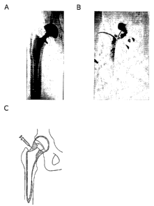
Figure 7 depicts aseptic loosening of a hip prosthesis. A is a radiograph of
loosened
prosthesis in situ. B is an arthrogram of a hip joint with a loosened
prosthesis. The
contrast medium is injected into the joint space under fluoroscopic guidance.
The
picture shows that a part of the area around the prosthesis (periprosthetic
space) is
filled with contrast medium. This proves that the prosthesis is loose in that
area.
17
CA 02556790 2006-08-17
WO 2005/084713 PCT/GB2005/000789
C shows a schematic representation of a hip joint with a loosened prosthesis.
The
grey area indicates the joint space, which is continuous with the
periprosthetic space.
When injecting a fluid into the joint space, this will spread through the area
which is
marked grey in the image.
Figure 2 shows the killing effect of infection with nitroreductase-encoding
adenoviral
vectors and subsequent exposure to the prodrug CB1954 at the concentrations
shown on interface cells from tissue taken from two revision surgery patients
as
described in Example 3. Figure 2a shows data from patient L1003 P3 and Figure
2b
shows data from patient L1002 P4.
Figure 3 shows the results of X-Gal staining of samples of intact interface
tissue
taken from patient LI014 infected with various doses of a Lac Z-encoding
adenoviral
vector, as described in Example 4.
The numbered wells contain tissue treated as follows:
1. Noninfected interface tissue
2. Interface tissue + 3.6 x 10⁴ pfu Ad.CMV.LacZ
3. Interface 3.6 pfu Ad.CMV.LacZ
tissue + x 105
4. Interface 3.6 pfu Ad.CMV.LacZ
tissue + x 106
5. Interface 3.6 pfu Ad.CMV.LacZ
tissue + x 10'
6. Interface 3.6 pfu Ad.CMV.LacZ
tissue + x 108
7. Interface 3.6 pfu Ad.CMV.LacZ
tissue + x 109
Figure 4 shows transduction of interface cells following incubation with six
different
concentrations of Ad.CMV.LacZ (0, 25, 50, 100, 200 and 400 pfu/ cell). After
three
days, cells were fixed and stained with X-gal reaction mix. The percentage of
transduced (blue) cells was counted. The figure shows the means and standard
deviations of 12 independent experiments.
Figure 5 shows the lack of toxicity of iotrolan (Isovist) contrast medium on
interface
cells. Interface cells were exposed to contrast medium (iotrolan) for 4 hours.
After 3
days of cell culturing viability of the cells was measured (n=12).
Figure 6 shows the effect of iotrolan on HAdVS-transduction of interface
cells. Cells
were exposed to different concentrations of Ad.CMV.LacZ : ((1) 0 pfu/cell, (~)
25
pfu/cell; (~) 100 pfu/cell; (~) 200 pfu/cell. (n=4)) and contrast medium for
four hours,
is
CA 02556790 2006-08-17
WO 2005/084713 PCT/GB2005/000789
after which the cells were fixed and stained with X-gal. Percentage of
transduced
cells was determined by counting blue cells.
Figure 7 shows pre- (A) and post-injection (B) images from Patient 1 showing
an
increased cement mass in the greater trochanteric region.
Figure 8 shows pre- (A) and post-injection (B) images from Patient 2.
Example 1 Procedure for treatment with CTL102 (Ad5-NTR and CB1954)
Materials
The drug product, CTL102 injection, is a sterile, clear or virtually clear,
aqueous liquid
solution containing CTL102 virions at a nominal mean potency of 2x10"
particles
ml-', buffered at pH 7.4.
CB1954 is formulated as a sterile solution in solvent (N-methyl pyrrolidone:
polyethylene glycol, 2:7 vlv with 17.8mg CB1954 ml-~). Just prior to use, the
prodrug
in solvent is diluted in sterile saline to a maximum final CB1954
concentration of 5mg
ml-'. .
To stabilise the prosthesis, low viscosity bone cement (Simplex~ P with
tobramycin
from Howmedica Inc, Rutherford, NJ, USA) is used. This radiopaque bone cement
is
a mixture of a liquid monomer component (2ml 97.4% methylmethacrylate, 2.6%
N,N-dimethyl-p-toluidine, 75ppm hydroquinone) and a polymer powder (6g
polymethylmethacrylate, 30g methylmethacrylate-styrene copolymer, 4g barium
sulphate, 1g tobramycin sulphate). The components are vacuum mixed (0.9 bar, 1
minute) immediately before use.
For arthrography, Hexabrix 320 (ioxaglate sodium meglumine, Guerbet, Roissy
Charles de Gaulle Cedex, France) contrast medium is used.
Procedure
Following careful flushing of the joint to remove synovial fluid and
inflammatory
exudate that may contain neutralising anti-adenovirus antibodies, 3x10⁹ pfu
CTL102
is injected intra-articularly resulting in delivery of vector to cells
throughout the
periprosthetic space. After 48 hours, to allow transduction of target cells
and
expression of the nitroreductase transgene, CB1954 (at a dosage of 24mg m-~)
is
19
CA 02556790 2006-08-17
WO 2005/084713 PCT/GB2005/000789
injected intra-articularly. To assure free access of CTL 102 and CB 1954 to
the
periprosthetic space it is preferred that patients are selected who have an
arthrogram
that shows contrast medium around the prosthesis. It is likely, therefore,
that patients
will usually undergo three arthrographies (one to assure access of contrast
medium,
one to inject the viral vector, and one to inject the CB 1954 prodrug).
In some circumstances after a number of days dead interface tissue may be
removed
by flushing or physical debridement, as appropriate. When the interface tissue
is
successfully diminished the prosthesis is refixated. To re-anchor the
prosthesis to the
bone, cement is injected in the periprosthetic space. For the flushing of the
periprosthetic space and injection of the cement a number of holes are drilled
through the bone into the periprosthetic space. This depends on the design of
the
prosthesis used. In many common designs, four is the minimum, because three
holes are necessary for the femoral component to fixate in 3D space and one is
necessary to fixate the acetabulum. As the bone biopsies are rather painful
and the
bone cannot be anaesthetised locally, these procedures are performed under
general
or spinal anaesthesia.
Example 2 Production of CTL102 (Ad5-NTR)
Materials and Methods
CTL102 was constructed as described in Djeha et al (2001) by homologous
recombination in PerC6 helper cells. The cells were transfected at 90%
confluence
with an equimolar mixture of the transfer vector pTX0375 and the backbone
vector
pPS1160 complexed with Lipofectamine transfection reagent (Life Technologies).
pTX0375 was constructed in two stages: (i) the CMV promoter/enhancer fused to
the
NTR gene was excised from pTX0340 as a 1.5-kb BamHl-partial Bglll fragment and
cloned into the unique BamHl site of pSW107, which is a pBluescript-based
vector
(Stratagene) that contains the human b-globin IVS II fused to the human
complement
2 gene polyadenylation sequence adjacent to the BamHl site. A plasmid,
pTX0374,
which contains the CMV.NTR fragment in the required orientation, was
identified by
PCR using the T3 primer (5'-ATTAACCCTCAC-TAAAG-3') which anneals to the
CMV promoter/enhancer, and an NTR primer, ECN2 (5'-TCTGCTCGGCCTGTTCC-
3'). (ii) The complete NTR expression cassette was excised from pTX0374 as a
2.5-
CA 02556790 2006-08-17
WO 2005/084713 PCT/GB2005/000789
kb Spel fragment and cloned into the unique Spel site of the E1-deleted
adenovirus
transfer vector pPS1128 in a left-to-right orientation with respect to Ad5
sequences.
pPS1128 is a pUC19-based plasmid that contains Ad5 sequences from the left-
hand
ITR to nucleotides (nt.) 359 fused to NT 3525-10589.
pPS1160 was constructed by Pacl linearisation of pPS1128, ligation with a
Pacl-compatible adaptor (5'-TACATCTAGATAAT-3' + 5'-P-TTATCTAGAT-GTA-
3') containing an Xbal site, followed by Xbal digestion to release a 7-kb Xbal
fragment containing Ad5 sequences 3524-10589. This was then cloned into Xbal-
linearised pPS1022, a pUC19-based plasmid containing Ad5 sequences from nt.
10589 to the right-hand ITR but lacking NT 28592 to 30470 (E3 region).
Recombinants containing the fragment in the required orientation were
identified by
PCR using primers flanking the Xbal site at 10589 (rightward, 5'-
TCGAGTCAAATACGTAGTCGT-3'; leftward, 5'-TGTTTCCGGAGGAATTTGCAA-3').
A plasmid, pPS1160/18, was confirmed to contain a single copy of the Xbal
fragment
(pPS1160/18) by Hindlll and Pstl digestion.
Transfected PerC6 cells were harvested following the appearance of extensive
CPE
(about 7-9 days after transfection) and recombinant virus released by three
freeze-
thaw cycles in infection medium (DMEM, 1 % FCS, 2 mM MgCl2 ). After two rounds
of
plaque purification on PerC6 cells the viruses were grown to large scale and
purified
by CsCI density centrifugation. Banded virus was dialysed against an excess of
storage buffer (10 mM Tris, pH 7.4, 140 mM NaCI, 5 mM KCI, 0.6 mM Na2 HP04 ,
0.9
mM CaCl2, 0.5 mM MgCl2 , and 5% sucrose), snap-frozen in aliquots in liquid
nitrogen, and stored at -280°C. Particle concentrations were determined
using the
BCA Protein Assay Reagent (Pierce, Rockford, IL) and the conversion factor 1
mg/ml
= 3.4x10'2 virus particles/ml. Infectious titres were determined by plaque
assay.
Genomic DNA was isolated from banded adenovirus by digestion with proteinase
K/SDS, phenol-chloroform extraction, and ethanol precipitation and
characterised by
restriction digestion.
Example 3 Killing of interface tissue from patients with CTL102 and
CB 1954
In order to demonstrate the feasibility of using a virally delivered enzyme-
prodrug
system to kill interface cells, cells taken from two patients during revision
surgery
were cultured in vitro, incubated with CTL102 at a range of MOIs and
subsequently
21
CA 02556790 2006-08-17
WO 2005/084713 PCT/GB2005/000789
exposed to CB1954. Cell viability was then determined using a metabolic
activity
assay.
Method
Interface tissue samples
For all experiments described, interface cells were used. Interface tissue was
removed from the periprosthetic space during revision-surgery by an orthopedic
surgeon and collected in sterile phosphate buffered saline (PBS). Connective
tissue
and fat were removed thoroughly and the interface tissue was digested for at
least
two hours at 37°C using collagenase 1A (1 mg/ml; Sigma, St Louis, MO,
USA). Cells
were then harvested by filtering the tissue/collagenase substance through a
200 pm
filter (NPBI, Emmer-Compascuum, The Netherlands). The cells were cultured in
75
cm2 flasks (Cellstar, Greiner, Alphen aan de Rijn, The Netherlands) with
Iscove's
modified Dulbecco's medium (IMDM; Biowitthaker, Verviers, Belgium),
supplemented
with glutamax (GibcoBRL, Paisley, UK), penicillin and streptomycin (Boehringer
Mannheim, Germany), and 10% fetal calf serum (FCS; GibcoBRL, Paisley, UK) at
37°C and 5% CO~.
Before each experiment interface cells were detached from the flasks using
0.25%
trypsin (GibcoBRL, Paisley, UK). The cells were counted in a barker counter
and
death cells were excluded by trypan blue. Cells were seeded in a 96 wells-
plate (flat
bottom) at a density of 5,000 cells per well. Cells were incubated overnight
to allow
attachment to the bottom. Before each experiment the wells were washed twice
with
IMDM. For the experiments passage 2 to 4 interface cells were used. Light
microscopy indicated that more than 95% of the cells were interface cells.
Transduction and cell killing assay protocol
Day 0: Interface cells from 2 patients were seeded at 5000 cells/well in IMDM
(10%
FCS) in 96 wells plates, 100p1 per well.
Day 1: Cells were infected with CTL102 (or diluent) at 0, 1, 5, 25, 100, 200
IU/cell in
IMDM (10% FCS), 50p1 per well.
Day 2: Cells were washed twice with in IMDM (10% FCS), hereafter cells were
incubated for 2 hr or 24 hr with CB1954 (or vehicle) at 0, 0.1, 0.5, 1, 5 and
50 pM in
IMDM (10% FCS, 10% HS), 50p1 per well.
Day 2/3: Cells were washed once with IMDM (10% FCS) and then incubated in
IMDM (10%FCS, 10% HS), 5pl per well.
22
CA 02556790 2006-08-17
WO 2005/084713 PCT/GB2005/000789
Day 4: Photographs were taken. Medium was refreshed with IMDM (10 % FCS),
10p1 WST reagent (Roche) was added and the plates were incubated for 2 hr.
Hereafter the absorbance at 415nm was measured.
Results
As shown in Figure 2A and 2B, virus and CB1954-dose dependent killing was
observed for cells from both patients. Importantly, efficient (90%) killing
was
observed with virus and CB1954 doses (200 virus pfu/cell and a CB1954
concentration of 50~,M) that is readily achievable in the clinic.
These results demonstrate that interface cells can be transduced by an HAdV-5-
vector and killed by the NTR/CB1954 approach. Human adenovirus 5 is capable of
infecting a broad range of dividing and non-dividing human cells including
fibroblasts
and macrophages (Djeha et al, 2001).
Killing of cells by GDEPT has been studied before in various cell lines, using
various
approaches. The NTR/CB1954 approach is attractive for clinical evaluation for
several reasons: (1 ) it generates a toxic agent that can kill both dividing
and non-
dividing cells, (2) induction of cell death occurs by a p53-independent
mechanism,
and (3) CB1954 is well-tolerated in man (Djeha et al, 2001 ). Cell killing by
the
NTR/CB1954 approach has been proved effective in a variety of human cancer
cells
(Chung-Faye et al, 2001; Bilsland et al, 2003, Green et al, 2003; McNeish et
al, 1998;
Shibata et al, 2002; Weedon et al, 2000; Wilson et al, 2002), but has not
previously
been studied in synovial or interface cells. The current study shows that
interface
cells can be effectively killed by the NTR/CB1954 approach.
For the current study passage 2 to 4 interface cells were used. These passages
were
used to maximally reduce culture artefacts. On the one hand, in very low
passages (0
and 1 ) there is a risk for presence of contaminating cells (especially
macrophages),
which decreases with higher passages. On the other hand, at higher passages
the
risk of substantial in vitro alteration/ growth selection exists (especially
at passages
higher than 4) (Zimmerman et al, 2001). In the current study, cultured
interface cells
of different patients were used. For the interpretation of the results the
data of all
patients were pooled. However, it must be noted that individual differences in
transducibility were observed.
23
CA 02556790 2006-08-17
WO 2005/084713 PCT/GB2005/000789
Example 4 Efficient infection of intact interface tissue with adenovirus
vectors
The experiment outlined in Example 3 confirmed that cultured interface cells
are Ad5-
infectable. However, when a cell is present within an intact tissue, access of
the
virus to the cell surface may be prevented, for instance by the extracellular
matrix
and by the low rate of virus diffusion through the extracellular space. In
view of this,
the infectability of fresh intact interface tissue was examined using a LacZ-
expressing
adenovirus and Xgal staining of LacZ-expressing tissue. Using this approach, a
virus
dose-dependent increase in gene expression was observed, with strong levels of
gene expression with the two highest virus doses tested (Figure 3).
Method
Interface tissue (LI014) was obtained from a revision operation of the hip of
a
rheumatoid arthritis patient. The tissue was cut in 7 pieces and the pieces
were put
in 10 ml round bottom tubes. Different concentrations of Ad.CMV.LacZ (0,
3.6x10⁴,
3.6x10⁵, 3.6x10⁶, 3.6x10', 3.6x10$, 3.6x10⁹ pfu) in 200p1 IMDM/10% FCS were
added. The tissues were incubated at 37°C for 2 hours, the tubes were
shaken every
to 15 minutes. Hereafter 5 ml IMDM/10%FCS was added and after an overnight
incubation the tissues were rinsed 3x with PBS and subsequently put in 5 ml
Xgal
colouring solution and incubated for 3.5 hours at 37 °C. The tissues
were rinsed 3x
with PBS and fixed in 10 % formalin.
Results
The tissues with the highest added amounts of Ad.CMV.LacZ have areas of dark
blue staining, which is evident down to an infection at 3.6x10' pfu
Ad.CMV.LacZ.
Demonstrating that infection of cells in intact interface tissue is effective.
Embedded paraffin sections of the tissues were examined microscopically and
the
presence of stained, infected cells was confirmed.
Example 5 Transduction of interface tissue and effect of contrast
medium
To test further the susceptibility of interface cells to human adenovirus 5
(HAdV-5)-
based vectors, primary cultures of interface cells were exposed to the HAdV-5
vector
24
CA 02556790 2006-08-17
WO 2005/084713 PCT/GB2005/000789
Ad.CMV.LacZ. Twenty-four hours post-infection the cells were stained with X-
gal
solution for (3-galactosidase reporter gene expression. The transduction
efficiency
increased with increasing vector concentration. At 400 plaque forming
unitslcell the
percentage of cells expressing the reporter gene was 88% (sd 4.0) (Fig. 4).
Thus
HAdV-5 vectors can transduce interface cells.
Materials and Methods
Adenoviral vectors
The Ad.CMV.LacZ (van der Eb et al, 2002) vector is identical to CTL102, but
the
E.coli IacZ gene replaces the ntrgene.
Transduction assays
To study the transducibility of interface cells by HAdV-5, interface cells
were infected
with Ad.CMV.LacZ vector (in concentrations of 0, 25, 50, 100, 200, 400
pfuicell).
Twenty-four hours post infection the cells were washed twice with IMDM, and
cultured for two days. Medium was refreshed each day. On day three, the
monolayer
cultures were washed twice with PBS and fixed with 0.2% glutaraldehyde and 2%
formaldehyde in PBS for 10 minutes at 4°C. Subsequently cells were
washed twice
with PBS and stained for ~i-galactosidase activity in 50 pl of reaction mix (1
mgiml X-
gal (Eurogentec, Seraing, Belgium), 5 mM potassium ferrocyanide, 5 mM
potassium
ferricyanide, 2 mM MgCl2 in PBS) for 2 hours at 37 °C. The percentage
of transduced
cells was assessed by counting at least 100 interface cells, using light
microscopy.
All conditions were tested in duplicate.
Effect of contrast medium on interface cells
Interface cells were seeded in 96-wells plates. Into each well 50 ~I of IMDMi
20%
FCS and 50 pl of a solution containing contrast medium and 0.9% NaCI in
various
concentrations (0, 12.5, 25, and 50% contrast medium) were added. The contrast
medium used was the low-osmolarity, nonionic dimer iotrolan (Isovist;
Schering,
Berlin, Germany). After four hours of exposure to the contrast medium, the
cells were
washed twice and incubated in IMDMi10% FCS. The cells were cultured for three
more days, changing the culture medium every day. On day four, cell viability
was
determined with the WST-1 cell viability assay kit (Roche, Mannheim, Germany)
according to the manufacturers protocol.
CA 02556790 2006-08-17
WO 2005/084713 PCT/GB2005/000789
Effect of contrast medium on HAdV-5 -transduction of interface cells
Interface cells were seeded in 96-wells plates. After overnight incubation
cells were
infected with Ad.CMV.LacZ (concentrations of 0, 25, 100, and 200 pfu/cell) in
IMDMI
20% FCS, 50 pl per well. Fifty pl lotrolan (Isovist) in 0.9% NaCI was added in
concentrations of 0, 25, 50, and 100%. (When diluted in the culture medium
these
concentrations decreased to 0, 12.5, 25, and 50%.) Four hours after infection,
the
cells were washed twice with IMD.M and incubated for the rest of the day in
IMDM/10% FCS at 37°C and 5% CO~. The Ad.CMV.LacZ transduced cells
were
cultured for three days after removal of the vector and contrast medium.
Subsequently, the cells were fixed and stained for ~i-galactasidase activity.
The
transduction rate was assessed as described above.
Statistical analysis
A univariate analysis of variance and Spearman's correlation was used to study
the
interaction between vector and prodrug and between vector and contrast medium
and to study the effect of CB1954 on viability of the cells. A Mann-Whitney
test for
independent groups was performed to determine the difference in cell killing
between
the cells that were exposed to contrast medium and the non-exposed cells. In
the
experiment to study the effect of transient exposure to contrast medium on
transduction of HAdV-5-vector Spearman's correlation between contact time and
viability and between delay time and viability was tested. For all statistical
analyses
p<0.05 was the level of statistical significance.
Results
Effect of contrast medium on interface cells
The toxicity of contrast medium (iotrolan) on interface cells was evaluated
(Fig. 5).
lotrolan does not affect the viability of the cells at any concentration
(p=0.563).
Adding of contrast medium to the interface cells for four hours does not lead
to killing
of the cells.
Effect of contrast medium on HAdV-5 transduction of interface cells
The effect of contrast medium (iotrolan) on HAdVS-transduction of interface
cells was
investigated with Ad.CMV.LacZ. Transducibility of the cells increases with the
concentration of HAdV-5 vector. However, the contrast medium has restraining
influence on the transduction efficiency. With higher concentrations of
iotrolan, the
HAdV-5 vector concentration has less effect on gene transfer efficiency. At a
contrast
26
CA 02556790 2006-08-17
WO 2005/084713 PCT/GB2005/000789
medium concentration of 50% none of the cells were transduced (Fig. 6). The
effect
of iotrolan on the transduction is statistically significant (p<0.001).
Furthermore,
differences between cells from different individuals (n=6) have been observed.
To evaluate the effect of contrast medium on cell killing by NTR/CB1954, the
previously described experiment for the efficiency of cell killing was
repeated in the
presence of contrast medium. The results showed that, in the presence of
contrast
medium, cells are not killed by the NTR/CB1954 approach (results not shown).
The
presence of Hexabrix 320 contrast medium also inhibited viral transduction
(data not
shown). In summary, the results from these experiments demonstrate the
incompatibility of viral administration in combination with the administration
of two
commonly used contrast media. This incompatibility may be due to the presence
of
iodine within the contrast media. Screening of all available contrast media
may allow
determination of a contrast medium compatible with viral transduction.
The influence of transient exposure to contrast medium on the transduction of
interface cells was investigated. Interface cells were exposed to contrast
medium for
0 to 120 minutes and the period between washing away of the contrast medium
and
performing the NTR/CB1954 cell killing approach was varied. Cell killing was
not
correlated with contact time (corr -0.033, p = 0.691 ) or length of period
between
washing away of the contrast medium and addition of the vector (corr -0.004, p
=
0.962). Killing of cells not exposed to contrast medium and those transiently
exposed was equivalent.
Discussion
In this study the influence of contrast medium on cell killing by NTR/CB1954
was
investigated in view of future clinical studies. Results show that the
contrast medium
does not seem to have any influence on the interface cells. However,
transduction of
the cells by an adenoviral vector, in the presence of contrast medium, is
almost
negligible. The adenoviral vector is inactivated by the presence of contrast
medium.
In a putative clinical study the viral vector will be injected in the joint
space. Normally,
contrast medium is used to verify the position of the needle in the joint. The
results of
this study however show that the use of contrast medium in combination with a
viral
vector is dissuaded. Thus, for a clinical study, we propose that alternative
methods
for the visualization of the needle should be employed such as injection of
air to
create an "air-arthrogram".
27
CA 02556790 2006-08-17
WO 2005/084713 PCT/GB2005/000789
In conclusion, this example shows that interface cells can be killed by the
NTR/CB1954 enzyme prodrug approach.
Example 6 Clinical outcomes
Data are available from the first two patients from a phase-1 study of 12
patients with
a loosened hip experiencing debilitating pain and significant comorbidity. On
day 1
the vector was injected into the hip joint and the prodrug injected on day 3,
as
described above. On day 10 three holes were drilled in the femur and one in
the
acetabulum. Biopsies are taken from the periprosthetic space and low viscosity
cement (Osteopal, Biomet Merck, Sjobo, Sweden) injected under fluoroscopic
guidance.
Patient 1 is an 82-year old female with loosening of both hip prostheses,
classified
ASA IV (mortality risk 20.3%, American Society of Anesthesiologists physical
status
classification, Saklad, 1941 ). There were no adverse effects from vector
injection
(3x10⁹ particles) and 24 hours post-injection there was no detectable virus
shedding.
Twelve hours after prodrug injection the patient experienced nausea, (WHO
grade 1 )
which was known as a reaction to the prodrug. Also hip pain increased, which
was
anticipated as the initial therapy is intended to cause more loosening. 16 ml
cement
was injected into periprosthetic space (see Figure 7B) indicating significant
destruction of interface tissue creating a void into which cement could now be
introduced. The patient was ambulated the day after surgery.
At two and four weeks after cement injection the patient had no pain in the
treated
hip, and was still improving. The maximum walking distance had increased from
4-5
metres to 30 metres. Subjective walking distance assessed by the patient (0: 0
metres, 100: unlimited walking distance) increased from 4 to 66. The patient's
pain
score (0: no pain, 100: unbearable pain) decreased from 81 preoperatively to
2. In
addition, she could now sleep on her side without pain, which she had been
unable
to do for four years. In terms of perceived dependency (0: completely
dependent on
others, 100: completely independent) the score decreased from 95 to 54.
Patient 2 is a 72-year-old woman with loosening of her left hip prosthesis and
an
ASA classification of II (mortality risk 2.8%). Again, there was no detectable
virus
shedding 24 hours after vector injection. 18m1 of cement was injected
following a
similar procedure (Figure 8B). Four weeks post-treatment the pain score had
28
CA 02556790 2006-08-17
WO 2005/084713 PCT/GB2005/000789
decreased from 43 to 22 (probably reflecting the presence of a post-operative
haernatoma, requiring 4-5 weeks to resolve). Specifically hip joint-related
pain
disappeared. Maximum walking distance increased from 500 to 2000 metres. By
the
3-month follow-up, the haematoma had completely resolved and pain score had
further decreased to 7. The patient continues to improve in terms of walking
performance and other activities.
The current study is the first to use in vivo intra-articular adenoviral
mediated gene
transfer in a clinical setting. The preliminary results suggest that gene
therapy and
cement injection for hip prosthesis refixation is clinically feasible.
References
1. Anderson WF (1998) Human gene therapy. Nature 392: (6679 Supply: 25-30.
2. Bilsland, A.E., et al. (2003). Selective ablation of human cancer cells by
telomerase-specific adenoviral suicide gene therapy vectors expressing
bacterial
nitroreductase. Oncogene 22: 370-380.
3. Calos MP (1996). The potential of extrachromosomal replicating vectors for
gene
therapy. Trends in Genetics 12: 463-466.
4. Chen L and Waxman DJ (2002) Cytochrome P450 gene-directed enzyme
prodrug therapy (GDEPT) for cancer. Curr Pharm Des 8: 1405-1416.
5. Chung-Faye, G., et al. (2001). Virus-directed, enzyme prodrug therapy with
nitroimidazole reductase: a phase I and pharmacokinetic study of its prodrug,
CB1954. Clin. Cancer Res. 7: 2662-2668.
6. Cotten M, Wagner E and Birnstiel ML (1992) Receptor-mediated transport of
DNA into eukaryotic cells. Meth Enzymol 217: 618-644.
7. Djeha, Thomson, Leung, Searle, Young, Kerr, Harris, Mountain, and Wrighton
(2001). Combined adenovirus-mediated nitroreductase gene delivery and
CB1954 treatment: a well-tolerated therapy for established solid tumors. Mol
Ther 3: 233-240.
29
CA 02556790 2006-08-17
WO 2005/084713 PCT/GB2005/000789
8. Eggelmeijer, Papapoulos, Van Paassen, Dijkmans, Valkema, Westedt, Landman,
Pauwels and Breedveld (1996). Arthritis Rheum 39: 396-4.02.
9. Emerson SG (1996). Ex vivo expansion of hematopoietic precursors,
progenitors,
and stem cells: the next generation of cellular therapeutics. Blood 87, 3082-
3088.
10. Flotte TR, Afione SA, Conrad C, McGrath SA, Solow R, Oka H, Zeitlin PL,
Guggino WB and Carter BJ (1993). Stable in vivo expression of the cystic
fibrosis
transmembrane conductance regulator with an adeno-associated virus vector.
Proc Natl Acad Sci USA 90: 10613-10617.
11. Friedlos, Quinn, Knox and Roberts (1992). The properties of total adducts
and
interstrand crosslinks in the DNA of cells treated with CB 1954. Exceptional
frequency and stability of the crosslink. Biochem Pharmacol 43: 1249-1254.
12. Goldring, Jasty, Roelke, Rourke, Bringhurst and Harris (1986). Formation
of a
synovial-like membrane at the bone-cement interface. Its role in bone
resorption
and implant loosening after total hip replacements. Arthritis Rheum 29: 575-
584.
13. Loosens PH, Schouten GJ, 't Hart BA, Brok HP, Kluin PM, Breedveld FC,
Valerio
D and Huizinga TW (1999). Feasibility of adenovirus-mediated nonsurgical
synovectomy in collagen-induced arthritis-affected rhesus monkeys. Hum Gene
Ther 10: 1139-1149.
14. Greaves and Gordon (2002). Macrophage-specific gene expression: current
paradigms and future challenges. Int J Hematol 76: 6-15.
15. Green, N.K., McNeish, I.A., Doshi, R., Searle, P.F., Kerr, D.J., Young,
L.S.
(2003). Immune enhancement of nitroreductase-induced cytotoxicity: studies
using a bicistronic adenovirus vector. Int. J. Cancer 104: 104-112.
16. Grove, Searle, Weedon, Green, McNeish and Kerr (1999). Virus-directed
enzyme prodrug therapy using CB1954. Anti-Cancer Drug Design 14: 461-4.72.
17. Hellman, Capello and Feinberg (1999). Omnifit cementless total hip
arthroplasty:
a 10-year average follow-up. Clin Orthop 364: 164-174.
CA 02556790 2006-08-17
WO 2005/084713 PCT/GB2005/000789
18. Hengge UR, Chan EF, Foster RA, Walker PS and Vogel JC (1995) Cytokine
gene expression in epidermis with biological effects following injection of
naked
DNA. Nature Genet 10: 161-166.
19. Hickman MA, Malone RW, Lehmann-Bruinsma K, Sih TR, Knoell D, Szoka FC,
Walzem R, Carlson DM and Powell JS (1994). Gene expression following direct
injection of DNA into liver. Human Gene Therapy 5: 1477-1483.
20. Hitt, MM, Addison CL and Graham, FL (1997) Human adenovirus vectors for
gene transfer into mammalian cells. Advances in Pharmacology 40: 137-206.
21. Keown WA, Campbell CR, Kucherlapati RS (1990). Methods for introducing DNA
into mammalian cells. Methods Enzymol 185: 527-37.
22. Knox RJ, Boland MP, Friedlos F et al (1988) Biochemical Pharmacology
37:4671-
4677.
23. Knox, Friedlos, Marchbank and Roberts (1991). Bioactivation of CB 1954:
reaction of the active 4-hydroxylamino derivative with thioesters to form the
ultimate DNA-DNA interstrand crosslinking species. Biochem Pharmacol 42:
1691-1697.
24. Kucherlapati and Skoultchi (1984) Introduction of purified genes into
mammalian
cells. CRC Crit. Rev. Biochem 16: 349-379.
25. Leung, Scammell, Lyons, Czachur, Gilbert, Freedholm, Malbecq, Miller, Carr
and
Checkley (1999). Alendronate prevents periprosthetic bone loss - 2 year
results.
Arthritis Rheum 42 (Supply: S270.
26. Mack KD, Walzem R and Zeldis JB (1994). Cationic lipid enhances in vitro
receptor-mediated transfection. Am J Med Sci 307: 138-143.
27. Mansky, Sulzbacher, Purdom, Nelsen, Hume, Rehli and ~strowski (2002). The
microphthalmia transcription factor and the related helix-loop-helix zipper
factors
TFE-3 and TFE-C collaborate to activate the tartrate-resistant acid
phosphatase
promoter. J Leukoc Biol 71: 304-310.
31
CA 02556790 2006-08-17
WO 2005/084713 PCT/GB2005/000789
28. McNeish, Searle, Young and Kerr (1997). Gene-directed enzyme prodrug
therapy for cancer. Advanced Drug Delivery Reviews 26: 173-184.
29. McNeish, I.A., et al. (1998). Virus directed enzyme prodrug therapy for
ovarian
and pancreatic cancer using retrovirally delivered E. coli nitroreductase and
CB1954. Gene Ther. 5: 1061-1069.
30. Meyer KB, Thompson MM, Levy MY, Barron LG and Szoka FC Jr. (1995).
Intratracheal gene delivery to the mouse airway: characterization of plasmid
DNA
expression and pharmacokinetics. Gene Therapy, 2, 450-460, 1995
31. Miller JL, Donahue RE, Sellers SE, Samulski RJ, Young NS and Nienhuis AW
(1994). Recombinant adeno-associated virus (rAAV)-mediated expression of a
human gamma-globin gene in human progenitor-derived erythroid cells. Proc
Natl Acad Sci USA 91:10183-10187.
32. Moolten FL et al (1986) Tumour chemosensitivity conferred by inserted
herpes
thymidine kinase genes : Paradigm for a prospective cancer control strategy.
Cancer Res 46:5276-5281.
33. Motyckova, Weilbaecher, Horstmann, Riemann, Fisher and Fisher (2001 ).
Linking osteopetrosis and pycdysostosis: regulation of cathepsin K expression
by
the microphthalmia transcription factor family. Proc Natl Acad Sci USA 98:
5798-
5803.
34. Mullen CA, Kilstrup M and Blaese RM (1992) Transfer of the bacterial gene
for
cytosine deaminase to mammalian cells confers lethal sensitivity to 5-
fluorocytosine : A negative selection system. PNAS USA 89:33-37.
35. NHS Centre for Reviews & Dissemination (1996). Total hip replacement.
Effective
Health Care. Volume 2. Number 7. Churchill-Livingstone.
36. N I H Consensus Statement Online (1994). Total Hip Replacement. September
12-14 1994, 12 (5): 1-31.
32
CA 02556790 2006-08-17
WO 2005/084713 PCT/GB2005/000789
37. Nishikawa M and Huang L (2001). Nonviral vectors in the new millennium
Delivery barriers in gene transfer. Human Gene Therapy 12:861-870.
38. Philip R, Brunette E, Kilinski L, Murugesh D, McNally MA, Ucar K,
Rosenblatt J,
Okarma TB and Lebkowski JS (1994). Efficient and sustained gene expression
in primary T lymphocytes and primary and cultured tumor cells mediated by
adeno-associated virus plasmid DNA complexed to cationic liposomes. Mol Cell
Biol 14: 2411-2418.
39. Plank C, Oberhauser B, Mechtler K, Koch C and Wagner E (1994). The
influence
of endosome-disruptive peptides on gene transfer using synthetic virus-like
gene
transfer systems. J Biol Chem 269: 12918-12924.
40. Ralston, Hacking, Willocks, Bruce and Pitkeathly (1989). Clinical,
biochemical
and radiographic effects of aminohydroxypropylidene bisphosphonate treatment
in rheumatoid arthritis. Ann Rheum Dis 48: 396-399.
41. Russell DW, Miller AD and Alexander IE (1994). Adeno-associated virus
vectors
preferentially transduce cells in S phase. Proc Natl Acad Sci USA 91: 8915-
8919.
42. Saklad M (1941 ) Grading of patients for surgical procedures.
Anesthesiology 2:
281-284.
43. Shanbhag, Hasselman and Rubash (1997). The John Charnley Award.
Inhibition of wear debris mediated osteolysis in a canine total hip
arthroplasty
model. Glin Orthop 344: 33-43.
44. Shibata, T., Giaccia, A.J., Brown, J.M. (2002). Hypoxia-inducible
regulation of a
prodrug-activating enzyme for tumor-specific gene therapy. Neoplasia 4: 40-48.
45. Sikes ML, O'Malley BW Jr, Finegold MJ, Ledley FD (1994). In vivo gene
transfer
into rabbit thyroid follicular cells by direct DNA injection. Human Gene
Therapy 5:
837-844.
33
CA 02556790 2006-08-17
WO 2005/084713 PCT/GB2005/000789
46. Strehle J, DeINotaro C, Orler R and Isler B (2000) The outcome of revision
hip
arthroplasty in patients older than age 80 years. Complications and social
outcome of different risk groups. J Arthroplasty 15:690-697.
47. Teitelbaum (2000). Bone resorption by osteoclasts. Science 289: 1504-1508.
48. Trubetskoy VS, Torchilin VP, Kennel SJ, Huang L (1992). Use of N-terminal
modified poly(L-lysine)-antibody conjugate as a carrier for targeted gene
delivery
in mouse lung endothelial cells. Bioconjugate Chem 3: 323-327
49. Ulrich-Vinther, Carmody, Goater, Sr~balle, O'Keefe, and Schwarz (2002).
Recombinant adeno-associated virus-mediated osteoprotegerin gene therapy
inhibits wear debris-induced osteolysis. J Bone Joint Surg 84A: 1405-1412.
50. Ustav M and Stenlund A (1991). Transient replication of BPV-1 requires two
viral
polypeptides encoded by the E1 and E2 open reading frames. EMBO J 10: 449-
457
51. van der Eb, M.M., et al. (2002). Gene therapy with apoptin induces
regression of
xenografted human hepatomas. Cancer Gene Ther. 9: 53-61.
52. Vile RG and Hart IR (1993). In vitro and in vivo targeting of gene
expression to
melanoma cells. Cancer Res 53: 962-967.
53. Walsh CE, Liu JM, ?Ciao ?C, Young NS, Nienhuis AW, Samulski RJ (1994).
Regulated high level expression of a human gamma-globin gene introduced into
erythroid cells by an adeno-associated virus vector. Proc Natl Acad Sci USA
89:
7257-7261.
54. Weedon, S.J., et al. (2000). Sensitisation of human carcinoma cells to the
prodrug CB 1954 by adenovirus vector-mediated expression of E. coli
nitroreductase. Int. J. Cancer 86: 848-854.
55. Wilson JM, Grossman M, Wu CH, Chowdhury NR, Wu GY and Chowdhury JR
(1992). Hepatocyte-directed gene transfer in vivo leads to transient
improvement
of hypercholesterolemia in low-density lipoprotein receptor-deficient rabbits.
J
Biol Chem 267: 963-967.
34
CA 02556790 2006-08-17
WO 2005/084713 PCT/GB2005/000789
56. Wilson, W.R., Pullen, S.M., Hogg, A., Helsby, N.A., Hicks, K.O., Denny,
W.A.
(2002). Quantitation of bystander effects in nitroreductase suicide gene
therapy
using three-dimensional cell cultures. Cancer Res. 62: 1425-1432.
57. Wolff JA, Malone RW, Williams P, Chong W, Acsadi G, Jani A and Felgner PL
(1990). Direct gene transfer into mouse muscle in vivo. Science 247:1465-1468.
58. Wooley PH and Schwarz EM (2004). Aseptic loosening. Gene Therapy 11: 402-
407.
59. Wu GY and Wu CH (1988). Receptor-mediated gene delivery and expression in
vivo. J Biol Chem 263:14621.
60. Weir N (1999) Non-viral vectors for gene therapy. In "Biotechnology - A
multi-
volume, comprehensive treatise", Volume 5a, Recombinant proteins, monoclonal
antibodies and therapeutic genes, Ed by A. Mountain, U.Ney and D,Schomburg,
Wiley VCH Verlag.
61. Zimmermann, T., et al. (2001). Isolation and characterization of
rheumatoid
arthritis synovial fibroblasts from primary culture--primary culture cells
markedly
differ from fourth-passage cells. Arthritis Res. 3: 72-76.