Note: Descriptions are shown in the official language in which they were submitted.
CA 02557544 2006-08-25
WO 2005/087135 PCT/US2005/005685
IMPLANTABLE OR INSERTAELE MEDICAL DEVICE RESISTANT TO
MICROBIAL GROWTH AND BIOFILM FORMATION
STATEMENT OF RELATED APPLICATION
[0001] This is a continuation-in-part of co-pending U.S. Patent Application
10/071,840,
filed February 8, 2002, and entitled "Implantable Or Insertable Medical Device
Resistant
To Microbial Growth And Biofilm Formation," which is incorporated by reference
herein
in its entirety.
FIELD OF THE INVENTION
[0002j The present invention relates to implantable or insertable medical
devices that
provide resistance to microbial growth on and in the environment of the device
and
resistance to microbial adhesion and biofilm formation on the device. In
another aspect,
the present invention relates to methods of manufacturing such implantable or
insertable
medical devices, particularly to methods of manufacturing such devices that
comprise at
least one matrix polymer region, an antimicrobial agent for providing
resistance to
microbial growth and/or a microbial adhesion/biofilm synthesis inhibitor for
inhibiting
the attachment of microbes and the synthesis and accumulation of biofilm on
the surface
of the medical device.
BACKGROUND OF THE INVENTION
[0003j Implantable or insertable medical devices such as stems made of
metallic,
polymeric or a composite of.metallic and polymeric materials frequently
occlude due to
microbial colonization and adhesion. This problem is particularly prevalent
with medical
devices that are adapted to remain implanted for a relatively long-term, i.e.,
from about 30
days to about 12 months or longer. Microbes such as bacteria often colonize on
and
around the medical device and, upon attaching to surfaces of the device,
proliferate and
form aggregates within a complex matrix consisting of extracellular polymeric
substances, typically polysaccharides. The mass of attached microorganisms and
the
associated extracellular polymeric substances is commonly referred to as a
biofilm or
slime. Antimicrobial agents have difficulty penetrating biofilms and killing
and/or
CA 02557544 2006-08-25
WO 2005/087135 PCT/US2005/005685
inhibiting the proliferation of the microorganisms within the biofilm. The
colonization of
the microbes on and around the device and the synthesis of the biofilm barrier
eventually
result in encrustation, occlusion and failure of the device.
[0004] Previous approaches to minimize this problem have included the use of
low
surface energy materials such as Teflon~ in implantable medical devices and
the use of
surface coatings on such medical devices. Surface coatings have typically
comprised
single antimicrobials or 1-2 antibiotics.
.[0005] For example, U.S. Patent No. 5,853,745 discloses an implantable
medical
device having a durable protective coating layer over an antimicrobial coating
layer. The
coating layers are formed by applying an antimicrobial coating layer to at
least a portion
of the surface of the medical device, applying a durable coating over the
antimicrobial
coating layer, and applying a resilient coating layer over the durable coating
layer.
[0006] U.S. Patent No. 5,902,283 discloses a non-metallic antimicrobial
impregnated
implantable medical device where the antimicrobial composition is applied to
the device
under conditions where the antimicrobial composition permeates the material of
the
device.
[0007j U.S. Patent No. 5,772,640 discloses polymeric medical devices that have
been impregnated and/or coated with chlorhexidine and triclosan by dipping or
soaking
the medical device in a solution of a hydrophobic or hydrophilic polymer
containing
chlorhexidine and triclosan.
[0008] Published International Application No. WO 99/47595 discloses a
plastics
material that can be used in certain medical applications comprising an
acrylic polymer
containing 5-50% of a rubbery copolymer and a biocidal compound. The patent
also
discloses adding antimicrobial agent to the polymer melt by means of a liquid
injection
system.
[0009] U.S. Patent No. 5,679,399 discloses membranes that may include one or
more
permeable or semipermeable layers containing substances such as biocides. The
layers
allow the transmission of environmental fluids inwardly and the outward
dispersion of the
biocides. These membranes may also include a sealing or coating to entrap
agents such
as biocides therein.
[0010] Of the previous approaches, coatings have met with the greatest success
2
CA 02557544 2006-08-25
WO 2005/087135 PCT/US2005/005685
because of their proximity to the bacterial environment and hence their active
approach to
preventing bacterial colonization and attachment. However, this approach has
proven
inadequate because of the potential for bacterial resistance to a single
narrow spectrum
active agent, because the amount of active agent that can be incorporated into
such
coatings is typically low, and/or because externally coated tubular devices
release active
agents to the environment external to the device but not intraluminally.
[0011] In an effort to alleviate the foregoing and other disadvantages of the
prior art,
Applicants have developed an implantable or insertable medical device suitable
for long-
term implantation and a method for manufacturing such a device, wherein the
device
provides resistance to microbial growth on and around the device and/or
biofilm
formation on the device. The device of the present invention, therefore,
overcomes the
disadvantages associated with the use of coatings as discussed above, and
provides a
reduced risk of biofilm fouling that eventually results in encrustation,
occlusion and
failure of the device.
SUMMARY OF THE INVENTION
[0012] One aspect of the present invention is directed to an implantable
medical
device comprising at least one biocompatible matrix polymer region and
bioactive agents
comprising an antimicrobial agent, a microbial attachment/biofilm synthesis
inhibitor, or
both. In some preferred embodiments, the medical device comprises multiple
distinct
matrix polymer regions. One or more barrier layers at least partially covering
a matrix
polymer region may also be provided in certain preferred embodiments of the
present
invention. Preferred antimicrobial agents include triclosan, chlorhexidine and
salts or
combinations thereof. Other antimicrobial agents include, but are not limited
to
nitrofurazone, benzalkonium chlorides, silver salts and antibiotics such as
rifampin,
gentamycin and minocyclin. Preferred microbial attachment/biofilm synthesis
inhibitors
include salicylic acid and salts and derivatives thereof. A radio-opacifying
agent rnay be
optionally included in a matrix polymer region, and one or more therapeutic
agents may
also be present. The matrix polymer and any barrier layer may preferably
comprise a
biodegradable or substantially non-biodegradable material such as an ethylene
vinyl
acetate copolymer, copolymers of ethylene with acrylic acid or methacrylic
acid;
metallocene catalyzed polyethylenes and polyethylene copolymers, ionomers,
elastomeric
CA 02557544 2006-08-25
WO 2005/087135 PCT/US2005/005685
materials such as elastomeric polyurethanes and polyurethane copolymers,
silicones and
mixtures thereof. Among medical devices in accordance with the present
invention are
biIiary, ureteral, urethral and pancreatic stems, stmt covers, catheters,
venous access
devices and devices bridging or providing drainage between a sterile and non-
sterile body
environment or between two sterile body environments. Pancreatic stems that
release a
buffering agent are among preferred pancreatic stems.
[0013] In another aspect, the present invention is directed to a method of
manufacturing an implantable or insertable medical device comprising providing
one or
more biocompatible matrix polymers and one or more bioactive agents comprising
an
antimicrobial agent and/or a microbial attachment/biofilm synthesis inhibitor;
processing
the one or more biocompatible matrix polymers and the one or more bioactive
agents
under conditions that substantially prevent preferential partitioning of any
of the bioactive
agents to a surface of any of the biocompatible matrix polymers and
substantially prevent
chemical modification of the one or more bioactive agents. Processing
preferably
comprises forming a homogenous mixture of the matrix polymer and any bioactive
agent
and optional radio-opacifying agent and/or therapeutic agent and shaping the
homogeneous mixture into at least a portion of an implantable or insertable
medical
device. Among preferred shaping processes are included extrusion and
coextrusion for
multiple layer structures or for partitioning of the medical device into
different polymer
matrix sections (e.g., sections of different durometer values).
BRIEF DESCRIPTION OF THE DRAWINGS
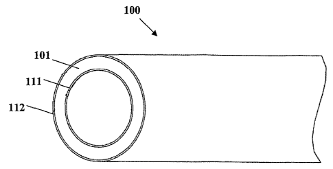
[0014] Fig. 1 is a simplified schematic representation (perspective view) of a
portion
of an implantable or insertable medical device in accordance with an
embodiment of the
present invention.
[0015] Fig. 2 is a simplified schematic representation (perspective view) of a
portion
of an implantable or insertable medical device in accordance with an
embodiment of the
present invention.
[0016] Fig. 3 is a graph showing bacterial attachment inhibition onto extruded
tubes
ontaining varying amounts of triclosan (TCN) and salicylic acid (SA).
[0017] Fig. 4 is a graph showing bacterial attachment inhibition onto extruded
tubes
containing varying amounts of triclosan (TCN) and salicylic acid (SA).
4
CA 02557544 2006-08-25
WO 2005/087135 PCT/US2005/005685
[0018] Fig. 5 is a graph showing the zone of bacterial (E. coli ATCC 25922)
growth
inhibition around extruded tubes containing varying amounts of triclosan (TCN)
and
salicylic acid (SA).
[0019] Fig. 6 is a graph showing the zone of bacterial (coagulase negative
staph #99)
growth inhibition around extruded tubes containing varying amount of triclosan
and
salicylic acid (SA).
[0020] Fig. 7 is a graph showing the amount of triclosan released as a
function of the
number of days of urine exposure.
[0021] Fig. 8 is a graph showing the concentration of triclosan released as a
function of
the number of days of urine exposure.
[0022] Fig. 9 is a graph showing the % of the total triclosan in the stmt as a
function of
the number of days of urine exposure.
[0023] Fig. 10 is a graph showing zone of inhibition size as a function of
triclosan release
concentration.
[0024] Fig. 11 is a graph showing zone of inhibition size as a function of
triclosan release
concentration in Fig. 11.
(0025] Fig. 12 is a ureteral stmt for use in connection with the present
invention.
[0026] As is typically the case with such figures, Figs. 1, 2 and 12 are
simplified
schematic representations presented fox purposes of illustration only, and the
actual
structures may differ in numerous respects including the relative scale of the
components.
DETAILED DESCRIPTION OF THE INVENTION
[0027] In one aspect, the present invention is directed to an implantable or
insertable
medical device comprising at least one biocompatible matrix polymer region, as
well as
one or multiple bioactive components, which comprise an antimicrobial agent
and/or a
microbial attachment/biofilm synthesis inhibitor.
[0028] The term "biocompatible" as used herein describes a material that is
substantially not toxic to the human body, and that does not significantly
induce
inflammation or other adverse response in body tissues.
[0029j The term "matrix polymer" as used herein refers to a polymeric material
that
forms at least a portion or region of the implantable or insertable medical
device of the
present invention. The matrix polymer is selected to be biocompatible and
provide
CA 02557544 2006-08-25
WO 2005/087135 PCT/US2005/005685
mechanical properties consistent with the intended function and operation of
the
implantable or insertable medical device. The matrix polymer also serves as a
repository
in which at least one and, in some preferred embodiments, both the
antimicrobial agent
and microbial attachment/biofilm synthesis inhibitor are dispersed and/or
dissolved. The
matrix polymer may also contain, as further optional components, a radio-
opacifying
agent and/or one or more therapeutic agents.
[0030] The term "antimicrobial agent" as used herein means a substance that
kills
and/or inhibits the proliferation and/or growth of microbes, particularly
bacteria, fungi
and yeast. Antimicrobial agents, therefore, include biocidal agents and
biostatic agents as
well as agents that possess both biocidal and biostatic properties. In the
context of the
present invention, the antimicrobial agent kills andlor inhibits the
proliferation andlor
growth of microbes on and around the surfaces of an implanted medical device.
[0031] The term "microbial attachment/biofilm synthesis inhibitor" as used
herein
means a substance that inhibits the attachment of microbes onto a surface and
the ability
of such microbes to synthesize and/or accumulate biofilm on a surface. In the
context of
the present invention, such a surface includes a surface of an implantable
medical device
exposed to a physiological environment, such as a physiological fluid, that
may be
conducive to the formation and accumulation of biofilm on the surface of the
medical
device. The microbial attachment/biofihn synthesis inhibitor may also have
substantial
antimicrobial activity as described herein. Likewise, the antimicrobial agent
may also
have substantial ability to inhibit microbial attachment/biofilm synthesis.
[0032] By "biofihn" is meant the mass of microorganisms attached to a surface,
such
as a surface of a medical device, and the associated exti~acellular substances
produced by
one or more of the attached microorganisms. The extracellular substances are
typically
polymeric substances and commonly comprise a matrix of complex
polysaccharides,
proteinaceous substances and glycopeptides. This matrix or biofilm is also
commonly
referred to as "glycocalyx."
[0033] Biofilm formation on the surfaces of implantable or insertable medical
devices adapted for long-term implantation, e.g., from about 30 days to 12
months or
longer, can result in eventual encrustation and failure of the device.
Further, the
proliferation of microbes within the biofilm can lead to localized infections
as well as
di~cult to treat systemic infections. The extracellular substances that
comprise the
6
CA 02557544 2006-08-25
WO 2005/087135 PCT/US2005/005685
biofilm matrix can act as a barrier that protects and isolates the
microorganisms housed in
the biofilm from normal immunological defense mechanisms, such as antibodies
and
phagocytes, as well as from antimicrobial agents including surfactants,
biocides and
antibiotics. The biofilm also facilitates the growth and proliferation of
microbes housed
within the biofilm.
[0034] The present invention substantially reduces the risk of biofihn
accumulation
on the surfaces of a medical device adapted for long term implantation, and
the resultant
likelihood of premature failure of the device due to encrustation and
occlusion by such
biofilm. In some preferred embodiments of the present invention, the medical
device is
intended to remain implanted for a relatively long period of from about 30
days to about
12 months or longer. However, it is understood that the device may be
implanted for a
period of 30 days or shorter as well.
[0035] The biocompatible matrix polymer of the device of the present invention
is
provided to serve as a repository in which the antimicrobial agent, the
microbial
attachment/biofilm synthesis inhibitor, or both, are dispersed and/or
dissolved. The
medical device of the present invention will preferably contain at least one
matrix
polymer which forms at least a single distinct portion or region of the
medical device.
Where only a single distinct matrix polymer region is provided in the medical
device, the
matrix polymer will preferably contain one or both of the antimicrobial agent
and the
microbial attachment/biofilm synthesis inhibitor. However, in other preferred
embodiments, the medical device will comprise two or more distinct matrix
polymer
regions. The distinct regions may exist, e.g., as coaxial layers or as
distinct sections
lengthwise along the longitudinal axis of the device (e.g., a stmt having
distinct end
regions of different durometer value with a transitional coextruded region in
between).
Where two or more distinct matrix polymer regions are present in a medical
device that
contains both the antimicrobial agent and the microbial attachment/biofilm
synthesis
inhibitor, it is not necessary that both bioactive agents be present in any
single one of
such multiple matrix polymer regions. Thus, the antimicrobial agent may be
present in a
first matrix polymer region and the microbial attachment/biofilm synthesis
inhibitor may
be present in a second matrix polymer region distinct from the first matrix
polymer
region. However, it is understood that one or both bioactive agents may be
present in one
or all of any distinct matrix polymer regions. Further, as discussed more
fully below,
7
CA 02557544 2006-08-25
WO 2005/087135 PCT/US2005/005685
where multiple distinct matrix polymer regions are present, the regions may be
separated
by barrier layers that at least partially cover a surface of the matrix
polymer region.
[0036] The amount of the antimicrobial agent present in a matrix polymer is
preferably an amount effective to kill and/or inhibit the growth of microbes
on and around
the implanted medical device. Preferred amounts of the antimicrobial agent
present in the
matrix polymer range from about 0.1% to about 25% by weight of the matrix
polymer.
Amounts of from about 10% to about 25% by weight of the matrix polymer are
particularly preferred.
[0037] The amount of the microbial attachment/biofihn synthesis inhibitor
present in a
matrix polymer is preferably an amount effective to inhibit the attachment of
microbes
onto and the synthesis and/or accumulation of biofilm by attached microbes on
a surface
of the implanted medical device. Preferred amounts of the microbial
attachment/biofilm
synthesis inhibitor present in the matrix polymer range from about 0.1% to
about 25% by
weight of the matrix polymer. Amounts of from about 10% to about 25% by weight
of
the matrix polymer are particularly preferred.
[003] The amount of antimicrobial agent and/or microbial attachment/biofilm
synthesis
inhibitor present in a matrix polymer will depend on, ihte~~ alia, on the
efficacy of the
bioactive agent employed, the length of time during which the medical device
is intended
to remain implanted, as well as the rate at which the matrix polymer or
barrier layer
releases the bioactive agent into the environment of the implanted medical
device. Thus,
a device that is intended to remain implanted for a longer period will
generally require a
higher percentage of the antimicrobial agent and/or microbial
attachment/biofilm
synthesis inhibitor. Similarly, a matrix polymer that provides faster release
of the
bioactive agent may require a higher amount of the bioactive agent. The amount
of
bioactive agent in the matrix polymer may be limited, of course, by the
propensity for
such bioactive agent to cause undesirable localized or systemic toxic reaction
and by the
potential impairment of the mechanical properties necessary for the proper
functioning of
the medical device.
[0039] In many instances, it is believed that the bioactive agent is released,
at least in
part, from a non-biodegradable matrix polymer region by a mechanism wherein
the
matrix polymer imbibes or contacts physiological fluid. The physiological
fluid dissolves
or disperses the bioactive agent reposed within the matrix, and the dissolved
or dispersed
CA 02557544 2006-08-25
WO 2005/087135 PCT/US2005/005685
bioactive agent then diffuses outwardly from the matrix polymer into the
physiological
environment where the device is implanted. Matrix polymers need not be
permeable to
aqueous fluids such as physiological fluids to provide release of bioactive
agent. Matrix
polymers with low permeability to aqueous fluids may adsorb such fluids at a
surface of
the polymer. In such matrix polymers, a concentration gradient is believed to
be set up at
the surface of the polymer and the bioactive agent is released via diffusion
based on its
affinity for the solid polymer relative to its solubility in the fluid or
aqueous phase.
Where the matrix polymer is biodegradable, similar diffusion processes may
also occur.
In a biodegradable matrix polymer, bioactive agent may also be released as the
biodegradable matrix polymer containing the reposed bioactive agent
biodegrades upon
contact with the physiological environment where the device is implanted.
Thus, in a
biodegradable polymer, bioactive agent may be released by diffusional
processes and
upon biodegradation of the polymer matrix.
[0040] The antimicrobial agent present in the matrix polymer can be any
pharmaceutically acceptable antimicrobial agent. By "pharmaceutically
acceptable" as
used herein is meant an agent that is approved or capable of being approved by
the United
States Food and Drug Administration or Department of Agriculture as safe and
effective
for use in humans or animals when incorporated in or on an implantable or
insertable
medical device. Preferred antimicrobial agents include, but are not limited
to, triclosan,
chlorhexidine, nitrofurazone, benzalkonium chlorides, silver salts and
antibiotics such as
rifampin, gentamycin and minocyclin and combinations thereof.
[0041] The microbial attachment/biofilm synthesis inhibitor can be any
pharmaceutically
acceptable agent that inhibits the attachment of microbes onto and the
synthesis and/or
accumulation of biofilm on a surface of an implantable or insertable medical
device.
Among preferred microbial attachment/biofilm synthesis inhibitors include, but
are not
limited to, non-steroidal anti-inflammatory drugs (NSAIDs) and chelating
agents such as
EDTA (ethylenediaminetetraacetic acid), EGTA (O,O'-bis(2-
aminoethyl)ethyleneglycol-
N,N,N',N'-tetraacetic acid) and mixtures thereof. Among preferred NSAIDs are
salicylic
acid and salts and derivatives thereof. Preferred salts of salicylic acid
include, but are not
limited to, sodium salicylate and potassium salicylate. Sodium salicylate is a
particularly
preferred salt for use as the microbial attachment/biofilm synthesis
inhibitor. Salicylic
acid is a particularly preferred microbial attachment/biofilm synthesis
inhibitor.
CA 02557544 2006-08-25
WO 2005/087135 PCT/US2005/005685
[0042] Some preferred combinations of antimicrobial agent and microbial
attachment/biofihn synthesis inhibitors present in a medical device in
accordance with the
present invention comprise triclosan and/or chlorhexidine in combination with
salicylic
acid or a salt thereof such as sodium salicylate. A particularly preferred
combination
comprises triclosan and salicylic acid or a salt thereof.
[0043] The presence of both an antimicrobial agent and/or a microbial
attachment/biofilm
synthesis inhibitor in a medical device in accordance with the present
invention can
provide distinct advantages in some embodiments over the use of, for example,
only an
antimicrobial agent. The use of such a dual mechanism for preventing microbial
attachment and colonization is believed to have a synergistic effect. The
synergy is
related to the different mechanism of action of each of the bioactive agents.
The
antimicrobial agent not only kills a large percentage of microbes approaching
a surface of
the device, it also reduces the burden of microbes upon which the microbial
attachment/biofilm synthesis inhibitor must act. Moreover, microbes that have
attached
to a surface produce a protective biofilm barrier after attachment. This
biofilm barrier
prevents or reduces the ability of antimicrobial agents from reaching the
microbes. The
antimicrobial agent is thereby rendered substantially less effective upon
formation of the
biofilm barrier. Therefore, if microbial attachment is prevented, biofilm
synthesis is
inhibited and the antimicrobial agent is rendered more effective.
[0044] The matrix polymer used in the implantable or insertable medical device
of
the present invention may be any biocompatible polymer suitable for use in
implantable
or insertable medical devices. The matrix polymer may be substantially non-
biodegradable or biodegradable.
[0045] Preferred substantially non-biodegradable biocompatible matrix polymers
include thermoplastic and elastomeric polymeric materials. Polyolefins such as
metallocene catalyzed polyethylenes, polypropylenes, and polybutylenes and
copolymers
thereof; vinyl aromatic polymers such as polystyrene; vinyl aromatic
copolymers such as
styrene-isobutylene copolymers and butadiene-styrene copolymers; ethylenic
copolymers
such as ethylene vinyl acetate (EVA), ethylene-methacrylic acid and ethylene-
acrylic acid
copolymers where some of the acid groups have been neutralized with either
zinc or
sodium ions (commonly known as ionomers); polyacetals; chloropolymers such as
polyvinylchloride (PVC); fluoropolymers such as polytetrafluoroethylene
(PTFE);
CA 02557544 2006-08-25
WO 2005/087135 PCT/US2005/005685
polyesters such as polyethyleneterephthalate (PET); polyester-ethers;
polyamides such as
nylon 6 and nylon 6,6; polyamide ethers; polyethers; elastomers such as
elastomeric
polyurethanes and polyurethane copolymers; silicones; polycarbonates; and
mixtures and
block or random copolymers of any of the foregoing are non-limiting examples
of non-
biodegradable biocompatible matrix polymers useful for manufacturing the
medical
devices of the present invention.
[0046] Among particularly preferred non-biodegradable polymeric materials are
polyolefins, ethylenic copolymers including ethylene vinyl acetate copolymers
(EVA)
and copolymers of ethylene with acrylic acid or methacrylic acid; elastomeric
polyurethanes and polyurethane copolymers; metallocene catalyzed polyethylene
(mPE),
mPE copolymers, ionomers, and mixtures and copolymers thereof; and vinyl
aromatic
polymers and copolymers. Among preferred vinyl aromatic copolymers are
included
copolymers of polyisobutylene with polystyrene or polymethylstyrene, even more
preferably polystyrene-polyisobutylene-polystyrene triblock copolymers. These
polymers are described, for example, in U.S. Patent No. 5,741,331, U.S. Patent
No.
4,946,899 and U.S. Serial No. 09/734,639, each of which is hereby incorporated
by
reference in its entirety. Ethylene vinyl acetate having a vinyl acetate
content of from
about 19% to about 28% is an especially preferred non-biodegradable material.
EVA
copolymers having a lower vinyl acetate content of from about 3% to about 15%
are also
useful in particular embodiments of the present invention as are EVA
copolymers having
a vinyl acetate content as high as about 40%. These relatively higher vinyl
acetate
content copolymers may be beneficial in offsetting stiffness from coextruded
barrier
layers. Among preferred elastomeric polyurethanes are block and random
copolymers
that are polyether based, polyester based, polycarbonate based, aliphatic
based, aromatic
based and mixtures thereof. Commercially available polyurethane copolymers
include,
but are not limited to, Carbothane~, Tecoflex~, Tecothane~, Tecophilic~,
Tecoplast~,
Pellethane~, Chronothane~ and Chronoflex~. Other preferred elastomers include
polyester-ethers, polyamide-ethers and silicone.
[0047] Among preferred biodegradable matrix polymers are included, but not
limited to,
polylactic acid, polyglycolic acid and copolymers and mixtures thereof such as
poly(L-
lactide) (PLLA), poly(D,L-lactide) (PLA); polyglycolic acid [polyglycolide
(PGA)],
poly(L-lactide-co-D,L-lactide) (PLLA/PLA), poly(L-lactide-co-glycolide)
(PLLA/PGA),
11
CA 02557544 2006-08-25
WO 2005/087135 PCT/US2005/005685
poly(D, L-lactide-co-glycolide) (PLA/PGA), poly(glycolide-co-trimethylene
carbonate)
(PGA/PTMC), poly(D,L-lactide-co-caprolactone) (PLA/PCL), poly(glycolide-co-
caprolactone) (PGAIPCL); polyethylene oxide (PEO), polydioxanone (PDS),
polypropylene fumarate, poly(ethyl glutamate-co-glutamic acid), poly(tert-
butyloxy-
carbonylmethyl glutamate), polycaprolactone (PCL) , polycaprolactone co-
butylacrylate,
polyhydroxybutyrate (PHBT) and copolymers of polyhydroxybutyrate,
poly(phosphazene), polyphosphate ester), poly(amino acid) and poly(hydroxy
butyrate),
polydepsipeptides, malefic anhydride copolymers, polyphosphazenes,
polyiminocarbonates, poly[(97.5% dimethyl-trimethylene carbonate)-co-(2.5%
trimethylene carbonate)], cyanoacrylate, polyethylene oxide, polyvinyl alcohol
(PVA),
polyvinylpyrrolidone (PVP), chemically modified celluloses such as
hydroxypropylmethylcellulose and regenerate cellulose, polysaccharides such as
hyaluronic acid, chitosan, alginates and modified starch such as pentastarch
and
hydroxyethyl starch, proteins such as gelatin and collagen, and mixtures and
copolymers
thereof, among others.
[0048] Particularly preferred biodegradable polymers comprise polylactic acid,
polyglycolic acid and copolymers and mixtures thereof.
[0049] The medical device of the present invention may also contain a radio-
opacifying
agent within its structure. For example, the radio-opacifying agent may be
present in or
on any of the matrix polymer regions or in or on an optional barrier layer
that at least
partially covers a surface of a matrix polymer region. Barrier layers are
described more
fully below. The radio-opacifying agent facilitates viewing of the medical
device during
insertion of the device and at any point while the device is implanted. A
radio-opacifying
agent typically functions by scattering x-rays. The areas of the medical
device that scatter
the x-rays are detectable on a radiograph. Among radio-opacifying agents
useful in the
medical device of the present invention are included, but not limited to,
bismuth
subcarbonate, bismuth oxychloride, bismuth trioxide, barium sulfate, tungsten
and
mixtures thereof. Where present, the radio-opacifying agent is preferably
present in an
amount of from about 0.5% to about 90%, more preferably from about 10% to
about 90%
by weight, of the matrix polymer. A particularly preferred amount of radio-
opacifying
agent is from about 10 to about 40% by weight of the matrix polymer.
[0050] The medical device of the present invention may also contain one or
more
12
CA 02557544 2006-08-25
WO 2005/087135 PCT/US2005/005685
therapeutic agents within its structure. For example, any therapeutic agent
may be
present in or on any of the matrix polymer regions or in or on any optional
barrier layer
that at least partially covers a surface of a matrix polymer region. The
therapeutic agent
may be any pharmaceutically acceptable synthetic or non-synthetic agent. A
therapeutic
agent includes genetic therapeutic agents, non-genetic therapeutic agents and
cells.
[0051] Exemplary non-genetic therapeutic agents include: (a) anti-thrombotic
agents
such as heparin, heparin derivatives, urokinase, and PPack
(dextrophenylalanine proline
arginine chloromethylketone); (b) steroidal and non-steroidal anti-
inflammatory agents
(NSAIDs) such as dexamethasone, prednisolone, corticosterone, hydrocortisone
and
budesonide estrogen, sulfasalazine and mesalamine, salicylic acid and salts
and
derivatives thereof, ibuprofen, naproxen, sulindac, diclofenac, piroxicam,
Icetoprofen,
diflunisal, nabumetone, etodolac, oxaprozin and indomethacin; (c)
chemotherapeutic
agents such as antineoplastic/antiproliferative/anti-mitotic agents including
paclitaxel, 5-
fluorouracil, cisplatin, vinblastine, vincristine, epothilones, endostatin,
angiostatin,
doxorubicin, methotrexate, angiopeptin, monoclonal antibodies capable of
blocking
smooth muscle cell proliferation, and thymidine kinase inhibitors; (d)
anesthetic agents
such as lidocaine, bupivacaine and ropivacaine; (e) anti-coagulants such as D-
Phe-Pro-
Arg chloromethyl ketone, an RGD peptide-containing compound, heparin, hirudin,
antithrombin compounds, platelet receptor antagonists, anti-thrombin
antibodies, anti-
platelet receptor antibodies, aspirin, prostaglandin inhibitors, platelet
inhibitors and tick
antiplatelet peptides; (~ vascular cell growth promoters such as growth
factors,
transcriptional activators, and translational promotors; (g) vascular cell
growth inhibitors
such as growth factor inhibitors, growth factor receptor antagonists,
transcriptional
repressors, translational repressors, replication inhibitors, inhibitory
antibodies, antibodies
directed against growth factors, bifunctional molecules consisting of a growth
factor and
a cytotoxin, bifunctional molecules consisting of an antibody and a cytotoxin;
(h) protein
lcinase and tyrosine kinase inhibitors (e.g., tyrphostins, genistein,
quinoxalines); (i)
prostacyclin analogs; (j) cholesterol-lowering agents; (k) angiopoietins; (1)
antimicrobial
agents such as triclosan, cephalosporins, [3-lactams, aminoglycosides and
nitrofurantoin;
(m) chemotherapeutic agents such as cytotoxic agents, cytostatic agents and
cell
proliferation affectors; (n) vasodilating agents; (o)agents that interfere
with endogenous
vasoactive mechanisms; (p) inhibitors of leukocyte recruitment, such as
monoclonal
13
CA 02557544 2006-08-25
WO 2005/087135 PCT/US2005/005685
antibodies; (q) cytokines; (r) hormones; (s) analgesics; (t) local anesthetic
agents; and (u)
antispasmodic agents.
[0052] Examples of non-steroidal anti-inflammatory drugs, not necessarily
exclusive of
those listed above, include aminoarylcarboxylic acid derivatives such as
enfenamic acid,
etofenamate, flufenamic acid, isonixin, meclofenamic acid, mefanamic acid,
niflumic
acid, talniflumate, terofenamate and tolfenamic acid; arylacetic acid
derivatives such as
acemetacin, alclofenac, amfenac, bufexamac, cinmetacin, clopirac, diclofenac
sodium,
etodolac, felbinac, fenclofenac, fenclorac, fenclozic acid, fentiazac,
glucametacin,
ibufenac, indomethacin, isofezolac, isoxepac, lonazolac, metiazinic acid,
oxametacine,
proglumetacin, sulindac, tiaramide, tolmetin and zomepirac; arylbutyric acid
derivatives
such as bumadizon, butibufen, fenbufen and xenbucin; arylcarboxylic acids such
as
clidanac, lcetorolac (the tromethamine salt thereof is sold under the
commercial name
Toradol~) and tinoridine; arylpropionic acid derivatives such as alminoprofen,
benoxaprofen, bucloxic acid, carprofen, fenoprofen, flunoxaprofen,
flurbiprofen,
ibuprofen, ibuproxam, indoprofen, ketoprofen, loxoprofen, miroprofen,
naproxen,
oxaprozin, piketoprofen, pirprofen, pranoprofen, protizinic acid, suprofen and
tiaprofenic
acid; pyrazoles such as difenamizole and epirizole; pyrazolones such as
apazone,
benzpiperylon, feprazone, mofebutazone, morazone, oxyphenbutazone,
phenybutazone,
pipebuzone, propyphenazone, ramifenazone, suxibuzone and thiazolinobutazone;
salicylic acid and its derivatives such as acetaminosalol, aspirin,
benorylate,
bromosaligenin, calcium acetylsalicylate, diflunisal, etersalate, fendosal,
gentisic acid,
glycol salicylate, imidazole salicylate, lysine acetylsalicylate, mesalamine,
morpholine
salicylate, 1-naphthyl salicylate, olsalazine, parsalmide, phenyl
acetylsalicylate, phenyl
salicylate, salacetamide, salicylamine o-acetic acid, salicylsulfuric acid,
salsalate and
sulfasalazine; thiazinecarboxamides such as droxicam, isoxicam, piroxicam and
tenoxicam; others such as s-acetamidocaproic acid, s-adenosylmethionine, 3-
amino-4-
hydroxybutyric acid, amixetrine, bendazac, benzydamine, bucolome,
difenpiramide,
ditazol, emorfazone, guaiazulene, nabumetone, nimesulide, orgotein, oxaceprol,
paranyline, perisoxal, pifoxime, proquazone, proxazole and tenidap; and
pharmaceutically
acceptable salts thereof.
[0053] Examples of steroidal anti-inflammatory agents (glucocorticoids) , not
necessarily
14
CA 02557544 2006-08-25
WO 2005/087135 PCT/US2005/005685
exclusive of those listed above, include 21-acetoxyprefnenolone,
aalclometasone,
algestone, amicinonide, beclomethasone, betamethasone, budesonide,
chloroprednisone,
clobetasol, clobetasone, clocortolone, cloprednol, corticosterone, cortisone,
cortivazol,
deflazacort, desonide, desoximetasone, dexamethasone, diflorasone,
diflucortolone,
difluprednate, enoxolone, fluazacort, flucloronide, flumehtasone, flunisolide,
fluocinolone
acetonide, fluocinonide, fluocortin butyl, fluocortolone, fluorometholone,
fluperolone
acetate, fluprednidene acetate, fluprednisolone, flurandrenolide, fluticasone
propionate,
formocortal, halcinonide, halobetasol priopionate, halometasone, halopredone
acetate,
hydrocortamate, hydrocortisone, loteprednol etabonate, mazipredone, medrysone,
meprednisone, methyolprednisolone, mometasone furoate, paramethasone,
prednicarbate,
prednisolone, prednisolone 25-diethylaminoacetate, prednisone sodium
phosphate,
prednisone, prednival, prednylidene, rimexolone, tixocoual, triamcinolone,
triamcinolone
acetonide, triamcinolone benetonide, triamcinolone hexacetonide, and
pharmaceutically
acceptable salts thereof.
[0054] Analgesic agents include narcotic and non-narcotic analgesics. Narcotic
analgesic
agents include alfentanil, allylprodine, alphaprodine, anileridine,
benzylmorphine,
bezitramide, buprenorphine, butorphanol, clonitazene, codeine, codeine methyl
bromide,
codeine phosphate, codeine sulfate, desomorphine, dextromoramide, dezocine,
diampromide, dihydrocodeine, dihydrocodeinone enol acetate, dihydromorphine,
dimenoxadol, dimepheptanol, dimethylthiambutene, dioxaphetyl butyrate,
dipipanone,
eptazocine, ethoheptazine, ethylmethlythiambutene, ethylmorphine, etonitazene,
fentanyl,
hydrocodone, hydromorphone, hydroxypethidine, isomethadone, lcetobemidone,
levorphanol, lofentanil, meperidine, meptazinol, metazocine, methadone
hydrochloride,
metopon, morphine, myrophine, nalbuphine, narceine, nicomorphine,
norlevorphanol,
normethadone, normorphine, norpipanone, opium, oxycodone, oxymorphone,
papaveretum, pentazocine, phenadoxone, phenazocine, pheoperidine, piminodine,
piritramide, proheptazine, promedol, properidine, propiram, propoxyphene,
rumifentanil,
sufentanil, tilidine, and pharmaceutically acceptable salts thereof. Non-
narcotic
analgesics include aceclofenac, acetaminophen, acetaminosalol, acetanilide,
acetylsalicylsalicylic acid, alclofenac, alminoprofen, aloxiprin, aluminum
bis(acetylsalicylate), aminochlorthenoxazin, 2-amino-4 picoline,
aminopropylon,
aminopyrine, ammonium salicylate, amtolmetin guacil, antipyrine, antipyrine
salicylate,
CA 02557544 2006-08-25
WO 2005/087135 PCT/US2005/005685
antrafenine, apazone, aspirin, benorylate, benoxaprofen, benzpiperylon,
benzydamine,
bermoprofen, brofenac, p-bromoacetanilide, 5-bromosalicylic acid acetate,
bucetin,
bufexamac, bumadizon, butacetin, calcium acetylsalicylate, carbamazepine,
carbiphene,
carsalam, chloralantipyrine, chlorthenoxazin(e), choline salicylate,
cinchophen,
ciramadol, clometacin, cropropamide, crotethamide, dexoxadrol, difenamizole,
diflunisal,
dihydroxyaluminum acetylsalicylate, dipyrocetyl, dipyrone, emorfazone,
enfenamic acid,
epirizole, etersalate, ethenzamide, ethoxazene, etodolac, felbinac,
fenoprofen,
floctafenine, flufenamic acid, fluoresone, flupirtine, fluproquazone,
flurbiprofen, fosfosal,
gentisic acid, glafenine, ibufenac, imidazole salicylate, indomethacin,
indoprofen,
isofezolac, isoladol, isonixin, ketoprofen, ketorolac, p-lactophenetide,
lefetamine,
loxoprofen, lysine acetylsalicylate, magnesium acetylsalicylate,
methotrimeprazine,
metofoline, miroprofen, morazone, morpholine salicylate, naproxen, nefopam,
nifenazone, 5' nitro-2' propoxyacetanilide, parsalmide, perisoxal, phenacetin,
phenazopyridine hydrochloride, phenocoll, phenopyrazone, phenyl
acetylsalicylate,
phenyl salicylate, phenyramidol, pipebuzone, piperylone, prodilidine,
propacetamol,
propyphenazone, proxazole, quinine salicylate, ramifenazone, rimazolium
metilsulfate,
salacetamide, saIicin, salicylamide, salicylamide o-acetic acid,
salicylsuIfuric acid,
salsalte, salverine, simetride, sodium salicylate, sulfamipyrine, suprofen,
talniflumate,
tenoxicam, terofenamate, tetradrine, tinoridine, tolfenamic acid, tolpronine,
tramadol,
viminol, xenbucin, zomepirac, and pharmaceutically acceptable salts thereof.
[0055] Local anesthetic agents include arnucaine, amolanone, amylocaine
hydrochloride,
benoxinate, benzocaine, betoxycaine, biphenamine, bupivacaine, butacaine,
butaben,
butanilicaine, butethamine, butoxycaine, carticaine, chloroprocaine
hydrochloride,
cocaethylene, cocaine, cyclomethycaine, dibucaine hydrochloride,
dimethisoquin,
dimethocaine, diperadon hydrochloride, dyclonine, ecgonidine, ecgonine, ethyl
chloride,
beta-eucaine, euprocin, fenalcomine, fomocaine, hexylcaine hydrochloride,
hydroxytetracaine, isobutyI p-aminobenzoate, Ieucinocaine mesylate,
Ievoxadrol,
lidocaine, mepivacaine, meprylcaine, metabutoxycaine, methyl chloride,
myrtecaine,
naepaine, octacaine, orthocaine, oxethazaine, parethoxycaine, phenacaine
hydrochloride,
phenol, piperocaine, piridocaine, polidocanol, pramoxine, prilocaine,
procaine,
propanocaine, proparacaine, propipocaine, propoxycaine hydrochloride,
pseudococaine,
I6
CA 02557544 2006-08-25
WO 2005/087135 PCT/US2005/005685
pyrrocaine, ropavacaine, salicyl alcohol, tetracaine hydrochloride, tolycaine,
trimecaine,
zolamine, and pharmaceutically acceptable salts thereof.
[0056] Antispasmodic agents include alibendol, ambucetamide, aminopromazine,
apoatropine, bevonium methyl sulfate, bietamiverine, butaverine, butropium
bromide, n-
butylscopolammonium bromide, caroverine, cimetropium bromide, cinnamedrine,
clebopride, confine hydrobromide, confine hydrochloride, cyclonium iodide,
difemerine,
diisopromine, dioxaphetyl butyrate, diponium bromide, drofenine, emepronium
bromide,
ethaverine, feclemine, fenalamide, fenoverine, fenpiprane, fenpiverinium
bromide,
fentonium bromide, flavoxate, flopropione, gluconic acid, guaiactamine,
hydramitrazine,
hymecromone, leiopyrrole, mebeverine, moxaverine, nafiverine, octamylamine,
octaverine, 4-diethylamino-2-butynylphenylcyclohexylglycolate (e.g., 4-
diethylamino-2-
butynylphenylcyclohexylglycolate hydrochloride, also lcnown as oxybutynin
chloride,
sold under the commercial name Ditropan~), pentapiperide, phenamacide
hydrochloride,
phloroglucinol, pinaverium bromide, piperilate, pipoxolan hydrochloride,
pramiverin,
prifinium bromide, properidine, propivane, propyromazine, prozapine,
racefemine,
rociverine, spasmolytol, stilonium iodide, sultroponium, tiemonium iodide,
tiquizium
bromide, tiropramide, trepibutone, tricromyl, trifolium, trimebutine, n,n-
ltrimethyl-3,3-
diphenyl-propylamine, tropenzile, trospium chloride, xenytropium bromide, and
pharmaceutically acceptable salts thereof.
(0057] Exemplary genetic therapeutic agents include anti-sense DNA and RNA as
well as
DNA coding for: (a) anti-sense RNA, (b) tRNA or rRNA to replace defective or
deficient
endogenous molecules, (c) angiogenic and other factors including growth
factors such as
acidic and basic fibroblast growth factors, vascular endothelial growth
factor, endothelial
mitogenic growth factors, epidermal growth factor, transforming growth factor
a and (3,
platelet-derived endothelial growth factor, platelet-derived growth factor,
tumor necrosis
factor a, hepatocyte growth factor and insulin-like growth factor, (d) cell
cycle inhibitors
including CD inhibitors, and (e) thymidine kinase ("TK") and other agents
usefi.il for
interfering with cell proliferation. Also of interest is DNA encoding for the
family of
bone morphogenic proteins ("BMP's"), including BMP-2, BMP-3, BMP-4, BMP-S,
BMP-6 (Vgr-1), BMP-7 (OP-1), BMP-8, BMP-9, BMP-10, BMP-11, BMP-12, BMP-13,
BMP-14, BMP-1 S, and BMP-16. Currently preferred BMP's are any of BMP-2, BMP-
3,
BMP-4, BMP-S, BMP-6 and BMP-7. These dimeric proteins can be provided as
17
CA 02557544 2006-08-25
WO 2005/087135 PCT/US2005/005685
homodimers, heterodimers, or combinations thereof, alone or together with
other
molecules. Alternatively, or in addition, molecules capable of inducing an
upstream or
downstream effect of a BMP can be provided. Such molecules include any of the
"hedgehog" proteins, or the DNA's encoding them.
[0058] Vectors of interest for delivery of genetic therapeutic agents include
viral vectors
such as adenoviruses, gutted adenoviruses, adeno-associated virus,
retroviruses, alpha
virus (Semliki Forest, Sindbis, etc.), lentiviruses, herpes simplex virus,
replication
competent viruses (e.g., ONYX-015) and hybrid vectors; and non-viral vectors
such as
artificial chromosomes and mini-chromosomes, plasmid DNA vectors (e.g., pCOR),
cationic polymers (e.g., polyethyleneimine, polyethyleneimine (PEI)), graft
copolymers
(e.g., polyether-PEI and polyethylene oxide-PEI), neutral polymers such as
polyvinylpyrrolidone (PVP) and SP1017 (SUPRATEK), lipids such as cationic
lipids,
liposomes, lipoplexes, nanoparticles, or microparticles, with and without
targeting
sequences such as the protein transduction domain (PTD).
[0059] Cells include cells of human origin (autologous or allogeneic),
including whole
bone marrow, bone marrow derived mono-nuclear cells, progenitor cells (e.g.,
endothelial
progenitor cells), stem cells (e.g., mesenchymal, hematopoietic, neuronal),
pluripotent
stem cells, fibroblasts, myoblasts, satellite cells, pericytes,
cardiomyocytes, skeletal
myocytes or macrophage, or from an animal, bacterial or fungal source
(xenogeneic),
which can be genetically engineered if desired to deliver proteins of
interest.
[0060] Among preferred therapeutic agents that may optionally be present in a
medical device of the present invention include, but are not limited to,
steroidal and non-
steroidal anti-inflammatory agents (NSAIDs) and chemotherapeutic agents such
as
antineoplastic/antiproliferative/anti-mitotic agents, cytotoxic agents,
cytostatic agents and
cell proliferation affectors. Examples of chemotherapeutic agents include
cisplatin,
methotrexate, doxorubicin, paclitaxel and docetaxel. Examples of steroidal
anti-
inflammatory agents include dexamethasone, hydrocortisone and prednisone.
[0061] The therapeutic agent may be applied onto or into the device or any
portion
thereof (the matrix polymer region or any optional barrier layer, for example)
by
contacting the device or portion thereof with a solution or suspension of the
therapeutic
agent, for example by spraying, dipping, and so forth, followed by evaporating
the
solvent or carrier liquid. The drug may also be incorporated during the
processing and/or
1~
CA 02557544 2006-08-25
WO 2005/087135 PCT/US2005/005685
shaping of any of the matrix polymers and/or optional polymeric barrier layers
used to
form the medical device of the present invention provided that the drug is
stable at the
conditions (e.g., temperature and pressure) required during such processing
and/or
shaping.
[0062] The amount of the therapeutic agent will be a therapeutically effective
amount.
As with the antimicrobial agent and microbial attachment/biofilm synthesis
inhibitor, the
amount of any therapeutic agent present in a medical device will depend, inter
alia, on
the particular therapeutic agent, the length of time during which the medical
device is
intended to remain implanted, and the rate at which the therapeutic agent is
released from
the matrix polymer and/or barrier layer. The amount of the therapeutic agent
may be
limited by the propensity of such agent to cause an undesirable localized or
systemic toxic
reaction and by the impairment of mechanical properties necessary for proper
functioning
of the device.
[0063] The medical device of the present invention may comprise a multilayer
structure
comprising from 2 to about 50 distinct layers, more preferably from about 2 to
about 20
layers formed by coextrusion as described more fully below. Preferred
multilayer
structures may have from about 2 to about 7 distinct layers. Particularly
preferred
multilayer structures have from about 3 to about 7 layers, with a 3 layer
construction
being especially preferred. As noted above, the medical device comprises one
or more
matrix polymer regions. The medical device can also comprise one or more
barrier
regions as well. Hence, in a multilayer construction, one or more of the
distinct layers
may be a barrier layer that least partially covers one or more matrix polymer
layers.
Thus, the medical device of the present invention may comprise one or more
layers
comprising one or more distinct matrix polymer layers and, if desired, one or
more barrier
layers.
[0064] Multilayer structures of the present invention need not comprise a
barrier layer.
For example, a medical device in accordance with the present invention may
comprise a
two-layer structure comprising a first matrix polymer layer containing the one
or more
bioactive agents and a further optional radio-opacifying agent and a second
layer on an
external surface of the first matrix polymer layer wherein the second layer
provides
lubricity. Such a lubricious layer may be desirable, for example, to
facilitate insertion
19
CA 02557544 2006-08-25
WO 2005/087135 PCT/US2005/005685
and implantation of the medical device (e.g., a hydrophilic coating layer such
as
HydroplusTM coating (Union Carbide)).
[0065] It is understood that the medical device of the present invention is
not limited to a
multiple layer structure and, indeed, a single layer structure such as an
annular tube
comprising a matrix polymer or lengthwise sections of differing matrix
polymers, an
antimicrobial agent and/or a microbial attachment/biofilm synthesis inhibitor
and an
optional radio-opacifying agent, is within the scope of the present invention.
[0066] Medical devices in accordance with the present invention having
multiple layer
structures may provide certain advantages relative to single layer devices,
however. For
example, a barrier layer can be provided to control the rate of release of
bioactive material
or therapeutic agent from an adjacent layer, such a matrix polymer layer. The
barrier
layer, as described more fully below, may also be advantageous in
substantially reducing
the partitioning of a bioactive agent to the surface of a matrix polymer layer
during
processing. Multiple, layers, such as distinct matrix polymer layers, may also
act as
reservoirs for different bioactive agents and/or combinations of a bioactive
agent, a radio-
opaque material and a therapeutic agent. Hence, the use of multiple layers may
be
advantageous in providing different release profiles of different bioactive
agents and/or
therapeutic agents. For example, the release characteristics of a particular
bioactive
and/or therapeutic agent may depend on its ability to diffuse from a
particular matrix
polymer. Thus, different compositions of matrix polymer and bioactive and/or
therapeutic agent may provide different release characteristics therefrom.
Some
compositions may result in relatively fast release while others may result in
a relatively
slower release profile. By appropriate selection and arrangement of distinct
layers of
matrix polymer containing bioactive and/or therapeutic agents, the release
profile of the
different bioactive and/or therapeutic agent from the device may be optimized
for a
particular application.
[0067] For example, in one embodiment of the present invention adapted to
provide
controlled release of a bioactive and any optional therapeutic agents, there
is provided a
multilayer structure comprising a first annular layer comprising a
biocompatible matrix
polymer, an antimicrobial agent, a microbial attachment/biofilm synthesis
inhibitor and,
optionally, a therapeutic agent. First and second barrier layers (also annular
in shape) are
disposed on the exterior and interior surfaces, respectively, of the first
annular layer. The
CA 02557544 2006-08-25
WO 2005/087135 PCT/US2005/005685
first and second barrier layers that enclose the first annular layer are
typically less
permeable than the biocompatible matrix polymer and, thereby, control the rate
of
diffusion of the bioactive and optional therapeutic agents from the device to
the external
environment.
[0068] A simplified schematic representation of this embodiment of the present
invention
is depicted in Fig. 1. Implantable or insertable medical device 100 in
accordance with
this embodiment of the present invention comprises an annular first matrix
polymer
region 101; an annular first polymeric barrier layer 111 at least partially
covering an
interior surface of first matrix polymer region 101 and, an annular second
polymeric
barrier layer 112 at least partially covering an exterior surface of first
matrix polymer
region 101. Annular first and second polymeric barrier layers 111 and 112,
respectively,
may have the same or a different composition.
[0069] The barrier layers preferably comprise polymeric materials. Any of the
non-
biodegradable and biodegradable polymers described hereinabove in relation to
the
matrix polymer may also form a barrier layer. Preferred barrier layer polymers
include,
but are.not limited to, ethylenic copolymers such as ethylene vinyl acetate
and
copolymers of ethylene with acrylic or methacrylic acid, elastomers including
elastomeric
polyurethanes and block and random copolymers thereof, metallocene catalyzed
polyethylene (mPE) and mPE copolymers, ionomers, silicones and mixtures
thereof.
Metallocene catalyzed polyethylenes and mPE copolymers, such as copolymers of
ethylene with octene, and ionomers and may be particularly preferred polymeric
barrier
layer materials to control partitioning of any bioactive agent such as
salicylic acid or
sodium salicylate to the surface of the matrix polymer during processing and
to provide
controlled release of active agents from the matrix polymer.
[0070] A barrier layer and any contacting matrix polymer layer or region will
preferably
comprise different polymeric materials. Different polymeric materials will
generally
provide different rates of diffusion or release of bioactive agent. Thus, less
permeable
barrier layers may be provided to control the rate of release of a bioactive
agent from a
contacting matrix polymer region which may be more permeable to diffusion of a
bioactive agent. For example, where an EVA copolymer having a vinyl acetate
content of
from about 19% to about 28% is used as the matrix polymer, an EVA copolymer
having a
lower vinyl acetate content of from about 3% to about 15% may be useful to
form the
21
CA 02557544 2006-08-25
WO 2005/087135 PCT/US2005/005685
contacting barrier layer, or vice versa. The relative rigidity or stiffness of
lower vinyl
acetate content barrier layers may be offset somewhat where employed by the
use of
higher vinyl acetate content matrix polymer layers or regions, or vice versa.
(Using
triclosan as a specific example, the rate of release for triclosan is faster
using stiffer or
higher durometer EVA, and slower with lower durometer EVA.) While two barrier
layers are provided in medical device 100 depicted in Fig. l, it is understood
that a
medical device of the present invention may comprise an annular matrix polymer
region
provided with no barrier layer, or with a single barrier layer at least
partially covering an
exterior or interior surface of the annular matrix polymer region. It is also
understood
that while annular matrix polymer regions and annular barrier layers may be
preferred in
some embodiments of the present invention, neither any matrix polymer region
nor any
barrier layer need be annular.
[0071] In the medical device depicted in Fig. l, and the above-described and
other
modifications thereof in accordance with the present invention, the first
matrix polymer
region preferably comprises a biocompatible matrix polymer as described
herein, an
antimicrobial agent, a microbial attachment biofilm synthesis inhibitor and,
as further
optional components, one or more of a radio-opacifying agent and a therapeutic
agent.
[0072) Another embodiment of the present invention comprising a mufti-layer
structure
will now be described. In this embodiment, the device is designed to provide
slower
release of a bioactive andlor therapeutic agent from a first matrix polymer
composition
relative to release of a bioactive and/or therapeutic agent from a second
matrix polymer
composition. In this embodiment, there is provided an annular layer of the
first matrix
polymer composition between distinct annular layers of the second matrix
polymer
composition. In such a multilayer configuration, each surface of the second
matrix
polymer composition that would otherwise be exposed to the external
environment is
provided with a barrier layer. Similarly, barrier layers are provided between
the annular
layer of the first matrix polymer composition and the annular layers of the
second matrix
polymer composition. The resulting structure comprises seven layers, three of
which
form distinct matrix polymer regions and four of which form barrier layers
covering at
least a portion of one or more surfaces of the matrix polymer regions. In this
configuration, the bioactive and/or therapeutic agent from the annular layer
comprising
the first matrix polymer composition would have to diffuse through its own
barrier layer,
22
CA 02557544 2006-08-25
WO 2005/087135 PCT/US2005/005685
into and through an annular layer comprising the second matrix polymer
composition and
through another barrier layer before reaching the external environment. This
multi-layer
configuration provides a slower release of bioactive and/or therapeutic agent
from the
annular layer of the first matrix polymer composition relative to the rate of
release of
bioactive and/or therapeutic agent from the annular layer of the second matrix
polymer
composition.
[0073] A simplified schematic representation of this embodiment of the present
invention
is depicted in Fig. 2. Implantable or insertable medical device 200 in
accordance with
this embodiment of the present invention comprises annular first matrix
polymer region
201; annular first polymeric barrier layer 211 at least partially covering an
interior
surface of first matrix polymer region 201; annular second polymeric barrier
layer 212 at
least partially covering an exterior surface of first matrix polymer region
201; annular
second matrix polymer region 202 at least partially covering an exterior
surface of
annular second polymeric barrier layer 212; annular third polymeric barrier
layer 213 at
least partially covering an exterior surface of annular second matrix polymer
region 202;
annular third matrix polymer region 203 disposed on an interior surface of
annular first
polymeric barrier layer 211; and annular fourth polymer barrier layer 214 at
least partially
covering an interior surface of annular third matrix polymer region 203.
[0074] Annular first, second, and third matrix polymer regions 201, 202 and
203,
respectively, may have the same or different compositions. In a preferred
embodiment,
annular second and third matrix polymer regions 202 and 203, respectively,
have the
same composition which is different from the composition of annular first
matrix polymer
region 201. In this preferred embodiment, it is also preferred that annular
first and
second polymeric barrier layers 211 and 212, respectively, have the same
composition
and annular third and fourth polymer barrier layers 213 and 214, respectively,
have the
same composition. In this embodiment, it is particularly preferred that the
annular first
and second polymeric barrier layers 211 and 212, respectively, have a
composition
different from that of the annular third and fourth polymeric barrier layers,
213 and 214,
respectively. However, more broadly, annular first, second, third and fourth
polymeric
barrier layers 211, 212, 213 and 214, respectively, may have the same or
different
compositions. Similarly, annular first, second and third matrix polymer
regions 201, 202
and 203, respectively may have the same or different compositions.
23
CA 02557544 2006-08-25
WO 2005/087135 PCT/US2005/005685
[0075] Another embodiment of the present invention may also be described with
reference to Fig. 2. In this embodiment, the medical device has two matrix
polymer
regions and three polymeric barrier layers. This embodiment of the present
invention
may be envisioned by removing, from the medical device depicted in Fig. 2,
annular third
matrix polymer region 203 and annular fourth polymer barrier layer 214,
thereby
resulting in a five layer structure comprising two distinct matrix polymer
regions (201,
202) and three polymeric barrier layers (211, 212, 213) at least partially
covering one or
more surfaces of the distinct matrix polymer regions.
[0076] It is understood that other configurations of barrier layers and matrix
polymer
regions are within the scope of the present invention. For example, again with
reference
to Fig. 2, a five layer structure within the scope of the present invention
may be
envisioned by removing annular third and fourth polymeric barrier layers, 213
and 214,
respectively. In this embodiment, the resulting five layer structure will
comprise three
distinct matrix polymer regions (201, 202, 203) separated from each other by
two barrier
layers (211, 212) disposed on inner and outer surfaces of annular first matrix
polymer
region 201.
[0077] In the medical device depicted in Fig. 2, and the above-described and
other
modifications thereof in accordance with the present invention, any of the
first, second
and optional third matrix polymer regions preferably comprises a biocompatible
matrix
polymer as described herein and either or both of an antimicrobial agent
andlor a
microbial attachment biofilm synthesis inhibitor and, as further optional
components, one
or more of a radio-opacifying agent and a therapeutic agent.
[0078] The present invention is not to be construed as limited in any way by
the
simplified schematic representations of the embodiments of the present
invention as
depicted in Figs. 1 or 2. Thus, a medical device in accordance with the
present invention
can be a single layer or multilayer construction; may have one or multiple
matrix polymer
regions and may have none, one or multiple barrier layers. Moreover, neither
any matrix
polymer region nor any barrier layer need be annular as depicted in the
Figures. Further,
where a barrier or other layer is provided in addition to a matrix polymer
layer, any of a
bioactive agent, a radio-opacifying agent and a therapeutic agent may be
provided in or
on such barrier or other layer.
[0079] Further optimization of release profiles can be obtained by providing a
multilayer
24
CA 02557544 2006-08-25
WO 2005/087135 PCT/US2005/005685
structure having both biodegradable and substantially non-biodegradable
layers. Matrix
polymer layers having different rates of biodegradation can, for example,
provide
different release profiles of bioactive and/or therapeutic agents. By
appropriate selection
and placement of such biodegradable layers, release profiles can be optimized
based on
the desired time-dependent requirements for release of such bioactive and/or
therapeutic
agents.
[0080] Multiple layers may also be provided to act as barrier layers to
separate, at least
temporarily, otherwise incompatible polymers, bioactive agents, therapeutic
agents and
radio-opacifying agents. For example, such materials or agents may not be
compatible
with another such material or agent under the processing conditions employed
to
manufacture the medical device. As a speciftc example, an antimicrobial agent
such as
chlorhexidine may react with a microbial attachment/biofilm synthesis
inhibitor such as
salicylic acid when mixed with an EVA copolymer under certain conditions in a
twin
screw extruder. The resultant chemical modification of the compounds may
render them
ineffective for their intended purpose. As another example, a radio-opacifying
agent such
as bismuth subcarbonate may react with an antimicrobial agent such as
salicylic acid
under certain processing conditions necessary for a particular matrix polymer.
[0081] The use of a barrier layer is also advantageous in substantially
reducing or
preventing the preferential partitioning of a bioactive agent to the surface
of a medical
device during or subsequent to processing. For example, a microbial
attachment/biofihn
synthesis inhibitor such as salicylic acid may preferentially partition to the
surface of a
matrix polymer such as an EVA copolymer during or subsequent to some of the
processing steps involved in formation of the medical device. This
preferential
partitioning may be referred to as "blooming." It is believed that blooming
may result, at
least in part, when the bioactive agent has limited solubility in the polymer,
particularly as
it is cooled after processing. Also, a bioactive agent that has greater
solubility in water
than in a matrix polymer may be more susceptible to blooming during processing
of the
matrix polymer and bioactive agent. It may, therefore, be desirable to control
moisture
content during processing of the bioactive agent and matrix polymer to prevent
blooming
of bioactive agent. In any event, blooming may result in the appearance of
crystals of the
bioactive agent, such as salicylic acid, on the surface of the device within
hours after
processing.
CA 02557544 2006-08-25
WO 2005/087135 PCT/US2005/005685
[0082] In one embodiment of the present invention adapted to substantially
reduce or
prevent blooming, there is provided a multilayer structure comprising a first
annular layer
comprising a biocompatible matrix polymer, an antimicrobial agent, a microbial
attachment/biofilm synthesis inhibitor; and annular first and second barrier
layers on the
exterior and interior surfaces, respectively, of the first annular layer (as
optional
components, a radio-opacifying agent and/or a therapeutic agent may also be
added to one
or more of the layers). Blooming or partitioning of a bioactive agent to a
surface of the
device can be effectively controlled by providing the first and second annular
barrier
layers in this embodiment. A medical device in accordance with this embodiment
comprising a three layer structure adapted to substantially reduce blooming
may have a
structure similar to that shown in Fig. l, described hereinabove.
[0083] In another aspect, the present invention is directed to a method of
manufacturing
an implantable or insertable medical device comprising (a) providing one or
more
biocompatible matrix polymers, one or more antimicrobial agents and/or one or
more
microbial attachment/biofilm synthesis inhibitors and, optionally, one or more
of a radio-
opacifying agent and/or a therapeutic agent; (b) processing the one or more
biocompatible
matrix polymers and the one or more bioactive agents, preferably under
conditions that
substantially prevent preferential partitioning of any of the bioactive agents
to a surface of
any of the biocompatible matrix polymers and that substantially prevent
chemical
modification of the one or more bioactive agents.
[0084] Processing typically comprises dry blending, mixing or compounding the
matrix
polymer, one or more bioactive agents, and further optional radio-opacifying
and/or
therapeutic agents to form a more homogeneous mixture thereof and shaping the
homogenous mixture into a matrix polymer region of an implantable or inseuable
medical
device. The mixing and shaping operations, as described more fully below, may
be
performed using any of the conventional devices known in the art for such
purposes. In
the following description, the one or more bioactive agents and further
optional radio-
opacifying and/or therapeutic agents will, at times, be collectively referred
to as
"additives" or "agents."
[0085] During processing, there exists the potential for one of more of the
polymer matrix
material, one or more bioactive agents and further optional radio-opacifying
and/or
therapeutic agents to become chemically modified by cross-reacting with one
another.
26
CA 02557544 2006-08-25
WO 2005/087135 PCT/US2005/005685
These undesirable cross-reactions may result from the incompatibility or
instability of
these agents at the elevated temperatures typically involved during the
processing. It is
also believed that excessive moisture content during processing may facilitate
chemical
modification of the agents.
[0086] Excessive moisture content can also facilitate blooming of a bioactive
agent to a
surface of a matrix polymer. Other processing conditions can also result, as
discussed
hereinabove, in blooming of one or more of the bioactive agents to the surface
of a matrix
polymer during and/or subsequent to processing.
[0087] Hence, processing is preferably performed under conditions that
substantially
prevent preferential partitioning of any of the agents and substantially
prevent chemical
modification of the agents. It is understood that some partitioning and
chemical
modification may be unavoidable during processing. Therefore, by
"substantially
prevent" is meant that no more than about 25% by weight, preferably less than
about 10%
by weight (based on the weight of the matrix polymer composition), of any
bioactive
agent is preferentially partitioned to a surface of a matrix polymer and/or
chemically
modified during processing.
[0088] Among the processing conditions that may be controlled during
processing to
substantially reduce the risk of partitioning and/or chemical modification are
the
temperature, moisture content, applied shear rate and residence time of the
mixture of
matrix polymer, one or more bioactive agents, and further optional radio-
opacifying
and/or therapeutic agents in a processing device.
[0089] Mixing or compounding a matrix polymer with one or more of the
bioactive
agents and further optional radio-opacifying and/or therapeutic agents to form
a
homogeneous mixture thereof may be performed with any device known in the art
and
conventionally used for mixing polymeric materials with additives. Where
thermoplastic
materials are employed, a polymer melt is formed by heating the various
agents, which
can then be mixed to form a more homogenous mixture. A common way of doing so
is to
apply mechanical shear to a mixture of the matrix polymer and additives.
Devices in
which the matrix polymer and additives may be mixed in this fashion include,
but are not
limited to, devices such as a single screw extruder, a twin screw extruder, a
banbury
mixer, a high-speed mixer, and a ross kettle.
[0090] Mixing may also be achieved by dissolving the matrix polymer with one
or more
27
CA 02557544 2006-08-25
WO 2005/087135 PCT/US2005/005685
of the bioactive agents and further optional radio-opacifying and/or
therapeutic agents in
a solvent system or forming a liquid dispersion of the same.
[0091] Any of the matrix polymer and/or additives may be precompounded or
individually premixed to facilitate subsequent processing. For example, a
radio-
opacifying agent may be precompounded with a matrix polymer and then mixed
with any
bioactive agent. Alternatively, the radio-opacifying agent such as bismuth
subcarbonate
may be preblended with any bioactive agent in a device such as a v-mixer with
an
intensifier bar before being mixed with the matrix polymer.
[0092] In some preferred embodiments, a more homogenous mixture of the matrix
polymer and additives is produced using a twin screw extruder, such as a twin
screw
extruder with a low-shear profile design. Barrel temperature, screw speed and
throughput
are typically controlled to prevent partitioning and chemical modification as
discussed
hereinabove.
[0093] The conditions necessary to achieve a more homogenous mixture of the
matrix
polymer and additives during compounding will depend, to some extent, on the
specific
matrix polymer as well as the type of mixing device used. For example,
different matrix
polymers will typically soften into a melt to facilitate mixing at different
temperatures. It
is generally preferred in some embodiments to mix the matrix polymer and
additives at a
temperature from about 60 °C to about 140 °C, more preferably
from about 70 °C to
about 100 °C, most preferably from about 80 °C to about 90
°C. These temperature
ranges have been found to result in formation of a more homogenous mixture of
the
matrix polymer and additives, while substantially preventing partitioning and
chemical
modification. Some combinations of matrix polymer and additive can be
processed at a
lower temperature than might othemvise be expected to result in homogeneous
mixing.
For example, while 70 °C may be a relatively low temperature for
processing an EVA
copolymer and an additive, an antimicrobial agent such as triclosan, which
melts at a
temperature of around 50 °C, may act as a plasticizer for the EVA,
facilitating use of a
lower temperature of about 70 °C. The ability to process the EVA at a
lower temperature
by virtue of an additive acting as a plasticizer advantageously reduces the
risk of chemical
modification of the additives if a higher temperature were otherwise required.
Higher
temperatures may be employed, however, during subsequent shaping of the
homogenous
mixture into a portion of a medical device as described herein. For example,
higher
28
CA 02557544 2006-08-25
WO 2005/087135 PCT/US2005/005685
temperatures may be necessary in localized portions of a coextrusion device
used to apply
a barrier layer onto one or more surfaces of the matrix polymer. However, the
time at
which the higher temperatures are encountered by the mixture are generally
kept to a
minimum.
(0094] The mixture of matrix polymer and additives can be shaped into at least
a portion
of a medical device in accordance with the present invention by means of any
process
conventionally used to shape polymeric materials such as thermoplastic and
elastomeric
materials. Among such shaping processes are included, but not limited to,
extrusion
including coextrusion, molding, calendaring, casting and coating. Among
preferred
shaping processes are extrusion and coextrusion processes.
[0095] Coextrusion is a particularly preferred shaping process wherein at
least a portion
of a medical device in accordance with the present invention is a multilayer
structure, for
example, comprising one or more distinct matrix polymer regions and one or
more barrier
layers at least partially covering a surface of a matrix polymer region. Among
preferred
coextruded multilayer structures are those having 3 to 7 distinct layers as
described
hereinabove. Especially preferred coextruded structures are those in which
each of the
matrix polymer regions and contacting barriers layers have annular shapes. For
example,
a three layer structure may be formed by coextruding annular polymeric barrier
layers
with an annular matrix polymer region such that the polymeric barrier layers
at least
partially cover interior and exterior surfaces of the matrix polymer region.
Two, five, and
seven layer constructions as described herein may be similarly formed by
coextrusion, as
can any multilayer construction having from 2 to about 50 layers. It is also
understood
that a medical device of the present invention may be formed by extruding a
single
annular matrix polymer containing one or more bioactive agents, an optional
radio-
opacifying agent and an optional therapeutic agent. Mufti-layer structures can
also be
formed by other processing and shaping techniques such as laminar injection
molding
(LIM) technology. Alternatively, co-extrusion may comprise two mixtures of
different
polymer matrix with or without additives, wherein, for example, the mixtures
are
extruded according to a gradient such that the resultant medical device has
sections with
differing percentages of the mixtures ranging from 0-100%. For example,
mixtures
having a polymer matrix with a different durometer value may be extruded
according to a
gradient from 0%-100% to form a device, such as a stmt, with opposing end
regions
29
CA 02557544 2006-08-25
WO 2005/087135 PCT/US2005/005685
having 100% of the respective mixtures and a transition region in between that
is a co-
extrusion of the two mixtures going from 100% of the one mixture at one end of
the
transition region to 100% of the other mixture at the other end of the
transition region.
Other lengthwise co-extrusions are contemplated to form sections along the
length of a
medical device that have different polymer matrices and/or different bioactive
agents.
(0096] The temperatures used for shaping the matrix polymer and any barrier
layers will,
of course, depend on the particular materials used and the shaping device
employed.
Shaping process conditions, as with the mixing or compounding process
conditions, may
also result in undesirable partitioning and/or cross-reactions. Therefore,
control of any
shaping process condition such as temperature, moisture content, shear rate
and residence
time may be desirable to avoid partitioning and/or cross-reactions.
[0097] For example, a three layer structure comprising an annular matrix
polymer region
and two barrier layers covering an interior and exterior surface,
respectively, of the matrix
polymer region may be formed by coextruding the matrix polymer containing the
one or
more bioactive agents and further optional radio-opacifying agent and/or
therapeutic
agent. In such a coextrusion process, the barrel and shaping die temperatures,
screw
speed and compression ratio, may be controlled to prevent undesirable
partitioning and
chemical modification of the one or more bioactive agents. For example,
coextrusion of a
19% vinyl acetate EVA copolymer as a matrix polymer, compounded with 10% by
weight triclosan, 10% by weight salicylic acid and 30% by weight bismuth
subcarbonate
may be coextruded with two layers of a metallocene catalyzed polyethylene
("mPE")
copolymer such as an ethylene-octene copolymer (24% octene co-monomer) serving
as
barrier layers. In such a coextrusion process, a screw speed of 35 rpm on a
3:1
compression ratio, using a 1" screw diameter with no mixing section and a
barrel
temperature of about 110 °C was found to be sufficient to substantially
prevent cross-
reactions and bioactive agent partitioning. As alluded to above, barrier layer
materials
may require higher processing temperatures during shaping than temperatures
employed
during compounding of the matrix polymer and additives. Consequently, it may
be
necessary to maintain portions of the extrusion apparatus at higher
temperatures than
employed during compounding. In this embodiment, a barrel temperature of about
110
°C and a shaping head temperature of about 150 °C are employed
to facilitate formation
of the mPE copolymer barrier layers. While these temperatures are higher than
the
CA 02557544 2006-08-25
WO 2005/087135 PCT/US2005/005685
temperature (about 70 °C) used to compound the EVA matrix polymer and
additives,
substantial partitioning and chemical modification of the bioactive agents
may,
nonetheless, be avoided, due in part to the short residence time at these
temperatures.
[0098] Other shaping processes, as mentioned above, include extrusion coating
and
solvent coating. For example, a barrier layer polymer could be extruded onto a
preformed
matrix polymer region. This process is distinguished from a coextrusion
process in which
the matrix polymer and barrier layers are shaped substantially simultaneously.
Alternatively, a barrier layer could be applied to a surface of a matrix
polymer by
applying a solvent solution or liquid dispersion of a barrier polymer onto a
surface ofthe
matrix polymer followed by removing the solvent or liquid dispersing agent,
e.g., by
evaporation. Such a solution or dispersion of the barrier polymer may be
applied by
contacting a surface of the matrix polymer with the solution or dispersion by,
for
example, dipping or spraying. The use of these other shaping processes is not
limited to
the application of a barrier layer to a matrix polymer region. Therefore, a
matrix polymer
region may also be formed onto a preformed substrate by similar methods.
[0099] The medical device of the present invention may be any implantable or
insertable
medical device, particularly one that may be susceptible to microbial growth
on and
around the surfaces of the device, including attachment of microbes onto and
the
synthesis by attached microbes of biofilm on the surface of the medical
device. Preferred
implantable medical devices include those adapted to remain implanted for a
relatively
long-term, i.e., for period of from about 30 days to about 12 months or
greater. However,
devices intended to remain implanted for about 30 days or less are also
included within
the scope of the present invention.
[0100] Examples of implantable medical devices include, but are not limited
to,
stems, stmt grafts, stmt covers, catheters, artificial heart valves and heart
valve scaffolds,
venous access devices, vena cava filters, peritoneal access devices, and
enteral feeding
devices used in percutaneous endoscopic gastronomy, prosthetic joints and
artificial
ligaments and tendons. Preferred medical devices include those adapted to
bridge or
provide drainage between two sterile areas of the body or between a sterile
and non-
sterile area of the body. Devices adapted to bridge or provide drainage
between a sterile
and a non-sterile area of the body are particularly susceptible to microbial
growth,
attachment and biofilm formation due to contamination of the sterile area from
microbes
31
CA 02557544 2006-08-25
WO 2005/087135 PCT/US2005/005685
normally present in the non-sterile area. Medical devices intended to be
implanted in or
bridge sterile body environments may be susceptible to microbial growth,
attachment and
biofilm formation, for example, from microbial organisms that are ordinarily
present in
the non-sterile area (i.e., non-pathogenic organisms), from those that are
present due to
disease, (i.e., pathogenic organisms) and from those introduced during the
insertion or
implantation of the medical device.
[0101] Stents include biliary, urethral, ureteral, tracheal, coronary,
gastrointestinal and
esophageal stems. Preferred stems include biliary stems, ureteral stems and
pancreatic
stems. The stems may be of any shape or configuration. The stems may comprise
a
hollow tubular structure which is pauicularly useful in providing flow or
drainage
through biliary and ureteral lumens. Stems may also be coiled or patterned as
a braided
or woven open network of fibers or filaments or, for example, as an
interconnecting open
network of articulable segments. Such stmt designs may be more particularly
suitable for
maintaining the patency of a body lumen such as a coronary artery. Thus, stems
adapted
primarily to provide drainage, in contrast to stems adapted primarily to
support a body
lumen, will preferably have a continuous wall structure in contrast to an open
network
wall structure.
[0102] Stent covers are also a preferred medical device of the present
invention. For
example, a stmt cover may comprise a thin wall tubular or sheath-like
structure adapted
to be placed over a stmt comprising an open mesh stmt of knitted, woven or
braided
design. A preferred stmt cover is adapted to be placed over a biliary stmt.
The biliary
stmt can be made of any material useful for such purpose including metallic
and non-
metallic materials as well as shape memory materials. Among useful metallic
materials
include, but are not limited to, shape memory alloys such as Nitinol~, and
other metallic
materials including, but not limited to, stainless steel, tantalum, niclcel-
chrome, or cobalt-
chromium, i.e., Elgiloy~. The biliary stmt can be made from a single strand or
multiple
strands of any such material and may be self expanding. The stmt cover may
comprise,
for example, a matrix polymer as described herein which comprises an
antimicrobial
agent such as triclosan, a microbial attachment/biofilm synthesis inhibitor
such as
salicylic acid and a radio-opacifying agent such as bismuth subcarbonate. A
particularly
preferred stmt cover comprises an elastomeric polyurethane or polyurethane
copolymer
as described above. The stmt cover is particularly advantageous in reducing
tissue growth
32
CA 02557544 2006-08-25
WO 2005/087135 PCT/US2005/005685
through an open mesh stmt while at the same time reducing microbial growth on
and
around the surface of the stmt, reducing attachment of microbes onto the stmt,
and
reducing the synthesis of biofilm on the surface of the stmt.
[0103] Another preferred medical device of the present invention is a
pancreatic stmt that
provides drainage from the pancreas to the duodenom. Normally, the pancreas
drains into
the duodenum by the pancreatic duct. Implantable pancreatic drainage devices
are
sometimes desired to alleviate problems such as strictures, sphincter
stenoses, obstructing
stones or to seal duct disruptions. However, when the pancreatic duct is
opened, or an
implantable medical device is placed in the pancreas, morphological changes
may occur
that may lead to pancreatitis.
[0104] It is believed that morphological changes in the pancreas upon
insertion of an
implantable medical device may be related to the pH difference between a
normal
pancreas and the duodenum into which the pancreas drains. The pancreas has a
higher
pH than the duodenum and excretes aqueous bicarbonate to buffer the duodenum.
The
implantation of a medical device into the pancreas may substantially reduce
the ability or
effectiveness of the pancreas to provide this buffering action, potentially
leading to
undesirable morphological changes in the pancreas.
[0105] It is believed that by providing a pancreatic stmt that releases a
buffering agent so
as to create a pancreatic pH level in the environment of the implanted medical
device,
undesirable morphological changes in the pancreas may be substantially reduced
or even
prevented. This may be accomplished by providing an agent in or on the
surfaces of a
pancreatic stmt such that when the pancreatic stmt is exposed to physiological
fluids, the
buffering agent is released from the stmt creating a locally higher pH
environment
around the device. Among buffering agents are included, but not limited to,
bicarbonate
salts such as sodium or potassium bicarbonate. Such buffering agents may be
incorporated, for example, in a matrix polymer in a manner described her
einabove with
respect to bioactive agents, or they may be applied as a coating on a surface
of a matrix
polymer, or they may be applied in or as a coating on any optional barrier
layer by any of
the methods described above.
[0106] The invention will be further described with reference to the following
non-
33
CA 02557544 2006-08-25
WO 2005/087135 PCT/US2005/005685
limiting Examples. It will be apparent to those skilled in the art that many
changes can be
made in the embodiments described in such Examples, consistent with the
foregoing
description, without departing from the scope of the present invention.
EXAMPLE 1
[0107] A single-layer matrix polymer structure is formed from a mixture
containing
an ethylene vinyl acetate (EVA) copolymer having a 19% vinyl acetate content,
10% triclosan by weight of the mixture as an antimicrobial agent, 10%
salicylic acid by
weight of the mixture as a microbial attachment/biofihn synthesis inhibitor,
and 30%
bismuth subcarbonate by weight of the mixture as a radio-opacifying agent. The
bismuth
subcarbonate is precompounded with the EVA copolymer (62.5% EVA/37.5% bismuth
subcarbonate) and added to the triclosan and salicylic acid bioactive agents.
Alternatively, the bismuth subcarbonate is preblended with the triclosan and
salicylic acid
in a v-mixer with intensifier bar before adding to the polymer. A v-blender
with an
approximately 13 rpm shell speed and a pin-type intensifier bar at about 120
rpm for
about 15 minutes produces a consistent, homogenous powder blend. The
triclosan,
salicylic acid and bismuth subcarbonate are compounded with the EVA copolymer
in an
18 mm screw diameter twin screw extruder with a low-shear profile screw
design. The
barrel temperature in the screw is about 70 °C with a screw speed of
about 200 rpm and a
throughput of about 3.5 kg/hr. Since 70 °C is a relatively low
processing temperature for
EVA, the triclosan acts as a plasticizer to facilitate compounding and
subsequent
extrusion. After compounding, the mixture is extruded into tubes in a standard
1" screw
diameter extruder with a 24:1 L/D, 3:1 compression ratio, low shear screw.
Maximum
barrel temperature is about 100 °C to prevent reaction between the
bismuth subcarbonate
and the salicylic acid. The screw speed is kept relatively low, around 20 rpm,
to keep the
shear rate low and prevent excess viscous heat dissipation.
EXAMPLE 2
[0108] A three-layer structure is formed having a matrix polymer region with
the same
composition and compounding as described in Example 1, and coextruded with
barrier
layers covering the inner and outer surfaces of the matrix polymer region. The
barrier
layers are formed of an ethylene-octene copolymer in which the octane co-
monomer
34
CA 02557544 2006-08-25
WO 2005/087135 PCT/US2005/005685
content is about 24%. Each of the barrier layers forms about 5% of the total
wall
thickness of the three-layer structure. Thicker or thinner barrier layers may
be provided
to retard or increase the release rate of the bioactive agents from the matrix
polymer. The
compounded matrix polymer and barrier layers are coextruded while controlling
the
screw speed and temperature to avoid overheating and undesirable cross-
reactions and the
consequent chemical modification of the bioactive agents and/or radio-
opacifying agent.
The coextrusion is performed using a screw speed of about 35 rpm on a 3:1
compression
ratio, a 1" diameter screw with no mixing section. A barrel temperature of
about 110 °C
was found to substantially prevent cross-reactions. The copolymer barrier
layers require
higher processing temperatures at the shaping die at the head of the extruder.
A shaping
die temperature of about 150 °G was found to provide adequate head
pressure, layer
quality and cross-reaction at the shaping die.
EXAMPLE 3
[0109] Approximately 2 cm lengths of extruded 19% vinyl acetate EVA copolymer
tubing containing varying amounts of triclosan (TCN), salicylic acid (SA) and
bismuth
subcarbonate (BsC) are incubated in phosphate buffered saline (PBS) at 37
°C for 0 ("no
treatment"), 3, 8 and 28 days. The purpose of incubation in PBS is to show
longevity of
SA inhibition on bacterial attachment after exposure, and release of SA from
the extruded
tube. After incubation in PBS, the tubes are exposed a solution containing
approximately
10-4 to 10-5 cfu/ml e. codi for about 4 hr at 37 °C with rotation at
about 100 rpm.
Subsequent to this exposure, the samples are rinsed in saline and "rolled"
following an
established pattern onto a standard Mueller-Hinton agar plate. The plates are
incubated
for about 18 to 24 hr to allow colonies to form. The colonies are counted and
expressed
as cfu per inch of tube.
[0110] Figure 3 shows the normalized inhibitory response of the tubes. The
amounts of
TCN, SA and BsC in the tubes are represented as %TCN/%SA/%BsC (wt% based on
weight of vinyl acetate EVA copolymer). Therefore, a tube having 10% TCN, 0%
SA
and 30% BsC is designated in Fig. 3 as "10/0130". Fig. 3 shows the inhibitory
response
of five tubes having varying weight percentages of TCN and SA and 30% BsC,
normalized to a 10/0/30 tube, given an inhibitory response value of 1. Fig. 3
shows that
tubes containing 10% TCN and varying amounts (1%, 3% and 10% SA) inhibited
CA 02557544 2006-08-25
WO 2005/087135 PCT/US2005/005685
bacterial attachment more effectively than tubes containing only TCN
("10/0/30") and
inhibited bacterial attachment more effectively than tubes containing neither
TCN nor SA
("0/0/30"). Fig. 3 also shows that, as the amount of SA increased fi~om 0% to
10%, with
TCN constant at 10%, the tubes were generally more effective at inhibiting
bacterial
attachment, suggesting that SA provides a synergistic effect. Fig. 3 further
shows that
incubation of the tubes in PBS prior to exposure to e. coli did not
significantly affect
bacterial inhibition, suggesting that effective amounts of TCN and SA remained
in the
extruded tubes after extended incubation in PBS, i.e., that the bioactive
agents were not
excessively or prematurely leached out of the extruded tubes. Fig. 4 shows
results (not
normalized) for similar tubes incubated for 3 and 8 days in PBS prior to
exposure to e.
coli.
FXAMPT,F 4
[0111] Approximately 2 cm lengths of extruded 19% vinyl acetate EVA copolymer
tubing containing varying amounts of triclosan (TCN), salicylic acid (SA) and
bismuth
subcarbonate (BsC) are inserted into an agar lawn of either e. coli (Fig. 5)
or staph (Fig.
6). The tubes are positioned so as to extend vertically upwardly from the
surface of the
agar lawn (similar to birthday candles). After 24 hours in the agar lawn, the
zone
(diameter) of bacterial growth inhibition around the tubes is measured. The
tubes are
repositioned in fresh agar plates every 24 hours, and the zone of bacterial
growth
inhibition is again measured. Fig. 5 shows the measurement results for the
tubes
positioned in the agar lawn of e. coli and Fig. 6 shows the results for the
tubes positioned
in the agar lawn of staph. Figs. 5 and 6 show that the zone of bacterial
growth inhibition
was larger as the percentage of TCN increased from 0% to a maximum of 10%. A
tube
containing no TCN, but varying amounts of SA did not effectively inhibit
bacterial
growth, suggesting that bacterial growth inhibition (as opposed to inhibition
of bacterial
attachment) is predominantly provided by TCN. Figs. 5 and 6 also show that,
for a given
percentage of TCN, the measured zone of bacterial growth inhibition is similar
over a
period extending to about 40 days or longer, suggesting that the TCN component
in the
extruded tubes effectively maintains its activity for extended periods of
time.
36
CA 02557544 2006-08-25
WO 2005/087135 PCT/US2005/005685
EXAMPLE 5
[0112] Ureteral stems are used, for example, in post endourological procedures
to act as a
scaffold in the event of ureteral obstruction secondary to the procedure.
Stems are also
used as palliative devices to provide patency in the presence of congenital
defects,
strictures or malignancies that cause a ureter obstruction. The ureteral stems
of the
present Example are formed based on the design of the Percuflex~ Ureteral
Stent, which
is commercially available from Boston Scientific, Natick, MA, USA. A schematic
diagram of such a stmt 10 is illustrated in Fig. 12. The stmt 10 is a tubular
polymer
extrusion containing a renal pigtail 12, a shaft 14 and a bladder pigtail 16.
The stmt 10 is
inserted into the ureter to provide ureteral rigidity and allow the passage of
urine. The
pigtails 12, 16 serve to keep the stmt 10 in place once positioned by the
physician. The
stmt 10 is further provided with the following: (a) a tapered tip 1 l, to aid
insertion, (b)
multiple side ports 18 (one numbered), which are arranged in a spiral pattern
down the
length of the body to promote drainage, (c) graduation marks 20 (one
illustrated), which
are used for visualization by the physician to know when the appropriate
length of stmt
has been inserted into the ureter, and (d) a Nylon suture 22, which aids in
positioning and
withdrawal of the stmt, as is known in that art. During placement, such
ureteral stems 10
are typically placed over a urology guidewire, through a cystoscope and
advanced into
position with a positioner. Once the proximal end of the stmt is advanced into
the
lcidney/renal calyx, the guidewire is removed, allowing the pigtails 12, 16 to
form in the
kidney and bladder.
[0113] Unlike the above Percuflex~ Ureteral Stent, however, the stems used in
the
present Example contain triclosan. A nonionic, broad spectrum, antimicrobial
agent,
triclosan has been used for more than twenty years in a variety of personal
care products
such as shower gels, soaps, mouthwash and toothpaste, detergents, lotions,
creams, and
cosmetics. It is also incorporated into plastic toys, polymers, and textiles.
Recently,
triclosan has been incorporated as an antimicrobial agent in a biodegradable
coated
VICRYL Plus antimicrobial Suture from Ethicon, Inc., a Johnson & Johnson
Company.
The antimicrobial effectiveness of triclosan is well documented and described
to be
immediate, persistent and broad-spectrum against most gram positive and gram
negative
aerobic and anaerobic bacteria, some yeast and fungi, even at a very low
concentration
(MIC < 0.3 ppm) in most organisms tested. See, Bhargava, H.N. PhD; Leonard,
Patricia
37
CA 02557544 2006-08-25
WO 2005/087135 PCT/US2005/005685
A.BS: Triclosan: Application and safety. AJIC American Journal of Infection
Control ,
vol. 24(3) June 1996, pp 209-218; Jones RD, Jampani HB, Newman JL, Lee AS
Triclosan: a review of effectiveness and safety in health care settings. A,m J
Infect Control
2000 Apr;28(2):184-96; Regos J, Zalc O, Solf R., Vischer WA, Weirich EG:
Antimicrobial spectrum of triclosan, a broad-spectrum antimicrobial agent for
topical
application. II. Comparison with some other antimicrobial agent. Dermatologia
1979;
158(1): 72-9.
[0114] The triclosan-containing ureteral stems contain the following
ingredients in the
following amounts: (a) either l2wt% or l8wt% triclosan (available under the
trade name
IrgacareTM MP from Ciba Specialty Chemicals), (b) 23 wt% bismuth subcarbonate
(Mallinclcrodt), (c) 0.45 wt% colorant, and (d) the balance ethylene vinyl
acetate
copolymer (Elvax 460, from DuPont). First, the triclosan, bismuth subcarbonate
and
colorant are preblended in a v-mixer with intensifier bar before adding to the
polymer. A
v-blender with an approximately 13 rpm shell speed and a pin-type intensifier
bar at about
120 rpm for about 15 minutes produces a consistent, homogenous powder blend.
The
blended triclosan, bismuth subcarbonate and colorant are then compounded with
the EVA
copolymer in an 18 mm screw diameter twin screw extruder with a low-shear
profile
screw design. The barrel temperature in the screw is about 70°C with a
screw speed of
about 200 rpm and a throughput of about 3.5 lcg/hr. After compounding, pellets
are
extruded into tubes of an appropriate diameter in a standard 1 " screw
diameter extruder
with a 24:1 L/D, 3:1 compression ratio, low shear screw at a screw speed of 20-
35 rpm.
Barrel temperature is 100-125°C. Although the compounding may not
be 100%
homogeneous, mixing the compounded pellets again before extrusion leads to a
more
uniform TCN loading.
[0115] The extruded material is then cut, provided with a tapered tip,
annealing at 155 °C
for 2 hrs, marlced with inlc (Formulab CFX-00) and coated with HydroplusT""
(Union
Carbide, Dow Chemical) coating, followed by side port formation, pigtail
formation by
hot air treatment, and the addition of the suture, as is known in the au.
EXAMPLE 6
[0116] Triclosan release tests are conducted by measuring flow-through release
by the
38
CA 02557544 2006-08-25
WO 2005/087135 PCT/US2005/005685
stmt (i.e., a 6 French, 39 cm length stmt, sterilized 1X in ethylene oxide
(Et0)) into
artificial urine (AU), such as that detailed in the British Standard for
simulated/artificial
urine formulation, at a flow rate of 0.5 ml/min. (Similar release results are
obtained with
mM PBS.) For this purpose, the stmt is placed inside a piece of tygon tubing
which
is connected to a peristaltic pump to deliver the AU media. The tubing and
stmt are
submerged in a 37°C water bath to simulate body temp. Release is
measured over a
period of 120 days and is presented as follows: in Fig. 7 as amount of
triclosan released
vs. days of urine exposure, in Fig. 8 as the concentration of triclosan vs.
days of urine
exposure, and in Fig. 9 as % of total triclosan released vs. days of urine
exposure. As can
be seen by referring to these figures, the stems exhibit a daily release rate
of 675 ~g /day
(0.94 ~g/ml effluent urine concentration) on the first day of release, and a
200 ~,g /day
(0.27 pg/ml) release rate after 30 days exposure. Following 30 days, the
release rate
slowly drops to about 32 p,g /day (0.04 ~ghnl) post 90 days. The total amount
of triclosan
released during ninety days was about 16 mg, or 14% of the stmt's total
triclosan content.
(Depending on the stmt size, e.g., 5-8 FR, 20-30 cm, and assuming a triclosan
concentration of 11 ~ 3 wt%, the overall triclosan content in the stmt will
generally range
from about 100 to 240 mg.) These data show that the stems continue to release
significant amounts of triclosan after long term (> 90 days) urine exposure.
[0117] As seen in Fig. 7, an increase in total triclosan loading from 12% to
18%
increased the first day release rate to 975 ~g /day (1.4 ~.g/ml effluent urine
concentration)
with the release rate being reduced to about 36 ~.g /day (0.05 pg/ml) post 90
days. For
18% TCN, the total TCN loading is 175.9 mg and the amount released is 25.29 mg
(for a
release of 14.38%). For 12% TCN loading, the TCN loading is 115.Smg and the
amount released is 16.66mg (for a % release of 14.42%). Thus, while the
absolute
amount of the triclosan released at 12% and 18% loadings are different, the %
of total
triclosan released at any time point for both loadings are the same, as seen
in Fig. 9. This
indicates that the % of triclosan release was independent of the total initial
loading of the
stmt. In general, however, as the drug-to-polymer ratio increases, the rate
(or %) of TCN
release is expected to increase as well. It is believed that this effect is
not observable in
this example, because the drug-polymer ratio is not sufficiently different and
the
difference between the amount of the TCN released and the total (e.g., ug vs
mg) is very
large.
39
CA 02557544 2006-08-25
WO 2005/087135 PCT/US2005/005685
EXAMPLE 7
[0118] The most common issues related to the use of ureteral stems and other
urinary
tract medical devices are infection and encrustation. The microbial etiology
of urinary
infections has been regarded as well established and reasonably consistent.
Most
pathogens responsible for urinary tract infection are enterobacteriaceae of
which
Escherichia coli remains the most predominant uropathogen (80%). Other less
common
strains include Proteus (i.e. Proteus mirabillis), Klebsiella (i.e. Klebsiella
pneumoniae),
Enterobacter (i.e. Ercte~°obacter Cloacae), Enterococcus (i.e.
Enterococcus faecalis),
Pseudomonas (i.e. P. aeruginosa) and Candida (i.e. Caszdida albicans) species
as well
as gram positive microbes such as Staphylococcus epidermidis, Staphylococcus
aureus
and Staphylococcus saprophyticus. The problem of encrustation of urinary
catheters or
stems stems from infection by Proteus mirabilis or other urease-producing
bacteria.
These organisms colonize device surfaces forming biofilm communities embedded
in a
polysaccharide matrix. Urease generates ammonia and raises the pH of urine.
Under
these conditions, crystals of magnesium and calcium phosphates form and lead
to
encrustation of the device. Thus, the selected test organisms for this Example
and the
Example to follow, E. coli and Proteus rnirabilis, represent two of the most
important
pathogens for the clinical use of ureteral stems.
[0119] This Example evaluates the in vitro antimicrobial activity of ureteral
stems (6Fr x
39 cm, 1X Et0 sterilized) containing 12% and 18% triclosan, which are exposed
to
synthetic urine flowing at the rate of 0.5 ml/min for >90 days. Test stems
that were not
exposed to urine served as positive controls. Stems that did not contain
triclosan wexe
used as negative controls. The antimicrobial activity of stems is assessed
using a standard
zone of inhibition test as described in Example 4 above, except that sample
sections
tested came from the flow-through study post >90 days, and they were only
tested once at
24hrs. Clinical isolates of Proteus mirabilis and Escherichia coli were used
as test
organisms.
[0120] Zone of inhibition data are presented in Fig. 10 for stems with 12 wt%
and 18
wt% TCN loads as a function of the concentration of triclosan released into
the agar
medium. It can be observed that the finished stmt as a positive control
exhibits a zone of
inhibition > 20 mm. As the stmt is exposed to urine, and triclosan is released
from the
CA 02557544 2006-08-25
WO 2005/087135 PCT/US2005/005685
stmt, this zone of inhibition decreases, yet is maintained to be > 3.5 mm for
stems after
continuous exposure to artificial urine fox 117 days. Zone of inhibition data
are also
presented in Fig. 11 as a function of triclosan release concentration.
EXAMPLE 8
[0121] In this Example, the relative encrustation potential is assessed for
several different
stmt configurations. A dynamic encrustation test (i.e., simulated clinical
conditions) in
artificial urine is conducted to determine the rate and extent of
encrustation. A clinical
isolate of Proteus mi~abilis (ATCC, #25933) was used as test organism. All non-
triclosan-containing stmt types were observed to encrust after 6 days of
exposure to
inoculated and uninoculated AU (lx Et0 sterilized). When visually assessed and
when
subjected to elemental analysis for magnesium and calcium, the triclosan-
containing
stems were not observed to encrust throughout the exposure period (15 days).
Hence,
incorporation of triclosan in the stems has the potential reduce encrustation
and therefore
preserves the patency of the stmt.
[0122] The invention may be embodied in other specific forms without departing
from its
proper scope. The described embodiments and Examples are to be considered in
all
respects only as illustrative and not restrictive. The scope of the invention
is, therefore,
indicated by the appended claims rather than by the foregoing description. All
changes
that come within the meaning and range of equivalency of the claims are to be
embraced
within their scope.
41