Note: Descriptions are shown in the official language in which they were submitted.
CA 02558305 2006-08-31
WO 2005/092013 PCT/US2005/009210
SEPARATIONS PLATFORM BASED UPON ELECTROOSMOSIS-DRIVEN
PLANAR CHROMATOGRAPHY
Cross-Reference To Related Applications
[0001] The application claims benefit of U.S. provisional patent application
No.
60/521,250, filed March 19, 2004.
Field of the Invention
[0002] The present invention generally relates to the separation of proteins,
peptides and glycans using electroosmosis-driven planar chromatography. The
present invention also relates to systems and methods for separating
biomolecules
using planar electrochromatography.
Background of Invention
[0003] The human proteome is known to contain approximately 30,000 different
genes. But, due to post-translational modifications and differential mRNA
splicing,
the total number of distinct proteins is most likely to be close to one
million. The
level of complexity, coupled with the relative abundances of different
proteins,
presents unique challenges in terms of separations technologies. Analytical
methods
for the simultaneous quantitative analysis of the abundances, locations,
modifications,
temporal changes and interactions of thousands of proteins are important to
proteornics. Two-dimensional or even multi-dimensional protein separations,
based
upon different physicochemical properties of the constituent proteins, are
favored over
single dimension separations in proteomics due to the increased resolution
afforded by
the additional dimensions of fractionation. Two-dimensional separation systems
can
be categorized by the type of interface between the dimensions. In "heart-
cutting"
methods a region of interest is selected from the first dimension and the
selected
region is subjected to second dimension separation. Systems that subject the
entire
first dimension to a second dimension separation, or alternatively sample the
effluent
from the first dimension at regular intervals and fixed volumes for subsequent
fractionation in the second dimension, are referred to as "comprehensive"
methods.
[0004] The principal protein separation technology used today is high-
resolution
two-dimensional gel electrophoresis (2DGE). High resolution 2DGE involves the
separation of proteins in the first dimension according to their charge by
isoelectric
CA 02558305 2006-08-31
WO 2005/092013 PCT/US2005/009210
focusing and in the second dimension according to their relative mobility by
sodium
dodecyl sulfate polyacrylamide gel electrophoresis. The technique is capable
of
simultaneously resolving thousands of polypeptides as a constellation pattern
of spots,
and is used for the global analysis of metabolic processes such as protein
synthesis,
glycolysis, gluconeogenesis, nucleotide biosynthesis, amino acid biosynthesis,
lipid
metabolism and stress response. It is also used for the analysis of signal
transduction
pathways, to detect global changes in signaling events, as well as to
differentiate
between changes in protein expression and degradation from changes arising
through
post-translational modification.
[0005] Polyacrylamide gels are mechanically fragile, susceptible to stretching
and
breaking during handling. Analysis using 2DGE produces a random pattern of
smudged, watery ink spots on a wobbly, sagging, gelatinous-like slab. Other
limitations include difficulty in automating the separation process, low
throughput of
samples, and difficulty in detecting low abundance, extremely basic, very
hydrophobic, very high molecular weight or very low molecular weight proteins.
While detection of proteins directly in gels with labeled antibodies or
lectins has been
accomplished, the approach is not generally applicable to every antigen and is
relatively insensitive. Consequently, proteins are usually electrophoretically
transferred to polymeric membranes before specific targets are identified. The
polyacrylamide gel also poses difficulties in the identification of proteins
by
microchemical characterization techniques, such as mass spectrometry, since
the gels
must be macerated and rinsed, the proteins must be incubated with proteolytic
enzymes, and peptides must be selectively retrieved and concentrated using a
reverse-
phase column prior to identification.
[0006] Integral membrane proteins play an important role in signal
transduction
and are thus primary drug targets pursued by the pharmaceutical industry. The
proteins typically contain one or more hydrophobic, transmembrane domains that
intermingle with the hydrophobic portion of lipid bilayer membranes. The 2DGE
technique is poorly suited for the fractionation of hydrophobic proteins,
particularly
proteins containing two or more alpha-helical transmembrane domains, because
the
technique is based upon aqueous buffers and hydrophilic polymers.
[0007] Two-dimensional liquid chromatography-tandem mass spectrometry (2D
LC/MS/MS) has been used as an alternative analytical approach for protein
2
CA 02558305 2006-08-31
WO 2005/092013 PCT/US2005/009210
separation. In 2D LC/MS/MS, a proteolytic digest of a complex protein sample
is
loaded onto a microcapillary column that is packed with two independent
chromatography phases, a strong ration exchanger and a reverse-phase material.
Peptides are iteratively eluted directly into a tandem mass spectrometer and
the
spectra generated are correlated to theoretical mass spectra obtained from
protein or
DNA databases. This peptide-based approach to proteomics generates large
number
of peptides from a specimen that exceeds the analytical capacity of the LC-MS
system. Consequently, strategies have been developed that retrieve a small
percentage
(3-5%) of the peptides from a complex digest, such as tryptic peptides
containing only
cysteine residues or only histidine residues. The remaining 95-98% of the
peptides
are discarded, thus prohibiting a comprehensive analysis of the sample.
Additionally,
such procedures are unable to distinguish among the various protein isoforms
exhibited in a proteome that arise from differential mRNA splicing and gost-
translational modification due to a combination of poor sequence coverage and
the
sequence scrambling arising from the fragmentation process itself.
[0008] Another technique applied to the analysis of peptides and proteins is
capillary electrochromatography (CEC), but its use has been limited to 1-D
capillary
separations of model analytes. CEC is a hybrid separation technique that
couples
capillary zone electrophoresis (CZE) with high-performance liquid
chromatography
(HPLC). In CEC, both chromatographic and electrophoretic processes determine
the
magnitude of the overall migration rates of the analytes. Unlike HPLC, where
the
dominant force is hydraulic flow, the driving force in CEC is electroosmotic
flow.
When a high voltage is applied, positive ions accumulate in the electric
double layer
of the particles in the column packing and move towards the cathode, dragging
the
liquid phase with them. The separation mechanism in CEC is based upon both
kinetic
processes (electrokinetic migration) and thermodynamic processes
(partitioning). This
combination reduces band broadening and thus allows for higher separation
efficiencies.
[0009] Electroosmotic flow depends upon the surface charge density, the field
strength, and the thickness of the electric double layer and the viscosity of
the
separation medium, which in turn depends upon the temperature. Electroosmotic
flow
is highly dependent upon pH, buffer concentration (ionic strength), the
organic
modifier and the type of stationary phase employed. CEC separations can be
3
CA 02558305 2006-08-31
WO 2005/092013 PCT/US2005/009210
performed isocratically, thus dispensing with the requirement for gradient
elution,
which in turn simplifies instrumentation requirements.
[0010] Other techniques for protein separations include the use of planar
electrophoresis and membrane electrophoresis, such as electrically-driven
cellulose
filter paper-based separation of proteins, where hydrophilic cellulose-based
filter
paper is utilized as the stationary phase and dilute aqueous phosphate buffer
as the
electrode buffer. Using this technique, plasma proteins could be separated in
the first
dimension by electrophoresis and in the second dimension by paper
chromatography.
The cellulose polymer is too hydrophilic to provide for significant binding of
proteins
to the solid-phase surface. Thus, the proteins interact minimally with filter
paper in
aqueous medium, and once the applied current is removed the separation pattern
will
degrade rapidly due to diffusion. In the case of cellulose acetate membranes,
electroosmosis is often minimized through derivatization of the acetate
moieties with
agents such as boron trifluoride and separations are subsequently achieved by
conventional isoelectric focusing. The cellulose acetate membranes are
considered
extremely fragile for diagnostic applications in clinical settings and the
generated
profiles of very hydrophilic proteins, such as urinary and serum proteins, are
poor
compared to those generated with polyacrylamide gels.
[0011] Another electrically-driven polymeric membrane-based separation process
includes electromolecular propulsion (EMP) which involves the use of complex
nonaqueous mobile phase buffers composed of four or more different organic
solvents
that are free of electrically conductive trace contaminants.
Summary of Invention
[0012] One aspect of the present invention provides a high resolution protein,
peptide and glycan separation system that employs a solid phase support and
simple
combinations of organic and aqueous mobile phases to facilitate the
fractionation of
biological species by a combination of electrophoretic and/or chromatographic
mechanisms. The separation system includes mechanical stability of the
separating
medium, accessibility of the analytes to post-separation characterization
techniques
(immunodetection, mass spectrometry), ability to fractionate hydrophobic
analytes
and large molecular complexes, and minimizes sample consumption, number of
manual manipulations and timelines for performing the actual fractionation.
4
CA 02558305 2006-08-31
WO 2005/092013 PCT/US2005/009210
[0013] In one aspect of the invention, a method of separating biomolecules is
provided. The method includes the steps of providing a sample comprising one
or
more biomolecules, loading the sample on a planar stationary phase, wherein
the
stationary phase is amphiphilic; contacting the stationary phase with a first
liquid
mobile phase, providing a first and a second electrode in electronic contact
with
opposing edges of the stationary phase; and creating an electrical field
between the
first electrode and the second electrode so as to cause the first liquid
mobile phase to
be advanced across the length of the stationary phase, whereby one or more
biomolecules are separated.
[0014] In one or more embodiments, the bi omolecule is selected from the group
consisting of proteins, peptides, amino acids, oligosaccharides, glycans and
small drug
molecules. In one or more embodiments, the pH, ionic strength and
water/organic
content of the mobile phase are selected to promote electroosmosis-driven
separation.
[0015] In one or more embodiments, the liquid mobile phase is an aqueous
mixure
containing a water miscible organic liquid. The liquid mobile phase may be
selected
from a group consisting of methanol-aqueous buffer; acetonitrile-aqueous
buffer;
ethanol-aqueous buffer; isopropyl alcohol-aqueous buffer; butanol-aqueous
buffer;
isobutyl alcohol-aqueous buffer; carbonate-aqueous buffer; furfuryl alcohol-
aqueous
buffer; and mixtures thereof.
[0016] In one or more embodiments, the arnphiphilic planar stationary phase
includes a hydrophobic polymer derivatized with ionic groups. The ionic group
is
selected from one or more of sulfonic acid, sulfopropyl, carboxymethyl,
phosphate,
diethylaminoethyl, diethylmethylaminoethyl, a~.lylamine and quartenary
ammonium
residues. The hydrophobic polymer is selected from the group consisting of
polyvinylidine difluoride, polytetrafluoroethylene, poly(methyl methacrylate),
polystyrene, polyethylene, polyester, polyureth ane, polypropylene, nylon and
polychlorotrifluoroethylene. The deriviatized hydrophobic polymer may be
particulate.
[0017] In one or more embodiments, the planar stationary phase includes a
silica,
alumina or titania based thin layer chromatography resin derivatized with
alkyl
groups, aromatic groups, or cyanoalkyl groups. The planar stationary phase may
include silica, alumina or titania-particles derivatized with alkyl, aromatic
or
cyanoalkyl groups
5
CA 02558305 2006-08-31
WO 2005/092013 PCT/US2005/009210
[0018] In one or more embodiments, the planar stationary phase includes pores
of
about 30 namometers to about 100 nanometers in diameter. The planar stationary
phase may be made up of particles having a diameter of about 3 microns to
about 50
microns.
[0019] In one or more embodiments, the separation method further includes the
step of applying a second electrical potential between the first electrode and
the
second electrode so as to cause a second liquid mobile phase to be advanced
across the
length of the stationary phase in a second direction, whereby one or more
biomolecules are separated. The pH, ionic strength and water/organic content
of the
mobile phase may be selected to promote electroosmosis-driven separation in
both the
first and second directions. Alternatively, the pH, ionic strength and
water/organic
content of the mobile phase may be selected to promote electroosmosis-driven
separation in one direction and chromatographic separation in another
direction.
[0020] In one or more embodiments, the first and second mobile phases have
different pHs. In one embodiment, the pH of the first mobile phase is acidic
and the
pH of the second mobile phase is basic; and in other embodiments, the pH of
the first
mobile phase is basic and the pH of the second mobile phase is acidic.
[0021] In one or more embodiments, the first and second mobile phase have
different organic content. In one embodiment, the first liquid mobile phase
has a
higher organic solvent concentration than the second liquid mobile phase; and
in other
embodiments, the first liquid mobile phase has a lower organic solvent
concentration
than the second liquid mobile phase.
[0022] In one or more embodiments, the first and second liquid mobile phases
have different ionic strengths.
[0023] In one or more embodiments, the separation method further includes the
step of detecting the separated biomolecules. Detection is selected from the
group
consisting of fluorescence, mass spectrometry, chemiluminescence,
radioactivity,
evanescent wave, label-free mass detection, optical absorption and reflection.
The
biomolecules are labeled with a detection agent prior to or after separation.
The
detection agent is selected from the group consisting of colored dyes,
fluorescent dyes,
chemiluminescent dyes, biotinylated labels, radioactive labels, affinity
labels, mass
tags, and enzymes.
6
CA 02558305 2006-08-31
WO 2005/092013 PCT/US2005/009210
[0024] In one or more embodiments, the separations method includes mass
tagging the biomolecules for differential analysis of protein expression
changes and
post-translational modification changes.
[0025] In another aspect of the invention, an electrochromatography system for
the separation of biomolecules includes a chamber having at least bottom and
side
walls defining a planar electrochromatography area, a first region within the
chamber
for containing a liquid mobile phase, a second region within the chamber for
containing a liquid mobile phase, a planar amphiphilic stationary phase
positioned
between the first and second regions within the chamber and in contact with
the liquid
mobile phase, first and second electrodes capable of electronic contact with
opposing
sides of the planar amphiphilic stationary phase, and a power source capable
of
generating an applied electric potential between the first and second
electrodes for
performing planar electrochromatography.
[0026] In one or more embodiments, the first and second electrodes and the
planar
stationary phase are in contact with a planar wick. The wick is selected from
a group
consisting of cellulose-based filter paper, Rayon fiber, buffer-impregnated
agarose
gel, and moistened paper towel. In one embodiment, the end of the wick is in
contact
with the liquid phase in the first region and second end of the wick is in
contact with
the liquid phase in the second region.
[0027] In one embodiment, a first wick is in contact with the liquid phase in
the
first region and the second wick is in contact with the liquid phase in the
second
region. In another embodiment, a first end of the stationary phase is in
contact with a
first wick and the first electrode, and an opposing end of the stationary
phase is in
contact with a second wick and the second electrode.
[0028] In one or more embodiments, the stationary phase is held between two
holders by mechanical fastener. The holders are frames with openings in the
center
for contacting the stationary phase with the liquid mobile phase. The holder
includes
alignment means for positioning the stationary phase held between two holders
by
mechanical means within the chamber. The alignment means is selected from a
group
consisting of holes, slots, pins, datum surfaces and datum features.
[0029] In one or more embodiments, the system further includes a dispenser for
dispensing a sample on the planar stationary phase. The dispenser is manual or
automated. The manual dispenser is selected from a group consisting of
pipette,
7
CA 02558305 2006-08-31
WO 2005/092013 PCT/US2005/009210
piezo-electric dispensing tip, solid pin, and quill pin. The automated
dispenser is an
automated pipetting dispenser or reagent spotting or printing instrument.
[0030] In one or more embodiments, the system further includes a controller
for
controlling the power supply unit, wherein the controlling means is selected
from a
group consisting of a computer, a programmable controller, a microprocessor,
and a
timer.
[0031] In another aspect of the invention, a kit for conducting
electrochromatography is provided. The kit includes a planar amphiphilic
stationary
phase for loading a sample comprising one or more biomolecules, at least one
buffer
solution, and an instruction booklet outlining instructions on how to use the
kit for
separating a sample containing two or more biomolecules using planar
electrochromatography.
[0032] In one or more embodiments, the kit further includes a wick, wherein
the
wick is selected from a group consisting of cellulose-based filter paper,
Rayon fiber,
buffer-impregnated agarose gel, and moistened paper towel.
[0033] In one or more embodiments, the kit further includes an impermeable
barrier to cover the stationary phase, wherein the impermeable barrier is
glass plate or
silicone oil.
[0034] In yet another aspect of the invention, a cassette is provided, which
includes a frame having a base, side walls and a cover and having an inlet
port and an
outlet port for introducing and removing a fluid, and a stationary phase
supported in
the frame, the stationary phase including an amphiphilic stationary phase. The
cassette may further include a pair of electrodes integral with the cover and
located at
first opposing side walls of the frame. The cassette may further include a
second
electrode pair integral with the cover and located at second opposing side
walls of the
frame.
Brief Description of Drawings
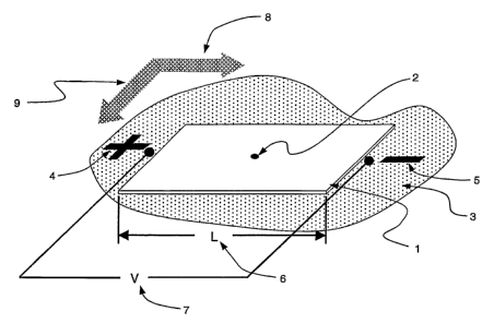
[0035] Fig. 1 is a schematic representation of a planar stationary phase in
contact
with a first mobile phase, having a sample spotted near the center and an
electric field
applied in a first direction in accordance with the present invention.
[0036] Fig. 2 illustrates a sample separated in one dimension in accordance
with
the present invention.
8
CA 02558305 2006-08-31
WO 2005/092013 PCT/US2005/009210
[0037] Fig. 3 illustrates a sample separated in two dimensions in accordance
with
one or more embodiments of the present invention.
[0038] Fig. 4 is a schematic representation of an apparatus in accordance with
one
or more embodiments of the present invention.
[0039] Fig. 5 is a schematic representation of an apparatus in accordance with
one
embodiment of the present invention.
[0040] Fig. 6 is a schematic representation of an apparatus in accordance with
a
second embodiment of the present invention.
[0041] Fig. 7 is a schematic representation of an apparatus in accordance with
a
third embodiment of the present invention.
[0042] Fig. 8 illustrates means for supporting the stationary phase with
respect to
alignment features in accordance with one or more embodiments of the present
invention.
[0043] Fig. 9 illustrates spotting of two samples on a stationary phase prior
to
simultaneous separation under nearly identical conditions.
[0044] Fig. 10 is an illustration of two simultaneous separations resulting
from
applying the two-dimensional separation method to two samples.
[0045] Fig. 11 is an illustration of cassette including a planar stationary
phase and
electrode pairs.
[0046] Fig. 12 illustrates a reagent loading and washing station that may be
used
in conjunction with a cassette to semi-automate the separations process.
[0047] Fig. 13 illustrates a planar electrochromatographic separations station
that
may be used in conjunction with a cassette to semi-automate the separations
process.
Detailed Description
[0048] System and methods for separation of biomolecules, e.g., proteins,
peptides, amino acids, oligosaccharides, glycans and even small drug
molecules, using
electroosmosis-driven planar chromatography are described. In electroosmosis-
driven
planar chromatography an amphiphilic polymeric membrane, amphiphilic thin-
layer
chromatography plate or similar planar substrate provides the stationary phase
for the
separation platform. The planar substrate surface is characterized by a
combination of
charge carrying groups (ion exchangers), non-covalent groups (counterions),
and
nonionic groups that facilitate chemical interactions with the analyte, e.g.,
proteins or
9
CA 02558305 2006-08-31
WO 2005/092013 PCT/US2005/009210
peptides. In a method for the separation of biomolecules using a planar
electrochromatographic system, electroosmotic flow is generated by application
of a
voltage across the planar support in the presence of a miscible organic
solvent-
aqueous buffer mobile phase. Charged ions accumulate at the electrical double
layer
of the solid-phase support and move towards the electrode of opposite charge,
dragging the liquid mobile phase along with them. Charged biomolecules are
separated due to both the partitioning between the liquid phase and the solid
phase
support and the effects of differential electromigration.
[0049] According to one or more embodiment of the present, upon completion of
separation in one direction, e.g., the first dimension separation, the solid
phase is
rinsed, incubated in a second organic solvent-aqueous buffer mobile phase and
then
fractionated in a direction that differs from the original direction of
separation (e.g.,
the second dimension separation). Typically, the second direction is
perpendicular to
the first direction. In one or more embodiments, both dimensions are separated
by the
partitioning effects between the liquid phase and solid support and effects of
electromigration. By adjusting the pH, ionic strength and organic solvent
concentration, electrophoretic separation in one dimension is obtained and
separation
in second dimension is obtained chromatographically.
[0050] Although the systems and methods described herein rnay be used for any
charged molecule, the invention is described with reference to the separation
of
proteins, peptides and glycans. Such description is for convenience only and
is not
intended to limit the invention. Application of the systems and methods
described to
other molecules will be apparent from the description which follows.
[0051] Fig. 1 shows a sample spotted near the center of a planar stationary
phase
in contact with a first mobile phase and an electric field applied in a first
direction in
accordance with one embodiment of the present invention. Referring to Fig. 1,
a
planar stationary phase 1, particularly in the form of a membrane, is wetted
by a first
mobile phase 3 shown as a puddle surrounding the membrane. A small volume of a
sample 2 is dispensed or spotted for example, by hand, on top of the
stationary phase,
near the center of the stationary phase. In other embodiments, spotting is
performed
by dispensing the sample with a pipette, a piezo-electric dispensing tip, a
solid or quill
pin. Spotting may be located anywhere on the membrane and location maybe
determined, in part, by the anticipated direction and extent of
electronugration of the
CA 02558305 2006-08-31
WO 2005/092013 PCT/US2005/009210
species. In another embodiment, precise location in spotting can be achieved
using a
Multiprobe liquid handling robot (PerkinElmer) capable of automated spotting
of
single locations or array spotting. An electric field characterized by
positive 4 and
negative 5 potentials is applied across a first direction 8 of stationary
phase 1. The
applied potential 7 and dimension of the length 6, across which the potential
is
applied, characterize the magnitude of the electric field.
[0052] Fig. 2 shows sample 2 on the planar stationary phase 1 after a period
of
separation in the first dimension 8. Sample 2 is separated into multiple spots
11, some
distinct and some overlapping. This first dimension separation occurs along a
line in
the direction of the applied potential 7.
[0053] Fig. 3 shows the separated sample on planar stationary phase 1 after
both a
separation in a first dimension 8 and a separation in a second dimension 9.
Prior to
the second dimension separation, first mobile phase 3 is removed and a second
mobile
phase 12 is applied to the stationary phase. A second electric field,
characterized by
positive 13 and negative 14 potentials, is applied across the stationary phase
in the
second dimension 9.
[0054] Fig. 4 is a schematic diagram of an apparatus for carrying out the
invention. Referring to Fig. 4, planar stationary phase 1 is placed on a
fixture or
support 16 and a mobile phase (not shown) is applied to stationary phase 1.
Support
16 may be solid, porous, or contain reservoirs or cavities to retain a supply
of mobile
phase to keep the stationary phase wet during separation. Exemplary support
materials include PTFE (Teflon), Macor machineable ceramic, glass, or other
compatible materials. Electrodes 17 and 18 are placed on top of stationary
phase 1,
with wire leads 21 connecting the electrodes to a power supply 22. In one
embodiment, the electrodes are made of non-reactive metals. Exemplary non-
reactive
metals include platinum, palladium, or gold. The electrodes may be in the
shape of
rectangular bars, wires, rods, or any other shape with sufficient length to
substantially
span the width of the stationary phase. In one embodiment in accordance with
the
present invention, power supply 22 is a high-voltage DC supply. Power supply
22
may be controlled by a computer, a programmable controller, a microprocessor,
a
timer or the like in order to precisely control the separation conditions for
more
reproducible results.
11
CA 02558305 2006-08-31
WO 2005/092013 PCT/US2005/009210
[0055] In some embodiments, connection pads 19 and 20 are placed between the
electrodes and the stationary phase to ensure a continuous electrical
connection along
the entire lengths of electrodes 17 and 18. In another embodiment of the
present
invention, connection pads 19 and 20 are made of filter paper.
[0056] In one or more embodiments, planar stationary phase 1 is rotated, e.g.,
by
about 90 degrees, after a separation in first dimension 8 to facilitate
another separation
in second dimension 9. Prior to separation in the second dimension 9, first
mobile
phase 3 is removed and a second mobile phase 12 is applied to the stationary
phase.
Electrodes 17 and 18 are placed on top of stationary phase 1, with wire leads
21
connecting the electrodes to a power supply 22. A second electric field is
applied
across the stationary phase in the second dimension 9.
[0057] In another embodiment, electrodes 17 and 18 are placed on top of planar
stationary phase 1 along second dimension 9 after a separation in first
dimension 8.
Prior to separation in the second dimension 9, first mobile phase 3 is removed
and a
second mobile phase 12 is applied to the stationary phase. A second electric
field is
applied across the stationary phase in the second dimension 9.
[0058] Fig. 5 shows an alternate embodiment of the present invention, where a
wick 23 is placed beneath planar stationary phase 1. Wick 23 is at least as
wide as
stationary phase 1 in the separation direction 9 and longer than stationary
phase 1 in
the separation direction 8. Wick 23 protrudes beyond the ends of the
stationary phase
and is placed in reservoirs 24 and 25 containing additional liquid mobile
phase.
Capillary action draws mobile phase from the reservoirs and into wick 23,
keeping the
wick and the adjacent stationary phase 1 soaked in liquid mobile phase at all
times
during separation. Electrodes 17 and 18 are applied to the top of stationary
phase 1.
In one embodiment, wick 23 is made of filter paper.
[0059] In an alternate embodiment, planar stationary phase l and wick 23 are
rotated, e.g., by about 90 degrees, after a separation in first dimension 8 to
facilitate
another separation in second dimension 9. Prior to separation in the second
dimension
9, first mobile phase 3 is removed and a second mobile phase 12 is applied to
the
stationary phase. Electrodes 17 and 18 are placed on top of stationary phase
1, with
wire leads 21 connecting the electrodes to a power supply 22. A second
electric field
is applied across the stationary phase in the second dimension 9.
12
CA 02558305 2006-08-31
WO 2005/092013 PCT/US2005/009210
[0060] Fig. 6 shows an alternate embodiment of a separation apparatus of the
present invention, where planar stationary phase 1 is placed directly on the
support 16.
Short wicks 26 and 27 are placed between electrodes 17 and 18 and stationary
phase
1. Wicks 26 and 27 extend from under electrodes 17 and 18 to the mobile phase
reservoirs 24 and 25. Wicks 26 and 27 do not extend beyond electrodes 17 and
18
toward the center of stationary phase 1. Capillary action of wicks 26 and 27
draws
liquid mobile phase from reservoirs 24 and 25 to stationary phase 1 but do not
provide
a parallel electrical conduction path across the separation area of stationary
phase 1.
[0061] Fig. 7 shows another embodiment of a separation apparatus in accordance
with the present invention. Referring to Fig. 7, a stationary phase 27 is
placed on the
support 16 without a wick. The length of stationary phase 27 is such that the
ends of
stationary phase 27 protrude into mobile phase reservoirs 24 and 25, beneath
the
surface of the liquid mobile phase. Capillary action of stationary phase 27
draws
liquid mobile phase from reservoirs 24 and 25 to the rest of stationary phase
27.
Electrodes 17 and 18 are applied to the top of stationary phase 27.
[0062] In another embodiment of the present invention, electrodes 17 and 18
are
placed in reservoirs 24 and 25. Electrodes 17 and 18 are in complete contact
with the
mobile phase and the liquid mobile phase conducts current to the stationary
phase.
[0063] Fig. 8 shows another means for holding a stationary phase to a
separation
apparatus in accordance with one or more embodiments of the present invention.
Referring to Fig. 8, stationary phase 36 is held between two rigid or semi-
rigid holders
28 and 29. Holders 28 and 29 are in the form of frames with large openings in
the
center where the stationary phase is exposed for application of sample, mobile
phase,
wicks, contact pads, or electrodes. The large openings also facilitate optical
access to
the stationary phase, allowing imaging the stationary phase after separation
is
completed. The stationary phase is clamped between the two holders in the
manner of
a sandwich using rivets, eyelets, screws, snap tabs, heat staking or other
mechanical
means to fix the two holders together. Alignment features 30 and 31, such as
holes,
slots, pins or the like, could be used to align stationary phase 36 on a
separation
apparatus in accordance with one or more embodiments of the present invention.
The
alignment feature allows precise registration to other instruments, such as
imaging
instruments, spot excising instruments, mass spectrometers, etc. Alignment
features
13
CA 02558305 2006-08-31
WO 2005/092013 PCT/US2005/009210
30 and 31 allow the precise coordinates of separated spots located using one
instrument to be transferred to another instrument.
[0064] The planar stationary phase support includes a frame for supporting a
planar stationary phase and a fastener for securing the planar stationary
phase to the
frame. The frame is open in a center portion for exposing a surface of the
planar
stationary phase, and the open center portion is substantially the size of the
planar
stationary phase to optimize contact of the planar stationary phase with
buffers and
other liquids. The frame may include a recess for receiving a planar
stationary phase.
The planar stationary phase rnay be either a polymer membrane or a silica,
alumina or
titania-based thin layer chromatography resin.
[0065] The planar stationary phase support may include two opposing frames, in
which the frames are configured to secure a planar stationary between the
opposing
frames. The planar stationary phase support may be secured to the frame by a
mechanical fastener. Exemplary mechanical fastener include rivets, eyelets,
screws,
snaps, tabs, clamps, and gaskets. The planar stationary phase may also be
secured
using a crimp or fold of a portion of the frame over an edge of the planar
stationary
phase. The planar stationary phase may be secured to the frame by a chemical
fastener, such as a thermal weld, heat stake, bonding agent or adhesive.
[0066] The planar stationary phase includes alignment of the planar stationary
~,0 phase relative to a predetermined location. Alignment is accomplished by
registration
of a feature or immobilizing the frame with respect to a predetermiend
location. Such
feature or immobilizing means is located at an edge of the frame or on a face
of the
frame. The frame may be aligned using an indentation or projection that is
positionable to register with a complimentary indentation or projection.
Exemplary
projections or indentations include holes, slots, and pins. The alignment
means may
be a spring set that is positionable to repeatably locate the frame relative
to a reference
location.
[0067] Figs. 9 and 10 show another embodiment in accordance with the present
invention where samples 32 and 33 are spotted on planar stationary phase 1 and
are
separated simultaneously into two-dimensional (2D) separation patterns 34 and
35.
When similar mobile phase, electric fields, temperature, and other operating
conditions are applied to a plurality of samples, multiple separation
patterns, as shown
in Figure 10, is obtained. This technique allows the assessment of
differential protein
14
CA 02558305 2006-08-31
WO 2005/092013 PCT/US2005/009210
expression, for example, where the differences in the separation patterns
correspond to
differences in protein contents between the samples.
[0068] Fig. 11 shows a portable cassette 50 that can be used in a planar
electrochromatographic separation apparatus. The cassette includes a frame 51
having
a base 52 and side walls 53. The planar stationary phase (not shown) is
supported
within the frame. The frame is equipped with an inlet port 55 and an outlet
port 56 for
introducing and removing a fluid from the cassette interior, such as a buffer
or
washing liquids. The cassette 50 further includes a cover 60. The cover 60 may
be
transparent to permit imaging or detection in real time or without the need to
remove
the stationery phase from the cassette. The cover 60 may also include
electrode pairs
58, 58' and 59, 59' as an integral component of the cover. The electrodes are
built in
to the cassette and are located near opposing side walls of the frame. The
electrodes
can be spring loaded or otherwise mounted so that they can be reversibly
engaged
with the stationary phase. This features permits the electric field to be
established in
two orthogonal directions. The cover also includes a sample loading port 61.
[0069] In other embodiments, cassette 50 is integrated into a semi-automated
process, as illustrated in Figs. 12 and 13. Fig. 12 shows a reagent loading
and
washing station including cassette 50 and pump station 62. Pump station 62
includes
automated pumps (not shown) for delivery of fluid, e.g., buffer solution and
washing
fluids, through conduits 63 from reservoir 64 to the cassette.
[0070] Fluids exit the cassette through conduit 66 and are stored in a
container
(not shown). Thus, buffer loading, stationary phase rinsing and other fluid
transfers
are carried out without movement or transfer of the planar stationary phase.
[0071] Fig. 13 shows a electrochromatographic separation station 65 that is
integrated with cassette 50 by connection to the first electrode pair 59, 59'.
Reagent
loading station 62 (not shown) is connected to the cassette through inlet and
outlet
parts 55, 59. In operation, a sample is manually loaded onto the planar
stationary
phase in the cassette through loading port 61 and the pump injects a first
buffer or
liquid mobile phase into the appropriate port of the cassette. A voltage then
is applied
and separation is performed in the first dimension. The pump station then
washes the
planar stationary phase to remove the first buffer and injects a second buffer
or liquid
mobile phase. The cassette is repositioned at electrochromatographic
separation
station 65 and is connected using the second electrode pair 58, 58'. The
second
CA 02558305 2006-08-31
WO 2005/092013 PCT/US2005/009210
separation in the second direction is then performed and the planar stationary
phase is
rinsed to remove the second buffer or mobile phase. The stationary phase is
then
manually stained or otherwise treated for detection.
[0072] In one or more embodiments, the separations system includes a cover.
First and second electrodes are integral with the cover and located at first
opposing
side walls of the chamber. Third and fourth electrodes may be integral with
the cover
and are located at second opposing side walls of the chamber.
[0073] Fully automated systems that incorporate the features of automated
proteomic systems are also contemplated.
[0074] As used herein, an "amphiphilic stationary phase" refers to a solid-
support
stationary phase exhibiting both non-polar and polar interactions with the
analyte, e.g.,
proteins, glycans or peptides. An amphiphilic stationary phase includes
regions,
phases or domains that are nonionic and/or hydrophobic in nature as well as
regions,
phases or domains that are highly polar and preferably ionic. The ionic
regions can be
positively or negatively charged. Hydrophobic groups favor the interaction and
retention of the protein during separation, while the ionic groups promote the
formation of the charged double layer used in electrokinetic separation. In
one
embodiment, the amphiphilic stationary phase for protein fractionation has a
combination of charge carrying groups (ion exchangers), non-covalent groups,
and
nonionic groups that facilitate chemical interactions with the analytes. In
another
embodiment, the amphiphilic stationary phase is predominantly hydrophobic, but
partially ionic in character.
[0075] Examples of amphiphilic stationary phase that can be used for protein
separation includes hydrophobic planar support derivatized with sulfonic acid,
sulfopropyl, carboxymethyl, phosphate, diethylaminoethyl,
diethylmethylaminoethy,
allylamine or quartenary ammonium residues or the like. Hydrophobic planar
supports derivatized with sulfonic acid, sulfopropyl, carboxymethyl, or
phosphate
residues enable cathodic electroosmotic flow, while hydrophobic planar
supports
derivatized with diethylaminoethyl, diethylmethylaminoethy, allylamine or
quartenary
ammonium residues enable anodic electroosmotic flow. Membranes, particulate
thin-
layer chromatography (substrates, large pore mesoporous substrates, grafted
gigaporous substrates, gel-filled gigaporous substrates, nonporous reversed
phase
packing material and polymeric monoliths are contemplated.
16
CA 02558305 2006-08-31
WO 2005/092013 PCT/US2005/009210
[0076] Membranes include polymeric sheets, optionally derivatized to provide
the
amphiphilic character of the planar stationary phase. Exemplary hydrophobic
membranes for membrane-based electrochromatography of proteins and peptides
include Perfluorosulfonic Nafion~ 117 membrane (Dupont Corporation), partially
sulfonated PVDF membrane, sulfonated polytetrafluoroethylene grafted with
polystyrene, polychlorotrifluoroethylene grafted with polystyrene, or the
like.
Sulfonation of polyvinylidene difluoride (PVDF) can be achieved by incubation
with
sulfuric acid at a moderately high temperature. The degree of sulfonation can
be
systematically varied, where ion exchange capability of 0.25 meqlg is
considered as
"moderate" sulfonation. In these membranes separation depends upon the
electrostatic
interaction of proteins with sulfonated residues in combination with
hydrophobic
interactions with aromatic residues in the substrate. At pH in the range from
about pH
2.0 to about pH 11.0, the protonated primary amine groups on the proteins will
interact with sulfonated residues on the membrane. This interaction is
diminished at
pH greater than about pH 11Ø Sulfonate residues will be protonated at a pH
less than
about pH 2..0 and will lead to a decline in the electroosmosis driving force
of the
separation.
[0077] In some embodiments, PVDF membranes, used for the isolation by
electroblotting of proteins separated by gel electrophoresis, can be
derivatized with
cationic functional groups in order to generate an amphiphilic membrane (e.g.,
Immobilon-CD protein sequencing membrane (Millipore Corporation)). For
example,
PVDF membrane can be etched with 0.5 M alcoholic I~OH and subsequently reacted
with polyallylamine under alkaline conditions. As another example, PVDF
membranes can be derivatized with diethylaminoethyl or quartenary ammonium
residues.
[0078] In some embodiments, the membrane is unsupported. In other
embodiments, the membrane is supported or semi-supported. For example, the
membrane can be held between two rigid or semi-rigid holders in the form of
frames
with large openings in the center. The membrane may also be rigidly supported
on a
solid support, for example, a glass plate. Membranes may be substantially non-
porous. In such instances, the mobile phase moves over the surface of the
membrane.
In other embodiments, the membrane may be porous, in which case the mobile
phase
moves through the pores andlor channels of the membrane. Separation occurs by
17
CA 02558305 2006-08-31
WO 2005/092013 PCT/US2005/009210
preferential interactions of the proteins with the hydrophobic surfaces or the
interstial
surfaces of the membrane.
[0079] As another example, a planar stationary phase useful for separation of
proteins include silica thin-layer chromatography plates derivatized with
alkyl groups
(e.g. C3_Cls surface chemistry), aromatic phenyl residues, cyanopropyl
residues or the
like. In these instances, the silanol groups provide the ion exchange
qualities of the
amphiphilic support and can be deprotonated at a pH of 8, leading to
electroosmosis
and thereby providing the ion exchange qualities of the amphiphilic support.
At pH
below pH 3, there will be a reduction or elimination in electroosmosis. In
some
embodiments, both hydrophobic groups, e.g., alkyl, and charged groups, e.g.,
sulfonic
acid, can be attached to the same silica particle. As a further example, a
stationary
phase support for the separation of peptides and proteins by planar
electrochromatography includes a gamma-glycidoxypropyltrimethoxysilane
sublayer
attached to the silica support of a thin-layer chromatography plate. A
sulfonated layer
is then covalently affixed between the sublayer and an octadecyl top layer.
For
separation of proteins in the 10 and 100 kDa range using a silica-based
stationary
phase, it is expected that derivitization with C8 and C4 groups, respectively,
may be
used. Phenyl functionalities are slightly less hydrophobic than C4
functionalities and
may be advantageous for the separation of certain polypeptides.
[0080] The planar stationary phase includes pores or connected pathways of a
dimension that permits unimpeded migration of the proteins. For particulate
stationary phases, such as silica thin-layer chromatography plates or
particulate-based
polymer membranes, the stationary phase consists of particles that form pores
of about
30-100 nanometers in diameter, although for some smaller peptides with
molecular
weights of 2,000 daltons or less, 10 nanometers pores may be acceptable.
Typical
absorbants commercially available for thin-layer chromatography are made of
particles that form pores sizes of only 1-6 nm, which precludes effective use
for
protein separations. The particles may have a diameter of about 3-50 microns,
with
the smaller diameter particles typically producing higher resolution protein
separations. For higher protein loads, large particle absorbents are
preferable. This is
particularly advantageous for the preparative scale isolation of proteins. The
size
distribution of the particles should be relatively narrow and particles are
preferably
spherical, rather than irregularly shaped. While the base material of the
particles can
18
CA 02558305 2006-08-31
WO 2005/092013 PCT/US2005/009210
be silica, synthetic polymers, such as polystyrene-divinylbenzene (or any of
the above
mentioned hydrophobic polymers) are also expected to be appropriate.
[0081] The liquid mobile phase typically includes an organic phase and an
aqueous phase. Exemplary mobile phases include methanol-aqueous buffer,
acetonitrile-aqueous buffer, ethanol-aqueous buffer, isopropyl alcohol-aqueous
buffer,
butanol-aqueous buffer, isobutyl alcohol-aqueous buffer, propylene carbonate-
aqueous buffer, furfuryl alcohol-aqueous buffer systems or the like. The basic
principles of electrochromatography provide the foundation for systematic
selection of
stationary phase supports, mobile phase buffers and operating conditions, and
allow
for the adaptation of the technology to a broad range of applications in
proteomics,
drug discovery and the pharmaceutical sciences. As with CEC, mobile phases
rich in
organic modulators will exhibit relatively little chromatographic retention
and in
mobile phases low in organic modulator, chromatographic retention will
dominate the
separation process.
[0082] In one embodiment of the present invention, the concentrations of
organic
modulators in liquid mobile phases are in the range of about 0% to about 60%.
In
another embodiment, the ionic strength of liquid mobile phases can be from
about 2
mM to about 150 mM. Exemplary liquid mobile phase formulations include 20 mM
ammonium acetate, pH 4.4, 20% acetonitrile; 2.5 mM ammonium acetate, pH 9.4,
50% acetonitrile; 25 mM Tris-HCI, pH 8.0/acetonitrile (40/60 mix); 10-25 mM
sodium acetate, pH 4.5, 55% acetonitrile; 60 mM sodium phosphate, pH2.5/30%
acetonitrile; 5 mM borate, pH 10.0, 50% acetonitrile; 5-20 mM sodium
phosphate, pH
2.5, 35-65% acetonitrile; 30 rnM potassium phosphate, pH 3.0, 60% acetonitrile
and
10 mM sodium tetraborate, 30% acetonitrile, 0.1 % trifluoroacetic acid; 20%
methanol,
80% 10 mM MES, pH 6.5, 5 mM sodium dodecyl sulfate; 20% methanol, 80% 10
mM MES, pH 6.5, 5 mM sodium phosphate, pH 7.0/methanol (4:1, v/v); 4 mM Tris,
47 mM glycine, pH 8.1; 20 mM sodium phosphate, pH 6.0, 150 mM NaCl; 20 rnM
Tris-HCI, pH 7.0, 150 mM NaCl; 5 mM sodium borate, pH 10.0; or the like.
[0083] In some embodiments, different cathode and anode buffers could be used
as a discontinuous buffer system for the separation of proteins. In certain of
these
embodiments, the amphiphilic stationary phase could be incubated in a buffer
that is
compositionally different from either electrode buffer. Additives, such as
carrier
ampholytes may be included in the buffer in which the stationary phase is
incubated.
19
CA 02558305 2006-08-31
WO 2005/092013 PCT/US2005/009210
In other embodiments, the composition of the mobile phase could be altered
temporally to provide a composition gradient that facilitates separation of
proteins.
[0084] In two-dimensional separation of proteins on an amphiphilic stationary
phase using planar electrochromatography, protein sample is applied on the
center of
the membrane (dry or pre-wetted with mobile phase) and the planar stationary
phase is
then incubated in a mobile phase. Once the proteins are electrophoretically
separated
in one direction, the planar stationary phase is washed and incubated in a
second
mobile phase, and then electrophoretically separated in a direction
perpendicular to
the first direction. In one embodiment in accordance with the present
invention, liquid
mobile phases can be adjusted to different pH values, concentrations of
organic
solvent, and ionic strengths to facilitate 2D separations of proteins on the
amphiphilic
substrate. For example, one mobile phase will have acidic pH (ca. pH 4.5) and
the
other basic pH (ca pH 8.5). The pH of the buffers will affect the total charge
of the
individual protein species and thus influence their electrokinetic migration.
Changes
to the concentration of organic solvent in liquid mobile phase will impact the
strength
of interaction of the proteins with the hydrophobic component of the
stationary phase.
Finally, the ionic strength of the buffer will change the separation
properties of the
proteins in the two dimensions. By manipulating pH, ionic strength and organic
solvent concentration, separation in one dimension will occur
electrophoretically and
separation in the other dimension will occur chromatographically.
[0085] Protein samples are prepared by first dissolving the proteins in the
mobile
phase or a weaker solvent of lower ionic strength. In some embodiments,
"biological
buffers", such as Good's buffers, are used for sample preparation. These
biological
buffers produce lower currents than inorganic salts, thereby allowing the use
of higher
sample concentrations and higher field strengths. Exemplary Good's buffers
include
N-(2-Acetamido)-2-aminoethanesulfonic acid (ACES), N-(2-
Acetamido)iminodiacetic
acid (ADA), N,N-Bis(2-hydroxyethyl)-2-aminoethanesulfonic acid (BES), N,N-
Bis(2-
hydroxyethyl)glycine (BICINE), Bis(2-
hydroxyethyl)iminotris(hydroxyhnethyl)methane (BIS-TRIS), N-Cyclohexyl-3-
aminopropanesulfonic acid (CAPS), N-Cyclohexyl-2-hydroxy-3-
aminopropanesulfonic acid (CAPSO), N-Cyclohexyl-2-aminoethanesulfonic acid
(CHES), 3-[N,N-Bis(hydroxyethyl)amino]-2-hydroxypropanesulfonic acid (DIPSO),
3-[4-(2-Hydroxyethyl)-1-piperazinyl]propanesulfonic acid (EPPS), 2-[4-(2-
CA 02558305 2006-08-31
WO 2005/092013 PCT/US2005/009210
Hydroxyethyl)-1-piperazinyl]ethanesulfonic acid (HEPES), 2-Hydroxy-3-[4-(2,-
hydroxyethyl)-1-piperazinyl]- propanesulfonic acid, monohydrate (HEPPSO), 2-
Morpholinoethanesulfonic acid, monohydrate (MES), 3-Morpholinopropanesulfonic
acid (MOPS), 2-Hydroxy-3-morpholinopropanesulfonic acid (MOPSO), Piperazine-
1,4-bis(2-ethanesulfonic acid) (PIPES), Piperazine-1,4-bis(2-ethanesulfonic
acid),
sesquisodium salt (PIPES, sesquisodium salt), Piperazine-1,4-bis(2-hydroxy-3-
propanesulfonic acid), dehydrate (POPSO), N-Tris(hydroxymethyl)methyl-3-
aminopropanesulfonic acid (TAPS), N-Tris(hydroxymethyl)methyl-2-hydroxy-3-
aminopropanesulfonic acid (TAPSO), Tris-(hydroxymethyl)aminomethane (TRIS), N-
Tris(hydroxymethyl)methyl-2-aminoethanesulfonic acid (TES), and N-
[Tris(hydroxymethyl)methyl]glycine (TRICINE). If salts are used to facilitate
extraction and isolation of the protein specimen, desalting of protein samples
may be
performed using reverse phase resins by organic solvent-based protein
precipitation or
by sample dialysis prior to sample fractionation by planar
electrochromatography.
[0086] In some embodiments, protein samples are prepared by first dissolving
the
proteins in HPLC solvent systems thereby avoiding the use of detergents,
chaotropes
and strong organic acids for protein dissolution. HPLC solvent systems include
buffered solutions containing organic solvents, such as methanol or
acetonitrile, may
be employed to prepare the biological specimens. For example, 60% methanol or
acetonitrile, 40% water containing 0.1 % formic acid or 60% methanol or
acetonitrile,
40% 50 mM ammonium carbonate, pH 8.0 are suitable sample solubilization
buffers.
In one embodiment, final protein concentration in the solubilization buffer is
from
about O.OSrng/ml to about 5 mg/ml. In another embodiment, final protein
concentration in the solubilization buffer is from about 0.4 mg/ml to about
0.6 mg/ml.
Extraction and solubilization of proteins can be facilitated by intermittent
vortexing
and sonication. Surfactants are well known to suppress peptide ionization in
mass
spectrometry and also to interfere with chromatographic separations,
particularly with
reversed-phase liquid chromatography. Buffered solutions containing organic
solvents are more compatible with liquid chromatography and mass spectrometry
and
thus facilitate characterization of the proteins after planar
electrochromatography.
Another important advantage of the buffered organic solvent extraction
procedure is
that it facilitates solubilization, separation and identification of integral
membrane
proteins, including proteins containing transmembrane-spanning helices.
21
CA 02558305 2006-08-31
WO 2005/092013 PCT/US2005/009210
[0087] Planar electrochromatographic separation of peptides and proteins is
performed by directly applying an electric field across the membrane or thin
layer
chromatography plate. In one embodiment, the planar surface may be interfaced
with
the electrical system through the use of wicks, also referred to as buffer
strips. A wick
is a solid or semisolid medium used to establish uniform electrical paths
between the
planar solid phase and the electrodes of a horizontal electrophoresis
apparatus. For
example, a wick may be composed of cellulose-based filter paper, Rayon fiber,
buffer-
impregnated agarose gel, moistened paper towel, or the like.
[0088] Application of an electric field in electrochromatographic systems
could
result in Joule heating which in turn could to lead to evaporation of liquid
mobile
phase from the membrane or plate surface. The evaporation of the mobile phase
could
result in decreased current, drying of the surface, and subsequent degradation
in the
quality of the separation. In one embodiment in accordance with the present
invention, the planar stationary phase is covered with a glass plate, silicone
oil or
other impermeable barrier to reduce the evaporation of the mobile phase as a
result of
Joule heating. Further, flow of the mobile phase across the membrane or plate
may be
impeded in the forward direction, causing the electroosmotic flow to drive the
liquid
mobile phase to the surface of the membrane or plate. This can result in poor
resolution separations and arcing of the electrophoretic device. Adjusting
mobile r
phase pH or ionic strength will aid in optimizing conditions for the
electrically driven
separation. In one embodiment, operating current for protein or peptide
separations is
from about 1~ ~A to about 500 mA and the electric field strength applied to
the
separation is from about 50 voltslcm to about 900 volts/cm. In another
embodiment,
the electric field strength applied to the separation is from 200 volts/cm to
about 600
volts/cm. In certain embodiments of the present invention, separations of
proteins can
be performed using constant voltage, constant current or constant power mode,
the
latter resulting in constant amount of Joule heating in the system.
[0089] In one or more embodiments, planar electrochromatography can be used
with other electrophoresis modalities, such as immobilized metal affinity
electrochromatography, immunoaffinity electrochromatography, zonal
electrophoresis, electromolecular propulsion, electrokinetic chromatography,
isoelectric focusing, nonequillibrium pH electrophoresis and micellar
electrokinetic
chromatography. In certain embodiments, a two component or dual phase planar
22
CA 02558305 2006-08-31
WO 2005/092013 PCT/US2005/009210
substrate can be created. For example, immobilized metal ion affinity
electrochromatography, followed by reverse-phase electrochromatography could
be
performed. One edge of the planar support, for example, a 1 cm strip along one
side
of the membrane, can be derivatized with metal-chelating groups (e.g.,
iminodiacetic
acid, nitrilotriacetic acid) while the rest of the membrane will possess
sulfonate ion
exchange characteristics. The membrane will be charged with a metal ion, such
as
Ni(II), Cu(II), Ca(II), Fe(II~ or Ga(III), and the chelating groups will
selectively
retain these metal ions. Protein sample can be applied as a discrete spot on
the
membrane and subjected to electrochromatographic separation along the length
of the
modified strip using a buffer appropriate for binding. In one embodiment,
Fe(III)- or
Ga(III)-charged membrane strips, 20 mM sodium acetate, pH 4.0 can be used.
Upon
completion of first fractionation, the membrane is rinsed in a second buffer
and
subjected to electrochromatography in a direction perpendicular to the
direction of
original separation. A comparison of the profile generated with the described
membrane to a profile generated from a membrane lacking the metal chelating
strip
will reveal metal-binding proteins as spots whose migration is altered between
the two
profiles. Other combined modalities of separation are envisioned, including
ration
exchange electrochromatography and reverse-phase electrochromatography.
[0090] Proteins, peptides and glycans may be detected after planar
electrochromatography using a variety of detection modalities well known to
those
skilled in the art. Exemplary strategies employed for general protein
detection include
organic dye staining, silver staining, radio-labeling, fluorescent staining
(pre-labeling,
post-staining), chemiluminescent staining, mass spectrometry-based approaches,
negative-staining approaches, contact detection methods, direct measurement of
the
inherent fluorescence of proteins, evanescent wave, label-free mass detection,
optical
absorption and reflection, or the like. In negative-staining approaches, the
proteins
remain unlabeled, but unoccupied sites on the planar surface are stained. In
contact
detection methods, another membrane or filter paper that has been imbibed with
a
substrate is placed in contact with the planar surface and protein species
resident on
the planar stationary phase interact with the substrate molecules to generate
a product.
In direct measurement of the inherent fluorescence of proteins, solid-phase
supports of
low inherent fluorescence are used. Exemplary detection methods suitable for
revealing protein post-translational modifications include methods for the
detection of
23
CA 02558305 2006-08-31
WO 2005/092013 PCT/US2005/009210
glycoproteins, phosphoproteins, proteolytic modifications, S-nitrosylation,
arginine
methylation and ADP-ribosylation. Exemplary methods for the detection of a
range of
reporter enzymes and epitope tags include methods for visualizing (3-
glucuronidase, (3-
galactosidase, oligohistidine tags, and green fluorescent protein. For optimal
performance of these detection technologies, it will be necessary to use solid-
phase
supports of low inherent fluorescence.
[0091] Protein samples that have undergone planar electrochromatography appear
as discrete spots on the strip that are accessible to staining or
immunolabeling as well
as to analysis by various detection methods. Exemplary detection methods
include
mass spectrometry, Edman-based protein sequencing, or other micro-
characterization
techniques. In one embodiment, proteins bound to the surface of the membrane
can
be labeled by reagents, such as, antibodies, peptide antibody mimetics,
oligonucleotide aptamers, quantum dots, Luminex beads or the like.
[0092] In some embodiments, cherniluminescence-based detection of proteins on
planar surfaces can be used prior to or after fractionation by planar
electrochromatography. In one embodiment, proteins can be biotinylated and
then
detected using horseradish peroxidase-conjugated streptavidin and the Western
Lightning Chemiluminescence kit (PerkinElmer). In another embodiment, proteins
may be fluorescently stained or labeled and the fluorescent dye subsequently
chemically excited by nonenzymatic means, such as the bis(2,4,6-
trichlorophenyl)oxalate (TCPO)-H202 reaction.
[0093] Separations of protein, using the method in accordance with one or more
embodiments of the present invention, can be achieved in a short duration.
Proteins
are spotted on a planar substrate, subjected to first dimension separation,
rinsed and
subjected to second dimension separation thereby providing access to the
proteins and
peptides on the surface of the stationary phase for detection. In one
embodiment,
SYPRO Ruby protein blot stain (Molecular Probes) is capable of detecting
proteins on
a surface within about 15 minutes. Additionally, the planar support itself
serves as a
mechanically strong support, allowing archiving of the separation profiles
without the
need for vacuum gel drying.
[0094] In certain embodiments of the present invention, planar
electrochromatography can be used to fractionate very large proteins, very
small
proteins, highly acidic proteins, highly basic proteins and hydrophobic
proteins. In
24
CA 02558305 2006-08-31
WO 2005/092013 PCT/US2005/009210
some embodiments, large mufti-subunit complexes can be fractionated on the
surface
of a membrane. In one embodiment, mobile phases containing high concentrations
of
organic solvents are used to separate hydrophobic integral membrane proteins.
In
another embodiment, planar electrochromatography can be used to separate
"electrophoretically silent" mutations, wherein proteins and peptides differ
only by an
uncharged amino acid residue. In a further embodiment, the planar
electrochromatography system can be used to fractionate intact proteins. This
is
advantageous with respect to the analysis of protein isoforms arising from
post-
translational modification or differential splicing.
[0095] Proteomics studies are often based upon the comparison of different
protein profiles. The central objective of differential display proteomics is
to increase
the information content of proteomics studies through multiplexed analysis.
Currently, two principal gel-based approaches to differential display
proteomics are
being actively pursued, difference gel electrophoresis (DIGE) and Multiplexed
Proteomics (MP). In one embodiment in accordance with the present invention,
planar electrochromatography can be used with difference gel electrophoresis
(DIGE)
to increase the information content of proteomics studies through multiplexed
analysis. Succinimidyl esters of the cyanine dyes (e.g., Cy2, Cy3 and Cy5) can
be
employed to fluorescently label as many as three different complex protein
populations prior to mixing and running them simultaneously on the same 2D gel
using DICE. Images of the 2D gels are acquired using three different
excitationlemission filter combinations, and the ratio of the differently
colored
fluorescent signals is used to find protein differences among the samples.
DIGE
allows two to three samples to be separated under identical electrophoretic
conditions,
simplifying the process of registering and matching the gel images. DIGE can
be used
to examine differences between two samples (e.g., drug-treated-vs-control
cells or
diseased-vs-healthy tissue). The principle benefit of the planar
electrochromatography
technology detailed in this disclosure with respect to DIGE is that protein
separations
can be achieved more quickly and samples are more readily evaluated by mass
spectrometry after profile differences are determined. One requirement of DIGE
is
that from about 1 % to about 2% of the lysine residues in the proteins be
fluorescently
modified, so that the solubility of the labeled proteins is maintained during
electrophoresis. Very high degrees of labeling can be achieved when
separations are
CA 02558305 2006-08-31
WO 2005/092013 PCT/US2005/009210
performed by the planar electrochromatography technique, due to the fact that
organic
solvents are employed in the mobile phase and sample buffers. High degrees of
labeling should in turn dramatically improve detection- sensitivity using the
DIGE
technology.
[0096] In another embodiment, planar electrochromatography can be used with
Multiplexed Proteomics to increase the information content of proteomics
studies
through multiplexed analysis. The Multiplexed Proteornics (MP) platform is
designed
to allow the parallel determination of protein expression levels as well as
certain
functional attributes of the proteins, such as levels of glycosylation, levels
of
phosphorylation, drug-binding capabilities or drug-metabolizing capabilities.
The MP
technology platform utilizes the same fluorophore to measure proteins across
all gels
in a 2DGE database, and employs additional fluorophores with different
excitation
and/or emission maxima to accentuate specific functional attributes of the
separated
species. With the MP platform, a set of 2D gels is fluorescently stained and
imaged to
reveal some functional attribute of the proteins, such as drug-binding
capability, or a
particular post-translational modification. Then, protein expression levels
are revealed
in the same gels using a fluorescent total protein stain. Differential display
comparisons are made by computer, using image analysis software, such as Z3
program (Compugen, Tel Aviv, Israel). All gels are imaged using the same
excitation/emission filter sets and resulting images are then automatically
matched,
with the option of adding some manual anchor points to facilitate the process.
Any
two images can then be re-displayed as a single pseudo-colored map. In
addition,
quantitative information can be obtained in tabular form, with differential
expression
data calculated. With a gel imaging platform similar profiles from different
gels, such
as total protein patterns, are matched by computer; while dissimilar ones from
the
same gel, such as total protein patterns and glycoprotein patterns, are
superimposed
and matched by computer. In MP the gels must be serially stained and imaged,
as
succeeding stains mask their predecessors in polyacrylarnide gels. In one
embodiment, planar electrochromatography can be used to assist MP in
simultaneous
imaging of multiple signals on profiles generated. Fluorescent dyes do not
have the
same strong tendency to mask one another on polymeric membranes.
[0097] In alternate embodiments, planar electrochromatrography can be used
with
MALDI-TOF MS for direct analysis of proteins. In this embodiment, proteins are
26
CA 02558305 2006-08-31
WO 2005/092013 PCT/US2005/009210
fractionated on solid phase supports followed by direct probing with MALDI-TOF
laser. In one embodiment, the planar electrochromatrography system in
accordance
with the present invention can be used with an orthogonal MALDI-TOF mass
spectrometer (e.g., PrOTOF 2000 PerkinElrner, Boston, MA, IJSA/MDS Sciex,
Concord, ON, Canada). The prOTOF 2000 MALDI O-TOF mass spectrometer is a
MS MALDI with orthogonal time of flight technology. The prOTOF's novel design
provides improved instrument stability, resolution, and mass accuracy across a
wide
mass range compared with conventional linear or axial-based systems. The more
accurate and complete protein identification achieved with the prOTOF 2000
reduces
the need for peptide sequencing using more complicated tandem mass
spectrometry
techniques such as Q-TOF and TOF-TOF. The instrument is particularly well
suited
for planar electrochromatography because the MALDI source is decoupled from
the
TOF analyzer. As a result, any discrepancies arising from the solid phase
surface
topography or differential ionization of the sample from the surface are
eliminated
before the sample is actually delivered to the detector. The presentation of
the
proteins bound to a solid phase surface facilitates removal of contaminating
buffer
species and exposure to protein cleavage reagents (e.g., trypsin) prior to
analysis by
mass spectrometry. The use of HPLC-based buffers in the fractionation process
minimizes the potential for downstream interference by detergents and
chaotropes
during mass spectrometry-based analysis.
[0098] Laser desorption of proteins by direct MALDI-TOF MS-based surface
scanning of carrier ampholyte isoelectric focusing gels, immobilized pH
gradient
isoelectric focusing gels, native polyacrylamide gels, and SDS-polyacrylamide
gels
can be achieved, with sub-picomolar detection sensitivities. The procedure is
currently quite slow, however, requiring a day to run the gel, two days to dry
it down
and two days to acquire spectra. In one embodiment of the present invention,
planar
electrochromatography can be used with MALDI-TOF MS for direct analysis of
proteins by providing proteins conveniently affixed to solid phase supports
and thus
suitably presented for direct probing by the MALDI-TOF laser. "Virtual" 2D
profiles
can be generated by 1D planar electrochromatographic separations followed by
desorbing proteins directly from the planar substrate using MALDI-TOF mass
spectrometry, in effect substituting mass spectrometry for SDS polyacrylamide
gel
electrophoresis. Analytical data obtained can be presented as a computer-
generated
27
CA 02558305 2006-08-31
WO 2005/092013 PCT/US2005/009210
image with 2D gel type appearance. In another embodiment, planar
electrochromatography can be used as a starting point for high throughput
peptide
mass fingerprinting and glycosylation analysis using chemical printing
techniques
such as piezoelectric pulsing where multiple chemical reactions are conducted
on
different regions of a spot by defined microdispensing of trypsin in-gel
digestion
procedures, and allowing peptide mass profiles and characterization of
glycosylation,
for example, to be achieved from the same spot. Defined microdispensing of
trypsin
and MALDI-TOF matrix solutions bypasses multiple liquid handling steps usually
encountered with in-gel digestion procedures, and thus streamlines protein
characterization methods.
[0099] In one or more embodiments, planar electrochromatography can be used
with mass tagging techniques for differential display proteomics where
relative
abundances of different proteins in biological specimens are correlated with
physiological changes. For example, Isotope-coded affinity tag (ICAT) peptide
labeling is one such technique useful for distinguishing between two
populations of
proteins using isotope ratios. ICAT reagent employs a reactive functionality
specific
for the thiol group of cysteine residues in proteins and peptides. Two
different isotope
tags are generated by using linkers that contain either eight hydrogen atoms
(do, light
reagent) or eight deuterium atoms (d8, heavy reagent). A reduced protein
mixture
from one protein specimen is derivatized with the isotopically light version
of the
ICAT reagent, while the other reduced protein specimen is derivatized with the
isotopically heavy version of the ICAT reagent. Next, the two samples are
combined,
and proteolytically digested with trypsin or Lys-C to generate peptide
fragments. The
combined sample can be fractionated by planar electrochromatography. The ratio
of
the isotopic molecular weight peaks that differ by 8 daltons, as revealed by
mass
spectrometry, provides a measure of the relative amounts of each protein from
the
original samples. Other mass tagging approaches include growth of cells in
either
1~N- or 15N-enriched medium, use of regular water (H2i6O) and heavy water
(H218O)
as the solvent during Glu-C proteolysis of samples, use of acetate (do) and
trideuteroacetate (d3) to acetylate primary amino groups in peptides, methyl
esterification of aspartate and glutamate residues using regular methanol (do)
or
trideuteromethanol (d3), 12C and 13C labeled tri-alanine peptides
iodoacetylated on
their N-termini, and use of 1,2.-ethanedithiol (do) and tetraalkyl deuterated
1,2-
28
CA 02558305 2006-08-31
WO 2005/092013 PCT/US2005/009210
ethanedithiol (d4) to measure differences between O-phosphorylation sites in
samples
using beta-elimination chemistry.
[0100] Recently, it has been demonstrated that the advantages of 2DGE and ICAT
labeling technology can be combined into a single differential display
platform.
Proteins from two different samples are labeled with heavy and light ICAT
reagents,
combined and then separated by 2DGE. The gel-separated proteins are detected
with
a sensitive protein stain, excised, treated with protease and identified by
peptide mass
profiling. Additionally, selected peptides can be evaluated further using
collision-
induced dissociation (CID) and sequence database searching. One important
application of ICAT differential display in 2D gels is for the assessment of
the relative
abundances of protein isoforms that arise from post-translational
modification. In one
embodiment of the present invention, 2D planar electrochromatography can be
combined with ICAT labeling into a single platform for differential display
proteomics using the ICAT reagents. Separations are much faster and the
proteins are
more amenable to downstream mass spectrometry-based analysis.
[0101] Mass tagging approaches based upon the same basic principles as the
ICAT strategy include growth of cells in either 14N- or 15N-enriched medium,
and the
use of regular water (Hal6O) and heavy water (H2180) as the solvent during Glu-
C
proteolysis of samples, leading to the incorporation of two 180 or two 160
atoms in the
C-terminal moiety of each proteolytic fragment. This results in a 4 dalton
difference
in mass between paired peptides. Acetate (do) and trideuteroacetate (d3) can
be
employed to acetylate primary amino groups in peptides. Similarly, methyl
esterification of aspartate and glutamate residues using regular methanol (do)
or
trideuteromethanol (d3) can be used as an isotope tagging strategy. laC and
13C
labeled tri-alanine peptides iodoacetylated on their N-termini for mass
tagging
experiments. Finally, 1,2-ethanedithiol (do) and tetraalkyl deuterated 1,2-
ethanedithiol
(d4) can be employed to measure differences between O-phosphorylation sites in
samples using beta-elimination chemistry. The pendant sulfhydryl group is then
reacted with biotin iodoacetamidyl-3,6-dioxaoctanediamine. In one embodiment
of
the present invention, 2D planar electrochromatography can be used with mass
tagging technologies as a separation platform for differential analysis of
protein
expression changes and post-translational modification changes.
29
CA 02558305 2006-08-31
WO 2005/092013 PCT/US2005/009210
[0102] In one or more embodiments, planar electrochromatography can be used
with inductively-coupled plasma mass spectrometry (ICP-MS) for the trace
elemental
analysis of metalloproteins, such as selenoproteins, zinc metalloenzymes,
cadmium-
binding proteins, cisplatin-binding drug targets, and myoglobins subsequent to
fractionation by planar electrochromatography. Laser ablation ICP-MS permits
trace
element analysis by combining the spatial resolution of an ultraviolet laser
beam with
the mass resolution and element sensitivity of a modern ICP-MS. UV laser
light,
produced at a wavelength of 193-266 nm is focused on a sample surface, causing
sample ablation. Ablation craters of 15-20 microns are routinely produced by
the
instrumentation. No special sample preparation is required for the procedure.
Ablated
material is transported in an argon carrier gas directly to the high
temperature
inductively-coupled plasma and the resulting ions are then drawn into a mass
spectrometer for detection and counting. A mass filter selects particles on
the basis of
their charge/mass ratio so that only specific isotopes are allowed through the
filter and
can enter the electron multiplier detector mounted at the end of the mass
spectrometer
(quadrupole, magnetic sector or time-of flight instrumentation). Detected
signals of
individual isotopes can be converted to isotopic ratios or, when standards are
measured along with the unknowns, to the actual element concentrations.
[0103] Laser ablation ICP-MS can be used for directly measuring phosphorous as
m/z 31 signal liberated from phosphoproteins on electroblot membranes. Using
Laser
ablation ICP-MS, 16 pmole of the pentaphosphorylated beta-casein can be
detected on
polymeric membranes. In another embodiment, planar electrochromatography can
be
used as a platform for the direct analysis of protein phosphorylation, without
the use
of radiolabels or surrogate dyes, such as Pro-Q Diamond phosphoprotein stain
(Molecular Probes).
[0104] The detection of low concentrations of phosphorous presents certain
analytical challenges for ICP-MS due to its poor ionization in the argon ICP
and the
presence of interfering polyatomic species directly at mass 31 (15N160 and
14Ni60iH)
and indirectly at mass 32 (1602 arid 32S). Phosphorous has a high first
ionization
potential of 10.487 electron volts (Wilbur and McCurdy, 2001). This translates
to a
poor conversion of phosphorous (P) atoms to P+ ions in the inductively coupled
plasma. In a well-optimized ICP-MS, this translates to a 6% conversion of P
atoms to
P+ ions, a relatively low response factor for ICP-MS. It is lcnown to one
skilled in the
CA 02558305 2006-08-31
WO 2005/092013 PCT/US2005/009210
art that phosphate groups in proteins and peptides readily bind certain
trivalent metal
ions, such as aluminum (III), gallium (III) and iron (III). Using ICP-MS, as
little as 1
part per billion (ppb) of these metal ions can be detected. The ionized
conversion of
aluminum, which has a first ionization potential of 5.986 electron volts, is
99°lo under
identical run conditions as described for phosphorous. Thus, detecting
aluminum
instead of phosphorous improves detection 16-fold. In addition, the specific
detection
of the trivalent metal ions shifts the detection window away from the cited
biological
background signal. The atomic masses of aluminum, gallium and iron are 26.98,
69.7
and 55.85, respectively. Among these three trivalent metal ions, the ferric
ion poses
problems due to polyatomic interferences arising from ArN, Ar0 and ArOH at the
interface region of the ICP-MS. Gallium is probably the most suitable metal
ion for
the proposed application. Both 69Ga, and ~lGa signal could be quantified by
the
method, minimizing the probability of overlapping signal from other molecular
species.
[0105] The detection of proteins using ICP-MS-based detection procedure
includes the following steps. First, proteins are separated by 2D planar
electrochromatography as described in accordance with one embodiment of the
present invention. The planar substrates are then incubated with 1 mM gallium
chloride, 50 mM sodium acetate, pH 4.5, 50 mM magnesium chloride. Next, the
planar substrates are washed repeatedly in 50 mM sodium acetate, pH 4.5, 50 mM
magnesium chloride to remove excess metal ions. The individual spots on the
planar
surface are subjected to laser ablation ICP-MS methods where gallium signal is
quantified rather than the phosphorous signal. Alternatively, the phosphorous
signal
can be read without incubating in the gallium solution. Sampling can be
performed by
single or mufti-spot analysis, straight line scans or rastering. To aid in
spot selection,
the proteins on the planar substrate can be stained with a total protein
stain, prior to
the incubation with the gallium ions.
[0106] In one or more embodiments, planar electrochromatography can be used
with protein microarrays for protein expression profiling and studying protein
function. Planar electrochromatography can be used to provide a relatively
simply
method for generating protein microarrays. Small planar surfaces may be
spotted with
a defined mixture of proteins that are subsequently fractionated by 2D planar
electrochromatography. Though the constituent proteins are not explicitly
assigned a
31
CA 02558305 2006-08-31
WO 2005/092013 PCT/US2005/009210
pre-determined coordinate in the resulting orthogonal matrix of spots thus
generated,
the identities of the spots can simply be determined by mass spectrometry, by
immunodetection or by systematic omission of each protein from the mixture in
subsequent separations. Once the location of each protein in the profile is
known, the
array may be used as conventional protein arrays, such as for profiling
autoantibody
responses in autoimmune disease and screening for other protein-protein,
receptor-
ligand, enzyme-substrate, enzyme-inhibitor or even protein-DNA interactions.
The
advantages of the arraying approach are that a dedicated pin-based or
piezoelectric
spotting device is not required and the membrane arrays are amenable to filter-
based
protein microarray techniques as described recently. For example, a filtration
approach that allows mufti-stacking of protein chips can be used for
simultaneously
probing with a particular reagent.
[0107] In one or more embodiments, planar electrochromatography can be used
for examination of biomarkers associated with specific proteins present in
plasma,
urine, lymph, sputum and other biological fluids. Serum albumin in particular
is a
high abundance blood protein with broad binding capability that serves as a
depot and
transport protein for numerous exogenous and endogenous circulating compounds.
Once plasma is fractionated into its constituent serum protein components
using
methods described in this invention, peptides associated with discrete
proteins, such as
albumin, haptoglobin, aa-macroglobulin or immunoglobulin, may be selectively
eluted
and identified by mass spectrometry. The peptides can be acid eluted with 0.2%
trifluoroacetic acid and can subsequently be concentrated using reversed phase
resin
prior to analysis. Using this technique, noncovalently bound peptides can be
isolated
from a variety of proteins, such as hsp 70, hsp 90 and grp 96. The advantage
of one
embodiment in accordance with the present invention is that it obviates the
need for
separating the peptides from the protein by a molecular weight cut-off
membrane.
Instead, the target protein remains affixed to the electrochromatography
substrate and
the peptides are eluted away from it.
[0108] In one or more embodiments, planar electrochromatography can be used
for the fractionation of complex oligosaccharides, glycoproteins, glycolipids,
proteoglycans, and oligosaccharides pre-derivatized with fluorophores (such as
8-
aminonaphthalene-1,3,6-trisulfonic acid (ANTS) and 2-aminoacridone (AMAC)).
Protein glycosylation is used for biochemical alterations associated with
malignant
32
CA 02558305 2006-08-31
WO 2005/092013 PCT/US2005/009210
transformation and tumorogenesis. Glycosylation changes in human carcinomas
contribute to the malignant phenotype observed downstream of certain oncogenic
events. Technologies that permit the rapid profiling of glycoconjugate
isoforms with
respect to oligosaccharide branching, sialyation and sulfation are invaluable
tools in
assessing the malignant nature of clinical cancer specimens.
[0109] The many features and advantages of the invention are apparent from the
detailed specification, and thus, it is intended to cover all such features
and advantages
of the invention which fall within the true spirit and scope of the invention.
Further,
since numerous modifications and variations will readily occur to those
skilled in the
art, it is not desired to limit the invention to the exact construction and
operation
illustrated and described, and accordingly, all suitable modifications and
equivalents
may be resorted to, falling within the scope of the invention. While the
foregoing
invention has been described in detail by way of illustration and example of
preferred
embodiments, numerous modifications, substitutions, and alterations are
possible.
33