Note: Descriptions are shown in the official language in which they were submitted.
DEMANDES OU BREVETS VOLUMINEUX
LA PRESENTE PARTIE DE CETTE DEMANDE OU CE BREVETS
COMPREND PLUS D'UN TOME.
CECI EST LE TOME 1 DE 2
NOTE: Pour les tomes additionels, veillez contacter le Bureau Canadien des
Brevets.
JUMBO APPLICATIONS / PATENTS
THIS SECTION OF THE APPLICATION / PATENT CONTAINS MORE
THAN ONE VOLUME.
THIS IS VOLUME 1 OF 2
NOTE: For additional volumes please contact the Canadian Patent Office.
CA 02558515 2006-09-01
- -
Method of enriching and/or separating prokaryotic DNA by means of a protein
which specifically
binds DNA containing non-methylated CPG motifs
The invention relates to a method of separating and/or enriching prokaryotic
DNA or of
depleting said DNA from physiological liquids using a protein which
specifically binds non-
methylated cytidine-phosphate-guanosine dinucleotides (CpG motifs) of DNA, as
well as to a kit
for carrying out said method.
Infections caused by bacteria are one of the most frequent causes of
inflammatory diseases.
For the prognosis of the clinical cause as well as, in particular, for timely
selection of suitable
therapeutic measures, early detection of the bacterial pathogens is of
decisive importance.
In the detection of bacterial pathogens use is made even today, above all, of
different methods
of cultivating cells. However, current studies clearly show the poor
suitability of culture-
dependent methods for detection of pathogens (Hellebrand W., KOnig-Bruhns C.,
Hass W.,
Studie zur Blutkulturdiagnostik im Jahr 2002, Poster Jahrestagung der
Deutschen Gesellschaft
fur Hygiene und Mikrobiologie, Gottingen 2004; Straube E (2003) Sepsis ¨
microbiological
diagnosis. Infection 31:284). According to these studies, it was possible to
determine pathogens
in only approximately 15-16 % of all blood cultures examined. As a result of
the disadvantages
of these methods, increased efforts were made to find alternatives, especially
during the past
decade, simultaneously with the rapid technological development in molecular
biology. First
reports on the use of culture-independent methods of detecting bacterial
pathogens, based on
the principal of the polymerase chain reaction (PCR), date back to the early
1990s. Thus, for
instance, Miller and colleagues (Miller N J Clin Microbiol. 1994
(Feb;32(2):393-7) were able to
show that culture-independent methods are superior to the classic techniques
of cultivation and
microscopy for detection of mycobacterium tuberculosis. However, further
molecular-biological
methods based on the detection of pathogen-specific nucleic acids have gained
importance
(e.g. M. Grijalva et al. Heart 89 (2003) 263-268; Uyttendaele M et al. Lett
Appl Microbiol.
2003;37(5):386-91; Saukkoriipi A et al. Mol Diagn. 2003 Mar;7(1):9-15;
Tzanakaki G et al.
FEMS Immunol Med Microbiol. 2003 Oct 24;39(1):31-6).
CA 02558515 2006-09-01
- 2 -
In addition to the high specificity of such molecular-biological methods, the
reduced time
expenditure is to be mentioned as a substantial advantage over conventional
culture-dependent
methods. However, the sensitivity of the detection of prokaryotic DNA directly
from body fluids
and not from pre-treated testing material as compared to the culture of
microorganisms has
been much too low so far. An amount of nucleic acids of bacteria sufficient
for the directed
detection of pathogens from testing material, which is not pre-treated, is
achieved to a limited
extent only also with respect to the 16S-rRNA analysis, by means of PCR of the
16S region on
the bacterial chromosome and the subsequent sequence analysis of the PCR
fragment,
because in most cases, several copies of the segment encoding 16S-rRNA are
found on the
chromosome. The direct specific detection of pathogens by means of 16S-rRNA
analysis
requires that only one species of pathogen is present in the sample to be
examined. If there are
different species of pathogens in the sample, specific detection by sequencing
of the 16S-rRNA
region is not possible, because the primers used are universal for most
bacteria. Further, it is a
prerequisite to the detection of pathogens by 16S-rRNA analysis that the
bacteria to be
detected are present in the metabolic phase and sufficiently express 16S-rRNA.
This is usually
not the case, in particular in patients subject to calculated antibiotic
therapy. Moreover,
expression of certain pathogenicity factors of bacteria does not occur at all
times, although the
corresponding genes are present in the bacterial genome. As a result,
erroneously negative
results are transmitted to the clinical physician. Thus, selective antibiotic
therapy may be
initiated either not at all or much too late. In such cases, the physician has
to rely on his
knowledge gained by experience and on general guidelines (such as those of the
Paul Ehrlich
Foundation) and will therefore effect a much too general antibiotic treatment.
The unspecific use
of antibiotics bears a number of risks, not only for the individual patient
(such as unnecessary
side effects in the form of renal damage etc.), but also for the entire
society (e.g. the
development of additional antibiotic resistances, such as MRSA (methicilline-
resistant
Staphylococcus aureus, etc.). Therefore, the detection of clinically
meaningful pathogenicity
factors and resistances of bacteria at the chromosomal level and at the
plasmid level, i.e.
ultimately on the DNA level, provides considerable advantages for the
diagnosis of many
infectious diseases but also of sepsis. This applies even more because, at
this level, a
distinction can also be made between pathogenic and commensal bacteria.
Most frequently, the detection of pathogen-specific nucleic acids is effected
by nucleic acid
amplification techniques (NAT), such as the amplification of the prokaryotic
DNA by means of
the polymerase chain reaction (PCR) or the ligase chain reaction (LCR),
respectively. The high
specificity and fast availability of the results is contrasted by the
susceptibility to interference by
contamination or by strongly reaction-inhibiting factors of clinical samples.
CA 02558515 2006-09-01
- 3 -
In a conventional PCR detection method, successful detection of pathogens in
the blood
theoretically requires at least 1 target DNA of the pathogen to be present in
10 pl of blood. This
corresponds to approximately 100 targets in 1 ml of blood or 1,000 targets in
10 ml of blood,
respectively. Things are different with regard to the blood culture for
detection of infection
pathogens. In this case, the lower detection limit is approximately 3-5
bacteria per 10 ml of
blood.
This detection limit is presently not reached yet by PCR methods, not even by
those which have
their target sequences in the vicinity of the 16S-rRNA region on the
chromosome. Although
several regions encoding 16S-rRNA are located on the bacterial chromosome, in
most cases 3
to 6, the prerequisite that at least one molecule of the template DNA is
located in the PCR
reaction mixture is not met.
Improved diagnostic safety is to be expected of PCR methods whose specific
target sequences
encode species-specific proteins, either in the chromosome or on plasmids of
the
microorganisms. The above remarks with respect to the detection limit also
apply here.
Especially under the action of a current antibiotic therapy, growth of the
pathogens can be
considerably decelerated, limited or blocked, even if the antibiotic employed
ultimately does not
have an optimal effect. This situation is often found especially in patients
who are already
receiving antibiotic treatment and in whom, therefore, no disease-causing
bacteria can be
grown from blood cultures or other samples (such as for example tracheal
smears, broncho-
alveolar lavages (BAL) etc.).
Due to insufficient sensitivity, the detection of pathogen-specific nucleic
acids without an
amplification step by direct detection of prokaryotic DNA (probe technique,
FISH technique) is of
diagnostic importance only at a sufficiently high germ count in the test
material.
The essential problems of the detection of prokaryotic DNA for identification
of bacterial
pathogens in body fluids consist, in addition to PCR-inhibiting ingredients in
the test material,
mainly in the low concentration of prokaryotic DNA and the resulting excess of
eukaryotic DNA
versus prokaryotic DNA. In this connection, in particular, competitive
processes in DNA analysis
as well as the quantity of prokaryotic DNA can be regarded as a hindrance to
qualitative and
quantitative detection of pathogens.
The usual methods of DNA isolation enrich the total DNA of a body fluid so
that the ratio of host
DNA to microbial DNA may be between 1:10-6 and 1:10-8. This difference makes
the difficulty in
detecting microbial DNA in body fluids quite easy to understand.
CA 02558515 2006-09-01
- 4 -
Prokaryotic DNA differs from eukaryotic DNA, for example, by the presence of
non-methylated
CpG motifs (Hartmann G et al., Deutsches Arzteblatt, Jg. 98/15:A981-A985
(2001). In the
prokaryotic DNA, 16 times more CpG motifs are present than in eukaryotic DNA,
which contains
such motifs only temporarily, for example in cancer cells or promoter regions.
These motifs are
not methylated in prokaryotic DNA, whereas the majority of them are methylated
in eukaryotic
DNA, which further augments their distinctiveness. Non-methylated CpG motifs
are non-
methylated desoxycytidylate-desoxyguanylate-dinucleotides within the
prokaryotic genome or
within fragments thereof.
It is further known that diagnostic statements for cancers can be derived from
different
methylation patterns within the human DNA (Epigenetics in Cancer Prevention:
Early Detection
and Risk Assessment (Annals of the New York Academy of Sciences, Vol 983)
Editor: Mukesh
Verma ISBN 1-57331-431-5). Methylated and non-methylated cytosines in the
genome allow
tissue-specific but also disease-specific patterns to be identified. The
specific methylation
patterns of a disease allow, on the one hand, diagnosis at a very early point
in time and, on the
other hand, molecular classification of a disease and the likely response of a
patient to a certain
treatment. For detailed information on this, see, for example, Beck S, Olek A,
Walter J.: From
genomics to epigenomics: a loftier view of life.", Nature Biotechnology 1999
Dec;17(12):1144,
on the homepage of Epigenomics AG (http://www.epigenomics.de), or WO
200467775.
Cross at el. showed that it is possible to separate differently methylated
genomic human DNA
by binding the methylated CpG motifs to a protein (Cross SH, Charlton JA, Nan
X, Bird AP,
Purification of CpG islands using a methylated DNA binding column, Nat Genet.
1994
Mar;6(3):236-44). Thus, this method serves to bind DNA containing methylated
CpG motifs.
Sufficient isolation of non-methylated and methylated DNA is not possible for
technical reasons,
because the protein used also weakly binds non-methylated DNA. It is also not
possible with
these methods to enrich non-methylated DNA, because the capacity of the
protein used is not
sufficient to separate non-methylated DNA to a sufficient extent in the case
of a high excess of
methylated DNA. Further, due to the binding of the methylated DNA, the initial
volume in which
the non-methylated DNA is present, remains unchanged so that no enrichment is
achieved.
Thus it would be desirable to separate non-methylated DNA from methylated DNA
and to be
able to enrich non-methylated DNA so as to separate prokaryotic DNA from
eukaryotic DNA or
differently methylated human DNA, respectively, from each other. In addition,
it would be
desirable and of great interest in terms of health economics if the isolation
and enrichment of
non-methylated DNA could also be obtained from a mixture (for example, full
blood) which is
characterized by a great excess of methylated DNA.
I
CA 02558515 2011-12-13
30071-4
- 5 -
It is known from Voo et al. that human CpG-binding protein (hCGBP) is capable
of
binding non-methylated CpG motifs. This publication describes the
transcription-activating factor hCGBP which has been shown to play a role in
the
regulation of gene expression in CpG motifs.
EP 02020904 shows a method which enables isolation and enrichment of
prokaryotic
DNA from a mixture of prokaryotic and eukaryotic DNA by binding the
prokaryotic
DNA to a protein which specifically binds non-methylated DNA.
Therefore, an embodiment of the present invention relates to a method of
separating
and/or enriching prokaryotic DNA from examination samples having a high
content of
eukaryotic DNA, in particular from patients with infections.
According to the invention, this is achieved by a protein binding non-
methylated CpG
motifs, said protein having a 25% to 35% homology, in particular approximately
27.6% homology, with wild type CPGB protein and is shortened with respect to
the
latter, to the length of the binding site at maximum.
In a particular embodiment, the present invention relates to a method of
separating
and/or enriching prokaryotic DNA in vitro, comprising the steps of: a)
contacting at
least one prokaryotic DNA, present in a solution which is a body fluid or
derived
therefrom, with a protein which specifically binds prokaryotic DNA and has the
amino
acid sequence of SEQ ID NO: 2, thereby forming a protein-DNA complex, and b)
separation of said complex.
In another particular embodiment, the present invention relates to a method of
separating and/or enriching non-methylated genomic DNA from a mixture of
non-methylated genomic and methylated genomic DNA in vitro, comprising the
steps
of: a) contacting the non-methylated genomic DNA, present in a solution which
is a
body fluid or is derived therefrom, with a protein which specifically binds
non-methylated genomic DNA and has the amino acid sequence of SEQ ID NO: 2,
thereby forming a protein-DNA complex, and b) separation of said complex.
CA 02558515 2012-10-05
30071-4
- 5a -
In another particular embodiment, the present invention relates to a method of
separating and/or enriching non-methylated DNA from a mixture of non-
methylated
and methylated DNA in vitro, comprising: a) contacting at least one non-
methylated
DNA, present in a solution which is a body fluid or a derivate therefrom, with
a protein
which specifically binds non-methylated DNA and has the amino acid sequence of
SEQ ID NO: 2, thereby forming a protein-DNA complex; and b) separating said
complex.
In another particular embodiment, the present invention relates to a kit for
enriching
and/or separating prokaryotic DNA by means of the method as described herein,
the
kit comprising a protein of the amino acid sequence of SEQ ID NO: 2 and
reagents to
carry out said method.
The human CPGB protein (cf. Voo et al., Mol Cell Biol. 2000 Mar; 20(6): 2108-
21) is
referred to hereinafter as wild type CPGB protein (or CPGbP656). The protein
according to the invention is referred to hereinafter as CPGbP181. The protein
described in EP 02020904, which is a shortened variant of the wild type CPGB
protein and served as the basis for the protein according to the invention, is
referred
to hereinafter as CPGbP241.
The invention will be described below with respect to the Figures, wherein:
Figure 1 shows the amino acid sequence of CPGbP181 (in bold print)
compared
with the wild type CPGB protein (CPGbP656) and CPGbP241 (printed in italics);
Figure 2 shows the DNA sequence and translation to the amino acid
sequence
of the complete CPG-binding protein CPGbP656, wherein the shortened
CPG-binding peptides CPGbP241 (bold) and CPGbP181 (italics) are shown;
I I
CA 02558515 2011-12-13
30071-4
- 5b -
Figure 3 shows a PCR of streptococci-DNA in human blood;
Figure 4 shows a nested PCR with the PCR products from the primary PCR
approach of Figure 3 as template;
Figure 5 shows a gel retardation experiment;
=
' I
CA 02558515 2006-09-01
- 6 -
Figure 6 shows a further gel-retarding experiment;
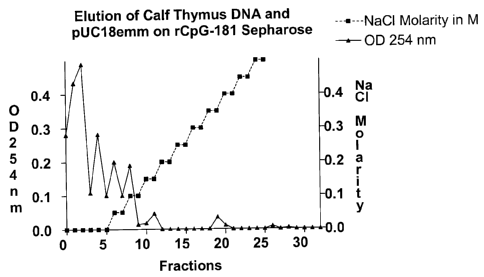
Figure 7 shows the elution of calf thymus DNA and pUC18emm by rCpG-181
sepharose,
and
Figure 8 shows the determination of the eluted DNA in the fractions by
measurement of
the extinction at 254 nm as a function of the NaCI gradient;
Figure 9 shows the results of PCR after enrichment of prokaryotic DNA from
a DNA
mixture of staphylococcus aureus and human DNA using coupled CpGbP-181
protein on CNBr sepharose, and
Figure 10 shows results of PCR after enrichment of prokaryotic DNA from
a DNA mixture
of staphylococcus aureus and human DNA using coupled CpGbP-181 protein
on AH-sepharose.
The wild type CPGB protein CPGbP656 binds non-methylated CpG motifs of
prokaryotic DNA,
thus forming a protein-DNA complex. This complex may be or become attached to,
for example,
a carrier, whereby separation and/or enrichment of DNA can be effected. The
present invention
is now based on the surprising finding that a protein which is shortened
relative to the wild type
CPGB protein (CPGbP656 comprising 656 amino acids) and presenting 25 % to 35
%, in
particular approximately 27.6 %, homology with the wild type CPGB protein, has
improved
binding properties over non-methylated CpG motifs of prokaryotic DNA than the
wild type CPGB
protein and variants thereof with a homology of 80% or more. An example of
such shortened
protein is CPGbP181 with 181 amino acids.
Prokaryotic DNA differs from eukaryotic DNA, for example, by the presence of
non-methylated
CpG motifs (Deutsches Arzteblatt, Jg. 98/15: A981-A985 (2001)). The invention
is based on the
finding that eukaryotic DNA and prokaryotic DNA differ in their proportion of
CpG motifs. In
prokaryotic DNA, CpG motifs are present with a 20-fold excess as compared to
eukaryotic DNA,
which contains such motifs only temporarily, e.g. in cancer cells or promoter
regions (Deutsches
Arzteblatt, Jg. 98/15: A981-A985 (2001)). In prokaryotic DNA, these motifs are
not methylated,
whereas most of them are methylated in eukaryotic DNA, which additionally
increases their
distinctiveness. Non-methylated CpG motifs are non-methylated desoxycytidylate-
desoxyguanylate dinucleotides in the prokaryotic genonne or in fragments
thereof.
CA 02558515 2006-09-01
- 7 -
The invention is further based on the finding that the protein according to
the invention
specifically binds to non-methylated CpG motifs. This specific binding
property of the protein
according to the invention is utilized in order to bind prokaryotic DNA and
thus to subsequently
enrich, separate and isolate it from a sample, e.g. with a majority of
eukaryotic DNA.
The term "DNA containing non-methylated CpG motifs" refers to both eukaryotic
and prokaryotic
DNA. Said DNA can be purified and dissolved again (e.g. non-methylated DNA
isolated from
tissues) or be present directly in the original source (e.g. body fluid, such
as blood, serum,
tracheal aspirate, urine, bronchoalveolar lavage, nose smear, skin smear,
puncture fluid).
According to a preferred embodiment, the DNA containing non-methylated CpG
motifs is
prokaryotic DNA, in particular bacterial DNA.
The term "homology" in the sense of the present invention relates to the
degree of identity of
two protein sequences. For example, a homology of 60 % means that 60 out of
100 amino acid
positions in the sequences are identical. The term "shortened" used in order
to characterize the
protein according to the invention means that the length of the amino acid
sequence of the
protein according to the invention (e.g. CPGbP181) is shorter than the length
of the amino acid
sequence of the wild type CPGB protein (CPGbP656). Shortening is effected at
the N-terminus
and at the C-terminus of the wild type protein sequence (Figure 1). The
maximum shortening is
represented by the DNA binding site of the protein.
The protein employed according to the invention may have a molecular weight
of, for example,
approximately 19,959 Dalton (native) or 21,444 Dalton (in plasmid pQE60). In
another preferred
embodiment the isoelectric point of the protein according to the invention is
approximately 10.09
(native protein) or 10.15 (in plasmid pQE60). A particularly preferred protein
employed
according to the invention has the amino acid sequence shown in SEQ ID No. 2
or in Fig. 1.
This protein has particularly good binding properties as compared to non-
methylated CpG
motifs of prokaryotic DNA.
The protein described in EP 02020904 (CPGbP241), which is a shortened variant
of the wild
type CPGB protein (CPGbP656) and served as the basis for the protein employed
according to
the invention (e.g. CPGbP181), has a length of 241 amino acids, a molecular
weight of
approximately 33,650 Dalton (native) or 28,138 Dalton (in plasmid pQE60) and
an isoelectric
point of 9.89 (native) or 9.88 (in plasmid pQE60). The cDNA and amino acid
sequence is shown
in Figs. 1 and 2.
CA 02558515 2006-09-01
- 8 -
The wild type CGBP protein has a length of 656 amino acids, 135 positively
charged residues
and 94 negatively charged residues, a molecular weight of approximately 75,684
Dalton and an
isoelectric point of 8.15. The cDNA and amino acid sequnce is shown in Fig.1.
The sequence comparison of the protein CPGbP181 according to SEQ ID No. 2 used
according
to the invention with the protein described in EP 01010904 (CPGbP241) is shown
in Figs. 1 and
2.
The protein employed according to the invention is preferably produced by
cloning the
corresponding cDNA sequence into a plasmid and by expression in Escherichia
coli. An E.coli
strain expressing the protein according to the invention was deposited with
the Deutsche
Sammlung fCir Mikroorganismen und Zellkulturen under No. DSM 16229 on February
16, 2004.
Alternatively, other methods of manufacture known in the art can be applied.
The use of plasmid
pQE9 represents an exemplary possibility, but any other suitable plasmid is
useful as a vector.
Expression in E.coli is also just an example. Expression in other prokaryotic
systems and also in
a eukaryotic system as well as chemical or enzymatic synthesis or purification
from a natural
source, such as e.g. tobacco plants, are further possible embodiments of
protein extraction. The
protein can be produced both on a laboratory scale (e.g. in an Erlenmeyer
flask) and on an
industrial scale (e.g. fermenter). For example, the protein according to the
invention can be
purified by binding histidine residues (His-tag), which are introduced at the
beginning or at the
end of the protein, to a suitable nickel-containing matrix, which is a method
known in the art.
Further possibilities of purification may be any type of fusion proteins
allowing purification via
suitable matrices (columns, gels, beads etc.). Other forms of tags may be
fusion peptides/fusion
proteins, e.g. streptavidin-tag, Myc-tag and others.
A preferred form of the protein used according to the invention is the native
form, but a
denaturated form is also suitable for binding non-methylated CpG motifs.
"Denaturated forms" in
the sense of the present invention are understood to be secondary structures
other than those
found in nature.
The native or denaturated form of the protein used according to the invention
is an exemplary
embodiment. The invention includes in vitro-synthesis as well as any other
chemical or
enzymatic modifications of the protein, such as e.g. incorporation of
disulfide bridges,
glycosilations, phosphorylations, acylations, amino acid exchanges as well as
fusion with
proteins or other molecules. Such modifications may be achieved, for example,
by
recombination and/or expression and/or chemical and/or enzymatic modification
of single or
multiple amino acids.
CA 02558515 2006-09-01
- 9
The protein used according to the invention has a multiplicity of advantages.
It is better in
binding prokaryotic DNA via non-methylated CpG motifs than the wild type CPGB
protein or
variants thereof with a homology of 80 % or more. This makes it possible to
specifically
separate and/or enrich the prokaryotic DNA of a mixture of prokaryotic and
eukaroytic DNA.
This ultimately enables quick and simple detection of pathogens as well as
early diagnosis of
infections which may be caused by bacterial pathogens. Conversely, the
invention can also be
used for depletion of microbial DNA in the sense of purification in the case
of clinical conditions
accompanied by non-physiological presence of bacteria or their cleavage
products in body
fluids, in particular blood, of patients. This applies even more because it is
well documented that
bacteria and also their cleavage products, such as, for example, bacterial
DNA, are responsible
for a multiplicity of biological effects detrimental to the patient.
Due to the good binding ability of the protein used according to the invention
to non-methylated
CpG motifs of prokaryotic DNA, the invention relates to a method of separating
and/or enriching
prokaryotic DNA, comprising the steps of:
a) contacting at least one prokaryotic DNA present in solution with a protein
which
specifically binds prokaryotic DNA and has 25 % to 35 (Yo homology with the
wild
type CGPB protein, thus forming a protein-DNA complex, and
b) separation of said complex.
The DNA can be purified and dissolved again or may be present directly in the
original source
(e.g. body fluid, such as blood, serum, tracheal aspirate, urine,
bronchoalveolar lavage, nose
smear, skin smear, puncture fluid).
Separation may be effected by different methods of separating, isolating or
enriching DNA
protein complexes or DNA polypeptide complexes that are well-known to the
person skilled in
the art. In doing so, use will be made preferably of methods in which the DNA-
binding protein is
or is being immobilized to a carrier in order to separate and/or enrich the
DNA from the sample
solution.
According to a preferred embodiment, separation is followed by a step of
separating the DNA
from the protein according to the invention in said complex. This may be
effected, for example,
by conventional methods of DNA purification known to the person skilled in the
art. In the most
simple case, separation is effected by changing the pH value or the salt
concentration (e.g. to 1
M NaCI) of the medium/buffer or by adding chaotropic reagents, etc.; i.e.
suitable parameters
CA 02558515 2006-09-01
- 10 -
which lead to the separation of the protein-DNA-complex. Such methods are
known to the
person skilled in the art.
According to a further preferred embodiment, the protein according to the
invention is coupled
For the solution of the prokaryotic DNA, any suitable solvent is basically
contemplated.
In particular, the embodiment according to which the DNA-binding protein is
immobilized to the
In order to increase the binding capacity and binding efficiency with respect
to non-methylated
CpG motifs of DNA, the invention provides a method enhancing the binding
capacity and
According to the invention, this is achieved by indirect coupling of the
protein to the matrix. This
method will be described hereinafter with reference to Figures 9 and 10.
In order to enhance the binding capacity and binding efficiency of the CpGbP-
181 protein with
respect to DNA containing non-methylated CpG motifs, the invention relates to
indirect binding
of the protein to the matrix via a spacer. By coupling the protein to the
matrix via a spacer, the
CA 02558515 2006-09-01
- 11 -
degree of mobility as well as the number of free binding sites of the CpGbP-
181 protein is
increased. Thus, increased binding capacity and binding efficiency are
achieved. This further
allows to reduce the amount of protein used.
Spacers in the sense of this invention are understood to be short chain-like
molecules, which
allow a spatial distance between the matrix and the protein used according to
the invention, e.g.
the CpGbP-181 protein. Such spacers are known in the art, e.g. from affinity
chromatography or
immobilization of proteins. Such chain-like molecules are composed of C and H
atoms as well
as optionally hetero atoms, e.g. N. These chain-like molecules are made up of
individual chain
members on the basis of the C atoms, e.g. CH2, and potentially present hetero
atoms, e.g. NH.
In particular, the spacer comprises 4 to 20, preferably 7 to 10 chain members.
A particularly
preferred spacer is derived from diamine hexane (NH2(CH2)6-NH2). Antibodies in
the sense of
the present invention are not to be considered as spacers.
A matrix in the sense of this invention relates to substances which function
as carriers for the
spacer and the protein. Carrier materials may be, for example, sepharose,
pearl cellulose, silica,
or similar substances known in the art.
Body fluids in the sense of the invention are understood to be all fluids
originating from the body
of a mammal, including humans, in particular such fluids in which disease
pathogens may
occur, such as blood, urine, liquor, pleural liquids, pericardial liquids,
peritoneal liquids as well
as synovial liquids. The description of the invention referring to human blood
is not to be
construed as [imitative, but only as an exemplary application.
Bacterial pathogens are preferably understood to be pathogens of sepsis, but
also any other
bacterial pathogens of infections. They may differ from commensal pathogens,
which are part of
the normal population of the organism and are sometimes also found in test
samples from
patients, but do not have any clinical significance.
When isolating total DNA from infected body liquids, the ratio of host-DNA to
pathogen-DNA
may be, in many cases, only 1:10-6 to 1:10-8 or even less. Through the
specific binding of
prokaryotic DNA to the protein according to the invention, the method
according to the invention
enables enrichment by 1 exponential unit and more.
The protein used according to the invention may be coupled directly or
indirectly to the carrier.
The type of coupling depends on the carrier and the carrier material. Suitable
carriers include, in
particular, membranes, microparticles and resins, or similar materials for
affinity matrices.
Suitable materials for binding the protein according to the invention, as well
as ¨ depending on
CA 02558515 2006-09-01
- 12 -
the type of material ¨ for carrying out such binding are well-known to the
person skilled in the
art. For indirect coupling, specific antibodies against the protein according
to the invention or the
polypeptide are suitable, for example, which are in turn bound to the carrier
by known methods.
One application of the method according to the invention consists in enriching
prokaryotic DNA.
A further application consists in the separation of prokaryotic DNA from a
mixture of eukaryotic
and prokaryotic DNA by binding the prokaryotic DNA to the protein used
according to the
invention, which has been immobilized, for example, to a matrix. The mixture
of the body's own
DNA and prokaryotic DNA is contacted with the affinity matrix by means of
suitable methods
and, in doing so, the prokaryotic DNA is bound to the immobilized protein; the
eukaryotic DNA
passes, for example, through a separating column and may be collected
separately. Affinity
matrices may be, for example, polymeric polysaccharides, such as agaroses,
other
biopolynners, synthetic polymers, or carriers having a silicate backbone, such
as porous glasses
or other solid or flexible carriers on which the DNA-binding protein used
according to the
invention is immobilized. After separation of prokaryotic DNA from eukaryotic
DNA has been
effected, the affinity matrix is rinsed with a suitable reagent, so that the
binding protein with the
coupled prokaryotic DNA is separated from the matrix and/or the prokaryotic
DNA is separated
from the binding protein and is available in a sufficient amount for further
process steps.
A further application of the method according to the invention consists in the
separation and
enrichment of prokaryotic DNA from eukaryotic DNA by binding the prokaryotic
DNA to the
protein according to the invention, which has been immobilized on
microparticles. In this
connection, all microparticles which allow the DNA-binding protein according
to the invention to
be immobilized are suitable. Such microparticles may consist of latex,
plastics (e.g. styrofoam,
polymer), metal, or ferromagnetic substances. Furthermore, use may also be
made of
fluorescent microparticles, such as those available from the Luminex company
for example.
After the prokaryotic DNA has been bound to the proteins used according to the
invention,
which are immobilized on microparticles, said microparticles are separated
from the mixture of
substances by suitable methods, such as filtration, centrifugation,
precipitation, sorting by
measuring the intensity of fluorescence, or by magnetic methods. After
separation from the
microparticles, the prokaryotic DNA is available for further processing.
Another application of the method according to the invention consists in the
separation and
enrichment of prokaryotic DNA from eukaryotic DNA by binding the prokaryotic
DNA to the
protein used according to the invention, which is subsequently separated from
other ingredients
of the mixture by electrophoresis.
CA 02558515 2006-09-01
- 13 -
A further application of the method according to the invention consists in the
separation and
enrichment of prokaryotic DNA from eukaryotic DNA by binding the prokaryotic
DNA to the
protein used according to the invention. The protein used according to the
invention is
subsequently bound to corresponding antibodies. The antibodies may be bound to
solid or
flexible substrates, such as glass, plastics, silicone, microparticles,
membranes, or may be
present in solution. After binding of the prokaryotic DNA to the protein
according to the invention
and binding of the latter to the specific antibody, separation from the
substance mixture is
effected by methods known to the person skilled in the art.
The method according to the invention may also be used in order to purify body
fluids to remove
prokaryotic DNA therefrom. In this connection, it is convenient for separation
to be effected
extra corporally, under sterile conditions, to allow the body fluids to be fed
back into the body
again, so that the body's own immune system is assisted in eliminating
infections by removing
the prokaryotic DNA contained in said body fluid.
Any suitable chemical, mechanical or electrochemical processes may be
considered for
extracorporal removal of prokaryotic DNA from body fluids. Further, the
combination with other
extracorporal methods, such as hemoperfusion, heart-lung machine or endotoxin
adsorbers, is
a further convenient application.
The protein used according to the invention can also be used to detect
prokaryotic DNA. In this
case, enrichment of the prokaryotic DNA is followed by a step of amplifying
said prokaryotic
DNA, for which all common methods of amplification are suitable (PCR, LCR; LM-
PCR, etc.).
The method according to the invention, in particular with the above-described
embodiments,
has the advantage that, by specific binding of non-methylated prokaryotic DNA,
rich in CpG
motifs to proteins with specific affinity for such structures, prokaryotic DNA
from the total DNA of
an infected host is successfully concentrated and thus the sensitivity of
detection of pathogen
DNA in body fluids is strongly enhanced.
The possibilities of separating prokaryotic DNA from eukaryotic DNA using a
specifically binding
protein are no more time-consuming than known methods of isolating total DNA.
However,
subsequent detection can then be effected only by PCR. A nested PCR will not
be required in
most cases, which makes it possible to save a considerable amount of time in
diagnostics.
The use of the protein of the invention to deplete prokaryotic DNA in
physiological body fluids
was already mentioned above. Depletion in the sense of the present invention
means that the
amount of prokaryotic DNA is reduced. This possibility of reducing prokaryotic
DNA also
CA 02558515 2006-09-01
- 14 -
enables the use of the proteins according to the invention in environmental
technology, waste
water management and air conditioning technology.
The invention further relates to a method of separating and enriching non-
methylated genomic
DNA from a mixture of non-methylated genomic and methylated genomic DNA. The
methylated
genomic DNA is separated by binding the non-methylated genomic DNA to the
CpGbP-181
protein coupled to a matrix. This procedure contributes substantially to the
simplified
examination of the methylation patterns of methylated genomic DNA and enables
the diagnosis
of diseases having a specific nnethylation pattern.
Moreover, the invention relates to a kit for enriching prokaryotic DNA by one
of the above-
described methods, said kit containing at least the protein according to the
invention, optionally
together with further reagents suitable to carry out said method.
In addition to the protein according to the invention, said kit may contain at
least one set of
primers, which are suitable to amplify genomic DNA of certain prokaryonts
under standard
conditions.
The invention will be explained in more detail below with reference to the
examples, without
limiting it thereto.
Example 1: Preparation of the protein according to the invention
The DNA sequence for the complete CPGbP protein was used to construct primers
1
(GGATCCGGTGGAGGGCGCAAGAGGCCTG ¨fw SEQ ID No. 3) and 2
(AAGCTTAGAGGTAGGTCCTCAT-CTGAG-rv SEQ ID No. 4) which amplify a shortened DNA
fragment encoding CPGbP-181, which is a shortened protein binding CPG. After
cleavage, the
DNA fragment was ligated into the pQE9 vector (Qiagen) using restriction
enzymes BamHI and
Hind III. An open reading frame forms in pQE9, in which frame a DNA fragment
encoding 6 x
His-Tag (pQE9[6HisCPGbP181]) is fused to the 5 end. The complete amino acid
sequence of
the encoding fusion protein 6His-CPGbP181 is shown hereinafter, the portion
indicated in bold
print representing the peptide CPGbP181 and the portions indicated in italics
indicating fused
foreign amino acids of plasmid pQE9.
Plasmid pQE9[6HisCPGbP181] was transformed to the E. coli expression strain
M15[pREP4]
(Qiagen). The clone is referred to hereinafter as M15[pCPGbP181], and the
expressed protein
is referred to as rCPGbP181. Expression of the protein rCPGbP181 occurred
according to the
following protocol: A colony of the expression strain M15[pCPGbP181] is grown
overnight in 2
CA 02558515 2006-09-01
- 15 -
ml Luria Medium with 100 pg/r1n1 ampicilline and 25 pg/ml kanamycine at 37 C
with shaking.
Then, the pre-culture is transferred to 200 ml preheated nutrient medium
containing the same
concentrations of antibiotics. After 3 hours of growth at 37 C with shaking,
IPTG is added to
induce expression, and incubation is continued for 5 hours. Thereafter, the
bacteria are
removed by centrifugation and the sediment is re-suspended in 5 ml 0.2 M tris
buffer, pH 7.5.
The bacteria are subjected to ultrasonic treatment in an iced bath for 5 x 1
min. After
centrifugation, the sediment is re-suspended in 10 ml 0.2 M tris, 2M urea, pH
7.5, and shaken
for 15 min. After centrifugation has been effected, the remaining sediment is
taken up in 0.2 M
tris, 6M guanidine hydrochloride, 0.001 M dithioeritrite (DTE), 0.02 M
imidazole, and suspended
therein. The inclusion bodies are dissolved at room temperature for 1 hour
with agitation. After
centrifugation, the crude protein is present in the supernatant and can be
applied directly to a 3
ml Ni-agarose column. The subsequent steps should be effected in the cooling
chamber at +4
to +6 C. First, the column is washed with 0.2 M tris, 6M guanidine
hydrochloride, 0.001 M
dithioeritrite (DTE), 0.02 M imidazole buffer, pH 7.5, until extinction has
reached the zero line.
From this point, rCPGbP181 can be obtained in different ways: 1. as a
denaturated protein,
dissolved in 6M guanidine hydrochloride or 6M urea, and 2. as a native
protein, soluble in
buffers at physiological concentrations. In the second case, however, the
yield is lower.
Purification according to method 1 (denaturated):
The protein rCPGbP181 is eluted from Ni-NTA agarose with an imidazole gradient
of 0 ¨ 0.5 M,
M in buffer 0.2 M tris, 6M guanidine hydrochloride, 0.001 M dithioeritrite
(DTE), 0.02 M
imidazole, pH 7.5, as the basic material. In doing so, rCPGbP181 is detached
from the column
at 0.2 ¨ 0.3 M imidazole. The protein thus obtained is dialyzed against 0.2 M
tris, 6M urea,
0.001 M dithioeritrite (DTE), pH 7.5, and frozen. During dialysis against
physiological buffers,
purified rCPGbP181 is thus precipitated.
Purification according to method 2 (native):
According to this method, the guanidine hydrochloride concentration is shifted
from 6 mol on Ni-
NTA agarose with the bound rCPGbP181 via a gradient up to 0 mol guanidine
hydrochloride.
The basis for this is the buffer 0.2 M tris, 0.5 M NaCI, 0.001 M
dithioeritrite (DTE), 0.02 M
imidazole, pH 7.5. In this case, a flow rate of 0.5 ml/min was selected.
Subsequently, an
imidazole gradient of 0 to 0.5 mol was applied for elution in buffer 0.2 M
tris, 0.5 M NaCI, 0.001
M dithioeritrite (DTE), pH 7.5, as basic material. In this case, too, a
substantial proportion of the
bound protein (20 A) was eluted at 0.2 to 0.3 mol imidazole. This native
rCPGbP181 eluate
remained dissolved in this buffer even after dialysis in PBS. However, it is
disadvantageous that
approximately 80 % of rCPGbP181 bound to Ni-NTA agarose remained on the column
under
these conditions and were subsequently extractable only under the denaturated
conditions of
CA 02558515 2006-09-01
- 16 -
method 1. This means that the yield of method 2 as used resulted only in 20 %
native
rCPGbP181 soluble in physiological buffers.
Example 2: Detection of pathogens by means of nested PCR:
Fresh, heparinized human blood, which contains streptococcus pyogenes with
103/m1
colony-forming units as pathogens, is used for detection of pathogens. The DNA
is isolated
by means of absorption to DNA-binding matrix using commercial kits for
isolation of total
DNA from body fluids according to modified instructions from the
manufacturers. For this
purpose, 200 pl of the total lysis buffer, which contains proteinase K and
SDS, is added to
100 pl of infected blood in Eppendorf tubes. The mixture is incubated at 37 C
for 30 min and
then heated to 95 C for 20 min. After cooling, 20 pg of mutanolysine are added
and
incubated at 37 C for another 60 min. After centrifugation, the mixture is
applied to the
centrifugation columns using DNA-binding matrix and the DNA is purified
according to
manufacturer's instructions. The purified DNA is placed in a final volume of
100p1 of 0.01 mol
tris buffer, pH 7.5, or in an equal amount of elution buffer from the
manufacturer. For
detection of pathogens, primers were selected to identify the streptolysin 0
gene (slo).
1. PCR. Amplification of a 465 bp fragment
Forward primer 1: 5'-AGCATACAAGCAAATTTTTTACACCG
Reverse primer 2: 5'-GTTCTGTTATTGACACCCGCAATT
Primer concentration 1mg/m1
Starting material: 5 pl isolated DNA
0.5 pl primer fw 1
0.5 pl primer rv 2
14 pl aqua dest
total 25 pl in Ready to go Kit (Amersham-Pharmacia)
Reaction:
5 min 95 C
40 cycles (30 sec. 95 C; 30 sec. 51 C; 3 min 72 C; 1 x 7 min 72 C).
The first PCR of streptococci-DNA in human blood is shown in Fig. 1 (10 pl
each of the 25 pl
starting material were separated. 1) PCR starting material containing 5 pl
template DNA; 2)
starting material containing 5 pl template, at a dilution of 1:10. 3) positive
control: 0.2 pl of
streptococci-DNA as template in the absence of eucaryotic DNA from blood. ST)
molecular
weight standard)
CA 02558515 2006-09-01
- 17 -
Result: The first primary PCR does not result in a positive reaction.
Therefore, a second
PCR (nested PCR) was subsequently carried out.
2. PCR (nested): Amplification of a 348 bp fragment in the above s/o-fragment.
Forward primer 3: 5'-CCTTCCTAATAATCCTGCGGATGT
Reverse primer 4: 5'-CTGAAGGTAGCATTAG TCTTTGATAACG
Primer concentration: 1mg/m1
Starting material: 5 pl from PCR1, sample 1, Fig. 1
0.5 pl primer fw 1
0.5 pl primer rv 2
14 pl aqua dest
total 25 pl in Ready to go Kit (Amersham-Pharmacia)
Reaction:
5 min 95 C
40 cycles (30 sec. 95 C; 30 sec. 54 C; 3 min 72 C; 1 x 7 min 72 C)
Figure 4 shows the nested PCR with the PCR products from the primary PCR
starting
material according to Fig. 3 as template. The samples correspond to those of
Figure 3.
Result: In the nested PCR, the desired slo-DNA fragment is amplified at a
concentration of 100
streptococci cells per 100 pl blood (sample 1). For 5 pl starting material in
the 1st PCR (Fig. 3),
this corresponds to about 5 to 10 templates. At a dilution of 1:10 (sample 2),
sensitivity is
exhausted (0.5 to 1 template).
These experiments show that successful PCR detection of pathogens in blood
requires isolation
of the total DNA from at least 1 to 5 ml blood. However, the total DNA
concentration is then too
large to be used directly in a PCR.
Other pathogen-specific nucleic acid detections without an amplification step
by direct detection
of the bacterial DNA, for example by DNA hybridization, are also too
insensitive, which is
primarily due to the high excess of human DNA relative to bacterial DNA. In
addition,
competitive processes during DNA analysis as well as the low quantity of
bacterial DNA are to
be regarded as hindrances to qualitative and quantitative analysis. The common
methods of
DNA isolation enrich the total DNA of a body fluid so that the ratio of host
DNA to microbial DNA
can be between 1:10-6 and 1:10-8. This difference makes it easy to understand
the difficulty in
detecting microbial DNA in body fluids.
CA 02558515 2006-09-01
- 18 -
Example 3: Determining the binding properties of rCPGbP181:
In gel retardation experiments both the binding of the denaturated and of the
native protein
rCpGbP181 to methylated and to non-methylated DNA molecules with CpG motifs
was
examined. The pUC18 plasmid of E. coli was used as the test DNA with an
inserted M-protein
gene segment of streptococcus dysgalactiae supsp. equisimilis (Geyer et. al
FEMS Immuno.
Med. Microbiol. 26:11-24, 1999). The plasmid preparation was divided and one
half was
methylated with the CpG methylase kit of New England BioLabs. Both
preparations were mixed
with rCPGbP181 (native or denaturated) and electrophoretically separated on
agarose gel. The
results are shown in Figures 5 and 6. Both the native form and the denaturated
form of
rCPGbP181 showed a higher affinity to non-methylated plasmid DNA, which
confirms the
selective binding property with respect to non-methylated CpG-rich DNA.
Description of the gel retardation experiment according to Figure 5: 5p1 (72
ng) methylated
pUC18emm DNA and 1 pl (142 ng) non-methylated pUC18emm DNA, respectively, were
mixed
with 5 pl (0.5 pg) native rCPGbP181 and filled up to a volume of 35 pl with
the following buffer:
0.01 M tris, 0.08M NaCI, 0.001M EDTA, 0.005M DTE, 5% glycerine, pH 7.8. After
incubation at
C for 30 min the mixtures were electrophoretically separated on 1.5% agarose.
Methylated
DNA was applied in lanes 1 and 3 and non-methylated DNA was applied in lanes 2
and 4. In
20 lanes 1 and 2 the DNA was mixed with native rCPGbP181. Lane 2 shows that
non-methylated
pUC18emm interacts with rCPGbp181; in contrast thereto, rCPGbP181 did not show
any
interaction with methylated pUC18emm (lane 1). Lanes 4 and 5 are the plasmids
without
addition of rCPGbP181 as controls.
Description of the gel retardation experiment of Figure 6 for non-methylated
and methylated
pUC18emm after incubation with denaturated rCPGbP181. The concentrations
correspond to
those of Figure 5. Methylated DNA was applied in lanes 1 and 3 and non-
methylated DNA was
applied in lanes 2 and 4. In lanes 1 to 4, the DNA was mixed with two
different batches of
denaturated rCPGbP181. Lanes 2 and 4 show that non-methylated pUC18emm also
interact
with denaturated rCPGbP181; however, rCPGbP181 did not show any interaction
with
methylated pUC18emm (lanes 1 and 3). Lane 5 is pUC18emm without rCPGbP181 as
control.
Example 4: Binding and separation of a mixture of calf thymus DNA and
bacterial DNA to
immobilized CPGbp181.
Purified CPGbp181 was coupled to aminohexyl sepharose (Amersham-Biosciences)
by means
of glutaraldehyde according to the protocol of Cambiasso et al. (Cambiasso, C.
et al.,
Immunochemistry 12-273-278, 1975). The concentration of immobilized protein
was 0.3 mg per
CA 02558515 2006-09-01
- 19 -
milliliter sepharose. 300 pl sepharose was placed in a spin-filter tube
containing inert fritting
material which absorbs neither DNA nor protein, but retains sepharose.
200 ng calf thymus DNA and 25 ng pUC18emm was dissolved in 100 pl 20 mM tris-
HCL buffer,
pH 7.5, and applied to the column thus prepared. After each step, the liquid
was centrifuged at
14,000 RPM for 0.5 min in an Eppendorf centrifuge in one fresh Eppendorf tube
each. Thus, a
two-step increase of the NaCI concentration was effected from 0 to 1M. DNA
precipitation was
effected in each tube by adding 10 pl 4 M acetate, ph 4.5, and 250 pl ethanol
abs., mixing and
centrifugation at 14,000 RPM for 15 min. Thereafter, the supernatant was
discarded and the
precipitate was washed with 300 pl 70 % ethanol. After discarding, the residue
was dried for
5 min in a vacuum centrifuge and then taken up in 15 I distilled water (PCR-
suitable). On the
one hand, extinction at 254 nm was measured for 10 pl each of the samples
(Figure 7). On the
other hand, PCR was effected with sequence primers for PUC18, using 3 pl of
each sample
(Figure 8).
The result (Figures 7, 8) shows that the eukaryotic calf thymus DNA is
initially washed from the
column between 0 to 0.1 M NaCI, while the prokaryotic DNA (pUC18emm) was
eluted in the
fraction at 0.3 M NaCI. This shows that eukaryotic DNA has a lower affinity to
CPGbP181 and,
thus, a clear separation of both DNA fractions was achieved.
Example 5: Enhancement of binding properties of the CpG-bP-181 protein, which
result from
indirect binding of this protein to a matrix via a spacer.
In order to examine binding properties, prokaryotic DNA from a DNA mixture of
staphylococcus
aureus and human DNA was enriched using the directly coupled CpGbP-181 protein
on CNBr
sepharose or using the indirectly coupled CpGbP-181 protein on sepharose (in
the following AH
sepharose) via a diaminohexyl spacer (AH).
First, the AH sepharose was incubated at room temperature for 15 min with
addition of
glutaraldehyde. Next, the AH sepharose was washed with 0.1 mol Na2HPO4. Then,
0.24 mg of
the CpGbP-181 protein was placed on the matrix. The binding of the CpGbP-181
protein to AH
sepharose was achieved by incubation at room temperature for 2 hours. The
excess CpGbP-
181 protein was removed.
After subsequent washing of the CpGbP-181-AH sepharose with 0.1 mol Na2HPO4
and addition
of 0.1 mol glycine, the CpGbP-181-AH sepharose was incubated at room
temperature for 2
hours in order to saturate free binding sites. Then, the CpGbP-181-AH
sepharose was again
washed with 0.1 mol Na2HPO4. In order to reduce the Schiff base and in order
to stabilize
CA 02558515 2006-09-01
- 20 -
binding, the CpGbP-181-AH sepharose was admixed with sodium borohydride and
incubated at
room temperature for 1 hour.
Next, the CpGbP-181-AH sepharose was washed with 0.1 mol Na2HPO4.
The storability of the CpGbP-181-AH sepharose at 4 C is achieved by addition
of 20% ethanol.
Next, the CpGbP-181-AH sepharose was portioned into columns. The columns
prepared with
CpGbP-181-AH sepharose were then washed with tris buffer and were available
for
separation/enrichment of DNA containing non-methylated CpG motifs.
2) Enrichment of the DNA mixture, followed by elution of the prokaryotic DNA
and determination
of the concentration of prokaryotic DNA by PCR.
The DNA mixture consisted respectively of 330 ng human DNA and 150 ng
prokaryotic DNA
(staphylococcus aureus DNA). The DNA mixture was placed on the columns
prepared with
CNBr sepharose or with AH sepharose, respectively, and incubated at room
temperature for 1
minute. Then, the columns were centrifuged and washed with 100 I tris buffer
(10 pM, pH 7).
The washing and centrifugation step was repeated 5 times.
The supernatant was carefully removed and then 100 pl elution buffer (10 pM
tris buffer, 0.5 M
NaCI, pH 7) each were added to the columns and centrifuged. The elution step
was repeated 5
times. Then, the individual fractions of each sample were precipitated by
addition of 10 pl 3 M
sodium acetate and 250 pl ethanol with subsequent mixing and centrifugation
(15 min at 15,000
g). The supernatant was carefully discarded and the pellet was washed with 1
pl ethanol (70%)
and centrifuged at 15,000 g. The supernatant was then removed again, the
pellet was dried in a
vacuum centrifuge and taken up in 30 pl DEPC water. 5 pl each were used for
PCR detection.
The PCR used universal primers for the 16s RNA gene. After carrying out the
PCR, 15 pl each
of the individual fractions were placed on 2% agarose gel.
Figure 9 (direct binding of the CpG-181 protein to CNBr sepharose) and Figure
10 (indirect
binding of the CpG-181 protein to sepharose via a spacer (AH)) show the
results of the PCR for
the individual fractions. It is clearly evident that the use of the AH spacer
allowed more
prokaryotic DNA to be enriched (fraction 1, elution fraction). This
characteristic improvement in
binding properties is useful in the methods according to the invention.
DEMANDES OU BREVETS VOLUMINEUX
LA PRESENTE PARTIE DE CETTE DEMANDE OU CE BREVETS
COMPREND PLUS D'UN TOME.
CECI EST LE TOME 1 DE 2
NOTE: Pour les tomes additionels, veillez contacter le Bureau Canadien des
Brevets.
JUMBO APPLICATIONS / PATENTS
THIS SECTION OF THE APPLICATION / PATENT CONTAINS MORE
THAN ONE VOLUME.
THIS IS VOLUME 1 OF 2
NOTE: For additional volumes please contact the Canadian Patent Office.