Note: Descriptions are shown in the official language in which they were submitted.
CA 02558650 2006-09-06
WO 2005/088583 PCT/US2005/007623
DEVICE AND METHOD FOR MEDICAL TRAINING AND EVALUATION
The present application claims the benefit of U.S. provisional application
number
60/551,090 filed March 8, 2004, which is incorporated herein by reference in
its entirety.
FIELD OF INVENTION
In one aspect, the present invention relates to devices and methods for
performing
and evaluating surgical and other medical treatment procedures, including
anatomical
structures and methods for use in training and evaluating individuals in
laparascopic
surgeries. The devices and methods may further be used in training and
evaluation of
non-laparascopic surgeries, particularly those that require precision and are
complicated
by the dynamic motion of the organs and body parts acted upon during the
procedure.
The devices and methods may further be used in radioscopic training and
evaluation. In
preferred aspects, devices of the invention can mimic anatomical, dynamic
and/or
mechanical properties of that area of the human anatomy on which training and
evaluation is being performed.
BACKGROUND OF THE INVENTION
Laparoscopic surgeries are performed by inserting laparoscopic tubes and
sleeves
into the body through small incisions. Various instruments and a video camera
(laparoscope) are then introduced into the body via the tubes and sleeves for
performing
and monitoring the surgery. The laparoscope and instruments allow the surgeon
to
explore the entire body cavity without making large standard openings,
dividing skin and
muscle, to access the cavity. The tubes and sleeves have diameters in the
order of 10
millimeters and, thus, laparoscopic procedures require only small incisions to
access the
surgical site. These incisions significantly reduce the trauma and the
required healing
compared to traditional surgery, resulting in decreased hospitalization and
patient
morbidity, lower analgesic dosages for pain control, better aesthetic results
and faster
CA 02558650 2006-09-06
WO 2005/088583 PCT/US2005/007623
recovery. Such procedures can be used in a wide variety of procedures, such as
urologic,
gynecological, chest and abdomen surgeries.
For example, in a conventional laparascopic technique on the abdomen, a Veress
needle is first inserted through the patient's abdominal wall and gas, usually
carbon
dioxide, is then injected through the needle to pressurize the abdominal
cavity and
distend the abdominal wall (insufflation). A pressure regulator gas
insufflator is typically
connected to the needle so that the pressure obtained does not go beyond 15
mmHg. Five
or six small (5-10 mm) incisions are then made in the abdomen. The laparoscope
and
surgical instruments are inserted through these incisions, typically through
laparoscopic
tubes and sleeves, into the inflated abdominal cavity. The surgeon is then
guided by the
laparoscope, which transmits a picture of the internal organs on a video
monitor.
Due to its complexity, however, laparoscopic surgical complications correlate
highly to the level of surgeon experience (W.A. Cooper, C.S. Fischer, R.J.,
Predictors of
laparoscopic complications after formal training in laparoscopic surgery,
JAMA, 270:
2689, 1993). Further, the difficulty of laparoscopy in general, the high
complexity of
urological applications, and the relatively infrequent incidence of urologic
cases make it
essential for urologists to have access to specialized training programs. To
meet this
demand, numerous urologic laparoscopy programs and short courses have been
established (Fahlenkamp, D., Rassweiler, J., Fornara, P. et al., Complications
of
Laparoscopic Procedures in Urology: Experience with 2, 407 Procedures at 4
German
Centers, Journal of Urology, 162: 765, 1999). Training programs use a sequence
of
theoretical, simulator and animal training followed by mentored surgery,
whereas short
courses address only the first three of these steps. Laparoscopy simulators
can be
classified as either physical devices of various construction or virtual
reality (VR)
simulators.
Traditional training devices present box architecture with flexible trocar
entry
ports. For example, a box trainer designed for basic inverted-motion
laparoscopy training
was developed (Muhgal, M. A cheap laparoscopic surgery trainer, Ann R Coll
Surgery,
2
CA 02558650 2006-09-06
WO 2005/088583 PCT/US2005/007623
England, 74: 256, 1992). Box trainers that allow trocar placement and
abdominal
insufflation were also reported (Monro, A., Park, K., Atkinsori, D. et al., A
laparoscopic
surgical simulator, J.R. Coll. Surg. Edimb., 39: 176, 1994; Kopchok, G.,
Cavaye, D.,
Klein, S. et al., Endoscopic Surgery Training: Application of an In Vivo
Trainer and In
Vivo Swine Model, Journal of Investigative Surgery, 6: 329, 1993). Seattle's
Simulab
Corporation, along with the University of Washington Center of Videoendoscopic
Surgery, provide a simulator with the purpose of replacing live-animal
training. This
simulator comprises a synthetic body model and procedure-specific packs and
allows
trainees to introduce surgical instruments and practice laparoscopy skills on
simulated
latex organs with standard instruments and laparoscopes. Specific procedure
packs are
available for general laparoscopic training in the initial phase for acquiring
through-the-
hole, inverted manipulation skills, depth perception under monitor vision, and
hand-eye
coordination, but fail to give a realistic anatomical perspective.
Recently, several virtual reality (VR) surgical simulators have become
available.
These trainers use a computer modeled human body and laparoscopic-like input
devices
(haptic interface) through which the trainee interacts with the model to
perform specific
surgical procedures. For example, MIST-VR is a laparoscopic trainer developed
by
Virtual Presence allowing the simulation of several laparoscopic procedures
(Wilson,
M.S., Middlebrook, A., Sutton, C. et al., MIST VR: a virtual reality trainer
for
laparoscopic surgery assesses performance, Annals of the Royal College of
Surgeons of
England, 79: 403, 1997; Gallagher, A.G., McClure, N., McGuigan, J. et al.,
Virtual
reality training in laparoscopic surgery: A preliminary assessment of
minimally invasive
surgical trainerreality (MIST VR), Endoscopy, 31: 310, 1999). While VR
simulators
potentially can provide training alternative, their application and utility is
presently
limited due to the high complexity required to realistically model human
organs
(Kneebone, R. Simulation in surgical training: educational issues and
practical
implications, Medical Education, 37: 267, 2003; Ahlberg, G., Heikkinen, T.,
Iselius, L.
et al., Does training in a virtual reality simulator improve surgical
performance? Surg
Endosc, 16: 126, 2002).
3
CA 02558650 2006-09-06
WO 2005/088583 PCT/US2005/007623
The use of live animals, while more realistic with respect to tissue
properties, is
limited by its high cost and animal death rate, especially at the beginning of
the learning
curve. In addition, the use of a live animal provides a limited period of time
within
which to one can practice surgical skills. Still further, animals present
different anatomy
and organ situs than humans.
Various simulation approaches also have been employed in various imaging
therapies and diagnostics, including for training and assessment of magnetic
resonance
imaging, various nuclear medicine therapies, and ultrasound procedures.
Computerized
Imaging Reference Systems, Inc. (Norfolk Virginia) markets certain devices for
those
simulation applications.
It would be desirable to have new devices and methods for medical personnel
residents to increase their level of experience in performing various surgical
and imaging
procedures including laparoscopic procedures.
SUMMARY OF THE INVENTION
The present invention features a device and method for use in training and
evaluating various medical procedures, particularly laparoscopic procedures,
radioscopic
procedures and procedures requiring precision that may be impacted by the
dynamic
motion of the organs, tissues and various body parts involved in or affecting
the
procedure.
In one embodiment, the present invention features a medical training and/or
evaluation device comprising a housing, an organ or tissue element, and
apparatus for
simulating respiration and/or pulmonary action on the organ or tissue element.
In one
preferred embodiment, the apparatus simulates respiration action and/or
pulmonary force
action of a human. As referred to herein, "simulate" or other similar term
means to create
a representation or model of, particularly to imitate. Preferably, the
apparatus exerts on
the organ or tissue element a force with a plurality of degrees of freedom. As
referred to
herein, a "plurality of degrees of freedom" or other similar term refers to
the minimum
4
CA 02558650 2006-09-06
WO 2005/088583 PCT/US2005/007623
number of coordinates required to specify completely the motion of a
particular element.
In particular, when used in connection with a particular anatomical structure,
it is
preferred that the corresponding structure in the present invention possess
the same
number of degrees of freedom as the corresponding real anatomical structure.
Thus, for
example, if the heart (synthetic or real) is located within the present
device, then it would
be located in a manner that would provide it with the same number of degrees
of freedom
as the heart in a live body.
The housing can correspond to any simple geometric shape, for example, a box
element/box-like shape. As used herein, a "box element" or other similar term
includes
any geometric shape having a base and typically four sides that suitably may
be
substantially perpendicular to the base. A box element may or may not include
a lid or
cover. In some embodiments, the housing is the shape of at least a portion of
a
mammalian anatomy, preferably a human anatomy. As used herein, "mammalian" or
other similar term includes any of the various warm-blooded vertebrate animals
of the
class Mammalia, including primates particularly humans. It is also
contemplated that the
housing may be provided in the shape of other living beings other than mammals
such as,
for example, fish (cold-blooded aquatic vertebrates of the superclass Pisces),
reptiles
(cold-blooded, usually egg-laying vertebrates of the class Reptilia) and birds
(warm-
blooded, egg-laying, feathered vertebrates of the class Aves). The housing
preferably
comprises one or more cavities for nesting one or more organ or tissue
elements. As
referred to herein, "nesting" or other similar terms refer to fitting,
containing or housing
within. The one or more cavities may be lined with materials that simulate the
lining of
the corresponding real cavity. In some embodiments, the housing further
includes a
skeletal system. The device may further comprise one or more walls through
which a
simulated laparascopic procedure can be performed. In a preferred embodiment,
the one
or more walls correspond to an abdominal wall and are fabricated of a material
that
allows insufflation. A preferred material for use in forming the housing
and/or walls is a
silicone material that simulates the corresponding part of the anatomy. In
some
embodiments, the device is adapted for training and/or evaluation of
radiological
procedures. In other embodiments, the device is adapted for training and/or
evaluation of
5
CA 02558650 2006-09-06
WO 2005/088583 PCT/US2005/007623
laparoscopic procedures. In yet other embodiments, the device is adapted for
procedures
that require particular precision and which may be complicated by the dynamic
motion of
the organs and body parts involved in the procedure.
In an exemplary embodiment, the training and/or evaluation device comprises a
housing in the shape of shape the human torso, one or more organ or tissue
elements
within the housing, and apparatus for simulating respiration and/or pulmonary
action on
the organ or tissue element. Preferably, the device is provided so as to
simulate the
corresponding anatomy in size, dimensions and proportions. The device
preferably
further comprises one or more cavities corresponding to one or more cavities
of the
human torso in which the one or more organs or tissue elements are located.
The device
may include one or more removable wall elements positioned above the one or
more
organs through which a simulated laparoscopic procedure can proceed. In one
embodiment, the one or more wall elements are adapted to correspond to a human
abdominal wall and, preferably are adapted to allow for insufflation within
the housing.
Further, the one or more wall elements are preferably pierceable with
laparoscopic
instruments. The device may further comprise apparatus to simulate a
circulatory system,
urinary system and/or digestive system. The housing may further include a
skeletal
system.
The one or more organs may be fastened to various portions of the device in a
manner that simulates the corresponding organ connections in the degrees of
freedom by
which the organ may move. In some embodiments, the outer housing further
houses real
or synthetic tissues and muscles that correspond to the tissues and muscles of
the live
body.
The outer housing is preferably fabricated of one or more materials that
simulate
the properties of the corresponding portions) of the body wall. For example,
some
suitable materials include, but are not limited to flexible urethane rubbers,
thermoplastic
polyurethanes and silicone rubbers. Preferably, the materials used in forming
the outer
housing possess tensile strength, elongation, hardness and/or tear strengths
similar to
6
CA 02558650 2006-09-06
WO 2005/088583 PCT/US2005/007623
those of the corresponding portions) of the body wall. It is particularly
desirable to
select materials that are "skin-like" in their properties. Preferably, the
outer housing is
fabricated of a silicone material. If desired, the outer housing may be
colored similar to
various colors of the human skin.
The device can be used for a variety of training and/or evaluation procedures.
In
one embodiment, the device is for training and/or evaluation of laproscopic
procedures.
In such embodiments, the device preferably includes one or more walls
positioned above
the one or more organs, such that the laparascopic procedure can take place
through the
one or more walls. Preferably, the one or more walls are fabricated of a
material that
provides properties similar to those of the corresponding body wall through
which the
laparascopic procedure is performed. In an exemplary embodiment, the one or
more
walls corresponds to the abdominal wall and is fabricated of a material that
allows
insufflation. In some embodiments, the one or more walls contain one or more
pre-
1 S formed openings through which a laparoscopic procedure may be performed.
In some
embodiments, the one or more walls are fabricated of a material that is
piercable by
laparoscopic instruments. In a preferred embodiment, particularly wherein the
one or
more walls are pierced by laparoscopic instruments, the one or more walls are
disposable
and removable. The walls may be removably fastened using any conventional
fastening
means that can be fastened and unfastened repeatedly. For example, fastening
means
may include, but are not limited to buttons, snaps, hooks, Velcro, clips and
mating lips
and recessed portions. Some materials useful in forming the one or more walls
include,
but are not limited to flexible urethane rubbers, thermoplastic polyurethanes
and silicone
rubbers. Preferably, the materials used in forming the one or more walls
possess tensile
strength, elongation, hardness and/or tear strengths similar to those of the
corresponding
body walls (e.g. the abdominal wall). In some embodiments, the one or more
walls are
fabricated of two or more layers of materials. For example, in an exemplary
embodiment, the inner layer is fabricated of a substantially or sanhighly
elastic material,
such as Cine Skin Silicone A/B with 50% part C (Elongation: 1000% (A/B+50%C),
and
the outer layer is fabricated of a material selected from those used in
forming the outer
7
CA 02558650 2006-09-06
WO 2005/088583 PCT/US2005/007623
housing. For example, the one or more walls may comprise two layers, wherein
the inner
layer is a more elastic material as compared to the outer layer.
In another exemplary embodiment, wherein the device is used for training
and/or
S evaluation of laproscopic procedures, the outer housing includes one or more
portions
through which a laparoscopic procedure is performed. For example, the one or
more
portions may include one or more openings through which the one or more organs
may
be accessed and through which a lapsarascope, laparoscopic instruments,
laparascopic
tubes and sleeves may be inserted and manipulated. In one embodiment, the
device
further includes one or more walls positioned between the one or more portions
of the
outer housing through which a laparoscopic procedure is performed and the one
or more
organs. During use, a laparascope and laparoscopic instruments are inserted
through and
manipulated through the one or more walls. The one or more walls contain one
or more
pre-formed openings through which a laparoscopic procedure may be performed.
In
some embodiments, the one or more walls are fabricated of a material that is
piercable by
laparoscopic instruments. In a preferred embodiment, the one or more walls are
located
so as to removably seal the one or more openings. For example, the wall may be
fastened
to the outer housing so as to seal the one or more openings. In both
embodiments, the
walls are preferably fastened using any conventional fastening means.
Preferably, the
walls are removably fastened and the fastening means are those that can be
fastened and
unfastened repeatedly. For example, fastening means may include, but are not
limited to
buttons, snaps, hooks, Velcro, clips and mating lips and recessed portions. In
some
embodiments, the one or more walls are disposable and removable, and
preferably are
fabricated of a material that simulates the properties of the corresponding
body walls)
through which the procedure is performed (e.g. flexible urethane rubbers,
thermoplastic
polyurethanes and silicone rubbers). In some embodiments, the one or more
walls are
fabricated of a material that allows insufflation.
In another exemplary embodiment, a training and/or evaluation device for use
in
laparoscopic procedures comprises an outer housing in the shape of a human
torso sized
and proportioned so as to simulate a human torso, one or more organs within
the outer
8
CA 02558650 2006-09-06
WO 2005/088583 PCT/US2005/007623
housing provided so as to move with multiple degrees of freedom in a manner
that
simulates motion of the corresponding organ in a live body, a disposable and
removable
wall, corresponding to the abdominal wall, positioned above the one or more
organs
through which the laparoscopic procedure will proceed and means for simulating
respiration and/or pulmonary motion. Preferably, the one or more disposable,
removable
walls are designed to simulate the properties of the abdominal wall. For
example, the one
or more disposable, removable walls may allow for insufflation in the outer
housing and
are preferably pierceable with laparoscopic instruments. The device may
further
comprise one or more cavities for housing the one or more organs. The one or
more
cavities are preferably positioned, sized and shaped in a manner that
simulates the
corresponding cavities of a human body. In one embodiment, the means for
simulating
respiration and/or pulmonary motion comprises one or more tubes through which
gases
and/or liquids may be circulated. In some embodiments, the device may further
comprise
a means for simulating the circulatory system, urinary system and/or digestive
system.
Such means may comprises one or more tubes through which gases and/or liquids
may
be circulated. If desired, a skeletal system may further be located within the
outer
housing. Preferably, the skeletal system is a synthetic skeletal system
fabricated of a
material that provides properties similar to those of a real skeletal system.
In another embodiment, the device is useful for training and/or evaluation of
radiological procedures. In such embodiments, the device is preferably
designed so as to
simulate the size, location and proportions of the corresponding parts) of the
anatomy,
and the various organs and tissues contained within the device. Further, the
components
of the device are preferably fabricated of materials that will allow for
realistic simulation
of the various radiological procedures. Thus, for example, when the device is
used for
generating images, the materials forming the various parts will allow for
accurate image
generation using X-ray, CT (Computed Tomography), MRI (Magnetic Resonance
Imaging), Ultrasound, Nuclear Magnetic Resonance Imaging and Interventional
Radiology devices.
9
CA 02558650 2006-09-06
WO 2005/088583 PCT/US2005/007623
The present invention also includes methods for training or evaluating a
medical
procedure comprising using the device described herein and performing a
medical
procedure. In particular, in one embodiment, methods comprise providing a
training
device comprising a housing in the shape of at least a portion of a mammalian
anatomy,
one or more organ or tissue elements within the housing, and apparatus for
simulating
respiration and/or pulmonary action on the organ or tissue elements, causing
respiration
and/or pulmonary action to be simulated within the device and performing the
medical
procedure. During such methods, the one or more organ or tissue elements
preferably
move as a result of the respiration and or/pulmonary action. The methods may
further
comprise inserting a laparoscope and one or more laparoscopic instruments into
the outer
housing and performing a laparoscopic procedure. In another embodiment, the
methods
further comprise imaging the one or more organ or tissue elements with
radiation. In
some embodiments, the laparoscopic training and/or evaluation device further
comprises
apparatus to simulating a circulatory system, urinary system and/or digestive
system and
the method further comprises, prior to inserting a laparoscope and
laparoscopic
instruments into the outer housing, causing the circulatory system, urinary
system and/or
digestive system to be simulated within the laparoscopic training and/or
evaluation
device. Preferably, the one or more organ or tissue elements move as a result
of such
simulation in a manner that mimics the motion of the corresponding organ or
tissue
elements during action by the circulatory system, urinary system and/or
digestive system
in a live body. As referred to herein, "mimic" or other similar terms refer to
simulating,
copying or imitating, particularly copying or imitating closely.
One exemplary embodiment provides a method for training and/or evaluating a
laparoscopic procedure comprising the steps of providing a laparoscopic
training and/or
evaluation device in accordance with any of the embodiments set forth, causing
respiration to be simulated within the laparoscopic training and/or evaluation
device,
wherein the one or more organs move as a result of such respiration in a
manner that
mimics the motion of the corresponding organs) during respiration in a live
body, and
inserting a laparoscope and laparoscopic instruments into the outer housing
and
performing a laparoscopic procedure. Preferably, the laparoscopic training
and/or
CA 02558650 2006-09-06
WO 2005/088583 PCT/US2005/007623
evaluation device further comprises one or more cavities in which the one or
more organs
are located in a manner that simulates the organ and cavity placement in a
live body. One
or more walls are preferably included above the one or more organs through
which the
laparoscopic procedure is performed and thus, the laparoscope and laparoscopic
instruments are inserted into the outer housing through the wall(s). In one
embodiment,
the wall includes one or more pre-formed openings, and thus, the laparoscope
and
laparoscopic instruments are inserted into the outer housing through the wall
by inserting
them through one or more of the pre-formed opening(s). In another embodiment,
the
laparoscope and laparoscopic instruments are inserted into the outer housing
by piercing
the wall and inserting the laparoscope and laparoscopic instruments through
the openings
formed in the wall. In some procedures, prior to inserting the laparoscope and
laparoscopic instruments into the outer housing, gas is injected into the
outer housing to
insufflate the interior of the outer housing. In an exemplary embodiment, one
or more
tubes through which liquids and/or gases may be circulated provide a mechanism
for
simulating respiration and the methods further involve causing respiration to
be simulated
by flowing one or more liquids and/or gases through the tube(s). If desired,
the
laparoscopic training and/or evaluation device may further comprise a means
for
simulating pulmonary motion, the circulatory system, urinary system and/or
digestive
system and the method further comprises, prior to inserting a laparoscope and
laparoscopic instruments into the outer housing, causing pulmonary motion, the
circulatory system, urinary system and/or digestive system to be simulated
within the
laparoscopic training andlor evaluation device, wherein the one or more organs
move as a
result of such motion andJor simulation in a manner that mimics the motion of
the
corresponding organs) during action by the pulmonary motion, circulatory
system,
urinary system and/or digestive system in a live body. In some embodiments,
the outer
housing or walls) also move as a result of the motion and/or simulation in a
manner that
mimics the motion of the corresponding body wall portions) during such
respiration,
pulmonary motion or action by the circulatory system, urinary system and/or
digestive
system. In some embodiments, the outer housing further houses a skeletal
system and the
method further comprises the step of inserting a laparoscope and laparoscopic
instruments into the outer housing through the skeletal system.
11
CA 02558650 2006-09-06
WO 2005/088583 PCT/US2005/007623
In another embodiment, the present invention provides methods for training
and/or evaluating a radiological procedure by providing a training and/or
evaluation
device in accordance with any of the embodiments set forth herein,
particularly a device
comprising an outer housing in the shape of one or more portions of a
mammalian
anatomy, one or more organ or tissue elements within the outer housing
provided so as to
move with multiple degrees of freedom, and means for simulating respiration.
The
method further comprises causing respiration to be simulated within the
training and/or
evaluation device, wherein the one or more organ or tissue elements move as a
result of
such respiration in a manner that mimics the motion of the corresponding
organs) and
tissue elements) during respiration in a live body and performing a
radiological
procedure. Such methods may further include causing pulmonary motion, the
circulatory
system, urinary system and/or digestive system to be simulated within the
training and/or
evaluation device, wherein the one or more organs move as a result of such
motion and/or
simulation in a manner that mimics the motion of the corresponding organs)
during
action by the pulmonary motion, circulatory system, urinary system and/or
digestive
system in a live body.
As discussed herein, a wide variety of actual or simulated organs or tissue
may be
manipulated and/or treated in accordance with the invention, including e.g.
actual or
simulated heart, lung, liver, kidney, prostrate, testes, ovaries, skeletal
muscle, epithelial
tissue, connective tissue, nerve tissue, breast tissue, kidneys, brain,
spleen, stomach,
intestines, and the like. As referred to herein, an "organ or tissue element"
or other
similar term includes such actual tissue or organ (as may obtained from an
animal,
particularly a mammal such as a cow, sheep, primate or the like) or simulated
tissue or
organ (which may be commercially available or constructed from materials as
disclosed
herein such as a silicone to approximately replicate e.g. such mammalian
(particularly
human) organ or tissue), including actual or simulated heart, lung, liver,
kidney, prostrate,
testes, ovaries, skeletal muscle, epithelial tissue, connective tissue, nerve
tissue, breast
tissue, kidneys, brain, spleen, stomach, intestines, and the like.
12
CA 02558650 2006-09-06
WO 2005/088583 PCT/US2005/007623
The present invention further includes kits for training and/or evaluating
laparoscopic and/or radiological procedures comprising one or more of the
devices
described herein. The one or more devices are preferably packaged in sterile
condition.
Other aspects, embodiments and advantages of the present invention will become
readily apparent to those skilled in the art are discussed below. As will be
realized, the
present invention is capable of other and different embodiments without
departing from
the present invention. Thus the following description as well as any drawings
appended
hereto shall be regarded as being illustrative in nature and not restrictive.
BRIEF DESCRIPTION OF THE DRAWING
For a fuller understanding of the nature and desired objects of the present
invention, reference is made to the following detailed description taken in
conjunction
with the accompanying drawing figures wherein like reference character denote
corresponding parts throughout the several views and wherein:
FIG. 1 shows one embodiment of the present device in the form of a box-like
outer housing having a wall through which a procedure may occur.
FIG. 2a-b shows one embodiment of the present device in the form of a
synthetic
torso. Fig. 2a shows a digitally manufactured woodblock that can be used in
forming the
torso. Fig. 2b shows one embodiment of a synthetic torso with an abdominal
cavity and
replaceable abdominal wall.
FIG. 3 shows one embodiment wherein a synthetic skeleton is placed within the
device.
FIG. 4a-b shows an embodiment of a replaceable abdominal wall. FIG. 4a shows
the wall as fastened on the outer housing and being pierced with a
laparoscopic
instrument. FIG. 4b shows the wall as a two-layer structure.
FIG. Sa-b shows an embodiment of a synthetic torso with a replaceable wall
removed (a) and attached (b) to the outer housing.
FIG. 6a-b shows the setup used to perform the example, wherein (a) shows a box-
simulator and (b) shows a synthetic torso in accordance with one embodiment.
FIG. 7 shows a drawing of the upper human body with the various body cavities.
13
CA 02558650 2006-09-06
WO 2005/088583 PCT/US2005/007623
FIG. 8a-b shows an embodiment wherein a synthetic torso was based on a 3D
model of a typical male, which was segmented and reconstructed.
DETAILED DESCRIPTION OF THE INVENTION
The present invention provides devices and methods for use in training
individuals in medical procedures. In particular, the devices and method are
useful in
performing laparoscopic surgeries, radioscopic procedures, and precise
procedures that
may be impacted by the dynamic motion of a live body.
Preferred devices of the invention include a housing; an organ and/or tissue
element; and apparatus that can simulate one or more forces by a live subject
(such as a
mammal, particularly a human, who may be male, female, child or adult) on the
organ
and/or tissue element. The one or more forces exerted on the organ and/or
tissue element
suitably are those forces that result from what is generally considered to be
involuntary
motion by a live mammal, such as motion that results from respiration,
pulmonary system
(particularly heartbeat), circulatory system, digestive system, and the like,
particularly
respiration and/or pulmonary action.
Referring now to the drawings, which depict illustrative embodiments of the
invention, preferred devices comprise an outer housing 1 that represents the
body wall of
the human anatomy. The body wall forms the framework that supports the body
and
encloses the cavities and organ and tissue elements of the body. The outer
housing 1
further includes means for housing one or more organ or tissue elements. It is
preferred
that the outer housing may 1 be opened and sealed repeatedly so as to add and
remove
organ or tissue elements as desired for each procedure. In some embodiments,
wherein
the device is used for performing laparoscopic procedures, the portion of the
outer
housing through which the laparoscope and various laparoscopy instruments are
inserted
and manipulated may be made with pre-formed openings 2 through which the
laparoscope and instruments are inserted and manipulated either directly
through the
openings 2 or through tubes and/or sleeves inserted through the openings 2. In
other
embodiments, the portion of the outer housing through which the laparoscope
and various
14
CA 02558650 2006-09-06
WO 2005/088583 PCT/US2005/007623
laparoscopy instruments are to be inserted and manipulated may be fabricated
of a
disposable wall 3 that mimics the properties of that portion of the body wall
so that a user
may make incisions and form openings through which the laparoscope,
instruments,
sleeves and tubes may be inserted.
In one embodiment, as shown in Fig. l, the outer housing 1 is in any simple
geometric shape, e.g. square, rectangular, oval, etc. and includes one or more
cavities 4
for housing one or more organ or tissue elements.
In another embodiment, the outer housing 1 is specifically designed so as to
replicate the shape of a mammalian anatomical structure, preferably a human
anatomical
structure. Thus, for example, it would be desirable to provide an outer
housing having a
shape that would be recognizable to a viewer as the shape of the human body or
portions) of the human body. For example, as shown in Figs. 2-6, the outer
housing 1
may be in the shape of a human torso and may contain one or more cavities 4
for housing
one or more organ or tissue elements.
The outer housing 1 may contain a plurality of pre-formed openings 2 through
which a laparoscope and instruments can be inserted and manipulated either
directly or
through tubes and/or sleeves inserted through the openings 2. The outer
housing 1 may
further contain a wall or sheet of material S that mimics the body wall, for
example the
abdominal wall, between the pre-formed openings 2 and the one or more organ or
tissue
elements within the outer housing 1. The wall or sheet of material 5 is
preferably formed
so as to allow for insufflation, which is often performed on abdominal
cavities during
laparoscopic procedures. In some embodiments, the wall or sheet of material 5
contains
one or more pre-formed openings through which a procedure takes place. In
another
embodiment, the wall or sheet of material is punctured using the laparoscopic
instruments
and, thus, is preferably replaceable and disposable. The outer housing l,
having pre-
formed openings may for fabricated of any materials. For example, in some
embodiments, the outer housing 1 is fabricated of metals or plastics. In these
CA 02558650 2006-09-06
WO 2005/088583 PCT/US2005/007623
embodiments, the body wall characteristics are simulated via the wall or sheet
of material
within the outer housing 1.
In other embodiments, the outer housing 1 does not contain a wall or sheet of
5 material 5 but, rather, the outer housing 1 itself is fabricated of a
material selected to have
the appearance, texture, tensile properties, elastomeric properties, density
and/or various
other properties of the body wall. At the very least, in this embodiment, the
portion of
the outer housing through which the procedure takes place is fabricated of a
material
selected to mimic the various desired properties of that portion of the body
wall. In
particular, it is desirable to design the portion of the outer wall 1 through
which the
procedure takes place of materials that will provide the surgeon with proper
tactile
feedback when the outer wall 1 is touched, cut, sutured or otherwise
manipulated with the
various instruments used during a laparoscopic procedure. Still further, the
elastic
properties of the materials preferably allows for respiratory motion
simulation and
1 S insufflation. Some useful materials include, but are not limited to,
flexible urethane
rubbers, thermoplastic polyurethanes and silicone rubbers. Some important
properties
that are considered in selecting suitable materials include tensile strength,
elongation,
hardness and tear strength. It is particularly desirable to select materials
that are "skin-
like" in their properties. In particularly preferred embodiments, a number of
"skin-like"
materials can further be selected based on the ease of the molding process
using such
materials, the resulting mechanical properties, the ability to vary these
properties by
changing the mixing ratios of the components, the color of the materials, and
the
availability to use die pigments for various color settings. For example, in
some
embodiments, it can be desirable to provide an outer wall 1 that is realistic
in appearance
and, thus, in some embodiments, the materials used in forming the outer wall 1
can
preferably be modified to provide a skin-like appearance by the use of colors
and dye
pigments if necessary. One particularly preferred commercially available
material for use
in forming the outer wall 1, or portions of the outer wall 1, is Cine Skin
Silicone A/B
from Burman Industries Inc (Van Nuys, CA). Cine Skin Silicone A/B is a room
temperature vulcanizing rubber having the following physical properties:
hardness, shore:
A 10; specific gravity: 1.14; tensile strength: 525psi (A&B only); elongation:
575%
16
CA 02558650 2006-09-06
WO 2005/088583 PCT/US2005/007623
(A&B only); elongation: 1000% (A/B+50%C); color: Translucent clear; viscosity:
SOOOOcps at 77°F. Other materials having similare physical properties
would also be
suitable. Other "skin-like" materials include: Ecoflex~Rubbers and Dragon
SkinTM from
Smooth-on (Easton, PA) (http://wwv.smooth-on.corn/liqrubr.htm); liquid
silicone rubber
from Stockwell Rubber Company (Philadelphia, PA) (http://www.stockwell.com/);
Duralco 4538D from Contronics Corporation (Brooklyn, NY)
In some preferred embodiments, wherein the outer housing 1 is fabricated at
least
in part of materials that mimic the corresponding portion of the body wall, a
plurality of
pre-formed openings 2 may be included. In other embodiments, no openings 2 are
present and the user makes incisions in the portion of the outer housing 1
through which
the procedure takes place. In such embodiments, the portion of the outer
housing 1
through which the procedure takes place is preferably disposable and
replaceable. Thus,
for example, the outer housing 1 may have an opening that is removably sealed
with a
1 S disposable wall (e.g. a disposable "abdominal" wall) 3 that can be
replaced after repeated
puncture with laparoscopic instruments. This disposable wall 3 can be fastened
to the
outer housing 1 using any conventional fastening means that can be fastened
and
unfastened repeatedly. For example, some exemplary fasteners include, but are
not
limited to Velcro, buttons, snaps, mating recesses/depressions and lips, and
hooks. Some
commercially available fasteners that are particularly suitable include
Flextite from
Minigrip/ZIPPAK (Orangeburg, NY) (http://www.minigri~
zippak.com/pvc zipper.ht~nl); and the reclosable fastener system (Hook and
loop) from
3M Corporation (St. Paul, MIA
http://www.3m.com/us/healthcare/personal care/fastenin~/reclosable.jhtml).
Such
fasteners should also allow for the making of incisions in the disposable wall
if required,
the placement and manipulation of instruments through such incisions and
abdominal
wall insufflation without becoming unfastened by such manipulations.
There are many body cavities, particularly in the torso and head, which are
shown
in Fig. 7. The most prominent cavity is the ventral cavity 6 within the torso
and the
dorsal cavity 7 within the body wall. The ventral cavity 6 is enclosed by the
rib cage and
17
CA 02558650 2006-09-06
WO 2005/088583 PCT/US2005/007623
the abdominal musculature and includes the thoracic cavity 7 and
abdominopelvic cavity
8. The thoracic cavity 7, which is enclosed by the rib cage and separated from
the
abdominopelvic cavity 8 by the diaphragm, in turn includes the pericardial
cavity which
surrounds heart and is formed by pericardial membrane, and the pleural cavity
which
surrounds the lungs and is formed by pleural membrane. The abdominopelvic
cavity 8 is
below the diaphragm, and includes the abdominal cavity 9 and the pelvic cavity
10.
Cavities in the upper torso and head include the dorsal cavity 11, which is
enclosed
completely by bones of the skull and vertebral column. Three membranes
surround the
internal structures of the dorsal cavity 11, the dura, arachnoid, and pia
mater. The dorsal
cavity 11 is further split into the cranial cavity 12 which is the cavity
within the skill
housing the brain, and the spinal cavity 13 which is the cavity formed by the
vertebrae
enclosing the spinal cord.
The outer housing 1 may be designed so as to contain any one or more of these
cavities discussed above.
Further, the human anatomy includes a number of organ systems, or groups of
organs that work together in a related function. Such organ systems are well
known to
one of skill in the art and may be included in the present device. In
particular, the organ
systems of the human anatomy include the integumentary system, skeletal
system,
muscular system, nervous system, endocrine system, digestive system,
cardiovascular
and lymphatic systems, respiratory system, urinary system and reproductive
system. The
integumentary system forms the outermost part of the body wall (the skin), and
includes
the epidermis and dermis. Accessory structures: include the hair, nails,
glands and
sensory endings. The skeletal system includes the bones, joints and ligaments.
The
muscular system includes the skeletal muscles and tendons. The nervous system
includes
the central nervous system (brain and spinal cord), the PNS and the sensory
structures.
The endocrine system includes the endocrine tissues. The digestive system
includes the
alimentary canal (mouth, esophagus, stomach, intestines, colon, rectum/anus)
and
accessory structures include the salivary glands, pancreas, liver and gall
bladder. The
cardiovascular and lymphatic systems include the heart, blood vessels
(arteries,
18
CA 02558650 2006-09-06
WO 2005/088583 PCT/US2005/007623
capillaries, veins), blood lymph nodes, vessels and lymph reticuloendothelial
system
(spleen, bone marrow, lymph nodes). The respiratory system includes the nose,
airways
(pharynx, larynx, bronchi, etc) and lungs. The urinary system includes the
kidneys,
ureters, bladder and urethra. The male reproductive system includes the gonads
(testis),
epididymis tube, vas deferens tube, urethra tube, prostate, seminal vesicles
and
bulbourethral glands. The female reproductive system includes the gonads
(ovaries),
uterine tubes, uterus, vagina and vestibular glands.
The outer housing 1, may be designed so as to contain any one or more of the
various organs and/or organ systems of a mammalian anatomy, particularly a
human
anatomy. Such organs andlor organ systems may include any of those discussed
above or
known to one of skill in the art. Thus, for example, the outer housing 1 may
contain one
or more of the following organs: bones, cartilage, tendons, ligaments,
skeletal muscles,
smooth muscles, heart, blood vessels, blood, brain, spinal cord, peripheral
nerves, nose,
trachea, lungs, mouth, esophagus, stomach, small and large intestines,
kidneys, ureters,
bladder, urethra, glands such as the hypothalamus, pituitary, thyroid,
pancreas and
adrenal glands, ovaries, oviducts, uterus, vagina, mammary glands, testes,
seminal
vesicles, penis, lymph, lymph nodes and vessels, white blood cells, T- and B-
cells.
Further the present device may be designed so as to contain any one or more of
the various tissues of a mammal. Such tissues are well known to one of skill
in the art
and may be included in the present device. In particular, it is known that
cells group
together in the body to form tissues, which are a collection of similar cells
that group
together to perform a specialized function. There are four primary tissue
types in the
human body: epithelial tissue, connective tissue, muscle tissue and nerve
tissue. The
cells of epithelial tissue pack tightly together and form continuous sheets
that serve as
linings in different parts of the body. Epithelial tissue can serve as
membranes lining
organs and helping to keep the body's organs separate, in place and protected.
Some
examples of epithelial tissue are the outer layer of the skin, the inside of
the mouth and
stomach, and the tissue surrounding the body's organs. There are many types of
connective tissue in the body. Generally, connective tissue adds support and
structure to
19
CA 02558650 2006-09-06
WO 2005/088583 PCT/US2005/007623
the body. Most types of connective tissue contain fibrous strands of the
protein collagen
that add strength to connective tissue. Some examples of connective tissue
include the
inner layers of skin, tendons, ligaments, cartilage, bone and fat tissue.
Blood is also
considered a form of connective tissue. Muscle tissue is a specialized tissue
that can
contract. Muscle tissue contains the specialized proteins actin and myosin
that allow
movement. Examples of muscle tissue are contained in the muscles throughout
the body.
Nerve tissue contains two types of cells: neurons and glial cells. Nerve
tissue has the
ability to generate and conduct electrical signals in the body. These
electrical messages
are managed by nerve tissue in the brain and transmitted down the spinal cord
to the
body.
Methods of the present invention include the use of the device to perform
various
procedures on one or more of the known organs, organ systems and/or tissues of
a
mammal, such as those organs, organ systems and/or tissues listed herein with
relation to
the human anatomy.
In some embodiments, the outer housing 1 is designed to contain only those
cavities, tissues and/or organs that are involved with the particular surgical
procedure
being practiced. For example, in performing a surgery on the upper urinary
tract, the
synthetic torso may be designed so as to provide only the pelvic cavity and
the pelvic
cavity may house only the organs of the upper urinary tract. However, it may
be
desirable to provide a device that can be used for multiple procedures. Thus,
it may be
desirable in some embodiments to provide an outer housing 1 that contains
all/most
cavities of the corresponding portion of the human anatomy. Further, it may be
desirable
to provide not only those organs that are being operated on, but also
surrounding organs
that may somehow impact how the procedure is performed. Still further, it may
be
desirable in some embodiments to provide all cavities with all organs,
regardless of
whether or not they may somehow impact how the procedure is performed.
The cavities and one or more organ or tissue elements within the outer housing
1
may be sealed by the use of one or more disposable walls 3, which simulates
the
CA 02558650 2006-09-06
WO 2005/088583 PCT/US2005/007623
corresponding portions of the body wall (e.g. the abdominal wall). The user
can then
make incisions through the one or more disposable walls 3 and perform the
procedure
through these incisions. The one or more disposable walls 3 can be replaced
after
repeated punctures with laparoscopic instruments. This disposable wall 3 can
be fastened
to the outer housing 1 using any conventional fastening means that can be
fastened and
unfastened repeatedly. For example, some exemplary fasteners include, but are
not
limited to Velcro, buttons, snaps, mating recesses and lips and hooks. In
alternate
embodiments, a plurality of pre-formed openings 2 may be included in that
portion of the
outer housing 1 through which a procedure will take place.
In particularly preferred embodiments, the device includes means for providing
simulated respiration that mimics the respiratory system through a live body.
For
example one or more tubes 14 (as depicted in Fig. Sa exiting the outer housing
1 via the
neck of a torso-shaped outer housing) may be located in the device in a manner
that
simulates the pathways through which respiration occurs in the human body. A
pump or
other type of device (not shown) that circulates air, other gases or liquids
in a manner that
simulates respiration is in connection with these tubes 14. Thus, the means
for simulating
respiration will provide a device that is in motion and organ and tissue
elements that are
in motion like a live body. For example, a Large Animal Volume Controlled
Ventilator
from Harvard Apparatus (Holliston, MA) (www.harvacda~parahis.com), or similar
type
of apparatus may be used. The Large Animal Volume Controlled Ventilator has a
volume that is adjustable from 30-70 cc/stroke; a rate that is adjustable from
7-50
strokes/min; and a phase that is adjustable from 25 to 50% of single stroke
cycle
continuously variable while the pump is in operation.
The outer housing 1 may further include a mechanism for simulating the
pulmonary system, digestive system, cardiovascular and lymphatic systems,
and/or
urinary systems, if desired. Thus, for example, further tubes 14 may be
located in the
outer housing 1 through which gases or liquids are pumped in a manner that
simulates the
urinary system. Still further, a animal or synthetic heart may be located in
the outer
housing 1 and can be provided with a means by which the heart is made to beat
like the
21
CA 02558650 2006-09-06
WO 2005/088583 PCT/US2005/007623
heart of a live human, thereby adding further dynamic motion that simulates
that of a live
body. For example, in one embodiment, the heart is in connection with tubes 14
through
which fluids or gasses can be made to flow so as to provide a beating heart.
Any fluids or
gases may be used, and such fluids and gases preferably have properties that
are similar
to that of human blood and urine.
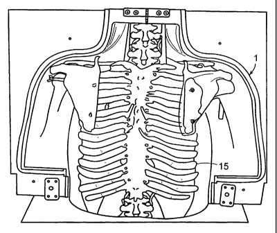
In some embodiments, the outer housing 1 further includes a skeletal system,
either real or synthetic, for example, as shown in Fig. 3. The skeletal system
will provide
an accurate environment in which the laparascope and various instruments must
be
inserted and manipulated. The skeletal system is preferably fabricated of
synthetic
materials. Synthetic skeletal systems are known and, thus, the design of the
skeletal
systems for use with the synthetic torso can be in accordance with these
conventional
skeletal systems. Such skeletal systems are designed so as to provide the
texture,
elastomeric properties, density and various other properties of the human
skeletal system
1 S 15. Materials useful in forming a synthetic skeletal system 15 include,
but are not limited
to plastics, preferably durable, unbreakable plastics, such as those available
through 3B
Scientific (Rudorffweg 6, 21031, Hamburg) (www.3bscientific.com). If a pre-
formed
skeleton is used, the outer housing 1 may be scaled to fit the size of the
skeleton.
Alternatively, pre-formed skeletons scaled to the size of the outer housing 1
can be used.
The outer housing 1, along with the disposable abdominal wall, cavity walls,
skeletal system, synthetic organs and various tissues are fabricated of
materials selected
to provide accurate simulation of the corresponding anatomical structures. For
example,
the materials may be selected to provide similar appearance, texture, tensile
properties,
elastomeric properties, density and/or various other properties of the
corresponding real
anatomical structures. In particular, it is desirable to design the outer
housing, disposable
abdominal wall, cavity walls, skeletal system, organ and tissue elements and
any other
incorporated elements so as to provide the surgeon with proper tactile
feedback when
these objects are touched, cut, sutured or otherwise manipulated with the
various
instruments used during a laparoscopic procedure. Further, in some
embodiments, it is
desirable to design the outer housing, disposable abdominal wall, cavity
walls, skeletal
22
CA 02558650 2006-09-06
WO 2005/088583 PCT/US2005/007623
system, organ and tissue elements and any other incorporated elements so as to
provide a
device that will provide an image, using various radiological procedures,
similar to those
provided using a live body. Thus, the outer housing, disposable abdominal
wall, cavity
walls, skeletal system, organs and other portions can be fabricated of any
material that
mimics the properties of the corresponding real anatomical structure. Further,
in some
embodiments the various parts of the device are preferably formed by molding
processes.
Thus, in some embodiments the materials are preferably easily moldable. Still
further,
the elastic properties of the materials preferably allows for respiratory
motion simulation,
pulmonary motion simulation, motion from the circulation of various fluids
through the
device, beating of a heart if included, and insufflation if performed. In
particular some
useful materials include, but are not limited to, flexible urethane rubbers,
thermoplastic
polyurethanes and silicone rubbers. Some important properties that are
considered in
selecting suitable materials include tensile strength, elongation, hardness
and tear
strength. It is particularly desirable to select materials that are "skin-
like" in their
properties. In particularly preferred embodiments, a number of "skin-like"
materials can
further be selected based on the ease of the molding process using such
materials, the
resulting mechanical properties, the ability to vary these properties by
changing the
mixing ratios of the components, the color of the materials, and the
availability to use die
pigments for various color settings. In some embodiments, for example, it can
be
desirable to provide an outer housing 1, organ and tissue elements and other
elements that
are realistic in color and, thus, in some embodiments, the materials can be
modified to
provide a realistic appearance by the use of colors and dye pigments if
necessary. One
particularly preferred commercially available material is Cine Skin Silicone
A/B,
discussed herein and other materials having similar properties. Other
commercially
available materials include Ecoflex~Rubbers, Dragon SkinTM, liquid silicone
rubber and
Duralco 4538D.
It is particularly beneficial to provide motion of the one or more organ or
tissue
elements within the outer housing 1 so as to closely simulate the atmosphere,
physically
and dynamically, within the human body. In particular, when performing a
laparoscopic
procedure on a live human, the organs and body walls of the subject are in
constant
23
CA 02558650 2006-09-06
WO 2005/088583 PCT/US2005/007623
motion due, in part, to respiration, pulmonary action, circulation of fluids
and the beating
of the heart. It is particularly beneficial to provide an accurate training
device on which
laparoscopic procedures can be performed, wherein the body walls, organ and
tissue
elements and other portions of the device simulate live motion, which has
multiple
degrees of freedom. This provides an accurate environment because the body
walls,
organs and tissue elements are not static nor do they move in limited degrees
of freedom
in a live human body. Thus, the device is provided with a means for simulating
the
respiration of air and/or the circulation of fluids through the outer housing
1 much like
the flow of air, blood, urine and other materials through the human body.
Still further the
a means for simulating pulmonary motion and the beating of the heart, which
provides
further motion, are preferably incorporated into the device as discussed
above.
In one preferred embodiment, the outer housing 1 is specifically designed so
as to
replicate the shape of the a mammalian anatomical structure. Thus, for
example, the
outer housing 1 may be recognized by a viewer as the shape of a human
anatomical
structure, e.g. the human torso or head. For example, as shown in Figs. 2-6,
the outer
housing 1 may be in the shape of a human torso. The synthetic torso is
preferably
designed so as to present a replica of the human anatomy, in physical, dynamic
and
mechanical properties. Thus, for example, the shape, proportions and structure
of the
synthetic torso are preferably designed to replicate the human anatomy. For
example, the
average dimensions of a male torso, based on a typical 6 foot tall male, are:
a height of
approximately 820 mm (32 inches), a width of approximately 510 mm (20 inches),
a
depth of approximately 250 mm (10 inches) and an external neck diameter of
approximately 140 mm (5.5. inches). Thus, for example, when providing a
synthetic
torso of an average adult male, wherein the dimensions are intended to
replicate those of
an average adult male, the dimensions may be as follows: height ranging from
about 24
to about 33 inches, width ranging from about 1 S to about 21 inches, depth
ranging from
about 7 to about 11 inches, and an external neck diameter ranging from about 4
to about 6
inches. If the outer housing 1 is provided in the shape, dimensions and
proportions of an
average female, the dimensions for the average adult female would be
approximately 10-
12% smaller than those of the average adult male. Thus, the dimensions for an
outer
24
CA 02558650 2006-09-06
WO 2005/088583 PCT/US2005/007623
housing 1 provided in the shape and dimensions of an average adult female may
be, for
example: height ranging from about 21 to about 30 inches, width ranging from
about 13
to about 19 inches, depth ranging from about 6 to about 10 inches, and an
external neck
diameter ranging from about 3.5 to about 5.5 inches. If the outer housing 1 is
provided in
the shape, dimensions and proportions of an average youth, the dimensions for
the
average adult youth would be approximately 15-20% smaller than those of the
average
adult male. Thus, the dimensions for an outer housing 1 provided in the shape
and
dimensions of an average youth may be, for example: height ranging from about
19 to
about 28 inches, width ranging from about 12 to about 18 inches, depth ranging
from
about 5.5 to about 9.5 inches, and an external neck diameter ranging from
about 3.2 to
about 5.1 inches. Further, these dimensions may be used in forming a device
having an
outer housing 1 of any geometrical shape (e.g. box-simulator) such that the
overall
dimensions are in proportion to those of a live male, female or youth.
The synthetic torso includes means by which one or more synthetic and/or
animal
organ or tissue elements may be incorporated. In a preferred embodiment, the
outer
housing includes one or more body cavities 4, like those contained in the
human
anatomy, for housing one or more organ or tissue elements. In one preferred
embodiment, the synthetic torso that includes cavities corresponding to the
thoracic
cavity 7 and abdominopelvic cavities 8. The abdominopelvic cavity 8 may
further be
split into the abdominal cavity 9 and the pelvic cavity 10. One or more of the
organ or
tissue elements of the human anatomy located in these cavities are then
located within
these cavities 4.
The one or more organ or tissue elements are preferably housed within the
cavities 4 in a manner that simulates the environment of the human body. In
particular,
one or more organ or tissue elements are preferably placed in the cavities 4
and are held
in proper position and allowed to move in accordance with organ and tissue
elements
within a live human body. When required, conventional fastening means that
will
withstand manipulation of the organ and tissue elements during a laparoscopic
procedure
may be used for portions of organ and tissue elements that are somehow
interconnected
CA 02558650 2006-09-06
WO 2005/088583 PCT/US2005/007623
to other portions of the anatomy. For example, the kidney may be held in place
in a
manner that simulates a live body by the use of, for example, water balloons
placed in
proximity to the kidney, particularly on top of the kidney. Water balloons can
be easily
sized and situated with relation to various organ and tissue elements so as to
simulate the
environment within a live body. Further, real organs can be used in some
applications
and can be obtained in a form that includes the surrounding tissues and fat.
Thus, for
example, one may wish to use the device of the present invention to learn and
practice a
procedure in which the kidney is detached. For such a procedure, one may, for
example,
obtain a real kidney with the tissues and fat that surround the kidney. The
kidney can
then be propped into proper position using any type of fastening means, such
as tape, and
by further placing one or more water balloons on top of the kidney.
In some embodiments, the one or more organ or tissue elements are in contact
with materials that line the cavities 4, in the manner that muscle tissue
lines the cavities
1 S in the human anatomy. Further, much like the abdominal and thoracic
cavities are
separated by the diaphragm, a wall of material may be located between the
abdominal
and thoracic cavities to simulate the diaphragm. Thus, the cavities 4 are
preferably lined
and separated with materials that mimic those lining human cavities such that,
for
example, if an organ or tissue element moves as a result of respiration or as
a result of
manipulation by a laparoscopic instrument and the organ or tissue element
contacts the
cavity wall or the diaphragm wall, the organ or tissue element will react as
it would in a
live body.
Further, the one or more organ or tissue elements in the cavities 4 may be
interconnected and connected to the cavities 4 in a manner that simulates the
human
anatomy by the use of tissue-like materials similar to those found in the
corresponding
anatomical structure. In particular, fastening means that provide the same
type of motion
as the corresponding anatomic connection means may be used. For example, if an
organ
is connected in a manner that allows any type of motion, not limited in its
degree of
freedom, then it could be connected in the present device using, for example,
a string-like
material with sufficient slack to allow for unlimited motion. Where motion is
limited to
26
CA 02558650 2006-09-06
WO 2005/088583 PCT/US2005/007623
particular degrees, hinge-like fastening mechanisms and the like could be used
to
simulate such limits on the motion.
In an exemplary embodiment, the device in the shape of a synthetic torso is
based
on the 3D model of a typical male, which was segmented and reconstructed from
the
Visible Human Data of the National Library of Medicine (National Library of
Medicine,
The Visible Human Project:
http://www.nlm.nih.~ov/research/visible/visible human.html) using transverse
slices
(Fig. 8) at an average of five-millimeter intervals (from section no. 4155 at
sagittal-
coronal coordinate z=463 voxel = 154 mm to section no. 2048 at z-3190 voxel -
1063
mm). This spans the entire torso including the thoracic, abdominal and pelvic
regions. A
manual segmentation method was used to define the outer body, pulmonary, and
abdominal cavities in these image slices. The volume created for the abdominal
cavity
includes all the peritoneal and retroperitoneal organs. The 3D model was
constructed
using engineering design software, Pro/Engineer (PTC, Inc.). The model was
used to
design and fabricate negative molds for casting the synthetic torso, as
depicted in Fig. 2.
These were digitally manufactured of woodblocks as shown in Fig. 2a.
In order to provide a realistic support and structure for the body,
respiratory
motion, pulmonary motion and various other motions a synthetic skeleton 15 was
placed
within the molds prior to casting the torso (Fig. 3). A disposable abdominal
wall 3 was
cast in a separate mold.
In some embodiments, the disposable abdominal wall 3 presents a two layer
structure (Fig. 4) having an inner layer 3a and an outer layer 3b. The two
layers may be
fabricated of the same or different materials. Preferably, the inner wall is
fabricated of a
highly elastic material, such as Cine Skin Silicone A/B with 50% part C
(Elongation:
1000% (A/B+50%C), and the outer layer is fabricated of any of those materials
described
above for use in forming the outer housing (e.g.Cine Skin Silicone A/B).
27
CA 02558650 2006-09-06
WO 2005/088583 PCT/US2005/007623
The disposable abdominal wall 3 is shown in Fig. Sb attached to the synthetic
torso using a hook and loop fastener 16 (Velcro) on the boundary of the
abdominal wall 3
and outer housing 1/synthetic torso. A lip and corresponding depression in the
peripheries of the disposable wall and the synthetic torso can further be used
to hold the
disposable wall 3 in place. As shown, the disposable wall 3 is hermetically
sealed over
the torso with a closed strip 17. This fastening means allows for the
insufflation of the
abdominal cavity as depicted in Fig. Sb. As shown in Fig. Sa, six flexible
tubes 14 are
included next to the spine from the left and right retroperitoneal fossae and
out through
the neck to simulate blood and urine flow to the animal organs placed within
the torso.
However, any number of tubes may be used in a variety of locations.
Preferably,
however, the number of tubes and their placement is selected so as to closely
mimic the
actual human anatomy.
The present device, in the form of an outer housing 1 having any geometric
shape
or the shape of the corresponding anatomical structure (e.g. synthetic torso)
provides a
realistic approximation of an actual surgical procedure and provides surgeons,
medical
students and residents with a method for learning and practicing various
procedures. In
particular, the present device provides components that are designed to
approximate the
properties of the corresponding anatomical structures (e.g. the organs,
tissues, abdominal
wall). Further, the present device is designed to provide proper shape,
proportion and
structure of the corresponding anatomical structures. Still further, the
present device
incorporates means for simulating respiration, pulmonary action, circulation
and the heart
beat of a live anatomy, thereby mimicking the constant motion that the various
parts of
the human anatomy undergo. This motion is not particularly limited in its
degrees of
freedom, and the motion provided by the present device simulates the motion of
a live
body in its degrees of freedom.
Because laparoscopic procedures are performed through small incisions and
often
through laproscopic sleeves and/or tubes, the use of laparoscopic instruments
is different
than that in an open surgery. In particular, the skill required to perform
laparoscopic
surgery is more difficult than one might expect due to inverted manipulation,
two-
28
CA 02558650 2006-09-06
WO 2005/088583 PCT/US2005/007623
dimensional visualization, and the loss of direct touch. The lack of normal
stereoscopic
vision results in the loss of depth perception while "through the hole"
slender instrument
manipulation constrains motion capabilities. One of the surgeon's most
important assets
is a highly developed sense of touch. Laparoscopy, however, separate the
surgeon's
hands from the surgical site.
The laparoscopic simulators presently available present certain limitations.
First,
they do not provide the anatomic perspective of the procedure that is being
performed.
Further, such simulators do not allow for changing the orientation of the
simulated body.
Still further, such simulators do not allow for unrestricted placement and
localization of
trocars. Further, such simulators do not provide the force-feedback
encountered in needle
and trocar insertion. Due to these unrealistic characteristics, the simulators
are only used
at the beginning of the training programs for gaining basic laparoscopic
skills.
Thus, the device of the present invention will provide a valuable training
step that
can be used alone and together with box simulators, live animal training, and
mentored
surgery.
In further embodiments, the present invention provides a device for training
and
evaluating radiological procedures. In particular, radiological procedures
generally
include X-ray, CT (Computed Tomography), MRI (Magnetic Resonance Imaging),
Ultrasound, Nuclear Magnetic Resonance Imaging and Interventional Radiology. X-
ray
procedures is the oldest radiological procedure which uses a small amount of
radiation on
a selected part of the body to generate an image it is commonly used to
generate images
of the chest, musculoskeletal system and the gastrointestinal system. CT is
similar to an
X-ray except that CT imaging provides cross-sectional images of specific areas
of the
body. CT is mainly used for brain, neck, spine, chest, abdominal and
musculoskeletal
imaging. MRI is similar to CT except that MR imaging uses a magnet and radio
waves to
generate an image. MRI has been used to generate images of the brain, spinal
cord, heart,
bones, joints, soft tissues and blood vessels. Ultrasound uses sound waves
passed
through the body to generate an image and is typically used in prenatal care.
Nuclear
29
CA 02558650 2006-09-06
WO 2005/088583 PCT/US2005/007623
Magnetic Resonance Imaging uses small amounts of radioactive compounds to
produce
images that help in gathering information about the function of various
organs. The
radioactive compounds, called tracers, are helpful in evaluating bone, liver,
heart, lung,
brain, kidney or endocrine problems. Interventional Radiology uses catheters
and coils
to open obstructed vessels, drain areas where fluids have accumulated, relieve
certain
forms of hypertension, remove foreign objects and manage gastrointestinal
bleeding
without surgery.
The present invention provides a device that can be used in training and
evaluating a variety of radiological procedures. In particular, as set forth
above, the
human body undergoes constant motion in multiple degrees of freedom. Thus,
when
training individuals in performing procedures, such as radiological
procedures, and in
practicing such procedures, it would be desirable to provide an accurate
approximation of
a live human body that possesses not only the physical properties of the human
anatomy,
but that also simulates the dynamic motion of the live human anatomy. The
present
invention provides such a device in the form of any geometric shape, a
synthetic torso or
other body part that is designed so as to simulate the proportion, structure,
dynamic
motion, and preferably the shape and various other properties of the
corresponding
human anatomy. Such features are discussed above. The device may further be
designed
so as to provide a user with radiological uses. Thus, for example, in training
andJor
practicing a Nuclear Magnetic Resonance Imaging procedure, a tracer may be
implanted
in the desired training/practice site (e.g. in the lungs). The synthetic
torso, which presents
the user with an accurate representation of the human anatomy (e.g. housing
the lungs,
surrounding organs, ribs, etc) and which simulates respiration and pulmonary
motion
(thereby providing lung motion) provides an accurate device on which an
imaging
procedure could take place.
Methods of the present invention comprise using the device to perform various
laparoscopic procedures in a realistic manner. While the devices and methods
are
described, in particular, in relation to a synthetic torso used in performing
laparoscopic
procedures on the torso area, it is to be understood that the device could
comprise various
CA 02558650 2006-09-06
WO 2005/088583 PCT/US2005/007623
other body parts and, further, that various other types of laparoscopic
procedures could be
performed using such synthetic training devices. Still further, while the
present device is
described in connection with the human anatomy, it is also understood that the
present
devices could be modified so as to simulate other animal, preferably mammalian
body
parts, to provide anatomical structures and methods for use in training and
evaluating
individuals in laparascopic surgeries performed on any animal. Still further,
while the
devices have been described in particular with relation to laparoscopic
procedures, the
devices would also present beneficial training and evaluation devices on which
individuals could perform any type of precise surgeries, particularly wherein
motion of
the organs and other body parts impacts the procedure.
The methods of the present invention will be further illustrated with
reference to
the following Examples which are intended to aid in the understanding of the
present
invention, but which are not to be construed as a limitation thereof.
EXAMPLE
The synthetic torso formed in accordance with Figs. 2-6 and 8 was used and
evaluated by medical students, residents, and attending urologists who
compared it to the
standard training box. The study showed that the synthetic torso gives a more
realistic
approximation of a real procedure and is particularly suited for laparoscopy
training.
Animal organs were used in the studies to allow for operating on real tissues.
The organs
were placed in situs, requiring appropriate instrument access and port
placement.
Further, induced respiration caused the organs to move as in a live human
body.
The trainee underwent numerous steps, beginning with the insertion of the
Veress
needle, insufflating the C02, determining the port sites and placing the
trocars. The
trainee then performed a variety of laparoscopic procedures that necessitated
dissecting
and developing tissue planes, excising and reconstructing tissue, suturing,
electrocautering, and performing other surgical maneuvers in an anatomically
consistent
setting. The synthetic torso provided the possibility of placing an animal
kidney in the
31
CA 02558650 2006-09-06
WO 2005/088583 PCT/US2005/007623
retroperitomeum for the trainee to operate on realistic tissues, thus
simulating various
surgeries such as renal biopsy, pyeloplasty, nephrectomy and even parial
nephrectomy.
Tests were run to determine whether the design of the simulator impacts the
subsequent performance of the trainee. Studies were performed by comparing
self
evaluation and trainer scores of a group of subjects randomized to
laparoscopic training
with either a standard box simulator or the synthetic torso of the present
invention.
Twenty five medical students, residents, and attending urologists particupated
in the
study. A 1 S minute instructional presentation given by a fellow provided a
basic
overview of laparoscopic surgery and described and demonstrated the tasks to
be
practiced. The participants were then randomized and allowed to work for two
hours,
during which the fellow was present to provide assistance.
In the case of the laparoscopic training box case, shown in Fig. 6A, a 10 mm
port
(LJSSC) was placed in the middle for camera access. Ports of 5 mm and 10 mm
(USSC)
were placed for instructional access. A large plastic Petri dish was used to
support and
immobilize a porcine kidney within the box.
With the synthetic torso, shown in Fig. 6B, a 10 mm port was placed at the
umbilicus for camera access. A second 10 mm trocar 4 cm lateral to the
umbilicus at the
mid-axillary line, and a S mm port 4 cm superior to the umbilicus were placed
for
instrument access. A porcine kidney was placed in the abdominal cavity in the
left
retroperitoneal fossa. A standard video cart was utilized for both simulators
and 10 mm
and 30° laparoscopes were connected to the camera and alight source
(Stryker
Instruments, Santa Barbara, CA).
The laparoscope and camera were held and manipulated by another participant
for
both the box simulator and the synthetic torso. The following tasks were
performed:
trocar placement, instrument selection and manipulation, needle and suture
manipulation,
suturing and intracorporal knot tying.
32
CA 02558650 2006-09-06
WO 2005/088583 PCT/US2005/007623
After each session, the participants were asked to complete a questionnaire
identifying the training model, the exercises performed, and the instructor.
The
questionnaire also queried the level of education and prior surgical
experience of the
subject. The subjects ranked their responses from "poor" to "excellent", and
from
S "strongly disagree" to "strongly agree". Participants also had the
opportunity to provide
written comments, suggestions and critiques. The questionnaires were then
converted to
an analog scale by assigning "strongly disagree" and "poor" a value of 1 and
"strongly
agree" and "excellent" a value of 5. The results were stratified by the
simulator used. A
mean and standard deviation were then calculated for each question. The means
were
compared and p-values calculated using student's standard t-test. All
statistical
computations were performed using STATA. Our level of statistical significance
was
0.05. The following Table 1 summarizes the statistical results for the
questions asked in
the survey.
33
CA 02558650 2006-09-06
WO 2005/088583 PCT/US2005/007623
TABLE 1: Summary Statistics of Survey Results
Box Synthetic
Simulator Torso
Simulator
Mean SD Mean SD P value
Ease of Use 2.9 0.73 4.0 0.95 0.008
Trainer Approximation 1.8 1.1 4.1 0.57 <0.001
Evaluationof
Anatomy
Internal View 2.7 1.3 4.3 0.65 <0.001
Overall 2.9 1.3 4.3 0.49 0.002
Importance of 4.0 1.1 4.7 0.49 0.064
Anatomy
Trainer External Appearance3.1 1.3 3.8 1.0 0.150
Design Trainer Port 2.9 1.1 4.2 0.72 0.500
Placement
Instrument Training3.6 0.97 4.1 0.90 0.239
Port Placement 3.1 1.3 4.0 0.77 0.069
Skills Training
Lab
Suturing 3.7 1.1 4.1 0.66 0.314
Knot Tying 3.5 1.3 4.1 0.79 0.203
Introduction 4.5 0.70 4.3 0.77 0.608
Definition of 4.1 1.1 4.3 0.86 0.724
Goals
InstructorTechnique 3.8 1.4 4.2 0.83 0.455
EvaluationDescription
Assistance 4.0 1.3 4.6 0.67 0.198
Based on these results, the synthetic torso was significantly easier to use,
provides
a better approximation of the real anatomy and provided a better internal view
(p=0.008,
<0.001, and <0.001, respectively). The participants also felt that it was a
better design
(p=0.002). Moreover, participants in both groups agreed or strongly agreed
that
approximating real anatomy was important and that port placement was important
for
training (p=0.064 and 0.500, respectively).
34
CA 02558650 2006-09-06
WO 2005/088583 PCT/US2005/007623
It was found that training utilizing the devices of the present invention
effectively
decreases errors in the operating room and teaches the trainee additional
skills. The
natural human body configuration allows the trainee to experience the correct
spatial
representation and distribution of trocars as are normally found in the human
abdomen
during laparoscopic surgery. The mixed synthetic/animal model with induced
respiratory
motion of the organs renders a more realistic model compared with existing box
simulators.
All documents mentioned herein are incorporated herein by reference in their
entirety.
The foregoing description of this invention is merely illustrative thereof,
and it is
understood that variations and modifications can be made without departing
from the
spirit or scope of the invention as set forth in the following claims.