Note: Descriptions are shown in the official language in which they were submitted.
CA 02558796 2013-08-14
= 55212-1
=
IN-VIVO VISUALIZATION SYSTEM
FIELD OF THE INVENTION
Embodiments of the present invention generally relate to medical devices.
Several embodiments are generally directed to medical catheters with steering
and/or
optical capabilities. Other embodiments are generally related to medical
systems, such as
in-vivo visualization systems, that are suitable for viewing and/or performing
diagnostic
and therapeutic modalities within the human body, such as in the binary tree.
BACKGROUND OF THE INVENTION
A challenge in the exploration and treatment of internal areas of the human
anatomy has been adequately visualizing the area of concern. Visualization can
be
especially troublesome in minimally invasive procedures in which small
diameter,
elongate instruments, such as catheters or endoscopes, are navigated through
natural
passageways of a patient to an area of concern either in the passageway or in
an organ
reachable through the passageway.
Ureteroscopy is one form of procedure that is performed to diagnosis and treat
urinary tract diseases and ureteral strictures. In conventional ureterscopy, a
ureteroscope
is inserted retrograde through the urinary tract such that diagnosis and
treatment of
urinary tract abnormalities occur under direct visualization. Ureteroscopes
are typically
7-10Fr. in diameter and include a sheath that encapsulates a fiber optic
element, an
illumination element and a working channel. The working channel allows for the
passage
of working devices, such as guidewires, stone retrieval baskets and lasers.
Some
ureteroscopes also incorporate a steering mechanism, which allows the distal
tip of the
scope to be deflected by the user in one or more planes. Steering is typically
achieved via
manipulation at the handle end of the scope, ex-vivo.
Problems, however, exist in the use of prior art ureteroscopes. For example,
after
each successive urological procedure, the scope must be cleaned and sterilized
before the
-1-
CA 02558796 2006-09-06
WO 2005/094665 PCT/US2005/009522
next use, which delays successive procedures unless multiple scopes are
purchased.
Furthermore, current uretero scopes are non-disposable and require extensive,
expensive
maintenance. Sterilization delays and costs associated with purchasing and/or
repairing
scopes have escalated costs for ureteroscopic procedures and other medical
procedures
that utilize similarly configured scopes.
Detailed information regarding other parts of the anatomy can be discerned
from
direct viewing of the anatomy provided through one or more of the elongate
instruments
used in other various medical procedures, such as colonoscopy, upper
endoscopy,
bronchoscopy, thoracoscopy, laparoscopy, and hysteroscopy. For use in these
procedures, various types of endoscopes configured for use in various
passageways of the
body, such as the esophagus, rectum or bronchus, can be equipped with direct
viewing
capability through the use of optical fibers extending through the length of
the scope, or
with digital sensors, such as CCD or CMOS. However, because endoscopes also
provide
a working channel through which other medical instruments must pass, optional
lighting
bundles and components to provide steering capability at its distal end, the
scope is
typically of a relatively large diameter, e.g., 5mm or greater. This large
diameter limits
the use of the endoscope to relatively large body lumens and prohibits their
use in smaller
ducts and organs that branch from a large body lumen, such as the biliary
tree.
Typically when examining small passageway such as the bile duct or pancreatic
duct, the endoscope is used to get close to a smaller passageway or region of
concern and
another instrument, such as a catheter, is then extended through the working
channel of
the endoscope and into the smaller passageway. Although the endoscope provides
direct
visualization of the large body passageway and entrance to adjoining ducts and
lumens,
after the smaller catheter has been extended from the endoscope into the
smaller duct or
lumen, direct visualization has heretofore been limited, and the physician
usually relies
on radiographical means to visualize the area of concern or probes blindly.
SUMMARY OF THE INVENTION
In accordance with aspects of the present invention, a medical visualization
system is provided. The system includes an endoscope having an endoscope
insertion
tube extending distally from an endoscope handle. The endoscope handle has an
access
port for accessing an interior lumen of the insertion tube. The endoscope
includes an
imaging device for viewing objects located at the distal end of the insertion
tube. The
system also includes a catheter assembly comprising a catheter extending
distally from a
-2-
CA 02558796 2013-08-14
55212-1
catheter handle. The catheter handle is selectively mounted to the endoscope
and has an
access port for accessing an interior lumen of the catheter, wherein the
catheter may be
inserted into the endoscope access port and routed through a portion of the
insertion tube
interior lumen. The system further includes an optical assembly comprising an
image
transmission cable having distal and proximal ends, wherein the image
transmission cable is
configured for insertion into the catheter access port and routable through a
portion of the
catheter interior lumen. The optical assembly is capable of obtaining images
located at the
distal end of the catheter and transmitting the images to the proximal end of
the cable. In
some implementations, the catheter handle is selectively connected to the
endoscope handle at
a position located distally of the endoscope access port.
In accordance with another aspect of the present invention, a medical
visualization system is provided. The system includes a disposable catheter
having a
proximal end and a distal end. The catheter defines one or more interior
lumens that extend
from the distal end to the proximal end. The system further includes a
disposable control
handle including an actuation device that effects distal end catheter
deflection. The control
handle is functionally connected to the proximal end of the catheter. The
system further
includes a reusable optical assembly that includes an optical handle and an
optical cable
extending therefrom. The optical cable is routable through a portion of the
interior catheter
lumen from a position exterior to the catheter.
In accordance with another aspect of the present invention, a medical
visualization system is provided. The system includes a disposable assembly
comprising a
catheter having one or more interior longitudinal lumens, a catheter handle
functionally
connected to the catheter and including a steering actuator, and at least one
steering wire
securely connected to the distal end of the catheter and to the steering
actuator. The system
further includes a reusable optical assembly that includes an optical handle
and an optical
cable extending therefrom. The optical handle includes a viewing device for
viewing images
transmitted thereto by the optical cable. The optical cable is sized and
configured to be routed
into one port of the hub, through one of the lumens of the catheter, and
positioned at the distal
end of the catheter, wherein the fiber optic cable transmits illumination
light from its proximal
end to its distal end while transmitting an image from its distal end to its
proximal end.
3
CA 02558796 2013-08-14
55212-1
In accordance with another aspect of the present invention, a catheter
assembly
is provided. The assembly includes a catheter having a proximal end and a
distal end. The
catheter includes at least one steering wire secured at or near the distal end
and extending
-3a-
CA 02558796 2006-09-06
WO 2005/094665 PCT/US2005/009522
outward of the proximal end of the catheter. The assembly also includes a
handle body
functionally connected to the proximal end of the catheter so that the
steering wire
extends therein and a deflection actuator carried by the handle body and
operatively
connected to the steering wire for selectively pushing or pulling the steering
wire to effect
bending of the distal end of the catheter. The assembly further includes a
steering wire
tension adjustment mechanism associated with the handle body. The mechanism is
capable of selectively adjusting the tension applied to the steering wire when
the steering
wire is in a static condition.
In accordance with another aspect of the present invention, a catheter handle
is
provided. The catheter handle is suitable for steering a catheter shaft having
a proximal
region and a distal region and at least one steering wire having a distal end
region secured
at or near the distal end region of the catheter shaft and a proximal end. The
catheter
handle includes a catheter housing having the proximal end of the catheter
shaft attached
thereto and a steering controller carried by the catheter housing and having
the proximal
end of the at least one steering wire connected thereto. The steering
controller is movable
from a first position to a second position. The steering controller is capable
of applying
tension to the at least one steering wire when the steering controller moves
from the first
position to the second position. The catheter handle further includes a lock
mechanism
for retaining the steering controller in the second position to prevent
movement thereof.
The lock mechanism includes a lever movable between an unlocked position and a
locked
position. The lever is associated with the steering controller such that
movement of the
lever to the locked position restricts movement of the steering controller.
In accordance with another aspect of the presnet invention, a medical
visualization
system is provided. The system includes a disposable catheter having a
proximal end and
a distal end. The catheter defines one or more internal lumens that extend
from the
proximal end to the distal end, wherein the catheter includes an optical cable
extending
from the distal end of the catheter to the proximal end of the catheter. The
system also
includes a reusable handle including an image viewing device functionally
connected to
an image transmission cable and a disposable hub functionally interconnecting
the
proximal end of the catheter with the handle. The system further includes a
first
connector that provides detachable connection between the catheter optical
cable and the
handle image transmission cable.
-4-
CA 02558796 2006-09-06
WO 2005/094665 PCT/US2005/009522
In accordance with another aspect of the present invention, a medical device
is
provided. The medical device includes a catheter having proximal and distal
ends. The
catheter defines one or more interior lumens that extend from the distal end
to the
proximal end. The medical device also includes a handle having a proximal end
and a
distal end and a steering assembly that deflects the distal end of the
catheter in at least
one plane. The steering assembly includes a first disposable sub assembly
including at
least one first steering wire and a second reusable sub assembly including at
least one
second steering wire and an actuator functionally connected to the second
steering wire
for selectively tensioning the second steering wire, wherein the actuator is
carried by the
handle. The medical device further includes an optical assembly comprising a
first
disposable sub assembly and a reusable second sub assembly, the first sub
assembly
being positioned within one of the internal lumens and including a first
imaging
transmission cable. The second sub assembly includes an image viewing device
positioned at the handle and a second image transmission cable. The medical
device
further includes a connector that detachably connects the first steering wire
to the second
steering wire and/or detachably connects the first imaging transmission cable
to the
second image transmission cable.
In accordance with another aspect of the present invention, a medical
visualization
system is provided. The system includes a reusable handle comprising an
eyepiece, a
catheter steering deflector, one or more steering wires connected to the
deflector and
extending outwardly of the handle, and an optical cable functionally connected
to the
eyepiece and extending outwardly of the handle. The system further includes a
disposable catheter having a proximal end and a distal end. The catheter
defines first and
second internal lumens that extend from the proximal end to the distal end.
The first
internal lumen and the second internal lumen are configured for receiving the
optical
cable and the steering wire, respectively, wherein the catheter includes
selective =
attachment structure positioned at or near the distal end of the second
internal lumen that
is capable of selectively coupling/decoupling the end of the steering wire to
the catheter.
In accordance with aspects of the present invention, a method of bifurcating
the
interior lumens of a catheter for connection to one or more fittings is
provided. The
method includes obtaining a connector having a central passageway and first
and second
branch passageway connected thereto, obtaining a catheter having first and
second
interior lumens extending longitudinally therethrough, and forming first and
second
-5-
CA 02558796 2006-09-06
WO 2005/094665 PCT/US2005/009522
openings in the outer surface of the catheter at selected, spaced locations
for accessing the
first and second interior lumens. The location of the first and second
openings
correspond to the intersections of the first and second branch passageways
with the center
passageway of the connector, respectively. The method further includes routing
the
catheter into the central passageway until the first and second openings
communicate
with the first and second branch passageways, respectively.
In accordance with another aspect of the present invention, a method of
examining a patient in-vivo is provided. The method includes providing an
endoscope
with an insertion tube having at least one channel. The endoscope has viewing
capabilities at the distal end of the insertion tube. The method also includes
providing a
catheter having at least one channel, providing an imaging device having an
image
transmission cable, and advancing the insertion tube into a passageway of a
patient under
direct visualization by the insertion tube. The method further includes
advancing the
catheter through the insertion tube to a position at or near the distal end of
the insertion
tube; and advancing the image transmission cable through the catheter channel
to a
position at of near the distal end of the catheter. =
In accordance with another aspect of the present invention, a method is
provided
for cannulating the papilla of a patient. The method includes providing an
optical device
having viewing capabilities, providing an endoscope with viewing capabilities
and at
least one channel, and providing a catheter having at least one channel. The
method also
includes placing the distal end of the endoscope into the duodenum of a
patient and
adjacent to the papilla and inserting the catheter into the channel of the
endoscope and
routing the catheter to the distal end of the endoscope. The method further
includes
advancing the optical device through the catheter channel to the distal end of
the catheter;
and advancing the catheter and optical device from the endoscope and through
the papilla
under visual inspection of the endoscope.
BRIEF DESCRIPTION OF THE DRAWINGS
The foregoing aspects and many of the attendant advantages of this invention
will
become more readily appreciated by reference to the following detailed
description, when
taken in conjunction with the accompanying drawings, wherein:
FIGURE 1 is an assembly view of an optical catheter system according to one
embodiment of the invention;
-6-
CA 02558796 2006-09-06
WO 2005/094665 PCT/US2005/009522
FIGURE 2 is a perspective end view of the distal tip of the catheter
illustrated in
FIGURE 1;
FIGURE 3 is perspective end view of the distal tip of the catheter illustrated
in
FIGURE 1, where the sheath of the catheter has been removed to expose the
elongated,
internal body of the catheter;
FIGURE 4 is a cross-sectional view of the elongated body of the catheter
illustrated in FIGURE 3, taken along the line 4-4 in FIGURE 3;
FIGURE 5 is a cross-sectional view of an alternative embodiment of a catheter
of
the system illustrated in FIGURE 1, where the cross-section is taken along a
longitudinal
axis of the catheter;
FIGURE 6 is an assembly view of an optical catheter system according to
another
embodiment of the invention;
FIGURE 7 is an assembly view of an optical catheter system according to a
further embodiment of the invention;
FIGURE 8 is a perspective view of one embodiment of a handle of the optical
catheter system illustrated in FIGURE 7;
FIGURE 9 is an assembly view of an optical catheter system according to
another
embodiment of the invention;
FIGURE 10 is an assembly view of an optical catheter system according to a
further embodiment of the invention;
FIGURE 11 is an assembly view of an optical catheter system according to an
additional embodiment of the invention;
FIGURE 12A is a partial longitudinal cross section view of another embodiment
of a catheter formed in accordance with aspects of the present invention;
FIGURE 12B is a partial longitudinal cross section view of another embodiment
of a catheter formed in accordance with aspects of the present invention;
FIGURE 13A is a partial longitudinal cross section view of another embodiment
of a catheter formed in accordance with aspects of the present invention;
FIGURE 13B is a partial longitudinal cross section view of another embodiment
of a catheter formed in accordance with aspects of the present invention;
FIGURE 14A is a partial view of one suitable embodiment of a catheter body
constructed in accordance with aspects of the present invention;
-7-
CA 02558796 2006-09-06
WO 2005/094665 PCT/US2005/009522
FIGURE 14B is a partial view of one suitable embodiment of a catheter formed
by taking the catheter body of FIGURE 14A and encasing said catheter body with
a
reinforcement sheath;
FIGURE 14C is a partial view of one suitable embodiment of a catheter formed
by taking the catheter of FIGURE 14B and encasing said catheter with an outer
sleeve;
FIGURE 15 is a cross sectional view of the catheter taken along lines 9-9 in
FIGURE 14B;
FIGURE 16 is a partial view of the distal end of another embodiment of a
catheter
that is suitable for used in the system illustrated in FIGURE 1;
FIGURE 17 is a partial view of the distal end of another embodiment of a
catheter
that is suitable for used in the system illustrated in FIGURE 1;
FIGURE 18 is a partial view of the distal end of another embodiment of a
catheter
that is suitable for used in the system illustrated in FIGURE 1;
FIGURE 19A is a perspective view of one suitable embodiment of a catheter
assembly suitable for use in an optical catheter assembly;
FIGURE 19B is a top view of the catheter assembly shown in FIGURE 19A;
FIGURE 19C is a perspective cross section view of the catheter assembly shown
in FIGURE 19A,
FIGURE 19D is a top cross section view of the catheter assembly shown in
FIGURE 19A;
FIGURE 20 is a planar view of one suitable embodiment of an optical assembly
suitable for use in an optical catheter assembly;
FIGURE 21 is a partial bottom view of the catheter assembly shown in
FIGURE 19A
FIGURE 22 is a cross sectional view of the imaging device cable of FIGURE 20
FIGURE 23A is a side view of the optical handle of FIGURE 20;
FIGURE 23B is a side view of the optical handle of FIGURE 20 showing the
detachable nature of its components;
FIGURE 24 is a perspective view of another catheter handle formed in
accordance
with aspects of the present invention;
FIGURE 25 is a top view of another catheter handle formed in accordance with
aspects of the present invention;
-8-
CA 02558796 2006-09-06
WO 2005/094665 PCT/US2005/009522
FIGURE 26 is a top view of another catheter handle formed in accordance with
aspects of the present invention;
FIGURE 27A-27B are partial perspective views of a distal portion of one
embodiment of a catheter formed in accordance with aspects of the present
invention,
several portions of FIGURE 27 is shown in cross-section;
FIGURE 28 is a perspective view of one embodiment of a catheter distal end cap
formed in accordance with aspects of the present invention;
FIGURE 29 is a perspective view of another suitable embodiment of a catheter
assembly suitable for use in an optical catheter assembly;
FIGURE 30 is a cross-sectional view of another embodiment of a catheter that
is
suitable for use with the catheter assembly shown in FIGURE 19A;
FIGURE 31 is a front elevational view of one representative embodiment of an
in-
vivo visualization system constructed in accordance with aspects of the
present invention;
FIGURE 32 is a lateral cross sectional view of an insertion tube of an
endoscope
shown in FIGURE 31;
FIGURE 33 is a perspective view of one embodiment of a catheter assembly
constructed in accordance with aspects of the present invention;
FIGURE 34 is a perspective view of the catheter assembly shown in FIGURE 33
with one housing half removed;
FIGURES 35A-35C are cross sectional views of suitable embodiments of a
catheter constructed in accordance with aspects of the present invention;
FIGURE 36A is a partial view of one suitable embodiment of a catheter body
constructed in accordance with aspects of the present invention;
FIGURE 36B is a partial view of one suitable embodiment of a catheter formed
by taking the catheter body of FIGURE 36A and encasing said catheter body with
a
reinforcement sheath;
FIGURE 36C is a partial view of one suitable embodiment of a catheter formed
by taking the catheter of FIGURE 36B and encasing said catheter with an outer
sleeve;
FIGURE 37 is a cross sectional view of the catheter taken along lines 39-39 in
FIGURE 38B;
FIGURES 38A-38C are cross sectional views of suitable embodiments of a
catheter constructed in accordance with aspects of the present invention;
-9-
CA 02558796 2006-09-06
WO 2005/094665 PCT/US2005/009522
FIGURES 39A-39C are cross sectional views of suitable embodiments of a
catheter constructed in accordance with aspects of the present invention;
FIGURE 40 is a partial perspective view of a catheter handle with the control
knobs removed to illustrate a lock lever;
FIGURE 41 is a partial cross sectional view of a catheter handle showing a
suitable embodiment of an irrigation port connected to irrigation lumens of
the catheter;
FIGURE 42 is a partial cross section view of the catheter handle showing the
steering mechanism and the optional locking mechanism;
FIGURE 43A is a front exploded perspective view of components of the locking
mechanism of FIGURE 42;
FIGURE 43B is a rear exploded perspective view of components of the locking
mechanism of FIGURE 42;
FIGURE 44 is a partial perspective view of the catheter handle of FIGURE 41
illustrating a suitable embodiment of an endo scope attachment device;
FIGURE 45 is a cross sectional view of one embodiment of a Y connector formed
in accordance with the present invention when assembled with a catheter;
FIGURE 46A is an end view of a distal end of another embodiment of a catheter
formed in accordance with the present invention;
FIGURE 46A is a partial side elevational view of the distal end of the
catheter
shown in FIGURE 46A;
FIGURE 47 is an end view of another embodiment of a catheter formed in
accordance with the present invention; and
FIGURE 48 is an end view of another embodiment of a catheter formed in
accordance with the present invention.
DETAILED DESCRIPTION OF THE PREFERRED EMBODIMENT
Embodiments of the present invention will now be described with reference to
the
drawings where like numerals correspond to like elements. Embodiments of the
present
invention are directed to systems of the type broadly applicable to numerous
medical
applications in which it is desirable to insert one or more steerable or non-
steerable
imaging devices, catheters or similar devices into a body lumen or passageway.
Specifically, several embodiments of the present invention are generally
directed to
medical visualization systems that comprise combinations of disposable and
resuable
components, such as catheters, functional handles, hubs, optical devices, etc.
-10-
CA 02558796 2006-09-06
WO 2005/094665 PCT/US2005/009522
Other embodiments of the present invention are generally directed to features
and
aspects of an in-vivo visualization system that comprises a catheter having a
working
channel through which a catheter having viewing capabilities is routed. As
will be
described in detail below, the catheter may obtain viewing capabilities by
being
constructed as a vision catheter or by having a fiberscope or other viewing
device
selectively routed through one of its channels. The catheter is preferably of
the steerable
type so that the distal end of the catheter may be steered from its proximal
end as it is
advanced within the body. A suitable use for the in-vivo visualization system
includes
but is not limited to diagnosis and/or treatment of the duodenum, and
particularly the
biliary tree.
Several embodiments of the present invention include medical devices, such as
catheters, that incorporate endoscopic features, such as illumination and
visualization
capabilities, for endoscopically viewing anatomical structures within the
body. As such,
embodiments of the present invention can be used for a variety of different
diagnostic and
interventional procedures. Although exemplary embodiments of the present
invention
will be described hereinafter with reference to duodenoscopes, it will be
appreciated that
aspects of the present invention have wide application, and may be suitable
for use with
other endoscopes (e.g., ureteroscopes) or medical devices, such as catheters
(e.g., guide
catheters, electrode catheters, angioplasty catheters, etc.). Accordingly, the
following
descriptions and illustrations herein should be considered illustrative in
nature, and thus,
not limiting the scope of the present invention. Additionally, the catheter
with vision
capabilities may be utilized alone, as well as in conjunction with a
conventional
endo scope.
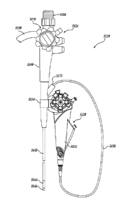
FIGURE 1 illustrates an optical catheter system 8 in accordance with one
embodiment of the present invention. The primary components of the system 8
include a
sterile, single-use, disposable catheter 10, a sterile, single-use, disposable
hub 20, and a
reusable handle 30. In the illustrated embodiment, the hub 20 is integral,
i.e.,
permanently part of, the disposable catheter 10 such that they together define
a sterile,
single-use, disposable catheter assembly. For example, the hub 20 may be
joined to the
catheter 10 with injection molding or adhesive bonding. The catheter assembly
defined
by the hub 20 and catheter 10 is preferably packaged in a sterile container or
package (not
illustrated) prior to use by a physician. In an alternative embodiment, the
hub 20 is
integral, i.e., permanently part of, the handle 30. In a further embodiment,
the hub 20 is
-11-
CA 02558796 2006-09-06
WO 2005/094665 PCT/US2005/009522
not integral with the catheter 10 or the handle 30, but connects to these
items with
connectors, such as male and female threaded connectors, quick lock
connectors, bayonet
connectors, snap connectors, or other known connectors.
As is illustrated in FIGURES 2-4, the catheter 10 includes an elongated,
preferably cylindrical, body 38 that extends the entire length of the catheter
10. In one
embodiment, the catheter body 38 has an outer diameter between approximately 5
and 12
French, and preferably between approximately 7 and 10 French. The catheter
body 38
may be constructed from any suitable material, such as Pebaxiil (polyether
block amides),
nylon, polytetrafluoroethylene (PTFE), polyethylene, polyurethane, fluorinated
ethylene
propylene (FEP), thermoplastic elastomers and the like, or combinations
thereof. The
body 38 may be formed of a single material using known techniques in the art,
such as
extrusion, or multiple materials by joining multiple extruded sections by heat
bonding,
adhesive bonding, lamination or other known techniques (e.g., juxtaposed
Nitinol tubes
wrapped with an adhesive bonding.
In some applications, e.g. urological, it is desirable that the catheter 10
have a
varying degree of stiffness from the distal (e.g., renal pelvis) end 18
towards the proximal
(e.g., bladder) end 16. The proximal end 16 should be stiff enough for the
device to
advance in the tract to the desired location (e.g., in the urinary tract to
the renal
pelvis/kidney area). The distal end 18 should be soft enough to provide a
reduction in
trauma during insertion but rigid enough to provide adequate support during
the
procedure and prevent collapse or kinking. According to an embodiment of the
present
invention for urological application, the distal portion of the catheter
(approximately 1-2
inches where the flexing occurs) is made more flexible (i.e., less stiff) than
the remainder
of the catheter to allow for steerability of the catheter in vivo. Several
techniques for
constructing a catheter having a more flexible distal portion than the
remainder of the
catheter will be described in more detail below.
In the embodiment shown in FIGURE 1, the catheter 10 includes a proximal
portion 42 that extends the majority of the catheter 10 and a distal portion
44. The
catheter 10 preferably varies in stiffness between the proximal portion 42 and
the distal
portion 44. More preferably, the proximal portion 42 is stiffer than the
distal portion 44.
This allows the catheter 10 to be easily advanced without compressing and with
minimal
twisting while providing deflection capabilities to the distal portion 42 for
deflecting the
distal end 18. In one embodiment, the proximal portion 42 has a durometer
value
-12-
CA 02558796 2006-09-06
WO 2005/094665 PCT/US2005/009522
between 35 and 85 shore D, preferable 60-80 shore D, and the distal portion 44
has a
durometer value between 5 and 55 shore D, preferable 25-40 shore D
As is illustrated in FIGURES 2 and 3, the catheter 10 may optionally include
an
inner sheath 56 and/or an outer sleeve 58 that encase the length of the
elongated body 38
or portions thereof. In one embodiment, the sheath 56 is a woven or layered
structure,
such as a braided design of fine wire or polymeric elements woven or coiled
together
along the longitudinal axis of the catheter with conventional catheter
braiding (e.g.,
2 wires having a diameter ranging from 0.001 to 0.010 inches wound. in a 2-
over, 2-under
helical fashion from the proximal to distal end of the catheter 10). This
allows the
catheter 10 to be advanced to the desired anatomical site by increasing the
column
strength of the assembly while also increasing the torsional rigidity of the
catheter.
Conventional coiled polymer or braid wire may also be used for this component
with coil
wire dimensions ranging in width from 0.002 to 0.120 inches and thickness from
0.002 to
0.10 inches. Braided ribbon wire (e.g., 0.002 x 0.005 inches; 0.003 x 0.012
inches) may
also be used for the sheath 56.
The outer sleeve 58 may comprise of any number of polymer jackets that are
laminated over the first sheath 56. Suitable materials for the sleeve 58
include, but
without limitation, polyethylene, such as polyethylene having a molecular
weight in the
range of 50,000 to 100,000; nylon, such as nylon 12, nylon 4-6, and nylon 6-6;
Pebax
(polyether block amides); polyurethane; polytetrafluoroethylene (PTFE),
particularly
fluorinated ethylene propylene (FEP) copolymers; and polyethylene impregnated
with
PTFE. The outer sleeve 58 may be used to vary the stiffness of the catheter,
if desired, or
to provide improved torque transfer and/or other desirable catheter
properties.
Additionally, the sleeve 58 may be used as one convenient method for securing
a more
flexible deflection section to the proximal section, as will be described in
detail below.
In one embodiment, as will be described in more detail below, the outer sleeve
58 is
coextruded, coated, or otherwise attached once the sheath 56 is applied, to
lock the
sheath 56 in place and secure it to the catheter body 38, thereby forming a
composite
catheter.
In several embodiments, the external surface of the catheter, for example, the
outer sleeve 58, can have a hydrophilic coating or a silicone coating to ease
the passage
of the device in vivo. Such a hydrophilic coating can be, for example, but
without
limitation, N-Vinyl Pyrrolidone, Poly Vinyl Alcohol, and Poly Vinyl
Pynolidone. The
-13-
CA 02558796 2006-09-06
WO 2005/094665 PCT/US2005/009522
hydrophilic coating can be accomplished by coating the device with a primer,
such as
Bayhydrol 110 (an anionic dispersion of an aliphatic polyester urethane resin
in
water/n-methyl-2pynolidone) and then bonding a primary layer over the primer.
The
primary layer can be, without limitation, an acrylamide or a polyurethane-
based
acrylamide. Alliphatic polyether and polyester polyurethanes also can be used
as
lubricous coatings.
In a further embodiment, the distal portion 44 of the catheter 10 may contain
a
preset curve detail that allows a physician to easily access various locations
(e.g., the
renal pelvis) with minimal manipulation via passive deflection (i.e., without
ex-vivo
steering mechanism actuation). In one embodiment, the durometer of the sleeve
58 varies
from 35 Shore D to 85 Shore D (preferably in the region of 70-80D) at the
proximal
end 16 to 20 Shore D to 55 Shore D (preferably in the region of 30-43D) at the
distal
end 18. Curves of various shapes and geometries may be preset to the distal
portion 44 of
the catheter 10 as desired. For example, these curves may be pre-baked into
the sleeve 58
at an elevated temperature below the melting point of the polymer. This pre-
baked curve
can vary between 10 and 270 degrees from vertical, depending upon the specific
application of the system 8. To insert the catheter 10, the curve should be
such that when
a dilator or stiff guidewire is insetted into a working channel of the
catheter 1 0 (described
below), the curve is straight, while once the dilator or guidewire is removed,
the distal
portion 44 reverts to the pre-baked curve providing access to a desired
location. In one
embodiment, the distal portion 44 of the sleeve 58 has a radiopaque marker
band 46
mounted thereon to provide confirmation of the location of the distal end 18
via
fluoroscopy.
Referring now to FIGURES 2-4, the elongated body 38 of the catheter 10 defines
a working channel 60 that extends the entire length of the catheter and allows
for the
passage of various treatment or diagnostic devices, such as guide wires, stone
retrieval
baskets, lasers, biospy forceps etc. The working channel 60 preferably has a
diameter
sufficient to accept up to a 4 French working device, such as a retrieval
basket device or
biopsy forceps. The elongated body 38 of the catheter 10 may also include
additional
channels 62, for use, e.g., as irrigation/insufflation channels or additional
working
channels for one or more of the instruments mentioned above. The channels 62
each
extend the entire length of the catheter 10 and, like the working channel 60,
allow the
passage of devices, liquids and/or gases to and from the treatment area. The
channels 62
-14-
,
CA 02558796 2006-09-06
WO 2005/094665 PCT/US2005/009522
each have a diameter similar to or smaller than main working channel 60. In
one
embodiment, the channels 62 each have a diameter of about 0.020 inches. The
catheter
may also include a channel 64 that extends the entire length of the catheter
through which
a fiberscope, fiber optic cables or other small diameter imaging devices
(e.g., 0.25inm-
1.5mm diameter) can be routed to the distal end of the catheter 10. It will be
appreciated
that one or more of the channels 62 may be eliminated or dimensioned to
accommodate
the necessary diameter needed for the working channel 60 and optic lumen.
As is illustrated in FIGURES 2-4, the catheter 10 also includes a pair of
control or
steering wires 68 that cause a distal portion 44 of the catheter 10 to deflect
in one or more
directions as indicated by the dashed lines in FIGURE 1. The steering wires 68
are
located on opposite sides of the catheter 10 and slide within grooves 70 in
opposite sides
of the elongated body 38. In other embodiments, the steering wires 68 may
reside in the
sheath 56 or outer sleeve 58. In yet another embodiment, the steering wires 68
may be
routed through dedicated steering wire lumens in the catheter. The steering
wires 68
extend from the distal end 18 of the catheter 10 to the opposing, proximal end
16 of the
catheter 10, and then through the hub 20. The steering wires 68 may be
attached to the
distal end 18 of the catheter 10 in a conventional manner, such as adhesive
bonding, heat
bonding, crimping, laser welding, resistance welding, soldering or other known
techniques, at anchor points such that movement of the wires causes the distal
end. to
deflect in a controllable manner. In one embodiment, the steering wires 68 are
attached
via welding or adhesive bonding to a fluoroscopy marker band 46 (see FIGURE 1)
fixedly attached to the distal end. In one embodiment, the band may be held in
place via
adhesive and/or an outer sleeve, as will be described in more detail below.
The steering
wires 68 preferably have sufficient tensile strength and modulus of elasticity
that they do
not deform (elongate) during curved deflection. In one embodiment, the
steering wires
are made from 304 stainless steel with an 0.008 inch diameter and have a
tensile strength
of approximately 325 KPSI. The steering wires 68 can be housed in a PTFE thin-
walled
extrusion (not shown) to aid in lubricity and prevent the catheter 10 from
binding up
during deflections, if desired.
In the illustrated embodiment shown in FIGURE 1, the steering wires 68
terminate in a wire connector 70, which may also be part of the hub 20. The
wire
connector 70 is a mechanical device that provides a detachable, preferably
quick-fit,
connection between the steering wires of the catheter 10 and the controller 74
or handle
-15-
CA 02558796 2006-09-06
WO 2005/094665 PCT/US2005/009522
steering wires (not illustrated) associted with the handle 30. Various types
of detachable
mechanical connectors, such as joints and linking elements, are capable of
forming a
connection that allows active deflection of the wires 68 via the controller 74
of the
handle 30. In the illustrated embodiment, the catheter 10 includes two
steering wires 68
that controllably steer the catheter distal end 18 within one plane. In
alternative
embodiments, the catheter 10 includes additional wires that allow a user to
steer the distal
end 18 in multiple planes. In a further embodiment, the catheter 10 only
includes one
control wire that allows the user to steer the distal end 18 in one direction.
In another
embodiment, such as described below, the steering wires 68 are not part of the
catheter 10. In such an embodiment, the catheter can be advanced over a
guidewire (not
shown) pre-placed in the region of interest.
Referring now to FIGURE 5, there is shown a cross-sectional view of an
alternative embodiment of a catheter 510 suitable for use with the optical
catheter
system 8. The catheter 510 illustrated in FIGURE 5 also includes additional
features and
inherent functions, as described further below. Unlike the catheter 10, the
catheter 510
has one large lumen 512 as opposed to multiple lumens. This is referred to as
a "loose
tube" configuration. The steering wires 568 run along the inner diameter of
the
catheter 510 to the distal end and are located within channels defined by an
internal
sleeve or liner 547. The liner 547 has a low co-efficient of friction to
facilitate the
passage of working devices through the catheter during surgery. The liner 547
has a wall
thickness from 0.0005 to 0.010 inches and is preferably formed from nitinol
tubing, a
polymer containing a degree of fluoroethylene such as, but not limited to,
FEP, PTFE or
PTFE impregnated thermoplastic elastomers like Pebax or is formed from a
polymer
having fluroethylene combined with thermoplastic materials such as polyamides,
polyurethane, polyethylene and block co-polymers thereof. The optical
assembly, any
working devices, and any irrigation tubes pass through the lumen 512 and
connect with
the hub as described above and below. In an alternative embodiment, the
elongated
body 538 of FIGURES 2-4 passes through the lumen 512, where the elongated body
538
routes any working devices, the optical assembly, and any irrigation tubes as
described
above.
The catheter 10 may be constructed in many different ways to achieve the
desired
result of a catheter having varying stiffness along its length, a few of which
will now be
described in more detail. FIGURE 12A is a longitudinal cross-section view of
one
-16-
CA 02558796 2006-09-06
WO 2005/094665 PCT/US2005/009522
embodiment of a catheter 1210 constructed in accordance with aspects of the
present
invention. As best shown in FIGURE 12A, the catheter 1210 comprises a catheter
body 1238 that is constructed with discrete proximal, deflection, and distal
tip
sections 1282, 1284, 1288. In this embodiment, the proximal section 1282 is
stiffer than
the deflection section 1284. Each section may be constructed in any suitable
manner,
such as extrusion or milling, with any suitable materials, such as
polyethylene, nylon,
Pebax (polyether block amides), polyurethane, polytetrafluoroethylene (PTFE),
thermoplastic elastomers, chosen for the desired application. The sections
1282, 1284,
and 1288 are then coupled together to form an integral body by encasing the
length of the
body 1238 or portions thereof with an outer sleeve 1258. The deflection
section may
contain one or both of section elements 1284 and 1288 to impart the required
deflection
at the distal end to the system. The outer sleeve 1258 may comprise one of any
number
of polymer jackets that are laminated, co-extruded, heat shrunk, adhesive
bonded, or
otherwise attached over the catheter body 1238. Suitable materials for the
sleeve 1258
include, but are not limited to, polyethylene, nylon, Pebax (polyether block
amides),
polyurethane, polytetrafiuoroethylene (PTFE), and thermoplastic elastomers to
name a
few. It will be appreciated that the sections 1282, 1284, and 1288 may also be
heat
bonded or adhesive bonded prior to outer sleeve attachment.
The catheter 1210 may optionally include an inner reinforcement sheath 1256,
for
example, a metallic braid, disposed between sections 1282, 1284, and 1288 of
the
elongated body 1238 and the outer sleeve 1258, as best shown in FIGURE 12B.
The
reinforcement sheath 1256 encases the length of the catheter body 1238 or
portions
thereof. In one embodiment, the reinforcement sheath extends from the proximal
end of
the catheter body to proximal an optional radio opaque band (not shown) at the
distal tip
section. The reinforcement sheath increases the kink resistance of the
deflecting
section 1284 to ensure that internal lumens remain patent during bending.
FIGURE 13A is a longitudinal cross section view of another embodiment of a
catheter 1310 constructed in accordance with aspects of the present invention.
As best
shown in FIGURE 13A, the catheter 1310 defines a proximal section 1382, a
deflection
section 1384, and a distal tip section 1388. The catheter 1310 comprises a
catheter
body 1338 and an outer sleeve 1358. The catheter body 1338 is a unitary core
that is
formed, preferably by extrusion, with one suitable material, such as nylon,
Pebaxii),
PTFE, etc. In one embodiment, the body 1338 is a PTFE extrusion. When
assembled,
-17-
CA 02558796 2006-09-06
WO 2005/094665
PCT/US2005/009522
the outer sleeve 1358 encases the length of the elongated body L 338 or
portions thereof.
The outer sleeve 1358 comprises a number of polymer jackets 13 58A, 1358B, and
1358C
that are laminated, co-extruded, heat shrunk, adhesive bonded, or otherwise
attached over
sections 1382, 1384, and 1388 respectively, of the catheter body 1338. The
stiffness
value of each jacket is specifically selected to achieve the desird results,
and may vary
upon different catheter applications.
In one embodiment, the jacket 1358A, which corresponds to the proximal
section 1382, is constructed of a material having a greater stiffness value
than the
jacket 1358B, which corresponds to the deflection section 1384_ Suitable
materials for
the sleeve 1358 include, but are not limited to, polyethylene, nylon, PebaxiD
(polyether
block amides), polyurethane, polytetrafluoroethylene (PTFE), to name a few. If
PTFE is
chosen for the body 1338, it may be necessary to etch or otherwise prepare its
outer
surface to promote suitable adhesion of the outer sleeve 1358.
The catheter 1310 may optionally include an inner reinforcement sheath 1356,
for
example, a metallic braid, disposed between the elongated bc=dy 1338 and the
outer
sleeve 1358, as best shown in FIGURE 13B. The reinforcarient sheath encases
the
length of the elongated body 1338 or portions thereof. Ia one embodiment, the
reinforcement sheath extends from the proximal end of the catheter body to
proximal an
optional radio opaque band (not shown) at the distal tip section. The
reinforcement
sheath increases the kink resistance of the deflecting section to ensure that
internal
lumens remain patent during bending.
FIGURES 14A-14C and 15 illustrate another embodiment of a catheter 1410
constructed in accordance with aspects of the present invention. As best shown
in
FIGURE 14A, the catheter includes a catheter body 1438 having a proximal
section 1482,
a deflecting section 1484, and a distal tip section 1488. In one embodiment,
the proximal
section 1482 is constructed of a material that is stiffer than the deflecting
section 1484.
The proximal section 1482 and the deflecting section 1484 may be extrusions
constructed
from any suitable material, such as polyethylene, nylon, Pbax (polyether
block
amides), polyurethane, polytetrafluoro ethylene (PTFE), and thermoplastic
elastomers, to
name a few. In one preferred embodiment for urological application, the
proximal
section is a multi-lumen, PTFE extrusion approximately 200 to 220 cm in
length, and the
deflecting section 1484 is a multi-lumen, Pebax extrusion approximately 2 to
10 cm in
length. The deflection section 1484 may be coupled to the prcodmal section
1482 via
-18-
CA 02558796 2006-09-06
WO 2005/094665 PCT/US2005/009522
suitable adhesive or joined by other techniques. The distal tip section 1488
may be
coupled to the distal end of the deflection section 1484 via suitable
adhesive. The distal
tip section 1488 may be constructed of any suitable material, such as
stainless steel or
engineering plastics, including but not limited to polyethylene, nylon, Pebax
(polyether
block amides), polyurethane, polytetrafluoroethylene (PTFE), and thermoplastic
elastomers. The catheter body 1438 may also include a radio opaque marker band
1446
that encircles a portion of the distal tip section 1488.
The catheter 1410 (see FIGURE 14B) also includes a reinforcement sheath 1456
that extends from the proximal end of the catheter to or immediately proximal
of the
radio opaque marker band 1446. The sheath 1456 may be a woven or layered
structure,
such as a braided design of fine wire or polymeric elements (0.001 inches to
0.010 inches
in diameter) woven or coiled together along the longitudinal axis of the
catheter with
conventional catheter braiding techniques. This allows the catheter to be
advanced to the
desired anatomical site by increasing the column strength of the assembly
while also
increasing the torsional rigidity of the catheter. The reinforced catheter
body shown in
FIGURE 14B is then encased by an outer sleeve 1458 comprising of one or more
sleeve
sections 1458A, 1458B, and 1458C, having the same or different stiffness
values, as best
shown in FIGURE 14C, to form the catheter 1410.
Returning to FIGURE 14A, the catheter also includes a plurality of steering
wires 1468 that extend through grooves or slots formed in the catheter body
from the
proximal end of the catheter past the deflecting section 1484. In one
embodiment, the
steering wires 1468 terminate at the radio opaque marker band 1446 to which
the steering
wires 1468 are joined by adhesive bonding, laser welding, resistance welding,
soldering
or other known techniques.
In several embodiments, it is preferable for the steering wires to be encased
with a
laminate structure 1496 for allowing the steering wires 1468 to move freely
within or
along the catheter body, and thus, make the mechanics of actuation as smooth
as possible.
As best shown in FIGURE 15, the laminate structure 1496 is formed by outer
jacket 1497
constructed of a thermoplastic polymer, such as polyurethane, Pebax ,
thermoplastic
elastomer etc. which encases an inner reinforcement member 1498, such as a
metallic
braid (e.g., stainless steel braid having, for example, a 0.0015" x 0.006"
helically wound).
Inside the reinforcement member 1498, is a layer 1499 of a friction reducing
material,
such as PTFE or FEP tubing, over which the aforementioned layers are formed.
The
-19-
CA 02558796 2013-08-14
. 55212-1
Inminsite structure 1496 begins at the proximal section 1482 and extends to
just proximate
the radio opaque marker band 1446, as best shown in FIGURE 14A.
As was described above, in several embodiments of the catheter, it is
desirable for
the deflection section or distal portion to be configured to deflect more
easily than the
proximal section or portion. In one embodiment, the deflection section or
distal portion
has a durometer value less than the proximal section. In other embodiments,
the
flexibility may be varied gradually (e.g., increasingly) throughout the length
of a catheter
tube from its proximal end to its distal end. In other embodiments, the
deflection section
may be an articulating joint. For example, the deflection section may include:
a plurality
of segments that allow the distal section to deflect in one or more
directions. For
examples of articulation joints that may be practiced with the present
invention, please
see co-pending U.S. Patent Application Nos. 10/406,149, 10/811,781, and
10/956,007. I
I
Other mechanical joints or configurations may be utilized that allow the
distal
portion of the catheter to flex or bend in one or more directions more easily.
Turning
now to FIGURE 16, there is shown one embodiment of a catheter 1610 formed in
accordance with aspects of the present invention. FIGURE 16 shows a partial -
view of the
distal portion 1646 of a catheter 1610 constructed from a metal or plastic
tube with
slots 1694 cut 180 degrees and spaced an even distance apart to form a
deflecting section.
The slots will allow the catheter 1610 to deflect in two directions or in a
single plane at
the distal end 1618. The proximal section of the tube is not slotted and may
be used as
the non-deflecting portion of the catheter. If preferred, the slotted section
may be used in
embodiments discussed above. The slotted section can be useful when the
catheter
profile is not symmetrical or is irregular. It will be appreciated that the
slots 1 694 can be
V-shaped, semi-circle, wave or any preferred configuration.
FIGURE 17 illustrates another embodiment of a catheter 1710 having a
deflectable distal portion. In this embodiment, the catheter is constructed
Erom a very
flexible plastic extrusion with multiple lumens. The two main lumens, the
working
6hanne11760 and the optical assembly channel 1762, are reinforced with cans
1796 to
minimize out-of plane deflection. As shown in FIGURE 17, the center of 13. oth
lumens
and both coils lie on the Y-axis to provide less resistance against deflection
in the x-
plane. When the steering wires (not shown) are pulled along the direction of -
the steering
wire slots, the catheter will tend to bend about the y-axis or in the x plane.
The
-20-
CA 02558796 2006-09-06
WO 2005/094665
PCT/US2005/009522
coils 1796 also prevent the lumen from kinking as the catheter deflection
radius becoanes
tighter. The catheter 1710 may further include an outer braid and outer layer-
, as
described in detail above.
FIGURE 18 illustrates yet another embodiment of a catheter 1810 havirn a
flexible distal portion 1846. In this embodiment, the multiple lumen
extrusioia is
preferred to be flexible. Slots 1894 are cut on both sides of the extrusion to
assist and
bias the catheter 1810 in the preferred direction of deflection. As was
described ab.ove,
coils 1896 may be used to support the main lumens, if preferred, but are not
required.
The coil or coils can be useful if the slot cuts are deep to penetrate the
main lumens. The
coils could be used to line the lumens such that the devices do not
inadvertently get
caught against the slots. The catheter may further include a braided sheath
and cuter
sleeve, as described above.
Returning now to FIGURES 1-4, the elongated body 38 of the catheter 10
includes a lumen 64 that holds an optical assembly 40 or portions thereof, as
described
briefly above. The optical assembly 40 is defined, e.g., by a cylindrical,
elongated
tubular member 24 and optic bundles 32, 34. The optical assembly 40 permits a
user of
the system 8 to view objects at or near the distal end 18 of the catheter 10.
In_ the
illustrated embodiment, the distal end 18 of the catheter includes a clear
lens or
window 22 that sealingly encloses the distal end of the lumen 64 to protect
the optic
bundles 32, 34 inside the lumen 16. The member 24 defines multiple lumens 26
that 'each
contain one fiber optic bundle 32, 34. The first fiber optic bundle 32
illuminates the area
or objects to be viewed, while the second fiber optic bundle 34 communicates
the
illuminated image to an eyepiece or ocular lens device 36 located at the
handle 30
through which a user can view the images communicated via the fiber optic
bundle. The
handle 30 can also be configured to connect to a camera or imaging system such
that
users can save images and view them on a display. The fiber optic bundles 32,
34 each
comprise one or more fiber optics cables, preferably multiple fiber optical
cables, but
may also include lenses, rods, mirrors, hollow or solid light guides, etc.
The
bundles 32, 34 are attached to the lens 22 with a clear adhesive, bond, or
other
connection, but can also abut the lens or be located adjacent the lens without
any
attachment. In an alternative embodiment, the lens 22 is not attached to the
distal en_d 18
of the catheter, but is instead attached directly to the elongated member 24
and fiber optic
bundles 32, 34.
-21-
CA 02558796 2013-08-14
. 55212-1
As will be appreciated, the optical components of the catheter 10 may take any
other forms and configurations. For example, the lumen 64 can include one
fiber optic
bundle for communicating images and one or more single illpmination fibers
that ar-e not
fixed relative to each other by the elongated member 24. That is, the fibers
can be freely
located in the lumen 64. Additionally, the elongated member 24 can have more
or less
lumens 26 that contain more or less fibers and/or bundles for illuminating
and/or
communicating images. For example, in an alternative embodiment, a single
fiber
replaces one or both of the bundles 32,34. Furthermore, the elongated body 38
need not
include the lumen 64. For example, one or more optical fibers or bundles of
fibers can be
molded in the elongated body 38. Alternatively, the elongated body 38 may
include two
lumens 64 for receiving separate fiber optic bundles 32 and 34, respectively.
Possible
alternative known configurations for the optical assembly 40 are described in
U.S. Patent
Nos. 4,782,819; 4,899,732; 5,456,245; 5,569,161; and 5,938,588.
In the illustrated embodiment, the f tubular optical assembly 40 is part of
the
disposable catheter assembly defined by the catheter 10 and hub 20. Hence, the
tubular
optical assembly 40 and its fiber optic bundles 32,34 extend from the distal
end 18 of the
catheter 10 to the opposing, proximal end 16 of the catheter 10, and then
through, the
hub 20. As is illustrated in FIGURE 1, the hub 20 includes a fiber optic
connector 72 in
which the fiber optic bundles 32,34 terminate. The fiber optic connector 72 is
a
mechanical device that provides a detachable optical connection between the
fiber of the
optical assembly 40 and the fiber or lens system of the handle 30. Thus, the
oTtical
assembly 40 extends continuously through the disposable catheter 10 and hub
20, without
interruption, to the fiber optic connector 72. In one embodiment, the fiber
optic
connector 72 is a detachable, simple point-to-point connection or splice. In
other
embodiments, the connector 72 is a more complex design having multi-port or
other types
of optical connections. For example, the connector 72 can be configured to
redistribute
(combine or split) optical signals, such as with an active or passive fiber
optic couplers,
e.g., splitters, optical combiners, X couplers, star couplers, or tree
couplers. The fiber
optic connecter 72 can also include a micro lens, graded-refractive-index
(GRIN) rods,
beam splitters, and/or optical mixers, and may twist, fuse, and taper together
the fiber
optic bundles 32, 34. In other embodiments, such as those described below, the
optical
assembly 40 is not part of the disposable catheter 10.
-22-
CA 02558796 2013-08-14
= 55212-1
=
Referring again to FIGURE 1, the handle 30 is generally an endoscopic handle
that connects to the connectors 70, 72 of the hub 20 such that a user of the
system can
view images communicated by the fibers of the catheter 10 and such that a user
can
controllably steer or deflect the distal end 18 of the catheter. The handle 30
includes one
or more shafts 78 that connect to and interact with the fiber optic connector
72 and the
wire connector 70. The handle 30 also includes a controller or actuator 74 by
which a
user can steer the distal end 18 of the catheter 10. In the illustrated
embodiment, the
handle 30 generally includes a pair of steering wires (not illustrated), each
of which is
associated with . one of the steering wires 68 of the catheter 10. The wires
of the
handle 30 are connected to the controller 74 at one end and are connected at
the other end
to the wires 68 via the connector 70. To steer the catheter 10, a user
actuates the
controller 74, which causes the wires 68 to deflect, which in turn forces the
distal end 18
of the catheter to deflect as illustrated in FIGURE 1. In the illustrated
embodiment, the
controller 74 is a user-operated mechanical slide or rotatable lever that is
adapted to pull
and release the wires 68 connected to the handle 30 by the connector 70. In an
alternative
embodiment, the controller 74 may take other forms, such as a rocker arn or
rotating
knob, adapted to pull and release the wires. In another alternative embodiment
in which
the catheter 10 has two or more pairs of steering wires, the handle 30
includes additional
actuators and corresponding controls to drive the additional pairs of steering
wires. In
one embodiment, the handle 30 includes a locking mechanism, such that when a
curve is
activated by the controller 74, the curve may be locked in place. The use of
wires to steer
a tip of a catheter is well-known. Suitable examples are set forth in U.S.
Patent
Nos.: 4,899,723; 5,273,535; 5,624,397; 5,938,588, 6,544,215, and International
Publication No. WO 01/78825 A2 .
As is described above, the handle 30 includes steering wires and fiber optics
that
connect to the steering wires 68 and fiber optic bundles 32,34 of the catheter
10 via the
connectors 70,72. As will be appreciated, the handle 30 may be battery powered
or
connect to a power supply. The handle 30 also includes a light source, or
connects to a
light source, that illuminates the fiber bundle 32. In addition, the handle 30
has an
eyepiece 80 for a user to view an image transmitted by the image bundle 34
from the
distal end 18.
-23-
CA 02558796 2006-09-06
WO 2005/094665 PCT/US2005/009522
Referring again to FIGURE 1, the hub 20 also includes connectors or ports 50
that
each communicate with one of the lumens 62 of the catheter 10, as well as a
connector or
port 52 that communicates with the working channel 60. The connectors 50, 52
are
preferably integral with the hub 20 and thus are disposable with the hub 20
and
,5 catheter 10.
In the illustrated embodiment, connector 72 is separate from the,
connector 70 and connects to two separate portions, shafts, or projections of
the
handle 30. In an alternative embodiment, the connectors 70 and 72 are combined
into a
single connector that interfaces with a single portion of the handle 30, such
that the optics
handle and actuator for steering are disconnectable as a unit and reusable.
In a further embodiment of a system 608 in which the connectors 670 and 672
are
separate connectors, such as is illustrated in FIGURE 6, the optical catheter
system 608
includes a first handle 630A that steers the catheter 610 and a second handle
or
component 630B having the eyepiece 680 through which the user can view images
communicated by the catheter optics. In this embodiment, the first handle 630A
connects
to the connector 670 and the second handle 630B connects to the connector 672
to couple
and decouple from the fiber bundle in the catheter 610. The handle 630A may be
disposable, while the handle 630B is reusable. The handle 630B includes a
sleeve 682,
such as an extrusion over the fiber optic/illumination fiber component of the
handle, to
protect fiber sterility and prevent damage during the procedure due to the
miniature
nature of the fiber.
As will be appreciated from the foregoing, the optical catheter system 8 (See
FIGURE 1) in accordance with one embodiment of the present invention includes
a
sterile, single-use, disposable optical catheter 10, a sterile, single-use,
disposable hub 20,
and a reusable handle 30 for viewing images and steering the catheter. Because
the
catheter 10 and hub 20 are disposed of after a procedure, delays and costs
associated with
cleaning, sterilizing, and maintaining conventional scopes are avoided.
Set forth below is a description of an exemplary clinical application of the
optical
catheter system 8 according to the invention. The sterile single-use catheter
10 and
hub 20 are removed from a factory package and then connected to the reusable
handle 30
via the connectors 70 and 72. A guidewire is advanced into the urinary tract
and the
catheter 10 with or without a dilator is inserted over the guidewire. The
guidewire may
be withdrawn. The catheter 10 is then steered with the controller 74 to
deflect the distal
end 18 to the desired location in the kidney. The connectors/ports 50 and 52
are then
-24-
CA 02558796 2006-09-06
WO 2005/094665 PCT/US2005/009522
associated with various working device and irrigation lines, as needed, and
the desired
treatment and/or diagnosis are performed. The catheter 10 is then withdrawn
and
discarded.
In an alternative embodiment of the optical catheter system 708 illustrated in
FIGURE 7, the optical assembly 740 is not attached to the distal end 718 of
the catheter
and instead extends from the distal end 718, through the hub 720, and into the
handle 730
without interruption. Additionally, the steering wires 768 extend from the
distal end 718,
through the hub 720, and into the handle 730 without interruption. When fully
inserted
into the catheter 710, the steering wires 768 each attach to the distal end
718 of the
catheter 710 such that movement of the wires causes the distal end 718 to
deflect in a
controllable manner. The steering wires 768 attach to the distal end 718 of
the catheter
with a detachable connection (not shown), such as a snap or quick lock
connection, that
permits the steering wires to be easily detached from the distal end 718 after
use of the
catheter such that the wires can be withdrawn from the catheter. In this
embodiment, the
system 708 does not include the optical and wire connectors, and the wires 768
and
optical assembly 740 are not disposable. That is, the wires 768 and optical
assembly 740
are part of the reusable handle 730. Hence, in this embodiment, the lumens and
channels
of the elongated body receive the elongated wires 768 and elongated optical
assembly 740 of the reusable handle 730b. The catheter 710 and hub 720 are
still
disposable.
FIGURE 8 illustrates an alternative embodiment of a handle 830 suitable for
use
with an optical catheter system 8. The handle 830 includes an optical portion
686 and a
snap-on, slide-on, or clip-on steering portion 688. The optical portion 686 is
the same as
that of the handle 30 (see FIGURE 1), but does not include the features for
steering the
catheter 10. The steering portion 688 is the same as that of the handle 30
(see
FIGURE 1), but does not include the optical features of the handle 30. The
steering
portion 688 may be disposable or reusable. The optical portion 680 is
reusable.
In a further embodiment of the optical catheter system 908 illustrated in
FIGURE 9, the connectors 970 and 972 are not part of the hub 920, but are
respectively
attached to the optical assembly 940 and the steering wires 968. The fibers of
the optical
assembly 940 are not attached to the distal end 918 of the catheter 910 and,
when inserted
into catheter, extend from the distal end 918, through the hub 920, and
terminate at the
connector 972, which is integral with the optical assembly. The reusable
handle 930 is
-25-
CA 02558796 2006-09-06
WO 2005/094665 PCT/US2005/009522
configured to connect directly to the connector 972 of the optical assembly
and functions
as described above. When fully inserted into the catheter 910, the steering
wires 968
each attach to the distal end 918 of the catheter 910 such that movement of
the wires
causes the distal end 918 to deflect in a controllable manner. The steering
wires 968
attach to the distal end 918 of the catheter with a detachable connection,
such as a snap or
quick lock connection, that permits the steering wires to be easily detached
from the
distal end 918 after use of the catheter such that the wires can be withdrawn
from the
catheter. When inserted into the catheter 910, the wires 968 extend from the
distal
end 918, through the hub 920, and terminate at the connector 970, which is
integral with
the wires. Hence, the wires 968 and the connector 970 form a control wire
assembly.
The handle 930 is configured to connect directly to the connector 970 of the
steering wire
assembly and function as described above. In this embodiment, the optical
assembly 940
(and its connector 972) and the wires 968 (and their connector 970) are both
disposable.
The optical assembly 940 and its connector 972, and the wires 968 and their
connector 970 may be sterilely packaged separately or in combination with the
catheter 910.
FIGURE 10 illustrates an additional embodiment of an optical catheter
system 1008 of the present invention. In this embodiment, the handle 1030 for
steering
the catheter 1010 is integral with the hub 1020 and catheter 1010, and are
together
packaged as a single-use, sterile, disposable assembly. The optical handle
1030B and its
optical assembly 1040 are reusable. Hence, the optical assembly 1040 is
received by the
hub 1020 and catheter 1010 for use, and then removed therefrom after the
procedure has
been performed. The steering wires of the handle 1030A are attached to the
distal
end 1018 of the catheter 1010 and extend from the distal end 1018, through the
hub 1020,
and into the handle 1030A without interruption. In this embodiment, the system
1008
does not include the optical fiber and steering wire connectors, and the
optical
assembly 1040 is part of, i.e., integral with, the reusable handle 1030B.
FIGURE 11 illustrates an additional embodiment of an optical catheter
system 1108 of the present invention. In this embodiment, the handle 1030A for
steering
the catheter 1110 is integral with the hub 1020 and catheter 1110, and are
together
packaged as a single-use, sterile, disposable assembly. The optical handle
1030B is
reusable and is connectable to the disposable optical assembly 1140 via a
connector 1172.
Hence, the optical assembly 1140 is disposable with the integral assembly
defined by the
-26-
CA 02558796 2006-09-06
WO 2005/094665 PCT/US2005/009522
handle 1130A, the hub 1120, and catheter 1110, and may also be packaged with
these
items. The optical assembly 1140 is received by the hub 1120 and catheter 1110
for use,
removed therefrom after the procedure has been performed, and then discarded
with the
handle 1130A, the hub 1120, and catheter 1110. The optical handle 1130B is
reused.
The steering wires of the handle 1130A are attached to the distal end 1118 of
the catheter
and extend from the distal end 1118, through the hub 1120, and into the handle
1130A
without interruption. In this embodiment, the system 1108 does not include the
steering
wire connector, and the optical assembly 1140 is not integral with the
reusable
handle 1130B.
FIGURES 19A-19D and 20 illustrate another embodiment of an optical catheter
system constructed in accordance with the present invention. As best shown in
FIGURES 19 and 20, the optical catheter system includes a sterile, single-use,
disposable
catheter assembly 1912 (See FIGURES 19A-19D) and a resuable optical system
2040
(See FIGURE 20). The catheter assembly 1912 includes a handle 1930A and a
catheter 1910. The optical system 2040 includes an optical handle 2030B
connected to
an optical cable 2042. The optical handle 2030B, in one embodiment, may
comprise an
image viewing device, such as an ocular 2080, and a coupler 2084.
As best shown in FIGURE 19, the catheter 1910 is functionally connected to the
catheter handle 1930B. The catheter 1910 may be any suitable catheter for use
in vivo,
such as any one of the catheters described in detail herein. The handle 1930A
includes a
handle housing 1932 to which a steering mechanism 1974, optional lock
mechanism 1976, and one or more ports 1958, 1960 are operatively connected. In
one
embodiment, the handle housing 1932 comprises an upper, proximal section 1934
and a
lower, distal hub 1936. In the embodiment shown in FIGURE 19A, the distal hub
1936
of the handle housing is Y-shaped. The Y-shaped hub 1936 includes a distal
stem
section 1938 to which the proximal end 1912 of the catheter 1910 is
functionally
connected. The Y-shaped hub 1936 further includes first and second branch
sections 1940 and 1942, the first branch section 1940 is connected to the
distal end of the
housing upper section 1934 while the second branch section 1942 includes an
opening
through which an interior channel of the catheter, such as the working
channel, may be
accessed. The first branch section 1940 may be connected to the upper section
1934 in
such a manner as to permit free or limited rotation of the Y-shaped hub 1936
with respect
to the housing upper section 1934 about a longitudinal axis of the handle
1930A. In one
-27-
CA 02558796 2006-09-06
WO 2005/094665 PCT/US2005/009522
embodiment, this may be accomplished by a circular flange (not shown) formed
at the
proximal end of the first branch section and being captured in a cooperating
slot (not
shown) formed by the distal end of the housing upper section.
In one embodiment, handle housing sections are formed by housing halves 1934A
and 1934B and 1936A and 1936B joined by appropriate removable fasteners, such
as
screws, or non removable fastening techniques, such as heat bonding,
ultrasonic welding
or adhesive bonding. As best shown in FIGURE 19A, the housing halves (only
1936B is
shown) of the Y-shaped hub 1936 define respective passageways 1948 and 1950
for
communicating with the remainder of the handle housing 1934 and exterior the
handle,
respectively. The handle 1930A
further includes a bifurcation 1954. The
bifurcation 1954 is preferably insert molded to connect the proximal end 1916
of the
catheter 1910 and its lumens to the working channel port 1958 and optical
assembly
port 1960. In embodiments where the bifurcation 1954 is insert molded, the
catheter
steering wires 1968 are sleeved with a PTFE sleeve or a metal sleeve or
similar coiled or
braided tube such that molten polymer from the bifurcation process will bond
to the
sleeve and allow the steering wire within the sleeve to move respectively
therein.
As was described above, the handle housing 1932 includes one or more
ports 1958 and 1960 for providing access to the respective channels of the
catheter 1910.
In the embodiment shown, the ports include, but are not limited to, a working
channel
port 1958 and an optical assembly port 1960. The ports may be defined by any
suitable
structure. For example, the working channel port 1958 and the optical assembly
port 1960 may be defined by fittings 1962 and 1964, respectively, such as luer
fittings,
that may be bonded or otherwise secured to the handle housing 1932 when
assembled. In
one embodiment, the housing halves may define cooperating structure that
securely locks
the fittings 1962 and 1964 in place when assembled. The fitting 1962 and 1964
are
connected to the appropriate catheter channels via tubing 1966, as best shown
in
FIGURE 19C. In one embodiment, the handle 1930A also includes a loop hub 1970
interconnected between the optical assembly port 1960 and the tubing 1966. The
loop
hub 1970 has an oversized chamber to allow the optical cable of the optical
system to be
deflected to account for the change (shortening) in catheter length when the
distal end of
the catheter is deflected by the steering wires 1968.
The catheter handle 1930A may also include a steering mechanism 1974, as best
shown in FIGURES 19A and 19B. The steering mechanism 1974 of the catheter
-28-
CA 02558796 2006-09-06
WO 2005/094665 PCT/US2005/009522
handle 1930A controls the deflection of the distal end 1918 of the catheter
1910. The
steering mechanism 1974 may be any known or future developed mechanism that is
capable of deflecting the distal end of the catheter by selectively pulling
one or more
steering wires 1968. In the embodiment shown in FIGURE 19A and 19B, the
steering
mechanism 1974 includes an activation lever 1980 for effecting 2-way steering
of the
catheter distal end in a single plane. By actuating the activation lever 1980
in one
direction the distal end will deflect in one direction. Turning the activation
lever 1980 in
the other direction will deflect the catheter distal end in the opposite
direction. It is
preferred that the catheter distal end will travel in a single plane when
sweeping from one
direction to the other. The activation lever 1980 is connected to the distal
end 1918 of the
catheter 10 via steering wires 1968 (See FIGURE 19C), respectively, that
extend through
the catheter 1910. While a manually actuated steering mechanism for effecting
2-way
steering of the distal end is shown, it will be appreciated that a manually
actuated steering
mechanism that effects 4-way steering may be practiced with and is therefore
considered
to be within the scope of the present invention.
Referring now to FIGURES 19A-19D, there is shown one embodiment of the
steering mechanism 1974 that may be practiced with the present invention. The
steering
mechanism 1974 includes the activation lever 1980 secured for rotation with a
pulley 1982. The pulley 1982 is rotatably supported by a boss 1984 integrally
formed or
otherwise positioned to extend into the interior of the handle housing 1932 in
a fixed
manner from the housing half 1934B. The pulley 1982 is either integrally
formed or
keyed for rotation with the activation lever 1980. The proximal ends of one
pair of
steering wires 1968 are connected to opposite sides of the pulley 1982 in a
conventional
manner. In the embodiment shown, the steering wires 1968 are placed into
respective
slots 1986 and secured thereto by suitable fasteners, such as set-screws 1988.
Each set-
screw pinches the steering wires 1968 against the pulley 1982 to secure it in
place. When
assembled, the pulley 1982 provides control of the distal end 1918 of the
catheter 1910 in
two directions_ In these embodiments, the catheter 1910 is straight in the
neutral
position.
It will be appreciated that the steering mechanism may be configured such that
the
direction of catheter deflection in both directions is either equal or such
that preferential
one side deflection is realized (e.g., 180 degree deflection in one direction
vs. 90 degree
deflection in the other, etc.). For equal directional deflection, the steering
wires 1968 are
-29-
CA 02558796 2006-09-06
WO 2005/094665 PCT/US2005/009522
of equal length when the catheter is in the neutral (i.e., straight or unbent)
position and
are attached to the pulley 1982 at positions located along an axis of the
pulley that is
perpendicular to the longitudinal axis of the catheter, as best shown in
FIGURE 19D. For
unequal angles of deflection, the steering wires are not equivalent in length
and the
steering wires are attached to the pulley in other positions around the
circumference
thereof. As will be appreciated, the catheter side related to the side with
the greater
steering wire displacement will deflect to the greater angle. In embodiments
where there
is only a single deflection of the shaft required, a single pull wire system
may be used.
The steering wire maybe attached to the pulley at a position proximal the
perpendicular
axis of the pulley to maximize the full swing of the pulley.
In other embodiments, it is also understood that changes could be made to the
design to achieve a mechanical advantage such as to increase the diameter of
the pulley
for a longer steering wire displacement length. Other configurations that
achieve a
mechanical advantage may also be used. For example, instead of the steering
wires
terminating at the pulley, the steering wires may be wrapped around pins
positioned on
the pulley and then anchored on the handle at points distal the pulley. In
this case, the
steering wires will displace up to twice its normal distance when compared to
the device
shown in FIGURE 19D. This feature may be used for larger diameter catheter
deflection
where longer steering wire displacement is utilized.
As best shown in FIGURES 19A-19D, the handle 1930A may further include a
lock mechanism 1976 that functions to lock the catheter 1910 in a desired
deflection
position or apply tension on the pulley 1982 during use. The lock mechanism
1976
includes a tension knob 1988 that is actuatable between a locked position,
selectively
tensioned positions, and an unlocked position. As best shown in FIGURE 19C,
the
tension knob 1988 is threaded onto a thread post 1990 extending from the
activation
lever 1980. The thread post 1990 extends through the handle housing to allow
the tension
knob 1990 to be externally mounted. In use, by tightening the tension knob
1990 on the
thread post 1990 against the handle housing 1932 will also bring the
activation lever 1980
into contact with the other handle housing half. The user can adjust the
tension of the
activation lever 1980, as desired, by rotation of the tension knob 1990.
Further tightening
of the tension knob 1990 will prevent rotation of the activation lever 1980,
thereby
locking the steering wires 1968 in place, and in turn, locking the deflected
position of the
catheter 1910.
-30-
CA 02558796 2006-09-06
WO 2005/094665 PCT/US2005/009522
In accordance with another aspect of the present invention, it may be
desirable to
adjust the tensioning of the steering wires after the handle 1930A has been
assembled.
Turning now to FIGURE 21, there is shown a handle having a tension adjustment
assembly 2188 accessible from exterior the housing through a window 2190. The
tension
adjustment assembly includes an adjustment screw 2192 cooperatingly engaged
with a
stationary nut 2194. The nut 2194 may be held stationary and non-rotatable,
for example,
via molded structure in the handle housing. When assembled, the steering wires
1968 are
threaded through the longitudinal lumen of the adjustment screw 2192. The
adjustment
screw 2192 is designed with teeth on the side of its head portion to allow a
user to rotate
the screw. Rotation of the screw to advance the adjustment screw 2192 in the
direction of
arrow A will increase steering wire tension while rotation of the screw for
advancing the
screw 2192 in the direction of arrow B will decrease tension on the steering
wires 1968.
Proper tension will allow quicker response of the steering wire to actuation
of the
activation lever.
As was discussed briefly above, a small diameter viewing device, such as a
fiberscope or other imaging device, may be slidably routed through one channel
(e.g.,
optical assembly channel) of the catheter 1910 to the distal end thereof. The
viewing
device permits the user of the optical catheter assembly to view objects at or
near the
distal end or tip of the catheter 1910. Turning now to FIGURE 20, there is
shown one
suitable embodiment of a viewing device or optical assembly 2040 formed in
accordance
with aspects of the present invention. The optical assembly 2040 includes a
fiber optic
cable 2072 connected to an optical handle 2030B comprising a coupler 2084 and
an
ocular or eyepiece 2080. The fiber optic cable 2072 is defined, for example,
by one or
more optical fibers or bundles 2032 and 2034 encased by a cylindrical,
elongated tubular
sleeve 2076, as best shown in FIGURE 22. The outer diameter of the fiber optic
cable 2072 is preferably between 0.4 mm and 1.2 mm, although other sizes may
be used
depending on its application and the lumen size of the catheter. The tubular
sleeve 2076
of the fiber optic cable 2072 may be constructed of any suitable material,
such as nylon,
polyurethane, polyether block amides, just to name a few. Additionally, a
metallic
hyptotube may be used.
In the illustrated embodiment, as best shown in FIGURES 20 and 22, the fiber
optic cable 2072 includes one or more centrally extending coherent imaging
fibers or
fiber bundles 2034 and one or more circumferentially extending illumination
fibers or
-31-
CA 02558796 2006-09-06
WO 2005/094665 PCT/US2005/009522
fiber bundles 2032 (which may not be coherent) that generally surround the one
or more
imaging fibers of fiber bundles 2034. The fibers or fiber bundles 2032 and
2034 may be
attached to the tubular sleeve 2076 via suitable adhesive. The distal end of
the fiber optic
cable 2072 includes a distal lens and/or window (not shown) that encloses the
distal end
to protect the fiber bundles. Alternatively, the optical assembly lumen of the
catheter 1910 (See FIGURE 19) may include a lens or window positioned at its
distal
end, as was described in detail above. The distal lens (not shown) also
projects the image
from the field of view onto the distal end of the image bundle 2034. The image
bundle 2034 then transmits the image from the distal end of cable 2072 to the
handle 2030B.
The optical assembly 2040 may have a stop collar or sleeve (not shown) to
limit
movement of the cable 2072 through the optical assembly channel of the
catheter and
limit the length by which the cable 2072 can extend beyond the distal end of
the
catheter 1910. The inner surface of the imaging channel of the catheter may
have color
markings or other calibration means to indicate to the user when inserting the
cable 2072
that the end of the catheter is approaching or has been reached.
The proximal end of the fiber optic cable 2072 is functionally connected to
the
coupler 2084 of the handle 2030B. In use, the illumination fibers or fiber
bundles 2032
illuminate the area or objects to be viewed, while the imaging fibers or fiber
bundles 2034
communicates the illuminated image to an image viewing device, such as an
eyepiece or
ocular lens device 2080, connected to the coupler 2084 through which a user
can view the
images communicated via the imaging fibers or fiber bundles 2034. The eyepiece
2080
may either be permanently or detachably connected to the coupler 2084 as shown
in
FIGURE 23A and 23B. In one embodiment, the eyepiece 2080 is detachably
connected
via a snap fit connector 2098; however, other selectively detachable
connectors may be
used, such as male and female threaded connectors, quick lock connectors,
bayonet
connectors, to name a few. In this embodiment, the coupler 2084 and cable 2072
can be
detached from the eyepiece 2080 after a procedure and discarded, while the
eyepiece 2080 may be sterilized and reused. The optical handle 2030B can also
be
configured to connect to a camera or imaging system such that users can save
images and
view them on display. It will be appreciated that the handle 2030B may include
other
known components, such as adjustment knobs (not shown), that adjust the
relative
positioning of the lenses and, thus, adjusts the focus of the image
transmitted through
-32-
CA 02558796 2006-09-06
WO 2005/094665 PCT/US2005/009522
them. The coupler 2084 may also includes a light post 2086 that is connected
to the
proximal end of the illumination fibers or fiber bundle 2032. The light post
2086 is
configured to be releasably connected to a light cable for supplying light
from a light
source external the optical assembly 2040 to the illumination fibers or fiber
bundle 2032.
In one embodiment, the optical assembly may optionally include a contamination
sleeve 2090 for protecting fiber sterility and preventing damage during the
procedure due
to the miniature nature of the fiber, as best shown in FIGURE 20. The
contamination
sleeve 2090 when attached to the handle extends from the coupler 2084 distally
to a
section of the optical cable 2072. The end of the contamination sleeve 2090
terminates in
a distal connector 2092. The distal connector 2092 is configured to connect to
the optical
assembly port of the steering handle 1930A, preferably in a sealable manner.
FIGURE 24 illustrates another embodiment of a catheter handle 2430 constructed
in accordance with aspects of the present invention that is suitable for use
with the
catheter 1910 described above and shown in FIGURE 19A. The catheter handle
2430 is
substantially similar in construction, materials, and operation as the
catheter
handle 1930A described above and shown in FIGURES 19A-19D, except for the
differences that will now be described. As best shown in FIGURE 24, the distal
hub
section 2436 of the handle housing 2432 is not formed as a Y-shaped distal hub
but
instead is formed as a tapering cylindrical body. In this embodiment, both
working
channel and optical channel ports/luer connectors 2458-2460 are located at the
proximal
end of the handle housing 2432. The connectors 2458 and 2460 are connected in
communication with the respective catheter channels via tubes (not shown).
Since the
Y-shaped distal hub is not required in this embodiment, the entire handle
housing can be
formed by two molded housing halves.
FIGURE 25 illustrates another embodiment of a catheter handle 2530 constructed
in accordance with aspects of the present invention that is suitable for use
with the
catheter 1910 of FIGURE 19A. The catheter handle 2530 is substantially similar
in
construction, materials, and operation as the catheter handle described above
and shown
in FIGURES 19A-19D, except for the differences that will now be described. The
catheter handle 2530 shown in FIGURE 25 includes the coupler 2584 and optical
cable
(not shown) of the optical assembly 2540, the coupler 2584 being slid, snapped
into,
molded, or otherwise mounted onto or within the handle 2530. The components of
the
optical assembly2540 are substantially similar in construction, materials, and
operation as
-33-
CA 02558796 2006-09-06
WO 2005/094665 PCT/US2005/009522
the components of the optical assembly described in FIGURES 20 and 23A, 23B.
The
light post 2588 may be included with the coupler 2584 and may be located in a
recessed
fitting at the rear of the handle. The working channel port 2558 is shown to
be side
mounted and distal to the activation lever 2580. In this embodiment, an ocular
(not
shown) can be removably attached to the coupler 2584 for direct viewing if a
monitor is
not available or connected to a monitor if preferred.
FIGURE 26 illustrates another embodiment of a catheter handle 2630 constructed
in accordance with aspects of the present invention that is suitable for use
with the
catheter 1910 described above and shown in FIGURE 19A... The catheter handle
2630 is
substantially similar in construction, materials, and operation as the
catheter handle 1930
described above and shown in FIGURES 19A-19D, except for the differences that
will
now be described. As best shown in FIGURE 26, the proximal portion 2690 of the
handle 2630 has been lengthened such that the handle can be gripped at either
the distal
and proximal portions to manipulate the activation lever 2680 with the thumb
or other
finger of the user. It is desirable that sufficient distance exist between the
working
channel port 2658 and the handle activation lever 2680, so that the user can
comfortable
hold the handle without blocking access to the working channel port for device
feed. The
optic assembly hub 2660 is not shown but can be positioned at the proximal
handle end or
exiting another side port at the Y-connector: It will be appreciated that the
distal
portion 2692 can be shortened such that the user uses and holds the proximal
end only.
Further, it will be appreciated that additional ports and hubs can be added,
removed or
repositioned as desired.
In accordance with another aspect of the present invention, it may be
desirable to
the user to provide a way to detect the orientation of the optical catheter
assembly once in
vivo. To that end, FIGURES 27A and 27B illustrate one suitable technique for
indicating
the orientation of optical catheter assembly when routed to a site within the
patient. As
best shown in FIGURE 27A, an indicator, such as a marker 2764, is placed on
the optical
cable 2772 of optical assembly 2740 to indicate a relative position, e.g.,
left side of the
optical catheter assembly, when assembled with the catheter to aid the user in
orientation
and manipulation of the system. For illustration proposes only, the selected
marking is
shown in FIGURE 27A at the distal end of the optic fiber cable 2772 and
oriented
coplanar with the deflection of the catheter distal end as indicated by arrows
A-A. In this
embodiment, an insert 2770, such as a metallic insert, is positioned at the
distal end of the
-34-
CA 02558796 2006-09-06
WO 2005/094665 PCT/US2005/009522
catheter optical assembly lumen and may be locked into place when the distal
end of the
catheter is formed. The insert 2770 is formed with the back end angle cut 2774
oriented
to the plane of deflection. The cable sleeve 2776 is also configured to have a
matching
front end angle cut 2778 so that when meshed, the marker 2764 is oriented to
indicate the
desired position on the image transmitted to the handle. The meshed cuts 2774,
2778 also
perform an anti-rotation function, that is, the cable 2772 is not allowed to
rotate with
respect to the catheter 2710 once meshed, as shown in FIGURE 27B. The cable
2772 in
this embodiment is made slightly longer than the catheter 2710 such that the
cable
deflects slightly in the loop hub chamber (see FIGURE 19C) when mated to
create a
constant force against the insert 2770. It will be appreciated that other
angles,
geometries, keyways, etc. may be used to inhibit rotation of the cable with
respect to the
catheter and to orient the indicator in the specified location.
In operation, when the distal end of the catheter is deflected, the lumen
length of
the catheter becomes shorter due to the radius of the deflection curve. The
insert 2770
prevents the cable 2772 from extending any further beyond the catheter distal
end. The
cable length is displaced by means of the fiber deflecting in the loop hub. As
the catheter
is straightened, the viscoelastic properties of the cable 2772 allows it to
relax to the center
of the loop hub, while still maintaining its position and contact with the
insert 2770 at the
distal end.
FIGURE 28 illustrates a distal end cap 2896 that may be practiced with one of
the
catheters described above. A hole 2858 through the cap for the working channel
is the
same or larger than the working lumen of the catheter body. The distal hole
2560 in the
cap for the optic fiber is size slightly smaller than the optical cable,
establishing a stop
mechanism for preventing the cable from exiting the cap yet providing a ledge
for the
cable to constantly abut against. The cable in this embodiment is made
slightly longer
than the catheter. The distal cap 2876 includes tapered sides 2898 to minimize
the cross
sectional area of the catheter distal end for reducing trauma when advanced in-
vivo.
FIGURE 29 illustrates another embodiment of a catheter assembly 2912 where a
balloon 2914 is mounted on the catheter 2910 at or near the distal end 2918
with an
accompanying inflation/deflation port 2962 at the proximal end of the handle.
It will be
appreciated that different types of balloons can be used for occlusion,
dilatation,
anchoring, or stabilizing yet still allow the working channel to remain patent
for other
uses. Other embodiments may include side ports for injections or suction.
Other features
-35-
CA 02558796 2014-08-27
= 55212-1
may also be included, including an additional working channel as well as
elevators, etc.
Complex curve deflection can also be achieved as well as four or multiple way
deflections.
FIGURE 30 illustrates a cross section of another embodiment of a catheter
3010. In this embodiment, it may be desired due to economies of manufacture
and in the
interests of reducing the overall outer diameter of the catheter to split the
elements of the
optical cable. As best shown in FIGURE 30, there is shown a multi-lumen
catheter having
separate lumens 3062A and 3062B to house the illumination and image fiber
bundles 3032
and 3034, respectively. By separating both optic cable components in this way,
a reduced
catheter outer diameter may be realized.
It will be appreciated that the optical catheter system in the various
embodiments described above could be used in other applications, such as a
colonoscope,
bronchoscope, gastroscope or similar visual device. Additionally, various
modifications to
the configurations, such as the number and dimension of working/optic
channels, the length of
the catheter, the materials used in construction, etc., may be made to
accommodate the
specific application without departing from the scope of the invention.
FIGURE 31 illustrates one exemplary embodiment of an in-vivo visualization
system 3120 constructed in accordance with the present invention. The
visualization system
3120 includes an endoscope 3124, such as a duodenoscope, to which a steerable
catheter
assembly 3128 is operatively connected. As will be described in more detail
below, the
steerable catheter assembly 3128 includes a catheter 3130 and a catheter
handle 3132. The
assembly 3128 may further include a viewing device 2040, such as a fiberscope
(See
FIGURES 20 and 23A-23B), or other small imaging device that is routed through
a channel of
the catheter 3130 for viewing objects at the distal end thereof While the
illustrative
embodiments described below will reference the catheter 3130 and the handle
3132, other
suitable catheters, catheter handles, and combinations thereof may be utilized
in the
visualization system 3120, such as those catheters and catheter/optical
handles described
above with regard to FIGURES 1-30.
-36-
CA 02558796 2014-08-27
55212-1
In one suitable use, the endoscope 3124 is first navigated down the
esophagus of a patient and advanced through the stomach and into the duodenum
to the
approximate location of the entrance to the common bile duct (also known as
the
papilla). After positioning the endoscope 3124 adjacent the common bile duct
entrance,
the catheter 3130 of the catheter assembly 3128 is advanced past the distal
end of the
-36a-
CA 02558796 2006-09-06
WO 2005/094665 PCT/US2005/009522
endoscope 3124 and into the common bile duct entrance. Alternatively, the
catheter 3130
may be routed prior to endoscope insertion. Once inside the common bile duct,
the
fiberscope allows a physician to view tissue in the bile duct, pancreatic duct
and/or
intrahepatics for diagnosis and/or treatment.
As best shown in FIGURE 31, one suitable embodiment of an endoscope 3124
includes an endoscope handle 3140 and an insertion tube 3142. The insertion
tube 3142
is an elongated flexible body that extends from the distal end of the
endoscope
handle 3140. In one embodiment, the insertion tube 3142 includes an
articulation
section 3144 disposed at its distal region, and a distal tip 3146. The
insertion tube 3142 is
constructed of well known materials, such as polyether block amides (e.g.,
Pebax(0),
polyurethane, polytetrafluoroethylene (PTFE), nylon, to name a few.
As best shown in the cross sectional view of FIGURE 32, the insertion tube
3142
defines. a working channel 3150 that extends the entire length thereof and
allows for the
passage of various treatment or diagnostic devices, such as guide wires,
biopsy forceps,
and the steerable catheter 3130 (FIGURE 31). The insertion tube 3142 also
includes one
or more lumens for the purpose of facilitating the insertion and extraction of
fluids, gases,
and/or additional medical devices into and out of the body. For example, the
insertion
tube 3142 may include an irrigation and/or insufflation lumen 3152 and an
optional
suction lumen 3154. The insertion tube 3142 further includes one or more
lumens for the
purpose of providing endoscopic viewing procedures. For example, the insertion
tube 3142 includes one or more lumens 3156 that extend the entire length of
the catheter
and allows for light and optical fiber bundles 3158 and 3160 to be routed to
the distal end
thereof. Alternatively, the insertion tube 3142 may include one or more LED's
and an
image sensor, such as a CCD or CMOS, for capturing images at the distal tip
and
transmitting them to the endoscope handle 3140. Finally, the insertion tube
3142
includes at least one pair of steering wires 3162A and 3162B, and preferably
two pairs of
steering wires 3162A, 3162B and 3164A, 3164B that are connected at the
insertion tube's
distal tip and terminate through the proximal end of the insertion tube 3142.
It will be
appreciated that the insertion tube 3142 may include other features not shown
but well
known in the art.
Returning to FIGURE 31, the proximal end of the insertion tube 3142 is
functionally connected to the distal end of the endoscope handle 3140. At the
proximal
end of the endoscope handle 3140, there is provided an ocular 3166 through
which a user
-37-
CA 02558796 2006-09-06
WO 2005/094665 PCT/US2005/009522
can view the images communicated by the optical fiber bundle 3160 (See FIGURE
32),
and a light cable 3168 for connecting to an external source of light. While
the endoscope
shown in FIGURE 31 includes an ocular, the endoscope may be of the electronic
type, in
which the ocular may be omitted and the images obtained from the distal end of
the
endoscope are transmitted to a video processor via the light cable 3168 or
other suitable
transmission means, and displayed by a suitable display device, such as a LED
monitor_
Light from the light source can be transmitted to the distal end of the
insertion tube 3142
via the light fiber bundle 3158. The endoscope handle 3140 also includes a
steering
mechanism 3170, as shown in the form of control knobs, that are connected to
the
steering wires 3162A, 3162B, and 3164A, 3164B (see FIGURE 32) in a
conventional
manner for deflecting the distal end of the insertion tube 3142 in one or more
directions.
The endoscope handle 3140 further includes a biopsy port 3172 connected in
communication with the working channel of the insertion tube 3142 for
providing access
to the working channel of the insertion tube 3142 from a position exterior the
endoscope
handle 3140.
The in-vivo visualization system 3120 further includes the steerable catheter
assembly 3128 which will now be described in more detail. As best shown in
FIGURES 33 and 34, one suitable embodiment of the catheter assembly 3128
includes a
catheter handle 3132 from which the catheter 3130 extends. The catheter 3130
includes
an elongated, preferably cylindrical, catheter body 3176 that extends the
entire length of
the catheter 3130 from the catheter proximal end 3178 to the catheter distal
end 3180. In
one embodiment, the catheter body 3176 has an outer diameter between
approximately 5
and 12 French, and preferably between approximately 7 and 10 French. The
catheter
body 3176 may be constructed from any suitable material, such as Pebax
(polyether
block amides), nylon, polytetrafluoroethylene (PTFE), polyethylene,
polyurethane,
fluorinated ethylene propylene (FEP), thermoplastic elastomers and the like,
or
combinations thereof. The body 3176 may be formed of a single material using
known
techniques in the art, such as extrusion, or multiple materials by joining
multiple extruded
sections by heat bonding, adhesive bonding, lamination or other known
techniques.
According to a preferred embodiment of the present invention, the distal
portion of the
catheter (approximately 1-2 inches where the flexing occurs) is made more
flexible (i.e.,
less stiff) than the remainder of the catheter.
-38-
CA 02558796 2006-09-06
WO 2005/094665
PCT/US2005/009522
In the embodiment shown in FIGURE 33, th e catheter body 3176 includes a
proximal section 3182 that extends the majority of the catheter 3130, a
deflection
section 3184, and a distal tip section 3188. The catheter 3130 preferably
varies in
stiffness between the proximal section and the distal tip section. More
preferably, the
proximal section 3182 is stiffer than the deflection section 3184. This allows
the catheter
to be easily advanced without compressing and with Tninimal twisting while
providing
deflection capabilities to the deflection section 3184 for deflecting the
distal end 3180. In
one embodiment, the proximal section 3182 has a durometer value between 35 and
85
shore D, preferable 60-80 shore D, and the deflection section 3184 has a
durometer value
between 5 and 55 shore D, preferable 25-40 shore D.
FIGURE 35A is a cross sectional view of one embodiment of the catheter
body 3176. The catheter body 3176 defines a workimg channel 3192 that extends
the
length of the catheter and allows for the passage of various treatment or
diagnostic
devices, such as guide wires, stone retrieval baskets, lasers, biopsy forceps
etc. In one
embodiment, the working channel 3192 preferably has a diameter sufficient to
accept up
to a 4-French working device, such as biopsy forceps. The catheter body 3176
may also
include a channel 3194 that extends the entire length of the catheter through
which a
fiberscope, fiber optic cable, optical assembly or other small diameter
viewing device
(e.g., 0.25mm-1.5mm diameter) can be routed to the distal end of the catheter
3130. The
catheter body 3176 may further include additional channels 3196, 3198 for use,
e.g., as
irrigation channels or additional working channels. The channels 3196, 3198
each extend
the entire length of the catheter and, like the working channel 3192, allow
the passage of
devices, liquids and/or gases to and from the treatment area. These channels
3196, 3198
each have a diameter similar to or smaller than the niain working channel, and
may be
symmetrically positioned to balance the remaining channels during extrusion.
Such
positioning of the channels balances out the wall thickness and stiffness in
two transverse
directions. Finally, the catheter body 3176 may include one or more steering
wire
lumens 3200 that extend the entire length of the catheter.
Referring to FIGURES 33 and 35A, the catheter 3130 further includes one or
more steering wires 3204 that cause the distal end 3180 of the catheter 3130
to deflect in
one or more directions. The steering wires 3204 arc routed through a
corresponding
number of steering wire lumens 3200, extend from the distal end 3180 of the
catheter 3130 to the opposing, proximal end 3182 of the catheter 3130, and
terminate in a
-39-
CA 02558796 2006-09-06
WO 2005/094665 PCT/US2005/009522
suitable manner with the steering mechanism, as will be described in detail
below. The
steering wires 3204 may be attached to the distal tip section 3188 of the
catheter 3130 in
a conventional manner, such as adhesive bonding, heat bonding, crimping, laser
welding,
resistance welding, soldering or other known techniques, at anchor points such
that
movement of the wires causes the distal end 3 180 to deflect in a controllable
manner. In
one embodiment, the steering wires 3204 are attached via welding or adhesive
bonding to
a fluoroscopy marker band (not shown) fixedly attached to the distal tip
section. In one
embodiment, the band may be held in place via adhesive and/or an outer sleeve,
as will be
described in more detail below. The steering wires 3204 preferably have
sufficient
tensile strength and modulus of elasticity that they do not deform (elongate)
during
curved deflection. In one embodiment, the steering wires are made from 304
stainless
steel with an 0.008 inch diameter and have a tensile strength of approximately
325 KPSI.
The steering wires 3204 can be housed in a PTFE thin-walled extrusion (not
shown) to
aid in lubricity and prevent the catheter 3130 from binding up during
deflections, if
desired.
In the illustrated embodiment shown in. FIGURE 35A, the catheter 3130 includes
two pairs of steering wires 3204 that controllably steer the catheter 3130 in
two
perpendicular planes. In alternative embodiments, the catheter 3130 includes
one pair of
steering wires 3204 that allow the user to steer the distal tip in one plane.
In one
embodiment, two steering wires may be provided and are located on opposite
sides of the
catheter 3130 and slide within grooves, as opposed to steering wire lumens
3200, formed
in the elongated body 3176 or either the sheath or outer sleeve, if included,
as will be
described in more detail below. In a further ernbodiment, the catheter 3130
only includes
one steering wire 3204 that allows the user to steer the distal tip in one
direction. In
another embodiment, the steering wires may be omitted, and thus, the catheter
3130 can
be of a non-steerable type. In such an embodiment, the catheter can be
advanced over a
guidewire (not shown) pre-placed in the bile or pancreatic duct.
In one embodiment, the catheter 3130 may also include an outer sleeve 3208
that
encases the length of the elongated body 3176, as shown in cross section in
FIGURE 35B, or sections thereof. The outer sleeve 3208 may comprise one of any
number of polymer jackets that are laminated, co-extruded, heat shrunk,
adhesive bonded,
or otherwise attached over the catheter body 3 176. Suitable materials for the
sleeve 3208
include, but are not limited to, polyethylene, nylon, Pebaxe (polyether block
amides),
-40¨
CA 02558796 2006-09-06
WO 2005/094665
PCT/US2005/009522
polyurethane, polytetrafluoroethylene (PTFE), thermoplastic elastomers to name
a few.
The outer sleeve 3208 may be used to vary the stiffness of the catheter, if
desired, or to
provide improved torque transfer and/or other desirable catheter properties.
Additionally,
the sleeve 3208 may be used as one convenient method for securing a more
flexible
deflection section to the proximal section, as will be described in detail
below. In several
embodiments, the external surface of the sleeve 3208 may have a hydrophilic
coating or a
silicon coating to ease the passage of the device in-vivo, as was described in
detail above
with reference to FIGURES 2-4.
In other embodiments, the catheter 3130 may optionally include an inner
reinforcement sheath 3210 disposed between the elongated body 3176 and the
outer
sleeve 3208. The reinforcement sheath encases the length of the elongated body
3176 or
portions thereof, as shown in FIGURE 35C. The sheath 3210 may be a woven or
layered
structure, such as a braided design of fine wire or polymeric elements (0.001
inches to
0.010 inches in diameter) woven or coiled together along the longitudinal axis
of the
catheter with conventional catheter braiding techniques. This allows the
catheter to be
advanced to the desired anatomical site by increasing the column strength of
the assembly
while also increasing the torsional rigidity of the catheter. Conventional
coiled polymer
or braid wire may also be used for this component with coil wire dimensioning
ranging in
width from 0.002 to 0.120 inches and thicknesses from 0.002 to 0.10 inches.
Braided
ribbon wire may also be used for the sheath. In one embodiment, as will be
described in
more detail below, the outer sleeve 3208 is coextruded, coated, or otherwise
attached
once the reinforcement layer 3210 is applied, to lock the reinforcement layer
in place and
secure it to the catheter body 3176, thereby forming a composite ca_theter.
The catheter may be constructed in many different ways to achieve the desired
result of a catheter having varying stiffness along its length. For example,
the catheter
may be constructed in a substantially similar manner to the catheters
described above
with reference to FIGURES 12A-18.
FIGURES 36A-36C, and 37 illustrates one suitable embodiment of a
catheter 3630 constructed in accordance with aspects of the presetut invention
that may be
used with the visualization system described above. As best shown in FIGURE
36A, the
catheter includes a catheter body 3676 having a proximal section 3682, a
deflecting
section 3684, and a distal tip section 3686. In one embodiment, the proximal
section 3682 is constructed of a material that is stiffer than the deflecting
section 3684.
-41-
CA 02558796 2006-09-06
WO 2005/094665 PCT/US2005/009522
The proximal section 3682 and the deflecting section 3684 may be extrusions
con_structed
from any suitable material, such as polyethylene, nylon, Pebax (polyethr
block
amides), polyurethane, polytetrafluoroethylene (PTFE), and thermoplastic
elastorners, to
name a few. In one preferred embodiment, the proximal section is a multi-
lumen, PTFE
extrusion approximately 200 to 220 cm in length, and the deflecting section
3684 is a
multi-lumen, Pebax114 extrusion approximately 2 to 10 cm in length. The
dflection
section 3684 may be coupled to the proximal section 3682 via suitable adhesive
(:.r joined
by other techniques. The distal tip section 3686 may be coupled to the distal
end of the
deflection section 3684 via suitable adhesive. The distal tip section 3686 may
be
constructed of any suitable material, such as stainless steel or engineering
plastics,
including but not limited to polyethylene, nylon, Pebax (polyether block
amides),
polyurethane, polytetrafluoroethylene (PTFE), and thermoplastic elastomers _
The
catheter body 3676 may also include a radio opaque marker band 3692 that
encircles a
portion of the distal tip section 3686.
The catheter 3630 (see FIGURE 36B) also includes a reinforcement sheath 3688
that extends from the proximal end of the catheter to or immediately proximal
of the
radio opaque marker band 3692. The sheath 3688 may be a woven or layered
structure,
such as a braided design of fine wire or polymeric elements (0.001 inches to
0.010 inches
in diameter) woven or coiled together along the longitudinal axis of the
catheter with
conventional catheter braiding techniques. This allows the catheter to be
advanced to the
desired anatomical site by increasing the column strength of the assembly
while also
increasing the torsional rigidity of the catheter. The reinforced catheter
body shown in
FIGURE 36B is then encased by an outer sleeve 3690 comprising of one or more
sleeve
sections 3690A, 3690B, and 3690C, having the same or different stiffness
values, as best
shown in FIGURE 36C, to form the catheter 3630.
Returning to FIGURE 36A, the catheter also includes a plurality of steering
wires 3694 that extend through channels of the catheter body from the proximal
end of
the catheter past the deflecting section 3684. In one embodiment, the steering
wires 3694
terminate at the radio opaque marker band 3694 to which the steering wires 3
694 are
joined by adhesive bonding, laser welding, resistance welding, soldering or
other known
techniques. In this embodiment, the catheter body includes openings 3695
formed in the
outer surface thereof just proximal the radio opaque marker band 3694 via any
suitable
method, such as skiving. These openings 3695 communicate with the steering
wire
-42-
CA 02558796 2006-09-06
WO 2005/094665
PCT/US2005/009522
channels so that the steering wires 3694 may exit the extruded catheter body
and connect
to the radio opaque marker band 3694, as shown.
In some instances where the catheter body is not extruded or otherwise
constructed of PTFE or other friction reducing materials, it may be desirable
to ncase the
steering wires 3694 with a laminate structure 3696 for allowing the steering
wires 3694 to
move freely within the catheter body, and in particular, the deflecting
section 3684, and
thus, make the mechanics of actuation as smooth as possible. As best shown in
FIGURE 37, the laminate structure 3696 is formed by outer jacket 3697
constructed of a
thermoplastic polymer, such as polyurethane, Pebax , thermoplastic elastomer
etc. which
encases an inner reinforcement member 3698, such as a metallic braid (e.g.,
stainless
steel braid having, for example, a 0.0015" x 0.006" helically wound). hiside
the
reinforcement member 3698, is a layer 3699 of a friction reducing material,
such as PTFE
or FEP tubing, over which the aforementioned layers are formed. In embodiments
where
the proximal section 3682 is extruded or otherwise formed with a friction
reducing
material, the laminate structure 3696 begins at the intersection of the
proximal
section 3682 and the deflecting section 3684 and extends to just proximate the
radio
opaque marker band 3694, as best shown in FIGURE 36A.
In accordance with one embodiment of the present invention, the multi-lumen
catheters described herein may be extruded using known materials, such as
PTFE, Nylon,
Pebax , to name a few. The catheters may be extruded using mandrels. In
several
embodiments of the present invention, the mandrels may be constructed from
suitable
materials, such as stainless steel, stainless steel with PTFE coating, or a
phertol plastic,
such as Cellcorelp. In the embodiment shown in FIGURE 35A, the multi-lumen
catheter 3130 has eight lumens that include a working channel 3192, a
flberscope or
viewing device channel 3194, and four smaller steering wire lumens 3200 spaced
90 degrees apart. To balance out the wall thicknesses and stiffuesses in the
traverse
directions during extrusion, left and right lumens 3196, 3198 may also be
formed using
separate mandrels. These lumens 3196, 3198 may be used for air/gas irrigation
and
insufflation.
The catheter 3130 shown in FIGURE 35B may optionally include an outer
sleeve 3208. The sleeve may be constructed of suitable materials by
casextrusion,
heatshrinking processes, such as reflow, or spray coating. The outer sleeve
3208 may
provide additional rigidity, improved torque transfer, etc. In one embodiment,
the outer
-43-
CA 02558796 2006-09-06
WO 2005/094665
PCT/US2005/009522
sleeve may be applied for facilitating the attachment of a flexible distal
section, such as a
deflection section, that has a lower durometer value than the remaining
catheter body. In
such an embodiment, one suitable material that may be used includes, but is
not limited
to, PebaxiD (polyether block amide). In other embodiments, the catheter 3130
may
include a reinforcement layer 3210 or sheath between the catheter body 3176
and the
outer sleeve 3208, as best shown in FIGURE 35C. The reinforcement may be any
known
catheter reinforcement structure, such as wire coil or braid. In such as
embodiment, the
outer sleeve 3208 is coextruded, coated, or otherwise attached once the
reinforcement
layer 3210 is applied, to lock the reinforcement layer in place. It will be
appreciated that
the reinforcement layer 3210 may extend the entire length of the catheter or
portions
thereof. In one embodiment, the reinforcement layer 3210 extends over the
deflection
section. It will be appreciated that if the body is extruded from PTFE, its
outer surface
should be etched or otherwise prepared for appropriate bonding with the outer
layer.
In accordance with another embodiment, the catheter may be built up using a
catheter core 3820, an optional reinforcement layer 3824, and an outer sheath
or
jacket 3826, as best shown in FIGURES 38A-38C. The catheter core 3820 is an
open-
lumen core that is extruded from suitable materials, such as nylon, PTFE,
Pebax , etc.,
with the use of mandrels. In this embodiment, the mandrels (not shown) are
placed and
configured to produced a plurality of open-lumens 3892, 3894, 3896, 3898, and
3899
when extruded. The mandrels may be constructed from metal, Cellcoreli), or
PTFE.
Once the open-lumen core has been extruded, the mandrels are kept in place and
the core
is either coextruded to add the outer sleeve 3826, as shown in FIGURE 38B, or
braided
and coextruded to add a reinforcement layer 3824 and an outer sleeve 3826, as
shown
best in FIGURE 38C. As was discussed above, the outer sleeve 3826 may function
to
lock the braid in place and/or to facilitate attachment of a distal section,
such as a
deflection section, having, for example, a lower stiffness value, if desired.
The mandrels (not shown) can then be removed after coextrusion. In one
embodiment, the mandrels are constructed of a phenol plastic, such as Cellcore
. To
remove these mandrels, the mandrels are pulled from one or both ends. Due to
the
"necking down" effect inherent to the Cellcore material, the cross sectional
areas of the
mandrels decrease when pulled in tension, thereby allowing the mandrels to be
removed
from the built-up catheter. In one embodiment, this property of Cellcore may
be used
to the manufacture's advantage by using such a material for the steering wire
lumen
-44-
CA 02558796 2013-08-14
, 55212-1
mandrels. However, instead of completely removing the mandrels from the
steering wire
lumens, tension forces may be applied to the steering wire mandrels, and the
mandrels
may be drawn to a decreased diameter that will be sufficient to function as
the steering
wires. Thus, to be used as steering wires, the drawn mandrels are then
connected to the
distal end of the catheter in a conventional manner. While the latter
embodiment was
described as being coextmded to form the outer sheath, the outer sheath may be
formed
on the catheter core by a heat shrink process or spraycoating.
It will be appreciated that not all of the lumens in the latter embodiments
need to
be formed as open-lumens. Thus, as best shown in FIGURE 39A-39C, only the
steering
wire lumens 3999 are formed as open-lumens. This will create over sized lumens
for the
steering wires and provided the largest possible lumen diameters for the
lumens 3992,
3994, 3996, and 3998.
As was described above, in several embodiments of the catheter, it is
desirable for
the deflection section to be configured to deflect more easily than the
proximal section.
In one embodiment, the deflection section has a durometer value less than the
proximal
section. In other embodiments, the flexibility may be varied gradually (e.g.,
increasingly)
throughout the length of a catheter tube from its proximal end to its distal
end. In other
embodiments, the deflection section may be an articulating joint For example,
the
deflection section may include a plurality of segments that allow the distal
section to
deflect in one or more directions. For examples of articulation joints that
may be
practiced with the present invention, please see co-pending U.S. Patent
Application
Nos. 10/406,149, 10/811,781, and 10/956,007.
Other methods that may be used were described above with reference
to FIGURES 16-18.
Returning to FIGURE 33 and 34, the catheter 3130 is functionally connected to
the catheter handle 3132. The handle 3132 includes a handle housing 3220 to
which a
steering mechanism 3224, one or more ports 3226, 3228, 3230, and an endoscope
attachment device 3234 is operatively connected. In one embodiment, the handle
housing 3220 is formed by two housing halves 3220A and 3220B joined by
appropriate
removable fasteners, such as screws, or non removable fasteners, such as
riveting, snaps,
heat bonding or adhesive bonding. In the embodiment shown, the proximal end of
the
catheter 3130 is routed through a strain relief fitting 3238 secured at the
distal end of the
handle housing 3220 and terminates at a Y connector 3242, as best shown in
-45-
CA 02558796 2006-09-06
WO 2005/094665
PCT/US2005/009522
FIGURES 34 and 45. The Y connector 3242 may be secured to the handle housing
3220
via any suitable means, such as adhesive bonding. Similarly, the proximal end
of the
catheter 3130 is securely coupled to the Y connector 3242 via suitable means
known in
the art, such as adhesive bonding. The Y connector 3242 includes first and
second
branch fittings 3244 and 3246 that define respective passageways 3248 and 3250
for
communicating with the catheter working channel and the catheter imaging
device
channel, respectively, through openings 3251 and 3252 located on the outer
surface of the
catheter, as best shown in FIGURE 45.
In embodiments of the present invention, the openings 3251 and 3252 may be
formed by skiving the outer surface of the catheter. This process may be done
manually
using known mechanical techniques, or may be accomplished by laser micro-
machining
that removes a localized area of material from the outer surface of the
catheter to expose
one or more catheter channels. When assembled, the proximal ends of the
catheter
channels are plugged by adhesive or the proximal end of the catheter is capped
to prohibit
access to the channels.
As was described above, the handle housing 3220 includes one or more
ports 3226, 3228, 3230 for providing access the respective channels of the
catheter 3130.
In the embodiment shown, the ports include, but are not limited to, a working
channel
port 3226, an imaging device port 3228, and an irrigation/suction port 3230.
The ports
may be defined by any suitable structure. For example, the working channel
port 3226
and the imaging device port 3228 may be defined by fittings 3254 and 3256,
respectively,
that may be bonded or otherwise secured to the handle housing 3220 when
assembled. In
one embodiment, the housing halves may define cooperating structure that
securely locks
the fittings 3254 and 3256 in place when assembled. With regard to the
irrigation/suction
port 3230, a luer style fitting 3258 is preferably used for defining the port
3230. The
fitting 3258 defines a passageway 3260 for fluidly connecting the port 3230
with the
appropriate catheter channels, as best shown in FIGURE 41. The fitting 3258
works in
conjunction with a barrel connector 3264 that ensconces the catheter 3130. The
barrel
connector 3264 defines a cavity 3266 that surrounds the perimeter of the
catheter 3130
and is fluidly connected to the appropriate catheter channels (irrigation
channels) via
inlets 3270. As such, the port 3230 is connected in fluid communication with
the
irrigation channel via passageway 3260 and cavity 3266. In one embodiment, the
inlets 3270 are formed by skiving the outer surface of the catheter. This
process may be
-46-
CA 02558796 2006-09-06
WO 2005/094665
PCT/US2005/009522
done manually using known mechanical techniques, or may be accomplished by
laser
micro-machining that removes a localized area of material from the outer
surface of the
catheter to expose one or more catheter channels. The working channel port
3226 and the
imaging device port 3228 are connected in communication with the branch
fittings 3254
and 3256 of the Y connector, respectively, via appropriate tubing 3272, and
best shown in
FIGURE 34.
The catheter handle 3132 also includes a steering mechanism 3224. The steering
mechanism 3224 of the catheter handle 3132 controls deflection of the distal
end 3180 of
the catheter 3130. The steering mechanism 3224 may be any known or future
developed
mechanism that is capable of deflecting the distal end of the catheter by
selectively
pulling the steering wires. In the embodiment shown in FIGURES 33 and 34, the
steering mechanism 3224 includes two rotatable knobs for effecting 4-way
steering of the
catheter distal end in the up/down direction and in the right/left direction.
This
mechanism 3224 includes an outer knob 3280 to control up/down steering and an
inner
knob 3284 to control right/left steering. Alternatively, the inner knob 3284
may function
to control right/left steering and an outer knob 3280 may function to control
up/down
steering. The knobs are connected to the distal end of the catheter 3130 via
the steering
wires 3204, respectively, that extend through the catheter 3130. While a
manually
actuated steering mechanism for effecting 4-way steering of the distal is
shown, it will be
appreciated that a manually actuated steering mechanism that effects 2-way
steering may
be practiced with and is therefore considered to be within the scope of the
present
invention. =
Referring now to FIGURE 42, there is shown one embodiment of the steering
mechanism 3224 that may be practiced with the present invention. The steering
mechanism 3224 includes inner and outer pulleys 3288 and 3290, and control
knobs 3280
and 3284. The inner pulley 3288 for left and right bending control is mounted
via an
inner bore 3294 for rotation on a shaft 3296 integrally formed or otherwise
positioned to
extend into the interior of the handle housing 3220 in a fixed manner from the
housing
half 3220A. The inner pulley 3288 is integrally formed or keyed for rotation
with one
end of an inner rotary shaft 3300. The opposite end of the inner rotary shaft
3300 extends
outside the handle housing 3220 to which the control knob 3280 is attached for
co-
rotation. In one embodiment, the end 3304 of the inner rotary shaft 3300 is
configured to
be keyed with a cooperatingly configured control knob opening. The control
knob 3280
-47-
CA 02558796 2006-09-06
WO 2005/094665 PCT/US2005/009522
may then be retained thereon via a threaded fastener. The proximal end of one
pair of
steering wires 3204 are connected to opposite sides of the inner pulley 3288
in a
conventional manner.
The outer pulley 3290 for up and down bending control is rotatably fitted over
the
inner rotary shaft 3300 for independent rotation with respect to the inner
pulley 3288.
The outer pulley 3290 is integrally formed or keyed for rotation with one end
of an outer
rotary shaft 3310. The outer rotary shaft 3310 is concentrically arranged in a
rotational
manner over the inner rotary shaft 3300. The opposite end of the outer rotary
shaft 3310
extends outside the handle housing 3220 to which the control knob 3284 is
attached for
co-rotation. The rotary shafts 3300, 3310 are further supported for rotation
within the
housing 3220 by a boss 3316 integrally formed or otherwise positioned to
extend
inwardly into the handle housing 3220 from the housing half 3220B. It will be
appreciated that other structure may be provided that rotatably supports the
pulleys 3288,
3290 and shafts 3300, 3310 within the handle housing 3220. When assembled, the
proximal ends of the second pair of steering wires 3204 are fixedly connected
in a
conventional manner to the outer pulley 3290, respectively.
In one embodiment, a thrust plate 3320 is positioned between the inner and
outer
pulleys 3288, 3290 for isolating rotary motion therebetween. The thrust plate
3320 is
restricted from rotation when assembled within the housing 3220.
The steering mechanism 3224 may further includes a lock mechanism 3340 that
functions to lock the catheter 3130 in a desired deflection position during
use. The lock
mechanism 3340 includes a lever 3344 that is actuatable between a locked
position and
an unlocked position. In the embodiment shown in FIGURE 40, detents 3346 are
provided, and may be molded into the exterior housing half 3220B to index the
movement between the locked and unlocked positions. A small protuberance (not
shown) may be included to signal the user that the lever 3344 has changed
positions.
Referring now to FIGURES 42, 43A, and 43B, the lock mechanism 3340 further
includes a lever member 3350 and a pulley member 3354 that are housed within
the
handle housing 3220 when assembled.
The lever member 3350 includes a
throughbore 3358 that is size and configured for receiving the outer rotary
shaft 3310 in a
rotationally supporting manner. The lever member 3350 includes a boss section
3362
that is sized and configured to be rotationally supported by the inwardly
extending
boss 3316 when assembled. The boss section 3362 is configured at one end 3364
to be
-48-
CA 02558796 2006-09-06
WO 2005/094665 PCT/US2005/009522
keyed for rotation with one end of the lock lever 3344. The lever member 3350
further
includes a flange 3366 integrally formed at the other side of the boss section
3362. The
end face 3368 of the flange 3366 .defines a cam profile that armularly extends
around the
perimeter of the flange 3366. In the embodiment shown, the cam profile is
formed by
varying the thickness of the flange. The pulley member 3354 includes a boss
section 3370 that is sized and configured for receiving the lever member 3350
therein.
The pulley member 3354 includes an inwardly extending flange 3374 that defines
a cam
profile on the lever member facing surface 3378 of the flange 3374. Similar to
the lever
member 3350, the cam profile of the pulley member 3354 is formed by varying
the
thickness of the flanges as it armularly extends. The inwardly extending
flange 3374
further defines a throughbore 3380 that is sized and configured for receiving
the outer
rotary shaft 3310 in a rotationally supporting manner. When assembled, the
pulley
member 33254 is restricted from rotating with respect to the housing 3220 but
allowed to
linearly translate, as will be described in more detail below.
When assembled, the lever member 3350 is inserted within the pulley
member 3354, the cam profiles mate, and the lever 3344 is keyed for rotation
to the lever
member 3350. The cam profiles on the lever member 3350 and the pulley member
3354
are specifically configured to transmit a rotary motion of the lever 3344 into
translational
movement of the pulley member 3354. Thus, when the lever member 3350 rotates
by
movement of the lever 3344 from the unlocked position to the locked position,
the pulley
member 3354 moves away from the lever member 3350 in a linear manner by
coaction of
the cam profiles. Therefore, the lever member 3350 acts like a cam, and the
pulley
member 3354 acts like a follower to convert rotary motion of the lever 3344
into linear
motion of the pulley member. The linear movement of the pulley member 3354
causes
the inner pulley 3288 to frictionally engage the housing 3220 and the thrust
plate 3320
while the outer pulley 3290 frictionally engages the thrust plate on one side
and the
pulley member of the other. The friction present between the engaged surfaces
prohibits
rotation of the inner and outer pulleys 3288 and 3290, and thus, locks the
distal end of the
catheter in a deflected position.
To change the deflection of the distal end of the catheter from one position
to
another, the lock lever 3344 is moved from the locked position to the unlocked
position.
This, in turn, rotates the lever member 3350 with respect to the pulley member
3354.
Due to the configuration of the cam profiles of the lever and pulley members,
the pulley
-49-
CA 02558796 2006-09-06
WO 2005/094665 PCT/US2005/009522
member 3354 is capable of moving toward the lever member 3350. This alleviates
the
friction between the engagement surfaces and allows the inner and outer
pulleys 3288 and
3290 to rotates by turning the control knobs 3284 and 3280.
In accordance with aspects of the present invention, the catheter assembly
3128
can be mounted directly to the endoscope handle 3140 so that a single user can
manipulate both the endoscope 3124 and the catheter assembly 3128 using two
hands. In
the embodiment shown, the catheter handle 3132 is attached to the endoscope
3124 via
the endoscope attachment device, such as the strap 3234. The strap 3234 can be
wrapped
around the endoscope handle 3140, as best shown in FIGURE 31. The strap 3234
includes a number of notches 3366 into which the head of a housing projection
3368 is
selectively inserted to couple the catheter handle to the endoscope, as best
shown in
FIGURE 44. The strap 3234 allows the catheter handle 3132 to rotate around the
shaft of
the endoscope 3124, if desired. The strap 3234 is positioned such that when
used to
attach the handle 3132 to the endoscope 3130, the longitudinal axes of the
both handles
are substantially aligned, as shown best in FIGURE 31. Additionally, the strap
orientation and the location of the ports on the catheter handle 3132 allow
for
manipulation of diagnostic or treatment devices and viewing devices through
the catheter
without interfering with control and use of the endoscope. As a result of
directly
connecting the catheter assembly 3128 to the endoscope 3124, as shown in
FIGURE 31,
the catheter 3130 creates a loop, known as a service loop, prior to entrance
into the biopsy
port 3172. In one embodiment, the catheter may include a proximally located
stop sleeve
or collar (not shown), which limits the minimum diameter of the service loop
and the
extension of the catheter 3130 beyond the distal end of the conventional
endoscope.
Alternatively, a mark or indicia may be placed on the catheter 3130 and used
to prevent
over insertion of the catheter 3130.
In embodiments of the present invention that form a service loop by directly
connected the catheter handle 3132 to the endoscope 3124, the catheter 3130 is
preferably
constructed to be suitably longer than conventional catheters to compensate
for the
service loop. In several of these embodiments, the catheter handle 3132 is
preferable
mounted below the biopsy port 3172 of the endoscope 3124 and the catheter 3130
is
preferably looped upward and into the biopsy port 3172. In this configuration,
the
catheter 3130 is accessible and can be gripped by the user just above the
biopsy port for
catheter insertion, withdrawal, and/or rotation.
-50-
CA 02558796 2013-08-14
= 55212-1
While the embodiment above shows a handle connected below the biopsy port
and longitudinally oriented with respect to the catheter, other configurations
are possible.
For example, the handle can be attached to the endoscope so that the
longitudinal axis of
the catheter handle is substantially transverse to the longitudinal axis of
the endoscope
handle. Additionally, the catheter handle may be mounted proximally or
distally of the
biopsy port or may be mounted directly on the biopsy port so that the
longitudinal axis of
the catheter is coaxial with the biopsy port.
As was discussed briefly above, a small diameter viewing device, such as a
fiberscope or other vision device, may be slidably routed through one channel
(e.g.,
imaging device channel) of the catheter 3130 (FIGURE 33) to the distal end
thereof. The
viewing device permits the user of the catheter assembly to view objects at or
near the
distal end or, tip of the catheter. For a detailed description of one viewing
device that may
be utilized by the visualization system, please see the optical assembly
described above
with regard to FIGURES 20 and 23A-23B. For other examples of imaging devices
that
may be practiced with embodiments of the present invention, please see the
description of
the fiber optic cable in co-pending U.S. Application No. 10/914,411, filed
August 9, 2004
to which priority as been claimed, and the guidewire scope described in U.S.
Published
Patent Application Number 2004/0034311 Al.
The imaging device 3370 may have a stop collar or sleeve (not shown) to limit
movement of the cable 3372 through the imaging device channel of the endoscope
and
limit the length by which the cable 3372 can extend beyond the distal lip of
the
catheter 3130. The inner surface of the imaging channel of the catheter may
have color
markings or other calibration means to indicate to the user when inserting the
cable 3372
that the end of the catheter is approaching or has been reached.
One suitable method of operation of the in-vivo visualization system 3120 will
now be described in detail with reference to the aforementioned FIGURES. The
insertion
tube 3142 of the endoscope 3124 is first navigated down the esophagus of a
patient under
endoscope visualization. The insertion tube 3142 of the endoscope 3124 is
advanced
through the stomach and into the duodenum at the bottom of the stomach. The
biliary
tree comprises the cystic duct from the gall bladder, the hepatic duct from
the liver and
the pancreatic duct from the pancreas. Each of these ducts joins into the
common bile
duct. The common bile duct intersects with the duodenum a slight distance
below the
-51-
CA 02558796 2006-09-06
WO 2005/094665 PCT/US2005/009522
stomach. The papilla controls the size of the opening at the intersection
between the bile
duct and duodenum.
The papilla must be crossed in order to reach the common bile duct to perform
a
biliary procedure. The insertion tube 3142 of the endoscope 3124 is navigated
under
direct visualization so that the exit port of the working channel 3150 is
directly across
from the papilla or so that the port is slightly below the papilla. After
positioning the
distal end of the insertion tube 3142 in the proper position, the catheter
3130 with the
imaging device 3370 is advanced through the working channel 3150 the endoscope
3124
such that the distal end of the catheter 3130 emerges from the endoscope and
cannulates
the papilla. The endoscope 3124 provides viewing of the catheter 3130 as it
emerges
from the endoscope 3124 and is advanced to enter the papilla. After
catmulating the
papilla, the catheter 3130 may be advanced into the common bile duct. Once
advanced
into the common bile duct, the fiber optic cable 3372 of the viewing device
3370 located
within the catheter 3130 allows a physician to view tissue in the bile duct
for diagnosis
and/or treatment.
Alternatively, once the insertion tube 3142 of the endoscope 3124 is in place
next
to the papilla, a conventional guidewire and sphinctertome may be advanced
together
through the endoscope and through the papilla to enter the common bile duct
and
pancreatic duct. It may be necessary for the physician to use the
sphinctertome to enlarge
the papilla. The sphinctertome may then be removed from the patient while
leaving the
conventional guidewire in place. The catheter 3130 and the fiber optic cable
3372 of the
viewing device 3370 may then be advanced together over the conventional
guidewire
through the papilla and into the common bile duct. Once inside the common bile
duct,
the fiber optic cable 3372 of the viewing device 3370 allows a physician to
view tissue in
the bile duct for diagnosis and/or treatment.
It will be appreciated that the selection of materials and use of insertable
and
removable optics in the catheter allow for the catheter to be constructed as a
single use
device. Once the procedure is performed, the optics can be removed and
sterilized for
reuse while the catheter may be removed from the endoscope and discarded.
While the steerable catheter assembly 3128 has been described above for use
with
an endoscope, it will be appreciated that the catheter assembly may be used
with other
devices, or may be used as a stand-alone device or in conjuction with the
viewing
device 3370.
-52-
CA 02558796 2006-09-06
WO 2005/094665 PCT/US2005/009522
FIGURES 46A-46B illustrates the distal end of an alternative embodiment of a
catheter 4630 formed in accordance with aspects of the present invention. In
this
embodiment, the catheter 4630 has a multi-lumen design with one or more (shown
as
three) steering wire lumens 4640 around its perimeter. Steering wires (not
shown) extend
from the proximal end of the catheter to the distal region of the catheter and
terminate in
an anchored connection at or near the distal end thereof. Deflection of the
distal end of
the catheter may be effected by the steering wires in a manner well known in
the art. The
catheter 4630 includes other lumens, for example, a guide wire lumen 4660, a
working
channel lumen 4662, and a fiberscope or other viewing device lumen 4664. As
shown,
the guide wire lumen 4660 is offset from the longitudinal axis of the
catheter.
In use, the tip of the catheter is advanced beyond the end of the endo scope
and is
steered in the direction of the papilla. The guide wire is then advanced
through the
papilla and the catheter is advanced to cannulate the papilla. Once in the
biliary tree, and
with visualization provided via the fiberscope or other viewing device, the
guide wire is
advanced again and steered to the target site. The catheter is once more
advanced over
the guide wire and positioned for use of the accessory instruments at the
therapy site
while simultaneously viewing such site with the fiberscope.
In an alternative embodiment, instead of extruding the catheter body, a
catheter 4730 may be constructed with an outer sheath 4758 encasing a bundle
4770 of
smaller diameter tubes, as best shown in FIGURE 47. Each tube of the bundle of
tubes
may be formed using any known technique, such as extrusion. Each tube extends
the
length of the catheter and may be used for a specific function, such as
steering wire
lumens, device working channel, optic channel, fluid or air infusion channel,
or section
channel, etc. Each tube is preferably separately constructed with materials
specifically
selected to maximize performance, lubricity, flexibility, and/or other
desirable
characteristics. When assembled, one or more steering wires 4774 are routed
through a
corresponding number of steering tubes 4776 of the catheter. The steering
wires 4774
may be connected to the distal end of the catheter via adhesive, heat bonding,
crimping,
or other known techniques. In one embodiment, the steering wires may be
attached to a
radio opaque marker band 4780 for use in fluoroscopy.
Alternatively, as best shown in FIGURE 48, a catheter 4830 may be formed from
a steering sheath 4854, such as a steering guide catheter of appropriate
dimensions, by
filling the central longitudinal lumen 4856 with a bundle of tubes. The
steering
-53-
CA 02558796 2015-06-18
.55212-1
sheath 4854 typically includes an outer sleeve or jacket 4858 with an internal
sleeve or
liner 4862. The steering wires 4874 typically run along the inner surface of
the catheter
to the distal end and are located within channels 4877 defined by the internal
sleeve or
liner 4862. The liner preferably has a low coefficient of friction to
facilitate the passage
of wires, and may be formed from a polymer containing PTFE or PTFE impregnated
thermoplastic elastomers, or may be constructed of thermoplastic materials,
such as
polyamides, polyurethane, polyethylene, and block copolymers thereof.
The principles, preferred embodiments, and modes of operation of the present
invention have been described in the foregoing description. However, the
invention
which is intended to be protected is not to be construed as limited to the
particular
embodiments disclosed. Further, the embodiments described herein are to be
regarded as
illustrative rather than restrictive. Variations and changes may be made by
others, and
equivalents employed, without departing from the scope of the present
invention as
claimed. Therefore, the scope of the claims should not be limited by the
preferred
embodiments set forth in the examples, but should be given the broadest
interpretation
consistent with the description as a whole.
- 54 -