Note: Descriptions are shown in the official language in which they were submitted.
CA 02559254 2006-09-11
WO 2005/089864 PCT/US2005/007618
-1-
REAL-TIME OPTIMIZATION OF RIGHT TO LEFT VENTRICULAR TIMING
SEQUENCE IN BI-VENTIRCULAR PACING OF HEART FAILURE PATIENTS
The present invention relates generally to implantable cardiac monitoring
devices
S and more particularly to cardiac monitoring systems including at least two
cardiac wall
motion sensors (e.g., tensiometric sensors, accelerometer sensors, and the
like) and
associated methods for measuring cardiac wall motion to assess ventricular
synchrony.
Evaluation of ventricular synchrony is of interest for both diagnostic and
therapeutic applications. During normal cardiac function the cardiac chambers
observe
consistent time-dependent relationships during the systolic (contractile)
phase and the
diastolic (relaxation) phase of the cardiac cycle. During cardiac dysfunction
associated
with pathological conditions or following cardiac-related surgical procedures,
these time-
dependent mechanical relationships are often altered. This alteration, when
combined
with the effects of weakened cardiac muscles, reduces the ability of the
ventricles to
generate contractile strength resulting in hemodynamic insufficiency.
Ventricular dyssynchrony following coronary artery bypass graft (CABG) surgery
is a problem encountered relatively often, requiring post-operative temporary
pacing.
Atrio-biventricular pacing has been found to improve post-operative
hemodynamics
following such procedures. A widely accepted, standardized method for
selecting pacing
sites and pacing intervals that provide the greatest hemodynamic benefit to
the patient
during the critical recovery phase, however, has not been available.
Chronic cardiac resynchronization therapy (CRT) has been clinically
demonstrated
to improve indices of cardiac function in patients suffering from congestive
heart failure.
Cardiac pacing may be applied to one or both ventricles or mrtltiple heart
chambers,
including one or both atria, to improve cardiac chamber coordination, which in
turn is
thought to improve strolce volume and pumping efficiency. Clinical follow-up
of patients
undergoing resynchronization therapy has shownn improvements in hemod~mamic
measures of cardiac function, left ventricular volumes, and wall motion.
However, not all
patients respond favorably to cardiac resynchronization therapy. Physicians
are
challenged in selecting patients that will benefit and in selecting the
optimal pacing
intervals between the atria and ventricles (A-V intervals) and between the
ventricles (V-V
CA 02559254 2006-09-11
WO 2005/089864 PCT/US2005/007618
-2-
intervals), also collectively referred to herein as "A-V-V" intervals, applied
to
resynchronize the heart chamber contractions.
Selection of pacing intervals may be based on echocardiographic studies
performed to determine the settings resulting in the best net output, ox other
selected !
hemodynamic response. In the InSync III clinical trial conducted to evaluate
resynchronization therapy, the A-V-V intervals were optimized individually in
patients by
shortening the A-V interval to maximize LV filling without truncating the
atrial
contribution as observed by echocardiography and to maximize stroke volume.
Acute
increases in stroke volume have been related to chronically sustained clinical
benefits.
Echocardiographic approaches for optimizing resynchronization therapy provide
only an open-loop method for selecting pacing intervals. After evaluating the
hemodynarnic effect of varying combinations of pacing intervals, a clinician
must
manually select and program the desired parameters. Furthermore, an
echocardiographic
procedure for optimizing resynchronization therapy can require substantial
time and
personnel. A technician is required to program A-V-V timing schemes while a
sonographer interprets the effects on the heart. A period of hemodynamic
stabilization is
generally desired prior to evaluating the hemodynamic effects of a particular
timing
scheme. However, the time required to reach hemodynamic stability may be
uncertain.
Echocardiographic assessments of ventricular synchrony or the hemodynamic
response to
resynchronization therapy are further limited, therefore, in that measurements
are available
only at a particular time point and may be affected by the patient's condition
on that
particular day.
Numerous algorithms for optimizing the A-V interval during dual chamber pacing
to improve cardiac function or hemodynamic status have been described
including
automatic algorithms based on an implantable sensor of hemodynamic function.
Reference is made, for example, to U.S. Pat. No. 5,700,283 to Salo; and U.S.
Pat. No.
5,626,623 issued to Kieval et al. Examples of implantable sensors proposed or
known for
measuring hemodynamic function include impedance sensors for measuring cardiac
output, intracardiac blood pressure sensors, acoustical sensors for monitoring
heart sounds,
and Doppler ultrasound sensors for monitoring flow. Reference is made, for
example, to
U.S. Pat. No. 5,334,222 to Salo et al.; and U.S. Pat. No. 6,477,406 issued to
Turcott.
CA 02559254 2006-09-11
WO 2005/089864 PCT/US2005/007618
-3-
Multichamber pacing systems having automated selection of pacing intervals
have
also been proposed. A four-chamber pacing system that includes impedance
sensing for
determining the timing of right heart valve closure or right ventricular
contraction and
adjusting the timing of delivery of left ventricular pace pulses is generally
disclosed in
U.S. Pat. No. 6223,082 issued to Bakels, et al., incorporated herein by
reference in its
entirety. Programmable coupling intervals selected so as to provide optimal
hemodynamic
benefit to the patient in an implantable multichamber cardiac stimulation
device are
generally disclosed in U.S. Pat. No. 6,473,645 issued to Levine, incorporated
herein by
reference in its entirety. Improvement in cardiac function is based on a
generic
physiological sensor. Such automated systems have not been put to clinical use
to date.
Implantable sensors for monitoring heart wall motion have been described or
implemented for use in relation to the right ventricle. A sensor implanted in
the heart
mass for monitoring heart function by monitoring the momentum or velocity of
the heart
mass is generally disclosed in U.S. Pat. No. 5,454,838 issued to Vallana et
al. A catheter
for insertion into the ventricle for monitoring cardiac contractility having
an acceleration
transducer at or proximate the catheter tip is generally disclosed in U.S.
Pat. No. 6,077,236
issued to Cunningham. Implantable leads incorporating accelerometer-based
caxdiac wall
motion sensors are generally disclosed in U.S. Pat. No. 5,628,777 issued to
Moberg, et al.
A device for sensing natural heart acceleration is generally disclosed in U.S.
Pat. No.
5,693,075, issued to Plicchi, et al. A system fox myocardial tensiometery
including a
tensiometric element disposed at a location subject to bending due to cardiac
contractions
is generally disclosed in U.S. Pat. No. 5,261,418 issued to Ferek-Petric et
al. All of the
above-cited patents are hereby incorporated herein by reference in their
entirety.
Detection of peak endocardial wall motion in the apex of the right ventricle
fox
optimizing A-V intervals has been validated clinically. A system and method
for using
cardiac wall motion sensor signals to provide hemodynamically optimal values
for heart
rate and AV interval are generally disclosed in U.S. Pat. No. 5,549,650 issued
to Bornzin,
et al., incorporated herein by reference in its entirety. A cardiac
stimulating system
designed to automatically optimize both the pacing mode and one or more pacing
cycle
parameters in a way that results in optimization of a cardiac performance
parameter,
including for example heart accelerations, is generally disclosed in U.S. Pat.
No.
5,540,727, issued to Tockman, et al.
CA 02559254 2006-09-11
WO 2005/089864 PCT/US2005/007618
-4-
The present invention is directed toward providing an automated method for
assessing ventricular synchrony in ambulatory patients. Such methods may be
advantageously put to use in managing therapies delivered by an implantable
medical
device (IMD) to improve hemodynamic performance in a closed control loop. In
one
family of inventive embodiments, implantable cardiac monitoring systems and
associated
methods are provided wherein two or more lead-based accelerometers are
deployed within
or are coupled to the heart (e.g., epicardial deployment) for monitoring
ventricular
synchrony. Preferably, at least one accelerometer is positioned in operative
relation to the
right ventricle in order to measure a right ventricular wall motion signal,
and a second
accelerometer is positioned in operative relation to the left ventricle in
order to measure a
left ventricular wall motion signal.
The two or more accelerometer signals are processed in order to identify the
time
at which an inflection (e.g., fiducial point) occurs on each respective
signal. The time
differential between the fiducial points on each respective signal is measured
as a metric
of ventricular synchrony.
In a preferred embodiment, a method for measuring ventricular synchrony
includes
detecting the R-wave from an EGM signal and defining a sensing window of time
relative
to the detected R-wave. Raw accelerometer signal segments defined by the
sensing
window are averaged for a predetermined number of consecutive cardiac cycles.
The
averaged signal segment for a given accelerometer signal is reversed in time
to define a
filter template used by a matched filter. A raw accelerometer signal is passed
through the
matched filter to obtain a processed accelerometer signal having an improved
signal-to-
noise ratio. A fiducial point on the processed accelerometer signals obtained
from each of
the accelerometers is identified for a given cardiac cycle. A fiducial point
may be, for
example, a peak amplitude, peak slope, threshold crossing, or the like. The
time
difference between the occurrence of the fiducial points on each of the
processed
accelerometer signals is measured as a metric of ventricular synchrony.
The metric of ventricular synchrony may be redetermined periodically such that
trends in ventricular synchrony may be determined. Measured ventricular
synchrony
metrics may be stored in the memory of an associated implantable device and
made
available during a device interrogation operation for review by a clinician.
Trends in
CA 02559254 2006-09-11
WO 2005/089864 PCT/US2005/007618
-5-
ventricular synchrony may be used for diagnostic purposes, disease assessment,
evaluation
of therapy response, and optimizing treatments.
The evaluation of ventricular synchrony in accordance with the methods of the
present invention may be utilized in a closed-loop method for optimizing a
therapy
delivered by an IMD. As such, the implantable cardiac monitoring device
provided for
monitoring ventricular synchrony may further include therapy delivery
capabilities such as
drug delivery or cardiac resynchronization therapy. In one embodiment,
ventricular
synchrony metrics or trends are used to optimize cardiac pacing intervals
applied during
cardiac resynchronization therapy.
The present invention may be realized in an implantable cardiac monitoring
system
including an implantable device coupled to two or more lead-based
accelerometers
deployed within a patient's heart and including a sensing electrode fox
sensing an EGM or
ECG signal. The implantable device includes EGM/ECG sensing circuitry for
receiving
an EGMIECG signal and detecting R-waves; wall motion sensing circuitry for
receiving at
least two accelerometer signals and a processing unit embodied in hardware or
softwaxe
for processing the accelerometer signals to determine a ventricular synchrony
metric. The
implantable device will generally include memory for storing ventricular
synchrony metric
results and telemetry circuitry for receiving programming and interrogation
commands
and transmitting stored data to an external' device. The implantable device
may further
include therapy delivery capabilities controlled by a control system, which
may utilize
ventricular 'synchrony measurement results in setting therapy delivery
parameters.
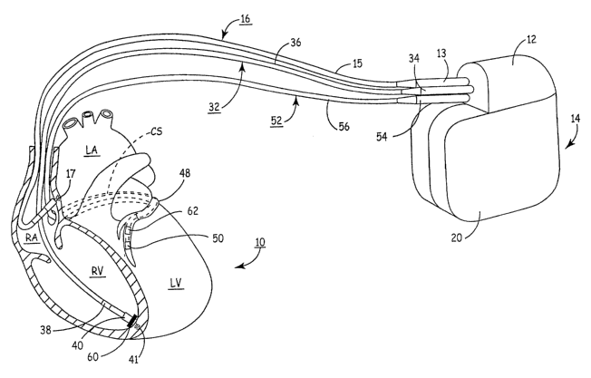
FIG. 1 depicts an IMD in which the present invention may be implemented.
FIG. 2 is a schematic block diagram of an exemplaxy multi-chamber pacemaker or
implantable pulse generator (IPG), such as that shown in FIG. 1, that provides
delivery of
a resynchronization therapy and is capable of automatically monitoring
ventricular
synchrony in accordance with the present invention.
FIG. 3 is a schematic diagram summarizing steps included in a method for
determining a metric of ventricular synchrony based on at least two
accelerometer signals.
FIG. 4 is a flow chart summarizing steps included in a method for measuring
ventricular synchrony as shown in FIG. 3 and providing additional details
regarding the
CA 02559254 2006-09-11
WO 2005/089864 PCT/US2005/007618
-6-
processing methods that may be used for converting a raw accelerometer signal
into a
processed signal useful for determining a synchrony metric.
FIG. S is a sample recording of a raw accelerometer signal wherein a series of
peaks are observed on the acceleration signal corresponding to the active
ejection phase of
each cardiac cycle.
FIG. 6 is a flow chart summarizing steps included in a general method for
monitoring ventricular synchrony and optionally adjusting a therapy based on a
measurement of ventricular synchrony.
As indicated above, the present invention is directed toward providing methods
and apparatus for monitoring ventricular synchrony in an ambulatory patient
using at least
a pair of transducers and automatically storing pacing therapy timing
information and/or
dynamically controlling said pacing therapy timing to maintain ventricular
synchrony. A
ventricular synchrony metric determined in accordance with the present
invention is useful
1 S for optimizing inter-ventricular pacing intervals during chronic
resynchronization therapy
(CRT) delivery for treating heart failure or for managing other cardiac
therapies such as
medical therapies. As such, the present invention may be embodied in an
implantable
medical device (IMD) having ventricular synchrony monitoring capabilities and
may
further include CRT delivery capabilities.
While the benefits of the present invention are expected to be particularly
advantageous when put to use in a fully IMD system, aspects of the present
invention may
also be beneficial when practiced in conjunction with external devices such as
temporary
pacemakers used to restore ventricular synchrony following coronary arterial
bypass graft
(CABG) surgical procedures. Therefore, methods described herein are not
limited to use
2S with implantable systems, however, for the sake of illustration the present
invention will
be described in the context of an IMD system.
FIG. 1 depicts an IMD in which the present invention may be implemented. The
IMD 14 is embodied as a mufti-chamber cardiac pacemaker or implantable pulse
generator
(IPG). The mufti-chamber IPG 14 is provided for restoring ventricular
synchrony by
delivering pacing pulses to one or more heart chambers as needed to control
the heart
activation sequence. The IPG 14 is shown in communication with patient's heart
10 by
way of three leads 16, 32 and S2. The heart 10 is shown in a partially cut-
away view
CA 02559254 2006-09-11
WO 2005/089864 PCT/US2005/007618
_7_
illustrating the upper heart chambers, the right atrium (RA) and left atrium
(LA), and the
lower heart chambers, the right ventricle (RV) and left ventricle (LV), and
the coronary
sinus (CS) extending from the opening in the right atrium laterally around the
atria to form
the great cardiac vein 48, which branches to form inferior cardiac veins.
The IPG 14 is implanted subcutaneously in a patient's body between the slcin
and
the ribs. Three transvenous endocardial leads 16, 32 and 52 connect the IPG 14
with the
RA, the RV and the LV, respectively. Each lead has at least one electrical
conductor and
pace/sense electrode. A remote indifferent can electrode 20 is formed as part
of the outer
surface of the housing of the IPG 14. The pace/sense electrodes and the remote
indifferent
can electrode 20 can be selectively employed to provide a number of unipolar
and bipolar
pacelsense electrode combinations for pacing and sensing functions.
The depicted bipolar endocardial RA lead 16 is passed through a vein into the
RA
chamber of the heart 10, and the distal end of the RA lead 16 is attached to
the RA wall by
an attachment mechanism 17. The bipolar endocardial RA lead 16 is formed with
an in-
line connector 13 fitting into a bipolar bore of connector block 12 that is
coupled to a pair
of electrically insulated conductors within lead body 15 and connected with
distal tip RA
pace/sense electrode 19 and proximal ring RA pace/sense electrode 21 provided
for
achieving RA pacing and sensing of R.A electrogram (EGM) signals.
Bipolar, endocardial RV lead 32 is passed through the RA into the RV where its
distal ring and tip RV pace/sense electrodes 38 and 40 are fixed in place in
the apex by a
conventional distal attachment mechanism 41. The RV lead 32 is formed with an
in-line
connector 34 fitting into a bipolar bore of connector block 12 that is coupled
to a pair of
electrically insulated conductors within lead body 36 and connected with
distal tip RV
pace/sense electrode 40 and proximal ring RV pace/sense electrode 38 provided
for RV
pacing and sensing of RV EGM signals. RV lead 32 further includes an RV wall
motion
sensor 60. RV wall motion sensor 60 may be positioned into or proximate the RV
apex
for detecting motion or acceleration of the RV apical region. Implantation of
an
acceleration sensor in the right ventricle is generally disclosed in U.S. Pat.
No. 5,693,075
issued to Plicchi, et al., incorporated herein by reference in its entirety.
In this illustrated embodiment, a unipolar, endocardial LV CS lead 52 is
passed
through the RA, into the CS and further into a cardiac vein to extend the
distal LV CS
pace/sense electrode 50 alongside the LV chamber to achieve LV pacing and
sensing of
CA 02559254 2006-09-11
WO 2005/089864 PCT/US2005/007618
_g_
LV EGM signals. The LV CS lead 52 is coupled at the proximal end connector 54
fitting
into a bore of connector block 12. A small diameter unipolar lead body 56 is
selected in
order to lodge the distal LV CS pace/sense electrode 50 deeply in a cardiac
vein branching
from the great cardiac vein 48.
Coronary sinus lead 52 is provided with a wall motion sensor 62 capable of
generating a signar proportional to the acceleration of the left ventricular
free wall.
Sensors 62 and 60 are preferably embodied as uniaxial, biaxial, or triaxial
sensors (e.g.,
accelerometers). In particular, sensor 62 is preferably contained in a capsule
of a
relatively small size and diameter such that it may be included in a coronary
sinus lead
without substantially increasing the lead diameter or impairing the ability to
steer the lead
to a left ventricular pacing and sensing site. Sensors 60 and 62 may
alternatively be
provided as another type of sensor such as an optical sensor, acoustical
sensor or a sensor
having piezoelectric, inductive, capacitive, resistive, or other elements
which directly or
indirectly produce a variable signal proportional to myocardial wall
acceleration, velocity,
displacement or force (including sensors that sense variations in the
foregoing).
Capacitive diaphragmatic-type sensors, cantilevered-type sensors, impedance-
injection
sensing circuits, and the like can all be utilized according to the present
invention provided
they are rendered of biocompatible material and sufficiently robust to
withstand the
dynamic forces, chemical forces, and macrophage response from phagocytes and
the like.
With respect to impedance-injection sensing circuits, a substantially
continuously injected
signal having appropriate frequency and inter-electrode sensing vector can be
utilized to
detect motion of at least one of the LV and RV. However, for consistency of
the text
hereof the foregoing sensors and transducer will be chiefly referred to as an
accelerometer
sensor, with the understanding that all suitable sensors for transducing
cardiac wall motion
are covered hereby. Furthermore, although the lead 52 is described herein
primarily as
being deployed through at least a portion of the great cardiac vein, lead 52
can also
represent an epicardial lead adapted to couple to any appropriate or suitable
location on a
portion of the epicardium of the LV chamber.
Sensor 62 is preferably located on CS lead 52 such that when CS lead 52 is
positioned for LV pacing and sensing, sensor 62 is located approximately over
the left
ventricular free wall mid-lateral to mid-basal segments. However, the depicted
positions
of the leads and electrodes shown in FIG. 1 in or about the right and left
heart chambers
CA 02559254 2006-09-11
WO 2005/089864 PCT/US2005/007618
-9-
are approximate and merely exemplary. For example, a left ventricular wall
motion sensor
62 may alternatively be located on CS lead 52 such that sensor 62 is
positioned in the
coronary sinus, in the great cardiac vein, or in any accessible inferior
cardiac vein.
Furthermore, it is recognized that alternative leads and pace/sense electrodes
that are
adapted for placement at pacing or sensing sites on or in or relative to the
RA, LA, RV and
LV may be used in conjunction with the present invention.
In a four chamber embodiment, LV CS lead 52 could bear a proximal LA CS
pace/sense electrode positioned along the lead body to lie in the larger
diameter coronary
sinus adjacent the LA for use in pacing the LA or sensing LA EGM signals. In
that case,
the lead body 56 would encase an insulated lead conductor extending proximally
fiom the
more proximal LA CS pace/sense electrodes) and terminating in a bipolar
connector 54.
In one embodiment of the present invention, and as shown in FIG. l, one
accelerometer is positioned relative to the right ventricle for measuring
right ventricular
wall motion and a second accelerometer is positioned relative to the left
ventricle for
measuring left ventricular wall motion. As will be described in greater detail
below,
signals from a right ventricular accelerometer and signals from a left
ventricular
accelerometer may be processed and analyzed to obtain a metric of synchrony
between the
right an left ventricles. Placement of accelerometers or other types of wall
motion sensors
relative to the right ventricle and left ventricle are not limited to the
positions shown in
FIG. 1 however and could alternatively be positioned at other right and left
ventricular
locations using transvenous or epicardial lead-based accelerometers.
Furthermore, the present invention may be practiced with multiple
accelerometers
positioned at more than one site in the right and/or left ventricle. The
methods taught
herein for measuring ventricular synchrony may be applied to measuring the
synchrony of
wall motion between multiple sites in the right and left ventricles and may
also be used for
measuring the synchrony between multiple sites within one ventricle. Thus
inter-
ventricular synchrony as well as infra-ventricular synchrony may be assessed
using
multiple accelerometers or other wall motion sensors and the methods to be
described
below.
FIG. 2 is a schematic block diagram of an exemplary multi-chamber IPG 14, such
as that shown in FIG. l, that provides delivery of a resynchronization therapy
and is
capable of automatically monitoring ventricular synchrony in accordance with
the present
CA 02559254 2006-09-11
WO 2005/089864 PCT/US2005/007618
-10-
invention. The IPG 14 is preferably a microprocessor-based device.
Accordingly,
microprocessor-based control and timing system 102, which varies in
sophistication and
complexity depending upon the type and functional features incorporated
therein, controls
the functions of IPG 14 by executing firmware and programmed software
algorithms
stored in associated RAM and ROM. Control and timing system 102 may also
include a
watchdog circuit, a DMA controller, a block mover/reader, a CRC calculator,
and other
specific logic circuitry coupled together by on-chip data bus, address bus,
power, clock,
and control signal lines in paths or trees in a manner known in the art. It
will also be
understood that control and timing functions of IPG 14 can be accomplished
with
dedicated circuit hardware or state machine logic rather than a programmed
microcomputer.
The IPG 14 includes interface circuitry 104 for receiving signals from sensors
and
pacelsense electrodes located at specific sites of the patient's heart
chambers and
delivering cardiac pacing to control the patient's heart rhythm and
resynchronize heart
IS chamber activation. The interface circuitry 104 therefore includes a
therapy delivery
system 106 intended for delivering cardiac pacing impulses under the control
of control
and timing system 102. Delivery of pacing pulses to two or more heart chambers
is
controlled in part by the selection of programmable pacing intervals, which
can include
atrial-atrial (A-A), atrial-ventricular (A-V), and ventricular-ventricular (V-
V) intervals.
Physiologic input signal processing circuit 108 is provided for receiving
cardiac
electrogram (EGM) signals for determining a patient's heart rhythm.
Physiologic input
signal processing circuit 108 additionally receives signals from left
ventricular wall
acceleration sensor 62, and RV wall acceleration sensor 60, and processes
these signals
and provides signal data to control and timing system 102 for further signal
analysis. For
purposes of illustration of the possible uses of the invention, a set of lead
connections are
depicted for making electrical connections between the therapy delivery system
106 and
the input signal processing circuit 108 and sets of pace/sense electrodes,
acceleration
sensors, and any other physiological sensors located in operative relation to
the RA, LA,
RV and LV.
Control and timing system 102 controls the delivery of bi-atrial, bi-
ventricular, or
multi-chamber cardiac pacing pulses at selected intervals intended to improve
heart
chamber synchrony. The delivery of pacing pulses by IPG 14 may be provided
according
CA 02559254 2006-09-11
WO 2005/089864 PCT/US2005/007618
-11-
to programmable pacing intervals, such as programmable conduction delay window
times
as generally disclosed in U.S. Pat. No. 6,070,101 issued to Struble et al.,
incorporated
herein by reference in its entirety, or programmable coupling intervals as
generally
disclosed in above-cited U.S. Pat. No. 6,473,645 issued to Levine. Selection
of the
programmable pacing intervals may be based on a determination of ventricular
synchrony
derived from LV wall motion sensor 62 and RV wall motion sensor 60 signals as
will be
described in greater detail below.
The therapy delivery system 106 can optionally be configured to include
circuitry
for delivering cardioversion/defibrillation therapy in addition to cardiac
pacing pulses for
controlling a patient's heart rhythm. Accordingly, Ieads in communication with
the
patient's heart could additionally include high-voltage cardioversion or
defibrillation
shock electrodes.
A battery 136 provides a source of electrical energy to power components and
circuitry of IPG 14 and provide electrical stimulation energy for delivering
electrical
impulses to the heart. The typical energy source is a high energy density, low
voltage
battery 136 coupled with a power supply/POR circuit 126 having power-on-reset
(POR)
capability. The power supply/POR circuit 126 provides one or more low voltage
power
(Vlo), the POR signal, one or more reference voltage (VREF) sources, current
sources, an
elective replacement indicator (ERI) signal, and, in the case of a
cardioversion/defibrillator
capabilities, high voltage power (Vhi) to the therapy delivery system 106. Not
all of the
conventional interconnections of these voltages and signals are shown in FIG.
2.
Current electronic mufti-chamber IPG circuitry typically employs cloclced CMOS
digital logic ICs that require a clock signal CLK provided by a piezoelectric
crystal 132
and system cloclc 122 coupled thereto as well as discrete components, e.g.,
inductors,
capacitors, transformers, high voltage protection diodes, and the like that
are mounted with
the ICs to one or more substrate or printed circuit board. In FIG. 2, each CLK
signal
generated by system clock 122 is routed to all applicable clocked logic via a
clock tree.
The system clock 122 provides one or moxe fixed frequency CLK signal that is
independent of the battery voltage over an operating battery voltage range for
system
timing and control functions and in formatting uplink telemetry signal
transmissions in the
telemetry I/O circuit 124.
CA 02559254 2006-09-11
WO 2005/089864 PCT/US2005/007618
-12-
The RAM registers included in microprocessor-based control and timing system
102 may be used for storing data compiled from sensed EGM signals,
acceleration signals,
and/or relating to device operating history or other sensed physiologic
parameters for
uplink telemetry transmission upon receipt of a retrieval or interrogation
instruction via a
downlink telemetry transmission. Criteria for triggering data storage can be
programmed
via downlinked instructions and parameter values. Physiologic data, including
acceleration data, may be stored on a triggered or periodic basis or by
detection logic
within the physiologic input signal processing circuit 108. In some cases, the
IPG 14
includes a magnetic field sensitive switch 130 that closes in response to a
magnetic field,
and the closure causes a magnetic switch circuit 120 to issue a switch closed
(SC) signal to
control and timing system 102 which responds in a magnet mode. For example,
the patient
may be provided with a magnet 116 that can be applied over the subcutaneously
implanted
IPG 14 to close switch 130 and prompt the control and timing system to deliver
a therapy
and/or store physiologic data. Event related data, e.g., the date and time and
current pacing
parameters, may be stored along with the stored physiologic data for uplinlc
telemetry in a
later interrogation session.
Uplink and downlink telemetry capabilities are provided to enable
communication
with either a remotely located external medical device or a more proximal
medical device
on or in the patient's body. Stored EGM, or LV acceleration data as well as
real-time
generated physiologic data and non-physiologic data can be transmitted by
uplink RF
telemetry from the IPG 14 to the external programmer or other remote medical
device 26
in response to a downlinlc telemetered interrogation command. As such, an
antenna 128 is
connected to radio frequency (RF) transceiver circuit 124 for the purposes of
uplinl~/downlink telemetry operations. Telemetering both analog and digital
data between
antenna 128 and an external device 26, also equipped with an antenna 118, may
be
accomplished using numerous types of telemetry systems known in the art for
use in
implantable devices.
The physiologic input signal processing circuit 108 includes electrical signal
amplifier circuits for amplifying, processing and sensing events from
characteristics of the
electrical sense signals or sensor output signals. The physiologic input
signal processing
circuit 108 may thus include a plurality of cardiac signal sense channels for
sensing and
processing cardiac signals from sense electrodes located in relation to a
heart chamber.
CA 02559254 2006-09-11
WO 2005/089864 PCT/US2005/007618
-13-
Each such channel typically includes a sense amplifier circuit for detecting
specific
cardiac events and an EGM amplifier circuit for providing an EGM signal to the
control
and timing system 102 for sampling, digitizing and storing or transmitting in
an uplink
transmission. Atrial and ventricular sense amplifiers include signal
processing stages for
detecting the occurrence of a P-wave or R-wave, respectively and providing an
atrial sense
or ventricular sense event signal to the control and timing system 102. Timing
and control
system 102 responds iri accordance With its particular operating system to
deliver or
modify a pacing therapy, if appropriate, or to accumulate data for uplink
telemetry
transmission in a variety of ways known in the art. Thus the need for pacing
pulse
delivery is determined based on EGM signal input according to the particular
operating
mode in effect.
Input signal processing circuit 108 includes signal processing circuitry for
receiving accelerometer or other wall motion sensor signals and may include
amplifiers
and filters for processing an analog accelerometer signal. Alternatively,
accelerometer
signals may be digitized and averaging and filtering of such signals may be
performed by
microcomputer 102 or other dedicated digital circuitry. Accelerometer signal
processing
circuitry is further provided for detection and/or determination of one or
more acceleration
signal characteristics such as maximum peak amplitude, slope, integral,
threshold crossing
or other time domain signal characteristic that may be used in deriving a
ventricular
synchrony metric as will be described below. Acceleration data from LV wall
motion
sensor 62 and RV wall motion sensor 60 are made available to control and
timing system
102 via LV MOTION signal line and RV MOTION signal line, respectively, for
determining a synchrony metric. The synchrony metric may further be used by
control
and timing system 102 for identifying pacing intervals producing optimal
ventricular
synchrony.
FIG. 3 is a schematic diagram summarizing steps included in a method for
determining a metric of ventricular synchrony based on at least two
accelerometer signals.
A first raw accelerometer signal 202 is received from accelerometer 60,
positioned to
measure ventricular wall motion at a desired site, e.g., right ventricular
wall motion as
shown in FIG. 1. The raw accelerometer signal 202 is processed by processing
unit 206 to
produce a processed accelerometer signal 210. A fiducial point 2I4 on the
processed
signal 210 and the time point 215 at which it occurs is identiried. The
fiducial point 214
CA 02559254 2006-09-11
WO 2005/089864 PCT/US2005/007618
-14-
may be a peak amplitude as shown in FIG. 3 or alternatively any characteristic
feature of
the processed signal such as a peak slope or a threshold crossing.
In a similar method, a second raw accelerometer signal 204 is received from a
second accelerometer 62 positioned at a second location for measuring
ventricular wall
motion, e.g. left ventricular free wall motion as shown in FIG. 1. The raw
signal 204 is
processed by processing unit 208 in the same manner as raw signal 202 to
produce a
processed signal 212. An analogous fiducial point 216 on processed signal 212
and the
time point 217 at which it occurs are identified such that the time difference
(0) 220 may
be determined between the fiducial point 214 occurring on the first processed
signal 210
and the analogous fiducial point 216 occurnng on the second processed signal
212.
The time difference 220 between analogous fiducial points 214,216 provides a
synchrony metric between the two ventricular sites at which the accelerometers
60 and 62
are located. In the example embodiment shown in FIG. l, a measure of synchrony
between right apical and left ventricular free wall motion may be obtained.
FIG. 4 is a flow chart summarizing steps included in a method for measuring
ventricular synchrony as shown in FIG. 3 and providing additional details
regarding the
processing methods that may be used for converting a raw accelerometer signal
into a
processed signal useful for determining a synchrony metric. An EGM or ECG
signal is
sensed at step 255 using transvenous or subcutaneous sensing electrodes such
that the R-
wave may be detected at step 260 for each cardiac cycle during a synchrony
monitoring
session. Any R-wave detection method may be used for the proposes of the
present
invention and such methods are well known in the art of cardiac pacing.
As described above, two or more accelerometers or other wall motion sensors
are
deployed at desired monitoring sites within the patient's heart.
Simultaneously to
EGM/ECG sensing, the two or more accelerometer signals are sensed at step 265.
At step
270 a sensing window is set defining an accelerometer signal segment of
interest within
each cardiac cycle. The sensing window set at step 270 is preferably set for
each cardiac
cycle relative to the R-wave detected at step 265 for the same cardiac cycle.
The sensing
window may alternatively be set relative to other EGM/ECG events such as a T-
wave or a
P-wave or other atrial or ventricular senses or pace events. The sensing
window is set so
as to select a period during the cardiac cycle during which ventricular
syncluony is to be
evaluated. While the active ejection phase can be selected for evaluating
ventricular
CA 02559254 2006-09-11
WO 2005/089864 PCT/US2005/007618
-15-
synchrony, other phases may be of greater interest and value for extracting
meaningful
cardiac wall motion signals. In particular, the isovolumic contractile phase
or the
isovolumic relaxation phase appear to provide the most significant cardiac
wall motion
signals. However, other phases (e.g., the filling phase) can be use depending
on the
application of the synchrony metric.
In FIG. 5 a sample recording of a raw accelerometer signal is depicted wherein
a
series of peaks are observed on the acceleration signal corresponding to the
active ejection
phase of each cardiac cycle. An R-wave detected at a time point indicated by
arrow 304 is
used to set a sensing window 306, relative to R-wave detection time 304, for
isolating the
active ejection phase of the acceleration signal 302, during which a peak
acceleration
signal 308 is observed. The acceleration signal segment occurring during
sensing window
306 will undergo further processing as described in the flow chart of FIG. 4
for
determining a synchrony metric.
At step 275 of FIG. 4, a predetermined number of consecutive acceleration
signal
segments defined by the sensing window set at step 270 are averaged for a
given
accelerometer signal. Any number of one or more consecutive signal segments
may be
selected for averaging at step 275. At step 280, a template can be used for
filtering the
raw signal in a matched filter is defined. However, in lieu of a template, an
appropriately
configured conventional detection, also known as a matched filter, could be
utilized.
Presently, however, such matched filters are typically approximated by filters
with far less
numbers of coefficients and such approximated matched filters can also be used
in
practicing the present invention. The rilter template, as will be described in
greater detail
below, is defined by reversing in time an averaged signal segment for a given
accelerometer signal. A raw signal segment for each accelerometer signal may
then be
processed through a matched filter at step 283 using the filter template
defined at step 280
for the corresponding accelerometer signal to improve the signal-to-noise
ratio of the raw
signal.
While other filtering methods may be performed, such as band pass filtering,
matched filtering is preferred to achieve optimal signal-to-noise improvement.
In
performing matched filtering, the filter is designed to have a frequency
response matching
the frequency spectrum of the signal. The filter templates defined at step 283
therefore
CA 02559254 2006-09-11
WO 2005/089864 PCT/US2005/007618
-16-
involves deftning a filter template for each accelerometer signal received by
reversing in
time the averaged signal segment determined for each accelerometer signal.
A raw acceleration signal received at step 265 may be defined by Equation 1
below:
(1) a(t) = A~ s(t - tau) + n(t)
wherein a(t) is the raw acceleration signal including the acceleration signal
contributions
from noise, n(t), and ventricular wall motion defined by A * s(t -tau) wherein
A is the
strength of the myocardial acceleration and tau is the delay of the mechanical
response of
the myocardium following the electrical activation.
A filter template, h(t), may be defined according to Equation Z:
(2) h(t) = s(-t)
wherein s(-t) is the average of a predetermined number of signal segments
determined at
step 275 reversed in time.
After filtering each raw signal segment at step 283 using a matched filter
employing the respective filter template defined by Equation 2 above, a
processed signal
for each raw accelerometer signal received is obtained as was described above
in
conjunction with FIG. 3. The signal to be filtered can comprise the raw signal
segment
obtained from a single cardiac cycle (and not the averaged signal segments
found at step
275). In which case a a time difference is determined for a single cardiac
cycle at step
285. Otherwise, a time difference can be determined for an averaged value of
raw signals
(over a plurality of cardiac cycles). Thus, the time difference for a single
cardiac cycle or
a plurality of cardiac cycles can calculated from the raw signals or from pre-
filtered
signals. At step 285, the selected fiducial points are identified on each
processed
accelerometer signal. The time difference between these fiducial points is
measured as a
metric of synchrony at step 290 as described previously.
FIG. 6 is a flow chart summarizing steps included in a general method for
monitoring ventricular synchrony and optionally adjusting a therapy based on a
measurement of ventricular synchrony. At step 405 of method 400, synchrony
monitoring
is initiated. Monitoring of ventricular synchrony may be initiated by a user
to occur on a
continuous or periodic basis. For example, a synchrony metric may be
determined on an
hourly, daily, weekly or other periodic basis.
CA 02559254 2006-09-11
WO 2005/089864 PCT/US2005/007618
-17-
At step 410, a synchrony metric is determined from two or more accelerometers
signals according to the methods described above in conjunction with FIGS. 3
through' S.
The synchrony metric is stored at step 415 in associated device memory such
that trends in
ventricular synchrony may be recognized. Stored data is available for
uplinking to an
external device for display and review by a clinician.
The methods described herein for determining a metric of ventricular synchrony
may be utilized for monitoring purposes only by the associated IMD. Therefore
method
400 may include only the steps for determining and storing synchrony metrics.
However,
if the associated medical device includes therapy delivery capabilities, the
stored
synchrony metrics may be evaluated at step 420 to determine if a worsening
trend in
ventricular synchrony is indicated. If increased ventricular dyssynchrony is
indicated, as
determined at decision step 420, parameters used to control therapy delivery
by the
implantable device may be adjusted at step 425.
If the implantable device is a cardiac pacing device capable of dual chamber,
bi-
ventricular or mufti-chamber pacing, the various A-V and V-V intervals used to
control
the timing of pacing pulses or other pacing control parameters may be adjusted
at step 425
in an attempt to improve ventricular synchrony. If the implantable device is a
drug
delivery device, an adjustment may be made to the dosage of the delivered
drug. After
adjusting therapy parameters at step 425, method 400 may return to step 405 to
initiate
another measurement of ventricular synchrony to determine if the therapy
parameter
adjustment had the desired effect. If not, parameters may be adjusted until an
improvement in ventricular synchrony is observed based on the trend of stored
ventricular
metrics. When no increase in ventricular dyssynchrony is indicated as
determined at
decision step 420, method 400 returns to step 405 to initiate synchrony
monitoring at the
next user-initiated ox scheduled periodic monitoring time.
With respect to filtering of the raw sensor output signal(s), those of shill
in the art
of signal processing will appreciate that a matched filter can be approximated
by a lower
order filter using a variety of known techniques.
Thus a system and method for monitoring ventricular synchrony in ambulatory
patients has been described which allows for chronic monitoring of ventricular
synchrony
and closed-loop control of therapies delivered by implantable devices to
improve cardiac
CA 02559254 2006-09-11
WO 2005/089864 PCT/US2005/007618
-18-
mechanics. The detailed embodiments described herein are intended to be
illustrative,
rather than limiting, with regard to the following claims.