Note: Descriptions are shown in the official language in which they were submitted.
CA 02559320 2006-07-05
WO 2005/070330 PCT/US2005/000264
VENTRICULAR PARTITIONING DEVICE
FIELD OF THE INVENTION
[0001] The present invention relates generally to the field of treating
congestive
heart failure and more specifically, to a device and method for partitioning a
patient's
heart chamber and a system for delivering the treatment device.
BACKGROUND OF THE INVENTION
[0002] Congestive heart failure (CHF) is characterized by a progressive
enlargement of the heart, particularly the left ventricle and is a major cause
of death
and disability in the United States. Approximately 500,000 cases occur
annually in
the U.S. alone. As the patient's heart enlarges, it cannot efficiently pump
blood
forward with each heart beat. In time, the heart becomes so enlarged the heart
cannot adequately supply blood to the body. Even in healthy hearts only a
certain
percentage of the blood in a patient's left ventricle is pumped out or ejected
from the
chamber during each stroke of the heart. The pumped percentage, commonly
referred to as the "ejection fraction", is typically about sixty percent for a
healthy
heart. A patient with congestive heart failure can have an ejection fraction
of less
than 40% and sometimes lower. As a result of the low ejection fraction, a
patient
with congestive heart -failure is fatigued, unable to perform even simple
tasks
requiring exertion and experiences pain and discomfort. Further, as the heart
enlarges, the internal heart valves such as the mitral valve, cannot
adequately close.
An incompetent mitral valve allows regurgitation of blood from the left
ventricle back
into the left atrium, further reducing the heart's ability to pump blood
forwardly.
[0003] Congestive heart failure can result from a variety of conditions,
including
viral infections, incompetent heart valves (e.g. mitral valve), ischemic
conditions in
1
CA 02559320 2006-07-05
WO 2005/070330 PCT/US2005/000264
the heart wall or a combination of these conditions. Prolonged ischemia and
occlusion of coronary arteries can result in myocardial tissue in the
ventricular wall
dying and becoming scar tissue. Once the myocardial tissue dies, it is less
contractile (sometimes non-contractile) and no longer contributes to the
pumping
action of the heart. It is referred to as hypokinetic. As the disease
progresses, a
local area of compromised myocardium may bulge out during the heart
contractions,
further decreasing the heart's ability to pump blood and further reducing the
ejection
fraction. In this instance, the heart wall is referred to as dyskinetic or
akinetic. The
dyskinetic region of the heart wall may stretch and eventually form an
aneurysmic
bulge.
[0004] Patients suffering from congestive heart failure are commonly grouped
into
four classes, Classes I, II, III and IV. In the early stages, Classes I and
II, drug
therapy is presently the most commonly prescribed treatment. Drug therapy
typically
treats the symptoms of the disease and may slow the progression of the
disease, but
it can not cure the disease., Presently, the only permanent treatment for
congestive
heart disease is heart transplantation, but heart transplant procedures are
very risky,
extremely invasive and expensive and are performed on a small percentage of
patients. Many patient's do not qualify for heart transplant for failure to
meet any one
of a number of qualifying criteria, and, Furthermore, there are not enough
hearts
available for transplant to meet the needs of CHF patients who do qualify.
[0005] Substantial effort has been made to find alternative treatments for
congestive heart disease. For example, surgical procedures have been developed
to dissect and remove weakened portions of the ventricular wall in order to
reduce
heart volume. This procedure is highly invasive, risky and expensive and is
commonly only done in conjunction with other procedures (such as heart valve
2
CA 02559320 2006-07-05
WO 2005/070330 PCT/US2005/000264
replacement or coronary artery by-pass graft). Additionally, the surgical
treatment is
usually limited to Class IV patients and, accordingly, is not an option for
patients
facing ineffective drug treatment prior to Class IV. Finally, if the procedure
fails,
emergency heart transplant is the only presently available option.
[0006] Other efforts to treat CHF include the use of an elastic support, such
as an
artificial elastic sock placed around the heart to prevent further deleterious
remodeling.
[0007] Additionally, mechanical assist devices have been developed as
intermediate procedures for treating congestive heart disease. Such devices
include
left ventricular assist devices and total artificial hearts. A left
ventricular assist device
includes a mechanical pump for increasing blood flow from the left ventricle
into the
aorta. Total artificial heart devices, such as the Jarvik heart, are usually
used only
as temporary measures while a patient awaits a donor heart for transplant.
[0008] Recently, improvements have been made in treating patient's with CHF by
implanting pacing leads in both sides of the heart in order to coordinate the
contraction of both ventricles of the heart. This technique has been shown to
improve hemodynamic performance and can result in increased ejection fraction
from the right ventricle to the patient's lungs and the ejection fraction from
the left
ventricle to the patient's aorta. While this procedure has been found to be
successful
in providing some relief from CHF symptoms and slowed the progression of the
disease, it has not been able to stop the disease.
SUMMARY OF INVENTION
[0009] The present invention is directed to a ventricular partitioning device
and
method of employing the device in the treatment of a patient with congestive
heart
failure. Specifically, the ventricular chamber of the CHF patient is
partitioned by the
3
CA 02559320 2011-11-10
device so as to reduce its total volume and to reduce the stress applied
to the heart and, as a result, improve the ejection fraction thereof.
[009A] Various embodiments of this invention provide a system for
increasing the ejection fraction of a patient's heart chamber comprising a
partitioning apparatus, said apparatus comprising: a membrane component
which has a proximal face and a distal face and which is configured to
partition said patient's heart chamber into a main operational portion and
a secondary, non-operational portion; and a support component which is
adapted to be disposed in said non-operational portion of said heart
chamber defined in part by said distal face of said membrane and which is
configured to extend from said membrane to a region of a heart wall in
said heart chamber; at least one resilient member at a distal extremity
of said support component to non-traumatically engage said region of said
patient's heart wall.
[009B] Other embodiments of this invention provide a system for
treating a patient's heart comprising a partitioning apparatus, said
partitioning apparatus comprising: a central hub component; an expandable
frame component having a plurality of ribs with free proximal ends and
distal ends secured to said central hub; a plurality of securing elements
at said proximal ends of said ribs; a membrane component secured to said
expandable frame ribs; and a support component extending from a distal side
of the frame for non-traumatically engaging a region of the patient's heart
wall defining in part the heart chamber being partitioned, wherein the
support component has at least one resilient member at a distal extremity
of said support component to non-traumatically engage said region of said
patient's heart wall.
4
CA 02559320 2006-07-05
[0010] A ventricular partitioning device embodying features of the invention
has a
reinforced membrane component, preferably self expanding, which is configured
to
partition tie patient's ventricular heart chamber into a main productive
portion and a
secondary non-productive portion, and a support or spacing component extending
from the distal side of the reinforced membrane for non-traumatically engaging
a
region of the patient's ventricular wall defining in part the secondary non-
productive
portion to space a central portion of the reinforced membrane from the heart
wall.
The partitioning device preferably includes a centrally located hub secured to
the
reinforced membrane. The partitioning membrane of the device may be reinforced
by a radially expandable frame component formed of a plurality of ribs.
[0011] The ribs of the expandable frame have distal ends secured to the
central
hub, preferably secured to facilitate abduction of the free proximal ends of
the ribs
away from a centerline axis. The distal ends of the ribs may be pivotally
mounted or
formed of material such as superelastic NiTi alloy which allow for compressing
the
ribs into a contracted configuration and when released allow for their self
expansion..
The ribs also have free proximal ends configured to engage and preferably
penetrate
the tissue of the heart wall so as to secure the peripheral edge of the
membrane to
the heart wall and fix the position of the membrane with respect thereto. The
free
proximal ends of the ribs may have tissue penetrating tips such as barbs or
hooks.
The partitioning membrane is secured to the ribs of the expandable frame,
preferably
on the proximal or pressure side of the expandable frame.
[0012] The supporting component or stem of the device has a length configured
to extend to the heart wall (typically about 5 mm to about 50 mm, preferably
about 15
4a
CA 02559320 2006-07-05
WO 2005/070330 PCT/US2005/000264
to about 35 mm), to support and space the membrane from the heart wall. While
only one supporting component or stem is described herein, a plurality of such
components may be utilized. The supporting component or stem may have at least
one inner lumen extending therein for delivery of therapeutic or diagnostic
agents
through the ports provided along the length thereof. The stem is provided with
one
or more flexible bumper-type elements on its distal end to non-traumatically
engage
the weakened ventricular wall and maintain the reinforced membrane, preferably
the
central portion thereof, spaced a desired distance from the weakened
ventricular
wall.
[0013] The partitioning membrane in the expanded configuration has radial
dimensions from about 10 to about 160 mm, preferably about 50 to about 100 mm,
as measured from the center line axis.
[0014] The partitioning device may be delivered percutaneously or
intraoperatively. It is relatively easy to install and provides substantial
improvement
in the ejection fraction of the patient's heart chamber. These and other
advantages
of the invention will become more apparent from the following detailed
description of
the invention and the accompanying exemplary drawings.
BRIEF DESCRIPTION OF THE DRAWINGS
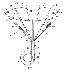
[0015] Figure 1 is a schematic perspective view of a ventricular partitioning
device embodying features of the invention.
[0016] Figure 2 is an elevational view of a delivery system for the
partitioning
device shown in Figure 1
[0017] Figure 3 is an enlarged view of the encircled region 3-3 shown in
Figure 2.
[0018] Figure 4 is a simplified view with parts removed similar to that shown
in
Figure 3 with the delivery catheter connected to the partitioning device.
CA 02559320 2006-07-05
WO 2005/070330 PCT/US2005/000264
[0019] Figure 5 is an end view of the hub which is secured in the proximal end
of
the stem of the partitioning device shown in Figure 1.
[0020] Figure 6 is a schematic view of a patient's left ventricular chamber
illustrating the partitioning device shown in Figure 1 disposed within the
chamber
separating a working portion of the chamber from an non-working portion of the
chamber.
[0021] Figure 7 is a schematic perspective view of an alternative design
embodying features of the invention with a pair of bumper elements on the
distal end
of the stem of the partitioning device.
[0022] Figure 8 is, a schematic perspective view of another alternative design
embodying features of the invention with three bumper elements on the distal
end of
the stem of the partitioning device.
[0023] Figure 9 is a schematic perspective view of another alternative design
embodying features of the invention with four bumper elements on the distal
end of
the stem of the partitioning device.
[0024] Figure 10 is a schematic perspective view of a fourth alternative
design
embodying features of the invention with a plurality of bumper elements on the
distal
end of the stem of the device provided with hooks which fix the end to the
interior
surface of the patient's ventricular wall.
[0025] Figure 11 is a schematic perspective view of another alternative design
embodying features of the invention with a membrane underlying a plurality of
bumper elements on the distal end of the stem of the partitioning device.
[0026] Figure 12 is a schematic perspective view of another alternative design
embodying features of the invention with a helical coil bumper element on the
distal
end of the stem of the partitioning device.
6
CA 02559320 2006-07-05
WO 2005/070330 PCT/US2005/000264
[0027] Figure 13 is a schematic perspective view of yet another alternative
design
embodying features of the invention with an inflatable balloon secured to the
underside of the partitioning device to space and support the partitioning
device from
the heart wall.
DETAILED DESCRIPTION OF EMBODIMENTS OF THE INVENTION
[0028] Figures 1-5 illustrate a partitioning device 10 which embodies features
of
the invention and which includes a partitioning membrane 11, a stem 12 and a
radially expandable reinforcing frame 13 formed of a plurality of ribs 14.
Preferably
the membrane 11 is secured to the proximal or pressure side of the frame 13 as
shown in Figure 1. The distal ends 15 of the ribs 14 are secured to the
central hub
16 and the proximal ends 17 of the ribs 14 are unsecured and are configured to
radially extend away from a center line axis 18 which extends through'the hub
16.
Radial expansion of the free proximal ends 17 unfurls the membrane 11 secured
to
the frame 13 so that the membrane presents a relatively smooth pressure side
surface. Stem 12 extends distally from the hub 16 and has a distal end 19
which has
a flexible, J-shape bumper element 20 to provide a yielding engagement with a
heart
wall when deployed within a patient's heart chamber. The frame 13 and attached
membrane 11 are collapsible toward the centerline axis 18 for delivery through
a
catheter.
[0029] The proximal or free ends 17 of ribs 14 are provided with sharp tip
elements 21 which are configured to hold the frame 13 and the membrane 11
secured thereto in a deployed position within the patient's heart chamber.
Preferably, the sharp tip elements 21 of the frame 13 penetrate into tissue of
the
patient's heart wall in order to secure the reinforced membrane 11 so as to
partition
the ventricular chamber in a desired manner.
7
CA 02559320 2006-07-05
WO 2005/070330 PCT/US2005/000264
[0030] As shown in Figure 1, the stem 12 is provided with an inner lumen 22
for
delivery of fluid to the non-operative portion of the ventricular chamber and
discharge
ports 23 are provided in the stem. The hub 16 is secured within the inner
lumen 22
in the proximal end of stem 12 suitable means such as a friction fit, an
adhesive
bond or a pin. The hub 16 has a deployment pin 24, as shown in Figure 5, which
as
will be described later allows the partitioning device 10 to be deployed
within the
patient's heart chamber and released from a delivery system used to place the
device. The distal ends of the reinforcing ribs 14 are secured to the hub 16
in a
suitable manner. They may be secured to the surface defining the inner lumen
or
the hub may be provided with channels or bores in the wall of the hub into
which the
distal ends of the ribs may be secured. The ribs 14 are preshaped so that when
not
constrained (as shown in Figures 1 and 2), the free proximal ends 17 thereof
expand
to a desired angular displacement (9) away from a center line axis 18 which is
about
20 to about 90 , preferably about 50 to about 80 .
[0031] Figures 2-4 illustrate a suitable delivery system 30 with a
partitioning
component device 10 as shown in Figure 1. The delivery system 30 includes a
control handle 31 with a delivery catheter 32 having a deploying coil screw 33
secured to the distal end 34 for releasing the partitioning device 10 from the
delivery
system 30. The delivery catheter 32 has a an inner lumen 35 through which
therapeutic or diagnostic fluids may be delivered. The delivery catheter 32
extends
through the handle 31 and the proximal end of the catheter 32 is secured to
torquing
knob 36 to allow rotation of the catheter by rotating knob 36. An injection
port 37 is
provided in fluid communication with the delivery catheter 32 for injecting
therapeutic
or diagnostic fluids through the inner lumen 35.
8
CA 02559320 2006-07-05
WO 2005/070330 PCT/US2005/000264
[0032] The delivery system 30 may be introduced into a patient's body through
guiding catheter or cannula 40 which has an inner lumen 41: A radiopaque
marker
(not shown) may be provided on the distal end of the guiding catheter 40 to
aid in
fluoroscopically guiding the catheter to the desired location. The
partitioning device
is slidably disposed within the inner lumen 41 with the free proximal ends 17
of
the ribs 14 in a constricted configuration. The guiding. catheter 40 is
percutaneously
introduced in a conventional fashion into the patient's vasculature and
advanced
therein until the distal end 42 of the guiding catheter. 40 is position close
to the
desired location for the partitioning device 10 within the patient's heart
chamber such
as the left ventricle. The delivery system 30 is advanced distally within the
inner
lumen 41 until the J-shaped bumper 20 extends out the distal end 42 of the
guiding
catheter 40 and engages the ventricular wall. With the delivery system 30 held
in
place and the bumper 20 engaging the ventricular wall, the guide catheter 40
is
pulled proximally until the free ends 17 of ribs 14 are released from the
distal end 42
so that anchoring tip elements 21 on the free proximal ends 17 of ribs 14
penetrate
into tissue of the patient's heart wall as shown in Figure 6 to secure the
partitioning
device 10 within the patient's heart chamber. With the partitioning device 10
properly positioned within the heart chamber, the delivery catheter 32 is
rotated
counter-clockwise to disengage the delivery system 30 from the hub 16. Upon
the
counter-clockwise rotation of the delivery catheter 32, the helical coil screw
33
attached to the distal end 34 of the delivery catheter 32 rides on the
deployment pin
24 secured within the inner lumen 22 of the hub 16. The delivery system 30 and
the
guide catheter 40 may then be removed from the patient. The proximal end of
the
guide catheter 40 is provided with an injection port 43 to inject therapeutic
or
diagnostic fluids through the inner lumen 41.
9
CA 02559320 2006-07-05
WO 2005/070330 PCT/US2005/000264
[0033] Figure 6 illustrates the placement of partitioning device 10 within a
patient's left ventricle 45. The membrane 11 secured to the proximal side of
ribs 14
partitions the patient's heart chamber 45 into a main productive or
operational portion
46 and a secondary, essentially non-productive portion 47. The operational
portion
46 is much smaller than the original ventricular chamber 45 and provides for
an
improved ejection fraction. The partitioning increases the ejection fraction
and
provides an improvement in blood flow. Over time, the non-productive portion
47 fills
initially with thrombus and subsequently cellular growth. Bio-resorbable
fillers such
as polylactic acid, polyglycolic acid, polycaprolactone and copolymers and
blends
may be employed to fill the non-productive portion 47. Fillers may be suitably
supplied in a suitable solvent such as DMSO. Other materials which accelerate
tissue growth may be deployed in the non-productive portion 47.
[0034] Figures 7-12 illustrate distal ends 19 of the partitioning devices
having
alternative bumper elements for providing non-traumatic contact with a
weakened
ventricular wall. In Figure 7 the distal end 19 of stem 12 has a pair of J-
shaped
bumpers 50 and 51. In Figure 8 the distal end 19 has three J-shaped bumpers
52,
53 and 54. Figure 9 illustrates a distal end 19 having three J-shaped bumpers
55,
56, 57 and 58. Figure 10 depicts a slight change, where the distal end 19 has
four
wire J-shaped bumpers 59-62 (not shown in drawing) with sharp tips 63-66 (not
shown) for securing the ends of the bumpers in heart tissue. A further
alternative is
illustrated in Figure 11 where a membrane 68 is applied to the J-shaped
bumpers In
Figure 12, the distal end 19 of stem 12 is provided with a coiled bumper 70
for
engaging a ventricular wall.
[0035] Another modification is shown in Figure 13 wherein an inflatable
balloon
80 is provided on the distal side of the frame 13 to support and space the
partitioning
CA 02559320 2006-07-05
WO 2005/070330 PCT/US2005/000264
device 10 from a patient's ventricular wall in lieu of the stem with flexible
bumpers,
as shown in the partitioning devices previously described.
[0036] The ribs 14 of the partitioning device have a length of about 1 to
about 8
cm, preferably, about 1.5 to about 4 cm for most left ventricle deployments.
To
assist in properly locating the device during advancement and placement
thereof into
a patient's heart chamber, the distal extremity of one or more of the ribs
and/or the
stem may be provided with markers at desirable locations that provide enhanced
visualization by eye, by ultrasound, by X-ray, or other imaging or
visualization
means. Radiopaque markers may be made with, for example, stainless steel,
platinum, gold, iridium, tantalum, tungsten, silver, rhodium, nickel, bismuth,
other
radiopaque metals, alloys and oxides of these metals.
[0037] The membrane 11' may be formed of suitable biocompatible polymeric
material which include ePTFE (expanded polytetrafluoroethylene), Nylon, PET
(polyethylene terephthalate) and polyesters such as Hytrel. The membrane 11 is
preferably foraminous in nature to facilitate tissue ingrowth after deployment
within
the patient's heart. The delivery catheter and the guiding catheter may be
formed of
suitable high strength polymeric material such as PEEK (polyetheretherketone),
polycarbonate, PET, Nylon, and the like. Braided composite shafts may also be
employed. To the extent not otherwise described herein, the various components
of
the partitioning device and delivery system may be formed of conventional
materials
and in a conventional manner as will be appreciated by those skilled in the
art.
[0038] While particular forms of the invention have been illustrated and
described
herein, it will be apparent that various modifications and improvements can be
made
to the invention. Moreover, individual features of embodiments of the
invention may
be shown in some drawings and not in others, but those skilled in the art will
11
CA 02559320 2006-07-05
recognize that individual features of one embodiment of the invention can be
combined with any or all the features of another embodiment. Accordingly, it
is not
intended that the invention be limited to the specific embodiments
illustrated. It is
intended that this invention to be defined by the scope of the appended claims
as
broadly as the prior art will permit.
12