Note: Descriptions are shown in the official language in which they were submitted.
CA 02559476 2012-02-17
TREATMENT OF FIBROSIS USING FXR LIGANDS
10001]
FIELD OF THE INVENTION
[0002) The present invention relates to the prevention, treatment, and/or
reversal of
fibrosis. In particular, this invention relates to the novel use of ligands
specific for farnesoid
X receptor (F_XR) in patients with fibrotic liver, intestinal, or renal
diseases who do not also
suffer from a cholestatic condition, in order to inhibit the development and
progression of
fibrosis in those tissues where FXR is expressed.
BACKGROUND OF THE INVENTION
[00031 Fibrosis is characterized by an excessive accumulation of collagen in
the
extracellular matrix of the involved tissue. It is a Ion- standing and
challenging clinical
problem for which no effective treatment is currently available. The
production of collagen
is a highly regulated physiological process, the disturbance of which may lead
to the
development of tissue fibrosis. The formation of fibrous tissue is part of the
normal
beneficial process of healing after injury. In some cases, however, an
abnormal accumulation
of fibrous material can severely interfere with the normal function of the
affected tissue or
even cause the complete loss of function of the affected organ.
[0004) Liver fibrosis, for instance, represents a major medical problem with
significant
morbidity and mortality. In a variety of liver diseases, chronic injury leads
to progressive
fibrosis that the liver is able to compensate for over as long as 20-30 years;
eventually,
however, patients begin to experience symptoms and signs of liver failure due
to severe
fibrosis and cirrhosis. Worldwide chronic viral hepatitis infections,
particularly by Hepatitis
B and C virus, represent the major cause of liver fibrosis; however, within
the United States
chronic alcohol consumption has traditionally been the leading cause of
hepatic fibrosis and
cirrhosis. Currently, with the rapid increase in the prevalence of obesity in
the general
population, non-alcoholic fatty liver disease (NAFLD) is becoming the most
prevalent
condition associated with liver fibrosis and may become the leading cause of
liver fibrosis
CA 02559476 2006-09-12
WO 2005/089316 PCT/US2005/008575
associated morbidity and mortality in coming years. Other known causes of
liver fibrosis
include parasitic infection, autoimmune diseases, iron or copper storage
disorders, and biliary
obstruction. Liver fibrosis can be classified as a wound healing response to a
variety of
chronic stimuli that is characterized by an excessive deposition of
extracellular matrix
proteins, of which type I collagen predominates. This excess deposition of
extracellular
matrix proteins disrupts the normal architecture of the liver resulting in
structural and
functional damages to the organ. If left untreated, liver fibrosis can
progress to liver cirrhosis
ultimately leading to organ failure and death. Many other debilitating and
potentially fatal
diseases also lead to fibrosis of organs such as the intestine, kidney, heart,
and lung.
[0005] Because of the pivotal role of collagen production during fibrosis,
many studies
have focused on the regulation of collagen expression and proliferation of
fibroblasts, the
major cell type responsible for collagen synthesis. In the liver, the hepatic
stellate cell (HSC)
is the primary fibrogenic cell type.
[0006] A variety of compounds have been identified as anti-fibrosis agents via
different
mechanisms of action, including the suppression of collagen expression. For
example,
pantethine (D-bis-(N-pantothenyl-fl-aminoethyl)-disulfide) has been reported
to be effective
for the inhibition of hepatic fibrosis (U.S. Patent No. 4,937,266); a
hydrazine derivative,
benzoic hydrazide, has been shown to be a powerful antifibrotic agent (U.S.
Patent Nos.
5,374,660 and 5,571,846); the use of angiotensin inhibitors in combination
with nitric oxide
stimulators to inhibit the progression of fibrosis is disclosed in U.S. Patent
Nos. 5,645,839
and 6,139,847; U.S. Patent No. 6,005,009 describes methods using certain
pyridoxal benzoyl
hydrazones or their analogs for inhibiting fibrosis; U.S. Patent No. 6,117,445
describes the
use of Al adenosine receptor antagonists and/or P2X purinoceptor antagonists
for treating or
preventing fibrosis and sclerosis. More recently, somatostatin agonists,
hepatocyte growth
factors (HGFs), chymase inhibitors, and antagonists of IL-13 have been
reported to
effectively inhibit fibrosis (U.S. Patent Nos. 6,268,342, 6,303,126,
6,500,835, and
6,664,227).
[0007] The farnesoid X receptor (FXR), also known as the bile acid receptor
(BAR) and
NR1H4, is a member of the nuclear receptor superfamily of ligand-activated
transcription
factors and forms, with retinoid X receptor (RXR), a heterodimer receptor
crucial for bile
acid homeostasis (Forman et al., Cell 81:687-693, 1995; Lu et al., J. Biol.
Chem., 17:17,
2001). FXR is expressed in various tissues including the liver, kidney,
intestine, colon,
ovary, and adrenal gland (Forman et al., Cell 81:687-693, 1995).
2
CA 02559476 2006-09-12
WO 2005/089316 PCT/US2005/008575
[0008] Containing a conserved DNA-binding domain (DBD) and a C-terminal ligand-
binding domain (LBD), FXR binds to and becomes activated by a variety of
naturally
occurring bile acids, including the primary bile acid chenodeoxycholic acid
(CDCA) and its
taurine and glycine conjugates (Makishima et al., Science 284:1362-1365, 1999;
Parks et al.,
Science 284:1365-1368, 1999; Wang et al., Mol. Cell., 3:543-553, 1999). Upon
activation,
the FXR-RXR heterodimer binds the promoter region of target genes and
regulates the
expression of several genes involved in bile acid homeostasis. For example,
the activation of
FXR in the liver leads through the direct induction of the nuclear receptor
short heterodimer
partner (SHP) to the reduced expression of CYP7A, a gene encoding an enzyme
catalyzing
the rate-limiting step in bile acid synthesis (Schwartz et al., Curr. Opin.
Lipidol., 9:113-119,
1998); whereas the activation of FXR in the intestine leads to increased
expression of a bile
acid-binding protein (I-BABP), which is involved in the active transport of
bile acids in the
ileum (Kanda et al., Biochem. J., 330:261-265, 1998). For a more detailed list
of FXR-
regulated genes, see, e.g., WO 03/016288, pages 22-23.
[0009] Because of the importance of FXR in bile acid homeostasis, FXR-
activating ligands
have been proposed for use to treat a variety of cholestatic liver diseases
and conditions
where the normal enterohepatic bile flow is blocked or has otherwise ceased
(see, e.g., WO
02/072598 and WO 03/090745).
[0010] While not intending to be bound to any particular theory, the present
inventor
revealed that FXR activation can down-regulate collagen synthesis and
resulting fibrosis
through a mechanism involving SHP and other FXR target genes. Thus, FXR-
activating
ligands are effective anti-fibrosis agents in tissues and organs where FXR is
present, such as
liver, kidney, intestine, etc. The present disclosure provides a new method
for preventing,
treating and/or reversing fibrosis, based on the surprising discovery of
previously unknown
properties of FXR-activating ligands.
BRIEF SUMMARY OF THE INVENTION
[0011] In one aspect, this invention provides a method for inhibiting fibrosis
in a subject
not suffering from an underlying cholestatic condition. This method comprises
the step of
administering to the subject an effective amount of a ligand specific for the
famesoid X
receptor (FXR), in order to inhibit fibrosis that might occur in an organ
where FXR is
expressed. The FXR ligand used in the claimed method is not chenodeoxyxholic
acid
(CDCA) or ursodeoxycholic acid (UDCA); in the alternative, the ligand has an
EC50 no
3
CA 02559476 2006-09-12
WO 2005/089316 PCT/US2005/008575
greater than 5 gM in a cell-free FXR assay or in a cell-based FXR
transactivation assay. In a
preferred embodiment, the ligand has an EC5o no greater than 1 M.
[0012] In some embodiments, the cholestatic condition is defined as having
abnormally
elevated serum levels of alkaline phosphatase, y-glutamyl transpeptidase
(GGT), and 5'
nucleotidase. In one exemplary embodiment, the abnormally elevated serum level
is greater
than about 125 IU/L for alkaline phosphatase, greater than about 65 IU/L for
GGT, and
greater than about 17 lU/L for 5' nucleotidase. In other embodiments, the
cholestatic
condition is defined as presenting with at least one clinical symptom in
addition to having
abnormally elevated serum levels of alkaline phosphatase, GGT, and 5'
nucleotidase. In one
exemplary embodiment, the clinical symptom is itching (pruritus).
[0013] In some embodiments, the fibrosis to be inhibited by the method of this
invention is
liver fibrosis, kidney fibrosis, or intestinal fibrosis. In other embodiments,
the subject is not
suffering from a cholestatic condition such as primary biliary cirrhosis,
primary sclerosing
cholangitis, drug-induced cholestasis, hereditary cholestasis, or intrahepatic
cholestasis of
pregnancy. In yet other embodiments, the subject is not suffering from a
cholestatic
condition associated with a disease or condition such as primary liver and
biliary cancer,
metastatic cancer, sepsis, chronic total parenteral nutrition, cystic
fibrosis, or granulomatous
liver disease.
[0014] In some embodiments, the FXR ligand is 6ECDCA, tauro-6ECDCA, 6EUDCA,
GW4064, 6a-MeCDCA, 6a-PrCDCA, fexaramine, or guggulsterone.
[0015] In some embodiments, the fibrosis to be inhibited is liver fibrosis
associated with a
disease such as hepatitis B; hepatitis C; parasitic liver diseases; post-
transplant bacterial, viral
and fungal infections; alcoholic liver disease (ALD); non-alcoholic fatty
liver disease
(NAFLD); non-alcoholic steatohepatitis (NASH); liver diseases induced by
methotrexate,
isoniazid, oxyphenistatin, methyldopa, chlorpromazine, tolbutamide, or
amiodarone;
autoimmune hepatitis; sarcoidosis; Wilson's disease; hemochromatosis;
Gaucher's disease;
types III, IV, VI, IX and X glycogen storage diseases; a1-antitrypsin
deficiency; Zellweger
syndrome; tyrosinemia; fructosemia; galactosemia; vascular derangement
associated with
Budd-Chian syndrome, veno-occlusive disease, or portal vein thrombosis; or
congenital
hepatic fibrosis.
4A
CA 02559476 2012-07-31
[0016] In other embodiments, the fibrosis to be inhibited is intestinal
fibrosis associated
with a disease such as Crohn's disease, ulcerative colitis, post-radiation
colitis, or
microscopic colitis.
[0017] In some further embodiments, the fibrosis to be inhibited is renal
fibrosis associated
with a disease such as diabetic nephropathy, hypertensive nephrosclerosis,
chronic
glomerulonephritis, chronic transplant glomerulopathy, chronic interstitial
nephritis, or
polycystic kidney disease.
[0018] In another aspect, this invention provides a kit for inhibiting
fibrosis in a subject not
suffering from a cholestatic condition. The fibrosis to be inhibited occurs in
an organ where
farnesoid X receptor (FXR) is expressed. This kit comprises an effective
amount of a
ligand specific for FXR and an instructional material teaching the
indications, dosage, and
schedule of administration of the ligand to the patient. The FXR ligand in the
claimed kit is
not chenodeoxyxholic acid (CDCA) or ursodeoxycholic acid (UDCA); in the
alternative,
the ligand has an EC50 no greater than 5 [tM in a cell-free FXR assay or in a
cell-based FXR
transactivation assay. In a preferred embodiment, the ligand has an EC50 no
greater than
I M.
[0019] In some embodiments, the kit is used for inhibiting liver fibrosis,
kidney fibrosis, or
intestinal fibrosis. In other embodiments, the kit comprises an FXR ligand
such as
6ECDCA, tauro-6ECDCA, 6EUDCA, GW4064, 6a-MeCDCA, 6a-PrCDCA, fexaramine,
or guggulsterone In yet other embodiments, the FXR in the claimed kit is
presented in a
pharmaceutical composition suitable for oral or intravenous administration.
10019a] In another aspect, this invention provides use of 6-ethyl-
chenodeoxycholic acid for
alleviating symptom(s) of fibrosis in a subject, the use comprising the step
of selecting the
subject who is not suffering from a cholestatic condition. Also provided is
the use of 6-
ethyl-chenodeoxycholic acid in the manufacture of a medicament for such
alleviating of
symptom(s) of fibrosis.
[0019b] In some embodiments, the cholestatic condition is defined as having
abnormally
elevated serum levels of alkaline phosphatase, 7-glutamyl transpeptidase
(GGT), and 5'
nucleotidase. In further embodiments, the cholestatic condition is further
defined as
presenting with at least one clinical symptom. In some embodiments, the
symptom is
itching (pruritus).
CA 02559476 2012-07-31
[0019c] In further embodiments, the fibrosis is liver fibrosis, kidney
fibrosis, or intestinal
fibrosis. In other embodiments, the cholestatic condition is primary biliary
cirrhosis,
primary sclerosing cholangitis, drug-induced cholestasis, hereditary
cholestasis, or
intrahepatic cholestasis of pregnancy. In some embodiments, the subject is not
suffering
from a cholestatic condition associated with a disease or condition that is:
primary liver and
biliary cancer, metastatic cancer, sepsis, chronic total parenteral nutrition,
cystic fibrosis, or
granulomatous liver disease.
[0019d] In some further embodiments, the subject has liver fibrosis associated
with a
disease that is: hepatitis B; hepatitis C; parasitic liver diseases; post-
transplant bacterial,
viral and fungal infections; alcoholic liver disease (ALD); non-alcoholic
fatty liver disease
(NAFLD); non-alcoholic steatohepatitis (NASH); liver diseases induced by
methotrexate,
isoniazid, oxyphenistatin, methyldopa, chlorpromazine, tolbutamide, or
amiodarone;
autoimmune hepatitis; sarcoidosis; Wilson's disease; hemochromatosis;
Gaucher's disease;
types III, IV, VI, IX and X glycogen storage diseases; al-antitrypsin
deficiency; Zellweger
syndrome; tyrosinemia; fructosemia; galactosemia; vascular derangement
associated with
Budd-Chiari syndrome, veno-occlusive disease, or portal vein thrombosis; or
congenital
hepatic fibrosis.
[0019e] In some embodiments, the subject has intestinal fibrosis associated
with a disease
that is: Crohn's disease, ulcerative colitis, post-radiation colitis, or
microscopic colitis. In
other embodiments, the subject has renal fibrosis associated with a disease
that is: diabetic
nephropathy, hypertensive nephrosclerosis, chronic glomerulonephritis, chronic
transplant
glomerulopathy, chronic interstitial nephritis, or polycystic kidney disease.
BRIEF DESCRIPTION OF THE DRAWINGS
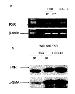
[0020] Figure 1 shows the expression of FXR in the primary cultures of HSCs
and HSC-
T6, at mRNA level by RT-PCR (panel a) and at protein level by Western blot
analysis
(panel b). Panel b also demonstrates that the amount of FXR in HSC increases
over time
during culture and its increase parallels the expression of a-smooth muscle
actin (aSMA), a
marker of HSCs differentiation into myofibroblast-like cells.
[0021] Figure 2 shows the expression of NTCP, BSEP, CYP7A1, and SHP in HSC
(panel
a) and the expression of these genes regulated by FXR ligands (panel b). The
results of
5a
CA 02559476 2012-07-31
quantitative RT-PCR in Figure 2b illustrates that exposure to 6-ECDCA (a
synthetic FXR
ligand) and to CDCA (a natural FXR ligand) leads to a 2-fold increase of SHP
and BSEP
mRNA and a 50-70% reduction of NTCP and CYP7A1 mRNA.
5b
CA 02559476 2006-09-12
WO 2005/089316 PCT/US2005/008575
[0022] Figure 3a shows results of RT-PCR and quantitative RT-PCR, indicating
that
exposure of HSCs to FXR ligands 6-ECDCA (1 AM), CDCA (20 GM), or GW4064 (100
nM)
reduces the expression of type I collagen as measure by assessing al mRNA
expression by
methods. Figure 3b shows results of Northern blot analysis, which confirm the
results of
panel a.
[0023] Figure 4 shows results of HSC proliferation assays, indicating that 6-
ECDCA does
not prevent HSCs proliferation induced by thrombin, PDGF, or TGF'31, as
assessed by
determining [3H]-thymidine incorporation (panels a and b) or cell counting
(panel c).
Furthermore, FXR ligands do not drive HSCs to apoptosis (panel d).
[0024] Figure 5 shows FXR ligands-mediated inhibition of collagen al release,
as
measured by determining hydroxyproline concentrations in cell supernatants
(panels a and
b).
[0025] Figure 6 shows that SHIP overexpression in HSC-T6 abrogates al
expression on
resting HSC-T6, as measured by QRT-PCR and Northern blot analysis, and
prevents al
induction caused by thrombin, TGF,l31, and PDGF.
[0026] Figure 7 shows that abrogation of SHP expression, by specific small
interference
RNA (siRNA), reverses al mRNA inhibition caused by FXR ligands. Silencing of
SHP also
preventes inhibition of a1 expression induced by FXR ligands on HSCs treated
with
mitogenic factors such as thrombin, TGF(3 and PDGF. Results of Northern blot
analysis
confirming the effect of SUP on a1 mRNA are also shown in Figure 7.
[0027] Figure 8 shows the levels of collagen deposition, hydroxyproline, and
a1 collagen
mRNA in the livers of BDL rats treated or untreated with 6ECDCA.
DEFINITIONS
[0028] "Fibrosis" refers to a condition involving the development of excessive
fibrous
connective tissue, e.g., scar tissue, in a tissue or organ. Such generation of
scar tissue may
occur in response to infection, inflammation, or injury of the organ due to a
disease, trauma,
chemical toxicity, and so on. Fibrosis may develop in a variety of different
tissues and
organs, including the liver, kidney, intestine, lung, heart, etc.
[0029] The term "inhibiting" or "inhibition," as used herein, refers to any
detectable
positive effect on the development or progression of a disease or condition.
Such a positive
effect may include the delay or prevention of the onset of at least one
symptom or sign of the
6
CA 02559476 2006-09-12
WO 2005/089316 PCT/US2005/008575
disease or condition, alleviation or reversal of the symptom(s) or sign(s),
and slowing or
prevention of the further worsening of the symptom(s) or sign(s).
[0030] As used herein, a "cholestatic condition" refers to any disease or
condition in
which bile excretion from the liver is impaired or blocked, which can occur
either in the liver
or in the bile ducts. Intrahepatic cholestasis and extrahepatic cholestasis
are the two types of
cholestatic conditions. Intrahepatic cholestasis (which occurs inside the
liver) is most
commonly seen in primary biliary cirrhosis, primary sclerosing cholangitis,
sepsis
(generalized infection), acute alcoholic hepatitis, drug toxicity, total
parenteral nutrition
(being fed intravenously), malignancy, cystic fibrosis, and pregnancy.
Extrahepatic
cholestasis (which occurs outside the liver) can be caused by bile duct
tumors, strictures,
cysts, diverticula, stone formation in the common bile duct, pancreatitis,
pancreatic tumor or
pseudocyst, and compression due to a mass or tumor in a nearby organ.
[0031] Clinical symptoms and signs of a cholestatic condition include: itching
(pruritus),
fatigue, jaundiced skin or eyes, inability to digest certain foods, nausea,
vomiting, pale stools,
dark urine, and right upper quadrant abdominal pain. A patient with a
cholestatic condition
can be diagnosed and followed clinically based on a set of standard clinical
laboratory tests,
including measurement of levels of alkaline phosphatase, y-glutamyl
transpeptidase (GGT), 5'
nucleotidase, bilirubin, bile acids, and cholesterol in a patient's blood
serum. Generally, a
patient is diagnosed as having a cholestatic condition if serum levels of all
three of the
diagnostic markers alkaline phosphatase, GGT, and 5' nucleotidase, are
considered
abnormally elevated. The normal serum level of these markers may vary to some
degree
from laboratory to laboratory and from procedure to procedure, depending on
the testing
protocol. Thus, a physician will be able to determine, based on the specific
laboratory and
test procedure, what is an abnormally elevated blood level for each of the
markers. For
example, a patient suffering from a cholestatic condition generally has
greater than about 125
I[J/L alkaline phosphatase, greater than about 65 IU/L GGT, and greater than
about 17 IU/L
5' nucleotidase in the blood. Because of the variability in the level of serum
markers, a
cholestatic condition may be diagnosed on the basis of abnormal levels of
these three markers
in addition to at least one of the symptoms mentioned above, such as itching
(pruritus).
[0032] A "ligand" specific for FXR refers to a natural or synthetic compound
that binds to
FXR and is thereby capable of specifically stimulating ligand-dependent FXR
transcriptional
7
CA 02559476 2006-09-12
WO 2005/089316 PCT/US2005/008575
activity differentiated from the baseline level determined in the absence of
any ligand. In this
application, the term "an FXR ligand" is interchangeable with "an FXR-
activating ligand."
[00331 The term "effective amount" as used herein refers to an amount of
compound (e.g.,
an FXR-activating ligand) that produces an acute or chronic therapeutic effect
upon
appropriate dose administration. The effect includes the prevention,
correction, inhibition, or
reversal of the symptoms, signs and underlying pathology of a
disease/condition (e.g.,
fibrosis of the liver, kidney, or intestine) and related complications to any
detectable extent.
The exact amount and dosing schedule will depend on the purpose of the
treatment, and will
be ascertainable by one skilled in the art using known techniques (see, e.g.,
Lieberman,
Pharmaceutical Dosage Forms (vols. 1-3, 1992); Lloyd, The Art, Science and
Technology of
Pharmaceutical Compounding (1999); and Pickax, Dosage Calculations (1999)).
[0034] The term "organ" refers to a differentiated structure (as in a heart,
lung, kidney,
liver, etc.) consisting of cells and tissues and performing some specific
function in an
organism. This term also encompasses bodily parts performing a function or
cooperating in
an activity (e.g., an eye and related structures that make up the visual
organs). The term
"organ" further encompasses any partial structure of differentiated cells and
tissues that is
potentially capable of developing into a complete structure (e.g., a lobe or a
section of a
liver).
DETAILED DESCRIPTION OF THE INVENTION
[0035] For the first time, ligands specific for the farnesoid X receptor
(FXR), particularly
those capable of activating FXR at a low concentration, are shown to be
effective in treating
or preventing fibrosis in tissues or organs such as liver, kidney, and
intestine, in patients who
are not suffering from a cholestatic condition.
[0036] Without being bound to any particular theory, the present inventor
discovered that
FXR plays an important role in regulating the synthesis of collagen primarily
via the actions
of SHP that FXR directly regulates in a ligand-dependent fashion. This
discovery therefore
allows the use of FXR-activating ligands for the effective prevention,
treatment, and/or
reversal of fibrosis in tissues where FXR is expressed, particularly in
patients who are not
suffering from any condition for which the use of FXR ligands has been
previously
suggested, e.g., in cholestatic conditions where the anti-cholestatic
therapeutic effect of an
FXR ligand may also indirectly inhibit fibrosis.
8
CA 02559476 2006-09-12
WO 2005/089316 PCT/US2005/008575
1. Identification of Patient Population
[0037] The present invention relates to the prophylactic and therapeutic use
of FXR ligands
in patients who: (1) suffer from fibrosis or certain diseases/conditions that
are known to lead
to fibrosis in a tissue or organ in which FXR is expressed; and (2) do not
suffer from a
cholestatic condition that may secondarily cause liver fibrosis, where such
patients are treated
with an FXR ligand to inhibit ongoing liver fibrosis or prevent the
development of liver
fibrosis. The description below allows for determination if a patient falls
within the
population suitable for treatment pursuant to the present invention.
A. Expression of FXR in an Organ
[0038] One must first determine the status of FXR expression in an organ or a
tissue prior
to determining whether an FXR ligand may be used to effectively inhibit
fibrosis in this
organ. The detection of FXR expression can be accomplished at two different
levels: nucleic
acid level and polypeptide level.
1. FXR Expression at Nucleic Acid Level
[0039] The polynucleotide sequence encoding human FXR has been identified by
Forman
et at. (Cell 81:687-93, 1995) and available as GenBank Accession No. NM
005123. Based
on this information, FXR gene expression can be detected at nucleic acid level
in a human
patient sample. A variety of methods of specific DNA and RNA measurement using
nucleic
acid hybridization techniques are commonly used (e.g., Sambrook and Russell,
Molecular
Cloning, A Laboratory Manual (3rd ed.) 2001). Some methods involve an
electrophoretic
separation (e.g., Southern blot for detecting DNA and Northern blot for
detecting RNA), but
detection of DNA or RNA can be carried out without electrophoresis as well
(such as by dot
blot, or in situ hybridization if the detection is made within a target
tissue). The presence of
nucleic acid encoding FXR in the cells of a particular organ can also be
detected by
polymerase chain reaction (PCR) or PCR-based methods, e.g., real-time PCR and
reverse
transcription polymerase chain reaction (RT-PCR), using sequence-specific
primers.
2. FXR Expression at Protein Level
[0040] The expression of FXR in an organ can be confirmed by detecting FXR
protein in a
tissue sample from this organ. The amino acid sequence of human FXR can be
determined
based on its coding sequence, e.g., GenBank Accession No. NM 51023, and is set
forth in
publications such as WO 00/76523. Various immunological assays (such as enzyme-
linked
immune absorbent assay (ELISA), Western blot, and immunohistochemistry) can be
used by
9
n
CA 02559476 2006-09-12
WO 2005/089316 PCT/US2005/008575
those skilled in the art to measure the level of FXR gene product,
particularly using
polyclonal or monoclonal antibodies that react specifically with the FXR
polypeptide, (e.g.,
Harlow and Lane, Antibodies, A Laboratory Manual, Chapter 14, Cold Spring
Harbor, 1988;
Kohler and Milstein, Nature, 256:495-497, 1975). Such techniques require
antibody
preparation by selecting antibodies with high specificity against the FXR
polypeptide or an
antigenic portion thereof. The methods of raising polyclonal and monoclonal
antibodies are
well established and their descriptions can be found in the literature, see,
e.g., Harlow and
Lane, supra; Kohler and Milstein, Eur. J Immunol., 6:511-519, 1976.
Production of Antibodies against FXR
[0041] Methods for producing polyclonal and monoclonal antibodies that react
specifically
with an immunogen of interest are known to those of skill in the art (see,
e.g., Coligan,
Current Protocols in Immunology Wiley/Greene, NY, 1991; Harlow and Lane,
Antibodies: A
Laboratory Manual Cold Spring Harbor Press, NY, 1989; Stites et al. (eds.)
Basic and
Clinical Immunology (4th ed.) Lange Medical Publications, Los Altos, CA, and
references
cited therein; Goding, Monoclonal Antibodies: Principles and Practice (2d ed.)
Academic
Press, New York, NY, 1986; and Kohler and Milstein Nature 256: 495-497, 1975).
Such
techniques include antibody preparation by selection of antibodies from
libraries of
recombinant antibodies in phage or similar vectors (see, Huse et al., Science
246: 1275-1281,
1989; and Ward et al., Nature 341: 544-546, 1989).
[0042] In order to produce antisera containing antibodies with desired
specificity, the
polypeptide of interest (e.g., human FXR) or an antigenic fragment thereof can
be used to
immunize suitable animals, e.g., mice, rats, rabbits, goats, horses, or
monkeys. A standard
adjuvant, such as Freund's adjuvant, can be used in accordance with a standard
immunization
protocol. Alternatively, a synthetic antigenic peptide derived from that
particular polypeptide
can be conjugated to a carrier protein and subsequently used as an immunogen.
[0043] The animal's immune response to the immunogen preparation is monitored
by
taking test bleeds and determining the titer of reactivity to the antigen of
interest. When
appropriately high titers of antibody to the antigen are obtained, blood is
collected from the
animal and antisera are prepared. Further fractionation of the antisera to
enrich antibodies
specifically reactive to the antigen and purification of the antibodies can be
performed
subsequently, see, Harlow and Lane, supra, and the general descriptions of
protein
purification provided above.
10,
CA 02559476 2006-09-12
WO 2005/089316 PCT/US2005/008575
[00441 Monoclonal antibodies are obtained using various techniques familiar to
those of
skill in the art. Typically, spleen cells from an animal immunized with a
desired antigen are
immortalized, commonly by fusion with a myeloma cell (see, Kohler and
Milstein, Eur. J.
Immunol. 6:511-519, 1976). Alternative methods of immortalization include,
e.g.,
transformation with Epstein Barr Virus, oncogenes, or retroviruses, or other
methods well
known in the art. Colonies arising from single immortalized cells are screened
for production
of antibodies of the desired specificity and affinity for the antigen, and the
yield of the
monoclonal antibodies produced by such cells maybe enhanced by various
techniques,
including injection into the peritoneal cavity of a vertebrate host.
[0045] Additionally, monoclonal antibodies may also be recombinantly produced
upon
identification of nucleic acid sequences encoding an antibody with desired
specificity (e.g.,
specifically recognizing human FXR) or a binding fragment of such antibody by
screening a
human B cell cDNA library according to the general protocol outlined by Huse
et al., supra.
The general principles and methods of recombinant polypeptide production
discussed above
are applicable for antibody production by recombinant methods.
Immunoassays for Detecting FXR Expression
[0046] Once antibodies specific for FXR are available, the presence and amount
of FXR in
a sample, e.g., a small section of tissue, can be measured by a variety of
immunoassay
methods (such as ELISA or Western blot) providing qualitative and quantitative
results to a
skilled artisan. For a review of immunological and immunoassay procedures in
general see,
e.g., Stites, supra; U.S. Patent Nos. 4,366,241; 4,376,110; 4,517,288; and
4,837,168.
(a) Labeling in Immunoassays
[0047] Immunoassays often utilize a labeling agent to specifically bind to and
label the
binding complex formed by the antibody and the target protein (e.g., human
FXR). The
labeling agent may itself be one of the moieties comprising the
antibody/target protein
complex, or may be a third moiety, such as another antibody, that specifically
binds to the
antibody/target protein complex. A label may be detectable by spectroscopic,
photochemical,
biochemical, immunochemical, electrical, optical or chemical means. Examples
include, but
are not limited to, magnetic beads (e.g., DynabeadsTm), fluorescent dyes
(e.g., fluorescein
isothiocyanate, Texas red, rhodamine, and the like), radiolabels (e.g., 3H,
1251, 35s, 14C, or
32P), enzymes (e.g., horse radish peroxidase, alkaline phosphatase, and others
commonly used
11
CA 02559476 2006-09-12
WO 2005/089316 PCT/US2005/008575
in an ELISA), and colorimetric labels such as colloidal gold or colored glass
or plastic (e.g.,
polystyrene, polypropylene, latex, etc.) beads.
[0048] In some cases, the labeling agent is a second antibody bearing a
detectable label.
Alternatively, the second antibody may lack a label, but it may, in turn, be
bound by a labeled
third antibody specific to antibodies of the species from which the second
antibody is
derived. The second antibody can be modified with a detectable moiety, such as
biotin, to
which a third labeled molecule can specifically bind, such as enzyme-labeled
streptavidin.
[0049] Other proteins capable of specifically binding immunoglobulin constant
regions,
such as protein A or protein G, can also be used as the label agents. These
proteins are normal
constituents of the cell walls of streptococcal bacteria. They exhibit a
strong non-
immunogenic reactivity with immunoglobulin constant regions from a variety of
species (see,
generally, Kronval, et al. J Immunol., 111: 1401-1406 (1973); and Akerstrom,
et al.,
J ImmunoL, 135:2589-2542 (1985)).
(b) Immunoassay Formats
[0050] Immunoassays for detecting a target protein of interest (e.g., FXR)
from samples
maybe either competitive or noncompetitive. Noncompetitive immunoassays are
assays in
which the amount of captured target protein is directly measured. In one
preferred
"sandwich" assay, for example, the antibody specific for the target protein
can be bound
directly to a solid substrate where the antibody is immobilized. It then
captures the target
protein in test samples. The antibody/target protein complex thus immobilized
is then bound
by a labeling agent, such as a second or third antibody bearing a label, as
described above.
[0051] In competitive assays, the amount of target protein in a sample is
measured
indirectly by measuring the amount of an added (exogenous) target protein
displaced (or
competed away) from an antibody specific for the target protein by the target
protein present
in the sample. In a typical example of such an assay, the antibody is
immobilized and the
exogenous target protein is labeled. Since the amount of the exogenous target
protein bound
to the antibody is inversely proportional to the concentration of the target
protein present in
the sample, the target protein level in the sample can thus be determined
based on the amount
of exogenous target protein bound to the antibody and thus immobilized. See,
e.g., Karlson et
al., Lab. Invest., 70:705-710 (1994).
[0052] In some cases, western blot (immunoblot) analysis is used to detect and
quantify the
presence of FXR in the samples. The technique generally comprises separating
sample
12
CA 02559476 2006-09-12
WO 2005/089316 PCT/US2005/008575
proteins by gel electrophoresis on the basis of molecular weight, transferring
the separated
proteins to a suitable solid support (such as a nitrocellulose filter, a nylon
filter, or a
derivatized nylon filter) and incubating the samples with the antibodies that
specifically bind
the target protein. These antibodies may be directly labeled or alternatively
may be
subsequently detected using labeled antibodies (e.g., labeled sheep anti-mouse
antibodies)
that specifically bind to the antibodies against FXR. See, e.g., Pineda et
al., J. Neurotrauma,
18:625-634 (2001); Bowler et al., J. Biol. Chem., 277:16505-16511 (2002).
[0053] Various in situ immunochemical staining methods using antibodies
against FXR are
also useful for demonstrating the presence of FXR in a tissue sample.
[0054] Other assay formats include liposome immunoassays (LIA), which use
liposomes
designed to bind specific molecules (e.g., antibodies) and release
encapsulated reagents or
markers. The released chemicals are then detected according to standard
techniques (see,
Monroe et al., Amer. Clin. Prod. Rev., 5: 34-41 (1986)).
[0055] In addition, functional assays may also be performed for detecting the
presence of
FXR in a tissue sample. Assays for detecting the biological activity of FXR
are generally
described in a later section.
B. Diagnosing Fibrosis
[0056] Fibrosis is a pathophysiological process in response to tissue injury
due to viral or
bacterial infection, inflammation, autoimmune disease, trauma, drug toxicity,
and so on.
During this process, an excess amount of collagen is expressed and fibrous
material forms in
the extracellular space of the affected tissue. Thus, fibrosis can be
generally recognized
based on the distinct morphology of fibrous tissue in a biopsy of the organ in
which fibrosis
is suspected. Other means for detecting the presence of fibrosis or developing
fibrosis
include computerized axial tomography (CAT or CT) scan, ultrasound, magnetic
resonance
imaging (MRI), and monitoring the level of one or more serum markers known to
be
indicative of fibrosis (e.g., various types of collagens).
[0057] The precise manner of diagnosing fibrosis also varies depending on the
organ where
the fibrotic process takes place. For instance, biopsies are generally
effective for diagnosing
fibrosis of most organs, whereas endoscopy involving a fiber optic instrument
(e.g., a
sigmoidoscope or a colonoscope) can be a less traumatic alternative to detect
fibrosis of
certain organs such as the intestine.
13
CA 02559476 2006-09-12
WO 2005/089316 PCT/US2005/008575
1. Biopsy for Detecting Liver Fibrosis
[0058] Standard procedures have been established for obtaining biopsy from a
given organ.
For example, a liver specimen can be obtained during exploratory surgery, but
is more often
obtained by inserting a biopsy needle through the skin and into the liver.
Before this
procedure, termed percutaneous liver biopsy, is performed, the person receives
a local
anesthetic. Ultrasound or CT scans may be used to locate the abnormal area
from which the
specimen is to be taken.
[0059] In transvenous liver biopsy, a catheter is inserted into a neck vein,
threaded through
the heart, and placed into one of the hepatic veins that drain the liver. The
needle of the
catheter is then inserted through the wall of the vein into the liver. This
procedure is less
likely to injure the liver than is percutaneous liver biopsy. It is especially
useful in people
who bleed easily, which is a complication of severe liver disease.
[0060] Upon obtaining a liver biopsy, the sample is examined and given a score
to indicate
the presence and level of fibrosis in the sample. Most frequently used scoring
systems
include the METAVIR or modified HAI (ISHAK) scoring system. The Knodell
scoring
system can also be used for analyzing the liver sample. The criteria used in
scoring liver
samples are well established and known to those of skilled in the art. For
example, the
METAVIR system provides five gradings: FO indicates the absence of fibrosis;
F1 indicates
portal fibrosis without septa; F2 indicates portal fibrosis and some septa; F3
indicates septal
fibrosis without cirrhosis; and F5 indicates the presence of cirrhosis. See,
e.g., Bedossa and
Poynard, Hepatology 24:289-293, 1996.
[0061] Biopsy is not only useful for the diagnosis of liver fibrosis, it can
also aid physicians
to assess the effectiveness of fibrosis treatment/prevention methods of the
present invention
by monitoring the progression of fibrosis using methodologies known in the
art. See, e.g.,
Poynard et al., Lancet 349:825, 1997.
2. Serum Markers for Liver Fibrosis
[0062] There are numerous known serum markers whose level can be indicative of
the
presence and/or severity of liver fibrosis. Blood tests measuring markers,
e.g., hyaluronic
acid, laminin, undulin (type IV collagen) pro-peptides from types I, II, and
IV collagens,
lysyl oxidase, prolyl hydroxylase, lysyl hydroxylase, PIIINP, PICP, collagen
VI, tenascin,
collagen XIV, laminin P1, TIMP-1, MMP-2, a2 macroglobulin, haptoglobin, gamma
glutamyl transpeptidase, "y globulin, total bilirubin, apolipoprotein Al,
etc., according to the
14
CA 02559476 2006-09-12
WO 2005/089316 PCT/US2005/008575
established methods can thus be useful for both the diagnosis of fibrosis and
monitoring of
fibrosis progression in the liver.
3. Other Markers
[0063] Additional markers, such as nucleic acid markers, can be used for
detecting and/or
monitoring fibrosis. For instance, Wnt-4 has recently been indicated in
laboratory
experiments as a gene that plays an important role in renal fibrosis, where
its mRNA
expression is significantly increased in the fibrotic tissue in the kidney
(see, e.g., Surendran et
al., JPediatr. 140:119-24, 2002). The quantitative detection of gene
expression of this type
of markers can be useful in the diagnosis and monitoring of fibrosis.
C. Identifying Patients with Elevated Risk of Developing Fibrosis
[0064] Because the method of the present invention is also effective for the
prevention of
the onset of fibrosis or the slowing of its progression after onset, patients
with heightened risk
of fibrosis fall within the patient population suitable for treatment using
the method of the
present invention. Such patients are identified based on prior diagnosis of
certain diseases
and conditions known to lead to fibrosis. The following sections describe the
means to
diagnose some of these diseases and conditions. There are, however, additional
diseases/conditions that are known to elevate a patient's risk of developing
fibrosis later in
life and that can be readily diagnosed by a physician. The treatment of
patients suffering
from any of these diseases/conditions with an FXR ligand to prevent, inhibit,
or reverse
fibrosis is within the contemplation of the present inventor and within the
scope of the
present invention. Such treatment may be warranted for a short through
lifetime course, as is
warranted for a given patient with a given disease/condition and as determined
by one skilled
in the art of treating such patients.
1. Liver Fibrosis
[0065] The following are some examples of diseases known to significantly
increase a
patient's risk of developing liver fibrosis: (i) chronic liver infections
(including chronic
hepatitis B and hepatitis C viral infection; schistosomiasis and other
parasitic liver diseases;
post-transplant bacterial, viral and fungal infections); (ii) alcoholic liver
disease; (iii) non-
alcoholic fatty liver disease (NAFLD) or non-alcoholic steatohepatitis (NASH);
(iv) drug and
chemical induced liver diseases (including methotrexate, isoniazid,
oxyphenistatin,
methyldopa, chlorpromazine, tolbutamide, and amiodarone); (v) autoimmune
disease
(including autoimmune hepatitis, sarcoidosis, and lupoid hepatitis); (vi)
storage diseases
CA 02559476 2006-09-12
WO 2005/089316 PCT/US2005/008575
resulting from inborn errors of metabolism (including Wilson's disease,
hemochromatosis,
Gaucher's disease, types III, IV, VI, IX and X glycogen storage diseases, al-
antitrypsin
deficiency, Zeliweger syndrome, tyrosinemia, fructosemia, and galactosemia);
(vii) vascular
derangement (including Budd-Chiari syndrome, veno-occlusive disease, and
portal vein
thrombosis); and (viii) congenital hepatic fibrosis.
Hepatitis B
[0066] Hepatitis B causes inflammation of the liver due to the infection by
hepatitis B virus
(HBV, a DNA virus belonging to the family of Hepadnaviridae). An acute HBV
infection
usually lead to recovery, but rarely can also lead to acute liver failure, and
sometimes to
chronic infection. The chronic infection can result in a healthy carrier state
or progress
through fibrosis to cirrhosis and its complications, including liver cancer.
[0067] Acute hepatitis B is the initial, rapid onset, short duration illness
that results from
infection with HBV. About 70% of adults with acute hepatitis B have few or no
symptoms,
whereas the remaining 30% develop significant symptoms two to four months
following
exposure to the HBV. The most common symptoms of acute hepatitis B are
fatigue, loss of
appetite, nausea, vomiting, dark urine, light stools, and abdominal pain over
the region of the
liver. Jaundice often accompanies these other symptoms.
[0068] The diagnosis of chronic hepatitis B can be made, by definition, only
after six
months from the onset of acute hepatitis B. Most individuals with chronic
hepatitis B
infection remain asymptomatic for many years, even up to two or three decades.
During this
time, the patient's liver blood tests usually are at most mildly abnormal and
the inflammation
and scarring (i.e., fibrosis) of the liver progresses slowly. Occasionally,
however, these
individuals with otherwise inactive chronic hepatitis B may develop flares
(reactivation) of
acute symptoms, elevated liver blood tests, and inflammation of the liver.
These flares
resemble acute hepatitis and can cause more rapid progression of liver
fibrosis.
[0069] Besides the above-described symptoms, diagnosis of hepatitis B is
confirmed by
blood test detecting antibodies against HBV.
Hepatitis C
[0070] Infection by the hepatitis C virus (HCV, an RNA virus and a member of
the
Flaviviridae family) is one of the most significant health problems affecting
the liver. More
than 4 million Americans (1.3% of the U.S. population) and an estimated 170
million
individuals in the world (3% worldwide) are infected with HCV. About 85% of
individuals
16
CA 02559476 2006-09-12
WO 2005/089316 PCT/US2005/008575
initially infected with this virus will become chronically infected, usually
for decades. The
other 15% of HCV infected individuals simply have an acute infection.
[0071] At the beginning of an HCV infection, only about 25% of patients
exhibit the
characteristic symptoms of acute hepatitis. These symptoms include fatigue,
muscular aches,
poor appetite, and low-grade fever. Rarely, yellowing of the skin and/or eyes
(jaundice) also
occurs.
[0072] As the hepatitis becomes chronic, most individuals remain asymptomatic
and can
only be diagnosed through routine blood work when HCV antibodies are detected.
In well
compensated disease, infected individuals may exhibit no symptoms despite the
progressive
liver inflammation, necrosis, and fibrosis that is a ubiquitous feature of the
chronic infectious
process. Other patients may experience chronic or intermittent fatigue and a
diminished
sense of well-being as a result of advancing disease. On the other hand,
fatigue has been
described in some individuals with relatively mild disease.
[0073] A number of diagnostic tests are currently available for HCV infection.
Screening
tests are done to determine the presence of antibodies to HCV in the blood.
The enzyme
immunosorbent assay (EIA) is the conventional, initial screening test to
diagnose HCV
infection by measuring specific antibodies to HCV antigens. This test,
therefore, is referred
to as the anti-HCV antibody test. Patients who have elevated liver enzymes
(ALT/AST)
and/or any of the risk factors for HCV can be diagnosed to have HCV with a
greater than
95% certainty when the EIA is positive.
[0074] When an individual with low risk of HCV infection is tested positive by
EIA,
confirmatory testing is conducted using a specialized assay that likewise
tests for antibodies
against the HCV proteins. This assay is called the Recombinant Immunoblot
Assay (RIBA).
[0075] Since HCV is an RNA virus, several diagnostic assays are based on the
detection of
the HCV RNA in a person's blood. These tests are referred to as molecular
tests because they
examine the virus at the molecular level. The two most common systems for
measuring HCV
RNA are the reverse transcription polymerase chain reaction (RT-PCR) assay and
the
branched chain DNA (bDNA) assay. Recently, a third type of assay, called
transcription-
mediated amplification (TMA), has been become available.
Alcoholic Liver Disease
[0076] Alocholic liver disease (ALD) is a chronic liver disease caused by
excessive
consumption of alcohol. The symptoms of ALD are usually non-specific, and do
not
17
in
CA 02559476 2006-09-12
WO 2005/089316 PCT/US2005/008575
necessarily indicate the severity of the underlying liver damage. General ALD
symptoms
include fatigue, nausea and vomiting, diarrhea, or abdominal pains. Many
patients, even with
advanced ALD marked by progressive liver fibrosis and toxicity, may have no
symptoms and
their condition is only diagnosed by liver blood tests. Only in the more
advanced stages of
decompensated ALD (severe alcoholic hepatitis or cirrhosis) will the sufferer
present with
more specific liver-related symptoms such as jaundice, ascites, hematemesis,
or
encephalopathy.
[0077] The diagnosis of ALD is established based on a history of alcohol
abuse, blood tests
showing the presence and severity of liver damage. Ultrasound scan of the
liver can help
assess the severity of disease and exclude other conditions with similar
symptoms. Liver
biopsy is the most reliable means to determine the present and stage of ALD.
Non-Alcoholic Fatty Liver Disease
[0078] Non-alcoholic fatty liver disease (NAFLD) refers to a wide spectrum of
liver
diseases ranging from simple fatty liver (steatosis), to non-alcoholic
steatohepatitis (NASH),
to cirrhosis. All of the stages of NAFLD have in common the accumulation of
fat in the
hepatocytes. In NASH, the fat accumulation is associated with varying degrees
of
inflammation (hepatitis) and scarring (fibrosis) of the liver. NAFLD and NASH
occur in
individuals who do not consume excessive amounts of alcohol. Yet, in many
respects, the
histological picture of an NAFLD biopsy is similar to what can be seen in
liver disease
caused by alcohol abuse. NAFLD and NASH are considered the primary fatty liver
diseases.
The secondary fatty liver diseases include those that occur in other types of
liver disease.
Thus, alcoholic liver disease (ALD) is the most frequent secondary fatty liver
disease.
Secondary fatty liver can also occur in chronic viral hepatitis C (HCV),
chronic viral hepatitis
B (HBV), chronic autoimmune hepatitis (AIH), and Wilson's disease.
[0079] The symptoms of NAFLD and NASH are identical. They are usually not
dramatic
and tend to be non-specific (as can also be observed in other diseases). The
symptoms are
minimal in most patients, who may, however, experience occasional, vague right
upper-
quadrant abdominal pain. This pain characteristically is dull and aching,
without a
predictable pattern of occurrence. It is not an intense, sudden, and severe
pain, as might
occur with, for example, gallstones. The abdominal pain in NAFLD and NASH is
thought to
be due to the stretching of the liver covering (capsule) when the liver
enlarges and/or when
there is inflammation in the liver. In contrast to ALD, hepatitis B, or
hepatitis C, symptoms
18
CA 02559476 2006-09-12
WO 2005/089316 PCT/US2005/008575
of severe, acute liver failure (e.g., jaundice, intense fatigue, loss of
appetite, nausea,
vomiting, and confusion) are not observed in NAFLD or NASH. Obesity and
related
conditions (e.g., diabetes, hypertension) are frequent seen among those
suffering from
NAFLD or NASH, and the classic signs of insulin resistance often dominate the
physical
exam in NAFLD and NASH. Acanthosis nigricans, a dark pigmentation of the skin
of the
armpits and neck, can be a sign of insulin resistance and is frequently seen
in children with
NASH. When the liver is palpated, it usually feels normal. However, when very
large
amounts of fat accumulate in the liver, it can be become quite large with a
soft, rounded edge
that can be easily felt by the doctor.
[0080] In addition to the symptoms described above, a diagnosis of NAFLD or
NASH is
made based on the following criteria: clinical and/or biochemical signs of
insulin resistance;
chronically elevated ALT; signs of fatty liver on ultrasound; exclusion of
other causes of
elevated ALT and fatty liver. Only a liver biopsy, however, can establish a
definite diagnosis
and determine the severity of NAFLD or NASH.
Parasitic Liver Diseases
[0081] Various parasitic diseases are known to damage the liver and lead to
fibrosis or even
cirrhosis. Clonorchiasis, for instance, is an infection by the liver fluke
Clonorchis sinensis.
Patients initially infected with this parasite usually have no symptoms until
the worm load
reaches more than 500. Common symptoms are chills, diarrhea, fever, lower
abdominal pain,
jaundice, and swelling of the liver. To diagnose the disease, a medical
history should be
taken including questions on diet, travel, regions where previously resided. A
physical
examination should include gentle palpation of the liver. Further testing
includes endoscopy
and examination of stool sample for eggs.
[0082] O. tenuicollis (0. felineus) and O. viverrini are two other parasites
that are closely
related to Clonorchis sinensis and can lead to permanent liver damage. The
diagnostic
methods are similar to that described above. Close comparison of the
morphology of the
eggs and adult worms is necessary to distinguish the infections by these
parasites.
[0083] Schistosomiasis is another parasitic disease of liver, gastrointestinal
tract, and
bladder caused by schistosomes, trematode worms that parasitize people who
come into
contact with contaminated water.
[0084] There are three main species of these trematode worms (flukes) --
Schistosoma
haematobium, S. japonicuin, and S. mansoni -- that cause disease in humans.
Within days
19
CA 02559476 2006-09-12
WO 2005/089316 PCT/US2005/008575
after infection, a patient may develop a rash or itchy skin. Fever, chills,
cough, and muscle
aches can begin within 1-2 months of infection, even though most people have
no symptoms
at the early phase of infection. Eggs of the parasites travel to the liver or
pass into the
intestine or bladder. Rarely, eggs are found in the brain or spinal cord and
can cause seizures,
paralysis, or spinal cord inflammation. For people who are repeatedly infected
for many
years, the parasite can damage the liver, intestines, lungs, and bladder.
[0085] The diagnosis of schistosomasis involves examination of a patient's
stool or urine
samples for the eggs and/or the adult parasite. A blood test has been
developed to detect
antibodies against this parasite. Medical history reflecting possible exposure
to contaminated
water is also helpful for making a proper diagnosis.
Autoimmune Hepatitis
[0086] Autoimmune hepatitis, also known as lupoid hepatitis, involves
inflammation of the
liver caused by immune cells that mistake the liver's normal cells for a
foreign tissue or
pathogen. A person with autoimmune hepatitis has autoantibodies circulating in
the
bloodstream that cause the immune system to attack the liver. This disease is
associated with
other autoinimune diseases, including: thyroiditis, type 1 diabetes,
ulcerative colitis,
hemolytic anemia, and proliferative glomerulonephritis.
[0087] Symptoms of autoimmune hepatitis may include dark urine, loss of
appetite, fatigue,
general discomfort, uneasiness, or ill feeling (malaise), abdominal
distention, generalized
itching, pale or clay-colored stools, nausea, and vomiting.
[0088] Diagnosis can be made based on several criteria such as liver biopsy
showing
chronic hepatitis and fibrosis, abnormal liver function tests, as well as
tests associated with
autoimmune hepatitis, e.g., positive antinuclear antibodies, positive anti-
smooth muscle
antibody, positive anti-liver kidney microsomal antibody, positive anti-
mitochondrial
antibody, elevated sedimentation rate, elevated serum IgG.
Sarcoidosis
[0089] Another autoimmune disease that affects the liver is sarcoidosis.
Sarcoidosis is a
disease that causes small lumps, or granulomas, due to chronic inflammation to
develop in a
great range of body tissues. Sarcoidosis can appear in almost any body organ,
but most often
starts in the lungs or lymph nodes. It also affects the eyes, liver and skin;
and less often the
spleen, bones, joints, skeletal muscles, heart and central nervous system
(e.g., brain and
spinal cord). In the majority of cases, the granulomas clear up with or
without treatment. In
CA 02559476 2006-09-12
WO 2005/089316 PCT/US2005/008575
cases where the granulomas do not heal and disappear, the tissues tend to
remain inflamed
and become fibrotic.
Neonatal Liver Diseases
[0090] Neonatal liver diseases refer to severe liver disorders that occur in
newborns in the
neonatal period (i.e., the first 60 days of life). The possible causes of
these disorders may
include viral infection, hereditary metabolic diseases, neoplasia, and
vascular problems. The
infants affected frequently have jaundice, do not gain weight and grow
normally, and have
enlarged liver and spleen. The infants cannot absorb vitamins for proper
growth.
[0091] In addition to the above symptoms, the diagnosis of neonatal liver
diseases is aided
by liver biopsy, especially in the cases where the condition is not caused by
viral infection.
Wilson's Disease
[0092] Wilson's Disease is an inherited autosomal recessive disorder in which
too much
copper accumulates in the body. Although the accumulation of copper begins at
birth,
symptoms of the disorder appear later in life, between the ages of 6 and 40. A
diagnostic
feature of Wilson's Disease is what is called a Kayser-Fleischer ring, a deep
copper-colored
ring around the edge of the cornea. It represents copper deposits in the eye.
[0093] The most significant clinical consequence for about 40 percent of
patients with
Wilson's Disease is liver disease. In other patients, the first symptoms are
neurological or
psychiatric or both, and include tremor, rigidity, drooling, difficulty with
speech, abrupt
personality change, grossly inappropriate behavior, and inexplicable
deterioration of
performance at school or work, neurosis or psychosis.
[0094] Wilson's Disease can also be diagnosed by genetic testing to identify
both copies of
mutated gene, which has been localized to chromosome 13 between 13g14.3-g21.l.
Hemochromatosis
[0095] Hemochromatosis is an inherited disorder of excessive body accumulation
of iron.
It is common among the white population, affecting approximately 1 in 400
individuals of
European ancestry. Hemochromatosis patients are believed to absorb from their
diet
excessive amounts of iron, which becomes accumulated over time in the liver,
bone marrow,
pancreas, skin, and testicles.
[0096] Patients with early hemochromatosis have no symptoms, and the disease
may be
discovered when elevated iron blood levels are noted by routine blood testing.
In males,
21
CA 02559476 2006-09-12
WO 2005/089316 PCT/US2005/008575
symptoms may not appear until 40-50 years of age. Iron deposits in the skin
cause darkening
of the skin. Since females lose iron through menstrual blood loss, they
develop organ
damage from iron accumulation 15- 20 years later than men on average.
[0097] Iron deposits in the pituitary gland and testicles cause shrinkage of
the testicles and
impotence. Iron deposits in the pancreas cause a decrease in insulin
production resulting in
diabetes mellitus. Iron deposits in the heart muscle can cause heart failure
as well as
abnormal heart rhythms. Iron accumulation in the liver causes scarring of the
liver (fibrosis
and cirrhosis) and an increased risk of developing liver cancer.
[0098] Initial screening for hemochromatosis involves tests for levels of
blood iron and
ferritin, the latter is a blood protein that serves as an indicator of the
amount of iron stored in
the body. Blood iron and ferritin levels are high in patients with. Since
ferritin can also be
elevated in certain infections, such as viral hepatitis and other
inflammations in the body,
ferritin increase alone is not sufficient to accurately diagnose
hemochromatosis.
[0099] The most accurate test for hemochromatosis is measuring the iron
content of liver
tissue obtained by a biopsy. A biopsy involves the removal of a sample of
liver tissue for
analysis and is usually performed with a needle under local anesthesia. After
numbing the
skin and the underlying tissues, the doctor inserts a needle into the liver
through the right
lower rib cage, sometimes under ultrasound guidance. The tissue obtained by
the needle is
studied under a microscope for liver damage or cirrhosis. The amount of iron
in the liver is
usually significantly elevated in hemochromatosis.
[0100] Finally, genetic testing can effectively confirm a diagnosis of
hemochromatosis.
The gene for hereditary hemochromatosis, HFE, was identified in 1996 and can
be identified
in blood testing of 90 percent of patients with northern European ancestry.
Glycogen Storage Diseases
[0101] Glycogen storage diseases (GSD), also'known as glycogenoses, are
genetically
linked metabolic disorders that involve the enzymes regulating glycogen
metabolism and are
characterized by deposition of an abnormal type of quantity of glycogen in the
tissues. GSDs
often manifest the symptoms early in a patient's infancy or childhood. In some
cases,
however, the conditions may go undetected until adulthood or even old age.
Varying by type,
there are four major symptoms that typically lead a doctor to suspect GSDs:
low blood sugar,
enlarged liver, retarded growth, and an abnormal blood biochemistry profile. A
definitive
diagnosis is obtained by biopsy of the affected organ or organs, where the
biopsy sample is
22
CA 02559476 2006-09-12
WO 2005/089316 PCT/US2005/008575
tested for its glycogen content and assayed for enzyme activity. There are DNA-
based
techniques for diagnosing some GSDs from more easily available samples, such
as blood or
skin. These DNA techniques can also be used for prenatal testing.
[0102] In certain types of GSDs, disruption of glycogen metabolism often leads
to the
accumulation of abnormal metabolic by-products, which can damage organs such
as the liver
and the kidneys. Among all GSDs, types III, IV, VI, IX, and X are the most
relevant to the
onset of liver fibrosis.
[0103] Type III glycogen storage disease (Cori's disease) is characterized by
the absence
of debranching enzyme, amylo-1,6-glucosidase which causes the accumulation of
a
polysaccharide of the limit dextrin type. The structure of glycogen stored in
the liver and
muscle is abnormal and the amount is markedly increased. Most noticeable is
the short outer
branch of the glycogen, thus only a small portion of this abnormal glycogen is
functionally
active as an accessible source of glucose. Symptoms of this disorder include
enlargement of
the liver, hypoglycemia, ketosis, hyperuricemia, hyperlipemia, etc. In youths
affected by this
disease, growth is impaired, puberty is often delayed, and bones maybe
weakened by
osteoporosis. Blood platelets are also affected and frequent nosebleeds and
easy bruising are
common. Primary symptoms improve with age, but after age 20-30, liver tumors,
chronic
renal disease, and gout may appear. The diagnosis of this condition is based
on the above
symptoms and confirmed by examining of the glycogen structure.
[0104] Type IV glycogen storage disease (Andersen's disease) is characterized
by the
absence of branching enzyme (a-1,4 to a-1,6), with the result that the
glycogen constructed in
type IV GSD has very long outer branches and is insoluble. As the abnormal
glycogen
accumulates in the cells, cell death leads to organ damage. Infants born with
GSD IV appear
normal at birth, but are diagnosed with enlarged livers and failure to thrive
within their first
year. Infants who survive beyond their first birthday develop cirrhosis of the
liver by age 3-5
and die as a result of chronic liver failure. The diagnosis of this disease is
aided by the
detection of the characteristic abnormal glycogen structure.
[0105] Type VI glycogen storage disease (Hers' disease) is caused by liver
phosphorylase
deficiency, which blocks the first step of glycogenolysis. In contrast to most
other GSDs,
which involve autosomal mutations, type VI GSD is linked to the X chromosome.
In this
disease, phosphorylase deficiency results in increased amount of glycogen in
the liver.
Symptoms include enlargement of the liver, hypoglycemia, ketosis,
hyperuricemia,
23
CA 02559476 2006-09-12
WO 2005/089316 PCT/US2005/008575
hyperlipemia, etc. Low blood sugar is one of the key symptoms. Mildly retarded
growth can
occur in affected youths.
[0106] Type IX glycogen storage disease is caused by liver glycogen
phosphorylase
kinase (PhK) deficiency and, symptom-wise, is very similar to type VI GSD. The
main
differences are that the symptoms may not be as severe and may also include
exercise-related
problems in the muscles, such as pain and cramps. The symptoms abate after
puberty with
proper treatment. Most cases of GSD IX are linked to the X chromosome and
therefore
affect males. Enzymatic testing and measuring glycogen content provides a
definitive
diagnosis.
[0107] An enzyme that activates glycogen phosphorylase to stimulate glycogen
breakdown
in various tissues, PhK is a tetrameric enzyme made up of four different
subunits ((x(3y6) that
are responsible for various subtypes of GSD IX, that differ both in tissue
affected
(liver/muscle/RBC/Cardiac tissue) and in mode of inheritance. The genes for a,
0, and y
subunits have been cloned and mapped to X chromosome (a), chromosome 16g12
(a), and
chromosome 7p12 (y).
[0108] The most common form of PhK deficiency is the X-linked form, and it
mainly
affects the liver. Clinically patients with this form of PhK deficiency
present in infancy with
hepatomegaly, mild hypoglycemia, growth retardation, hyperlipidemia,
hyperketosis, and
delayed motor development. The symptoms improve with age, and adult patients
have
normal stature and normal liver.
[0109] The autosomal recessive form of PhK deficiency affects both liver and
muscle
depending on whether mutation has occurred in the a or 0 subunit of the
enzyme. Symptoms
could range from mild myopathy with muscle cramping to severe myopathic form.
[0110] Type X glycogen storage disease is an autosomal recessive disease
caused by a
deficiency of a cyclic adenosine monophosphate (AMP) -dependent
phosphoglycerate mutase
and presents symptoms similar to GSDs VI and IX. The gene involved in this
condition has
been mapped to chromosome 7p12-pl3.
al-Antitrypsin Deficiency
[0111] al-antitrypsin deficiency is a hereditary disease in which a lower-than-
normal level
of al-antitrypsin is present in the lungs. al-antitrypsin is a protein that is
made in the liver
and then released into the bloodstream. In normal lungs, al-antitrypsin
protects the lungs
24
CA 02559476 2006-09-12
WO 2005/089316 PCT/US2005/008575
from the harmful effects of neutrophil elastase. In a patient suffering from
al-antitrypsin
deficiency, damage to lung tissues by neutrophil elastase may lead to
emphysema and
breathing difficulty. The most noticeable symptom of this disorder is the
shortness of breath
during daily activities. Liver diseases associated with this disease include
those with early
onset, such as hepatitis or neonatal jaundice, or those with late onset, such
as cirrhosis and
primary cancer of the liver (Hepatoma).
[0112] al-antitrypsin deficiency can be diagnosed based on symptoms such as
shortness of
breath and a chronic cough. Blood test for al-antitrypsin level and pulmonary
function test
can also aid the diagnosis. Since this disease is caused by an autosomal
recessive mutation,
the most definitive diagnosis is based on results of genetic testing.
Gaucher's Disease
[0113] Gaucher's disease is caused by a genetic defect in an enzyme
glucocerebrosidase.
This enzyme helps the body break down the chemical glucocerebroside. The
defective
enzyme in patients with Gaucher's disease leads to the accumulation of
glucocerebroside in
the spleen, liver, and lymph nodes. Gaucher's disease is most common in
Ashkenazi Jews
(those of European origin), however, variants have been described in all
ethnic groups.
Depending on the precise type of the disease, affected patients may have
varying degrees of
symptoms. The most frequent early sign of Gaucher's disease is enlargement of
the spleen.
There can be associated fatigue, anemia, and a low count of platelets. Severe
bone
involvement can lead to pain and collapse (aceptic necrosis) of the bone of
the hips,
shoulders, and spine. Poor lung and brain function, and even seizures, can
occur.
[0114] The diagnosis of Gaucher's disease is confirmed by a special test in
which the
activity of ,6-glucocerbrosidase of fibroblasts activity is measured. Patients
with Gaucher's
disease have less than 15% of the normal level of glucocerebrosidase. Because
of the genetic
nature of the disease, diagnosis based on gene testing is also possible.
Zellweger Syndrome
[0115] Zellweger syndrome is a genetic disorder, also called the
cerebrohepatorenal
syndrome, characterized by the reduction or absence of peroxisomes in the
cells of the liver,
kidneys, and brain. Zellweger syndrome is one of a group of disorders called
the
leukodystrophies, all of which affect the myelin sheath, the fatty covering
which acts as an
insulator on nerve fibers in the brain. The most common features of Zellweger
syndrome
include an enlarged liver, high levels of iron and copper in the blood, and
vision disturbances.
CA 02559476 2006-09-12
WO 2005/089316 PCT/US2005/008575
Some affected infants may show prenatal growth failure. Symptoms at birth may
include
lack of muscle tone and an inability to move. Other symptoms may include
unusual facial
characteristics, mental retardation, seizures, and an inability to suck and/or
swallow.
Jaundice and gastrointestinal bleeding may also occur.
[01161 This disease is caused by mutations in any of several different genes
involved in
peroxisome formation. These genes lie on at least two different chromosome
locations
including chromosome 2 (region 2pl5) and chromosome 7 (region 7g21-q22). Thus,
its
diagnosis can be confirmed by genetic testing.
Tyrosinemia
[0117] Hereditary tyrosinemia is a genetic inborn error of metabolism
associated with
severe liver disease in infancy. The disease is inherited in an autosomal
recessive fashion.
The clinical features of the disease tend to fall into two categories: in the
acute form of the
disease, abnormalities appear in the first month of life. Babies may show poor
weight gain,
enlarged liver and spleen, distended abdomen, swelling of the legs and
increased tendency to
bleeding, particularly nose bleeds. Jaundice may or may not be prominent. In a
more
chronic form of tyrosinemia, enlargement of the liver and spleen are
prominent, the abdomen
is distended with fluid, weight gain may be poor, and vomiting and diarrhea
occur frequently.
Affected patients usually develop cirrhosis and its complications. In older
patients, there is
an increases risk of liver cancer.
[0118] In diagnosing this disease, liver tests are often used. Low serum
albumin and
clotting factors are frequently found. The liver enzymes transaminases may be
mildly to
moderately elevated, but the bilirubin is increased to a variable extent.
Because of the
biochemical defect, abnormal products may be measured in the urine which
confirm
diagnosis. These are parahydroxy phenylactic acid and parahydroxy
phenylpyruvic acid. In
addition, succinylacetone and succinylacetoacetate are found in the urine.
There may be
hypoglycemia and evidence of loss of certain substances in the urine including
sugar, protein,
and amino acids. The basic biochemical defect is an abnormality in a key
enzyme in the
metabolism of an essential amino acid, phenylalanine. The enzyme is
fumarylacetoacetate
hydrolase (FAH), which is markedly reduced in affected patients. Prenatal
diagnosis is
possible and can be performed by measuring succinylacetone in the amniotic
fluid or
fumarylacetoacetate hydrolase (FAH) in amniotic fluid cells.
26
CA 02559476 2006-09-12
WO 2005/089316 PCT/US2005/008575
Fructosemia
[0119] Fructosemia, also known as fructose intolerance or fructose aldolase B-
deficiency,
is a metabolic disease caused by the absence of an enzyme, 1-
phosphofructaldolase (i.e.,
fructose aldolase B). Hereditary fructose intolerance is inherited as an
autosomal recessive
disease. It may be as common as 1 in 20,000 in some European countries. In
fructose-
intolerant people, ingestion of fructose (fruit sugar) and sucrose (cane or
beet sugar, table
sugar) produces complicated chemical changes that cannot be corrected because
of the
absence of the enzyme 1-phosphofructaldolase. Ingestion of fructose causes
profound
hypoglycemia and progressive liver damage. The diagnosis of this condition is
based on the
fructose intolerant symptoms, test results that measure the level of fructose
aldolase B, and
genetic analysis to identify mutation(s) in the gene.
Galactosemia
[0120] Galactosemia is a rare hereditary disease leading not only to cirrhosis
in infants, but
more seriously, to early devastating illness if not diagnosed quickly. This
disease is caused
by elevated levels of galactose in the blood resulting from a deficiency of
the liver enzyme,
GALT (galactose-l-phosphate uridyl transferase), required for its metabolism.
Galactosemia
is inherited as an autosomal recessive trait. There are two forms of the
disease, GALT
deficiency (classic galactosemia) and galactose kinase deficiency. Of the two,
the GALT
deficiency is the most severe. The GALT gene is in chromosome 9pl3.
[0121] People with galactosemia are unable to metabolize the simple sugar
galactose. If an
infant with galactosernia is given milk, galactose builds up in the infants
system causing
damage to the liver, brain, kidneys and eyes. Individuals with galactosernis
cannot tolerate
any form of milk (human or otherwise) or any other galactose-containing food.
Exposure to
milk products will result in liver damage, mental retardation, cataract
formation, and kidney
failure. Typically, a newborn infant with galactosemia, upon being fed milk,
will develop
jaundice, vomiting, lethargy, irritability, and convulsions. The liver is
enlarged and the blood
sugar may be low. Continued feeding of milk products to the infant leads to
cirrhosis of the
liver, cataract formation in the eye resulting in partial blindness, and
mental retardation.
[0122] The symptoms of galactosemia include jaundice, vomiting, poor feeding,
poor
weight gain, lethargy, irritability, convulsions, and opacities in the lenses
of the eyes. The
signs detected include hepatomegaly, hypoglycemia, aminoaciduria, cirrhosis,
ascites,
cataracts, and mental retardation.
27
CA 02559476 2006-09-12
WO 2005/089316 PCT/US2005/008575
[0123] The diagnosis is usually based on the demonstration of a lack of
activity of the
enzyme GALT in erythrocytes. Prenatal diagnosis is also feasible by direct
measurement of
the enzyme. DNA-based testing is also possible for diagnosing the condition.
Chronic Inflammatory Condition
[0124] Chronic inflammatory hepatic condition is a progressive liver disease
and can lead
to fibrosis or death if complete liver failure occurs. Cause of this condition
may be bacterial
or viral infection, exposure to toxic agents, or in some cases, unknown.
[0125] Clinical signs of this disease can range from mild to severe. Typical
symptoms may
include fatigue, weight loss, nausea, vomiting, increased urination and
defecation, fluid
collecting in the abdomen (ascites), jaundice, blood in the stool, and
abnormal neurological
behavior. A definitive diagnosis of chronic inflammatory hepatic disease is
made by
examination of a biopsy specimen.
Vascular Derangement
[0126] Vascular disorders may also contribute to the heightened risk of liver
fibrosis. The
most frequent abnormality of circulation to affect the liver is congestive
heart failure, which
leads to reduced outflow of blood from the liver. Other causes of hepatic
congestion include
constrictive pericarditis, obstruction of the inferior vena cava and hepatic
veins (Budd-Chiari
syndrome), occlusion of the small hepatic veins (veno-occlusive disease), and
portal vein
thrombosis. Increased resistance to hepatic venous outflow results in
congestive
hepatomegaly, dilation of hepatic venules and sinusoids, and hypoxia. The
hypoxia in turn
leads to hepatocyte damage with possible fibrosis and cirrhosis.
Drug Toxigity
[0127] Toxins such as alcohol, drugs, or poisons can cause hepatitis directly
(by damaging
liver tissue) or indirectly (by reducing defenses or stimulating an autoimmune
response); both
can lead to liver fibrosis.
[0128] Alcohol is primarily metabolized by the liver, producing various
metabolites that
can cause liver damage. The risk of hepatic toxicity increases if more than 40
grams of
alcohol, or about four drinks, are consumed per day.
[0129] Numerous medications can damage the liver, ranging from mild,
asymptomatic
alteration in liver chemistries to hepatic failure and death. Liver toxicity
may or may not be
dose-related. Dilantin (an anti-convulsant), methotrexate (a drug used to
treat various
28
CA 02559476 2006-09-12
WO 2005/089316 PCT/US2005/008575
neoplastic diseases, psoriasis and rheumatoid arthritis), chlorpromazine (an
anti-psychotic
drug), and isoniazid (an anti-tuberculosis agent) are examples of drugs that
can cause "viral-
like" hepatitis.
[0130] Both environmental and industrial toxins can cause a wide variety of
changes in the
liver. Hepatic damage is not necessarily dose-dependent and can range from
mild,
asymptomatic inflammation to fulminant failure or progressive fibrosis and
cirrhosis.
[0131] Patients with risk of developing liver fibrosis due to their exposure
to drugs or
toxins are generally identified by review of their medical history and
continued monitoring of
their liver function.
Congenital Hepatic Fibrosis
[0132] Congenital hepatic fibrosis (CHF) is a rare hereditary disorder
characterized by
periportal fibrosis with irregularly shaped proliferating bile ducts,
intrahepatic portal
hypertension, and esophageal varices. CHF is associated with an impairment of
renal
functions, usually caused by an autosomal recessive polycystic kidney disease
(ARPKD).
The disease is inherited in an autosomal recessive fashion, but sporadic cases
do occur. The
typical liver abnormalities include hepatomegaly, portal hypertension, and
hepatic fibrosis.
Many patients with CHF also show bleeding from the gastrointestinal tract
(e.g., from
stomach and intestines). Diagnosis of CHF is made based on these symptoms,
especially the
association with ARPKD. Genetic testing is also a possible means for
diagnosing the
condition.
2. Intestinal Fibrosis
[0133] Several diseases are known to increase a patient's risk of developing
intestinal
fibrosis, including: Crohn's disease, ulcerative colitis, post-radiation
colitis, and microscopic
colitis.
Crohn's Disease
[0134] Crohn's disease is a chronic inflammatory disease of the intestines. It
primarily
causes ulcerations (breaks in the lining) of the small and large intestines,
but can affect the
digestive system anywhere from the mouth to the anus. It also is called
granulomatous
enteritis or colitis, regional enteritis, ileitis, or terminal ileitis. The
cause of Crohn's disease is
not yet understood. It has traditionally been classified as an autoimmune
disease and some
29
CA 02559476 2006-09-12
WO 2005/089316 PCT/US2005/008575
scientists now suspect that infection by certain bacteria, such as strains of
mycobacterium,
may be the cause of this disease.
[0135] Common symptoms of Crohn's disease include abdominal pain, diarrhea,
and
weight loss. Less common symptoms include poor appetite, fever, night sweats,
rectal pain,
and rectal bleeding. The symptoms of Crohn's disease are dependent on the
location, the
extent, and the severity of the inflammation. The different subtypes of
Crohn's disease and
their symptoms are:
(1) Crohn's colitis is inflammation that is confined to the colon. Abdominal
pain and bloody diarrhea are the common symptoms. Anal fistulae and peri-
rectal abscesses also can occur.
(2) Crohn's enteritis refers to inflammation confined to the small intestine
(the first part, called the jejunum or the second part, called the ileum).
Involvement of the ileum alone is referred to as Crohn's ileitis. Abdominal
pain and diarrhea are the common symptoms. Obstruction of the small
intestine also can occur.
(3) Crohn's terminal ileitis is inflammation that affects only the very end of
the small intestine (terminal ileum), the part of the small intestine closest
to
the colon. Abdominal pain and diarrhea are the common symptoms. Small
intestinal obstruction also can occur.
(4) Crohn's entero-colitis and ileo-colitis are terms to describe inflammation
that involve both the small intestine and the colon. Bloody diarrhea and
abdominal pain are the common symptoms. Small intestinal obstruction also
can occur.
[0136] Crohn's terminal ileitis and ileo-colitis are the most common types of
Crohn's
disease. Up to one third of patients with Crolm's disease may have one or more
of the
following conditions involving the anal area:
(1) Swelling of the tissue of the anal sphincter, the muscle at the end of the
colon that controls defecation.
(2) Development of ulcers and fissures (long ulcers) within the anal
sphincter.
These ulcers and fissures can cause bleeding and pain with defecation.
(3) Development of anal fistulae (abnormal tunnels) between the anus or
rectum and the skin surrounding the anus). Mucous and pus may drain from
the openings of the fistulae on the skin.
CA 02559476 2006-09-12
WO 2005/089316 PCT/US2005/008575
(4) Development of peri-rectal abscesses (collections of pus in the anal and
rectal area). Peri-rectal abscesses can cause fever, pain and tenderness
around
the anus.
[0137] The diagnosis of Crohn's disease is suspected in patients with fever,
abdominal pain
and tenderness, diarrhea with or without bleeding, and anal diseases.
Laboratory blood tests
may show elevated white cell counts and sedimentation rates, both of which
suggest infection
or inflammation. Other blood tests may show low red blood cell counts
(anemia), low blood
proteins, and low body minerals, reflecting loss of these elements due to
chronic diarrhea.
[0138] Barium x-ray studies can be used to define the distribution, nature,
and severity of
the disease. Barium is a chalky material that is visible by x-ray and appears
white on x-ray
films. When barium is ingested orally (Upper GI Series), it fills the
intestine and pictures (x-
rays) can be taken of the stomach and the small intestines. When barium is
administered
through the rectum (Barium Enema), pictures of the colon and the terminal
ileum can be
obtained. Barium x-rays can show ulcerations, narrowing, and, sometimes,
fistulae of the
bowel.
[0139] Direct visualization of the rectum and the large intestine can be
accomplished with
flexible viewing tubes (colonoscopes). Colonoscopy is more accurate than
barium x-rays in
detecting small ulcers or small areas of inflammation of the colon and
terminal ileum.
Colonoscopy also allows for small tissue samples (biopsies) to be taken and
sent for
examination under the microscope to confirm the diagnosis of Crohn's disease.
Colonoscopy
also is more accurate than barium x-rays in assessing the degree (activity) of
inflammation.
[0140] Computerized Axial Tomography (CAT or CT) scanning is a computerized x-
ray
technique that allows imaging of the entire abdomen and pelvis. It can be
especially helpful
in detecting abscesses.
[0141] Most recently, video capsule endoscopy has been added to the list of
diagnostic tests
for diagnosing Crohn's disease. For video capsule endoscopy, a capsule
containing a
minature video camera is swallowed. As the capsule travels through the small
intestine, it
sends video images of the lining of the small intestine to a receiver carried
on a belt at the
waist. The images are downloaded and then reviewed on a computer. The value of
video
capsule endoscopy is that it can identify the early, mild abnormalities of
Crohn's disease.
Video capsule endoscopy maybe particularly useful when there is a strong
suspicion of
31
CA 02559476 2006-09-12
WO 2005/089316 PCT/US2005/008575
Crohn's disease but the barium x-rays are normal. (Barium x-rays are not as
good at
identifying early, mild Crohn's disease.)
Ulcerative Colitis
[0142] Ulcerative colitis is another chronic inflammatory condition that is
closely related to
Crohn's disease but usually involves only the rectum, or rectum and sigmoid
colon at the
distal end of the colon. These are called ulcerative proctitis and procto-
sigmoiditis,
respectively. Collectively, Crohn's disease and ulcerative colitis are
frequently referred to as
inflammatory bowel disease (IBD).
[0143] Common symptoms of ulcerative colitis include rectal bleeding and
diarrhea, but
there is a wide range of symptoms among patients with this disease.
Variability of symptoms
reflects differences in the extent of disease (i.e., the amount of the colon
and rectum that are
inflamed) and the intensity of inflammation. Generally, patients with
inflammation confined
to the rectum and a short segment of the colon adjacent to the rectum have
milder symptoms
and a better prognosis than patients with more widespread inflammation of the
colon. The
different types of ulcerative colitis are classified according to the location
and the extent of
inflammation:
(1) Ulcerative proctitis refers to inflammation that is limited to the rectum.
In
many patients with ulcerative proctitis, mild intermittent rectal bleeding may
be the only symptom. Other patients with more severe rectal inflammation
may, in addition, experience rectal pain, urgency (sudden feeling of having to
defecate and a need to rush to the bathroom for fear of soiling), and tenesmus
(ineffective, painful urge to move one's bowels).
(2) Proctosigmoiditis involves inflammation of the rectum and the sigmoid
colon (a short segment of the colon contiguous to the rectum). Symptoms of
proctosigmoiditis, like that of proctitis, include rectal bleeding, urgency,
and
tenesmus. Some patients with proctosigmoiditis also develop bloody diarrhea
and cramps.
(3) Left-sided colitis involves inflammation that starts at the rectum and
extends up the left colon (sigmoid colon and the descending colon).
Symptoms of left-sided colitis include bloody diarrhea, abdominal cramps,
weight loss, and left-sided abdominal pain.
(4) Pancolitis or universal colitis refers to inflammation affecting the
entire
colon (right colon, left colon, transverse colon and the rectum). Symptoms of
32
CA 02559476 2006-09-12
WO 2005/089316 PCT/US2005/008575
pancolitis include bloody diarrhea, abdominal pain and cramps, weight loss,
fatigue, fever, and night sweats. Some patients with pancolitis have low-grade
inflammation and mild symptoms that respond readily to medications.
Generally, however, patients with pancolitis suffer more severe disease and
are more difficult to treat than those with more limited forms of ulcerative
colitis.
(5) Fulminant colitis is a rare but severe forin of pancolitis. Patients with
fulminant colitis are extremely ill with dehydration, severe abdominal pain,
protracted diarrhea with bleeding, and even shock. They are at risk of
developing toxic megacolon (marked dilatation of the colon due to severe
inflammation) and colon rupture (perforation). Patients with fulminant colitis
and toxic megacolon are treated in the hospital with potent intravenous
medications. Unless they respond to treatment promptly, surgical removal of
the diseased colon is necessary to prevent colon rupture.
[0144] The diagnosis of ulcerative colitis is suggested by the symptoms of
abdominal pain,
rectal bleeding, and diarrhea. As the first step, stool specimens are
collected for analysis to
exclude infection and parasites, since these conditions can cause colitis that
mimics ulcerative
colitis. Blood tests may then be conducted and show anemia and an elevated
white blood cell
count or sedimentation rate (commonly referred to as SED rate). An elevated
white blood
cell count and SED rate both reflect ongoing inflammation in the colon.
Confirmation of
ulcerative colitis requires a test to visualize the large intestine. Flexible
tubes inserted
through the rectum (sigmoidoscopes and colonoscopes) permit direct
visualization of the
inside of the colon to establish the diagnosis and to measure the extent of
the colitis. Small
tissue samples (i.e., biopsies) can be obtained during the procedure to
determine the severity
of the colitis. Knowledge of the extent and severity of the colitis is
important in choosing
among treatment options. A barium enema x-ray may also indicate the diagnosis
of
ulcerative colitis. During a barium enema, a chalky substance is administered
into the rectum
and injected into the colon. Barium is radio-paque and can outline the colon
on x-ray
pictures. A barium enema is less accurate and useful than direct visualization
techniques in
the diagnosis of ulcerative colitis.
Post-Radiation Colitis
[0145] Post-radiation colitis is a type of persistent colon irritation that
occurs in patients
who have been previously exposed to a significant amount of irradiation, such
as those who
33
CA 02559476 2006-09-12
WO 2005/089316 PCT/US2005/008575
have received radiotherapy for treating cancers. Although the general symptoms
are similar
to those of non-radiation related irritated colon conditions, such as pain and
chronic diarrhea,
patients suffering from post-radiation colitis are easily identified based on
their medical
history.
Microscopic Colitis
[0146] Microscopic colitis (MC) encompasses the two morphologically distinct
entities of
collagenous colitis (CC) and lymphocytic colitis (LC). Patients with MC
generally present
with chronic diarrhea, which can be associated with cramping and bloating.
Endoscopic and
radiological examinations are usually normal. Histological assessment reveals
inflammation
consisting predominantly of lymphocytic infiltration, and a thickened
subepithelial collagen
band is diagnostic of CC. Both LC and CC can be associated with autoimmune
diseases such
as celiac disease, diabetes, arthritis, and thyroiditis, yet the precise
mechanisms involved in
the pathogenesis remain unclear.
3. Renal Fibrosis
[0147] A variety of kidney diseases and conditions are known to increase a
patient's
likelihood of developing renal fibrosis, eventually leading to end-stage renal
disease and the
need for dialysis and transplant. These diseases and conditions include:
diabetic
nephropathy, hypertensive nephrosclerosis, chronic glomerulonephritis, chronic
transplant
glomerulopathy, chronic interstitial nephritis, polycystic kidney disease, and
other less
common diseases affecting the kidney.
Diabetic Nephropathy
[0148] Diabetic nephropathy is a kidney disease associated with long-standing
diabetes.
Also known as Kimmelstiel-Wilson disease (or syndrome), it affects the network
of tiny
blood vessels (the microvasculature) in the glomerulus, a key structure in the
kidney that is
composed of capillary blood vessels and is critically necessary for the
filtration of the blood.
[0149] The symptoms of this disease include excessive filtration of protein
into the urine
(proteinuria), frothy urine (signifying protein in urine), high blood pressure
(hypertension),
leg swelling (worse after walking/standing), itching, nausea/vomiting,
unexplained weight
loss, fatigue/lethargy, increased need to urinate at night, and requiring less
pills or insulin to
control diabetes.
34
CA 02559476 2006-09-12
WO 2005/089316 PCT/US2005/008575
[0150] Diabetic nephropathy generally causes progressively impaired kidney
function. In
its severe form, this disease can lead to kidney failure and end-stage renal
disease, and a
patient may require chronic kidney dialysis or a kidney transplant. Diabetic
nephropathy is
also referred to as intercapillary glomerulonephritis.
Hypertensive Nephrosclerosis
[0151] Hypertensive nephrosclerosis is the hardening (sclerosis) of the kidney
in
connection with hypertension. The kidney plays an important role in regulating
blood
pressure. Kidney diseases may affect the function of the kidneys and disrupt
such regulation,
resulting in elevated blood pressure. On the other hand, kidney damages may
result from
hypertension for a prolonged period, as high blood pressure can affect the
cardiovascular
system by causing the blood vessels to narrow and thicken.
[0152] At its early stage, hypertensive nephrosclerosis may not display any
significant
symptoms for a long time. When present, common symptoms include: high blood
pressure,
headache, neck discomfort, fatigue, nausea or vomiting, and protein in urine
(proteinuria).
Chronic Glomerulonephritis
[0153] Glomerulonephritis is an inflammatory condition that affects
predominantly the
glomeruli, the filtering heads of the nephrons in the kidney. Chronic
glomerulonephritis
usually leads to end-stage kidney disease.
[0154] The general symptoms of glomerulonephritis include blood or protein in
urine,
frothy urine (usually indicative of protein in urine), dark or pink-colored
urine, leg swelling,
systemic diseases such as diabetes or autoimmune diseases with systemic
manifestations,
e.g., unexplained weight loss, arthritis, or skin rash.
[0155] There are a number of different conditions that may cause
glomerulonephritis or
result from glomerulonephritis. Some of these conditions are discussed below.
One example
of a glomerulonephritis-related condition is IgA nephropathy, a kidney disease
where Ig A
deposits inside the glomeruli within the kidney. The IgA deposits prevent this
filtering
process, leading to the symptoms of blood and protein in the urine and
swelling in the hands
and feet. This disease causes glomerular inflammation that ultimately results
in the
impairment or even the complete loss of kidney function.
[0156] Autoimmune diseases can also give rise to glomerulonephritis. One such
example
is lupus nephritis (or glomerulonephritis secondary to lupus). In other cases,
infections by
CA 02559476 2006-09-12
WO 2005/089316 PCT/US2005/008575
bacteria (e.g., Streptococcus) or viruses (e.g., HIV or HBV), particularly in
children under the
age of ten, can cause post-infection glomerulonephritis.
[0157] Glomerulonephritis also relates to focal segmental glomerulosclerosis
(FSGS), an
illness that occurs when scar tissue forms in some of the glomeruli of the
kidney. The term
"focal" means that some of the glomeruli become scarred, while others remain
normal. The
term "segmental" means that only part of an individual glomerulus is damaged.
Symptoms of
FSGS include foamy urine, swelling of the body (i.e., generalized edema, from
retained
fluids), weight gain, and poor appetite.
[0158] A diagnosis can be made based on: a urinalysis, which shows protein,
with or
without small amounts of blood; a renal biopsy, which shows evidence of
scarring; and an
immunofluorescence microscopy test, which shows deposits of IgM.
[0159] There are two types of Membranoproliferative glomerulonephritis, which
are
kidney disorders with similar symptoms that result in disrupted or decreased
kidney function,
caused by inflammation and changes in the microscopic structure of kidney
cells. Symptoms
include: blood in the urine, dark urine, cloudy urine, decrease in urine
volume, swelling of
any part of the body, changes in mental status (e.g., decreased alertness,
decreased
concentration). A physical examination will reveal these symptoms to a varying
degree. A
diagnosis is aided by urinalysis and confirmed by kidney biopsies.
[0160] Rapidly progressive glomerulonephritis is a form of kidney disease that
causes
damage to the internal structures of the kidneys and rapid loss of function,
with crescent-
shaped abnormalities showing on a biopsy of the kidney. Common symptoms
include:
edema, dark or smoke-colored urine, blood in the urine, decreased urine
volume, fever,
muscle aches, joint aches, shortness of breath, cough, general ill-feeling,
abdominal pain, loss
of appetite, diarrhea, and the like. To diagnose this condition, a physical
examination
combined with blood tests and urinalysis can reveal many of the above
symptoms, as well as
increased BUN and creatinine, decreased creatinine clearance, and/or the
presence of anti-
glomerular basement membrane antibodies and anti-neutrophil cytoplasmic
antibodies
(ANCAs). A kidney biopsy confirms crescentic glomerulonephritis.
[0161] Scleroderma is an autoimmune disease of the connective tissue, also
called
systemic sclerosis. This condition is characterized by the fibrosis in the
skin and organs of
the body. The diagnosis of scleroderma is based on the fmding of the clinical
features of the
illnesses. Nearly all patients with scleroderma have blood tests that suggest
autoimmunity,
36
CA 02559476 2006-09-12
WO 2005/089316 PCT/US2005/008575
antinuclear antibodies (ANAs). A particular antibody, the anticentromere
antibody, is found
almost exclusively in the limited, or CREST, form of scleroderma. Anti-Scl 70
antibody
(antitopoisomerase I antibody) is most often seen in patients with the diffuse
form of
scleroderma.
[0162] Vasculitis is a general term for a group of uncommon diseases that
feature the
inflammation of the blood vessels, leading to the damages to the walls of
various blood
vessels. Laboratory testing of blood or body fluids in a patient with active
vasculitis
generally indicates inflammation in the body. Depending on the degree of organ
involvement, a variety of organ function tests may be abnormal and thus
indicative of the
condition. The diagnosis of vasculitis is ultimately established after a
biopsy of involved
tissue (e.g., kidney) demonstrates the pattern of blood vessel inflammation.
Depending upon
the situation, an alternative to biopsy can be an x-ray test of the blood
vessels, e.g., an
angiogram.
[0163] Wegener's granulomatosis (WG) is a rare disease that affects many
different
organs including the respiratory system (sinuses, nose, windpipe, and the
lungs) and the
kidneys. One of the main features of the disease is an inflammation of the
blood vessels (or
vasculitis). The inflammation narrows the blood vessels and reduces the blood
flow to the
affected organs, subsequently damages affected tissues and organs.
[0164] The precise cause of WG remains unknown but is thought to relate to an
autoimmune condition. In fact, auto-antibodies are often detected in some WG
patients. One
of the most common symptoms of WG is a chronic runny nose and other cold-like
symptoms
that do not respond to standard treatment. The cold symptoms gradually worsen
and could
lead to sinusitis (inflammation of the sinuses), middle ear infection (otitis
media), cough,
coughing of blood, and inflammation of the lung (pleuritis and pneumonia).
Other symptoms
include fever, fatigue, loss of appetite, weight loss, joint pain, night
sweats, change in urine
color, and weakness. Kidney disease is the most serious development of WG.
[0165] The blood tests of WG patients often show anemia (low red cell count)
and high
white blood cell counts. If the kidneys are involved, red blood cells are seen
in the urine
when viewed under a microscope. Also, blood tests aimed at measuring kidney
function may
show abnormalities. Chest X-rays are used to determine if the lungs are
involved. Kidney
biopsy and CT scans of sinuses or lungs are also important tools used in
diagnosing WG.
37
CA 02559476 2006-09-12
WO 2005/089316 PCT/US2005/008575
[0166] A specific type of antibody called anti-neutrophil cytoplasmic antibody
(ANCA) is
seen in the blood of about 90% of the patients with WG. The ANCA is a type of
self-
antibodies against an individual's own white blood cells (i.e., the
neutrophils). These anti-
neutrophil cytoplasmic antibodies are also found in other inflammatory
conditions and
diseases (such as HIV infection). The ANCA test is useful for confirming a
diagnosis of
WG, but cannot be used by itself to make a diagnosis.
[0167] Polyarteritis nodosa (PAN) is a rare autoimmune disease characterized
by
spontaneous inflammation of the arteries of the body. The most commonly
involved organs
include intestines and kidneys. Impaired function or pain in any of these
organs can be a
symptom. Poor blood supply to the bowels can cause abdominal pain and
bleeding. Fatigue,
weight loss, and fever are also often observed in patients. The cause of PAN
is unclear,
though it has been reported following HBV infection.
[01681 The diagnosis of PAN is supported by tests that indicate inflammation
including
elevation of blood sedimentation rate and c-reactive protein. The white blood
cell count and
platelet count can be elevated, while the red blood count is decreased
(anemia). Some
patients may be positive for the HBV tests. Urine testing can show the
presence of protein
and red blood cells in the urine. In some cases, abnormalities can be observed
in nerve
function tests. The diagnosis is confirmed by a biopsy or an angiogram of
involved tissue,
which reveals the inflamed blood vessels.
[0169] In addition, Goodpasture syndrome is an autoimmune disease
characterized by a
combination of lung and kidney disease--specifically, pulmonary hemorrhage
(bleeding in the
lungs) and glomerulonephritis (inflammation of the glomerulus)--due to severe
inflammation
in the basement membranes of the alveolus of the lung and the glomerulus in
the kidney with
the formation of antibodies to components of the basement membrane at both
sites. Clinical
symptoms include cough with bloody sputum, bloody urine, decreased urine
output, fatigue,
hypertension, swelling (edema), and unexplained weight loss. The syndrome has
also been
named anti-glomerular basement membrane antibody disease.
Chronic Transplant Goomerulopathy
[0170] Chronic transplant glomerulopathy refers to a variety of conditions
that occur in
patients who have received a kidney transplant and have the characteristic
changes in kidney
structure including mesangial matrix expansion, mesangial proliferation,
basement membrane
thickening with double contours, and peripheral mesangial interposition,
sometimes
38
CA 02559476 2006-09-12
WO 2005/089316 PCT/US2005/008575
accompanied by focal segmental sclerosis. These changes are usually associated
with
marked proteinuria, often in the nephrotic range. Diagnosis of these
conditions is made based
on review of medical history (whether a patient is a transplant receipient),
urinalysis, and
kidney biopsy.
Chronic Interstitial Nephritis
[0171] Interstitial nephritis is a type of nephritis due to disorders of the
connective tissue
within the kidney, severe allergic reactions, exposure to toxic substances,
transplant rejection,
urinary blockage, or other factors, resulting in inflammation of the space
between the renal
tubules and may include inflammation of the tubules. Symptoms of interstitial
nephritis may
include fever, pain in the kidney area, increased or decreased urine output,
fever, mental
status changes (ranging from drowsiness to confusion to coma), nausea or
vomiting, rash,
swelling of the body, weight gain due to fluid retention, and blood or protein
in the urine.
[0172] An examination of a patient suffering from interstitial nephritis may
reveal edema
or fluid overload, or signs of volume depletion, with abnormal sounds heard
when listening
with a stethoscope to the heart or lungs. The blood pressure commonly is high.
A urinalysis
often shows small amounts of protein and sometimes red blood cells, renal
tubular cells, and
other abnormalities. WBCs and WBC casts in the urine (particularly
eosinophils) are often
seen. CBC may demonstrate eosinophilia (higher than normal eosinophil count).
Urine
specific gravity and osmolality show there is a failure to concentrate urine
even when water
intake is restricted. Urine pH may show a failure to acidify urine
appropriately. Arterial
blood gases and blood chemistry may show metabolic acidosis. BUN and
creatinine levels
are used to assess level of kidney functioning. RBC - urine shows increased
red blood cells
indicating kidney disease. Finally, a kidney biopsy can confirm the diagnosis
of interstitial
nephritis and is used to evaluate the extent of damage to the kidney.
Polycystic Kidney Disease
[0173] Polycystic kidney disease (PKD) is a disorder that is characterized by
the growth of
numerous cysts in the kidneys. The cysts are filled with fluid. PKD cysts can
replace much
of the mass of the kidneys, thereby reducing kidney function and leading to
kidney failure.
When PKD causes kidneys to fail, which usually happens only after many years,
the patient
requires dialysis or kidney transplantation. About one-half of people with the
primary form
of PKD progress to kidney failure or end-stage renal disease (ESRD).
39
CA 02559476 2006-09-12
WO 2005/089316 PCT/US2005/008575
[0174] PKD can cause cysts in the liver and problems in other organs, such as
the heart and
blood vessels in the brain. These complications help doctors distinguish PKD
from the
usually harmless "simple" cysts that often form in the kidneys in later years
of life.
[0175] There are two major inherited forms of PKD and a non-inherited form.
Autosomal
dominant PKD is the most common, inherited form. Symptoms usually develop
between the
ages of 30 and 40, but they can begin as early in childhood. About 90 percent
of all PKD
cases are autosomal dominant PKD. The most common symptoms are pain in the
back and
the sides (between the ribs and hips), and headaches. The dull pain can be
temporary or
persistent, mild or severe. People with autosomal dominant PKD also can
experience the
following problems: urinary tract infections; hematuria (blood in the urine);
liver and
pancreatic cysts; abnormal heart valves; high blood pressure; kidney stones;
aneurysms
(bulges in the walls of blood vessels) in the brain; and diverticulosis (small
sacs on the
colon).
[0176] To diagnose autosomal dominant PKD, a doctor typically observes three
or more
kidney cysts using ultrasound imaging. The diagnosis is strengthened by a
family history of
autosomal dominant PKD and the presence of cysts in other organs.
[0177] In most cases of autosomal dominant PKD, the person's physical
condition appears
normal for many years, even decades, so the disease can go unnoticed. Physical
checkups
and blood and urine tests may not lead to diagnosis. Once cysts have formed,
however,
diagnosis is possible with imaging technology. Ultrasound is used most often.
Since
ultrasound imaging employs no injected dyes or radiation and is safe for all
patients,
including pregnant women. It can also detect cysts in the kidneys of a fetus.
[0178] More powerful imaging methods such as CAT scan and MRI also can detect
cysts.
The advancement in molecular technology has also made DNA testing a
possibility to
confirm a diagnosis of autosomal dominant PKD before cysts develop.
[0179] Autosomal recessive PKD is a second inherited form of the disease. It
is relatively
rare. Autosomal recessive PKD is caused by a genetic defect that is different
from the one
that causes autosomal dominant PKD. Parents who do not have the disease can
have a child
with the disease if both parents carry the abnormal gene and both pass the
gene to their baby.
The chance of this happening (when both parents carry the abnormal gene) is
one in four. If
only one parent carries the abnormal gene, the baby cannot get the disease.
CA 02559476 2006-09-12
WO 2005/089316 PCT/US2005/008575
[0180] The symptoms of autosomal recessive PKD can begin in the earliest
months of life,
even in the womb, so it is often called "infantile PKD." Children born with
autosomal
recessive PKD usually develop kidney failure within a few years. The severity
of the disease
varies. Babies with the worst cases die hours or days after birth. Children
with an infantile
version may have sufficient renal function for normal activities for a few
years. People with
the juvenile version may live into their teens and twenties and usually suffer
liver problems
as well.
[0181] Children with autosomal recessive PKD display symptoms including high
blood
pressure, urinary tract infections, and frequent urination. The disease
usually affects the
liver, spleen, and pancreas, and causes low blood- cell counts, varicose
veins, and
hemorrhoids. Because kidney function is crucial for early physical
development, children
with autosomal recessive PKD are usually smaller than average size.
[0182] In diagnosing this disease, ultrasound imaging of the fetus or newborn
baby can
reveal cysts in the kidneys, but does not distinguish between the cysts of
auto- somal
recessive and autosomal dominant PKD. An ultrasound examination of relatives'
kidneys can
be helpful in making the correct diagnosis. For example, a parent or
grandparent with
autosomal dominant PKD cysts could help confirm the diagnosis of autosomal
dominant
PKD in a fetus or child. It is extremely rare, although not impossible, for a
person with
autosomal recessive PKD to become a parent. Because autosomal recessive PKD
tends to
scar the liver, ultrasound imaging of the liver also aids in the diagnosis.
[0183] Similar to the diagnosis of autosomal dominant PKD, autosomal recessive
PKD can
also be definitively diagnosed based on DNA analysis.
[0184] Acquired cystic kidney disease (ACKD) is a non-inherited form of PKD
and tends
to occur in later years of life. ACKD often develops in association with long-
term kidney
problems (e.g., kidney damage and scarring), especially in patients who have
kidney failure
and who have been on dialysis for a long time. About 90 percent of people on
dialysis for 5
years develop ACKD. Patients with ACKD can have any underlying kidney disease,
such as
glomerulonephritis or the kidney disease caused by diabetes.
[0185] The cysts of ACKD may bleed. Thus, the first noticeable symptom of ACKD
is
blood in the urine, or hematuria. Diagnosis of ACKD is confirmed using
ultrasound, CAT
scan, or an MRI of the kidneys. In addition, kidney tumors, including kidney
(renal) cancer,
41
CA 02559476 2006-09-12
WO 2005/089316 PCT/US2005/008575
can also develop in people with ACKD. Although renal cancer is rare, it occurs
at least twice
as often in ACKD patients as in the general population.
H. The Exclusion of Cholestatic Conditions
A. Cholestatic Conditions
[0186] Although various cholestatic conditions are likely to lead to liver
fibrosis, the
present invention does not encompass the treatment/prevention of liver
fibrosis in a patient
who is already suffering from a cholestatic condition, such as primary biliary
cirrhosis,
primary sclerosing cholangitis, drug-induced cholestasis, hereditary
cholestasis, and
intrahepatic cholestasis of pregnancy, cholestasis associated with total
parenteral nutrition,
sepsis, and cystic fibrosis. The following describes how to exclude patients
with these
cholestatic conditions when practicing the present invention.
B. Diagnosis of Cholestatic Conditions
[0187] The typical symptoms of a cholestatic condition include itching
(pruritus), fatigue,
jaundiced skin or eyes, inability to digest certain foods, nausea, vomiting,
pale stools, dark
urine, and right upper quadrant abdominal pain. Organ failure may occur in
cases of sepsis
(but not from cholestasis itself), and rash or fever may result in some cases
of drug-induced
cholestasis.
[0188] The diagnosis of a cholestatic condition is generally based on the
detection of
elevated levels of conjugated bilirubin, alkaline phosphatase, y-
glutamyltranspeptidase
(GGT), 5' nucleotidase, bile acids, and cholesterol in a patient's blood. For
each of the above-
named conditions, specific diagnostic criteria may apply.
[0189] Primary biliary cirrhosis (PBC) is a chronic disease characterized by
slow,
progressive inflammation and destruction of the small bile ducts within the
liver. The
inflammation and destruction interfere with the excretion of bile, cause
scarring, and
eventually lead to cirrhosis. In the early stages of PBC, the main problem is
the build up of
substances (like bile acids, cholesterol) in the blood, which are normally
excreted into the
bile. Many PBC patients have no symptoms of disease and are diagnosed by
finding an
abnormality on routine liver blood tests. Itching and fatigue are common
symptoms. Other
signs include jaundice, cholesterol deposits in the skin, fluid accumulation
in the ankles and
abdomen, and darkening of the skin. Several other disorders are often
associated with PBC.
The most common is impaired functioning of the tear and salivary glands,
causing dry eyes or
mouth. Arthritis and thyroid problems may also be present. Renal stones and
gallstones may
42
CA 02559476 2006-09-12
WO 2005/089316 PCT/US2005/008575
develop. Bone softening and fragility leading to fractures can occur in late
stages of the
disease.
[01901 PBC diagnosis is based on several indications: the patient may have
symptoms
(such as itching) suggesting bile duct damage; laboratory tests, such as the
alkaline
phosphatase activity test, may confirm the diagnosis. The test for anti-
mitochondrial
antibodies (AMA) is particularly useful as it is positive in nearly all PBC
patients.
Infrequently, the bile ducts are X-rayed to rule out possibilities of other
causes of biliary tract
disease, such as obstruction. A liver biopsy is useful in confirming the
diagnosis and in
giving information on the severity and extent of liver damage.
[01911 The criteria for a definitive diagnosis of PBC have been established to
identify all
patients with classic PBC and exclude any patient with a questionable
diagnosis. A definitive
diagnosis of PBC is made in a patient who has all three of the following:
cholestatic liver
tests (alkaline phosphatase and GGT elevated more than ALT and AST); AMA
positive at a
titer of greater than or equal to 1:40; and positive reading of a diagnostic
or compatible liver
biopsy.
[01921 In a patient suffering from primary sclerosing cholangitis (PSC), the
bile ducts
inside and outside the liver become inflamed and scarred. As the scarring
increases, the ducts
become blocked, which leads to the buildup of bile in the liver and damages
liver cells.
Various causes of PSC have been speculated, including bacterial or viral
infection or
abnormalities of the immune system.
[01931 The main symptoms of PSC are itching, fatigue, and jaundice. An
infection in the
bile ducts can cause chills and fever. PSC is diagnosed through
cholangiography, which
involves injecting dye into the bile ducts and taking an X ray image.
Cholangiography can be
performed as an endoscopic procedure (endoscopic retrograde
cholangiopancreatography,
ERCP), through radiology or surgery, or with magnetic resonance imaging (MR1).
[01941 Drug-induced cholestasis refers to blockage of the bile flow from the
liver due to
certain medication. Many drugs can cause this type of cholestasis. Some more
common
culprits include: gold salts, nitrofurantoin, anabolic steroids, oral
contraceptives,
chlorpromazine, prochlorperazine, sulindac, cimetidine, erythromycin,
tobutamide,
imipramine, ampicillin, and other penicillin-based antibiotics. Other
medications can also
unexpectedly cause cholestasis in some individuals. Symptoms of drug-induced
cholestasis
are similar to other cholestatic conditions, namely, itching, jaundiced skin
or eyes, very dark
43
CA 02559476 2006-09-12
WO 2005/089316 PCT/US2005/008575
urine, very pale stools, fever or rash from drug sensitivity, right upper
quadrant abdominal
pain, and nausea/vomiting. A diagnosis of drug-induced cholestasis is made
based on blood
tests revealing elevated bilirubin and alkaline phosphatase levels in addition
to a careful
review of medical history.
[01951 Hereditary cholestasis is an inherited form of cholestatic condition,
an autosomal
recessive disease. With many symptoms similar to those of the non-hereditary
type of
cholestasis, this condition is diagnosed and distinguished from the non-
hereditary type based
on the early onset of the symptoms and family medical history. Genetic testing
is the most
reliable method for identifying patients with this condition. For instance,
ATP8B1 (FIC1)
and ABCB 11 (BSEP) have been identified as two genes involved in hereditary
cholestasis
(see, e.g., van Mil et al., Semin Liver Dis. 21:535-44, 2001; Chen et al.,
JPediatr. 140:119-
24, 2002).
[01961 Intrahepatic cholestasis of pregnancy (ICP) is a cholestatic condition
seen in
pregnant women. Women ICP may show symptoms such as anorexia, fatigue, greasy
stools,
dark urine, and epigastric discomfort. Urinary tract infections are more
common in women
with ICP than unaffected pregnant women. Finally, a deficiency of vitamin K
can develop in
women who have a prolonged course of ICP. The diagnosis of ICP is based on
blood tests
showing elevated levels of bile acids and certain liver enzymes (e.g.,
alkaline phosphatase,
GGT, 5' nucleotidase). The presence of itching without a primary rash also
helps to confirm
the diagnosis. A liver biopsy or ultrasound is rarely needed to establish the
diagnosis.
[01971 Cholestasis associated with total parenteral nutrition is a type of
cholestasis that
occurs in patients who receive 100% of their nutrition parenterally. Although
the clinical
features maybe similar to other cholestatic conditions, these patients are
easy to identify as
they are being given liquid nutrition through a catheter intravenously.
[01981 Potentially a life-threatening condition, sepsis is also referred to as
a "blood stream
infection." This condition reflects the body's response to an infection and
features the
presence of infectious organisms (such as bacterium, virus, fungus, yeast,
parasite, etc.) or
their toxins in the blood or in other tissue of the body. Sepsis may be
associated with clinical
symptoms of systemic illness, such as fever, chills, malaise, low blood
pressure, and reduced
mental alertness. Diagnosis of sepsis is based on blood cultures to detect the
presence of
bacteria or yeasts, which may have spread from another site in the body.
44
CA 02559476 2006-09-12
WO 2005/089316 PCT/US2005/008575
[0199] Cystic Fibrosis (CF), caused by a genetic defect inherited in an
autosomal recessive
fashion, is a chronic, progressive, and frequently fatal disease of the body's
mucus glands.
The clinical features of this disease include: chronic infections of the
lungs, emphysema,
progressive respiratory insufficiency, gastrointestinal problems (including
pancreas and
liver), pancreatic insufficiency (with no secretion of trypsin and other
digestive enzymes into
the intestine), intestinal obstruction at birth, continuing deficiency of
pancreatic enzymes,
biliary tract obstruction, constriction of the common bile duct, cirrhosis of
the liver, recurrent
episodes of pain in the right lower part of the abdomen, adenocarcinoma of the
ileum, heart
problems such as cor pulmonale, and reproductive problems such as male
infertility.
Laboratory tests are necessary for diagnosing CF. A CF patient often shows
positive sweat
test results, lack of trypsin in the stool (and high level of trypsin in blood
serum). The gene
implicated in CF has been identified, thus DNA testing is the most reliable
diagnostic tool for
this condition.
III. FXR Ligands
A. Assays for Identifyigg FXR Ligands
[02001 Several assay systems have been established for identifying FXR
ligands,
particularly those with high potency to activate FXR. For example, a candidate
compound
can be tested in a cell-free co-regulator recruitment assay to determine if
the compound is an
FXR-activating ligand and its efficacy. Briefly, this system utilizes the
binding between FXR
and a co-regulator protein or peptide. Co-regulators are nuclear proteins
known to be
recruited to FXR upon FXR's binding to its ligand (e.g., SRC1). The ligand-
dependent
recruitment of a co-regulator protein or peptide to FXR is measured by various
methods such
as fluorescence resonance energy transfer (FRET), fluorescence polarization or
luminescent
proximity assays. Either a human FXR or rat FXR may be used for this purpose.
For a
detailed description of this assay system, see, e.g., Maloney et al., J Med.
Chem., 43:2971-
2974, 2000; Pellicciari et al., J Med. Chem., 45:3569-3572, 2002; Cui et al.,
J. Bio. Chem.,
277:25963-25969, 2002; and Jones et al., Methods Enzymol., 364:53-71, 2003.
[0201] Alternatively, candidate compounds can be tested for their binding
potency to FXR
in cell-free assays such as gel filtration or scintillation proximity assays
where radioligands
are used, see, e.g., Jones et al., Methods Enzymol., 364:53-71, 2003.
[0202] Another assay system useful for testing a compound for its FXR ligand
properties is
a whole cell model (e.g., in hepatic stellate cells) involving a reporter gene
(such as luciferase
CA 02559476 2006-09-12
WO 2005/089316 PCT/US2005/008575
or a-galactosidase) controlled by a transcription regulatory element
responsive to a ligand
activated FXR. Either human or rat FXR can be used in the assay. The level of
reporter
activity indicates a test compound's effectiveness as an FXR activating
ligand. For a detailed
description of such a reporter gene-based screening system, see, e.g., Goodwin
et al., Mol.
Cell., 6:517-526, 2000; Cui et al., J Bio. Chem., 277:25963-25969, 2002.
[02031 In either of the two classes of assay systems described above, the
potency of a
particular FXR ligand is measured by its EC50 (i.e., the concentration of a
ligand necessary to
produce 50% of the maximum value of a measured effect) demonstrated during the
assay.
The FXR ligands suitable for use in the present invention are those with an
EC50 no greater
than 5 AM, preferably no greater than 2 M, more preferably no greater than 1.5
AM, and
most preferably no greater than 1 AM, as determined in a cell-free FXR assay
or a cell-based
transactivation assay using a human or rat FXR according to the methods
described in the
references named above.
[02041 In addition, there are established methods for the screening of a
ligand specific for
FXR and not for other nuclear receptors, particularly RXR. For example, WO
00/76523
describes an assay system in which the recombinant RXR is mutated by a single
point
substitution (RXRD322P) to eliminate the RXR ligand-binding site, such that
the use of FXR-
RXRD322P heterodimer permits unambiguous identification of compounds that are
capable of
modulating FXR activity.
[02051 Compounds of similar or dissimilar chemical structures have
demonstrated their
ability to specifically bind FXR. For instance, W000/40965, W000/76523,
W003/015771,
W003/015777, W003/016280, W003/016288, W003/030612, and W003/043581 provide a
long list of such compounds as potential candidates for FXR-activating
ligands.
B. Examples of Known FXR-Activating Ligands
[0206] A growing list of known FXR-specific ligands includes chenodeoxyxholic
acid
(CDCA), 6ECDCA, GW4064, 6a-MeCDCA, 6a-PrCDCA, fexaramine, lithocholic acid
(LCA), cholate (CA), ursodeoxycholic acid (UDCA), and deoxycholic acid (DCA)
(see, e.g.,
Pellicciari et al., J Med. Chem., 45:3569-3572, 2002). Among the FXR ligands,
those with a
lower EC50, e.g., no greater than 5 M, preferably no greater than 2 M, more
preferably no
greater than 1.5 AM, and most preferably no greater than 1 M, when tested in a
cell-free
assay or a cell-based transactivation assay using a human or rat FXR, are
effective for the
practice of this invention. An FXR ligand exhibiting an EC50 no greater than
0.2 M or no
46
A
CA 02559476 2006-09-12
WO 2005/089316 PCT/US2005/008575
greater than 0.1 M, such as 6ECDCA, is particularly effective for the
treatment method of
this invention (see, e.g., Fiorucci et al., Gastroenterology 127:1497-1512,
2004). These FXR
ligands can be chemically synthesized according to well known methods or some
of them can
be purchased from commercial suppliers such as Sigma-Aldrich (USA), Erregierre
(Italy),
and Hengchanlong Pharmaceuticals (China).
IV. Pharmaceutical Compositions and Administration
[0207] The present invention also provides pharmaceutical compositions
comprising an
effective amount of an FXR ligand for treating fibrosis in both prophylactic
and therapeutic
applications. Pharmaceutical compositions of the invention are suitable for
use in a variety of
drug delivery systems. Suitable formulations for use in the present invention
are found in
Remington's Pharmaceutical Sciences, Mack Publishing Company, Philadelphia,
PA, 17th
ed. (1985). For a brief review of methods for drug delivery, see, Langer,
Science 249: 1527-
1533 (1990).
[0208] The pharmaceutical compositions of the present invention can be
administered by
various routes, e.g., oral, subcutaneous, intramuscular, intravenous, or
intraperitoneal. The
referred routes of administering the pharmaceutical compositions are oral,
subcutaneous, and
intravenous at daily doses of about 0.01 - 5000 mg, preferably 5-500 mg, of
the FXR ligand
for a 70 kg adult human per day. The appropriate dose may be administered in a
single daily
dose or as divided doses presented at appropriate intervals, for example as
two, three, four, or
more subdoses per day.
[0209] For preparing pharmaceutical compositions containing an FXR ligand,
inert and
pharmaceutically acceptable carriers are used. The pharmaceutical carrier can
be either solid
or liquid. Solid form preparations include, for example, powders, tablets,
dispersible
granules, capsules, cachets, and suppositories. A solid carrier can be one or
more substances
that can also act as diluents, flavoring agents, solubilizers, lubricants,
suspending agents,
binders, or tablet disintegrating agents; it can also be an encapsulating
material.
[0210] In powders, the carrier is generally a finely divided solid that is in
a mixture with
the finely divided active component, e.g., an FXR ligand. In tablets, the
active ingredient
(FXR ligand) is mixed with the carrier having the necessary binding properties
in suitable
proportions and compacted in the shape and size desired.
[0211] For preparing pharmaceutical compositions in the form of suppositories,
a low-
melting wax such as a mixture of fatty acid glycerides and cocoa butter is
first melted and the
47
CA 02559476 2006-09-12
WO 2005/089316 PCT/US2005/008575
active ingredient is dispersed therein by, for example, stirring. The molten
homogeneous
mixture is then poured into convenient-sized molds and allowed to cool and
solidify.
[0212] Powders and tablets preferably contain between about 5% to about 70% by
weight
of the active ingredient of FXR ligand. Suitable carriers include, for
example, magnesium
carbonate, magnesium stearate, talc, lactose, sugar, pectin, dextrin, starch,
tragacanth, methyl
cellulose, sodium carboxymethyl cellulose, a low-melting wax, cocoa butter,
and the like.
[0213] The pharmaceutical compositions can include the formulation of the
active
compound of an FXR ligand with encapsulating material as a carrier providing a
capsule in
which the FXR ligand (with or without other carriers) is surrounded by the
carrier, such that
the carrier is thus in association with the compound. In a similar manner,
cachets can also be
included. Tablets, powders, cachets, and capsules can be used as solid dosage
forms suitable
for oral administration.
[0214] Liquid pharmaceutical compositions include, for example, solutions
suitable for oral
or parenteral administration, suspensions, and emulsions suitable for oral
administration.
Sterile water solutions of the active component (e.g., an FXR ligand) or
sterile solutions of
the active component in solvents comprising water, buffered water, saline,
PBS, ethanol, or
propylene glycol are examples of liquid compositions suitable for parenteral
administration.
The compositions may contain pharmaceutically acceptable auxiliary substances
as required
to approximate physiological conditions, such as pH adjusting and buffering
agents, tonicity
adjusting agents, wetting agents, detergents, and the like.
[0215] Sterile solutions can be prepared by dissolving the active component
(e.g., an FXR
ligand) in the desired solvent system, and then passing the resulting solution
through a
membrane filter to sterilize it or, alternatively, by dissolving the sterile
compound in a
previously sterilized solvent under sterile conditions. The resulting aqueous
solutions may be
packaged for use as is, or lyophilized, the lyophilized preparation being
combined with a
sterile aqueous carrier prior to administration. The pH of the preparations
typically will be
between 3 and 11, more preferably from 5 to 9, and most preferably from 7 and
8.
[0216] The pharmaceutical compositions containing FXR ligands can be
administered for
prophylactic and/or therapeutic treatments. In therapeutic applications,
compositions are
administered to a patient already suffering from fibrosis of an organ where
FXR is expressed,
in an amount sufficient to prevent, cure, reverse, or at least partially slow
or arrest the
symptoms of the disease and its complications. An amount adequate to
accomplish this is
48
CA 02559476 2006-09-12
WO 2005/089316 PCT/US2005/008575
defined as a "therapeutically effective dose." Amounts effective for this use
will depend on
the severity of the disease or condition and the weight and general state of
the patient, but
generally range from about 0.1 mg to about 2,000 mg of the compound per day
for a 70 kg
patient, with dosages of from about 5 mg to about 500 mg of the compound per
day for a 70
kg patient being more commonly used.
[0217] In prophylactic applications, pharmaceutical compositions containing
FXR ligands
are administered to a patient susceptible to or otherwise at risk of
developing fibrosis in an
organ where FXR is expressed, e.g., liver, kidney, intestine, etc., in an
amount sufficient to
delay or prevent the onset of the fibrostic symptoms. Such an amount is
defined to be a
"prophylactically effective dose." In this use, the precise amounts of the FXR
ligand again
depend on the patient's state of health and weight, but generally range from
about 0.1 mg to
about 2,000 mg for a 70 kg patient per day, more commonly from about 5 mg to
about 500
mg for a 70 kg patient per day.
[0218] Single or multiple administrations of the compositions can be carried
out with dose
levels and pattern being selected by the treating physician. In any event, the
pharmaceutical
formulations should provide a quantity of an FXR ligand sufficient to
effectively inhibit
fibrosis in the patient, either therapeutically or prophylatically.
V. KITS
[0219] The invention also provides kits for preventing, treating, or reversing
fibrosis
according to the method of the present invention. The kits typically include a
pharmaceutical
composition that contains an effective amount of a ligand specific for FXR and
capable of
stimulating FXR's transcriptional activity, as well as informational material
containing
instructions of how to dispense the pharmaceutical composition, including
description of the
type of patients who may be treated (e.g., a person at risk of developing
liver fibrosis in an
organ where FXR is expressed but not suffering from a cholestatic condition),
the schedule
(e.g., dose and frequency) and route of administration, and the like.
EXAMPLES
[0220] The following examples are provided by way of illustration only and not
by way of
limitation. Those of skill in the art will readily recognize a variety of non-
critical parameters
that could be changed or modified to yield essentially similar results.
49A n
CA 02559476 2006-09-12
WO 2005/089316 PCT/US2005/008575
Example 1: FXR Ligand-Mediated Suppression of Collagen Type al expression in
Hepatic Stellate Cells (HSC)
[0221] Liver fibrosis leading eventually to cirrhosis is a scarring process of
the liver that
includes components of both increased fibrogenesis and wound contraction.
Hepatic stellate
cells (HSCs) are recognized as the main cell type responsible for liver
fibrogenesis. In
chronic liver disease, HSCs acquire an "activated" phenotype, which includes
increased
proliferation, contractility, fibrogenesis, matrix degradation, chemotaxis,
and cytokine release
(Friedman, J. Biol. Chem. 275:2247-2250, 2000). The current paradigm
postulates that the
activated state of HSCs is achieved through the transformed microenvironment,
which is
supported in part by the growth factors Platelet-Derived Growth Factor (PDGF)
and
Transforming Growth Factor (TGF)-(3, reactive oxygen intermediates released by
hepatocytes
and by the fibrillar matrix generated by previously activated HSCs, as well as
in response to
stimulation with thrombin and its type I receptor (proteinase activate
receptor 1, or PAR-1)
(Fiorucci, et al., Hepatology, 39:365-75, 2004). The a-1 type of collagen I
(al) represents
the major collagen subtype found in the normal and cirrhotic liver (Friedman,
J. Biol. Chem.,
275:2247-2250, 2000). Collagen al is generated in the fibrotic and cirrhotic
liver by
activated HSCs.
[0222] Bile acids act as signaling molecules that regulate their own
biosynthesis and
transport by binding to and activating the farnesoid X receptor (FXR), also
known as NR1H4
and the bile acid receptor (BAR), a nuclear receptor expressed in tissues
exposed to bile
acids, such as liver, intestine, gallbladder, and kidney. FXR alters
transcription by binding
DNA sequences composed of two inverted repeats separated by one nucleotide (IR-
1) as a
heterodimer with the 9-cis-retinoic acid (9-cis-RA) receptor (RXR, also known
as NR2B1).
In hepatocytes, upon activation, FXR initiates a transcription of a cohort of
genes that
function to decrease the concentration of bile acids within the hepatocyte.
Specifically,
activated FXR induces the expression of the genes encoding BSEP, multidrug
resistance
protein 3 (MDR3; ABCB4), and MRP2. In addition, activation of FXR by both its
naturally
occurring ligands (e.g., chenodeoxycholic acid, CDCA) and synthetic ligands
(e.g., 6ECDCA
and GW4064) leads to a feedback repression of Na+/taurocholate co-transporting
polypeptide
(NTCP; SLC10A1), CYP7A1 and CYP8B1. These genes encode cholesterol 7a-
hydroxylase
and sterol 12a-hydroxylase, both of which are central to the synthesis of bile
acids from
cholesterol. The FXR-dependent suppression of CYP7A1 is mediated by the
transcriptional
repressor, short heterodimer partner (SHP; NROB2), an atypical nuclear
receptor that lacks a
50 n
CA 02559476 2006-09-12
WO 2005/089316 PCT/US2005/008575
DNA-binding domain. Thus, upon activation, FXR directly induces expression of
SHP,
which in turn interacts with liver receptor homolog-1 (LRH-1; NR5A2), a known
positive
regulator of CYP7A1 and represses its transcriptional activity. Studies
performed in mice
harboring a disrupted SHP gene have confirm the importance of the FXR-SHP-LRH-
1
cascade in suppression of CYP7A1 (see, e.g., Forman et al., Cell 81:687-693,
1995; Seol et
al., Mol. Endocrinol. 9:72-85, 1995; Sinal et al., Cell 102:731-744, 2000;
Ananthanarayanan
et al., J. Biol. Chem., 276: 28857-28865, 2001; Holt et al., Genes Dev.,
17:1581-91, 2003;
Kast et al., J Biol. Chem., 277:2908-2915, 2002; Goodwin et al., Mol. Cell
6:517-526,
2000; and Lu et al., Mol. Cell 6:507-515, 2000).
[0223] The goals of the study presented hereafter are: 1) to demonstrate
whether HSCs
express FXR; 2) to demonstrate whether FXR ligands modulate collagen al
expression and
synthesis in vitro; and 3) to define molecular intermediates of this effect.
Two types of HSCs
were used in this study, either freshly isolated cells in primary cultures or
an immortalized
cell line (HSC-T6) obtained from rat HSCs.
[0224] The results as shown in Figure 1 demonstrate that both primary cultures
of HSCs
and HSC-T6 express FXR, as assessed by measuring mRNA (Panel a) by reverse
transcription polymerase chain reaction (RT-PCR) and protein by Western blot
analysis
(Panel b). Panel b demonstrates that the amount of FXR in HSC increases over
time during
culture and its increase parallels the expression of a-smooth muscle actin
(aSMA), a marker
of HSC differentiation into myofibroblast-like cells. Thus, while HSCs acquire
their
differentiated phenotype, they also express FXR. Consistent with this, FXR
expression was
also detected in HSC-T6.
[0225] It was then assessed whether HSCs express genes that are known FXR
transcriptional targets. As shown in Figure 2 Panel a, NTCP, BSEP, CYP7A1, and
SHP
expression was detected in HSC. Furthermore, as shown in Figure 2 Panel b, the
expression
of these genes in HSC is regulated by FXR ligands. The quantitative RT-PCR
shown in
Figure 2b illustrates that exposure to 6ECDCA, a synthetic FXR ligand, (at a
concentration of
1 M) and to CDCA, a natural FXR ligand, (at a concentration of 20 M) results
in a 2-fold
increase of SHP and BSEP mRNA and a 50-70% reduction of NTCP and CYP7A1 mRNA.
[0226] As illustrated in Figure 3a, exposure of HSCs to FXR ligands 6ECDCA (1
MM),
CDCA (20 MM), and GW4064 (1001 M) reduces the expression of type I collagen as
measure
51
CA 02559476 2006-09-12
WO 2005/089316 PCT/US2005/008575
by assessing al mRNA expression by RT-PCR and quantitative RT-PCR. These
observations
have been confirmed by Northern blot analysis, as shown in Figure 3b.
[0227] The inhibitory effect FXR ligands exert on synthesis of al collagen in
vitro is not
related to inhibition of HSC proliferation or induction of HSC death, since,
as illustrated in
Figure 4a-c, 6ECDCA does not prevent HSC proliferation induced by thrombin,
PDGF, and
TGFO', as assessed by determining [3H]-thymidine incorporation (Panels a and
b) or cell
counting (Panel c). Furthermore, FXR ligand exposure does not result in any
HSC apoptosis
(Panel d).
[0228] As illustrated in Figure 5, FXR ligands also inhibit collagen al
release as measured
by determining hydroxyproline concentrations in cell supernatants, a measure
of collagen
release from HSCs.
[0229] Because the al gene lacks an IR that might be used by FXR to bind the
al
promoter, we have investigated mediators involved in the inhibition of al
expression induced
by FXR ligands in HSC and found evidence that SHP induction is strictly
required by FXR
ligands in order to inhibit al expression. Indeed, as illustrated in Figure 6,
SHP
overexpression in HSC-T6 abrogates al expression on resting HSC-T6 as measured
by QRT-
PCR (Panel a) and Northern blot analysis (Panel b), and prevents al induction
caused by
thrombin, TGF01 and PDGF.
[0230] In contrast, as illustrated in Figure 7, abrogation of SHP expression
by specific
small interference RNA (siRNA), reversed al mRNA inhibition caused by FXR
ligands
(Panel a). Silencing SHP also prevented inhibition of al expression induced by
FXR ligands
in HSCs treated with mitogenic factors such as thrombin, TGF and PDGF (Panel
7c). These
data were confirmed by Northern blot analysis.
[0231] In summary, data presented herein demonstrate that HSCs, the cells that
produce
collagen in the liver and are responsible for liver fibrosis, express FXR and
that exposure of
these cells to natural or synthetic ligands of FXR downregulates collagen al
mRNA and
secretion by a mechanism that involves the induction of SHP.
Materials and Methods
Real Time PCR
[0232] Quantitation of the expression genes was performed by Real-Time
Polymerase
Chain Reaction (Q-RTPCR). Total RNA was isolated (TRIzol reagent-Invitrogen)
from rat
hepatic stellate cells (HSC) or T6 cell line starved for 24 h and stimulated
with FXR ligand
52
CA 02559476 2006-09-12
WO 2005/089316 PCT/US2005/008575
6ECDCA 1 M for 18 hours. One g RNA was purified of the genomic DNA by DNasel
treatment (Invitrogen) for 15 min at room temperature. The DNaseI is
inactivated at 95 C for
minutes in presence of 2,5 mM EDTA. The RNA was random reverse-transcribed
with
Superscript III (Invitrogen) in 20 l reaction volume. One hundred ng template
was used in
25 l final volume reaction of Real-Time PCR contained the following reagents:
0.3 gM of
each primer and 12.5 ' l of 2X SYBR Green PCR Master MIX (Bio-Rad). All
reactions were
performed in triplicate and the thermal cycling conditions were: 2 minutes at
95 C, followed
by 50 cycles of 95 C for 10 seconds, and 60 C for 30 seconds in iCycler iQ
instrument
(Biorad, Hercules, CA). The mean value of the replicates for each sample was
calculated and
expressed as cycle threshold (CT: cycle number at which each PCR reaction
reaches a
predetermined fluorescence threshold, set within the linear range of all
reactions). The
amount of gene expression was then calculated as the difference (ACT) between
the CT value
of the sample for the target gene and the mean CT value of that sample for the
endogenous
control (Actin). Relative expression was calculated as the difference (AACT)
between the
ACT values of the test sample and of the control sample (WT) for each target
gene. The
relative quantitation value was expressed and shown as 2-AOCTAlI PCR primers
were
designed using software PRIMER3-OUTPUT using published sequence data from the
NCBI
database. Primers: Rat SHP: 5' cctggagcagccctcgt 3' and 5'
aacactgtatgcaaaccgagga 3'; Rat
FXR: 5' tggactcatacagcaaacagaga 3' and 5' gtctgaaaccctggaagtctttt 3'; Rat
Col1A1: 5'
tctccaagaggcagggttc 3' and 5' ggttagcttcggctcatgc 3'; Rat c-Jun: 5'
gaagcagagcatgaccttga 3'
.and 5' gacgtgagaaggtccgagtt 3'; Rat JunD: 5' atcttgggctgctcaaactc 3' and 5'
gccaccttagggtagaggaa 3'; Rat Actin: 5' ttaatgtcacgcacgatttc 3' and 5'
taccactggcattgttgatgg
3'.
Northern Blot Analysis
[02331 Levels of Collagen I alpha I were determined by Northern blot analyses
of total
RNA samples prepared from primary hepatic stellate cells (HSC), T6 and HepG2
cell lines.
For this purpose, 10 g total RNA was resolved by gel electrophoresis (1%
agarose
containing 0.98 M formaldehyde). Immediately after electrophoresis the RNA was
transferred to a positively charged Nylon membrane (Amersham Life Sciences
crop.). The
transferred RNA was cross-linked to the membrane by UV light. The membrane was
prehybridized for 4 hours in 6X SSC and 2% SDS and subsequently hybridized at
65 C for
20 h with 32P-labeled probes for collagen I alpha I or GAPDH (as internal
control).
53
CA 02559476 2006-09-12
WO 2005/089316 PCT/US2005/008575
Hybridized membranes were washed at a final stringency of 1X SSC, 1.0% SDS at
65 C and
exposed to Kodak AR-2 film at-80 C. The data are expressed relative to the
internal GAPDH.
Western Blot Analysis
[0234] Confluent cultures of HSC or T6 cell lines were serum starved for 48 h
and then
incubated for 18 h at 37 C in DMEM with or without either Thrombin (10
units/ml),
6ECDCA (1 M). Total lysates were prepared by cells solubilization in SDS
Laemmly
sample buffer (62.5 mM Tris-HCI, pH 6.8, 10% glycerol 2% SDS, 0.015%
Bromophenol
Blue), and 3-4X10⁵ cells were electrophoresed on 10% polyacrylamide gels.
Separated
proteins were then transferred to nitrocellulose membranes (BioRad), and the
membranes
were probed with primary antibodies to c-Jun, JunD, SHP, FXR, aSMA (Santa Cruz
Biotechnology). The anti-immunoglobulin G horseradish peroxidase conjugate
(Bio-Rad)
was added as the secondary antibody, and specific protein bands were
visualized using
enhanced chemiluminescence (ECL; Amersham corp.) following the manufacturer's
suggested protocol.
Co Immunoprecipitation Assay
[0235] To prepare extracts for immunoprecipitation, primary HSC cells, or T6
and T6 over-
expressing SHP cells were first washed three times with ice cold PBS and then
lysed by
sonication in EIA buffer (50 mM Hepes, pH 7, 250 mM NaCl, 0.1% NP-40, 5 mM
EDTA, 1
mM DTT, 1 mM PhenylMethylSulfonyl Fluoride, 1 mg/ml leupeptin, I mg/ml
aprotinin and
1 mg/ml pepstatin A). The lysates were clarified from membrane detrites by
centrifugation at
13,000g for 10 min, and the protein concentrations in the supernatant extracts
was adjusted to
1 mg/ml. From one to four mg total proteins or 107 cells lysates were
immunoprecipitated
with anti SHP, anti JunD or anti c-Jun (Santa Cruz Biotechnology, Santa Cruz,
California) or
anti CD28 as uncorrelated antibody (control) overnight at +4 C in the presence
of 10 Al
protein A sepharose (Amersham Pharmacia Biotechnology, Piscataway, New
Jersey). The
resultant immunoprecipitate was washed 5 times with E 1 A and then subjected
to SDS-PAGE
and immunoblotted with antibodies (reverse) used in immunoprecipitates.
Transduction of the Viral Vector Mediated SHP Gene in T6 Cells
[0236] The SHP coding sequence was cloned from rat primary hepatocyte. Briefly
one p.g
total RNA was retro-trascribed with SuperScript III reverse trascriptase
(Invitrogen) in 20 pl
reaction using 0.3 pM Random Hexamers. Two hundred cDNA template was used to
amplify the coding sequence of SHP with Pfu DNA polimerase (Stratagene) in 50
.d PCR
54
CA 02559476 2006-09-12
WO 2005/089316 PCT/US2005/008575
reaction using specific primers 5'-CATGAGCACCAGCCAACCAG-3' and 5'-
CTGGAACAGGTCACCTGAGC-3'. SHP coding sequence was first cloned in pCR2.1
vector (TOPO-TA cloning - Invitrogen) and then sub-cloned in retroviral vector
PINCO.
293T modified packaging cells (I)NX) were cultured in DMEM medium with 10% FBS
and
calcium phosphate transiently transfected with PINCO-SHP chimera and PINCO
alone as
negative control. 48 hours post-transfection the supernatant viral was
recuperated and used to
infect T6 cells. PINCO vector leads the EGFP (Emerald Green Fluorescence
Protein) gene
that allows the separation of the infected cells (green) from non-infected
cells. A pure
population of the T6 cells expressing SHP was obtained by FACS (Fluorescence
Activated
Cell Sorter) separation. The SHP expression was detected by Western Blot
analysis.
Example 2: Administration of FXR Ligands Results in Reduced Fibrosis in Bile
Duct
Ligated (BDL) Rats
[0237] BDL is a model of chronic cholestasis. In this model, however,
progressive liver
fibrosis leads to the development of cirrhosis 3-4 weeks after ligation and it
is therefore also
used as a model of liver fibrosis (Kountouras et al., Br. J. Exp. Pathol.,
65:305-311, 1984).
Because this model allows us to test the effect of anti-fibrotic remedies, we
administered rats
3 days after BDL with 6ECDCA at the dose of 1 and 3 mg/kg per os per day for 2
weeks.
The protocol's study was approved by the Animal Study Committee of the
University of
Perugia. Hepatic fibrosis was induced in 8-9 weeks old male Wistar rats
(Charles River,
Monza, Italy) by BDL. BDL was performed as originally described by Kountouras
et al.
(Br. J. Exp. Pathol. 65: 305-311; 1984). One week after BDL, rats were
randomized to
receive one of the following treatments, placebo ( subcutaneous injection of
100 L PBS) or
6ECDCA at the doses 1 and 3 mg/kg/day by oral route. Animals were then
followed for 3
weeks.
[0238] At the end of the study surviving rats were sacrificed under
pentobarbital sodium
anesthesia (50 mg/kg i.p) and terminally bled via cardiac puncture. The blood
was
centrifuged at 7250 g for 20 minutes at 4 C; the resultant serum was stored at
-20 C until
analysis (a maximum of 2 weeks). At the time of death, the bile duct ligature
was confirmed
to be intact with proximal dilatation of the common bile duct. After weight
determination,
specimens of livers were snap frozen in liquid nitrogen and stored at -70 C
for subsequent
analysis. For histologic examination portions of the right and left liver
lobes (10-15
mg/each) from each animal were fixed in 10% formalin, embedded in paraffin,
sectioned, and
stained with hematoxylin and eosin or Sirius red. For Sirius red staining, the
sections were
CA 02559476 2012-02-17
incubated for 30 minutes in 0.1 % Sirius red F3B (Sigma Chemical Co.)
containing saturated
picric acid and 0.1 % Fast Green. After rinsing twice with distilled water,
sections were
briefly dehydrated with 70% ethanol and coverslipped. Collagen surface density
from liver
samples was quantified using a computerized image analysis system as described
previously
(Image Acquisition System Ver.005, Delta Sistemi, Rome, Italy). The surface
density of
collagen in blinded specimens was measured at a video screen display
magnification
according to the method described by Rockey and Chung (Rockey, D.C. , Chung,
J.J. J. Clin.
Invest. 98:1381-1388, 1996) and expressed as a percent (the ratio of collagen
surface area per
total analyzed field surface). The average of the score taken from 10 random
fields was used
to generate a single score for each animal's liver.
[0239] As illustrated in Figure 8, in vivo delivery of 6ECDCA resulted in
significant
reduction of liver collagen deposition as measured by scoring of Sirius-red
staining (Figure
8a), liver hydroxyproline content (Figure 8b), and liver al mRNA by RT-PCR
(Figure 8c).
Quantitative analysis of Sirius Red stained collagen in the liver demonstrated
a reduction in
liver collagen content by 62% after treatment with 6ECDCA . In Figure 8 data
are mean
SE; * indicates P<0.01 versus sham operated and **, P<0.01 versus BDL.
"Central" and
"portal" refer to the central vein and the portal tract areas, as well as the
parenchymal area
immediately surrounding these spaces. "All" refers to all hepatic areas, as
visualized under
low magnification.
Example 3: Administration of F'R Ligands to Inhibit Fibrosis
[0240] The present invention can be practiced according to the following
example. A 58-
year old female patient, weighing about 60 kg, suffers from chronic Hepatitis
C Virus (HCV)
infection and is seeking treatment to inhibit development and progression of
liver fibrosis.
The patient's blood serum levels of alkaline phosphatase, GGT, and 5'
nucleotidase are
considered to fall within a range that is not indicative of a cholestatic
condition. After
assessment of liver fibrosis status and staging by performing liver biopsy
and/or measurement
of non-invasive serum markers, tablets containing 6ECDCA are prescribed to the
patient for
oral administration on a twice-per-day schedule. A total of 300 mg 6ECDCA is
taken each
day. The patient is on this schedule for the remainder of her life. The
development or
6
CA 02559476 2012-02-17
progression of the liver fibrosis can be monitored based on measuring serum
markers or
analyzing liver biopsy.
57
CA 02559476 2009-01-21
SEQUENCE LISTING IN ELECTRONIC FORM
This description contains a sequence listing in electronic form in ASCII
text format (file no. 49122-15 ca seglist vl 21Jan2009.txt).
A copy of the sequence listing in electronic form is available from the
Canadian Intellectual Property Office.
The sequences in the sequence listing in electronic form are reproduced in
the following Table.
SEQUENCE TABLE
<110> Intercept Pharmaceuticals, Inc.
<120> Treatment of fibrosis using FXR ligands
<130> 49122-15
<140> CA 2,559,476
<141> 2005-03-14
<150> 60/552,865
<151> 2004-03-12
<160> 14
<170> Patentln version 3.5
<210> 1
<211> 17
<212> DNA
<213> Rat
<400> 1
cctggagcag ccctcgt 17
<210> 2
<211> 22
<212> DNA
<213> Rat
<400> 2
aacactgtat gcaaaccgag ga 22
<210> 3
<211> 23
<212> DNA
<213> Rat
<400> 3
tggactcata cagcaaacag aga 23
<210> 4
<211> 23
<212> DNA
57a
CA 02559476 2009-01-21
<213> Rat
<400> 4
gtctgaaacc ctggaagtct ttt 23
<210> 5
<211> 19
<212> DNA
<213> Rat
<400> 5
tctccaagag gcagggttc 19
<210> 6
<211> 19
<212> DNA
<213> rat
<400> 6
ggttagcttc ggctcatgc 19
<210> 7
<211> 20
<212> DNA
<213> rat
<400> 7
gaagcagagc atgaccttga 20
<210> 8
<211> 20
<212> DNA
<213> rat
<400> 8
gacgtgagaa ggtccgagtt 20
<210> 9
<211> 20
<212> DNA
<213> rat
<400> 9
atcttgggct gctcaaactc 20
<210> 10
<211> 20
<212> DNA
<213> rat
<400> 10
gccaccttag ggtagaggaa 20
57b
CA 02559476 2009-01-21
<210> 11
<211> 20
<212> DNA
<213> rat
<400> 11
ttaatgtcac gcacgatttc 20
<210> 12
<211> 21
<212> DNA
<213> rat
<400> 12
taccactggc attgttgatg g 21
<210> 13
<211> 20
<212> DNA
<213> rat
<400> 13
catgagcacc agccaaccag 20
<210> 14
<211> 20
<212> DNA
<213> rat
<400> 14
ctggaacagg tcacctgagc 20
57c