Note: Descriptions are shown in the official language in which they were submitted.
DEMANDES OU BREVETS VOLUMINEUX
LA PRESENTE PARTIE DE CETTE DEMANDE OU CE BREVETS
COMPREND PLUS D'UN TOME.
CECI EST LE TOME 1 DE 2
NOTE: Pour les tomes additionels, veillez contacter le Bureau Canadien des
Brevets.
JUMBO APPLICATIONS / PATENTS
THIS SECTION OF THE APPLICATION / PATENT CONTAINS MORE
THAN ONE VOLUME.
THIS IS VOLUME 1 OF 2
NOTE: For additional volumes please contact the Canadian Patent Office.
CA 02559768 2006-09-14
DESCRIPTION
Method of stirring solution
[TECHNICAL FIELD]
The present invention relates to a method of stirring a solution containing an
analyte
substance, when a carrier-immobilized selective binding substance is allowed
to react with
an analyte substance by bringing a carrier carrying an immobilized substance
selectively
binding to an analyte substance ("selective binding substance" in the present
description)
into contact with a solution containing an analyte substance. More
specifically, it relates
to a method of stirring a solution containing an analyte substance for
acceleration of the
reaction between a carrier-immobilized selective binding substance and the
analyte
substance.
[BACKGROUND ART]
Research for analysis of genetic information of various organisms is now under
progress. A great number of genes including those of human, the base sequences
thereof,
proteins coded by the gene sequences, and sugar chains produced secondary from
these
proteins are now being elucidated quite rapidly. The functions of the genes,
proteins, and
sugar chains with known sequence can be studied by various methods. Nucleic
acids, and
their relationship with various genes and biological functions, can be studied
by using
various nucleic acid/nucleic acid complementarity, for example by Northern or
Southern
blotting. The function and expression of proteins can be studied by using
protein/protein
reactions, for example by western blotting.
A new analytical method called DNA-microarray method (DNA chip method) was
developed recently as a method of analyzing expression of multiple genes
simultaneously
and is attracting attention. These methods are in principle the same as
conventional
methods in that they are the nucleic acid detecting and quantitative
determining method
based on nucleic acid/nucleic acid hybridization reaction. These methods are
applicable
to the methods of detecting and quantitatively determining proteins and sugar
chains, based
CA 02559768 2006-09-14
,
2
on specific protein/protein, sugar chain/sugar chain, or sugar chain/protein
interaction.
These methods are characteristic in that a planar glass substrate called
microarray or DNA
chip carrying multiple DNA fragments, proteins, and sugar chains immobilized
thereon
orderly at high density is used. Typical examples of the DNA chip method
includes a
method of hybridizing, for example, an expression gene in the cell under
investigation with
a fluorescent dye-labeled sample on a planar substrate, allowing complementary
nucleic
acids (DNA or RNA) to bind to each other, and scanning the reaction sites with
a
high-definition detection device (scanner) at high speed, and a method of
detecting a
response, for example in electric current, based on electrochemical reaction.
In this
manner, it is possible to estimate the amount of a particular gene in sample
rapidly.
Application of the DNA chip is not limited to gene expression analysis of
estimating the
amount of expressed gene, and it is highly expected as a means to detect
single nucleotide
polymorphism (SNP).
For example, a method of coating a flat substrate such as slide glass with
poly-L-lysine, aminosilane, or the like and immobilizing nucleic acids by
using a spotting
device called spotter was developed as a method of immobilizing a nucleic acid
on
substrate (Published Japanese Patent Application No. 10-503841).
cDNAs and the fragments thereof having hundreds to thousands of bases, which
were
used traditionally as the nucleic acid probe (nucleic acid immobilized on
substrate) for use
in DNA chip, are being replaced gradually by oligo DNAs (oligo DNAs are DNAs
having
a base number of 10 to 100), because oligo DNAs reduce the error during
hybridization
with analyte and are easily prepared in synthesizer. The oligo DNAs are bound
to the
glass plate covalently (Published Japanese Patent Application No. 2001-
108683).
Currently, DNA chips are mainly used as a research tool for analyzing a great
number
of genes at once by placing from tens of thousands of to several thousand of
genes on a
single chip. It is hoped that DNA chips will be used more widely in diagnostic
applications. Generally when the DNA chip is used in diagnostic application,
it is
predicted that the amount of the sample collected would be very small. Current
DNA
chips are still insufficient in sensitivity, and thus, it would be impossible
to analyze such a
CA 02559768 2006-09-14
3
sample. In addition, with a current DNA chip, the fluorescence intensity of
genes lower
in expression amount after hybridization is very low, and thus, the current
DNA chips still
have a problem that it is practically impossible to analyze such genes.
Accordingly,
current DNA chips have a problem that how to increase the fluorescent
intensity after
hybridization of the samples lower in quantity and genes lower in expression
amount. To
solve the problem above, it is critical to improve the efficiency of the
reaction between the
analyte DNA and the probe DNA. For acceleration of the reaction between
analyte DNA
and probe, natural diffusion of the analyte is insufficient, and it is thought
that accelerating
the reaction between probe and analyte efficiently by stirring the solution.
For example as a method of stirring an analyte solution, Published Japanese
Patent
Application Nos. 2003-248008 and 2003-339375 disclose a method of increasing
the
reaction efficiency with an analyte by agitating an analyte solution while
moving magnetic
beads in the analyte solution by magnetic force. Alternatively, Published
Japanese Patent
Application No. 2003-339375 discloses a method of increasing the signal after
hybridization, by bringing an analyte solution containing beads into contact
with a DNA
chip, sealing the solution, for example, with a cover glass, forcing the beads
to drop in the
gravitational direction while rotating the chip, and thus agitating the
analyte solution.
However, the methods described in Published Japanese Patent Application Nos.
2003-248008 and 2003-339375 still had the following problems.
That is, generally when an analyte solution is sealed with a common cover
glass on a
flat plate-shaped DNA chip, the clearance between the cover glass and the DNA
chip is
approximately 10 pm at most. Thus, it caused a problem that fine particles
larger in
diameter than the clearance, when added, are held in the clearance between the
DNA chip
and the cover, prohibiting movement of the fine particles and making the
stirring
ineffective. In addition, use of fine particles of several lam in diameter,
caused a problem
that the fine particles do not move in the analyte solution efficiently
because of the solution
resistance even if forced by gravity or the like, resulting in insufficient
stirring. Contact
of the fme particles with the DNA probe-immobilized carrier seems to be one
reason for
insufficient characteristic of stirring even the migration of the fine
particles is forced by
CA 02559768 2013-08-01
76199-249
4
gravity. Alternatively, the solution may be stirred by agitating the fine
particles in the
reaction solution, by expanding the clearance between the cover glass and the
DNA chip
for example with an 0-ring, increasing the size of the agitating fine
particles, and forcing
the movement of the particles by gravitational or magnetic force. However, the
cover
glass and the DNA chip are both flat in shape for sealing, and thus, the fine
particles move
through the DNA probe-immobilized region. As a result, the fine particles
often damaged
the probe DNA-immobilized region, causing problems such as difficulty of data
analysis
and decrease in signal intensity because of separation of the probe by
collision of the fine
particles to the probe-immobilized surface.
[DISCLOSURE OF THE INVENTION]
The present invention provides a method of stirring a solution comprising
contacting a
selective binding substance immobilized on the surface of a carrier with a
solution
containing an analyte substance reactive with the selective binding substance,
and mixing
the fine particles or air bubbles into the solution containing an analyte
substance, and
moving the fine particles or air bubbles without allowing contact thereof with
the selective
binding substance-immobilized surface.
76199-249 CA 02559768 2013-08-01
4a
Specific aspects of the invention include:
- a method of stirring a solution comprising contacting a selective binding
substance immobilized on a surface of a carrier with a solution containing an
analyte
substance reactive with the selective binding substance, and mixing fine
particles or air
bubbles into the solution containing an analyte substance, and moving the fine
particles or air
bubbles without allowing contact thereof with the selective binding substance-
immobilized
surface, wherein the carrier has a structure such that the fine particles or
air bubbles do not
become in contact with the selective binding substance-immobilized surface;
- a method of stirring a solution comprising contacting a selective binding
substance immobilized on a surface of a carrier with a solution containing an
analyte
substance reactive with the selective binding substance, and mixing fine
particles or air
bubbles into the solution containing an analyte substance, and moving the fine
particles or air
bubbles without allowing contact thereof with the selective binding substance-
immobilized
surface, wherein the analyte substance-containing solution is contained in a
container having a
structure such that the fine particles or air bubbles do not become in contact
with the selective
binding substance-immobilized surface; and
- a method of stirring a solution comprising contacting a selective binding
substance immobilized on a top face of convexes of a carrier with a solution
containing an
analyte substance reactive with the selective binding substance, mixing fine
particles into the
solution containing the analyte substance, and moving the fine particles or
air bubbles,
wherein the solution is stirred by movement of the fine particles, the analyte
substance-containing solution is contained in a container, and the minimum
width of the fine
particles is greater than the minimum distance between the selective binding
substance-immobilized surface and the container for solution.
[BRIEF DESCRIPTION OF THE DRAWINGS]
Figure 1 is crosssectional schematic view of an embodiment of the present
invention.
CA 02559768 2013-08-01
76199-249
4b
Figure 2 is crosssectional schematic view of another embodiment of the present
invention.
Figure 3 is a schematic view illustrating a carrier.
Figure 4 is a crosssectional schematic view illustrating a carrier.
Figure 5 shows an example of a DNA chip-fixing jig.
Figure 6 is a conception diagram of a carrier having a support layer and a
selective binding substance-immobilized layer.
Figure 7 is a reaction scheme when a selective binding substance is
immobilized on a PMMA surface.
CA 02559768 2006-09-14
Figure 8 is a schematic view of the jig used in Example 9.
Figure 9 shows the change in fluorescence intensity when the target
concentration is
altered.
[EXPLANATION OF REFERENCES]
1 Carrier-immobilized selective binding substance (DNA)
2 Fine particles (beads)
3 Carrier
4 Reaction container for solution
11 Flat area
12 Convex-concave area
13 DNA chip
14 Object lens
Laser excitation light
16 Spring for fixing microarray to jig
31 Selective binding substance-immobilized layer
32 Support layer
41 PMMA
42 DNA
51 Magnet
52 Direction of magnet reciprocating motion
53 Substrate
[BEST MODE FOR CARRYING OUT THE INVENTION]
Hereinafter, the method of stirring a solution according to the present
invention will
be described.
The first method of stirring a solution according to the present invention is
a method
of comprising contacting a selective binding substance immobilized on the
surface of a
carrier with a solution containing an analyte substance reactive with the
selective binding
CA 02559768 2006-09-14
6
substance, mixing fine particles or air bubbles into the solution containing
an analyte
substance, and moving the fine particles or air bubbles without allowing
contact thereof
with the selective binding substance-immobilized surface.
In the first method of stirring a solution according to the present invention,
the
solution should be stirred by mixing the fine particles or air bubbles into
the solution
containing an analyte substance and moving the fine particles or air bubbles.
Also in the first method of stirring a solution according to the present
invention, the
solution is stirred by moving the fine particles or air bubbles without
allowing contact
thereof with the selective binding substance-immobilized surface. It becomes
possible to
prevent damage on the surface caused by contact of the fine particles or air
bubbles with
the probe-immobilized surface, by restricting the movable range of the fine
particles or air
bubbles.
It is preferable to use a carrier in such a structure that the fine particles
or air bubbles
do not become in contact with the selective binding substance-immobilized
surface.
Preferably, the carrier has convex-concave surface, and the selective binding
substance is
immobilized on the top face of the convexes.
It is also preferable to use a container holding a solution in such a
structure that the
fine particles or air bubbles do not become in contact with the selective
binding
substance-immobilized surface.
The second method of stirring a solution according to the present invention is
a
method of stirring a solution comprising contacting a selective binding
substance
immobilized on the top face of convexes of a carrier with a solution
containing an analyte
substance reactive with the selective binding substance, mixing fine particles
or air bubbles
into the solution containing the analyte substance, and moving the fine
particles or air
bubbles.
Air bubbles or fine particles are used in the first and second methods of
stirring a
solution according to the present invention. When air bubbles and fine
particles are
compared, use of fine particles is preferable both in the first and second
methods of stirring
a solution according to the present invention, because it is possible to
control the density
CA 02559768 2006-09-14
7
easily by selecting the size and kind of the material.
In the method of stirring a solution according to the present invention, the
size of the
fine particles (maximum diameter of fine particles) is preferably 10 gm or
more. When a
size of the fine particles is smaller than 10 gm, there is the case that most
of the effects by
stirring with fine particles may not be provided. It is because when a size of
fine particles
is smaller than 10 gm, there is the case the fine particles may not almost
move, even if an
external field (magnetic field, gravity, or vibration) is applied, because of
the resistance of
the solution. The size of the fine particles is particularly preferably 20 gm
or more.
Fine particles in any shape may be used in the method of stirring a solution
according
to the present invention. The fine particle is particularly preferably
spherical, i.e.,
bead-shaped. Favorably, when the fine particle is bead-shaped, the particles
move
smoothly in the reaction solution without stagnation by their own rotation,
consequently
allowing favorable stirring of the analyte solution. The fine particles are
most preferably
spherical fine particles (beads) having a diameter of 20 to 300 gm. When the
bead
diameter is in the range above, the beads migrate easily by gravity or
acceleration in the
solution because of the weight thereof even if there is resistance by the
reaction solution,
making the solution stirred sufficiently, and thus, use of such beads gives a
favorable
result.
In the method of stirring a solution according to the present invention, the
material for
the fine particles is not particularly limited. Examples of the materials for
fine particles
include metals, glass, ceramics, and polymers (such as polystyrene,
polypropylene, and
nylon). Among them, beads of a material higher in density than water (such as
glass,
quartz, or zirconia ceramic) are preferable, because the beads migrate easily
in solution
assisted by acceleration by gravity or vibration. Alternatively, magnetic
beads may also
be used. In particular, beads of zirconia ceramic are higher in density and
most preferable,
because they migrate easily by acceleration by gravity or vibration.
Alternatively, glass,
quartz, and zirconia ceramics are also favorable, because smaller amounts of
bead
components are dissolved and released into the analyte solution.
Beads of a zirconia ceramic (yttria-stabilized zirconia) are particularly
favorable,
CA 02559768 2006-09-14
8
because the beads have a density of 6 g/cm3, higher than that of quartz glass
at 2.2 g/cm3,
and are thus higher in stirring efficiency; and allow easier handling, because
they are
resistant to disturbance, for example when a solution containing them is
placed in a
container and shaken for sealing.
In the method of stirring a solution according to the present invention, the
solution is
stirred preferably by moving fine particles. More preferably in the method of
stirring a
solution according to the present invention, the fine particles are forced to
move by gravity,
magnetic force, or vibration of carrier, or by combination thereof. Among
them, a
method of moving beads by gravity while rotating the carrier along a vertical
face is
preferable, because the method is easier to perform and gives a sufficiently
advantageous
effect. The rotational velocity then is preferably 0.1 to 30 rpm. When a
rotational
velocity is more than 30 rpm, there is the case that gravity may be applied to
fine particle
in the opposite direction, before it moves completely in one direction. As a
result, the
distance of the reciprocal movement of the fine particles in the analyte
solution shortens,
there is the case that the enough effect of the stirring may not be shown.
Alternatively
when a rotational velocity is less than 0.1 rpm, there is the case that the
total period may
shorten when the fine particles are moving in solution, consequently the
period of stirring
the analyte solution may shorten and there is the case that the enough effect
may not be
shown. For that reason, the preferable range of rotational velocity is 0.5 to
5 rpm. In a
favorable method, fine particles in solution are agitated, with additional
acceleration by
horizontal vibration of the carrier.
In the method of stirring a solution according to the present invention, a
container for
solution is used preferably. More preferably in the method of stirring a
solution
according to the present invention, the solution is stirred by movement of
fine particles and
the minimum width of the fine particles is greater than the minimum distance
between the
selective binding substance-immobilized surface and the container for
solution.
Preferably in the method of stirring a solution according to the present
invention, the
maximum width of the fine particles is 10 pm or more and less than the
difference in
height between the top face of convexes and the concave area.
CA 02559768 2006-09-14
9
Preferably in the method of stirring a solution according to the present
invention, the
solution is stirred by movement of fine particles and the carrier has convex-
concave
surface, and the selective binding substance is immobilized on the top face of
convexes
and the fine particles move in the concave area.
Preferably in the method of stirring a solution according to the present
invention, the
carrier has a flat area and a convex-concave area, the selective binding
substance is
immobilized on the top face of the convexes, the height of the top face of the
convexes is
almost the same, and the difference in height between the flat area and the
top face of the
convexes is 50 gm or less.
Hereinafter, favorable shapes of the carrier on which the selective binding
substance is
immobilized will be described below.
Preferably, the selective binding substance-immobilized carrier for use in the
stirring
method according to the present invention has a convex-concave area, and the
selective
compatible substance is immobilized on the top face of the convexes. It is
possible to
obtain favorable results such as low noise and higher S/N ratio by using a
carrier in such a
structure, because there is no analyte non-specifically adsorbed thereon that
is observed
during detection. Specifically, the reason for low noise is as follows: When a
carrier
having a selective binding substance immobilized on the top face of the
convexes is
scanned in a device called scanner, because the laser beam is focused on the
top face of the
convexes in the convex-concave area, the laser beam is defocused in concave
area,
preventing the undesirable fluorescence (noise) of the analyte non-
specifically adsorbed in
concave area.
As for the height of the convex in the convex-concave area, the top faces
thereof
preferably have almost the same height. The height almost the same described
above
means a height that does not cause any significant difference in fluorescence
intensity level
when a fluorescent labeled analyte is allowed to react with a selective
binding substance
immobilized on the surfaces of convexes slightly different in height and the
bound analytes
on respective surfaces are scanned with a scanner. Concretely, the height
almost the same
mean a difference in height of 50 gm or less. The difference in height is more
preferably
CA 02559768 2006-09-14
30 [im or less; and still more preferably, the height is the same. The same
height in this
patent application includes the errors due to the fluctuations that may occur
during
production or the like. When a difference in height between the highest top
face of the
convexes and the lowest top face of the convexes is greater than 5011m, laser
beam on the
top faces of the convexes different in height may blur, and consequently,
signal intensity
from the analyte bound in reaction with the selective binding substance
immobilized on the
top face of the convexes may weaken.
The top face of the convex is preferably, substantially flat. The term
"substantially
flat" means that the convex top face does not have an irregularity of 20 pm or
more in
height.
The carrier for use in the stirring method according to the present invention
preferably
has a flat area. The height of the convex top faces in the convex-concave area
and the
height of the flat area are preferably, almost the same. That is, the
difference in height
between the flat area and the convex top face is preferably less than 50 Rin
or less. When
a difference in height between the convex top face and the flat area is 50 pn
or more,
detectable fluorescence intensity may weaken. The difference in height between
the flat
area and the convex top face is more preferably 30 gm or less; and most
preferably, the
height of the flat area and the height of the convex are the same as each
other.
Typical examples of the carriers for use in the stirring method according to
the present
invention are shown in Figures 3 and 4. There is a flat area indicated by 11
around a
convex-concave area, and a selective binding substance (e.g., nucleic acid) is
immobilized
on the top face of the convexes indicated by 12 in the convex-concave area. It
is possible
to focus the scanner excitation light on the top face easily by using the flat
area. Often for
focusing the excitation light of a scanner on the carrier surface, the carrier
is fixed to a jig
as shown in Figure 5, and the focal point of the laser beam is previously
adjusted in height
to the surface of the jig. It is thus possible to focus the scanner laser beam
on the top face
of the convexes on the carrier easily, by pressing the flat area of the
carrier used in the
stirring method according to the present invention to the surface of the jig.
In the method of stirring a solution according to the present invention, the
multiple
CA 02559768 2006-09-14
11
selective binding substance-immobilized convexes on the carrier on which the
selective
binding substance is immobilized means the region on which a selective binding
substance
(e.g., nucleic acid) essential for data acquisition is immobilized, and the
region on which
only a dummy selective binding substance is immobilized is not included.
In the method of stirring a solution according to the present invention, the
carrier on
which the selective binding substance is immobilized preferably has the
convexes having
the almost same area of the top face. When the areas of the convex top faces
are almost
the same, the multiple regions on which the selective binding substance is
immobilized
have almost the same area, which is advantageous in the later analysis. The
phrase "the
areas of the respective top faces of convexes are preferably almost the same"
means the
value of the largest top face area divided by the smallest top face area of
all the convexes is
1.2 or less.
In the method of stirring solution according to the present invention, the
area of the
convex top face on the carrier on which the selective binding substance is
immobilized is
not particularly limited, but is preferably 1 mm2 or less and 10 pm2 or more,
for reduction
of the amount of the selective binding substance used and from the point of
easiness in
handling.
In the method of stirring a solution according to the present invention, the
height of
the convexes in the convex-concave area of the carrier favorably used is
preferably 10 pm
or more and 500 pm or less. For the reason described below, it is particularly
preferably
50 pm or more and 300 pm or less. When a convex height is lower than the value
above,
the nonspecifically adsorbed analyte sample in the area other than the spots
may be
dietected and consequently S/N ratio may become deteriorated. When a convex
height
is 500 pm or more, it may cause problems such as easier cracking and breakage
of the
convex.
In the first method of stirring a solution according to the present invention,
the
movement range of the fine particles or air bubbles is restricted. Typical
shape of the
container holding the carrier and the solution for that purpose will be
described with
reference to Figure 1.
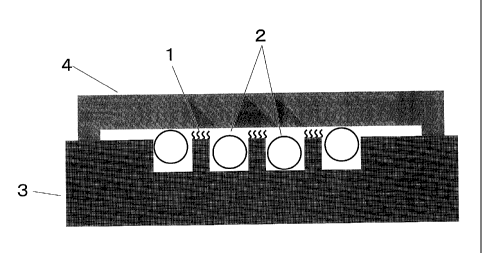
CA 02559768 2006-09-14
12
In Figure 1, reference numeral 1 denotes a probe DNA (selective binding
substance).
Reference numeral 2 denotes fine particles (beads in this case), and reference
numeral 3
denotes a probe DNA-immobilized carrier. Those components denoted by reference
numerals 1, 2, and 3 become in contact with a solution containing a target DNA
(analyte
substance). Reference numeral 4 denotes a container holding liquid, for
example made of
a material such as slide glass, cover glass, metal, or plastic, and the target
DNA-containing
solution is held between the container and the carrier. In the example of
Figure 1, the
probe DNA is immobilized on the convexes of the carrier. The minimum distance
between the top face of the convexes on carrier (selective binding substance-
immobilized
face) and the solution-containing container is smaller than the diameter of
the fine particles,
preventing contact of the fine particles with the probe DNA-immobilized face
and damage
of the face by the fine particles. When the fine particles are, for example,
elliptical in
shape, and when the minimum distance between the top face of the convexes and
the
container is smaller than the minimum width of the fine particles, it is
possible to prevent
contact of the probe-immobilized face with the fine particles.
A condition to realize the situation of Figure 1 concretely is placing a
solution
containing an analyte DNA (analyte solution) dropwise on a carrier which has
convex-concave structure, adding fine particles into the solution while
preventing
deposition thereof on the top face of the convexes, covering it with a cover
glass equivalent
to container, and sealing the cover glass, for example, with an adhesive tape
or agent for
prevention of spill or vaporization of the analyte solution. In this way,
there is formed a
space containing the analyte solution having a thickness of from several to
dozens of um
between the cover glass face and the top face of the convexes. When the size
of the fine
particles is greater than the distance between the cover glass face and the
top face of the
convexes, the fine particles do not damage the top face of the convexes. It is
possible to
guide the fine particles to pass through only the concave area in the convex-
concave area
and stir the analyte solution without the fine particles becoming in contact
with the top face
of the convexes, for example, by using a carrier in such a shape and rotating
the carrier in a
vertical plane. Preferably to form the space containing an analyte solution
between the
CA 02559768 2006-09-14
13
top face of the convexes and the container reliably, for example, a plate
having the corners
on the plate face higher by 5 to 100 pm than the other face or a plate having
its central area
lowered by 5 to 100 pm is used, and the plate is connected to the selective
binding
substance-immobilized carrier with the central area of the plate facing the
convex-concave
area of the carrier. An example of the plate is shown in Figure 1(4). Such a
container
can be prepared, for example, by treating glass with hydrofluoric acid,
bonding a film or
adhesive tape at 2 to 4 corners of a flat plate, preparing a plate having the
shape shown in
Figure 1(4), for example by injection molding, or forming raised dots at the
corners of a
plate by screen printing.
In the method of stirring a solution according to the present invention, a
container
holding solution in such a structure that the fine particles or air bubbles do
not become in
contact with the selective binding substance-immobilized surface is preferably
used.
The carrier in Figure 1 has convex-concave shape. It is also possible to
obtain a
similar effect by forming convex-concave structure on the container for
solution. A
typical example thereof is shown in Figure 2. In such a case, a probe DNA is
placed
under the container convexes. In this case too, the distance between the probe
DNA-immobilized face and the container convexes is preferably smaller than the
minimum width of the fine particles. Another typical example thereof is the
case where
both the carrier and the container have an convex-concave structure.
It is possible to obtain the following effects and consequently to increase
the intensity
of the fluorescence after hybridization more than that in conventional
methods, by stirring
a target DNA-containing analyte solution with fine particles while using the
carrier having
a convex-concave area or the container having a convex-concave area.
When hybridization is performed in combination of a common flat plate-shaped
DNA
chip and a cover glass placed thereon, the distance between the cover glass
and the DNA
chip is approximately 10 pm at most. Use of fine particles larger in diameter
results in
clogging between the DNA chip and the cover glass, causing problems such as
prohibition
of movement of the fine particles and decrease in the advantageous effects of
mixing fine
particles. On the other hand, use of finer particles having a diameter of
several pm, which
CA 02559768 2006-09-14
14
is resistant to clogging between the cover glass and the DNA chip, for
prevention of
clogging and for movement of the fine particles by acceleration by gravity or
vibration,
only results in insufficient movement of the fine particles in the analyte
solution, because
the solution resistance becomes greater as the fine particles become smaller
in size.
Accordingly, there is still a problem that it is not possible to obtain
favorable stirring effect
with fine particles. Alternatively, expansion of the distance between the
cover glass and
the carrier for example with an 0-ring and use of larger fine particles for
sufficiently
stirring cause the problem that the fluorescence intensity after hybridization
deteriorates,
presumably because the chip surface is damaged by the fine particles and the
probe on the
probe-immobilized surface is separated by collision of the fine particles.
As in the favorable embodiments of the present invention, it is possible to
increase the
size of fine particles at least up to the height of the concave area and
convex in the
convex-concave area, as shown in Figures 1 and 2, by using a carrier having a
convex-concave area or a container having a convex-concave area. Thus, it is
possible to
obtain advantageous effects allowing sufficient stirring of the analyte
solution with larger
fine particles and preventing damage on the probe DNA-immobilized face, by
stirring a
target DNA-containing analyte solution with fine particles while using a
carrier having a
convex-concave area or a container having a convex-concave area.
In the method of stirring solution according to the present invention, the
material for
the container favorably used is not particularly limited. Examples of the
materials for the
container favorably used in the present invention include glass, plastics, and
the like.
When the shape of the container is flat plate, a glass plate such as cover
glass or slide glass
is favorably used, and when the container has convex-concave shape, a plastic
material
such as polymethyl methacrylate or polycarbonate, which is injection moldable,
is
preferable from a point of productivity.
The material for the carrier for use in the present invention is not
particularly limited.
Favorable materials for the carrier include glass and various polymers
(polystyrene,
polymethyl methacrylate, and polycarbonate).
When the carrier material is glass, for immobilization of a selective binding
substance,
CA 02559768 2006-09-14
the carrier may be treated with a silane coupling agent for generation of
functional groups
on the surface and the selective binding substance such as DNA may be
immobilized on
the carrier by using the functional groups. It is possible to form amino
groups on the
surface of glass by using, for example, an aminoalkylsilane, and to
immobilize, for
example, DNA thereon by the electrostatic force between the plus charge of the
amino
group and the minus charge of DNA.
In the present invention, use of a solid material containing a polymer having
a
structural unit represented by the following General Formula (1) particularly
as the carrier
surface for immobilization of a selective binding substance is advantageous,
because the
signal after hybridization becomes greater.
[Formula 1]
R1
CH2 C __
C=0
(1)
X X=0, NR3, CH2
R2
In General Formula (1), Rl, R2, and R3 each represent an alkyl or aryl group
or a
hydrogen atom. The polymer having a structural unit represented by the
following
General Formula (1) used is a homopolymer or a copolymer. At least one type of
monomer is used as the raw material for the polymer, and the monomer is
present as a
double bond for polymerization or a functional group for polycondensation,
ketone or
carboxylic acid or the derivative thereof. The polymer more preferably has the
structure
represented by General Formula (1).
When the polymer having a structural unit represented by the following General
Formula (1) is a copolymer, the polymer preferably contains the structural
unit represented
by the following General Formula (1) in an amount of 10% or more with respect
to the
total amount of all monomers. When the content of the structural unit
represented by
General Formula (1) is 10% or more, it is possible to form more carboxyl
groups on the
surface and immobilize more probe nucleic acids in the steps described below,
leading to
CA 02559768 2006-09-14
16
improvement in the S/N ratio.
The polymer in the present invention is a compound having a number-averaged
polymerization degree of 50 or more. The number-averaged polymerization degree
of the
polymer is preferably in the range of 100 to 10,000, particularly preferably
200 or more
and 5,000 or less. The number-averaged polymerization degree can be determined
easily
by measuring the molecular weight of a polymer according to a common method by
GPC
(gel permeation chromatography).
In General Formula (1), R1 and R2 each represent an alkyl or aryl group or a
hydrogen
atom, and may be the same as or different from each other. The alkyl group may
be a
straight-chain or branched group, and preferably has a carbon number of 1 to
20. The
aryl group preferably has 6 to 18 carbon atoms, more preferably 6 to 12 carbon
atoms.
The functional group X is selected arbitrarily from 0, NR3, and CH2. R3 is a
functional
group defined similarly to R1 and R2.
In the present invention, the polymer on the carrier surface for
immobilization of a
selective binding substance is preferably a polymer having a functional group.
Favorable
examples of the polymers having a functional group include polyalkyl
methacrylates
(PAMA) such as polymethyl methacrylate (PMMA), polyethyl methacrylate (PEMA)
and
polypropyl methacrylate, and the like. Among them, particularly preferable is
polymethyl
methacrylate. Alternatively, polyvinyl acetate, polycyclohexyl methacrylate or
polyphenyl methacrylate, or the like may also be used. Yet alternatively, a
copolymer in
combination of the polymer components above or a copolymer in combination of
the
polymer components and one or more other polymer components may also be used.
The
other polymers include polystyrene.
When the polymer is a copolymer, the rate of a carbonyl group-containing
monomer,
for example alkyl methacrylate, is preferably 10 mol % or more in all
components. It is
because it is possible in this way to form a greater number of carboxyl groups
on the
surface, to immobilize a greater amount of a probe nucleic acid, and
consequently to
improve the S/N ratio. The ratio of the monomer in the polymer structural
units is more
preferably 50 mole % or more.
CA 02559768 2006-09-14
17
For immobilization of a selective binding substance on a carrier containing a
polymer
having at least one structural unit represented by the following General
Formula (1), it is
preferable to pre-treat the carrier, forming carboxyl group on the carrier
surface. The
methods of forming carboxyl groups on the carrier surface include alkali or
acid treatment,
ultrasonication in hot water, exposure of the carrier to oxygen plasma, argon
plasma, or
radiation ray, and the like; but immersion of the carrier in alkali or acid
for generation of
surface carboxyl groups is preferable from the points of smaller damage on
carrier and
productivity. More specifically, the support may be immersed in an aqueous
sodium
hydroxide or sulfuric acid solution @referable concentration: 1 to 20N)
preferably at a
temperature of 30 C to 80 C for 1 to 100 hours.
A thermoplastic copolymer containing an acid anhydride unit may be used as the
polymer. The thermoplastic copolymer preferably has an acid anhydride unit
(i). The
acid anhydride unit (i) is a unit present on the skeletons of the main and
side chains or at
the terminals of a thermoplastic copolymer (A). The structure of the acid
anhydride unit
(i) is not particularly limited, and examples thereof include (meth)acrylic
anhydride unit,
glutaric anhydride unit, maleic anhydride unit, itaconic anhydride unit,
citraconic
anhydride unit, aconitic anhydride unit and the like; maleic and glutaric
anhydride units are
preferable; and among them, a glutaric anhydride unit represented by the
following
General Formula (2) is particularly preferable.
[ Formula 2]
R4 . 5
(2)
(in the Formula, R4 and R5 each independently represent a hydrogen atom or an
alkyl
group having 1 to 5 carbon atoms).
CA 02559768 2006-09-14
18
The structure of the thermoplastic copolymer is not particularly limited, if
it has an
acid anhydride unit (i), but the copolymer preferably contains an unsaturated
carboxylic
acid group (ii) represented by the following General Formula (3).
Formula 3]
R6
(3)
coo
(wherein, R6 represents a hydrogen atom or an alkyl group having 1 to 5 carbon
atoms)
The unsaturated carboxylic acid unit (ii) is an unit obtained by
copolymerization of an
unsaturated carboxylic acid monomer, and the unsaturated carboxylic acid
monomer used
then is not particularly limited, and any unsaturated carboxylic acid monomer
copolymerizable with other vinyl compound may be used. Favorable unsaturated
carboxylic acid monomers include the compounds represented by the following
General
Formula (4):
[ Formula 4]
R6
CH2=- (4)
COOH
(wherein, R6 represents a hydrogen atom or an alkyl group having 1 to 5 carbon
atoms),
maleic acid, and the hydrolysate of maleic anhydride, and the like; acrylic
acid and
methacrylic acid are preferable, and methacrylic acid is more preferable, from
the point of
heat stability. These monomers may be used alone or in combination of two or
more.
The thermoplastic copolymer (A) is not particularly limited, if it contains an
acid
anhydride unit (i), but preferably contains an unsaturated alkylcarboxylate
esher unit (iii)
CA 02559768 2006-09-14
19
represented by the following General Formula (5):
(Formula 5]
14 7
( C H2 _____________ (5)
cOOR
(wherein, R7 represents a hydrogen atom or an alkyl group having 1 to 5 carbon
atoms; R8
represents an aliphatic or alicyclic hydrocarbon group having 1 to 6 carbon
atoms or an
aliphatic or alicyclic hydrocarbon group having 1 to 6 carbon atoms
substituted with at
least one hydroxyl group or halogen atom).
The unsaturated alkylcarboxylate ester unit (iii) is an unit obtained by
copolymerization of
an unsaturated alkylcarboxylate ester monomer, and the unsaturated alkyl
carboxylate ester
monomer is not particularly limited, and examples thereof include the
following
compounds represented by General Formula (6):
Formula 6]
127
(6)
cooR8
Presence of the carboxyl groups and the acid anhydrides on the carrier surface
enables
immobilization of a selective binding substance having an amino group or a
hydroxyl
group on the carrier surface by covalent bonding. When there are carboxyl
groups on the
carrier surface, various condensing agents such as dicyclohexylcarbodiimide
and
N-ethyl-5-phenylisoxazolium-3'-sulfonate are used for acceleration of the
reaction of these
groups. Among them, 1-ethy1-3-(3-dimethylaminopropyl) carbodiimide (EDC),
which is
CA 02559768 2006-09-14
less toxic and easily removed from the reaction system, is one of the
condensation agents
most effective for the condensation reaction of a selective binding substance
with the
carboxyl groups on the support surface. The condensation agent, for example
EDC, may
be used as it is mixed into a solution of the selective binding substance, or
alternatively, a
support carrying carboxyl groups previously formed on the surface is immersed
in a
solution of EDC and thus the surface carboxyl groups are activated. Use of the
condensing agent, which is used as mixed with a solution of a selective
binding substance,
is advantageous, because it is effective in increasing the reaction yield and
immobilizing a
greater amount of the selective binding substance on carrier.
When the carboxyl groups on support surface are reacted with the amino group
of a
selective binding substance by using such a condensation agent, the selective
binding
substance is immobilized on the support surface by amide bond, while when the
carboxyl
groups on the support surface are reacted with the hydroxyl group of a
selective binding
substance, the selective binding substance is immobilized on the support
surface by ester
bond. The temperature when a sample containing a selective binding substance
is
allowed to react with the carrier is preferably 0 to 95 C and more preferably
15 C to 65 C.
The processing period is normally 5 minutes to 24 hours and preferably 1 hour
or more.
On the other hand, if the polymer has acid anhydride groups on the surface,
the acid
anhydride groups react with, for example, the amino group of the selective
binding
substance, forming covalent bonds, with or without such a condensing agent
added.
Thus by immobilizing a selective binding substance preferably on the polymer
surface
it is possible to reduce non-specific adsorption of analyte, immobilize the
selective binding
substance covalently, tightly and at high density, and obtain a carrier higher
in the
hybridization efficiency with the analyte, presumably because the spatial
degree of
freedom of the immobilized selective binding substance is higher than that of
the substance
immobilized on glass.
When the carrier is prepared with a polymer containing a structural unit
represented
by General Formula (1) or (2), it is possible to produce fine concave-convex
structured
carrier more simply in a greater amount, for example by injection molding or
hot
CA 02559768 2006-09-14
21
embossing, than when the carrier is prepared with glass, ceramic, metal, or
the like. In
particular, injection molding, which allows easier mass production, is used
favorably.
By immobilizing a selective binding substance on the polymer surface of the
carrier
favorably used in the present invention according to the method described
above, it is
possible to immobilize a selective binding substance covalently, tightly and
at high density
while reducing non-specific adsorption of the analyte. It is possible to
obtain a carrier
higher in hybridization efficiency with the analyte, presumably because the
spatial degree
of freedom of the immobilized selective binding substance is higher than that
formed on
glass.
The support carrying an immobilized selective binding substance thus obtained
may
be treated additionally after immobilization of the selective binding
substance. It is
possible, for example, to modify the immobilized selective binding substance
by treatment
such as heat treatment, alkali treatment, or surfactant treatment.
It is common that by using the selective binding substance-immobilized
carrier, a
fluorescent-labeled analyte and a carrier-immobilized selective binding
substance are
allowed to react in hybridization reaction, and the fluorescence from the
product is
determined in a device called scanner. The scanner deflects an excitation
laser beam with
an object lens and focuses the laser beam. However, when there is
autofluorescence of
the surface of the support, the fluorescence may cause noise and lead to
deterioration in
detection accuracy. For
prevention thereof and also for prevention of the
autofluorescence of the carrier itself, it is preferably to make the surface
of the polymer
having a structural unit represented by General Formula (1) or (2) appear
black in color, by
adding a black substance that does not emit light by laser irradiation. It is
possible to
reduce the autofluorescence of the carrier during detection, by using such a
black carrier.
The black carrier gives a favorable selective binding substance-immobilized
carrier lower
in noise and thus higher in S/N ratio.
The blackened support means a support of which the blackened area has a
uniformly
low spectroscopic reflectance not in a particular spectral pattern (e.g.,
without any
particular peaks) and a uniformly low spectroscopic transmissibility not in a
particular
CA 02559768 2006-09-14
22
spectral pattern in the visible light range (wavelength: 400 to 800 nm).
In the present invention, the carrier has preferably a spectroscopic
reflectance of 7%
or less in the wavelength range of visible light (wavelength: 400 nm to 860
nm) and
preferably a spectroscopic transmissibility of 2% or less in the same
wavelength range.
The spectroscopic reflectance is a spectroscopic reflectance including the
regular reflected
light from the support, as determined in an optical illuminator-detector
system compatible
with the condition C of JIS Z8722.
In the present invention, the support may be blackened by adding a black
substance to
the support, and favorable examples of the black substances include carbon
black, graphite,
titanium black, aniline black, oxides of metals such as Ru, Mn, Ni, Cr, Fe, Co
and Cu,
carbides of metals such as Si, Ti, Ta, Zr and Cr, and the like. Among the
black substances,
carbon black, graphite, titanium black are preferably contained; and carbon
black is used
particularly preferably, because it is easily dispersed uniformly in polymer.
These black substances may be contained alone or in combination of two or
more.
As for the shape of the carrier in the present invention, a selective binding
substance-immobilized layer of a polymer having at least one structural unit
represented by
the following General Formula (1) formed on a support layer resistant to heat
deformation
such as of glass or metal is preferable, because it is effective in preventing
deformation of
the carrier by heat or external force. An example of such structure is shown
in Figure 6.
Polypropylene, glass, or a metal such as iron, chromium, nickel, titanium, or
stainless steel
is preferable for the support layer. In addition, the surface of the support
layer is
preferably finished in a plasma treatment with argon, oxygen, or nitrogen gas
or treated
with a silane-coupling agent, for improvement in adhesion between the support
layer and
the layer carrying an immobilized selective binding substance. Examples of the
silane-coupling agents include 3 -
aminopropyltriethoxysilane,
3 -aminopropyltrimethoxysilane, 3 -
aminopropyldiethoxymethyl silane,
3 -(2-aminoethylaminopropyl) trimethoxysilane, 3 -(2-
aminoethylaminopropyl)
dimethoxymethylsilane, 3 -
mercaptopropyltrimethoxysilane,
dimethoxy-3-mercaptopropylmethylsilane, and the like.
CA 02559768 2006-09-14
23
A layer carrying an immobilized selective binding substance is formed on the
support layer by any one of known means, for example, by spin coating with or
dipping in
a solution of a polymer dissolved in an organic solvent. More conveniently,
the layer
carrying an immobilized selective binding substance may be adhered to the
support with an
adhesive.
In the present invention, the "selective binding substance" means a substance
that can
selectively bind to an analyte substance directly or indirectly, and typical
Examples thereof
include nucleic acids, proteins, saccharides, and other antigenic compounds.
Particularly preferable as the "selective binding substances" is a nucleic
acid. The
nucleic acid may be DNA, RNA, or PNA. Single strand nucleic acids having a
particular
base sequence selectively hybridizes with and binds to a single strand nucleic
acid having
the base sequence complementary to the base sequence or the part thereof, and
thus are
included in the "selective binding substances" according to the present
invention.
Examples of the proteins include antibodies, antigen-binding antibody
fragments such
as Fab fragments and F (ab') 2 fragments, and various antigens. Antibodies and
their
antigen-binding fragments that selectively bind to respective complementary
antigens and
antigens that selectively bind to respective complementary antibodies are also
included in
"selective binding substances". Polysaccharides are preferably as the
saccharides, and
examples thereof include various antigens.
Alternatively, an antigenic substance other than protein or saccharide may be
immobilized.
The selective binding substance for use in the present invention may be a
commercially available product or a substance prepared from living cell or the
like.
The selective binding substance for use in the present invention is preferably
a nucleic
acid, and among nucleic acids, preferable are nucleic acids having a length of
10 to 100
bases called oligonucleic acids, which are easily prepared in synthesizer and
allows
modification of the amino group on the nucleic acid terminal for
immobilization thereof on
the carrier surface. Further, the length of the oligonucleic acid is
preferably 20 to 100
bases, because the hybridization efficiency is lower with an oligonucleic acid
having less
CA 02559768 2006-09-14
24
than 20 bases, and particularly preferably in the range of 40 to 100 bases,
for ensuring the
stability of hybridization efficiency.
Examples of the analyte substances to be processed in the method of stirring a
solution
according to the present invention include, but are not limited to, nucleic
acids to be
evaluated, such as genes of pathogenic bacteria and viruses and causative
genes of genetic
diseases, or the partial region thereof; various antigenic biological
components; antibodies
to pathogenic bacteria and viruses; and the like.
In the method of stirring a solution according to the present invention,
examples of the
samples containing the analyte substances above include, but are not limited
to, body fluids
such as blood, serum, blood plasma, urine, feces, spinal fluid, saliva, and
various tissue
fluids and various foods and drinks or diluents thereof, and the like.
In addition, the analyte nucleic acid may be prepared by labeling a nucleic
acid
extracted from blood or cell according to a common method or by amplifying the
nucleic
acid by a nucleic acid-amplifying method such as PCR by using it as a
template. It is
possible to improve measurement sensitivity drastically, when an analyte
prepared by a
nucleic acid-amplifying method such as PCR using a nucleic acid as a template
is used.
When an amplified nucleic acid product is used as the analyte substance, it is
possible to
label the amplified nucleic acid by performing amplification in the presence
of a
nucleotide-3-phosphate labeled with a fluorescent material or the like. When
the analyte
substance is an antigen or antibody, the analyte substance, antigen or
antibody, may be
directly labeled by a common method, or alternatively, the analyte substance,
antigen or
antibody, may be first bound to a selective binding substance; after washing
of the support,
the antigen or antibody is allowed react with a labeled antibody or antigen
that reacts in the
antigen-antibody reaction; and then, the labels bound to the support is
analyzed.
Preferably in the method of stirring a solution according to the present
invention, a
selective binding substance is allowed to react with an analyte substance.
The step of allowing an immobilized substance to react with an analyte
substance in
the method of stirring a solution according to the present invention may be
performed
entirely, similarly to that in conventional methods. The reaction temperature
and period
CA 02559768 2006-09-14
may be selected arbitrarily, for example, according to the chain length of the
nucleic acid
to be hybridized and the kinds of the antigen and/or the antibody involved in
the immune
reaction, but the reaction is generally carried out at approximately 35 C to
70 C
approximately for 1 minute to more than ten hours in the case of nucleic acid
hybridization,
and generally, at room temperature to approximately 40 C for approximately 1
minute to
several hours in the case of immune reaction.
The method of stirring a solution according to the present invention was found
to have
the following advantages, in addition to the improvement in intensity of the
signal after
hybridization. Conventional methods of DNA-chip hybridization caused a problem
of
difficulty in data analysis, because the fluorescence intensity after
hybridization is lower
and distribution of fluorescence intensity on the spot where a probe DNA was
immobilized
is donut-shaped. However, the method of stirring a solution according to the
present
invention has an advantage that it improves the fluorescence intensity
drastically and
prevents the donut-shaped distribution of the fluorescence intensity on the
spot.
EXAMPLES
The present invention will be described in more detail with reference to the
following
Examples. It should be understood that the present invention is not restricted
by the
following Examples.
Example 1
(Preparation of DNA-immobilized support)
A mold for injection molding was prepared according to a known LIGA
(Lithographie
Galvanoformung Abformung) process, and a PMMA substrate having the shape
described
below was prepared by injection molding. The PMMA used in this Example had an
average molecular weight of 50,000 and contained carbon black (#3050B,
manufactured
by Mitsubishi Chemical Corp.) at a ratio of 1 wt %, and the substrate was
black in
appearance. When the spectroscopic reflectance and transmissibility of the
black
substrate were determined, the spectroscopic reflectance was 5% or less at a
wavelength in
the visible light range (wavelength: 400 to 800 nm), and the transmissibility
was 0.5% or
less at a wavelength in the same range. The substrate had a uniformly flat
spectrum
CA 02559768 2006-09-14
26
without a particular spectral pattern (e.g., peaks) both in spectroscopic
reflectance and
transmissibility in the visible light range. The spectroscopic reflectance is
a spectroscopic
reflectance including regular reflectance from the support, as determined by
using a device
equipped with an optical illuminator-detector system (CM-2002, manufactured by
Minolta
Camera) compatible with the condition C of JIS Z 8722.
The shape of support was 76 mm in length, 26 mm in width, and 1 mm in
thickness,
and the surface was flat except in the central area of the substrate. A
recessed area of 10
mm in diameter and 0.2 mm in depth 0.2 mm is formed on the center of the
carrier, and
64 (8x8) convexes having a top face diameter of 0.2 mm and a height of 0.2 mm
were
formed in the recess. The difference between the height of convex top face
(average of
the heights of 64 convexes) in the convex-concave part and the height of the
flat area was 3
pm or less, when determined. In addition, the variation in height of the 64
convex top
faces (difference in height between the highest and the lowest convex top
faces), and the
difference between the height of convex top face in the convex-concave
surfaced area and
the height of the flat area, when determined, were both 3 pm or less. Further,
the pitch of
the convexes in the convex-concave surfaced area (distance between a convex
center to
another convex center next to it) was 0.6 mm.
The PMMA carrier was immersed in aqueous 10 N sodium hydroxide solution at 65
C
for 12 hours. The carrier was washed with purified water, aqueous 0.1 N HC1
solution,
and purified water in that order, forming carboxyl groups on the carrier
surface.
(Immobilization of probe DNA)
A DNA having the sequence shown by sequence number 1 (60 base, 5' terminal
aminated) was prepared. The DNA having the sequence of sequence number 1 has
an
aminated 5'-terminal.
The DNA was dissolved in purified water to a concentration of 0.3 nmol/gl, to
give a
stock solution. For spotting on the carrier, prepared was a solution of the
probe diluted
with PBS (a solution of 8 g of NaCl, 2.9 g of Na2HPO4-12H20, 0.2 g of KC1, and
0.2 g of
KH2PO4 dissolved in 1 L of purified water containing hydrochloric acid for pH
adjustment,
pH: 5.5) to a final concentration of 0.03 nmol/gl, containing additionally
CA 02559768 2006-09-14
27
1-ethy1-3-(3-dimethylaminopropyl)carbodiimide (EDC) at a final concentration
of 50
mg/ml, for condensation of the carboxyl groups on the carrier surface with the
terminal
amino group of the probe DNA. The mixture solution was then spotted on the top
face of
the convexes of the carrier with a glass capillary. The carrier was then
placed in a tightly
sealed plastic container, incubated under the condition of 37 C and a humidity
of 100% for
approximately 20 hours, and the washed with purified water.
Figure 7 shows the reaction scheme.(Preparation of sample DNA)
A DNA having a sequence of sequence number 4 (968 bases), which hybridizes
with
the probe DNA immobilized on the DNA-immobilized carrier, was used as the
analyte
DNA. The preparative method is as follows:
DNA's of sequence Nos. 2 and 3 were prepared. These DNA's were respectively
dissolved in purified water to a concentration of 100 pM. The DNA was
amplified in
PCR reaction (Polymerase Chain Reaction) by using a plasmid DNA (Takara Bio
Inc.,
product number: 3100), (sequence number 5: 2264 base) as the template and the
DNAs
having sequences of sequence numbers 2 and 3 as the primers.
The PCR condition is as follows: ExTaq (2 I), 10xExBuffer (40 pl), and dNTp
Mix
(32 pl) (these reagents were attached to the Product Number RROO1A
manufactured by
Takara Bio Inc.), a solution of sequence No. 2 (2 pl), a solution of sequence
No. 3 (2 pl),
and a solution of template (sequence No. 5) (0.2 pl) were mixed and diluted
with purified
water to a total volume of 400 1. The liquid mixture was divided into four
micro tubes,
and the PCR reaction was performed by using a thermal cycler. The product was
purified
by ethanol precipitation and dissolved in 40 pl of purified water.
Electrophoretic analysis
of part of the solution after PCR reaction confirmed that the base length of
the amplified
DNA was approximately 960 bases and the DNA of sequence No. 4 (968 bases) was
amplified.
Then, a 9-base random primer (manufactured by Takara Bio Inc., product number:
3802) was dissolved to a concentration of 6 mg/ml, and 2 IA thereof is added
to the DNA
solution purified after the PCR reaction.
The solution was heated at 100 C and quenched on ice. 5 1 of the buffer
attached to
CA 02559768 2006-09-14
28
Klenow Fragment (manufactured by Takara Bio Inc., Product Number 2140AK) and
2.5 pA
of a dNTP mixture (containing dATP, dTTP, and dGTP each at a concentration of
2.5 mM
and dCTP at a concentration of 40011M) were added thereto. Further, 2 [11 of
Cy3-dCTP
(manufactured by Ainersham Pharmacia Biotech, Product Number PA53021) was
added.
After addition of 10U of Klenow Fragment to the solution, the mixture was
incubated at
37 C for 20 hours, to give a Cy3-labeled sample DNA. Use of the random primer
during
labeling resulted in fluctuation in the length of the sample DNA. The longest
sample
DNA is the DNA of sequence No. 4 (968 bases). Electrophoretic analysis of part
of the
sample DNA solution showed the most intensive band in the area approximately
corresponding to 960 bases and bands slightly smeared in the area
corresponding to shorter
base lengths. The product was then purified by ethanol precipitation and
dried.
The labeled analyte DNA was dissolved in 400 ill of a solution containing 1 wt
%
BSA (bovine serum albumin), 5xSSC (5xSSC is a solution 20xSSC (manufactured by
sigma) diluted four times with purified water, 10xSSC is a solution of 20xSSC
diluted
twice with purified water, 20xSSC diluted twice is 10xSSC, that diluted 100
times is
0.2xSSC), 0.1 wt % SDS (sodium dodecylsulfate), and 0.01 wt % salmon sperm DNA
solution (concentrations above; final concentrations), to give a stock
solution for
hybridization.
In the following Examples and Comparative Examples, the stock solution above
diluted 200 times with 1 wt % BSA, 5xSSC, 0.01 wt % salmon sperm DNA, and 0.1
wt %
SDS solution (all, final concentrations) was used as the analyte solution
during
hybridization, unless specified otherwise. The concentration of the analyte
DNA in the
solution was determined to be 1.5 ng/ 1.
(Surface-modification of glass beads)
g of glass beads having a diameter of 150 [..tm were immersed in 10 N NaOH
solution and then, washed with purified water. Then, APS (3-
aminopropyltriethoxysilane;
manufactured by Shin-Etsu Chemical Co., Ltd.) was dissolved in water to a
concentration
of 2 wt %, and the glass beads were immersed therein for 1 hour, and, after
removal, dried
at 110 C for 10 minutes. In this way, amino groups were introduced on the
surface of the
CA 02559768 2006-09-14
29
glass beads.
Then, 5.5 g of succinic anhydride was dissolved in 335 ml of 1-methyl-2-
pyrrolidone.
50 ml of 1 M sodium borate (containing 3.09 g of boric acid and sodium
hydroxide for pH
adjustment in 50 ml of purified water, pH: 8.0) was added to the succinic acid
solution.
The glass plate above was immersed in the liquid mixture for 20 minutes. After
immersion, the glass plate was washed with purified water and dried. In this
manner,
amino groups on the glass plate surface and succinic anhydride were allowed to
react with
each other, introducing carboxyl groups on the glass surface.
(Hybridization)
The analyte DNA was applied on the probe DNA-immobilized carrier obtained
above,
for hybridization. Specifically, 50 pJ of the solution for hybridization was
applied
dropwise onto the carrier carrying the probe nucleic acid immobilized on the
convexes
prepared above; 2 mg of the surface-modified glass beads were added to the
concave area
of the carrier ; and the support was covered with a cover glass. In addition,
the cover
glass was sealed with a paper bond, for preventing vaporization of the
hybridization
solution. A cover glass carrying photoresists having a thickness of 8 [tm and
a width of 1
mm formed by photolithography on two opposing sides among four sides was used.
In
this way, the distance (gap) between the carrier convex and the cover glass
was kept 8 pm
during hybridization. It was fixed in a plastic container on the revolving
plate of a
microtube rotator (manufactured by As One, product number: 1-4096-01), and
incubated
under the condition of 65 C and a humidity of 100% for 10 hours. The
rotational
frequency of the rotator then was 3 rpm, and the angle was in the direction
perpendicular to
the revolving plate of the rotator. In addition, the probe DNA-immobilized
face of the
carrier was placed in the direction perpendicular to the revolving plate of
the rotator.
After incubation, the cover glass was removed from the carrier, and the
carrier was washed
and dried.
(Measurement)
The carrier after treatment was placed in a scanner for DNA chip
(GenePix4000B,
manufactured by Axon Instruments), and the fluorescence intensity therefrom
was
CA 02559768 2006-09-14
determined under the conditions of a laser output of 33% and a photomultiplier
gain of 500.
The results are summarized in Table 1. The fluorescence intensity is an
average of the
fluorescence intensity in the spot.
Although glass beads were used in the present Example, results similar to
those in
Table 1 were obtained when ceramic beads or Teflon (registered trademark)
beads were
used.
Comparative Example 1
An experiment was performed without added glass beads. The experiment
procedure
was similar to that in Example 1, except that no glass bead was mixed during
hybridization.
Results are summarized in Table 1.
It was found that the fluorescence intensity was lower than that in Example 1.
In
addition, the fluorescence intensity distribution on carrier convexes was
uneven
(donut-shaped) in Comparative Example 1, but the fluorescence intensity
distribution on
carrier convexes was almost uniform in Example 1.
Comparative Example 2
An experiment was performed with a flat PMMA carrier having no convex-concave
area. The experiment procedure was similar to that in Example 1, except that
(1) a flat
carrier was used, (2) a probe DNA was spotted in a special-purpose machine
(Gene Stamp
II, manufactured by Nippon Laser & Electronics Co., Ltd.), and (3) an opening
for bead
stirring between the carrier and the cover glass was formed by bonding a
polyester film
having a thickness of 200 pm and a width of 1 min on four sides of the cover
glass and
beads and an analyte solution were mixed in the opening for hybridization.
Comparison
with the results in Example 1 reveals that the fluorescence intensity is
lower. Results are
summarized in Table 1. It was also confirmed that there was damage on the spot
that was
not found in Example 1. It seemed that the beads were the cause of the damage
on the
probe-immobilized face during hybridization.
When the experiment was repeated in another Comparative Example after the
diameter of the beads was changed to 1 pm, the fluorescence intensity was
further lower at
approximately 1,500. Apparently, it is because of the phenomenon that the
beads were
CA 02559768 2006-09-14
31
less mobile by resistance of the hybridization solution.
Example 2
An experiment on stirring efficiency was performed by using air bubbles. The
experiment procedure was similar to that in Example 1, except that 0.9 jtL of
air bubble
was injected with a microsyringe, instead of adding glass beads, when a cover
glass is
placed in the hybridization step. The carrier was fixed in such a direction
that the
revolving plate of rotator tilted into the vertical direction and the probe-
immobilized face
of the carrier became in parallel, and rotated, allowing air bubbles to
migrate only around
the sealed analyte solution. In this way, air bubbles were kept separated from
the
probe-immobilized face. Results are summarized in Table 1. Advantageous
effects
similar to those in Examples were observed.
Example 3
An experiment similar to Comparative Example 2 was performed, by using a
container for solution 4 having the crosssectional structure shown in Figure 2
instead of a
glass cover. That is, the convex-concave structure was formed not on the
carrier but on
the cover. The container and the flat PMMA carrier were placed carefully in
the spatial
relationship shown in Figure 2. In subsequent hybridization while the beads
are agitated,
it was possible to move the glass beads without contact thereof with the probe
DNA-immobilized face 1, because the distance between the probe DNA-immobilized
face
and the container for solution 4 was smaller than the diameter of the glass
beads 2.
Results are summarized in Table 1. The fluorescence intensity obtained was
similar to
that in Example 1. Considering the results in Comparative Example 2 as well,
it seems
important that the beads do not become in contact with the probe-immobilized
face. In
the method of the present Example, accurate positioning of the cover convexes
and the
probe-immobilized region is important.
Comparative Example 3
An experiment was performed without stirring with beads by using a flat PMMA
carrier having no concave-convex area. The experiment was done by operation
and
measurement in a similar manner to Comparative Example 2, except that no bead
was
CA 02559768 2006-09-14
32
added to the hybridization solution and there was no rotation. Results are
summarized in
Table 1.
Table 1.
Comparative Comparative Comparative
Example 1 Example 2 Example 3
Example 1
Example 2 Example 3
Target concentration
1.5 1.5 1.5 1.5 1.5 1.5
(ng/uL)
Substrate shape convex- convex- convex-
Flat plate Flat plate Flat
plate
concave concave concave
Gap (p.m) 8 8 8 8 200 8
Stirring method Bead Air bubble Bead No Bead No
Rotation Yes Yes Yes Yes Yes No
Fluorescence
12000 8800 11800 2900 3900 700
intensity
Noise 45 50 300 50 300 300
Example 4
Five kinds of glass beads in Example 1 were used in the experiment. The
experiment
procedure was similar to that in Example 1, and the diameter of the glass
beads used was
10, 20, 50, 100, or 200 jim. Results are summarized in Table 2.Comparative
Example 4
An experiment was performed in a similar manner to Example 1, except that the
diameter of the glass beads was changed to 300 1.1m or 400 j.im. The results
are
summarized in Table 2.
Table 2
Comparative
Example 4
Example 4
Size (um) 10 20 50 100 200 300 400
Fluorescence intensity 8200 10500 12700 12000 12500
3100 3000
As apparent from the table, beads of 10 to 200 i.tm in diameter were
distinctively
higher in stirring efficiency, but the beads of Comparative Example 4 of 300
or 400 lam in
diameter were not distinctively effective. Because the distance between the
carrier
concave and the cover glass was 208 pin, it seems that the beads of 300 or 400
pm forcibly
added could not migrate as they are held in the opening between the cover
glass and the
CA 02559768 2006-09-14
33
carrier. The results in Examples and Comparative Examples reveal that
difficulty in bead
movement leads to decrease in fluorescence intensity. In addition, the results
in Example
4 indicates that the bead size is preferable 10 gm or more and more preferably
20 gm or
more.
Example 5
An experiment was performed in a similar manner to Example 1 by using a
carrier in
the shape having the following characteristics. A recessed area of 10 mm in
diameter and
0.3 mm in depth is formed on the center of the substrate, and 64 (8x8)
convexes having a
top face diameter of 0.2 mm and a height of 0.3 mm were formed in the recess.
Other
characteristics of the carrier and the experimental procedure were similar to
those in
Example 1. The diameter of the glass beads used was 10, 20, 50, 100, 200, or
300 gm.
Results are summarized in Table 3.
Comparative Example 5
An experiment was performed in a similar manner to Example 5, except that the
diameter of the glass beads used was 400 gm. The results are summarized in
Table 3.
Table 3
Comparative
Example 5
Example 5
Size (um) 10 20 50 100 200 300 400
Fluorescence intensity 8800 11000 13000 12000 12700
12000 2700
Beads of 10 to 300 gm in diameter were distinctively higher in stirring
efficiency, but
the beads of 400 gm in diameter were not distinctively effective. Because the
distance
between the carrier concave and the cover glass was 308 gm, it seems that the
beads of 400
gm forcibly added could not migrate as they are held between the cover glass
and the
carrier. The result in Examples and Comparative Examples reveals that
difficulty in bead
movement leads to decrease in fluorescence intensity. In addition, the results
in Example
indicates that the bead size is preferable 10 gm or more and more preferably
20 gm or
more.
CA 02559768 2006-09-14
34
Example 6
An experiment was performed by using a carrier having convexes varying in
height.
The convexes on the injection-molded PMMA substrate used in Example 1 were
polished
with a polishing paper, to make variation in the height of the convex top
faces.
Specifically, a support (support A) having four convexes lower by 30 gm than
other
convexes (standard convex) and a support (support B) having four convexes
lower by 50
pm than other convexes were prepared. The difference in height between the
face of the
convexes other than the lower convexes (standard convex) and the face of the
flat area was
3 pm or less. A probe DNA for spotting was prepared in a similar manner to
Example 1.
Then, the probe DNA solution was spotted on the faces of four standard convex
and four
lower convexes in a similar manner to Example 1, and further, a hybridization
analyte
DNA was prepared and hybridized in a similar manner to Example 1.
Hybridization and
measurement were performed in a similar manner to Example 1. The average of
the
fluorescence intensities from the faces of standard convexes and the average
of the
fluorescence intensities from the faces of the lower convexes are summarized
in Table 4.
Table 4
Example 6
Carrier a Carrier b
Standard Convex area Standard Convex area
convex area lower by 30 um convex area lower by
30 m
Fluorescence intensity 13000 120000 12600 8900
The results show that a S/N ratio similar to that in Examples 1 and 2 could be
obtained
even on a substrate where there is some fluctuation in the height of convexes
(50 pm or
less).
Example 7
The case where there is some difference in height between the top face of the
convexes and the flat area was also studied. The convexes on the injection-
molded
PMMA substrate used in Example 1 were polished with a polishing paper, to make
two
supports respectively having differences in height by 30 pm (support C) and 50
pm
CA 02559768 2006-09-14
(support D) between the faces of the flat area and the convex top face.
Namely, the
support C has convexes higher by 30 gm than the flat area. A probe DNA for
spotting
was prepared and spotted onto the face of the convexes; an analyte DNA was
prepared; and
glass beads are surface-modified in a similar manner to Example 1.
Hybridization was
performed in a similar manner to Example 1, except that a silicon sheet
(thickness: 60 pm)
was bonded instead of forming a polymer on the cover glass. The number of the
convexes on the carrier on which the DNA solution was spotted was four. The
average of
the fluorescence intensities of the DNA-bound spots (4 areas) was determined.
The results are summarized in Table 5.
Table 5
Example 7
Carrier c Carrier d
Fluorescence intensity 11500 8700
The results show that a S/N ratio equivalent to that in Example 1 could be
obtained
even where there is some difference in height between the flat area top face
and the convex
top face (50 gm or less).
Example 8
The substrate sealed with a cover glass and paper bond in Example 1 was placed
in a
Voltex shaker (manufactured by Scientific Industries, Inc.), and hybridization
stirring was
performed by movement f glass bead by vibration. The experiment procedure was
similar
to that in Example 1, except that the rotator was replaced with a Voltex
shaker. Results
are summarized in Table 6. High fluorescence intensity was observed.
Example 9
In the experiment, the hybridization solution was stirred by adding magnetic
beads
during hybridization and moving the magnetic beads while changing the external
magnetic
field. A device shown in Figure 8, in which a magnet moves reciprocally, was
first
prepared by the inventors. The experiment procedures, (preparation of
DNA-immobilized carrier) (immobilization of probe DNA) (preparation of analyte
DNA)
CA 02559768 2006-09-14
36
and (measurement), were similar to those in Example 1. Hybridization was
performed in
a similar manner to Example 1, except that 1 mg of magnetic beads having a
diameter of
50 pm (manufactured by Trial Corp.) were added into the carrier concave,
replacing glass
beads, and the rotator was replaced with the self-made machine described
above. Results
are summarized in Table 6. High fluorescence intensity was observed.
Comparative Example 6
An experiment was performed in a similar manner to Example 9 by using a flat
PMMA carrier having no convex-concave area. The experiment procedure was
similar to
that in Example 1, except that (1) a flat carrier was used, (2) a probe DNA
was spotted in a
special-purpose machine (Gene Stamp II, manufactured by Nippon Laser &
Electronics
Co., Ltd.), (3) an opening for bead stirring between the carrier and the cover
glass was
formed by bonding a polyester film having a thickness of 200 pm and a width of
1 mm on
four sides of the cover glass and magnetic beads and an analyte solution was
mixed in the
opening for hybridization, and (4) the carrier was placed in the self-made
machine shown
in Figure 8 with the cover glass face facing downward. With the cover glass
face placed
downward, the magnetic beads, which are attracted onto the cover glass face,
are expected
to stir the solution without contact with the opposing probe-immobilized face.
Results are
summarized in Table 6. Only a fluorescence intensity lower than that in
Example 9 was
obtained. In addition, there was damage observed on the spot where there was
none in
Example 9. In Comparative Example 6 the magnetic beads were attracted by a
magnet
aggregated and solidified over the entire width of 200 pm of a polyester film;
and the
aggregate, which was forcibly migrated, became in contact with the probe-
immobilized
face.
Table 6
Comparative
Example 8 Example 9
Example 6
Kind of bead Glass Magnetic material Magnetic material
External force Vibration Magneitic force Magneitic force
Fluorescence intensity 9900 7800 2800
CA 02559768 2006-09-14
37
Example 10
An experiment was performed in a similar manner to Example 1, except that
beads of
yttria-stabilized zirconia (containing yttria in an amount of 2.5 mol % with
respect to
zirconia) having a diameter of 125 um were used. As a result, the fluorescence
intensity
after hybridization was almost similar. However, the beads were less mobile
even in
movement of the solution when 2 mg of beads were mixed and a cover glass is
placed
thereon, and thus, it was easier to place the carrier. It is because the
density of the
zirconia beads is 6.05 g/cm3 thrice higher than that of glass.
Example 11
An experiment was performed in a similar manner to Example 1, except that the
concentration of the analyte DNA was adjusted to 0.73, 0.29, or 0.15 ng/uL.
Results are
summarized in Figure 9. The results in Example 1 and Comparative Example 1
were also
shown in Figure 9.
Comparative Example 7
An experiment was performed in a similar manner to Example 1, except that the
concentration of analyte DNA was adjusted to 0.73, 0.29, or 0.15 ng/uL.
Results are
summarized in Figure 9. The results in Example 1 and Comparative Example 1
were also
shown in Figure 9.
It was thus possible to determine the stirring efficiency by using beads under
four
conditions different in analyte concentration.
Example 12
An experiment for detecting SNP (single nucleotide polymorphism) with a DNA
chip
was performed. The experiment procedures, (preparation of DNA-immobilized
carrier),
(preparation of analyte DNA), (surface-modification of glass beads)
(hybridization) and
(measurement), were similar to those in Example 1. The concentration of the
analyte
DNA was 1.5 ng/gL. However, hybridization was performed at 42 C. The probe DNA
used was 5'-terminal-aminated DNAs having sequence numbers 6 and 7 prepared.
The
DNA's having sequence numbers 6 and 7 are different from each other only by
one base.
The 10-base T sequence from 5'-terminal of the two probes is not complimentary
with the
6 CA 02559768 2006-09-14
38
analyte DNA, while the other region in the DNA of sequence number 6 (20 bases)
is
completely complimentary with the analyte DNA. The two kinds of DNA's were
immobilized on the carrier convexes by a procedure similar to that in Example
1. Results
are summarized in Table 7. It is possible to detect difference of only one
base between
two kinds of probe DNAs by the method according to the present invention.
Table 7
Example 12
Sequence number 6 7
Fluorescence intensity 10500 3200
[Industrial Applicability]
The present invention provides a method of stirring a solution that
accelerates the
reaction of a carrier-immobilized selective binding substance with an analyte
substance and
detects a trace amount of analyte at high signal intensity and high S/N ratio.
Thus, it is
possible to improve the signal intensity and the SiN ratio (i.e., sensitivity)
of a selective
binding substance-immobilized carrier such as DNA chip, by using the stirring
method
according to the present invention, and thus, the method is allows analysis of
a trace
amount of clinical sample. The present invention enables diagnosis and
examination in
the clinical setting by using a selective binding substance-immobilized
carrier such as
DNA chip.
DEMANDES OU BREVETS VOLUMINEUX
LA PRESENTE PARTIE DE CETTE DEMANDE OU CE BREVETS
COMPREND PLUS D'UN TOME.
CECI EST LE TOME 1 DE 2
NOTE: Pour les tomes additionels, veillez contacter le Bureau Canadien des
Brevets.
JUMBO APPLICATIONS / PATENTS
THIS SECTION OF THE APPLICATION / PATENT CONTAINS MORE
THAN ONE VOLUME.
THIS IS VOLUME 1 OF 2
NOTE: For additional volumes please contact the Canadian Patent Office.