Note: Descriptions are shown in the official language in which they were submitted.
CA 02559778 2006-09-13
WO 2005/093388 PCT/CA2005/000458
TITLE OF THE INVENTION
[0001] REMOVABLE MICROFLUIDIC FLOW CELL
FIELD OF THE INVENTION
[0002] The present invention relates generally to the field of
microfluidics. More specifically, the present invention relates to a
removable microfluidic flow cell that enables to drive fluids.
BACKGROUND OF THE INVENTION
[0003] Microfluidic devices for driving fluids are known in the
art. These devices generally comprise a circuitry or flow network for
driving fluids such as reagents to a particular reaction area or
chamber. Detection of the foregoing reaction is usually burdensome
since standard detection techniques cannot be used given the relative
complexity of such microfluidic devices.
[0004] Microarrays involve bimolecular interactions where
one partner is in solution and the other one is attached to a surface
(Howbrook et al., 2003 Drug Discovery Today, 8:642-651; Kusnezow
and Hoheisel, 2003, J. Mol. Recogni. 16:165-176). For positive
interaction to take place, there should be an encounter between the
solution phase partner and the surface phase partner. Such an
encounter could be driven by several phenomena such as diffusion,.
electrostatic attraction, magnetic confinements, and forced or directed
flow. In most conventional microarrays, diffusion is the major driving
force. However, this is a slow process requiring between 3 to 16 hours
(Maughan et al., 2001, J. Pathol., 195:3-6). A system using
electrostatic attraction demonstrated faster hybridization on arrays
made on electrodes (U.S. Patent 6,099,803). However, in these
systems low ionic strength solutions must be used. Wang et al.
demonstrated that dynamic DNA hybridization can be achieved by
CA 02559778 2006-09-13
WO 2005/093388 PCT/CA2005/000458
2
flowing analytes through a microarray surface using an especially
designed array combined with microfluidic circuitry (Wang et al., 2003,
Anal. Chem., 75:1130-1140).
[0005] Over the last decade, DNA microarrays have
become a powerful tool for genomic and proteomic research.
Microarrays allow up to several thousands of nucleic acid probes to be
spotted onto very small solid supports (millimeter scale); generally
glass slides (Bryant et al., 2004, Lancet Infect. Dis., 4:100-111; Heller,
2002, Annu. Rev. Biomed. Eng., 4:129-153; Maughan et al., 2001, J.
Pathol., 195:3-6; Pirrung, 2002, Angew. Chem. Int. Ed., 41:1276-
1289).
[0006] Recent efforts were conducted to adapt the
microarray technology for rapid identification of biomolecules using
signal transduction; the biomolecule binds to a specific probe attached
onto the solid support (Mikhailovich at al., 2001, J. Clin. Microbiol.,
39:2531-2540; Chizhikov et al., 2001, Appl. Environ. Microbiol.,
67:3258-3263; Chizhikov et al., 2002, J. Clin. Microbiol., 40:2398-2407;
Wang et al., 2002, FEMS Microbiol. Lett., 213:175-82; Loy et al., 2002,
Appl. Environ. Microbiol., 68:5064-5081; Wilson et al., 2002, Mol. Cell.
Probes, 16:119-127). Such rapid identification is important for
diagnostic and forensic purposes, for food and water testing as well as
for rapid pathogen detection and identification. Classical DNA
microarrays such as Affymetrix's GenechipTM or custom glass-slide
technology require hybridization times of up to 18 hours for nucleic
acids detection. These methods are thus not fit for rapid molecular
testing..
[0007] To speed up the hybridization reaction, several
approaches to provide active hybridization systems, or to increase the
hybridization dynamics in passive systems have been developed.
Electric fields have been used to attract nucleic acid analytes onto
CA 02559778 2006-09-13
WO 2005/093388 PCT/CA2005/000458
3
capture probes immobilized on electrode surfaces (US patent
6,245,508; US patent 6,258,606; Weidenhammer et al., 2002, Clin.
Chem., 48:1873-1882; Westin et al., 2001, J. Clin. Microbiol., 39:1097-
1104). Such a system allows for rapid DNA hybridization (in the order
of minutes), but requires expensive hybridization equipment and
reader devices.
[0008] Flow-through systems, where targets flow over the
probes, increase the probability of interactions between the analyte
and the probe. Wang et al. disclosed the use of microfluidic circuitries
associated with microarrays, and demonstrated that smaller
hybridization chambers, in combination with flow-through hybridization,
enhanced the hybridization kinetics (Wang et al., 2003, Anal. Chem.,
75:1130-1140).
[0009] Microfluidics is an emerging technology allowing to
move very small volumes in microscopic tubing adapted for different
applications. Channels and chambers are microfabricated in a base of
silicon, hard plastic or soft elastomers such as PDMS (Poly-
dimethylsiloxane) (Bousse et al., 2000, Annu. Rev. Biophys. Biomol.
Struct.; 29:155-181; Anderson et al., 2000, Anal. Chem.; 72:3158-
3164). Fluid propulsion and control valves are designed to allow
sequential displacement of liquids into the various segments of the
circuits. Numerous microfluidic systems have been set-up for
hybridization purposes using different microfluidic technologies (Wang
et al., 2003, Anal. Chem., 75:1130-1140; Lenigk et al., 2002, Anal.
Biochem., 311:40-49; Fan et al., 1999, Anal. Chem. 71:4851-4859).
However, these technologies are complex, expensive to prototype, and
require custom made systems for the arraying of bioprobes and
detection of hybridization signals.Noerholm et al. developed a
microfluidic circuit engraved in a plastic polymer (Noerholm et al.,
2004, LabChip 4:28-37). The microarray was spotted directly onto the
plastic surface of the engraved hybridization chamber. Thus, this.
CA 02559778 2006-09-13
WO 2005/093388 PCT/CA2005/000458
4
system requires a special microarray support, and consequently,
cannot be read on commercially available array scanners. Spute and
Adey (WO 03/05248 Al) described a three-dimensional fluidic
structure for hybridization, but this system requires several layers of
microfluidic structures.
[0010] Microarrays constitute a promising technology for the
rapid multi-detection of nucleic acids with potential applications in all
fields of genomics including microbial (e.g. bacteria, viruses, parasites
and fungi) human, animal and plant genetic analysis. Currently,
hybridization protocols on microarrays are slow, need to be performed
by skilled personnel, and are therefore not suited for rapid diagnostic
applications such as point of care testing. The merging of microfluidic
and microarray technologies provides an elegant solution to automate
and speed up microarray hybridization and detection. Such an
association has already been described but requires a complex and
expensive microfluidic platform.
[0011] There thus remains a need to provide an improved
microfluidic flow cell, an improved microfluidic device, an improved
microfluidic method and an improved microfluidic system.
[0012] There thus remains a need for a rapid, efficient,
reliable and low cost method for performing microarray analyses.
[0013] The present invention seeks to meet these and other
needs.
SUMMARY OF THE INVENTION
[0014] An object of the invention is therefore to provide an
improved microfluidic flow cell, an improved microfluidic device, an
improved microfluidic method and an improved microfluidic system.
CA 02559778 2006-09-13
WO 2005/093388 PCT/CA2005/000458
[0015] More specifically, in accordance with an aspect of the
present invention there is provided a microfluidic flow cell for
removably interfacing with a removable-member for performing a
reaction therebetween, the microfluidic flow cell comprising:
5 at least one reaction portion defining with the
removable-member a reaction chamber when the microfluidic flow cell
and the removable-member are in an interfaced position thereof; and
at least one fluid-receiving portion for receiving a fluid
therein and being in fluid communication with the reaction chamber;
wherein when in the interfaced position, the
microfluidic flow cell is adapted to allow for the fluid in the fluid-
receiving portion to flow to the reaction chamber.
[0016] In an embodiment, the microfluidic flow further comprises
a conduit providing fluid communication between the fluid-receiving
portion and the reaction chamber.
[0017] In an embodiment, the microfluidic flow further comprises
a plurality of separate fluid-receiving portions each receiving a
respective fluid, each of the separate fluid-receiving portions being in
fluid communication with a common reaction chamber. In an
embodiment, the microfluidic flow cell further comprises a plurality of
separate conduits, each separate conduit providing fluid
communication between a respective fluid-receiving portion and the
common reaction chamber. In an embodiment, the plurality of
separate conduits meet at a valve for fluid communication therewith,
this valve being in fluid communication with the common reaction
chamber. In an embodiment, the fluid communication between the
reaction chamber and the valve is provided by a common channel.
[0018] In an embodiment, the reaction portion comprises a
reaction cavity. In an embodiment, this cavity comprises a structure
selected from the group consisting of indentations and at least one
CA 02559778 2006-09-13
WO 2005/093388 PCT/CA2005/000458
6
groove.
[0019] In an embodiment, the fluid-receiving portion comprises a
reagent chamber, the fluid comprising a reagent.
[0020] In an embodiment, the fluid-receiving portion comprises a
fluid-receiving chamber formed within the microfluidic flow cell.
[0021] In an embodiment, the fluid-receiving portion comprises a
fluid-receiving cavity defining a fluid-receiving chamber with the
removable-member when the microfluidic flow cell and the removable-
member are in the interfaced position.
[0022] In an embodiment, the conduit is formed within the
microfluidic flow cell. In another embodiment, the microfluidic flow cell
further comprises a conduit cavity, the conduit-cavity defines the
conduit when the microfluidic flow cell and the removable-member are
in the interfaced position.
[0023] In an embodiment, the at least one of said plurality of
conduits is formed within the microfluidic flow cell. In another
embodiment, the at least one of the plurality of conduits is defined by a
conduit in the microfluidic flow cell when the microfluidic flow cell and
the removable member are in the interfaced position.
[0024] In an embodiment, the valve is formed within the
microfluidic flow cell. In another, embodiment, the microfluidic flow cell
further comprises a valve-cavity; the valve-cavity defines the valve
when the microfluidic flow cell and the removable-member are in the
interfaced position.
[0025] In an embodiment, the common channel is formed within
CA 02559778 2006-09-14
WO 2005/093388 PCT/CA2005/000458
7
26 JANUARY 2006 26 -01 06
the microfluidic flow cell. In another embodiment, the microfluidic flow
cell further comprises a common channel-cavity; the common channel-
cavity defines the common channel when the microfluidic flow cell and
the removable-member are in the interfaced position.
[0026] In an embodiment, the microfluidic flow cell further
comprises a plurality of separate fluid-receiving portions, each fluid-
receiving portion of the plurality being in fluid communication with a
common channel, the common channel being in communication with the
reaction chamber. In another embodiment, the separate fluid-receiving
10' portions comprise a pair of elongate bores meeting at a common part
of the common channel. In an embodiment, the common part comprises
fa valve. In another embodiment, the common channel is formed within
the microfluidic flow cell. In an embodiment, the microfluidic flow cell
comprises a common channel-cavity; the common channel-cavity defines
the common channel when. , the microfluidic flow cell and the removable-
member are in the interfaced position. In an embodiment, the pair of
elongate bores are formed within the microfluidic flow cell. In an
embodiment, the elongate bores are formed by complementary
elongate bore portions, defined by the microfluidic flow cell and the
removable-member when in the interfaced position. In an embodiment,
the valve is formed within the microfluidic flow cell. In another
embodiment, the microfluidic flow cell further comprises a valve-cavity;
the valve-cavity defines the valve when the microfluidic flow cell and
the removable-member are in the interfaced position.
[0027] In an embodiment, the microfluidic flow cell further
comprises a dispensing portion in fluid communication with the
reaction chamber. In an embodiment, the dispensing portion is in fluid
communication with the external environment of said microfluidic flow
cell. In an embodiment, the dispensing portion comprises a dispensing
channel formed within the microfluidic flow cell. In another
embodiment, the dispensing portion comprises a dispensing channel,
t~DED STJ!
CA 02559778 2006-09-13
WO 2005/093388 PCT/CA2005/000458
8
the microfluidic flow cell further comprises a dispensing channel-cavity;
the dispensing channel-cavity defines the dispensing channel when the
microfluidic flow cell and the removable-member are in the interfaced
position.
[0028] In an embodiment, the microfluidic flow cell comprises
hydrophobic material. In another embodiment, the said microfluidic
flow cell comprises a substrate. In an embodiment, the substrate
comprises elastomeric material. In an embodiment, the elastomeric
material comprises PDMS.
[0029] In an embodiment, the removable-member comprises a
support for performing a reaction thereon. In an embodiment, this
support comprises hydrophobic material. In an embodiment, the
support is functionalized to allow for the binding of probes thereon. In
an embodiment, the support comprises glass. In an embodiment, the
support comprises a microarray. In an embodiment, the microarray
comprises bioprobe spots. In an embodiment, the bioprobe spots are
selected from the group consisting of DNA, RNA, oligonucleotides,
oligonucleotide analogs, proteins, peptides, organic molecules, sugars,
drugs and a combination thereof.
[0030] In an embodiment, the microfluidic flow cell further
comprises a plurality of fluid-receiving portions and a plurality of
channels in fluid communication therewith, the channels being in
communication with the reaction chamber. In an embodiment, the
plurality of channels access individual spots of the microarray. In an
embodiment, plurality of channels access individual groups of spots of
the microarray.
[0031] In an embodiment, the removable-member comprises an
enclosure. In an embodiment, the enclosure comprises a removable
seal.
CA 02559778 2006-09-14
WO 2005/093388 PCT/CA2005/000458
JANUARY 2006 26-0 1.0'6
[0032] In an embodiment, the microfluidic flow cell is adapted to
be actuated so as to provide for the fluid In the fluid-receiving portion to
flow to the reaction chamber. In an embodiment, this actuation is
provided by forces selected from the group consisting of: gravity,
centrifugation, capillary force, centripetal force, gas-pressure, electro-
osmosis, DC and AC electrokihetics, electrophoresis, electrowetting,
magnetic force, acoustic force, pneumatic drive force, mechanical
micropump force, positive and negative displacement force, thermal
force, electrochemical bubble. generation force, and combinations
thereof.
[0033] In an embodiment, the fluid is initially in dry form and is
adapted to be liquefied.
[0034] In an embodiment, the microfluidic flow cell further
comprises at least one vent, this vent being in fluid communication with
the ambient environment and with the reaction chamber. In another
embodiment, this - vent is in fluid communication with the ambient
environment and, with the fluid-receiving portion. In another
embodiment, this vent is in fluid communication with the ambient
environment and with the conduit. In another embodiment, this vent is
in fluid' communication with the ambient environment and with the
valve. In another embodiment, this vent is in fluid communication with
the ambient environment and with the common channel. In another
embodiment, this vent is in fluid communication with the ambient
i environment and with the common channel. In another embodiment, this
vent is in fluid communication with the ambient environment and with
the dispensing portion.
[0035] In another embodiment, the removable-member
comprises an auxiliary microfluidic flow cell.
[0036] In another embodiment, the removable-member
AMENDED S
CA 02559778 2006-09-13
WO 2005/093388 PCT/CA2005/000458
comprises a support comprising a support cavity defining said reaction
chamber when in said interfacing position, said reaction cavity
comprising a fluid outlet in communication with said reaction chamber.
[0037] In accordance with another aspect of the present
5 invention, there is provided a microfluidic device comprising:
a microfluidic flow cell in combination with a removable-
member;
at least one reaction chamber defined by the microfluidic
flow cell and the removable-member when in an interfaced position
10 thereof for performing a reaction therein; and
at least one fluid-receiving chamber for receiving a fluid
therein and being in fluid communication with the reaction chamber;
wherein the microfluidic flow device is adapted to allow
for'the fluid in said fluid-receiving chamber to flow to said reaction
chamber.
[0038] In accordance with a further aspect of the
present invention, there is provided a microfluidic system for driving
fluids, the system comprising:
at least one microfluidic device comprising:
a microfluidic flow cell comprising at least one
reaction portion and at least one fluid-receiving portion for receiving a
fluid therein;
a removable-member for interfacing with the
microfluidic flow cell as to perform a reaction therebetween;
a reaction chamber for performing a reaction therein,
the reaction chamber being defined by the reaction portion when
interfaced with the removable-member, the reaction chamber being in
fluid communication with the fluid-receiving portion; and
a force-providing device for providing an external
force onto the microfluidic device so as to provide for the fluid in said
fluid-receiving portion to flow to said reaction chamber.
CA 02559778 2006-09-13
WO 2005/093388 PCT/CA2005/000458
11
[0039] In an embodiment, the force-providing device
comprises a centrifuge device. In an embodiment, the centrifuge
device comprises a rotatable platform for positioning a plurality of said
microfluidic devices thereon. In an embodiment, the platform
comprises microfluidic device receiving portions. In an embodiment,
the microfluidic device receiving portions comprise slots, the
removable member comprising a glass support slide to be received by
the slot. In an embodiment, the rotatable platform comprises a disc. In
an embodiment, this disc comprises a central portion for operatively
communicating with an actuator to be rotated thereby. In an
embodiment, this central portion comprises an opening, the actuator
comprises a hub mounted to a motor. In an embodiment, the disc
comprises a waste reservoir positioned near the periphery thereof. In
an embodiment, the microfluidic device comprises a dispensing portion
for dispensing fluid therethrough, the microfluidic device being
positioned on the disc with the dispensing portion facing the waste
reservoir, whereby during operation of the disc, the waste reservoir
collects dispensed fluid.
[0040] In an embodiment, the microfluidic system further
comprising a reaction detecting/analyzing device for detecting and/or
analyzing the reaction occurring in the reaction chamber.
[0041] In accordance with yet another aspect of the present'
invention, there is provided a method for driving fluids used in a
reaction within a microfluidic structure, the method comprising:
[0042] (a) providing a microfluidic structure comprising a
microfluidic flow network interfaced with a removable-member for
defining a reaction chamber therebetween, the reaction chamber being
in fluid communication with the network;'
[0043] (b) placing at least one sample fluid product within
CA 02559778 2006-09-13
WO 2005/093388 PCT/CA2005/000458
12
the network and at least one reacting product in one of the network
and the reaction chamber;
[0044] (c) actuating the microfluidic flow network so that
products in the network are driven to the reaction chamber for
providing a reaction therein; and
[0045] (d) removing at least a part of the removable-
member from the network with a result of the reaction being provided
on either the removable-member or the network or both.
[0046] In an embodiment, this method further comprises:
(e) detecting and/or analyzing the reaction.
[0047] In an embodiment, (e) is performed before (d) so
that the reaction is detected and/or analyzed within the reaction
chamber. In an embodiment, the reaction is detected and/or analyzed
on either the removable-member or the network or both.
[0048] In an embodiment, the at least one sample fluid
product comprises a reagent. In another embodiment, the at least one
sample fluid product comprises a liquid phase analyte. In an
embodiment, the at least one of the sample fluid product and the
reacting product is initially provided as a dry product, the method
comprising liquefying this dry product prior to step (b). In an
embodiment, the at least of one of the sample fluid product and the
reacting product is initially provided as a dry product, the method
comprising liquefying the dry product after the placing in step (b).
[0049] In an embodiment, the reacting product comprises a
fluid. In another embodiment, the reacting product comprises a solid
substance. In an embodiment, the reacting product comprises
CA 02559778 2006-09-13
WO 2005/093388 PCT/CA2005/000458
13
bioprobes.
[0050] In an embodiment, the removable member
comprises a support, the network is interfaced on the support. In an
embodiment, placing the at least one reacting product in said reaction
chamber in step (b) comprises placing said reacting product on this
support prior to interfacing said network on said support thereby
defining said reaction chamber.
[0051] In an embodiment, the reaction comprises a
hybridization reaction.
[0052] In an embodiment, actuating comprises subjecting
the microfluidic flow network to a force selected from the group
consisting of: gravity, centrifuge, capillary force, centripetal force, gas-
pressure, electro-osmosis, DC and AC electrokinetics, electrophoresis,
electrowetting, magnetic force, acoustic force, pneumatic drive force,
mechanical micropump force, positive and negative displacement
force, thermal force, electrochemical bubble generation force, and
combinations thereof.
[0053] In an embodiment, the network comprises a series of
fluid-receiving portions from a proximal to distal position relative to the
reaction chamber, step (b) comprising placing a respective said
sample fluid in each of the series of fluid-receiving portions, the
actuating in step (c) causing fluid products in the series of the fluid-
receiving portions to be sequentially driven to the reaction chamber
from the most proximal positioned to the most distal positioned fluid-
receiving portion. In an embodiment, the actuating in step (c)
comprises centrifugation, the sequential driving of fluids being caused
by a progressive augmentation of centrifugation speed.
[0054] In an embodiment, the actuating in step (c)
CA 02559778 2006-09-13
WO 2005/093388 PCT/CA2005/000458
14
comprises centrifugation. In an embodiment, this centrifugation step
comprises:
[0055] placing the interfaced network and removable-
member on a rotatable platform; and
[0056] actuating the platform so as to apply centrifugal force
on the fluid products in the network.
[0057] In an embodiment, step (c) further comprises
dispensing fluid-waste from the microfluidic structure via a dispensing
portion thereof.
[0058] In an embodiment, the method further comprises
collecting fluid waste during centrifugation via a fluid-waste-collecting
portion formed on the rotatable platform.
[0059] A particular embodiment of the present invention
relates to a microfluidic device that enables to drive liquid phase
analytes, molecules or other solutions over microarrays of
biomolecules.
[0060] The present invention relates to a removable
microfluidic system. More precisely, the present invention relates to a
microfluidic platform comprising a microarray of bioprobes covered by
an elastomeric substrate engrafted with a microfluidic network. Fluids
are moved through this network by external forces. The substrate is
reversibly bound to the microarray allowing watertightness of the
system. The microfluidic substrate can be removed off the microarray
allowing it to be analysed externally in a commercial scanner (e.g.
scanner based on confocal microscopy).
CA 02559778 2009-11-09
WO 2005/093388 PCT/CA2005/000458
[0061] The present invention further relates to a device that
increases reaction reproducibility, reaction efficiency, and which
reduces reaction times and reagent volumes.
[0062] The present invention also relates to a rapid and
5 simple removable fluidic system enabling to drive liquid phase analytes
and other solutions over microarrays. In one embodiment, fluids are
driven into an elastomerid material engraved with microfluidic circuitry
juxtaposed above the microarray. In a preferred embodiment, the
microarray is engraved on glass, plastics or any other support. In a
10 more preferred embodiment, the elastomeric material is
polymethylsiloxane (PDMS).
[0063] The present invention also relates to a microfluidic
system comprising a connected waste reservoir located outside the
slide support or any other support, to allow complete drying of the
15 support prior to its analysis. The slide support may be made of glass,
plastics or any other material. In a particularly preferred embodiment,
the waste reservoir is a groove surrounding a, disk-shaped slide
support in a microfluidic system driven by centrifugal force. Each
microfluidic system is preferably sealed to prevent carryover
contamination by aerosols.
[0064] The present description refers to a number of
documents. -
[0065] Further scope and applicability will become apparent
from the detailed description given hereinafter. It should be understood
however, that this detailed description, while indicating preferred
embodiments of the invention, is given by way of illustration only, since
various changes and modifications within the spirit and scope of the
invention will become apparent to those skilled in the art.
CA 02559778 2006-09-13
WO 2005/093388 PCT/CA2005/000458
16
BRIEF DESCRIPTION OF THE FIGURES
[0066] Having thus generally described the invention,
reference will be made to the accompanying drawings, showing by way
of illustration only an illustrative embodiment thereof and in which:
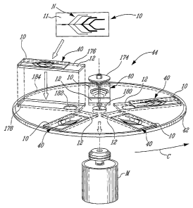
[0067] Figure 1 is top plan view of a microfluidic flow cell in
accordance with an embodiment of the present invention;
[0068] Figure 2 is a schematic illustration of a microfluidic
system in accordance with an embodiment of the present invention;
[0069] Figure 3 is a top plan view of a microfluidic device in
accordance with another embodiment of the present invention;
[0070] Figure 4 is a lateral view of the microfluidic device of
Figure 3;
[0071] Figure 5 is a perspective view of the microfluidic
device of Figure 3 showing the removable microfluidic flow cell and
support in a separated position in accordance with an embodiment of
the present invention;
[0072] Figure 6 is a lateral view of a microfluidic device in
accordance with a further embodiment of the present invention;
[0073] Figure 7 is a lateral view of a microfluidic device in
accordance with yet another embodiment of the present invention;
[0074] Figure 8 is a lateral view of a microfluidic device in
accordance with yet a further embodiment of the present invention;
[0075] Figure 9 is a lateral view of a microfluidic device in
CA 02559778 2006-09-13
WO 2005/093388 PCT/CA2005/000458
17
accordance with still another embodiment of the present invention;
[0076] Figure 10 is a lateral view of a microfluidic device in
accordance with still a further embodiment of the present invention;
[0077] Figure 11 illustrates a comparison 'between the
sensitivity of labeled oligonucleotide detection in no-flow hybridization
(circles) vs flow-through hybridization (squares) ' using a
complementary 15-mer capture probe;
[0078] Figure 12 illustrates a comparison between the
sensitivity of labeled amplicon detection in no flow hybridization
(circles) versus flow-through hybridization (squares); the amplicons
(368 bp) were generated using a pair of PCR primers targeting
Staphylococcus aureus tuf sequences; the S. aureus-specific capture
probe was a 20-mer fully complementary to internal sequences of the
368-bp amplicon;
[0079] Figure 13 illustrates flow-through hybridization of Cy-
labelled tuf gene amplicons. These amplicons were labelled by PCR
amplification of genomic DNA purified from four staphylococcal species
and hybridization was performed by using a microarray of capture
probes targeting these four staphylococcal amplicons; panels: A)
Hybridization to the S. aureus amplicons; B) Hybridization to the S.
epidermidis amplicons; C) Hybridization to the S. haemolyticus
amplicons; and D) Hybridization to the S. saprophyticus amplicons;
and
[0080] Figures 14A to 14D illustrate flow-through
hybridization of Cy-labelled tuf gene amplicons. These amplicons were
labelled by PCR amplification of 1 ng of genomic DNA purified from
four staphylococcal species and hybridization was performed by using
a microarray of capture probes targeting these four staphylococcal
CA 02559778 2006-09-13
WO 2005/093388 PCT/CA2005/000458
18
species. Panels are as follows: 14A) Hybridization to the S. aureus
amplicons; 14B) Hybridization to the S. epidermidis amplicons; 14C)
Hybridization to the S. haemolyticus amplicons; and 14D) Hybridization
to the S. saprophyticus amplicons. The graphs show the fluorescence
intensity for each hybridizations. Standard deviations are for the results
of five hybridizations.
[0081] Other objects, advantages and features of the
present invention will become apparent upon reading of the following
non-restrictive description of embodiments with reference to the
accompanying drawing, which are exemplary and should not be
interpreted as limiting the scope of the present invention.
DESCRIPTION OF THE EMBODIMENTS
[0082] With reference to the appended drawings,
embodiments of the invention will be herein described.
[0083] With reference to Figures 1 and 2, there is shown a
microfluidic flow cell 10 which can be removably interfaced with a
removable member 12 such as a support, which can be a slide, for
example.
[0084] The microfluidic flow cell 10 comprises a reaction
portion 14, which defines a reaction chamber with the support 12, as
will be described herein. Furthermore, the microfluidic flow cell 10
includes fluid-receiving portions 16, 18, 20, and 22. Each fluid-
receiving portion 16, 18, 20, and 22 comprises a respective fluid-
receiving chamber made of two similar elongated bores 24 and 26
(only one pair of bores are referenced here) within the microfluidic cell
body 11. The elongate bores 24 and 26 of each fluid-receiving
chamber defined by the fluid-receiving portions 16, 18, 20, and 22
meet at a common area 28, 30, 32, and 34 respectively along a
CA 02559778 2006-09-13
WO 2005/093388 PCT/CA2005/000458
19
common canal 38, which is in fluid communication with the reaction
chamber 14.
[0085] When the support 12 and the microfluidic flow cell 10
are in a interfaced position as shown in Figure 2, the microfluidic cell
10 is adapted to cause fluid in the fluid-receiving portions 16, 18, 20,
and 22 to flow to the reaction chamber 14.
[0086] In this non-limiting embodiment, the microfluidic flow
cell 10 in combination with the support 12, defines a microfluidic device
40, which is placed on a rotatable platform or disc 42. This rotatable
platform rotates as shown by arrow C thus applying centrifugal force to
the microfluidic flow cell 10 comprising fluid within the fluid-receiving
portions 16, 18, 20, and 22. As shown, chambers 16, 18, 20, and 22
are positioned within the body 11 of the microfluidic flow cell 10 from a
proximal to a distal position relative to the reaction chamber 14. In this
way, when centrifugal forces are applied upon the microfluidic flow cell
10, fluids will flow towards the reaction chamber 14 from the most
proximal chamber, 16 to the most distal chamber 22 as the speed of
the rotational disc 42 will increase, thus increasing the centrifugal
forces.
[0087] In the embodiment, shown in Figures 1 and 2, the
microfluidic flow cell 10 is a PDMS substrate unit 11 with an engraved
microfluidic network N, is applied to the support 12, in the form of a
glass slide on which nucleic acid capture probes have been arrayed
(not shown). The glass slide 12 with the PDMS microfluidic flow cell
10 is placed on a compact disc support 42 that can hold five slides 12
in this case, thus defining a microfluidic system 44. This microfluidic
system 44 can be designed to accomodate any number of slides.
[0088] During operation, the prehybridization buffer in
chamber 16 is released first and flows over the hybridization chamber
CA 02559778 2006-09-13
WO 2005/093388 PCT/CA2005/000458
14 where the oligonucleotide capture probes are spotted onto the glass
support 12. Subsequently, the sample in chamber 18 is released at a
higher angular velocity. Then, the wash buffer in chamber 20 and the
rinsing buffer in chamber 22 start to flow sequentially at even higher
5 angular velocities. The wash and rinsing buffers are used to wash
away the nonspecifically bound targets after the hybridization reaction.
[0089] With reference to Figures 3, 4 and 5, there is shown
a microfluidic flow device 45 in accordance with another embodiment
of the invention.
10 [0090] Figures 3 and 4 show the microfluidic device 45
including a removable microfluidic flow cell 46 interfaced with
removable-member 48 in the form of a support. Figure 5 shows the
microfluidic flow cell 46 having been removed from support 48.
[0091] The microfluidic flow cell 46 includes a body 47
15 having a reaction portion 49 (see Figure 5) in the form of a cavity. The
reaction cavity 49 defines a reaction chamber 50 (see Figures 3 and 4)
when interfaced with the support slide 48. The reaction chamber 50
provides a space for the microarray 52 on the support slide 48.
Furthermore, the microfluidic flow cell 46 includes fluid-receiving
20 portions 54 and 56 in the form of cavities as shown in Figure 5. As
shown in Figures 3 and 4, these fluid-receiving cavities 54 and 56
define respective fluid-receiving chambers 58 and 60 when interfaced
with the slide support 48. Turning back to Figure 5, the microfluidic flow
cell 46 also includes conduit cavities 62, 64 and 66 that define
respective conduits 68, 70 and 72 as shown in Figures 3 and 4 (when
the flow cell 46 is interfaced with support 48). Conduits 68, 70 and 72
meet at a valve 74, which is defined by a valve cavity 76 (see Figure 5)
when the flow cell 46 and the support 48 are in the interfaced position.
Figure 5 shows a common channel cavity 78 in the flow cell 46 that
defines, when interfaced with support 48; a common channel 80 (see
CA 02559778 2006-09-13
WO 2005/093388 PCT/CA2005/000458
21
Figure 3 and 4) in fluid communication with the reaction chamber 50
and valve 74.
[0100] Air vents 82, 84, and 86 are in fluid communication
with the ambient environment. Air vents 82, 84, and 86 are
respectively in fluid communication with fluid-receiving chamber 58, via
conduit 88, with the valve 74, via conduit 70 and with the fluid-receiving
chamber 60, via conduit 90. Turning to Figure 5, conduits 88 and 90
are defined by conduit-cavities 92 and 94 when interfaced with support
48.
[0101] The microfluidic cell 46 also includes an evacuation
duct 96 in fluid communication with the reaction chamber, providing for
excess or waste fluid to flow there through into the ambient
environment or on the support 48 via aperture 97. With reference to
Figure 5, duct 96 is formed by a duct cavity 98 when interfaced with
support 48.
[0102] Hence, the removable microfluidic flow cell 46 and
the removable solid support 48 provide a microfluidic device 45 for
microarray analyses in accordance with an embodiment of the present
invention.
[0103] Figures 6 to 10 show a variety of non-limiting
embodiments of the microfluidic flow cell, removable member and
microfluidic devices in accordance with the present invention.
[0104] Figure 6 shows a microfluidic device 100 having a
microfluidic flow cell 102 being removably interfaced with a removable
member 104 in the form of a support. The microfluidic flow cell 102 has
a fluid-receiving portion 106 in the form of a cavity that defines a fluid-
receiving chamber 107 when interfaced with support 104. The
microfluidic flow cell 102 also includes a reaction portion 106 in the
CA 02559778 2006-09-13
WO 2005/093388 PCT/CA2005/000458
22
form of reaction-chamber cavity 108 that defines a reaction chamber
109, for performing a reaction therein, when interfaced with the support
104. A conduit-cavity 110 defines a conduit 111 with support 104. A
waste dispensing duct 115 may also be provided.
[0105] Figure 7 shows a microfluidic device 116 having a
microfluidic flow cell 118 removably interfaced with a removable
member 120 such as a support. The flow cell 118 includes a fluid-
receiving chamber 122 as well as a conduit 124, in fluid
communication therewith, both formed within the cell body 126. The
conduit 124 is in fluid communication with a reaction portion 128 in the
form of cavity and defining by a reaction chamber 130 when interfaced
with the support 120. A waste dispensing duct 121 may also be
provided.
[0106] Figure 8 shows a microfluidic device 132 having
removable member 134 in the form of a removably positioned on a
microfluidic flow cell 136. The reaction chamber 138 is defined by a
cavity 140, formed within the microfluidic flow cell 136 and by the
enclosure member 134 when interfaced therewith. Optionally, the
conduit 142 and the fluid-receiving portion 144 may be cavities
enclosed by the removable member 134 or may be fully formed within
the microfluidic flow cell 136.
[0107] Figure 9 shows a microfluidic flow device 146 having
a removable member 148 in the form of seal which, when interfaced
with the microfluidic flow cell 150 at the cavity 152 thereof, defines the
reaction chamber 154. Again as before, the conduit 156 and the fluid-
receiving portion 158 may be cavities enclosed by the removable seal
member 148 or may be fully formed within the microfluidic flow cell
150. The seal 148 may be mounted to the microfluidic flow cell 150 by
a variety of adhesive materials as is known in the art.
CA 02559778 2006-09-13
WO 2005/093388 PCT/CA2005/000458
23
[0108] Figure 10 shows a microfluidic flow device 160
having a support member 162 comprises a cavity 164 that defines a
reaction chamber 163 when interfaced with the microfluidic flow cell
166. The microfluidic flow cell 166 includes a reaction portion 163 that
comprises an,exit aperture 165 in communication with a fluid-receiving
portion 168 via a conduit 170 and.an enclosing portion 167. Again, in
this embodiment, the conduit 170 and fluid-receiving portion 168 are
formed within the microfluidic flow cell body 172, yet it can be
contemplated by the skilled artisan to define cavities within the
microfluidic flow cell surface 172 that provides chambers when
interfaced with the support 162.
[0109] In another non-illustrated embodiment, two
microfluidic flow cells of the present invention can be removably
interfaced with each other, as such one of the two cells acts as a
removable member.
[0110] The microfluidic flow cells of the present invention
are adapted to provide for fluids to flow the reaction chambers of the
present invention by applying an external force onto the microfluidic
flow cells such as gravity, centrifuge, capillary force, gas pressure,
electro-osmosis, electrokinetics, electrowetting, magnetic pump force
or any combination of the foregoing as will be understood by the skilled
artisan.
[0111] In an embodiment, the present invention describes a
removable microfluidic flow cells adaptable to arrays printed onto a
surface or surrounded by such a surface. The different solutions
required for biochemical reactions are driven onto the slide by
microfluidic circuitry or network (such as N) engraved into an
elastomeric substrate juxtaposed onto the surface surrounding the
microarray. External forces can be applied to move the fluids; access
to various parts of the circuitry or network is valve-controlled. Non-
CA 02559778 2006-09-13
WO 2005/093388 PCT/CA2005/000458
24
limiting examples of such external forces are pumps, magnetic,
electrokinetic, electro-osmotic and centrifugal. In one embodiment,
centrifugal forces can be produced by a motor or a centrifuge and
move the fluids into the microfluidic channels and chambers engraved
in the surface of the elastomeric substrate positioned above the
microarray. The present invention comprises a microfluidic device
having one or more individual chambers connected with one or several
reaction chamber(s). The channels and chambers of the microfluidic
system of the present invention may access individual spots or group
of spots (rows, columns, blocks of spots) of the microarray or the entire
microarray. Chamber and channel volumes are generally kept as small
as possible to reduce the amount of sample and reagents that must be
used.
[0112] In an embodiment, the devices, methods and
systems of the present invention comprise microarray surfaces that are
functionalized with an appropriate coating allowing for the binding of
probes. The slide format can be adapted to standard microarray
equipment used in proteomic or genomic laboratories.
[0113] Each chamber may contain buffers and samples
necessary for the reaction(s) to proceed. Small volumes of the fluid
sample comprising the biomolecules are forced to flow into the
microfluidic circuitry or network positioned directly above the
immobilized probes of the microarrays. The close proximity between
the solution phase analytes and the bound probes speeds-up the
kinetic interactions, thereby reducing reaction time.
[0114] In a particular embodiment of the present invention, a
standard microscope glass-slide is chemically functionalized to
covalently bind bioprobes. The microfluidic device may be. used to
drive fluids over spot-bearing microarrays. The bioprobes spots may
be composed of DNA, RNA, oligonucleotides, oligonucleotide analogs,
CA 02559778 2006-09-13
WO 2005/093388 PCT/CA2005/000458
proteins, peptides, organic molecules, sugars, drugs or combinations
thereof, or any binding partner of a substrate present in the test
sample. Various reaction steps can be performed with the bound
molecules of the microarray including exposure to liquid reagents or
5 reactants, washing reagents, hybridization or detection reagents. The
progress or outcome of the reaction can be monitored at each spot of
the microarray in order to characterize molecules immobilized on the
slide.
[0115] Presently, most custom microarrays are printed onto
10 standard microscope glass slides; this format being required by most
of the current commercially available instruments used to scan for
detection signals (e.g., fluorescent signal) indicative of positive
interactions with particular probes spotted on the slide. In one
embodiment of the invention, the removable microfluidic platform, unit
15 or cell was thus designed to fit standard glass slides. However, any
microarray format flat surface (e.g. glass support, plastic support) can
be used in accordance with the present invention. Furthermore, such a
system may be used in concomitance with independant or integrated
microfluidic systems for test sample preparation (e.g. for nucleic acid
20 extraction) and/or target amplification (e.g. nucleic acid amplification by
polymerase chain reaction) for molecular diagnostics. Such a system
may also be a micro total analysis system.
[0116] The present invention provides microfluidic flow cells
that can be adapted to interface with glass microscope slides and
25 similar planar surfaces. This microfluidic interface system enables the
delivery of sample, interacting reagents (e.g., hybridization solutions,
binding solutions and the like), wash solutions, and detection reagents
to selected positions on the array.
[0117] In an embodiment, grooves or indentations on the
surface of the microfluidic flow cells of the invention are aligned with
CA 02559778 2006-09-13
WO 2005/093388 PCT/CA2005/000458
26
spots on the microarray; so that when the microfluidic interface system
is sealed onto the microarray surface, the indention and grooves form
channel(s), reagent reservoir(s) and/or reaction chamber(s) containing
the spots of the microarray(s).
[0118] In one particular embodiment, a soft elastomeric
material (e.g., PDMS) is selected to make the microchambers and
channels of the microfluidic interface system. PDMS-based elastomers
are low cost materials which can be molded and which seal reversibly
with flat and smooth surfaces such as glass.
[0119] In a further particular embodiment, centrifugal forces
.are used to move the fluids into the microfluidic channels and
chambers positioned above the microarray.
[0120] In yet a further particular embodiment, a standard
microscope glass slide support is designed to fit into a centrifugation
system. The centrifugation system may be a custom device or a
classical bench centrifuge. The centrifugation system comprises a step
by step motor, controlled by a computer.
[0121] The controlled delivery of fluids to one or more
selected regions of the microarray slide may be accomplished by
choosing the appropriate size and shape of the channels and
chambers of the microfluidic system, and by selecting the optimal
centrifugal force and the optimal time over which the centrifugal force
will be applied to deliver the fluids over the microarrays.
[0122] In yet a further particular embodiment of the present
invention, the microfluidic system is used for the analysis of nucleic
acids including but not limited to molecular diagnostic assays on
microarrays for infectious disease agents which typically require rapid,
sensitive, automated, high throughput and inexpensive systems.
CA 02559778 2006-09-13
WO 2005/093388 27 PCT/CA2005/000458
[0123] As mentioned herein above, the slide can be made of
glass, glass being the most commonly used support material for
custom microarrays of nucleic acids and proteins. The glass slide is
specifically coated to optimize the binding of nucleic acids or nucleic
acid analogs (e.g. peptide nucleic acid, locked nucleic acid).
Microarrays of nucleic acid probes are printed onto the glass slides
using an arrayer positioned to fit directly under the hybridization
chambers of the invention when the microfluidic circuitry or network
(such as N shown in Figure 2) engraved elastomeric material is placed
above the slide supports of the invention. Microarrays may include
numerous different probes and can be used to perform expression
profile experiments. The corresponding hybridization chamber(s) can
therefore be designed to accommodate the required volumes, in order
to be used as an automated hybridization platform. In a particular
embodiment, the array is linear and made-up of spotted probes, and is
fitted to be used for diagnostic purposes. The hybridization chamber(s)
can also be designed to accommodate smaller volumes allowing flow-
through hybridization, thus enhancing the hybridization kinetics. This
reduces the hybridization time and/or increases the sensitivity of the
reactions required for detection of hybrids.
[0124] In comparison with passive hybridization, the
microfluidic device of the present invention allows for about 6-fold
increase in the hybridization kinetics as demonstrated for a 20-mer
oligonucleotide as well as for a 368-bp amplicon (see Example 1).
Furthermore, it was possible to detect and discriminate 4 clinically
relevant Staphylococcus species using a 15-minute hybridization
process. This is at least 16 times faster than the times generally
required for passive hybridization. The removable microfluidic system
of the present invention allows to automate and speed up reaction
processes using conventional microarrays and provides for the rapid
detection and identification of nucleic acids or other biomolecules
present in a sample (proteins, cofactors, drugs and the like). The
CA 02559778 2006-09-13
WO 2005/093388 PCT/CA2005/000458
28
removable microfluidic flow cells as well as microfluidic devices of the
present invention can be used in a variety of applications such as in
the biomedical field (detection of the presence of pathogens or disease
associated markers), in the forensic field (identification of individuals),
in basic research as well as in other industrial applications. Finally, the
removable microfluidic flow cells and devices of the present invention
can be applied in any type of microarray analysis.
[0125] In one non-limiting embodiment, the substrate
comprising the body of the microfluidic flow cell of the present
invention is a soft elastomeric material capable of reversibly binding to
the microarray by Van der Waals forces without the need for any glue
or clamp. In a particularly preferred embodiment, the soft substrate is
made of PDMS. The microfluidic circuitry of network is engraved into
the substrate using classical microfabrication technologies such as
photolithography and computer numerically controlled (CNC)
machining. Various types of valves may be included in the microfluidic
circuitry or network. Valves are designed to control the release of fluids
from the different reservoirs. For example, the valves can be
electromagnetically actuated microvalves (Canapu et al., 2000, J.
Microelectromech. Syst., 9:181-189), air driven pressure valves (e.g.,
to control the venting of air in specific regions of the microfluidic
circuitry, therefore modulating the backpressure that opposes fluid
movements) (Unger et al., 2000, Science, 288:113), hydrogel valves
(Liu et al., 2002, J. MEMS, 11:45-53), and centrifugal valve (Madou at
a/., 2001, Sensor Actuat. A, 91:301-306). Alternatively, movements of
fluids in the microfluidic system may be driven without the use of
valves. For example, fluids can be moved by sequential flow of
different liquids separated by air bubbles' or other non-mixing
boundary.
[0126] Turning back to Figures 1 and 2, a microfluidic
system 44 is illustrated. The microfluidic flow cell 10 comprises a
CA 02559778 2006-09-13
WO 2005/093388 PCT/CA2005/000458
29
circuitry or network N engraved into a PDMS substrate 11. The PDMS
substrate 11 is aligned and reversely bound to a microarray (not
shown) printed onto a glass slide 12 by applying pressure to form a
functional microfluidic device 40. After printing the oligonucleotide
microarray onto the glass slide 12 using a commercial arrayer, the
PDMS microfluidic circuit N is superposed onto the glass slide 12 in
such a way that the PDMS engraved hybridization chamber 14 is
above the microarray. The microfluidic device 40 (glass slide 12 and
PDMS 11) was introduced into a custom-made plastic disc shape
support 42 comprising an opening and fixed on the actuator hub 174 of
a motor M. The disc 42 is rotated, as shown by arrow C, to drive
sample and buffers directly onto the glass surface 176 using
centrifugal forces to move the liquid reagent into the chamber 14 and
microfluidic channels 30 (Madou et al., 2001, Sensor Actuat. A,
91:301-306). At the end of the process, the PDMS fluidic circuits N
were pealed off the glass slide 12 and the microarray was analysed
using commercial instruments. This system 44 allows for dynamic DNA
hybridization (flow-through) generated by centrifugal forces. In the
present invention, such a microfluidic system was able to discriminate
nucleic acid sequences including single nucleotide polymorphisms
(SNPs) in a fraction of time required by conventional microarray
technology.
[0127] If centrifugal forces are used to drive the fluids in the
microfluidic chambers and channels, the valves (such as 38, 30, 32
and 34) can be designed to burst at different rotational speeds (Figure
1). The circuitry or network N may comprise a hybridization chamber
14, a pre-hybridization buffer reservoir 16, a sample inlet 18, a washing
reservoir 20, and a rinsing reservoir 22, all connected together by
different sized channels 38A, 38B, 38C, 38D and 38E (Figures 1 and
2). The chamber 14 is in fluid communication with a dispensing portion
39 in communication with the ambient environment. The different
buffers and the sample are forced to flow-through the hybridization
CA 02559778 2006-09-13
WO 2005/093388 PCT/CA2005/000458
chamber 14 positioned above the microarray on support 12, by varying
the rotation speed of the centrifugal system 42. The architecture of the
hybridization chamber 14 may be adapted to enhance the turbidity of
the fluid, thus enhancing the hybridization kinetics. Again, movements
5 of fluids in the microfluidic system may alternatively be driven without
the use of valves as described above.
[0128] The methods, systems and devices of the present
invention use a microarray support to connect the microfluidic system
to the device providing the force to move the fluids. The force can be
10 generated by pneumatic drive, mechanical micropumps, electro-
osmosis, electrophoresis, gas-pressure, positive and negative
displacement, thermal, electrochemical bubble generation, acoustics,
magnetic, DC and AC electrokinetics, and centripetal forces. In 'a
particular embodiment, the support is a disc 42 adaptable to a
15 rotational device 174 providing the centrifugal forces to move the
fluids. In a more particular embodiment the support is a disc 42
comprising microfluidic device receiving portions 178 such as slots
accommodating standard microscope slides 12. Each slot 178 is
placed at the same distance from the disc center 180, allowing for
20 equal centrifugal forces to be applied to each slotted slide. The disc is
designed to be fixed on the hub 174 of a motor M. Each slide 12 may
comprise an aperture 182, to facilitate removal of, the slides 12 after
centrifugation. In a related embodiment, a waste reservoir 184 such as
a furrow is engraved into the support disc 42 to collect the hybridization
25 waste liquid following centrifugation, allowing the slides 12 to dry
completely. In a another related embodiment, the microfluidic system
44 comprising disc 42 is sealed (not shown) to avoid aerosols
generation during the spinning of the disc 42 .
[0129] The force-providing devices of the present invention
30 can be any device, such as a pump, a heater, a motor, a magnetic
device, a mechanical device, or an electrical device. The device
CA 02559778 2006-09-13
WO 2005/093388 PCT/CA2005/000458
31
providing the centrifugal forces to force the fluids to the microarray
support is preferably a motor. The motor may be a step by step motor,
or a computer-driven or a programmable,, commercially available
bench centrifuge.
[0130] Although the microfluidic flow cells (10, 46) of the
present invention have been designed, in a particular embodiment, to
interface with slides (12, 48) bearing microarrays (such as 52) of
biomolecules, they may also be used to provide a fluid interfacing with
a support bearing various types of molecular probes or samples. The
probes or samples could be on bead or particles located on the
support. It is to be understood that the application of the present
invention is not to be limited to the use with microarray slides. This
invention could be applied to detect/analyse any reaction signals which
may be optical, electrical, mechanical, chemical, magnetic or any
other measurable property of said reaction.
[0131] The present invention is illustrated in further detail by
the following non-limiting examples.
EXAMPLES
EXAMPLE 1: Removable fluidic system to drive microarray reagents
using centrifugal force
Materials and methods
Selection of PCR primers and capture probes
[0132] All chemical reagents were obtained from Sigma-
Aldrich Co. (St-Louis, MI) and were used without further purification
unless otherwise noted. Oligodeoxyribonucleotide capture probes,
which were 5'-modified by the addition of two nine carbon spacers and
an amino-linker, were synthesized by Biosearch Technologies (Novato,
CA). The amino-linker modification permits the covalent attachment of
CA 02559778 2006-09-13
WO 2005/093388 PCT/CA2005/000458
32
probes onto a functionalized glass surface. Four capture probes were
used: S. aureus targeting probe (5'-CGTATTATCAAAAGACGAAG-3'),
S. epidermidis targeting probe (5'-CAIAGCTGAAGTATACGTAT-3'), S.
haemolyticus targeting probe (5'-CAAAATTTAAAGCAGACGTATA-3')
and S. saprophyticus targeting probe (5'-
AAAGCGGATGTTTACGTTTT-3'). Primer pairs TstaG422 - (5'-
AAAGCGGATGTTTACGTTTT-3') and TstaG765 (5'-
TIACCATTTCAGTACCTTCTGGTAA-3') were used to amplify all
staphylococcal species. Used genomic DNAs were purified from
strains S. aureus ATCC 43300, S. epidermidis ATCC 14990,
S. haemolyticus ATCC 29970 and S. saprophyticus ATCC 35552.
Fabrication of the elastomeric flow cell
[0133] The microfluidic structures were fabricated using
PDMS replicating techniques (Duffy at al., 1998, Anal. Chem.,
70:4974-4984). A novel 2-level SU-8 process was developed in order
to achieve the desired 2-level PDMS fluidic structures that provide
sufficient volume for reagent storage while also enabling the proper
flow rate for reagent manipulation and for hybridization in the shallow
hybridization chamber.
SU-8 mold fabrication
[0134] SU-8 is a negative tone photoresist that has attracted
significant interest for the fabrication, as well as for applications
requiring very thick photoresist layers. Due to its excellent UV
transparency, standard UV lithography can be used to craft LIGA-like
MEMS devices. SU-8 photoresists come in different viscosities: the
lower viscosity products are more suited for the fabrication of thin
structures (up to 2 pm); the more viscous SU-8 photoresists are better
suited for thick layers (mm scale). Two types of the photoresist, SU-8
25 and SU-8 100, available from Microchem Inc. (Newton, MA), were
used. SU-8 25 was used for the microchannel structures and SU-8 100
CA 02559778 2006-09-14
WO 20051093388 PCT/CA2005/000458
33 26 . JP UARY 230a 2 6=.01.0 6
was used for the much larger reagent chambers. In the first step, SU-8
25 was processed on a 15 cm silicon (Si) wafer (Addison Engineering,
San Jose, CA) to obtain the structures for the microchannels (25 pm in
depth) and the alignment marks for the second SU-8 layer.
Subsequently, a thick layer (250pm) of SU-8 100 was spin-coated over
the substrate on which the molds for the microchannels had been
created. This thicker layer was used to define the mold for the much
larger reagent reservoirs. Since crosslinked ' SU-8 photoresists have
lower optical transparency than their unexposed surroundings, the
alignment marks can be readily observed even when they are
completely covered with a thick layer of the unexposed photoresist. In
the pattern design, compensations were made for possible alignment
errors between the two layers of photoresist.. The channels and
chambers overlapped in the connection areas to avoid possible
disconnections caused by misalignment. Six identical molds were
simultaneously fabricated onto the 15 cm Si wafer for faster replication.
Polymerization molding of the flow cell
[01351 PDMS was purchased from Dow Corning (Midland,
MI). The base (Sylguard 184 silicone elastomer) and the curing agent
(silicone resin solution) were thoroughly mixed in a weight proportion of
10:1. Low temperature curing (e.g. 65 C) in a convection oven was
preferred over high temperature baking due to the thickness of the
structures. High temperatures (e.g. 150 C) causes significant thermal
stress at the interface between the SU-8 patterns and the Si substrate
which can actually crack the substrate and peel off the SU-8
structures. Leveling of the PDMS on the substrate is required in order
to achieve a uniform thickness over all the flow cells. The appropriate
combination of , the macrostructures of the chambers and
microstructures of the channels is important for the performance of the
flow cells.
,AMENDED SHEET
CA 02559778 2006-09-13
WO 2005/093388 PCT/CA2005/000458
34
Preparation of glass slides
[0136] All chemical reactions were carried out in
polypropylene jars at room temperature unless specified otherwise.
The microscope glass slides used (VWR Scientific, West Chester, PA)
had a surface of 25 mm x 75 mm. After sonication in deionized water
for 1 hour, the slides were sonicated in 40 ml of NaOH (10%) for 1
hour, washed several times with deionized water and dried under a
stream of nitrogen. The slides were then sonicated in an
aminopropyltrimethoxysilane solution (2 ml water, 38 ml MeOH and 2
ml aminopropyltrimethoxysilane) for 1 hour, washed with methanol,
dried and baked at 110 C for 15 minutes. The amine modified slides
were activated by overnight sonication in 1,4-dioxane (40 mL)
containing 0.32 g (2 mmol) of carbonyldiimidazole' as the coupling
agent, followed by washing with dioxane and diethyl ether, and drying
under a stream of nitrogen.
Microarray production
[0137] The probes were diluted two-fold by the addition of
Array-it Microspotting Solution PIusTM (Telechem International,
Sunnyvale, CA), to a final concentration of 5 pM. The capture probes
were spotted in triplicate, using a VIRTEK SDDC-2TM arrayer (Bio-Rad
Laboratories, Hercules, CA) with SMP3 pins (Telechem International).
Upon spotting, each spot had a volume of 0.6 nL and a diameter
ranging between 140 to 150 pm. After spotting, the slides were dried
overnight, washed by immersion in boiling 0.1% Igepal CA-630 for 5
minutes, rinsed in ultra-pure water for 2 minutes, and dried by
centrifugation under vacuum for 5 minutes (SpeedVacTM plus; Thermo
Savant, Milford, MA). The slides were subsequently stored at room-
temperature in a dry, oxygen-free environment.
PCR amplification and amplicon labeling
[0138] Universal PCR primers targeting conserved areas of
CA 02559778 2006-09-13
WO 2005/093388 PCT/CA2005/000458
the tuf gene were used to amplify a 368-bp fragment from S. aureus,
S. epidermidis, S. haemolyticus and S. saprophyticus purified genomic
DNAs. Fluorescent dyes were incorporated during assymetrical PCR
amplification. Cy-5 dCTP (Amersham Biosciences, Baie d'Urfe,
5 Quebec, Canada) was mixed at concentrations of 0.02 pM in a 50 pl
PCR mixture containing: 0.05 mM dATP, 0.02 mM dCTP, 0.05 mM
dGTP, 0.05 mM dTTP, 5 mM KCI, 1 mM Tris-HCI (pH 9), 0.01 % Triton
X-100, 2.5 mM MgCl2, 0.5 Unit of Taq DNA polymerase (Promega,
Madison, Wisconsin), 0.2 pM of primerTstaG765, 0.005 pM of primer
10 TstagG422 and 1 ng of purified staphylococcal genomic DNA. Thermal
cycling for PCR amplification (180 seconds at 94 C followed by 40
cycles of 5 seconds at 95 C, 30 seconds at 55 C, and 30 seconds at
72 C) was carried out on a PTC-200 DNA Engine ThermocyclerTM (MJ
Research, Reno, NV).
15 DNA microarray hybridization and data acquisition
[0139] PCR amplicons labeled with Cy5-dCTP were
denatured at 95 C for 5 minutes. Denatured labeled amplicons (5 pl)
were mixed with hybridization buffer (15 pl) (8X SSPE, 0.04% PVP
and 40% formamide).
20 [0140] Passive hybridization was performed in a 20 pl Hybri-
wellTM self-sticking hybridization chambers (15 mm x 13 mm) (Sigma-
Aldrich). Hybridization buffer containing the labeled sample was
introduced in the chambers and hybridization was conducted for 5
minutes at room temperature. After hybridization, the microarrays were
25 washed at room temperature (5 minutes) with 2X SSPE containing
0.1% SDS and rinsed once (5 minutes) with 2X SSPE at room
temperature. The microarrays were dried by centrifugation at 1348 x g
for 3 minutes.
[0141] Flow-through hybridization was performed in the
CA 02559778 2006-09-13
WO 2005/093388 PCT/CA2005/000458
36
flow-cell as described above for passive hybridization. A hybridization
unit consisting of a glass slide 'and flow-cell was placed onto a home
made plastic disc support, and the support fixed to the hub of a step by
step motor controlled by, a computer. The labeled sample was
prepared the same way as for a passive hybridization. Sample (2 pl)
and washing and rinsing buffer (10 pl) were loaded onto the
microfluidic unit just before spinning the disc. The disc was spun at
different speeds in order to sequentially burst the centrifugal valves
and allow the pre-hybridization buffer, sample, washing and rinsing
buffer to flow-through a.140 nl hybridization chamber respectively. The
disc was subsequently spun at 1000 rotation per minute (rpm) for 1
minute to dry the slide.
[0142] Slides were scanned using a ScanArray 4000XLTM
(Packard Bioscience Biochip Technologies; Billerica, MA) and
fluorescent signals were analyzed using its QuantArrayTM software.
Results
Assembly of the microfluidic unit
[0143] The assembled microfluidic unit is shown in Figures
1 and 2. The flow cell is aligned with, and adhered to, the glass slide to
form a DNA hybridization detection unit, up to 5 of which can be
mounted onto the disc platform (Figure 2). The design of the
microfluidic network and the microarray layout is such that a
hybridization chamber is positioned right above the oligonucleotide
capture probes spotted onto the glass slide when the two parts are put
together. The reagents are positioned to be sequentially pumped
through the hybridization chamber by centrifugal force beginning with
chamber 16. This flow sequence is achieved by optimizing the balance
between the capillary force and centrifugal pressure (Madou et al.,
2001, Sensor Actuat. A, 91:301-306). The pre-hybridization buffer
(chamber 16) is released first and flows over the 140 nI hybridization
CA 02559778 2006-09-13
WO 2005/093388 PCT/CA2005/000458
37
chamber (chamber 14) where the oligonucleotide capture probes are
spotted onto the glass support. The sample containing the labeled
PCR amplicons (chamber 18) is then released at higher angular
velocity and flows over reaction chamber 14. Then, the wash buffer
(chamber 20) and the rinsing buffer (chamber 22) flow sequentially at
even higher angular velocities. The wash and the rinsing buffers are
used to remove the nonspecifically bound targets following the
hybridization process. Pre-hybridization buffer, sample, washing buffer
and rinsing buffer were collected in a waste reservoir which is a groove
surrounding the disc. This system was enclosed in a box during
spinning of the disc to avoid the spread of aerosols carrying PCR
amplicons.
Flow-through versus passive hybridization
[0144] Cy3-labeled 20-mer oligonucleotides were hybridized
both in a passive way using a standard commercially available
hybridization chamber of 20 pl, and with the flow-through method using
the microfluidic platform in a 140 nl of hybridization chamber as
described hereinabove. For passive hybridization, 20 pl of different
concentrations (i.e. 0.025, 0.125, 0.25, 1.25 and 2.5 nM) of Cy3-
labeled oligonucleotides were hybridized to their complementary
probes, spotted onto a microarray on glass support using Hybri-wellTM
self-sticking hybridization chambers (Sigma-Aldrich). Following
hybridization at room temperature for 5 minutes, the slides were
washed and rinsed as described above in the method section. For
flow-through hybridization, 2 pl of different concentrations (i.e. 0.025,
0.125, 0.25, 1.25 and 2.5 nM) of Cy3-labeled oligonucleotide were
hybridized to their complementary probes as described for the passive
method. Samples of the different concentrations of oligonucleotides (2
pl) were loaded into the sample inlet of the microfluidic unit.
Prehybridization buffer, sample, washing buffer and rinsing buffer were
loaded respectively into chambers 16, 18, 20 and 22 of the
hybridization unit shown in Figures 1 and 2. Loading of the reagents
CA 02559778 2006-09-13
WO 2005/093388 38 PCT/CA2005/000458
was performed immediately before spinning the disc platform to avoid
reagent evaporation. A rotation speed of 300 rpm was selected to
release the content of the pre-hybridization chamber 16 and obtain a
sample flow rate of about 400 nI/min in the hybridization chamber (i.e.
reaction chamber), which corresponds to a hybridization time of 5
minutes (identical to the hybridization time used in the passive
hybridization experiments). Subsequently, the sample chamber
(chamber 18) was released into the reaction chamber by rotating the
disc at 412 rpm to achieve a flow rate of 400 nl/min. Following the
hybridization step, the rotation speed of the platform was further
increased to 585 and 764 rpm in order to sequentially burst the
centrifugation valves, thereby releasing respectively into the
hybridization chamber 10 pl of washing buffer and 10 pl of rinsing
buffer, both of which flowed through the hybridization chamber with an
average flow rate of 2 pl/min resulting in a total time of about 15
minutes for the entire hybridization process, including a 30 seconds
drying step (high rotation speed). The PDMS microfluidic flow cells
were pealed off. Following passive or flow-through hybridization, the
microarrays were scanned. The fluorescence intensity was spotted
against concentration of oligonucleotide (Figure 11). It was observed
that flow-through hybridization in a 140 nl chamber was more sensitive
than passive hybridization in a larger volume chamber (i.e. 20 pl). The
passive and flow-through hybridizations were also performed using a
368-bp Cy-labeled amplicon that is derived from tuf gene sequences.
The results of these experiments show that flow-through hybridization
is more sensitive than passive hybridization as observed with a
complementary oligonucleotide (Figures 11 and 12).
Analytical sensitivity of the microfluidic platform
[0145] In all the experiments described above, the standard
procedure was to perform PCR amplification using universal primers
targeting conserved areas of the tuf gene to amplify a 368-bp fragment
from S. aureus, S. epidermidis, S. haemolyticus and S. saprophyticus
CA 02559778 2006-09-13
WO 2005/093388 PCT/CA2005/000458
39
purified genomic DNAs. One ng of genomic DNA purified from different
strains of staphylococci was used for each PCR reaction.
Approximately, 20% of the amplified PCR reaction mixture was used
for each hybridization. To evaluate the minimal quantity of bacterial
genome required to have a clear and unambiguous signal using the
microfluidic platform, hybridization of PCR amplicons amplified from
the equivalent of 10, 100, 1000 or 10000 genome copies was
performed. It was found that the equivalent of as little as 100 genome
copies of starting material was enough to discriminate each of the four
different staphylococcal amplicons.
Detection and specific identification of four clinically important
staphylococcal species
[0146] Microarrays of species-specific capture probes
targeting each of the four staphylococcal species (i.e. S. aureus, S.
epidermidis, S. haemolyticus and S. saprophyticus) printed in duplicate
were prepared. The microarrays were then hybridized with the different
staphylococcal amplicons generated by PCR amplification of genomic
DNA purified from each of these four staphylococcal species. The
results demonstrate that it was possible to detect and discriminate the
four different staphylococcal tuf amplicons without any ambiguity
(Figures 13 and 14). It is worth noting that there is a difference of only
a single nucleotide polymorphism (SNP) between the S. epidermidis-
specific probe and the S. aureus-amplicon sequence, showing that this
system is able to distinguish a SNP after only 5 minutes of
hybridization.
Discussion
[0147] In the genomic field, microarrays have become the
CA 02559778 2006-09-13
WO 2005/093388 PCT/CA2005/000458
standard for profiling gene expression. Thousands of genes are
routinely studied for their expression using the microarray technology.
Several groups have attempted to adapt this technology to the rapid
detection of microbial targets for diagnostic purposes (Mikhailovich et
5 al., 2001, J. Clin. Microbiol., 39:2531-2540; Westin et al., 2001, J. Clin.
Microbiol., 39:1097-1104; Wang et al., 2003, Anal. Chem.,. 75:1130-
1140; Bavykin et al., 2001, Appl. Environ. Microbiol., 67:922-928;
Bekal et al., 2003, J. Clin. Microbiol., 41:2113-2125). Even though
such systems most often require highly advanced biochips, the
10 detection performance lacks both sensitivity and specificity (Lenigk et
al., 2002, Anal. Biochem., 311:40-49; Wang et al., 2003, Anal. Chem.,
75:1130-1140). As demonstrated hereinabove, very small amounts of
liquid can be precisely and directly moved onto a glass slide surface
from buffer chamber(s) to hybridization chamber(s) using a microfluidic
15 elastomeric flow-cell juxtaposed above the slide. This technology
allows to dramatically reduce the volumes of reagents required during
microarray hybridization. For identical concentrations of a 20-mer
oligonucleotide or 368-bp amplicon, the flow-though hybridization
method gave signals which were of an order of magnitude higher than
20 those obtain with passive hybridization (Figures 11 and 12). Theses
results confirm previous observations, obtained with a more complex
microfluidic device (Wang et al., 2003, Anal. Chem.,' 75:1130-1140).
The capture probes and buffer compositions were designed in order to
achieve efficient hybridization at room temperature, thereby reducing
25 the complexity of the device.
[0148] The system of the present invention is specific
enough to discriminate SNP at room temperature using a hybridization
period of less than 10 minutes. The results of the discrimination
specificity for four staphylococcal species (hybridization period of 5
30 minutes) are shown in Figures 13 and 14. Other probes have at least 3
distinct nucleotides as compared with others staphylococcal
amplicons. In addition to being specific, the present system is also
CA 02559778 2006-09-13
WO 2005/093388 PCT/CA2005/000458
41
sensitive. It is possible to generate a specific hybridization signal using
the amplified PCR reaction mixture containing the equivalent of as little
as 100 copies of staphylococcal genome as starting material. This
result is at least 10 times more sensitive than those obtained by other
groups using more complex microfluidic devices (Westin et al., 2001, J.
Clin. Microbiol., 39:1097-1104). It is worth noting that the PCR
products are not purified prior to their addition to the hybridization
buffer.
[0149] Overall, the present example describes an
affordable, easy to use, automated and rapid custom microarray
hybridization microfluidic platform. This microfluidic platform uses
standard glass slides totally compatible with commercial arrayers and
scanners. In this system the classical hybridization chamber or
coverslip is replaced by a low cost elastomeric material engrafted with
a microfluidic network. This elastomeric material sticks reversibly to the
,glass slide without any glue or chemical reaction thereby forming the
microfluidic unit. Placed in a plastic compact disc like support, the
microfluidic units are spun at different speeds to allow fluids to move.
Using the present system, it was demonstrated that it is possible to
detect and discriminate tuf sequence polymorphisms including SNP
present in four different staphylococcal species using a rapid
hybridization protocol of approximately 15 minutes.
[0150] It is to be understood that the invention is not limited
in its application to the details of construction and parts illustrated in
the accompanying drawings and described hereinabove. The
invention is capable of other embodiments and of being practised in
various ways. It is also to be understood that the phraseology or
terminology used herein is for the purpose of description and not
limitation. Hence, although the present invention has been described
CA 02559778 2006-09-13
WO 2005/093388 PCT/CA2005/000458
42
hereinabove by way of embodiments thereof, it can be modified,
without departing from the spirit, scope and nature of the subject
invention as defined in the appended claims.