Note: Descriptions are shown in the official language in which they were submitted.
CA 02559845 2006-09-08
WO 2005/087136 PCT/US2005/007417
REMOVABLE INTRAVASCULAR DEVICES AND METHODS
OF MAKING AND USING THE SAME
Field
This invention relates to intravascular devises and particularly to
intravascular
devices which may be installed and may optionally be subsequently removed.
Back rg ound
Certain intravascular devices may be left in a body lumen such as a blood
vessel for a period of time. For example, a vena cava filter may be implanted
in the
vena cava to capture blood clots and other embolic debris and to retain the
blood clots
and other embolic debris while they are lysed or until removed. It may be
desirable to
leave this filter in place for a period of time such as two or more weeks
after an
interventional procedure, and then to remove the filter. These filters are
often
retained in place by means of elongate members which contact the vessel wall.
The
vessel wall frequently encapsulates the portion of the elongate members which
contact the wall with endothelial growth. Thus, if removal is desired, it
becomes
necessary to free the elongate members from this endothelial growth.
Summary
One embodiment pertains to a filter which can be removed from a vessel that
has partially encapsulated it with minimal trauma to the vessel. The filter of
this
embodiment is a Greenfield style filter, though other filter configurations
and other
medical devices are contemplated. The ftlter has one or more elongate members
configured to anchor to a vessel wall. Each elongate member has a first end
and a
second end. Attached to the second end may be an anchoring member for securing
the filter to the vessel wall. Extending along the elongate member from the
second
end is a cutting edge. The cutting edge is directed towards a central elongate
axis of
the filter and consequently away from the nearest vessel wall.
A second embodiment pertains to a filter having one or more elongate
members with an inward facing cutting edge similar to the first embodiment.
The
filter includes an inward facing cutting edge on a portion of the elongate
member
which has a reduced profile, so that the overall profile of that portion of
the elongate
member, including the cutting edge, is no greater than that of another portion
of the
elongate member.
CA 02559845 2006-09-08
WO 2005/087136 PCT/US2005/007417
Another embodiment pertains to a filter having one or more elongate members
similar to the first embodiment. Provided on these elongate members are two or
more
cutting edges which may be generally aligned and facing inwards. These two or
more
cutting edges may be spaced apart from each other, providing gaps
therebetween.
Yet another embodiment pertains to a filter having one or more elongate
members similar to the first embodiment. At the end of each of the elongate
members
is a anchoring member comprising a hook, as will be described in more detail
below.
On the portion of the hook that is generally parallel to the elongate member
is another
cutting edge facing generally inwards.
Yet another embodiment pertains to a method of manufacture. A filter having
elongate members is provided. A portion of the elongate members is worked to
form
an inward facing cutting edge. This may be done, for example, by electron
discharge
machining (EDM), by grinding, or by some other suitable process.
Yet another embodiment pertains to a second method of manufacture. A filter
having elongate members is provided. A blade having a cutting edge is attached
to an
elongate member. This may be done, for example, by laser welding. Additionally
or
alternatively, a slot may be formed in the elongate member and a cutting blade
is
partially inserted into the slot, leaving an inward facing cutting edge
exposed.
Yet another embodiment pertains to a method of use. A medical device
having elongate members such as a vena cava filter is implanted in the vena
cava, for
example. The elongate members retain the medical device in place and have
inward
facing cutting edges disposed thereon. After a period, endothelial gxowth may
partially encapsulate the elongate members in a process called neointimal
hyperplasia.
If removal is desired, the elongate members may be urged radially inward,
causing the
cutting edges to cut through the endothelial encapsulation. This defined
cutting will
be less traumatic compared to removal of an embodiment which lacks cutting
edges
and therefore must tear through any endothelial encapsulation. The retrieval
force
needed will likely be less. The medical device can then be compressed and
removed
from the vena cava.
The above summary of some embodiments is not intended to describe each
disclosed embodiment or every implementation of the present invention. The
figures
and detailed description which follow more particularly exemplify these
embodiments.
2
CA 02559845 2006-09-08
WO 2005/087136 PCT/US2005/007417
Brief Description of the Drawings
The invention may be more completely understood in consideration of the
following detailed description of various embodiments of the invention in
connection
with the accompanying drawings in which:
Figure 1 is a perspective view of a vena cava filter according to the
invention;
Figure 2 is a side view of a strut of the filter of Figure 1;
Figure 3 is a side view of an elongate member of an intravascular device
according to the invention;
Figure 4 is a top view of an elongate member of an intravascular device
according to the invention;
Figure 5 is a perspective view of a vena cava filter according to the
invention;
Figure 6 is a side view of a strut of the filter of Figure 5; and
Figure 7 is a diagrammatic view of a strut encapsulated in a vessel wall.
Detailed Description of Selected Embodiments
The following detailed description should be read with reference to the
drawings, in which like elements in different drawings are numbered
identically. The
drawings which are not necessarily to scale, depict selected embodiments and
are not
intended to limit the scope of the invention.
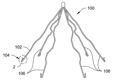
Figure 1 is a perspective view of a vena cava filter 100 having an elongate
member 102. Elongate member 102 may include an anchoring member 104 disposed
on the end. Detailed reference is made to elongate member 102, but filter 100
may
include additional elongate members 106, which may be configured substantially
like
elongate member 102. Filter 100 is selected to illustrate this embodiment, but
neither
this embodiment or any embodiment is limited to Filter 100. ~ther applications
are
contemplated. For example, the embodiment may be readily adapted to other vena
cava filters. Indeed, the embodiment may be readily adapted to any implantable
medical device retained in position by elongate members. Likewise, neither
this
embodiment or any embodiment is limited to medical devices retained by
generally
straight elongate members where an end of the elongate members is in contact
with
the vessel wall. Embodiments are contemplated where the elongate member
contacts
CA 02559845 2006-09-08
WO 2005/087136 PCT/US2005/007417
a vessel wall with a middle portion rather than an extremity. Such an elongate
member may be, for example, curved. Embodiments, therefore, are contemplated
with a wide variety of medical devices of numerous configurations.
In the present embodiment, elongate member 102 may be made from stainless
steel or other suitable biocompatible materials such as nickel-titanium
alloys.
Elongate member 102 has a generally circular cross-section. Other suitable
cross-
sections are contemplated. For example, elliptical or rectangular may be
equally or
more suitable in certain applications. Elongate member 102 may be, if desired
coated
with therapeutic agents. For example, elongate member 102 may be coated with
an
agent to resist neointimal hyperplasia.
Refer now to Figure 2, which is an enlarged side view of an end of elongate
member 102. Anchor member 104 can be seen in greater detail. Anchor member
104 has a hook shape with a barb 108 disposed on the end. The hook shape and
bard
108 may be used to retain filter 100 in a desired position. Disposed on
elongate
member 102 is an edge 110. Edge 110 generally faces in towards the center of
filter
100 and away from the vessel wall. Edge 110 may be disposed on a blade 112 or
may
be a shaped part of elongate member 102. For example, edge 110 may be formed
by
electron deposition machining elongate member 102. Edge 110 may start near the
hook end of elongate member 102 and extend up a portion of elongate member
102.
In one embodiment, edge 110 extends sufficiently up elongate member 102 so
that the
a portion of edge 110 has little chance of being encapsulated by a neointimal
hyperplasia process. In other words, edge 110 may extend far enough away from
the
vessel wall and up elongate member 102 to keep exposed. Edge 110 should be
sharp
enough to cut through vessel growth.
Figure 3 is a side view of an elongate member 202 of an intravascular device
of another embodiment. Elongate member 202 may be part of a vena cava filter
or
may be part of another intravascular device. Elongate member may include an
anchor
member 2.04 and includes an edge 210. Edge 210 is disposed in a cut-out 214 of
elongate member 204 and may be on a blade 212. The cut-out serves to reduce
the
overall cross section of the elongate member with the blade. In one
embodiment, cut-
out 214 is of sufficient depth so that the cross-section of the portion of
elongate
member 202 with edge 210 does not extend beyond the portion of elongate member
202 without a cut-out. Thus, the intravascular device may have a reduced
profile
when compressed for insertion or extraction.
4
CA 02559845 2006-09-08
WO 2005/087136 PCT/US2005/007417
Figure 4 is a top view of an elongate member 302 of an intravascular device
according to the invention. Elongate member 302 includes an anchor member 304,
which may be similar to anchors members previously described or may be another
suitable anchor member. Elongate member includes an inward facing edge 310 and
anchor member 304 includes an inward facing edge 3 I6. Thus, both edges should
face away from the portion of elongate member 302 and anchor member 304 which
are configured to contact the vessel wall. Edges 310 and 316 are susceptible
to several
contemplated variations. For example, in the pictured embodiment, the edges
are
substantially straight and are disposed on substantially straight portions of
elongate
member 302 and anchor member 304. In another embodiment, edges 310 and 316
may extend to join and form one continuous edge, curving between the elongate
member and the anchor member. In another embodiment, there may be a third edge
between edges 310 and 316, which may be disposed at a different angle and yet
still
away from the vessel wall. For example, this third edge may be disposed more
towards the direction in which the intravascular device may be retracted. In
another
embodiment, this third edge may smoothly join with edges 310 and 316.
Figure 5 is a perspective view of a thrombosis filter 400, which includes
several elongate members 402 having anchoring members 404. Figure 6 is a
perspective view of an elongate member 402. Elongate member 402 includes a
blade
412 having two or more separated, inward facing edges 410. Edges 410 may be
separate by a break 418 in the blade. Break 418 may be a complete gap between
two
sections of blade 412 or may be a partial removal of material. For example,
break 418
may be a v-shaped or u-shaped slot between two portions of the blade. In
another
embodiment, break 418 is a slight radial offset between two sections of blade
412 and
may not include a longitudinal gap. Break 418 may be created by removing
material
during the shaping of the blade or by removing material after the blade is
assembled
and joined. Break 418 may also be created by assembling the blade to the
elongate
member in several pieces.
Other embodiments are contemplated. One example embodiment of an
intravascular device has elongate members where an edge that faces generally
inwards is set in a cut-out of the elongate member to reduce the overall
profile, where
that edge also includes one or more gaps. In another embodiment, one or more
of the
elongate members may be coated with a therapeutic agent, such as an anti-
angiogenesis drug or other desired agent. In another embodiment, the
intravascular
CA 02559845 2006-09-08
WO 2005/087136 PCT/US2005/007417
device has elongate members with inward facing edges and anchor members
configured to easily break away from the device. The embodiments herein
described
are only a limited selection of the contemplated embodiments and serve to
illustrate
the invention and show the broad applicability of the invention to many
embodiments.
Figure 7 is a diagrammatic view of a portion of elongate member 102 after
vena cave filter 100 has been installed in a vena cave for a period of time
sufficient
for neointiminal hyperplasia to occur. Elongate member 102 includes anchor
member
104 and edge 110, which edge is disposed on blade 112. The wall of the vena
cave
includes the adventitia 120, the media 122 arid the intima 124. It is this
last layer,
intima 124, that encapsulates the anchor member and a portion of elongate
member.
As can be seen, edge 110 faces away from the wall of the body vessel and
towards the
vessel centerline. Egde 110 is configured to extend beyond the portion of
elongate
member 102 likely to be encapsulated by intima 124 and expected neointimal
hyperplasia.
When removal of vena cave filter 100 is desired, it may be accomplished by
the following process, or by another suitable process. Vena cave filter 100
may be
held to prevent undesired longitudinal motion, perhaps by grasping the filter
with a
retention device on the end of a guide wire or by other suitable method.
Elongate
member is urged inward. This urging may be accomplished by the action of a
catheter upon the elongate member, for example. When a catheter is slid over
the
vena cave filter, the inner lip of the catheter end will provide a force on
the elongate
member that will tend to move the elongate member inwards. When the elongate
member is urged inward, edge 102 may cut through intima 124, and thus provide
a
passage for the end of the elongate member and the anchor member through the
intima. By cutting rather than tearing a passage through the intima, trauma to
the
vessel wall may be reduced. Trauma is reduced because a cut is generally less
traumatic to tissue than a tear. Trauma is also reduced because a cut may be
created
using less force than a tear, and thus the surrounding tissue is subjected to
less force.
Also, less force needs to be delivered via the catheter. Thus, removal is
possible in
more situations. The urging of the elongate member may be done using one full
motion, or may be done using smaller, reciprocating motions if desired. By
providing
a configuration where the edge extends from the intima into the vessel lumen,
a spot
on the vessel wall is provided where the cut may be readily started. When the
intima
is cut, the end portion of elongate member 102 and anchor member 104 may be
6
CA 02559845 2006-09-08
WO 2005/087136 PCT/US2005/007417
readily removed from the vessel wall and the vena cave filter may then be
compressed
and removed.
Numerous advantages of the invention covered by this document have been set
forth in the foregoing description. It will be understood, however, that this
disclosure
is, in many respects, only illustrative. Changes may be made in details,
particularly in
matters of shape, size, and arrangement of parts or order of steps without
exceeding
the scope of the invention. The invention's scope is, of course, defined in
the
language in which the appended claims are expressed.
7