Note: Descriptions are shown in the official language in which they were submitted.
DEMANDES OU BREVETS VOLUMINEUX
LA PRESENTE PARTIE DE CETTE DEMANDE OU CE BREVETS
COMPREND PLUS D'UN TOME.
CECI EST LE TOME 1 DE 2
NOTE: Pour les tomes additionels, veillez contacter le Bureau Canadien des
Brevets.
JUMBO APPLICATIONS / PATENTS
THIS SECTION OF THE APPLICATION / PATENT CONTAINS MORE
THAN ONE VOLUME.
THIS IS VOLUME 1 OF 2
NOTE: For additional volumes please contact the Canadian Patent Office.
CA 02560581 2006-09-20
DSTY-R678
- 1 -
DESCRIPTION
PLURIPOTENT STEM CELL GROWING METHOD
Technical Field
The present invention relates to a growing method
and to a gene transfer method for pluripotent stem cells
such as ES cells, and to pluripotent stem cells prepared
by the methods.
Background Art
In order to continue to live, organisms have the
ability to rapidly replace and repair lost or damaged
cells and tissue, and this ability is known as
"regenerative capacity". Examples of "regenerative
capacity" in higher animals include the commonly known
phenomena of wound healing of skin and blood vessels, but
even parenchymal organs such as the liver and kidneys are
known to undergo cell growth and tissue reconstruction
for rapid restoration of tissue homeostasis in response
to tissue damage. Recent years have seen attempts to
utilize this innate "regenerative capacity" of biological
organisms to achieve cures or amelioration of various
diseases and wounds, and such new medical techniques are
coming to be known as "regenerative medicine".
Stem cells play a central role in practicing
"regenerative medicine". "Stem cells" can be generally
defined as undifferentiated cells having the ability to
differentiate into specialized cells or polyfunctional
cells, as well as having the ability to self-replicate,
allowing repeated generation of cells identical to
themselves. Unique stem cells are found in each tissue
and cell type, and for example, blood cells such as
erythrocytes, lymphocytes and megakaryocytes are produced
via progenitor cells derived from stem cells known as
"hematopoietic stem cells", while skeletal muscle cells
are produced from stem cells/precursor cells known as
CA 02560581 2010-02-02
f
=
- 2 -
=
"satellite cells" and "myoblasts". Additional types that . -
have been identified to date include neural stem cells
that are found in neural tissue such as the brain and
spinal cord and produce neurons and glial.cells,
5. epidermal stem cells that produce epidermal cells and
hair follicle cells, oval cells (hepatic stem cells): that
produce hepatocytes and bile duct cells, and cardiac.stem
cells that produce cardiomyocytes.
=
Some regenerative medicine treatments using stem
cells or precursor cells derived from such cells have
= 'already been implemented, and infusion graft methods with
hematopoietic stem cells or hematopOietic precursor cells
are well known for treatment of Conditions caused by a
lack or functional deficiency of blood cells, such as
leukemia and 1 vlastic anemia. However, stem cells
present in parenchymal organs such as the brain, heart or
liver are technicarry¨ctifficurt-tb¨ort-arri "from -n.¨v-1-n4
= = tissues .and/or to culture in vitro, and such stem cells
= = also generally have low proliferation potency. =Stem
. cells can also be recovered from tissues from. corpses,
but the medical uSe of cells obtained in this manner is
associated with ethical problems. Consequently, .
regenerative treatments for neutopathy, cardiopathy and
= the like will require the development of techniques for
generatingsdesired cell types using cells other than stem
cells present in Such target tissues.
First, methods of utilizing "pluripotent stem cells"
may be mentioned as strategies based on this approach.
"Pluripotent stem cells" are defined as cells capable of
prolonged or virtually indefinite proliferation in vitro
while retaining their undifferentiated state, exhibiting
normal karyotype (chromosomes) and having the capacity to
differentiate into all tell typeS of the three germ
= layers (ectoderm, mesoderm and endoderm) under the
appropriate,conditions. Currently the most commOnly
known pluripotent stem cells are embryonic stem cells (ES
cells) isolated from the. early embryo, and the analogous
=
=
CA 02560581 2006-09-20
- 3 -
embryonic germ cells (EG cells) isolated from fetal
primordial germ cells, both of which are the subjects of
ongoing research.
ES cells can be isolated as an undifferentiated stem
cell population by transferring the inner cell mass of. a
blastocyst-stage embryo to in vitro culture and repeating
the process of detaching and passaging the cell mass.
The cells have suitable cell density on feeder cells
prepared from primary cultured murine embryonic
fibroblasts (hereinafter, MEF cells) derived from murine
fetal tissue or stromal cells such as STO cells, and
repeated passaging with frequent replacement of the
culture medium can lead to establishment of a cell line
retaining the property of undifferentiated stem cells.
Another feature of ES cells is the presence of the enzyme
telomerase, which exhibits an activity of maintaining
chromosomal telomere length, and this enzyme confers to
ES cells the capacity for virtually unlimited cell
division in vitro.
ES cell lines produced in this manner are
"pluripotent" as they can be repeatedly grown and
passaged almost indefinitely while maintaining normal
karyotype, and they are capable of differentiating into
various different cell types. For example, when ES cells
are transplanted into an animal body subcutaneously,
intraabdominally or intratesticularly they form tumors
called "teratomas", but the tumors comprise a mixture of
different cells and tissues including neurons,
osteocytes, chondrocytes, intestinal cells, muscle cells
and the like. In mice, intrauterine transplantation into
a pseudopregnant mouse of an aggregate embryo generated
by infusion graft of ES cells into a blastocyst-stage
embryo or aggregation with an eight-cell stage embryo,
results in generation of a "chimeric mouse", which is an
offspring possessing differentiated cells derived from
the ES cells throughout the entire body or in parts of
its organs and tissues. This technique is often used as
CA 02560581 2010-02-02
' o '
.
.
¨ 4 -
a main method for generating "knockout mice" having
certain genes which are artificially disrupted or
modified.
It is also well known that ES cells are induced to
differentiate into diverse types of cells by in vitro
culturing as well. While the specific method differs
depending on the type of cell, it is common to employ a
method of inducing differentiation by forming an
"embryoid body" (hereinafter, "EB") which is .a cell mass
in an embryo-like state produced by aggregating ES cells
by suspension culture. Such a method can produce cells
=
having fetal stage endoderm, ectoderm and mesoderm
characteristics, as well as differentiated cells such as
blood cells, vascular endothelial cells, chondrocytes,
skeletal muscle cells, smooth muscle cells,
cardiomyocytesi glial cells, neurons, epithelial cells,
melanocytes, keratinocytes, adipocytes and the like.
Differentiated cells produced by in vitro culturing in
this fashion have essentially the same structural and
functional features as cells present in organs and
tissues, and transplant experiments using experimental
= animals have demonstrated that ES cell-derived cells
anchor to organs and tissues and function normally.
For reviews of ES cell properties and culturing
methods, and their in vivo and in vitro differentiating
abilities, refer to the following literature: Guide to
=
Techniques in Mouse Development (Wasserman et ai., =
=
Academic Press, 1993); Embryonic Stem Cell =
Differentiation in vitro (M.V. Wiles, Meth. Enzymol.
225:900, 1993); Manipulating =the Mouse Embryo:
ALaboratoryManual (Hogan et al., Cold Spring Harbor
= Laboratory Press, 1994)(Non-patent document 1); Embryonic
Stem Cells (Turksen, ed., Humana Press, 2002)(Non-patent
document 2).
EG cells can be produced by stimulating fetal germ
cells known as primordial germ cells on feeder cells such
as MEF cells or STO cells in the same manner as ES cells,
CA 02560581 2006-09-20
- 5 -
using Leukemia Inhibitory Factor (hereinafter, LIF) and
basic Fibroblast Growth Factor (hereinafter, bFGF/FGF-2),
or chemical agents such as forskolin (Matsui et al., Cell
70:841, 1992; Koshimizu et al., Development 122:1235,
1996). It has been confirmed that EG cells have
properties very similar to ES cells and have pluripotency
(Thomson & Odorico, Trends Biotechnol. 18:53, 2000).
Throughout the present specification, therefore, the term
"ES cells" may include "EG cells".
After Thomson et al. first established ES cells from
a primate (rhesus monkey) in 1995, the concept of
regenerative medicine using pluripotent stem cells began
to approach the realm of possibility (U.S. Patent No.
5,843,780; Proc. Natl. Acad. Sci. USA 92:7844, 1995).
Later, the researchers used similar methods to
successfully isolate and establish ES cell lines from
human early embryos (Science 282:114,1998). Research
groups in Australia and Singapore later submitted similar
reports (Reubinoff et al., Nat. Biotech. 18:399, 2000;
International Patent Publication No. W000/27995), and
currently 20 different human ES cell lines have been
registered at the U.S. National Institutes of Health
(NIH)(http://stemcells.nih.gov/registry/index). Also,
Gearhart and their colleagues have succeeded in
establishing a human EG cell line from human primordial
germ cells (Shamblott et al., Proc. Natl. Acad. Sci. USA
95:13726, 1998; U.S. Patent No. 6,090,622).
When these pluripotent stem cells are used to
produce research materials or regenerative medicine
products, it is essential that the passaging methods used
maintain the undifferentiated state and high
proliferation potency of the cells. MEF cells or similar
cells (such as STO cells) are usually used as feeder
cells for ES/EG cells to maintain the undifferentiated
state and high proliferation potency of the cells.
Addition of fetal bovine serum (hereinafter, FBS) to the
culture medium is also important, and it is crucial to
CA 02560581 2010-02-02
÷.
- 6 -
=
select an FBS product which is suited for the culturing
of the ES/EG cells, usually with the addition of FES at =
about 10-20%. Also, LIF has been identified as a factor
that maintains the'undifferentiated state of ES/EG cells
=
derived from mouse embryo (Smith & Hooper, Dev. Biol.
121:1, 1987; Smdth et al., Nature 336:688, 1988; Rathjen
et al., Genes Dev. 4:2308, 1990), and addition. of LIF to
culture can more effectively maintain the
undifferentiated state (see the following literature: .
Manipulating the Mouse Embryo: ' ALaboratory Manual (Hogan
et al., Cold Spring Harbor Laboratory Press, 1994 (Non-
patent document 1) and Embryonic Stem Cells (Turksen ed.,
= 1
Humana Press, 2002)(Non-patent document 2)). =
However, the culturing methods employed for these
classical ES/EG cells are not suitable methods when huMan
ES (or EG) cells are used for regenerative medicine o;
other practical purposes. One reason for this is that.
human-ES cells are unresponsive to LIF, and lack of
feeder cells causes death of the cells or loss of the
undifferentiated state and differentiation into different
= cell types (Thomson et al., Science 282:1145, 1998). The
=
use of feeder cells itself is another problem because
such co-culturing systems increase production cost and
= are poorly suited for large-scale culturing, while
, 25 separation and purification of the ES cells from the
feeder cells is required when the ES cells are to be
actually used. In the future, when human ES cells and
other pluripotent stem cells are utilized as cell sources
for regenerative medicine, and particularly for cell
transplantation therapy, the use of non-human animal cell
products such as MEF cells and FS will not be desirable
= because of risks including potential infection of the ES
cells by heterozoic viruses and contamination with
= antigenic molecules that may be recognized as
heteroantigens (Martin et al., Nature Med. 11:228, 2005).
Consequently, in order to refine ES/EG cell
culturing methods and modify them to be suitable for
CA 02560581 2012-05-28
- 7 -
future implementation, active efforts are being made to
develop FBS substitutes (International Patent Publication
No. W098/30679) and to utilize human cells as feeders
instead of MEF cells (Richards et al., Nature Biotech.
20:933, 2002; Cheng et al., Stem Cells 21:131, 2003;
Hovatta et al., Human Reprod. 18:1404, 2003; Amit et al.,
Biol. Reprod. 68:2150, 2003). Development of culturing
methods using no feeders is another alluring prospect.
Carpenter and coworkers have reported that seeding of ES
cells in a Matrigel- or Laminin-coated culturing plate
and addition of MEF cell conditioned medium to the
culture medium allows prolonged culturing of human ES
cells which retain their undifferentiated and
pluripotency (Xu et al., Nature Biotech. 19:971, 2001
(Non-patent document 3); International Patent Publication
No. W001/51616 (Patent document 1)). The same group also
succeeded in constructing a more effective ES cell
culturing system by developing a serum-free medium
containing added bFGF/FGF-2 or Stem Cell Factor
(hereinafter, SCF) (International Patent Publication No.
W003/020920 (Patent document 2)). An ES cell culturing
system using the same serum-free medium and requiring no
feeder has also been reported by an Israeli research
group (Amit et al., Biol. Reprod. 70:837, 2004 (Non-
patent document 4)). Recently, a method of maintaining
the undifferentiated state of human ES cells by addition
of bFGF/FGF-2 and the bone morphogenetic protein
antagonist Noggin has also been reported (Xu et al.,
Nature Methods 2:185, 2005). Separately, it has been
shown that simple addition of Glycogen Synthase Kinase
(GSK)-3 inhibitor to culture medium can efficiently
maintain the undifferentiated state of murine and human
ES cells without addition of growth factors or the like
and without using feeder cells (Sato et al., Nature Med.
10:55, 2004 (Non-Patent document 5)).
Thus, while new methods are being proposed for
culturing of pluripotent stem cells without the use of
* Trade-mark
CA 02560581 2006-09-20
- 8 -
feeder cells, actual implementation and industrial use of
such cells will require even greater convenience of
pluripotent stem cell growth effects and culturing
methods.
One well known factor that maintains the
undifferentiated state of murine ES/EG cells and
increases their proliferation potency is the LIF
mentioned above, and while the LIF-related IL-6 family of
molecules falls under this category (Yoshida et al.,
Mech. Dev. 45:163, 1994; Koshimizu et al., Development
122:1235, 1996), very few other examples have been
reported. Recently, serum-free medium containing added
bFGF/FGF-2 or SCF has been reported to notably promote
the proliferation potency of human ES cells
(International Patent Publication No. W003/020920 (Patent
document 2)).
Given the active, i.e., proliferating, nature of ES
cells in comparison to other cell types, few attempts
have been made to actually investigate their
proliferation potency; however, the needs of regenerative
medicine will require increased proliferation of such
cells.
One of the problems currently encountered in
culturing pluripotent stem cells is that the cells
generally form tight colonies and are therefore difficult
to handle for passaging and the like. Undifferentiated
ES/EG cells are usually found in a condition with the
cells firmly adhering to each other, forming colonies,
i.e. cell masses with indistinct boundaries between
cells. For provision of ES/EG cells for passaging or
differentiation-inducing experiments, it is therefore
necessary to disperse the colonies in as short a period
as possible by treatment with protease solutions of
trypsin or the like. When this is done, however,
dispersion of the ES/EG cell colonies into individual
cells requires relatively high-concentration protease
treatment and/or vigorous mechanical stirring, and such
CA 02560581 2006-09-20
- 9 -
procedures significantly reduce the viability and
adhesion ability of the ES/EG cells.
Moreover, since ES/EG cells undergo spontaneous
differentiation during continuous culturing in a
clustered condition, they must be dispersed to single
cells during passaging and the passaging must be carried
out before colonies grow to an excessive size. Murine ES
cells, for example, generally require each passaging to
be conducted for 2-3 days, and if the passaging is not
conducted by a suitable method, cells that have deviated
from their undifferentiated state may appear in the
cluster, rendering the cells unsuitable for use. This
cannot be overcome simply by adding a sufficient amount
of a factor that maintains the undifferentiated state of
ES/EG cells, such as the LIF mentioned above or GSK-3
inhibitors, and excessive colony growth and cells with a
differentiated phenotype are induced. Therefore, a
method of growing ES/EG cells without formation of
colonies, i.e., with the cells individually dispersed, is
expected to be highly useful for providing ES/EG cells
for industrial use. However, no such attempts or
successes can be found to date.
The present inventors have previously seeded F9
cells, an embryonal carcinoma cell line known to normally
proliferate by colony formation, on a culture plate
coated with E-cadherin (Nagaoka et al., Biotechnol. Lett.
24:1857, 2002 (Non-patent document 6)) and have found
that this prevents formation of cell colonies
(International Symposium on Biomaterials and Drug
Delivery Systems, 2002.04.14-16, Taipei, Taiwan; 1st
Meeting of the Japanese Society for Regenerative
Medicine, 2002.4.18-19, Kyoto, Japan). Specifically, F9
cells exhibited a dispersed cell morphology on a
culturing plate having E-cadherin, which is a known cell
adhesion molecule for F9 cells, immobilized on an
untreated polystyrene culturing plate (hereinafter, "E-
cad plate").
CA 02560581 2006-09-20
- 10 -
F9 cells exhibit a phenotype somewhat similar to ES
cells, expressing alkaline phosphatase (hereinafter, ALP)
or SSEA-1 and Oct-3/4, which are known as specific ES/EG
cell markers (Lehtonen et al., Int. J. Dev. Biol. 33:105,
1989, Alonso et al., Int. J. Dev. Biol. 35:389, 1991).
However, F9 cells do not require feeder cells or LIF for
maintenance of the undifferentiated state of the cells,
and therefore are different in their mechanism of
maintaining undifferentiation. Moreover, whereas ES
cells have triploblast differentiating potential to all
three germ layers, the differentiation of F9 cells is
limited to endodermal cells, and they are unable to form
chimeras. In =other words, although F9 cells are used as
an ES/EG cell model system in some experiments, they
differ from ES/EG cells in many aspects involving the
culturing method and culturing conditions.
Thus, it was not possible to predict, based on the
scientific evidence, whether the aforementioned E-cad
plate can be used in ES cell culturing methods that
require no feeder cells, whether ES cells cultured by
such methods can be passaged while maintaining their
undifferentiated state and pluripotency, and whether the
proliferation potency of the ES cells can be increased.
In fact, the proliferation potency of F9 cells cultured
on an E-cad plate is roughly equivalent to that of F9
cells cultured on a conventional cell culturing plate,
and no data had been obtained to suggest that the
proliferation potency of ES cells could thereby be
increased.
E-cadherin is known to be expressed by
undifferentiated murine ES cells, and it is also known
that intercellular adhesion is notably inhibited with ES
cells that lack E-cadherin gene expression due to gene
modification (Larue et al., Development 122:3185, 1996).
However, it has not yet been attempted to use E-cadherin
as an adhesion substrate in an ES/EG cell culturing
method.
CA 02560581 2006-09-20
- 11 -
In addition to the efficient culturing methods
described above, when pluripotent stem cells such as ES
cells are to be used as a laboratory material or for
production of regenerative medicine products, it is also
necessary to design methods for efficiently introducing
selected exogenous genes into the cells and expressing
them. In particular, one strategy for applying ES cells
in regenerative medicine for treatment of various
diseases is to modify the cell properties, such as
proliferation and differentiation potency or the drug
sensitivity, and this can be satisfactorily realized by
introducing and expressing appropriate exogenous genes in
the cells. In the case of murine ES cells, it is widely
known that genes can be artificially modified to produce
transgenic mice or knockout mice, for which efficient
gene transfer methods are especially useful.
Ordinary transfer of exogenous genes into cells is
frequently accomplished using agents such as calcium
phosphate, DEAE-dextran and cationic lipid preparations.
However, application of such methods to ES cells is known
to result in lower efficiency than for other cell types
(Lakshmipathy et al., Stem Cells 22:531, 2004 (Non-patent
document 8)). Methods using various viral vectors for
transfer of exogenous genes have also been reported. For
example, retroviral vectors (Cherry et al., Mol. Cell
Biol., 20:7419, 2000), adenovirus vectors (Smith-Arica et
al., Cloning Stem Cells 5:51, 2003), lentivirus vectors
(Amaguchi et al. J. Virol. 74:10778, 2000; Asano et al.,
Mol. Ther. 6:162, 2002; International Patent Publication
No. W002/101057), and Sendai virus vectors (Sasaki et
al., Gene Ther. 12:203, 2005; Japanese Unexamined Patent
Publication No. 2004-344001) are publicly known.
Nevertheless, the construction and preparation of viral
vectors require relatively complex and time consuming,
while biological safety is also an issue, depending on
the virus, and therefore such methods are neither
convenient nor universally employed.
CA 02560581 2010-02-02
, =
.
.
- 12 -
Consequently, exogenous gene transfer into ES cells
is most commonly carried out by a method known as
electroporation. This technique involves application of
an electrical pulse to cells to transiently open pores in
the cell membranes for introduction of an exogenous gene
into the cells, and it is a highly flexible method.
Recently, an improved technique called nucleofection has
been estblished, whereby an exogenous gene is
transferred directly into cell nuclei= to achieve
significantly higher expression efficiency (Lorenz et
al., Biotech. Lett. 26:1589, 2004; Lakshmipathy et al.,
Stem Cells 22:531, 2004 (Non-patent document 8)).
However, this method requires a special electrical pulse-
= generating device, and it is not easy to prepare the
optimal conditions. Furthermore, it is necessary to
first treat the cells with a protease such as trypsin to
disperse the individual Cells, and therefore the cell
toxicity is relatively high.
Thus, the most useful gene transfer methods for
pluripotent stem cells such as ES cells would be methods
using gene transfer agents that are inexpensive and
convenient to prepare, and would allow efficient transfer
of exogenous genes into cells being cultured in an
= incubator.
Non-patent document 1: Manipulating the Mouse
= Embryo: A Laboratory Manual (Hogan et al., Cold Spring
Harbor Laboratory Press, 1994).
Non-patent document 2: Embrycinic Stem Cells
(Turksen, ed. Humana Press, 2002).
Non-patent document 3: Xu. et al., Nature Biotech.
19:971, 2001.
Non-patent document 4: Amit et al., Biol. Reprod.
70:837, 2004.
Non-patent document 5: =Sato et al., Nature Med.
10:55, 2004. =
Non-patent document 6: Nagaoka et al., Biotechnol.
Lett. 24:1857, 2002.
CA 02560581 2006-09-20
- 13 -
Non-patent document 7: Nagaoka et al., Protein Eng.
16:243, 2003.
Non-patent document 8: Lakshmipathy et al., Stem
Cells 22:531, 2004.
Patent document 1: International Patent Publication
No. W001/51616.
Patent document 2: International Patent Publication
No. W003/020920.
Disclosure of the Invention
In light of these circumstances, it is an object of
the present invention to provide a method for culturing
pluripotent stem cells such as ES cells without using
feeder cells, wherein the proliferation potency of the
cells is augmented and the gene transfer efficiency is
increased.
In order to solve the problems described above, the
present inventors studied the possibilities of increasing
proliferation potency and increasing gene transfer
efficiency for ES cells, by culturing the cells in a
state without colony formation, or in other words, in a
dispersed state.
As mentioned above, the present inventors have
succeeded in culturing F9 cells, an embryonal carcinoma
cell line, without colony formation, i.e., in a dispersed
state. When a cell culturing plate which had E-cadherin
immobilized or coated on a solid phase surface (E-cad
plate) was prepared and F9 cells were seeded on the
plate, the F9 cells exhibited a dispersed cell morphology
without colony formation. The proliferation potency was
essentially the same for F9 cells cultured on the E-cad
plate and F9 cells cultured on an ordinary plate.
When it was attempted to seed ES cells on an E-cad
plate, virtually all of the cells adhered to the plate,
and they exhibited a dispersed cell morphology without
colony formation, similar to F9 cells. Most notably, the
proliferation potency of ES cells seeded on the E-cad
CA 02560581 2006-09-20
- 14 -
plate under these culturing conditions was significantly
higher than the proliferation potency of ES cells
cultured on an ordinary plate. Also, the exogenous gene
transfer efficiency and expression level were
significantly higher as well.
It was further confirmed that ES cells passaged
multiple times on an E-cad plate are still
undifferentiated and maintain their pluripotency if a
factor that maintains undifferentiation is added to the
liquid medium. In addition, it was demonstrated that ES
cells prepared by the aforementioned method can be
induced to differentiate into functional differentiated
cells such as neurons and cardiomyocytes by using known
methods, and that chimeric mice can be generated by
transplanting them into mouse early embryos, and the
present invention was thereby completed.
Therefore, according to a first mode of the
invention there is provided a novel method of growing
pluripotent stem cells, such as ES cells, which requires
no feeder cells. The method of the invention is
characterized in that the culturing vessel used has a
pluripotent stem cell-adhering molecule immobilized or
coated on a substrate solid phase surface at a
predetermined density, whereby the cells can be cultured
in a dispersed state for increased proliferation ability.
The pluripotent stem cells prepared in this manner retain
their undifferentiated state and pluripotency.
According to a second mode, the method of the
invention is characterized in that the culturing vessel
used has a pluripotent stem cell-adhering molecule
immobilized or coated on a substrate solid phase surface
at a predetermined density, whereby culturing of the
cells in a dispersed state can increase the gene transfer
efficiency into the cells.
As another working mode, the invention relates to
pluripotent stem cells having an undifferentiated state
and pluripotency, which are prepared by the method
CA 02560581 2010-02-02
- 15 -
disclosed by the invention. For the purposes of the =
present disclosure, the "undifferentiated" state of
pluripotent stem cells can be confirmed by expression of
at least one undifferentiation marker. . =
According to another working mode, the invention .
relates to differentiated cells produced, by appropriate
differentiation inducing treatment, from pluripotent stem
.cells prepared by the method disclosed for the invention.
The differentiated cells are not particularly restricted
as long as they are of a cell type whose differentiation -
can generally be induced from pluripotent stem cells. .
Specifically, there may be mentioned ectodermal cells or
ectoderm-derived cells, mesodermal cells or mesoderm-
. 'derived Cells, endodermal cells or endoderm-derived . =
cells, and the like.
= . According to another working mode, the invention
relates to a method of generating a chimeric embryo. or
= chimeric animal using pluripotent stem cells prepared by
the method disclosed for the invention, and to the
generated chimeric embryo or chimeric animal.
The present invention primarily relates to the
following aspects.
= (1) A growing method for pluripotent stem cells,
characterized by growing the pluripotent stem cells in a
dispersed state while maintaining their undifferentiated
state and pluripotency, using a liquid mediuM and a
culturing vessel having immobilized or coated on a
substrate solid phase surface a molecule which is
=
adhesive to the pluripotent stem cells, without using .
feeder cells. ==
(2) A gene transfer method for pluripotent stein
Cells, characterized by efficiently transferring a gene
into the pluripotent stem cells and I expressing it, using a
liquid medium and a culturing vessel having immobilized
or coated on a substrate solid phase surface a molecule
which is adhesive to the pluripotent stem cells. .
(3) The method of .(1) or (2) above, wherein the '
=
CA 02560581 2006-09-20
- 16 -
molecule which is adhesive to the pluripotent stem cells
is either a molecule that is expressed by the pluripotent
stem cells, or a molecule that is structurally homologous
with the molecule and has homophilic binding ability with
the pluripotent stem cells.
(4) The method of (3) above, wherein the molecule
which is adhesive to the pluripotent stem cells is a
molecule belonging to the cadherin family.
(5) The method of (4) above, wherein the molecule
belonging to the cadherin family is E-cadherin, or a
molecule which has structural homology with that
molecule, which comprises the EC1 domain and one or more
domains from among the EC2 domain, EC3 domain, EC4 domain
and EC5 domain of E-cadherin, and which has homophilic
binding ability with the pluripotent stem cells.
(6) The method of (5) above, wherein the E-cadherin
is derived from a mammal.
(7) The method of (6) above, wherein the E-cadherin
is derived from a human or mouse.
(8) The method of any one of (1) to (7) above,
wherein the molecule which is adhesive to the pluripotent
stem cells is fused with an immunoglobulin Fc region and
is immobilized on the substrate solid phase surface via
the Fc region.
(9) The method of any one of (1) to (8) above,
wherein the pluripotent stem cells are mammalian
embryonic stem cells (ES cells) or embryonic germ cells
(EG cells).
(10) Pluripotent stem cells produced by the method
of any one of (1) to (9) above.
Brief Explanation of the Drawings
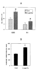
Fig. 1 is a pair of graphs showing adhesion of ES
cells (R1 and EB3 cell lines) to E-cad-Fc coated on a
polystyrene plate. The adhesion rate represents a
relative value in which 100% is the number of ES cells
adhering to a plate coated with gelatin (0.1%). BSA:
CA 02560581 2006-09-20
- 17 -
Group with ES cells adhered to plate coated with 0.1%
bovine serum albumin. *: with respect to gelatin group,
p<0.01.
Fig. 2 is a set of photographs showing the
morphology of ES cells seeded on an E-cad-Fc plate. The
cell images were taken two days after seeding the ES
cells on a plate coated with gelatin, type I collagen,
fibronectin or E-cad-Fc (indicated by Gel., Col., Fn. and
E-cad-Fc, respectively).
Fig. 3A is a graph showing the proliferation potency
of ES cells seeded on an E-cad-Fc plate. The ES cells
were seeded on a gelatin plate (indicated as Cont) or an
E-cad-Fc plate, and the cell counts on the third day were
compared. *: with respect to Cont group, p<0.01.
Fig. 3B is a graph showing BrdU uptake by ES cells
seeded on an E-cad-Fc plate. The ES cells (EB3) were
seeded on a gelatin plate (indicated as Cont) or an E-
cad-Fc plate, and labeled with BrdU. The BrdU taken up
into the cells after three days was detected by antibody
staining using a fluorescent dye. *: with respect to
Cont group, p<0.01.
Fig. 4A is a set of photographs showing expression
of a marker for an undifferentiated state of ES cells
seeded on an E-cad-Fc plate. The ES cells (EB3 cell
line) were seeded on a gelatin plate (indicated as Cont)
or an E-cad-Fc plate, and ALP activity was detected on
the 14th day of culturing. In this figure, LIF(+) and (-
respectively indicate addition/non-addition of LIF to
the culturing medium.
Fig. 4B is a set of photographs showing expression
of markers for undifferentiated ES cells seeded on an E-
cad-Fc plate. The ES cells (EB3 cell line) were seeded
on a gelatin plate (indicated as Cont) or an E-cad-Fc
plate, and Oct-3/4 protein was detected on the 14th day
of culturing. DAPI: Nuclear staining with DAPI. Merged:
Superposition of DAPI and Oct-3/4 antibody stain.
Fig. 5 is a set of photographs showing expression of
CA 02560581 2006-09-20
- 18 -
markers for undifferentiated ES cells seeded on an E-cad-
Fc plate. The ES cells were seeded on a gelatin plate
(indicated as Cont) or an E-cad-Fc plate, and Oct-3/4 and
Rex-1 gene expressions were examined by RT-PCR on the
14th day. The + and - symbols in the figure respectively
represent addition and non-addition of LIF to the
culturing medium.
Fig. 6 is a graph showing the LIF reactivity of ES
cells seeded on an E-cad-Fc plate. The ES cells (R1
line) were seeded on a gelatin plate (indicated as Cont)
or an E-cad-Fc plate and cultured with different
concentrations of LIF to from colonies, and the ALP
activity was detected for measurement of the proportion
of "undifferentiated" colonies. *: with respect to
Cont/LIF 1000 U/mL, p<0.05.
Fig. 7 is a set of photographs showing pluripotency
of ES cells passaged on an E-cad-Fc plate. The ES cells
(R1 line) seeded and passaged on a gelatin plate
(indicated as Cont) or an E-cad-Fc plate were recovered,
and an EB was formed in LIF-free medium to induce
differentiation. Samples recovered on day 16 after EB
formation ("EB" in the figure) were used for examination
of different differentiation marker gene expressions by
RT-PCR. As control groups there were used ES cells prior
to the aforementioned passaging and the 16th day EB
formed using those cells (1st and 2nd from left in the
figure, respectively). T/Bra: T/Brachyury, 13141:
hemoglobin, AFP: a-fetoprotein, TTR: transthyretin,
GAPDH: glyceraldehyde-3-phosphate dehydrogenase.
Fig. 8 is a pair of photographs showing pluripotency
of ES cells passaged on an E-cad-Fc plate. ES cells (R1
line) seeded on the E-cad-Fc plate were induced to
differentiate into neurons (upper) and cardiomyocytes
(lower). These photos show images of the cells fixed on
the 12th day after inducing differentiation and stained
with antibody for markers specific to neurons and
cardiomyocytes.
CA 02560581 2006-09-20
- 19 -
Fig. 9 is a pair of photographs showing germ-line
transmission of ES cells cultured on an E-cad-Fc plate.
A chimeric mouse generated from ES cells cultured on an
E-cad-Fc plate was crossed with a wild-type C57BL/6
mouse, and the progeny mice were subjected to PCR
analysis using two different microsatellite markers. M:
DNA size marker, ES: ES cells (EB3 line), B6: C57BL/6
mouse, #1-#4: individuals thought to have no
contribution of ES cells based on coat color, #5-#8:
individuals thought to have contribution of ES cells
based on coat color. The numbers on the vertical axis
represent DNA size (bp).
Fig. 10 is graph showing gene transfer/expression
efficiency for ES cells seeded on an E-cad-Fc plate. The
ES cells (EB3) were seeded on a gelatin plate (indicated
as Cont) or an E-cad-Fc plate, and they were subjected to
gene transfer with a GFP expression vector. The GFP in
the cells after one day was detected by antibody stain
using a fluorescent dye, and the fluorescent intensity
was measured. *: with respect to Cont group, p<0.01.
Fig. 11 is a set of photographs showing the
morphology of ES cells seeded on a human E-cad-Fc plate.
The cell images were taken two days after seeding the ES
cells on a plate coated with gelatin (Cont) or E-cad-Fc.
Best Mode for Carrying Out the Invention
(Definitions)
The term "pluripotent stem cells" as used throughout
the present specification refers to cells capable of
prolonged or virtually indefinite proliferation in vitro
while retaining their undifferentiated state, exhibiting
normal karyotype (chromosomes) and having the capacity to
differentiate into all three germ layers (ectoderm,
mesoderm and endoderm) under the appropriate conditions.
The term "pluripotent stem cells" includes, but is not
limited to, ES cells isolated from early embryo and their
analogous EG cells isolated from fetal-stage primordial
CA 02560581 2006-09-20
- 20 -
germ cells. Throughout the present specification, "ES
cells" will be used to include "EG cells".
The term "undifferentiated state" as used throughout
the present specification means the nature of pluripotent
stem cells exhibiting a state of undifferentiation that
can be confirmed based on one or more undifferentiated ES
cell markers such as ALP activity or Oct-3/4 gene
(product) expression, or based on expression of various
antigenic molecules. The state of undifferentiation of
pluripotent stem cells means that the pluripotent stem
cells are capable of prolonged or virtually indefinite
proliferation and exhibit normal karyotype (chromosomes),
while having the capacity to differentiate into all three
germ layers under the appropriate conditions.
The term "pluripotency" as used throughout the
present specification refers to the ability to
differentiate into a variety of cell types. The
differentiated cells are not particularly restricted as
long as they are of a cell type in which differentiation
can generally be induced from pluripotent stem cells.
Specifically, there may be mentioned ectodermal cells or
ectoderm-derived cells, mesodermal cells or mesoderm-
derived cells, endodermal cells or endoderm-derived
cells, and the like.
The term "liquid medium" as used throughout the
present specification includes any liquid medium that can
be used for conventional methods of passaging pluripotent
stem cells.
The term "pluripotent stem cell-adhering molecule"
as used throughout the present specification may refer to
a molecule that binds and adheres with affinity to
pluripotent stem cells, and it may be any of various
types such as a protein, peptide, saccharide chain, low
molecular compound (drug) or the like. As pluripotent
stem cell-adhering molecules, there are preferred
molecules that are expressed in the cells and have
homophilic binding ability, and as examples there may be
CA 02560581 2006-09-20
- 21 -
mentioned the cadherin family of molecules. E-cadherin
is known to be expressed by undifferentiated ES cells and
is therefore preferred for use, but there is no
particular restriction thereto. When the adhering
molecule is a protein or peptide molecule, a peptide
fragment thereof may be used as long as it has the same
adhering activity as the protein or peptide molecule.
A pluripotent stem cell-adhering molecule can be
used for the culturing method of the invention by being
immobilized or coated onto the solid phase surface of a
culturing vessel or culture substrate (hereinafter also
collectively referred to "culture substrate"). As
culture substrates for the invention there may be used
any ones that are conventionally used for cell culturing,
such as a plate or flask. These culture substrates may
be made of inorganic materials such as glass, or of
organic materials such as polystyrene or polypropylene,
but they are preferably sterilizable materials with high
heat resistance and moisture resistance.
The method applied for immobilizing or coating the
pluripotent stem cell-adhering molecule onto the solid
phase surface of the culture substrate may be a physical
method such as adsorption or a chemical method such as
covalent bonding, but an adsorption method is preferred
for ease of operation. Also, an artificial antigenic
molecule may be added to or fused with the adhering
molecule beforehand in order to utilize binding of
specific antibodies for the antigenic molecule. In this
case, the specific antibodies must be immobilized or
coated on the solid phase surface of the culture
substrate beforehand by a physical method such as
adsorption or a chemical method such as covalent bonding.
The culture substrate prepared in this manner can be
used directly for ordinary culturing of the pluripotent
stem cells. That is, an appropriate number of
pluripotent stem cells may be suspended in a commonly
employed liquid medium or cell culture medium, and the
CA 02560581 2006-09-20
- 22 -
mixture applied to the culture substrate. Subsequent
liquid medium replacement and passaging may also be
carried out in the same manner as in conventional
methods.
The term "homophilic binding" as used throughout the
present specification refers to cell-cell or cell-
substrate binding via adhesion molecules that involves
binding or association between the same type of adhesion
molecule.
The term "feeder cells" as used throughout the
present specification refers to separate cells, also
known as support cells, that are cultured beforehand and
perform the role of supplying nutrients and growth
factors which are missing in the medium used for
culturing cells which would be unable to survive and grow
on their own. "Feeder cells" include, but are not
limited to, MEF cells and stromal cells such as STO
cells.
The term "dispersed state" as used throughout the
present specification refers to a state of growing cells
adhered to a culture substrate surface, wherein no
distinct colonies are formed and the individual cells are
either not in contact with other cells or if partially in
contact, have a very small area of contact.
The term "gene" as used throughout the present
specification means genetic material, and refers to
nucleic acid including transcription units. A gene may
be of RNA or DNA, and may be a naturally occurring or
artificially designed sequence. Also, the gene need not
code for a protein necessarily, and for example, it may
code for functional RNA such as a ribozyme or siRNA
(short/small interfering RNA).
Other advantages and features of the invention in
addition to the effect described above will be explained
in the detailed description of the preferred embodiments
provided hereunder.
Unless otherwise specified, gene engineering methods
CA 02560581 2010-02-02
- 23 -
= employed in molecular biology and recombinant DNA
technology, as well as common cell.biology protocols and
conventional techniques, may. be employed for carrying out
the invention, with reference to standard literature in
=the field. These include, for example, Molecular
Cloning: ALaboratoryManual = 3rd Edition (Sambrook &
= Russell, Cold Spring Harbor Laboratory Press, 2001); =
Current Protocols in.Molecular Biology (Ausubel et al.
ed., John Wiley & Sons, 1987); Methods in Enzymology
= Series (Academic Press); PCR Protocols: Methods in
Molecular Biology (Bartlett & Stirling , eds., Humana
Press, 2003); Animal Cell Culture: A Practical Approach,
= 3rd Edition (Masters ed., Oxford University Press, 2000);
and Antibodies:l ALaboratoryMinual = (Harlow et al. & Lane
ed., Cold Spring Harbor Laboratory Press, 1987). The
reagents and kits used for the cell culturing and cell
biology experiments referred to throughout the present
specification are available from commercial vendors such
= as Sigma, Aldrich, Invitrogen/GIBCO, ClonteCh and
Stratagene. =
Also, ordinary methods for cell culturing and. .
= development and cell biology experiments using the
pluripotent stem cells may be carried out with reference
to standard literature in the field. These include Guide
to Techniques in Mouse Development (Wasserman et al. ed.,
Academic Press, 1993); Embryonic Stem Cell
Differentiation in vitro (M.V. Wiles, Meth. Enzytol.
225:900,= 1993); Manipulating the Mouse Embryo: -
ALaboratoryManual (Hogan et al. ed., Cold Spring Harbor
Laboratory Press, 1994); and Embryonic gtem Cells
(Turksen ed., Humana Press, 2002). The reagents and kits
used for the cell culturing and development and cell
= biology experiments referred to throughout the present
specification are available from commercial vendors such
as Invitrogen/GIBCO and Sigma.
Standard protocols have already been established for
generation, passaging and preservation of murine and
CA 02560581 2006-09-20
- 24 -
human pluripotent stem cells, and these may be carried
out using the pluripotent stem cells with reference to
the literature mentioned above, as well as an abundance
of other literature (Matsui et al., Cell 70:841, 1992;
Thomson et al., U.S. Patent No. 5,843,780; Thomson et
al., Science 282:114,1998; Shamblott et al., Proc. Natl.
Acad. Sci. USA 95:13726,1998; Shamblott et al., U.S.
Patent No. 6,090,622; Reubinoff et al., Nat. Biotech.
18:399, 2000; International Patent Publication No.
W000/27995). Methods are also known for establishing ES
cells or ES-like cell lines for other animal species such
as, for example, monkeys (Thomson et al., U.S. Patent No.
5,843,780; Proc. Natl. Acad. Sci. USA, 92, 7844, 1996),
rats (Iannaccone et al., Dev. Biol. 163:288, 1994; Loring
et al., International Patent Publication No. W099/27076),
chickens (Pain et al., Development 122:2339, 1996; U.S.
Patent No. 5,340,740; U.S. Patent No. 5,656,479), pigs
(Wheeler et al., Reprod. Fertil. Dev. 6:563, 1994; Shim
et al., Biol. Reprod. 57:1089, 1997) and the like, and
the ES cells used for the invention may be prepared
according to methods described for each.
ES cells are pluripotent stem cells isolated as an
aggregate of undifferentiated stem cells by extracting
the cell mass in the interior of the blastocyst-stage
embryo, known as an inner cell mass, and transferring it
to in vitro culture, with repeated detachment and
passaging of the cell mass. As murine ES cells, there
are known various lines including E14, D3, CCE, R1, Jl,
EB3 and the like, some of which may be obtained from the
American Type Culture Collection, Cell & Molecular
Technologies or Thromb-X. Currently, 50 human ES cell
lines have been established throughout the world, and 20
different lines are registered at the U.S. National
Institutes of Health (NIH)
(http://stemcells.nih.gov/registry/index.asp). Some of
these may be obtained from ES Cell International or the
Wisconsin Alumni Research Foundation.
CA 02560581 2006-09-20
- 25 -
ES cell lines are usually established by culturing
of early embryos, but ES cells can also be produced from
early embryos obtained by nuclear transfer of somatic
cell nuclei (Munsie et al., Curr. Biol. 10:989, 2000;
Wakayama et al., Science 292:740, 2001; Hwang et al.,
Science 303: 1669, 2004). There have also been proposed
methods for generating ES cells from blastocyst-stage
embryo-like cellular structures obtained by transfering
cell nuclei of desired animals into another species of
oocytes or denucleated oocytes divided into several
portions (known as cytoplasts or ooplastoids)
(International Patent Publication Nos. W099/45100;
W001/46401; W001/96532; U.S. Pregrant Publication Nos.
02/90722; 02/194637). There have also been reported, for
example, an attempt to produce ES cells from a
parthenogenetic embryo developed to the same stage as the
blastocyst-stage (U.S. Pregrant Publication No.
02/168763; Vrana K et al., Proc. Natl. Acad. Sci. USA
100:11911-6), and a method of fusing ES cells with
somatic cells to produce ES cells having the genetic
information of the somatic cell nuclei (International
Patent Publication No. W000/49137; Tada et al., Curr.
Biol. 11:1553, 2001). The ES cells used for the
invention include ES cells produced by such methods and
ES cells whose chromosomal DNA has been modified by
genetic engineering techniques.
EG cells used for the invention are produced by
stimulating fetal germ cells known as primordial germ
cells on feeder cells such as MEF cells, STO cells or
S1/S14-m220 cells with a chemical agent such as LIF,
bFGF/FGF-2 or forskolin in the same manner as ES cells
(Matsui et al., Cell 70:841, 1992; Koshimizu et al.,
Development 122:1235, 1996), and their properties are
very similar to those of ES cells (Thomson & Odorico,
Trends Biotechnol. 18:53, 2000). As with ES cells, EG
cells produced by fusing EG cells with somatic cells
(Tada et al., EMBO J. 16:6510, 1997; Andrew et al.) and
CA 02560581 2006-09-20
- 26 -
EG cells whose chromosomal DNA has been modified by
genetic engineering techniques, may also be used for the
method of the invention.
Moreover, pluripotent stem cells to be used for the
growing method of the invention are not limited to ES
cells or EG cells, but include all pluripotent stem cells
derived from a mammalian embryo or fetus, umbilical cord,
or adult tissue or blood, such as adult organs or bone
marrow, and having ES/EG cell-like features. For
example, ES-like cells obtained by culturing germ cells
under special culturing conditions exhibit features
extremely similar to ES/EG cells (Kanatsu-Shinohara et
al., Cell 119:1001, 2004), and may be used as pluripotent
stem cells. As another example there may be mentioned
multipotent adult progenitor/stem cells (MAPC) isolated
from bone marrow cells and having the potential to
differentiate into all three germ layers. Moreover,
pluripotent stem cells obtained by culturing root sheath
cells or keratinocytes (International Patent Publication
No. W002/51980), intestinal epithelial cells
(International Patent Publication No. W002/57430) or
inner ear cells (Li et al., Nature Med. 9:1293, 2003)
under special culturing conditions, and pluripotent stem
cells produced by treatment of blood mononuclear cells
(or stem cells contained in their cell fraction) with M-
CSF (Macrophage-Colony Stimulating Factor) + PMA (phorbol
12-myristate 13-acetate)(Zhao et al., Proc. Natl. Acad.
Sci. USA 100:2426, 2003) or CR3/43 antibody (Abuljadayel,
Curr. Med. Res. Opinion 19:355, 2003), are also all
included as long as their features resemble those of
ES/EG cells. In this case, features resembling ES/EG
cells may be defined as cell biology properties unique to
ES/EG cells, such as the presence of surface (antigenic)
markers specific to the cells and expression of genes
specific to the cells, as well as teratoma-forming
potential and chimeric mouse-forming potential.
The present invention relates to a method of
CA 02560581 2010-02-02
=
=
- 27 -
culturing pluripotent stem cells including ES cells and
is characterized by using molecules that adhere to
pluripotent stem cells (hereinafter referred to as
"pluripotent stem cell-adhering molecules"). The
pluripotent stein. cell-adhering =molecules used for
= carrying out the invention are used for the culturing =
method of the invention by being immobilized or coated on
the solid phase surface of a culturing vessel or culture
substrate (hereinafter also collectively referred to
culture substrate). Any culture substrate may be used as
the culture substrate of the invention as long as it
; allows culturing of= pluripotent stem cells, but
preferably it is one used in the prior art for cell
culturing. As examples of culture substrates for cell
culturing there may be mentioned a dish, plate, flask,
chamber slide, tube, cell factory,' roller bottle, spinner
flask, hollow fibers, microcarriers, beads and the like.
These culture substrates may be made of inorganic
materials such as glass, or of organic materials such as
polystyrene, but it is preferable to use materials such
as polystyrene that have high adsorption properties for
= proteins and peptides, or materials that have been
treated by, for example, hydrophilic treatment or
hydrophobic treatment for increased adsorption
i= 25 properties. Also preferred are sterilizable materials
with high heat resistance and moisture resistance. As an
= example of such a preferred substrate there may be
mentioned a polystyrene dish and/or plate with no special
cell culturing treatment (hereinafter referred to as
"untreated polystyrene plate"), most commonly used for
culturing of E. coli and the like, and such culture
substrates are commercially available.
A pluripotent stem cell-adhering molecule is a
molecule that binds and adheres with affinity to
pluripotent stem cells, and it may be any of various
types such =as a protein, peptide, saccharide chain, low
molecular compound, or a molecule composed of two or more=
ak 02560581 2006-09-20
- 28 -
of these. Few adhesion molecules have been reported for
undifferentiated ES/EG cells, but they are known to
express, for example, ICAM-1, VCAM-1 and NCAM belonging
to the immunoglobulin superfamily (Tian et al., Biol.
Reprod. 57:561, 1997). The pluripotent stem cell
adhesion molecule is preferably one that is expressed on
the cell membrane surfaces of the pluripotent stem cells
used, and more preferably it is a molecule with
homophilic binding ability. Homophilic binding for cell
adhesion means cell-cell or cell-substrate binding via
adhesion molecules that involves binding or association
between the same type of adhesion molecule. Known
adhesion molecules having such properties include NCAM,
L1, plexin and cadherin, among which members of the
cadherin family of molecules are preferably used from the
standpoint of adhesion strength. It has been reported
that E-cadherin is specifically expressed by
undifferentiated ES cells (Larue et al., Development
122:3185, 1996), and therefore this molecule is preferred
for use. However, the adhesion molecules to be used are
not limited to E-cadherin, and any of the cadherin family
of molecules or homophilic binding adhesion molecules
expressed by pluripotent stem cells may be used. Also,
gene modification of ES cells by a genetic engineering
technique resulting in expression of a full-length or
partial gene coding for a molecule with homophilic
binding ability, even if it is not normally expressed by
ES cells, may be carried out for use of the molecule in
the method of the invention.
Cadherins are adhesion molecules involved in Ca24-
dependent intercellular adhesion and binding known as
adhesive binding or adherens junction binding, and the
three types, E (epithelial)-cadherin, N (neural)-cadherin
and P (placental)-cadherin are well-known. These
cadherin molecules are membrane-bound glycoproteins
composed of 700-750 amino acid residues, and the
extracellular region comprises five repeating structures,
CA 02560581 2006-09-20
- 29 -
known as extracellular cadherin (EC) domains, consisting
of about 110 amino acid residues. For example, the
domains of human E-cadherin (amino acid sequence listed
as SEQ ID NO: 1) are EC1, EC2, EC3, EC4 and EC5,
respectively corresponding to amino acid residues 157-
262, 265-375, 378-486, 487-595 and 596-700 (where the
numbers are those of the residues of the amino acid
sequence listed as SEQ ID NO: 1). Also, the domains of
murine E-cadherin (amino acid sequence listed as SEQ ID
NO: 2) are EC1, EC2, EC3, EC4 and EC5, respectively
corresponding to amino acid residues 159-264, 267-377,
380-488, 489-597 and 598-702 (where the numbers are those
of the residues of the amino acid sequence listed as SEQ
ID NO: 2). These EC domains are homologous among
different cadherin molecules, with particularly high
tween the
dminlyaibisfliutnudaldTic: NCitrerlinal
(EC1, EC2). Currently, more than 50 cadherin molecules
are known to exhibit such similar structure, and these
have been grouped together as the cadherin family.
hRileow:Yobne
cadherins
Opin. Cell Biol. 7: 619, 1995; Marrs & Nelson, Int. Rev.
Cytol. 165:159, 1996; Yap et al., Annu. Rev. Cell Dev.
Biol. 13:119, 1997; Yagi & Takeichi, Genes Dev. 14:1169,
2000; Gumbiner, J. Cell Biol. 148:399, 2000; and
elsewhere.
E-cadherin (also, cadherin-1) is widely expressed in
epithelial cells such as parenchymal cells of internal
organs such as the liver, kidneys and lungs, and in
keratinocytes, and it is known to be an important
adhesion molecule for intercellular adhesion (see reviews
in Mareel et al., Int. J. Dev. Biol. 37:227, 1993; Mays
et al., Cold Spring Harb. Symp. Quant. Biol. 60:763,
1995; El-Bahrawy & Pignatelli, Microsc. Res. Tech.
43:224, 1998; Nollet et al., Mol. Cell. Biol. Res.
Commun. 2:77, 1999). Also, E-cadherin is abundantly
expressed on undifferentiated murine ES cells, and it is
known that ES cells lacking E-cadherin expression due to
CA 02560581 2006-09-20
- 30 -
genetic engineering have notably inhibited intercellular
adhesion (Larue et al., Development 122:3185, 1996).
Moreover, it can be confirmed that E-cadherin genes are
also expressed in human ES cell lines, based on data
stored at the public gene expression database at the U.S.
National Center for Biotechnology Information (NCBI).
The method of producing cadherin molecules such as
E-cadherin or other adhesion molecules for carrying out
the invention, if the molecule is a protein or peptide,
preferably involves production, purification and use of a
recombinant protein using molecular biological
techniques, although this is not restrictive. Other
methods with comparable results may be employed, and for
example, a pluripotent stem cell adhesion molecule may be
used after extraction and purification from living tissue
or cells, or a peptide may be chemically synthesized for
use.
Standard protocols have already been established for
methods of producing recombinant proteins and obtaining
genes coding for such proteins, as pluripotent stem cell
adhesion molecules, and reference may be made to the
literature cited above, although there is no restriction
thereto. Taking E-cadherin as an example, the E-cadherin
gene has already been isolated and identified for animals
including human (SEQ ID NO: 1), mouse (SEQ ID NO: 2) and
rat, and the respective nucleotide sequences are
accessible from public DNA databases such as NCBI
(Accession Nos.: (human) NM 004360; (mouse) NM 009864;
(rat) NM 031334). A person skilled in the art can
therefore design a primer or probe specific for the E-
cadherin gene of interest and use it in ordinary
molecular biological techniques to obtain and use cDNA
for the E-cadherin gene. Alternatively, cDNA for the E-
cadherin gene may be obtained from the RIKEN Gene Bank
(Tsukuba, Japan) or the American Type Culture Collection
(ATCC), or Invitrogen/ResGen. The gene coding for the
adhesion molecule used is preferably derived from the
CA 02560581 2006-09-20
- 31 -
same animal species from which the pluripotent stem cells
are derived, and for example, when the invention is
carried out using murine ES cells it is preferred to use
cDNA of murine E-cadherin. However, E-cadherin cDNA from
different species, such as human, monkey, cow, horse,
pig, sheep, bird (for example, chicken) or amphibian (for
example, Xenopus laevis) may be used.
An example of a suitable method for producing a
recombinant protein of an adhesion molecule to be used
for carrying out the invention is characterized by
transferring a gene coding for the molecule into
mammalian cells such as COS cells, 293 cells or CHO cells
and expressing it. Preferably, the gene is linked with a
nucleic acid sequence allowing transcription and
expression of the gene in a wide range of mammalian
cells, i.e., a promoter sequence, in a manner so that
transcription and expression are under the control of the
promoter. The transcribed and expressed gene is also
preferably linked to a polyA addition signal. As
preferred promoters there may be mentioned promoters from
viruses such as SV (Simian Virus) 40 virus,
cytomegalovirus (CMV) or Rous sarcoma virus, or 13-actin
promoter, EF (Elongation Factor)la promoter or the like.
The gene used to produce the recombinant protein
does not necessarily have to contain the full-length
region of the gene coding for the molecule, as it may be
a partial gene sequence as long as the protein or peptide
molecule encoded by the partial sequence has adhesion
activity equivalent to or exceeding that of the original
molecule. For example, an E-cadherin suitable for use
according to the invention may be a recombinant protein
constructed from partial sequences including 690-710
amino acid residues from the N-terminal coding for the
extracellular region, i.e., a protein comprising the EC1-
EC5 domains. Because the domain nearest the N-terminal
(EC1) of a cadherin molecule generally determines the
binding specificity, or homophilic binding property, of
CA 02560581 2006-09-20
- 32 -
the molecule (Nose et al., Cell 61:147, 1990), a protein
molecule containing at least EC1 and lacking one or more
of the other domains may be constructed and used. There
may also be used a protein having at least 80%,
preferably at least 85%, more preferably at least 90%,
and most preferably at least 95% amino acid level
homology with the aforementioned protein molecule, and
exhibiting adhesion activity.
The recombinant protein mentioned above may also be
produced as a fusion protein with another protein or
peptide. For example, it may be produced as a fusion
protein with an immunoglobulin Fc region or with GST
(Glutathione-S-Transferase) protein, MBP (Mannose-Binding
Protein), avidin protein, His (oligo histidine) tag, HA
(HemAgglutinin), Myc tag, VSV-G (Vesicular Stomatitis
Virus Glycoprotein) tag or the like, and a Protein A/G
column or a specific antibody column may be used for
convenient and efficient purification of the recombinant
protein. An Fc-fusion protein is particularly preferred
for carrying out the invention because it has a greater
ability to adsorb onto culture substrates made of
materials such as polystyrene.
Numerous genes coding for immunoglobulin Fc regions
have already been isolated and identified in mammals,
including humans. Many of their nucleotide sequences
have been reported, and for example, sequence data for
nucleotide sequences containing human IgGl, IgG2, IgG3
and IgG4 Fc regions are accessible from public DNA
databases such as NCBI, those sequences being registered
respectively as Accession Nos.: AJ294730, AJ294731,
AJ294732 and AJ294733. Thus, a person skilled in the art
can design a primer or probe specific for the Fc region
and use it in ordinary molecular biological techniques to
obtain and use cDNA coding for the Fc region. In this
case, the animal species and subtype of the gene coding
for the Fc region of interest is not particularly
limited, but preferably the gene codes for the Fc region
CA 02560581 2006-09-20
- 33 -
of human IgG1 or IgG2 or murine IgG2a or IgG2b, which
have strong binding affinity for Protein IA/G. Methods
for enhancing binding affinity for Protein A by
introducing mutations into Fc regions are known (Nagaoka
et al., Protein Eng. 16:243, 2003 (Non-patent document
7)), and Fc proteins with genetic modifications by such
methods may also be used.
Examples of methods for producing recombinant
proteins for E-cadherin, which is preferred for carrying
out the invention have been published in the literature
by the present inventors (Nagaoka et al., Biotechnol.
Lett. 24:1857, 2002 (Non-patent document 6); Protein Eng.
16:243, 2003 (Non-patent document 7)).
Also, there is commercially available is a purified
recombinant protein produced by introducing into murine
cells a fused gene obtained by linking cDNA having a
sequence coding for the Fc region of human IgG and an His
tag sequence to cDNA coding for the extracellular region
of murine or human E-cadherin, and expressing the
recombinant protein (Recombinant Human/Mouse E-cadherin-
Fc Chimera; R&D systems, Genzyme Techne), which may be
used as a mouse or human E-cadherin protein.
The method for immobilizing or coating the
pluripotent stem cell adhesion molecule onto the solid
phase surface of a culture substrate for carrying out the
method disclosed by the invention may be a physical
method such as adsorption or a chemical method such as
covalent bonding, but an adsorption method is preferred
for ease of execution. When the adhesion molecule is a
protein or peptide molecule, or when it is a high
molecular compound containing saccharide chains, the
molecule can be easily adsorbed by contacting a solution
of the molecule with the solid phase surface of a culture
substrate such as a plate and removing the solvent after
a prescribed period of time. More specifically, an
adhesion molecule solution prepared using a solvent such
as distilled water or PBS may be filtered and sterilized
CA 02560581 2006-09-20
- 34 -
and then contacted with a culture substrate such as a
plate, and it is allowed to stand for from a few hours to
a full day/night period to obtain a cell culture
substrate with the adhesion molecule immobilized or
coated thereon. This is preferably used after rinsing
several times with distilled water or PBS and replacing
with a balanced saline solution such as PBS.
An artificial antigenic molecule is preferably added
to or fused with the adhesion molecule beforehand because
this will allow utilization of binding with antibodies
specific for the antigenic molecule, and efficient
attachment of the adhesion molecules on the substrate
surface. In this case, the specific antibodies must be
immobilized or coated on the culture substrate surface
beforehand by a physical method such as adsorption or a
chemical method such as covalent bonding. For example,
for a recombinant protein obtained by fusing the IgG Fc
region to the adhesion molecule, the antibody attached to
the culture substrate beforehand may be one that
specifically recognizes the IgG Fc region. For a
recombinant protein obtained by fusing a protein or tag
sequence peptide to the adhesion molecule, an antibody
specific for the fused molecule may be attached to the
culture substrate beforehand for use.
The adhesion molecule immobilized or coated on the
solid phase surface of a cell culture substrate for
carrying out the invention may be of a single type, or
two or more different adhesion molecules may be used in
combination. In such cases, solutions of each adhesion
molecule may be mixed and the mixed solution applied in
the manner described above.
The concentration of the adhesion molecule solution
must be appropriately considered based on the adsorption
and/or affinity of the molecule and the physical
properties of the molecule, but for a recombinant protein
obtained by fusion of an Fc region with the extracellular
region of E-cadherin, the concentration is about 0.01-
CA 02560581 2006-09-20
- 35 -
1000 g/mL, preferably about 0.1-200 g/mL, even more
preferably 1-50 g/mL and most preferably 5-10 g/mL.
The pluripotent stem cells used to carry out the
invention are seeded on a culture substrate prepared in
the manner described above. The culturing method and
culturing conditions for the pluripotent stem cells may
be an ordinary culturing method and culturing conditions
for pluripotent stem cells, except for using the culture
substrate described above. Ordinary culturing methods
and culturing conditions for pluripotent stem cells are
described in the literature mentioned above, and
specifically, Guide to Techniques in Mouse Development
(Wasserman et al. eds., Academic Press, 1993); Embryonic
Stem Cell Differentiation in vitro (M.V. Wiles, Meth.
Enzymol. 225:900, 1993); Manipulating the Mouse Embryo: A
Laboratory Manual (Hogan et al. eds., Cold Spring Harbor
Laboratory Press, 1994); Embryonic Stem Cells (Turksen
ed., Humana Press, 2002), as well as other sources
(Matsui et al., Cell 70:841, 1992. Thomson et al., U.S.
Patent No. 5,843,780; Thomson et al., Science 282.114,
1998; Shamblott et al., Proc. Natl. Acad. Sci. USA
95:13726, 1998; Shamblott et al. U.S. Patent Publication
No. 6,090,622; Reubinoff et al., Nat. Biotech. 18:399,
2000; and International Patent Publication No.
W000/27995), although there is no particular restriction
to these.
The liquid medium used for the culturing of the
pluripotent stem cells may be any one that can be
employed in conventional methods of passaging pluripotent
stem cells. As specific examples, there may be mentioned
Dulbecco's Modified Eagle's Medium (DMEM), Glasgow
Minimum Essential Medium (GMEM), RPMI1640 medium and the
like, usually with addition of about 2 mM of glutamine
and/or about 100 M of 2-mercaptoethanol. There may also
be used KnockOut DMEM (Invitrogen), ES cell-qualified
DMEM (Cell & Molecular Technologies) and TX-WES (Thromb-
CA 02560581 2006-09-20
- 36 -
X), which are commercially available as ES cell culturing
media. Such media preferably contain FBS added to about
5-25%, but they may also be serum-free media, substituted
with, for example, 15-20% KnockOut Serum Replacement
(Invitrogen). MEF cell culture supernatant or medium
containing added bFGF/FGF-2, SCF and the like may also be
used, and detailed procedures therefor are publicly known
(Xu et al., Nature Biotech. 19:971, 2001; International
Patent Publication No. W001/51616; International Patent
Publication No. W003/020920; Amit et al., Biol. Reprod.,
70:837, 2004).
The liquid medium for culturing of the pluripotent
stem cells also preferably has substances and factors
added thereto which help maintain the undifferentiated
state of the pluripotent stem cells. The specific
substances and factors are not particularly restricted,
but LIF is preferred for murine ES/EG cells. LIF is a
protein factor that is publicly known from the published
literature (Smith & Hooper, Dev. Biol. 121:1, 1987; Smith
et al., Nature 336:688, 1988; Rathjen et al., Genes Dev.
4:2308, 1990), as well as by Access Nos. X13967 (human
LIF), X06381 (murine LIF) and NM 022196 (rat LIF), and
its recombinant proteins can be obtained, for example,
under the trade name of ESGRO (Chemicon). Addition of
GSK-3 inhibitor to the culture medium can efficiently
maintain the undifferentiated state of murine and human
ES cells without addition of other growth factors or
bioactive factors (Sato et al., Nature Med. 10:55, 2004).
In this case, any substance having activity of inhibiting
GSK-3 activity may be used, and there may be mentioned,
for example, the Wnt family of molecules (Manoukian &
Woodgett, Adv. Cancer Res. 84:203, 2002; Doble &
Woodgett, J. Cell Sci. 116:1175, 2003).
By seeding pluripotent stem cells that have been
maintained through passaging by conventional methods on
culture substrate prepared by the method described above
and culturing with the aforementioned culturing
CA 02560581 2006-09-20
- 37 -
conditions and method for carrying out the invention, it
is possible to accomplish passaging with the cells in a
dispersed state, while maintaining the original
undifferentiated state of the cells. Since the
pluripotent stem cells cultured in this state are not
physically inhibited during cell division, and/or the
cell growth-inhibiting mechanisms mediated by
intercellular contact do not function, and/or cell
survival is increased and the dead cell count is
decreased, significant cell proliferation and growth is
observed. In the case of culturing of murine ES cells by
the method of the invention, as one example, it is
possible to achieve a proliferation rate of at least 1.25
times, preferably at least 1.5 times and more preferably
at least 2 times compared to culturing by a conventional
method. Passaging to about 4 generations under these
conditions allows recovery of at least 3 times, and
preferably at least 10 times, the number of cells
recovered by conventional methods. The proliferation
rate may be indicated by indices such as the cell count
increase rate or doubling speed per unit of time, and the
methods of measurement and calculation used may be any
publicly known methods employed for common cell
experiments.
As explained above, the state of undifferentiation
of pluripotent stem cells means that the pluripotent stem
cells are capable of prolonged or virtually indefinite
proliferation and exhibit normal karyotype (chromosomes),
while having the capacity to differentiate into all three
germ layers under the appropriate conditions. Also, they
preferably have at least one of the other properties of
pluripotent stem cells such as telomerase activity
maintenance, teratoma formation, or ability to form
chimeras. Methods of examining cell character and
properties may be easily carried out using established
standard protocols with reference to the literature cited
above such as, for example, Guide to Techniques in Mouse
CA 02560581 2010-02-02
- 38 -
Development (Wasserman et al. eds., Academic Press,
1993); Embryonic Stem Cell Differentiation in vitro (M.V.
Wiles, Meth. Enzymol. 225:900, 1993); Manipulating the
Mouse Embryo:ALaboratoryManual (Hogan et al. eds.,
Cold Spring Harbor Laboratory Press, 1994); or Embryonic
Stem Cells (Turksen ed., Humana Press, 2002), but there
is no particular restriction to these methods.
Pluripotent stem cells in an undifferentiated state
may be defined as cells for which at least one and
preferably more marker molecules can be confirmed by at
least one, and preferably more than one, of the methods .
described below. Expression of various markers specific
to undifferentiated pluripotent stem cells is detected by
conventional biochemical or immunochemical methods.
Although there are no particular restrictions on the
method employed, immunochemical methods such as
immunohistological staining or immunoblot analysis are
preferred. There may be utilized, in such methods,
marker-specific polyclonal antibodies or monoclonal
antibodies that bind to undifferentiated pluripotent stem
cells. AntibOdies that target individual specific
markers are commercially available and may be
conveniently used. Specific markers for undifferentiated
pluripotent stem cells include ALP activity and Oct-3/4
or Rex-1/Zfp42 gene product expression. Various
antigenic molecules may also be used, which include the
undifferentiation markers SSEA-1 for murine ES cells,
SSEA-3 for human ES cells, or SSEA-4, TRA-1-60, TRA-1-81
gCTM-2 and the like. Expression of them is reduced or
eliminated upon differentiation of ES cells.
Alternatively, expression of undifferentiated
pluripotent stem cells markers can be confirmed by
molecular biological methods employed often in the prior
art for amplification, detection and analysis of mRNA
coding for desired marker proteins, such as reverse
transcriptase polymerase chain reaction (RT-PCR) or
hybridization analysis, without regard to the particular
CA 02560581 2006-09-20
- 39 -
method. Nucleic acid sequences for genes coding for
marker proteins specific to undifferentiated pluripotent
stem cells (for example, Oct-3/4, Rex-1/Zfp42 or Nanog)
are known, and marker-specific sequences necessary as
primers or probes can be easily determined working from
public databases such as NCBI.
Use as gene transfer method into pluripotent stem cells
According to another mode of the invention, the
method disclosed by the invention may be used as a method
for efficient transfer of a desired exogenous gene into
pluripotent stem cells. There are no particular
restrictions on the exogenous gene to be transferred, and
for example, it may be for a natural protein such as a
growth factor or receptor, an enzyme, a transcription
factor or the like, or an artificial protein generated by
modification using a genetic engineering method. The
transferred gene may also be functional RNA such as a
ribozyme or siRNA. The exogenous gene may even be a
marker gene for evaluation of gene transfer efficiency or
expression stability, such as a gene coding for GFP
(Green Fluorescent Protein) or P-galactosidase,
luciferase or the like.
As one preferred mode, the exogenous gene to be
transferred is linked to a nucleic acid sequence that
allows transcription and expression of the gene, i.e., a
promoter sequence, under control of the promoter in a
form allowing its transcription and expression. In such
cases the gene is also preferably linked to a polyA
signal sequence. As promoters that allow transcription
and expression of exogenous genes in pluripotent stem
cells, there may be mentioned promoters from viruses such
as SV40 virus, CMV or Rous sarcoma virus, or Ý3-actin
promoter, EFla promoter or the like. Depending on the
purpose, there may also be used a nucleic acid sequence
allowing transcription or expression of a specific gene
CA 02560581 2006-09-20
- 40 -
in certain cell/tissue types or in cells of a given stage
of differentiation, i.e., a cell/tissue-specific promoter
sequence or differentiation stage-specific promoter, or
Pol. III promoter for RNA expression. These promoter
sequences may be utilized from public DNA databases such
as NCBI, and ordinary molecular biological techniques may
be employed to construct gene vectors comprising desired
gene sequences. Vectors for these promoters may be
obtained from Invitrogen, Promega, Ambion and elsewhere.
The method for introducing the gene (vector) is not
particularly restricted, and there may be mentioned, for
example, transfection methods using calcium phosphate or
DEAE-dextran. Transfection methods for cell targets of
the gene transfer can also be applied using lipid
preparations that can be taken up into the cells and have
low cytotoxicity, such as LipofectAMINE (Invitrogen),
Superfect (Qiagen) or DOTMA (Roche), to form liposome-
nucleic acid complexes containing the target gene.
Alternatively, the gene of interest may be incorporated
into a viral vector such as a retrovirus or adenovirus
and the recombinant virus used to infect the cells. In
this case, the viral vector is a re-construct of the
nucleic acid sequence of full-length or partially
deficient or mutated viral DNA or RNA, with the gene of
interest incorporated in an expressible manner.
Use of pluripotent stem cells grown by method of the
invention
The pluripotent stem cells that have been grown by
the growing method according to the invention may then be
obtained efficiently and in large amounts as pluripotent
stem cells maintaining their undifferentiated state,
using publicly known cell recovery methods. The gene
transfer method of the invention allows efficient and
high-yield production of pluripotent stem cells having
the desired gene transferred and expressed therein. The
pluripotent stem cells obtained in this manner will
CA 02560581 2012-05-28
=
- 41 -
hereinafter be referred to as "pluripotent stem cells
prepared according to the invention".
As methods of recovering pluripotent stem cells
there may be mentioned methods using publicly known
enzyme treatment, which are ordinarily employed for
passaging of pluripotent stem cells. As a specific
example, there may be mentioned a method wherein the
medium is removed from a culturing vessel in which
pluripotent stem cells have been cultured, PBS is used
for rinsing several times, preferably 2-3 times, a
solution containing an appropriate protease (for example,
1 a solution containing a protease such as trypsin or
dispase) is added, culturing is carried out at 37 C for an
appropriate period, preferably about 1-20 minutes and
more preferably 3-10 minutes, and then the mixture is
suspended in an appropriate solution such as the
aforementioned ES cell culturing medium to obtain single
cells. Non-enzymatic methods may also be used, and for
example, there may be mentioned a method wherein the
medium is removed from a culturing vessel in which
pluripotent stem cells have been cultured, PBS is used
for rinsing several times, preferably 2-3 times, an
ethylenediamine tetraacetate (EDTA) solution it added to
a final concentration of 0.01-100 mM, preferably 0.1-50
mM and more preferably 1-10 mM, for treatment at 37 C for
an appropriate time, preferably about 1-60 minutes and
=
more preferably 10-30 minutes for detachment of the
cells, and then the mixture is suspended in an
= appropriate solution such as the aforementioned ES cell
culturing medium to obtain individual cells. The same
method may also be carried out using ethyleneglycol
bis(2-aminoethylether)tetraacetate (gGTA) instead of
EDTA.
= The present invention also provides differentiated
cells produced by appropriate differentiation-inducing
treatment from pluripotent stem cells prepared according
to the invention. The differentiated cells are not
Trade-mark
CA 02560581 2006-09-20
- 42 -
particularly restricted as long as they are of a cell
type whose differentiation can generally be induced from
pluripotent stem cells. Specifically, there may be
mentioned ectodermal cells or ectoderm-derived cells,
mesodermal cells or mesoderm-derived cells, endodermal
cells or endoderm-derived cells, and the like.
Ectoderm-derived cells are cells composing tissues
and organs such as neural tissue, the pineal body, the
adrenal medulla, plastids and epidermal tissue, but they
are not limited to these. Mesoderm-derived cells are
cells composing tissues and organs such as muscle tissue,
connective tissue, bone tissue, cartilage tissue, cardiac
tissue, vascular tissue, blood tissue, dermal tissue,
urinary organs and reproductive organs, but they are not
limited to these. Endoderm-derived cells are cells
composing tissues and organs such as digestive tract
tissue, respiratory organs, or thymus, thyroid,
parathyroid, bladder, middle ear, liver and pancreas
tissue, but they are not limited to these.
The pluripotent stem cells prepared according to the
invention and/or differentiated cells prepared from such
cells are useful for pharmacological evaluation or
activity evaluation of various physiologically active
substances (such as drugs) or novel gene products of
unknown function. For example, they may be utilized for
screening of substances and drugs involved with
functional regulation of pluripotent stem cells or
various differentiated cells, and/or substances or drugs
with toxicity or inhibitory action on pluripotent stem
cells or various differentiated cells. Currently, very
few screening methods have been established using human
cells, and differentiated cells derived from pluripotent
stem cells prepared according to the invention are useful
cell sources for conducting such screening methods.
The invention also relates to a method of generating
a chimeric embryos or chimeric animals using pluripotent
stem cells prepared by the method disclosed by the
CA 02560581 2010-02-02
=
=
- 43 -
invention, and to the generated chimeric embryos and
chimeric animals. Standard protocols have already been
established for generating chimeric embryos and chimeric
animals, and they can be easily generated with reference
to, for example, Manipulating the Mouse Embryo:
ALaboratoryManual (Hogan et al. eds., Cold Spring Harbor
= Laboratory Press, 1994), though there is no particular
limitation to this reference.
Examples
The present invention will now be explained in
greater detail by the following examples, with the
understanding that these are only examples for the
invention and are not intended to restrict its scope in
= any way.
Example 1: Preparation of recombinant E-cadherin protein
The methods for construction of a vector for
expression of a fusion protein comprising the murine E-
cadherin extracellular region and IgG Fc portion =(IgG/Fc)
(hereinafter referred to as "E-cad-Fc"), and for
production and purification of the protein were based on
methods reported by the present inventors (Nagaoka et
al., Biotechnol. Lett. 24:1857, 2002 (Non-patent document
6); Protein Eng. 16:243, 2003 (Non-patent document 7)).
First, an extracellular domain (E-cad-ECD)-coding DNA
=
fragment (corresponding to amino acid residues =1-699)
. from E-cadherin cDNA was amplified using as template cDNA
containing the full-length sequence for murine E-cadherin
(allotted by RIKEN; RDB No.1184) as template. A DNA
fragment coding for murine IgG/Fc was isolated from cDNA
obtained by preparing mRNA from a murine IgGl-expressing
hybridoma and performing reverse transcription with
Reverse Transcriptase. After confirming the nucleotide
sequences of both DNA fragments, they were incorporated . =
into expression vector pRC-CMV (Invitrogen) to =construct
expression vector pRC-E-cad-Fc containing the E-cad-ECD
CA 02560581 2006-09-20
- 44 -
and IgG/Fc sequences.
CHO-K1 cells (obtained from RIKEN, Tsukuba) were
used for production of E-cad-Fc protein. The linearized
pRC-E-cad-Fc (1.0 g) was mixed with 5.0 L of
LipofectAMINETm reagent (Invitrogen), and used for gene
transfer into the CHO-K1 cells according to the protocol
recommended in the product manual. Next, in order to
obtain a cell clone producing the E-cad-Fc protein
constitutively and in large amounts, the cells were
recovered after the second day from gene transfer and
seeded at 0.2 cells per well in a 96-well plate (IWAKI).
After culturing for 7 days in RPMI 1640 containing 400
g/mL of G418 (Invitrogen), the culture supernatants were
collected from wells with surviving cells and the E-cad-
Fc protein contents in the culture supernatants were
measured. Clones with the highest E-cad-Fc protein
production (4G7 lines) were isolated and used for the
following experiment. These cells were conditioned in
serum-free medium (CHO-S-SFM II; Invitrogen) and then
mass cultured using a spinner flask, and the culture
supernatant was collected.
The culture supernatant was filtered with a 0.45 m
membrane filter, and then concentrated using a 100 kDa-
pore size ultrafiltration membrane (YM100; Amicon) and a
stirred cell (Amicon 8200; Amicon). The concentrated
solution was dialyzed with 20 mM phosphate buffer (pH
7.2) and then purified by an ordinary method using a
Protein A column (Amersham Biosciences) and supplied for
the following experiment.
Example 2: Adhesion of ES cells to E-cad-Fc plate
The adhesion of ES cells to a cell culturing plate
coated with the E-cad-Fc protein (hereunder, "E-cad-Fc
plate") was examined. A PBS-diluted solution of the E-
cad-Fc protein was poured into untreated polystyrene
culturing plates of different sizes and treated for
CA 02560581 2010-02-02
- 45 -
=
coating overnight at 37 C. After rinsing and before
seeding of the cells, blocking treatment was performed
for 1-2 hours with 0.1% BSA solution to prevent non-
specific adhesion of the cells. As a control, plates
coated with BSA (0.1%), gelatin (0.1%), type I collagen
= (0.01%; KOKEN) or fibronectin (5.0 g/mL; KOKEN) were
used.
The ES cell lines used were EB3 cells (provided by
L.
Prof. Hitoshi Niwa of RIKEN), R1 cells (Nagy et al.,
Proc. Natl. Acad. Sci. USA 90:8424, 1993) and 129SV cells
(obtained from Dainippon Pharmaceutical Co. Ltd.), and
\
the experimental results showed no differences between
the different ES cell lines. These ES cells were
passaged according to the methods described in =
Manipulating the Mouse Embryo: ALaboptoryManual 1 (Hogan
et al. eds., Cold Spring Harbor Laboratory Press, 1994)
and Embryonic Stem Cells: Methods and Protocols (Turksen
ed., Humana Press, 2002), using KnockOut-DMEM
(Invitrogen) medium containing 10% FBS, 0.1 mM MEM non-
essential amino acid solution, 2 mM L-glutamine and 0.1
mM 2-meicaptoethanol (hereinafter referred to as ESM),
with addition of 1000 U/mI LIF (ESGRO; Chemicon), while
maintaining their undifferentiated states, and they were
supplied for experimentation. The ES cells passaged
under these conditions will= hereinafter be referred to as
"ES cells passaged under ordinary conditions".
The ES cells passaged under ordinary conditions were
rinsed twice with serum-free medium and treated with
= 0.25% trypsin solution containing 1 mM EDTA to obtain
single cells, which were suspended in ESM. Unless
otherwise specified, the same conditions were used
thereinafter for detachment of the ES cells from the
plate, passaging and other experiments. The purified E-
cad-Fc protein was coated onto an untreated 96-well plate
= 35 (IWAKI) by the method described above, and the cell
suspension prepared at 3.0 x 10⁵ cells/mL was seeded
CA 02560581 2006-09-20
- 46 -
therein at 100 L and cultured for 4 hours. After
rinsing with serum-free medium, the medium was replaced
with medium containing 10% Alamar Blue (Biosource
International) for 4 hours of reaction, after which the
absorbance was measured as an index of the viable cell
count.
The results are shown in Fig. 1. ES cells generally
have low adhesion abilities compared to fibroblasts and
epithelial cells, and they essentially fail to adhere
onto a polystyrene plate that is either untreated or
coated with BSA, and they form an aggregated cell mass in
the medium; however, after pretreatment of the culturing
plate with gelatin, collagen or fibronectin, they are
able to adhere in the same way as when seeded on an
ordinary cell culturing plate. When the adhesion ability
of ES cells was examined using a coated plate with
different E-cad-Fc concentrations, both the R1 cell line
and EB3 cell line exhibited satisfactory adhesion with
concentrations of 5.0 g/mL and above. Incidentally, ES
cells are also capable of adhering to an E-cad-Fc plate
under serum-free conditions, and the adhesion to the
plate is clearly independent of adhesion molecules in the
serum such as fibronectin.
Binding via E-cadherin is known to be Ca2+ dependent
(Mareel et al., Int. J. Dev. Biol. 37:227, 1993;
Takeichi, Curr. Opin. Cell Biol. 7: 619, 1995; Marrs &
Nelson, Int. Rev. Cytol. 165:159, 1996). In order to
examine the effect of chelating agent addition on
adhesion of ES cells to the E-cad-Fc plate, ES cells
cultured for 4 hours on an E-cad-Fc plate in the same
manner were treated for 30 minutes with ethylenediamine
tetraacetate (EDTA) or ethyleneglycol bis(2-
aminoethylether)tetraacetate (EGTA) at a 5 mM final
concentration, and after rinsing the cells with PBS, the
cell counts were measured with Alamar Blue in the same
manner as above. With treatment using EDTA which has low
metal ion selectivity, adhesion of the ES cells to both
CA 02560581 2006-09-20
- 47 -
E-cad-Fc and fibronectin was detached, but with treatment
using EGTA which has high Ca2+ selectivity, binding with
fibronectin was not inhibited and only binding with E-
cadherin was specifically detached. This effect was not
prevented even with addition of 5 mM Mg2+ for the EGTA
treatment. These results suggest that adhesion of ES
cells to an E-cad-Fc plate involves interaction between
E-cadherin molecules present on the surfaces of the ES
cells and E-cadherin molecules immobilized on the solid
phase surface of the E-cad-Fc plate.
Next, ES cells passaged under ordinary culturing
conditions were seeded in an E-cad-Fc plate or a 24-well
plate (IWAKI) coated with another substrate, and
culturing was carried out. It is known that ES cells
generally form tight, rounded colonies on feeder cells or
on an ordinary cell culturing plate. Here, ES cells
formed the same kind of distinct, tight colonies even
with an untreated polystyrene culturing plate coated with
gelatin, type I collagen or fibronectin (see Fig. 2).
However, it is of note that ES cells seeded on the E-cad-
Fc plate, whether EB3 cells or R1 cells, essentially
failed to form distinct colonies even 2 or 3 days after
seeding, and they were instead observed to increase in
number as individually dispersed cells.
Example 3: Culturing of ES cells using E-cad-Fc plate
In order to examine the proliferation potency of ES
cells on an E-cad-Fc plate, ES cells passaged under
ordinary conditions were recovered and 500 of the ES
cells were seeded on an E-cad-Fc plate or a gelatin plate
(96-well plate) and cultured for 3 to 4 days. After
rinsing the cells with serum-free medium, the cell counts
were measured with Alamar Blue in the same manner as
above. As a result, the number of ES cells cultured on
the E-cad-Fc plate with respect to the number of ES cells
cultured on the gelatin plate by day 3 of culturing was
significantly higher for both the EB3 and R1 cell lines
CA 02560581 2006-09-20
- 48 -
(see Fig. 3A). Also, the cell counts of the E-cad-Fc
plate groups with both ES cell lines were approximately 2
times greater by day 4 of culturing. When the ES cells
were recovered after four similar passages and the counts
were recorded, the numbers of E-cad-Fc plate-cultured ES
cells were more than 3-5 times greater than those
cultured on the ordinary gelatin plate. There was no
difference between the plates in terms of the adhesion
rate immediately after seeding of the ES cells,
suggesting that ES cells cultured on an E-cad-Fc plate
have higher proliferation potency and survival ability.
When the same experiment was conducted with F9 cells, no
difference was found in the cell proliferation potencies
of the E-cad-Fc plate cultured group and the ordinary
plate cultured group.
Next, the DNA synthesis of the ES cells was examined
using uptake of 5-bromo-2'-deoxyuridine (BrdU) as the
index. ES cells cultured for 3 days under the conditions
described above were recovered as single cells after
labeling with BrdU (10 M) for 30 minutes, and were re-
seeded in a 96-well plate. After 4 hours, the attached
ES cells were fixed with FixDenat solution (Roche Applied
Science) and rinsed with PBS, after which they were
reacted with anti-BrdU antibody (BMG 6H8; Roche Applied
Science) (100-fold dilution) and dyed using cyanogen 3
(Cy3)-labeled antibody (Jackson Immunoresearch
Laboratory) (1:1000 dilution). The cell nuclei were
stained with 4',6-diamidino-2-phenylindole (DAPI)
solution (0.1 g/mL). The antibody and dye stain images
were observed using an ArrayScanTM system (Cellomics). As
a result, the E-cad-Fc plate-cultured group exhibited
significantly higher BrdU uptake compared to the ordinary
plate-cultured group (see Fig. 3B).
It is known that ES cells generally undergo
spontaneous differentiation after forming large, close
colonies. Thus, when the three different murine ES cell
lines used in this experiment are cultured on an ordinary
CA 02560581 2006-09-20
- 49 -
plate, the colonies grow excessively large and produce
differentiated cells with different morphologies unless
they are passaged every 2 or 3 days. When cultured on an
E-cad-Fc plate, however, it was sufficient to passage
only about once ever 5-7 days if the initial seeding
count was reduced, and no excessive colony formation or
differentiated cells was observed under these culturing
conditions. Therefore, in order to confirm whether the
ES cells cultured on E-cad-Fc plate maintained their
undifferentiated state or whether the dispersed state and
undifferentiated state of the ES cells were maintained
even after multiple passaging, they were passaged several
times on the plate, and the properties of the resulting
ES cells were examined.
First, ALP activity and Oct-3/4 protein expression
were examined as indices of undifferentiated ES cells, in
order to confirm the differentiated state of the ES
cells. ALP activity was detected using a Sigma
Diagnostics Alkaline Phosphatase Kit (Sigma). After
rinsing the cultured ES cells (EB3 cell and R1 cell
lines) with PBS, they were fixed with citrate solution
containing 66% acetone/3% formalin and rinsed with PBS,
after which they were treated for 15 minutes with
naphthol AS-BI phosphate alkaline staining solution
included with the kit for color reaction (see Fig. 4A).
Oct-3/4 protein expression was examined by
immunostaining. Specifically, cultured ES cells were
fixed with 8% formaldehyde (Wako Pure Chemical Industries
Co., Ltd.) and rinsed with PBS, and then reacted with
anti-Oct-3/4 antibody (product of Santa Cruz)(1:200
dilution) and dyed with Alexa Fluor-labeled antibody
(Alexa-488; Molecular Probes)(1:1000 dilution). The cell
nuclei were stained with DAPI solution (0.1 g/mL). The
antibody and dye stain images were observed under a
fluorescent microscope (see Fig. 4B).
As a result, high ALP activity (Fig. 4A) and Oct-3/4
protein expression (Fig. 4B) were confirmed with the ES
ak 02560581 2006-09-20
- 50 -
cells cultured for 14 days on the E-cad-Fc plate,
similarly to ES cells cultured on a plate coated with
gelatin used as a control group (hereinafter, "gelatin
plate").
Expression of Oct-3/4 and Rex-1/Zfp42 genes as
undifferentiated ES cell markers was examined next. ES
cells cultured on an E-cad-Fc plate or gelatin plate for
14 days were recovered and total RNA was prepared using 1
ml of TRIZOL (Invitrogen). Next, cDNA was synthesized by
a common method using M-MLV reverse transcriptase
(Invitrogen), and this was used as a template for the
polymerase chain reaction (PCR) using the following
primers for amplification of each gene fragment.
Oct-3/4 [amplification size: 528 bp]
5'-primer: 5'-GAAGTTGGAGAAGGTGGAACC-3' (SEQ ID NO: 3)
3'-primer: 5'-GCCTCATACTCTTCTCGTTGG-3' (SEQ ID NO: 4)
Rex-1 [amplification size: 930 bp]
5'-primer: 5'-AAAGTGAGATTAGCCCCGAG-3' (SEQ ID NO: 5)
3'-primer: 5'-TCCCATCCCCTTCAATAGCA-3' (SEQ ID NO: 6)
Nanog [amplification size: 710 bp]
5'-primer: 5'-GAGGAAGCATCGAATTCTGG-3' (SEQ ID NO: 7)
3'-primer: 5'-AAGTTATGGAGCGGAGCAGC-3' (SEQ ID NO: 8)
GAPDH(glyceraldehyde-3-phosphate dehydrogenase)
[amplification size: 858 bp]
5'-primer: 5'-GGAAGOTTGTCATCAACGG-3' (SEQ ID NO: 9)
3'-primer: 5'-CTCTTGCTCAGTGTCCTTGC-3' (SEQ ID NO: 10)
PCR was carried out using a TaKaRa PCR Thermal
Cycler MP (TaKaRa), with TaKaRa Taq (TAKARA) as a
thermostable DNA polymerase. First, the cDNA-containing
PCR reaction solution was heated at 94 C and then a
heating cycle of 94 C:30 seconds -* 59 C:30 seconds -*
CA 02560581 2006-09-20
- 51 -
72 C:60 seconds was repeated 22 times, followed by final
heating at 72 C for 5 minutes and then cooling to 4 C.
The PCR product was electrophoresed on 1.5% agarose gel,
dyed with SYBR Green I (TAKARA) and detected with Typhoon
8600 (Amersham Biosciences).
The results are shown in Fig. 5. The ES cells
cultured on the ordinary gelatin plate showed high
expression of Oct-3/4, Rex-1 and Nanog in the presence of
LIF, with significant reduction of expression in the
absence of LIF. Similar results were found with the ES
cells cultured on the E-cad-Fc plate, with high
expression of Oct-3/4, Rex-1 and Nanog being exhibited in
the presence of LIF. The same samples were used to
examine expression of differentiation marker genes for
neurons, mesodermal cells and endodermal cells, such as
NeuroD3 or Sox-1, T/Brachyury, Flk-1, hemoglobin, a-
fetoprotein and transthyretin, but no differentiation
marker gene expression was found in the presence of LIF,
for ES cells cultured on either the gelatin plate or the
E-cad-Fc plate. As seen by the results in Figs. 4A, 4B
and 5, it was confirmed that the E-cad-Fc plate-cultured
ES cells proliferated without forming colonies and
exhibited a different morphology than by ordinary
culturing, yet while maintaining their undifferentiated
state.
It was then examined whether or not the reactivity
of E-cad-Fc plate-cultured ES cells for LIF had been
altered. ES cells passaged on an ordinary gelatin plate
and on an E-cad-Fc plate were cultured for 5 days with a
0-1000 U/mL LIF concentration without passaging, and then
both groups of cells were reseeded on a gelatin plate and
cultured for another 3 days (with a constant LIF
concentration during the culturing period). The ALP
activity of the ES cell colonies formed on the plates was
detected by the same method described above, and the
proportion of colonies maintaining an undifferentiated
ak 02560581 2006-09-20
- 52 -
state was measured. Colonies with ALP activity found in
at least 80% of the cells were judged as being
"undifferentiated".
The ES cells cultured on the gelatin plate had
formed oversized colonies by the end of the first 5 days
of culturing, but no cells were observed that could be
clearly judged to be differentiated cells. The ES cells
cultured on the E-cad-Fc plate proliferated without
forming colonies and maintained their dispersed state, as
in the previous experiment. After reseeding, the ES
cells cultured on the ordinary plate with 1000 U/mL LIF
concentration maintained ALP activity in virtually all of
the colonies, but the proportion of undifferentiated
colonies decreased with lower LIF concentration (see Fig.
6). The ES cells cultured on the E-cad-Fc plate
beforehand were able to maintain undifferentiated
characteristics in virtually all of the colonies, even
with an LIF concentration of 100 U/mL. No inhibition of
proliferation potency with lower LIF concentration was
found. This suggested that culturing with an E-cad-Fc
plate allows the amount of additional factors necessary
for ES cell growth, such as LIF, to be reduced compared
to the prior art methods.
It was next examined whether or not an E-cad-Fc
plate can be used for feeder-independent conditioning of
ES cells. ES/EG cells are usually passaged and
maintained by co-culturing with feeder cells, but their
feeder-dependent culturing state can be conditioned to a
feeder-independent state. However, this requires a high
density of ES cells and repeated passaging in medium
containing a high concentration of LIF (normally about 5-
to 10-fold). For example, in order for ES cells (R1
line) grown in a feeder-dependent state to be conditioned
to a feeder-independent state, it is necessary to seed at
a cell density of about 5.0 x 10⁴ cells/cm2 and add LIF at
1 x 10⁴ U/mL. However, it was possible to condition ES
cells to a feeder-independent state as in the prior art
ak 02560581 2006-09-20
- 53 -
by seeding ES cells on an E-cad-Fc plate at a cell
density of 500 cells/cm2 and passaging with ESM containing
1 x 10³ U/mL LIF. It was also confirmed that ES cells
prepared in this manner adequately retained their
undifferentiated state and pluripotency.
Example 4: Examination of differentiating ability of ES
cells cultured on E-cad-Fc plate
It was confirmed that ES cells passaged multiple
times on an E-cad-Fc plate still have pluripotency.
First, the ES cells were suspension cultured in the
absence of LIF to form an embryoid bodies (EB), and the
progression of spontaneous differentiation was examined
based on expression of several differentiation marker
genes. Specifically, in order to form an EB from the ES
cells, ES cells passaged at least 3 times on an E-cad-Fc
plate were recovered in the form of single cells, and
then a droplet containing 500 cells was prepared in 15 L
of LIF-free ESM for hanging-drop culture. The EB formed
in the hanging-drop culture was periodically collected
and RNA preparation and cDNA synthesis were accomplished
by the same methods described above. RT-PCR reaction was
conducted with this cDNA as template using the following
primers, for amplification of each marker gene fragment.
NeuroD3 [amplification size: 405 bp]
5'-primer: 5'-CATCTCTGATCTCGACTGC-3' (SEQ ID NO: 11)
3'-primer: 5'-CCAGATGTAGTTGTAGGCG-3' (SEQ ID NO: 12)
Sox-1 [amplification size: 407 bp]
5'-primer: 5'-GCACACAGCGTTTTCTCGG-3' (SEQ ID NO: 13)
3'-primer: 5'-ACATCCGACTCCTCTTCCC-3' (SEQ ID NO: 14)
T/Brachyury [amplification size: 528 bp]
5'-primer: 5'-TCCAGGTGCTATATATTGCC-3' (SEQ ID NO: 15)
3'-primer: 5'-TGCTGCCTGTGAGTCACAAC-3' (SEQ ID NO: 16)
CA 02560581 2006-09-20
- 54 -
Flk-1 [amplification size: 398 bp]
5'-primer: 5'-TAGGTGCCT0000ATACCCTGG-3' (SEQ ID NO:
17)
3'-primer: 5'-TGGCCGGCTCTTTCGCTTACTG-3' (SEQ ID NO:
18)
hemoglobin [amplification size: 415 bp]
5'-primer: 5'-AACCCTCAATGGCCTGTGG-3' (SEQ ID NO: 19)
3'-primer: 5'-TCAGTGGTACTTGTGGGACAGC-3' (SEQ ID NO:
20)
a-fetoprotein [amplification size: 997 bp]
5'-primer: 5'-TGCTCAGTACGACAAGGTCG-3' (SEQ ID NO: 21)
3'-primer: 5'-ACTGGTGATGCATAGCCTCC-3' (SEQ ID NO: 22)
transthyretin [amplification size: 440 bp]
5'-primer: 5'-AGTCCTGGATGCTGTCCGAG-3' (SEQ ID NO: 23)
3'-primer: 5'-TCAGAGGTCGGGCAGCCCAGC-3' (SEQ ID NO:
24)
The results are shown in Fig. 7. When LIF was
removed from the medium to induce spontaneous
differentiation of the EB from ES cells cultured on an
ordinary gelatin plate, expression of specific marker
genes of three germ layers including ectoderm (NeuroD3,
Sox-1), mesoderm (T/Brachyury, Flk-1, hemoglobin) and
endoderm (a-fetoprotein, transthyretin) was observed.
Even with the ES cells cultured on an E-cad-Fc plate,
expression of all three germ layer marker genes was
confirmed at about the same level as the gelatin plate
group.
The ability of the ES cells to differentiate to
neurons and cardiomyocytes was then examined. It has
been reported that co-culturing of ES cells with feeder
cells that are stromal cells seeded beforehand on a
culturing plate can induce their differentiation of ES
cells to neurons and/or cardiomyocytes (Yamane et al.,
CA 02560581 2006-09-20
- 55 -
Methods Mol. Biol. 184:261, 2002; Schroeder et al., Proc.
Natl. Acad. Sci. USA 100:4018, 2003). Thus, the ability
of ES cells to differentiate to neurons was examined in a
differentiation system using PA6 cells, and the ability
to differentiate to cardiomyocytes was examined in a
system using ST2 cells. PA6 cells or ST2 cells (both
obtained from RIKEN Cell Bank) were seeded on a 6-well
cell-culturing plate (CORNING) and cultured to confluency
using DMEM (Invitrogen) medium containing 10% FBS, for
use as feeder cells. Next, a culture medium of ES cells
as single cells was prepared, and the feeder cells were
rinsed twice with PBS and seeded at 2000 cells per well.
On the following day, the culture medium was replaced
with ESM containing 20% KnockOut Serum Replacement
(Invitrogen) for the neural differentiation system (PA6
feeder), and with ESM containing 10% FBS for the cardiac
differentiation system (ST2 feeder). The cells on day 12
of culturing were fixed with a 70% ethanol solution and
reacted with anti-Microtubule-Associated Protein-2 (MAP-
2) antibody (AB5622; Chemicon) or anti-sarcomere myosin
antibody (MF20; American Type Culture Collection) as the
primary antibody, and then with horseradish peroxidase-
labeled secondary antibody (Histofine Simple Stain PO(R)
or PO(M); Nichirei Biosciences), and finally color
reaction was conducted using ACE (3-amino-9-
ethylcarbazole) substrate solution (Nichirei
Biosciences), after which observation was performed with
an optical microscope. The results are shown in Fig. 8.
When ES cells cultured on an ordinary gelatin plate
were seeded on PA6 cells under these culturing
conditions, colonies formed to a size observable with the
naked eye within a few days of culturing, and at around
day 7 of culturing, a morphological change had occurred
to differentiated cells exhibiting a distinct neurite
structure. The cells were strongly positive for the
neuron marker MAP-2, indicating that the ES cells had
differentiated into neurons. The ES cells seeded on ST2
CA 02560581 2010-02-02
, =
-56-- =
cells formed cell colonies exhibiting autonomous beating
from day 12 of culturing, and the cell colonies= were
strongly positive for the cardiomyocyte marker sarcomere
myosin, clearly indicating that the ES cells had
differentiated into cardiomyocytes. Differentiation into
neurons and cardiomyocytes was also confirmed when using
the ES cells cultured on an E-cad-Fc plate, similar to
the control group.= These experimental results
demonstrate that ES cells cultured on an E-cad-Fc plate
retain their pluripotency in vitro.
The teratoma-forming ability of the ES cells was
examined next. A teratoma is a tumor, comprising =fetal
andmaturetissues from three germ layers= of
= endoderm, mesoderm and ectoderm, which is formed when ES
cells are transplanted into an animal such as a mouse,
and teratoma-forming ability is used as an indicator of
the pluripotency of ES cells.
ES cells (EB3 line) were seeded on a gelatin plate
and an E-cad-Fc plate and passaged 5 times every three
.20 days. The ES cells were injected into Balb/c nude mouse
testes (approximately 200 cells each) by an ordinary
= tests, and on day 60, teratoma formation was found in all
of the ES cell-transplanted testes, with no noticeable
= difference in tumor size between the gelatin plate-
.
cultured group and the E-cad-Fc plate-cultured group.
Also, upon preparing tissue sections by a common method
= and observing the histology, =the teratomas of both groups
had ectodermal tissue/cell formation including epidermal-
like tissue and neurons which were positive for different
neuron markers (III-tubulin, GFAP, neurofilament M, GAP-
43), mesodermal tissue/cell formation including bone,
= cartilage and skeletal muscle-like tissue and endoderMal
tissue/cells including intestinal and bronchoepithelial =
-
like tissue, and therefore the ES cells cultured on E-
cad-Fc plate were confirmed to have maintained teratoma-
forming ability.= =
=
=
CA 02560581 2010-02-02
, .
-57-.
Example 5: Examination of chimera-forming ability of ES
cells cultured on E-cad-Fc plate
It was determined whether ES cells passaged multiple
times on an E-cad-Fc plate retain chimera-forming
ability. ES cells (EB3 cell line) taken from the same
cell lot of frozen stock confirmed to have chimera-
forming ability were seeded on a gelatin plate and= E-cad-
Fe plate, and were passaged 5 times every three days.
The ES cells were injected into C57BL/6 mouse blastocysts
(approximately 100 cells each) by an ordinary method, and
these were transplanted into, the uteruses of
pseudopregnant ICR mice= (8-10 weeks old) and
brought to parturition. C57BL/6 mice are normally black-
haired, but some newborn individuals will have ES cell-.
= derived agouti-colored hair on a portion of the body (5-
80%); a total of four such chimeric mice were obtained
from the ES cells cultured on a gelatin plate, and a
total of seven were obtained from ES cells cultured on an
= E-cad-Fc plate.
The chimeric mice were then crossed with normal ICR
mice to produce offspring to confirm that the ES cell-
derived coat color was transmitted to the next
= generation. From two chimeric male mice generated by
= transplantation of ES cells cultured on an E-cad-Fc plate
there were obtained 14 and 17 pups, respectively, of
which 5 and 6, respectively, exhibited ES cell-derived
coat color among individuals exhibiting the coat color of
=the C57BL/6 mice whose blastocysts were used as hosts.
Analysis of mouse strain-specific microsatellite markers
confirmed that these individuals had a genotype derived
from the ES cells (Fig. 9).. Specifically, genomic DNA
was obtained from each young individual by a conventional
= method and the four microsatellite markers D4Mit72,
D4Mit116, D7Mit276 and D10Mit186 were detected to allow
genetic distinction between the 129SV mice derived from
ES cells and the C57BL/6 mice used for chimeric mice
generation and cross-breeding. As a result, in
CA 02560581 2012-05-28
- 58 -
individuals thought to have no genetic contribution by ES
cells based on coat color (#1-#4 in the photograph), the
same pattern for all of the microsatellite markers was
found as with C57BL/6 mice. On the other hand, in
individuals thought to have genetic contribution by ES
cells based on coat color (#5-#8 in the photograph), both
the C57BL/6 mouse pattern and the ES cell-specific
pattern was found, confirming that the ES cells genes had
been transmitted to these individuals.
Example 6: Examination of gene transfer rate in ES cells
cultured on E-cad-Fc plate
A conventional method with Lipofectamine 2000
(Invitrogen) was used for transfer of the GFP expression
vector pEGFP-N2 (Clontech) into ES cells that had been
cultured for 3 days on a gelatin plate or E-cad-Fc plate.
Single cells were recovered after one day, and were
reseeded in a 96-well plate. After 4 hours, the adhered
ES cells were fixed for 10 minutes with an 8% formalin
solution, and they were then treated with a 0.2% Tritori
X-100/PBS solution and an Image-iT FX signal enhancer
(Invitrogen). After rinsing with PBS and reaction with
anti-GFP monoclonal antibody (Nacalai Tesque), dyeing was
performed using Alexa Fluor 546-labeled anti-rat IgG
antibody. The cell nuclei were stained with DAPI
solution (0.1 g/mL). The antibody and dye stain images
were observed using an ArrayScanTM system (Cellomics). As
a result, significantly higher expression of GFP was
found in the E-cad-Fc plate-cultured group than in the
ordinary plate-cultured group, showing that the E-cad-Fc
plate-cultured ES cells had a higher gene transfer
efficiency and/or expression efficiency than the
ordinary-cultured ES cells (see Fig. 10).
Example 7: Preparation of human E-cad-Fc protein and
examination of usability
In order to construct a vector expressing a fusion
*Trade-mark
ak 02560581 2006-09-20
- 59 -
protein of the human E-cadherin extracellular region and
IgG/Fc (hereinafter referred to as hE-cad-Fc), cDNA from
the human squamous cell carcinoma line A431 was used as a
template for amplification of a DNA fragment
(corresponding to amino acid residues 1-697) coding for
the extracellular domain of human E-cadherin (hE-cad-
ECD). After confirming the nucleotide sequence, it was
inserted into an expression vector containing the IgG/Fc
sequence mentioned in Example 1 to construct pRC-hE-cad-
Fc. Construction and purification of the vector hE-cad-
Fc was accomplished according to the method described in
Example 1.
Adhesion and proliferation of murine ES cells on
cell culturing plates coated with the hE-cad-Fc protein
(hereunder, "hE-cad-Fc plates") were examined. The
method of preparing the hE-cad-Fc plates was the same as
described in Example 2 above. Specifically, a PBS-
diluted solution of the hE-cad-Fc protein was poured into
untreated polystyrene culturing plates and treated for
coating overnight at 37 C, for use as hE-cad-Fc plates.
When ES cells (EB3 and R1 lines) were seeded in the
plates, strong adhesion was exhibited as when using
plates coated with murine E-cad-Fc protein. Also, the ES
cells seeded in the hE-cad-Fc plates failed to form
distinct colonies even 2 or 3 days after seeding, and the
individual cells were observed to be in a dispersed and
actively proliferating state (Fig. 11). The
undifferentiated state and pluripotency of the ES cells
were also maintained, as with culturing using murine E-
cad-Fc plates.
Industrial Applicability
By using the growing method of the invention, it is
possible to produce pluripotent stem cells, such as ES
cells, efficiently and on a large scale without using
feeder cells. Moreover, since the pluripotent stem cells
can be cultured in a dispersed state, passaging and cell
CA 02560581 2006-09-20
- 60 -
recovery are greatly facilitated. It is also possible to
reduce the amount of factors such as LIF that are added
to liquid culturing medium. In addition, the method of
the invention allows efficient transfer of desired genes
into pluripotent stem cells such as ES cells, and allows
high levels of expression thereof. The pluripotent stem
cells produced in this manner can be utilized for
production of various types of functionally
differentiated cells by applying suitable known
differentiation-inducing systems, and are useful for
pharmacological evaluation or activity evaluation of
various physiologically active substances or novel gene
products of unknown function.
DEMANDES OU BREVETS VOLUMINEUX
LA PRESENTE PARTIE DE CETTE DEMANDE OU CE BREVETS
COMPREND PLUS D'UN TOME.
CECI EST LE TOME 1 DE 2
NOTE: Pour les tomes additionels, veillez contacter le Bureau Canadien des
Brevets.
JUMBO APPLICATIONS / PATENTS
THIS SECTION OF THE APPLICATION / PATENT CONTAINS MORE
THAN ONE VOLUME.
THIS IS VOLUME 1 OF 2
NOTE: For additional volumes please contact the Canadian Patent Office.