Note: Descriptions are shown in the official language in which they were submitted.
DEMANDES OU BREVETS VOLUMINEUX
LA PRESENTE PARTIE DE CETTE DEMANDE OU CE BREVETS
COMPREND PLUS D'UN TOME.
CECI EST LE TOME 1 DE 2
NOTE: Pour les tomes additionels, veillez contacter le Bureau Canadien des
Brevets.
JUMBO APPLICATIONS / PATENTS
THIS SECTION OF THE APPLICATION / PATENT CONTAINS MORE
THAN ONE VOLUME.
THIS IS VOLUME 1 OF 2
NOTE: For additional volumes please contact the Canadian Patent Office.
CA 02561221 2006-09-25
WO 2005/097207
PCT/US2005/009739
RNA interference Modulators of Hedgehog Signaling
and Uses Thereof
Background of the Invention
Pattern formation is the activity by which embryonic cells form ordered
spatial arrangements of differentiated tissues. The physical complexity of
higher
organisms arises during embryogenesis through the interplay of cell-intrinsic
lineage
and cell-extrinsic signaling. Inductive interactions are essential to
embryonic
patterning in vertebrate development from the earliest establishment of the
body
plan, to the patterning of the organ systems, to the generation of diverse
cell types
during tissue differentiation (Davidson, E., (1990) Development 108: 365-389;
Gurdon, J. B., (1992) Cell 68: 185-199; Jessell, T_ M. et al., (1992) Cell 68:
257-
270). However, the generation of complexity and the refinement of cellular
identity
and behavior that begin in embryogenesis continues throughout adulthood. Cell-
intrinsic and cell-extrinsic signaling and interactions continue to influence
cell
proliferation, differentiation, migration, and survival during adult
development.
Members of the Hedgehog family of signaling molecules mediate many
important short- and long-range patterning proces ses during invertebrate and
vertebrate embryonic, fetal, and adult development. In the fly, a single
hedgehog
gene regulates segmental and imaginal disc patterning. In contrast, in
vertebrates, a
hedgehog gene family is involved in the control proliferation, differention,
migration, and survival of cells and tissues derived from all three germ
layers. By
way of non-limiting example, hedgehog signaling is involved in left-right
asymmetry, CNS development, somites and limb patterning, chondrogenesis and
skeletogenesis, and spermatogenesis.
The first hedgehog gene was identified by a genetic screen in the fruit fly
Drosophila inelanogaster (Nasslein-V olhard, C. and Wieschaus, E. (1980)
Nature
287, 795-801). This screen identified a number of mutations affecting
embryonic
and larval development. In 1992 and 1993, the molecular nature of the
Drosophila
hedgehog (hh) gene was reported (CF., Lee et al_ (1992) Cell 71, 33-50), and
since
then, several hedgehog homologues have been isolated from various vertebrate
species. While only one hedgehog gene has been found in Drosophila and other
invertebrates, multiple Hedgehog genes are present in vertebrates.
-1-
CA 02561221 2006-09-25
WO 2005/097207
PCT/US2005/009739
The vertebrate family of hedgehog genes includes at least four members,
e.g., paralogs of the single Drosophila hedgehog gene. Exemplary hedgehog
genes
and proteins are described in PCT publications WO 95/18856 and WO) 96/17924.
Three of these members, herein referred to as Desert hedgehog (Dhh), Sonic
hedgehog (Ali) and Indian hedgehog (Ihh), apparently exist in all vertebrates,
including fish, birds, and mammals. A fourth member, herein referred to as
tiggie-
winkle hedgehog (Thh), appears specific to fish. Desert hedgehog (IAA) is
expressed
- principally in the testes, both in mouse embryonic development and in the
adult
rodent and human; Indian hedgehog (11th) is involved in bone development
during
embryogenesis and in bone formation in the adult; and Shh, which, is involved
in
multiple embryonic and adult cell types derived from all three lineages. Given
the
critical roles of hedgehog polypeptides and hedgehog signaling through
embryonic
and adult development, as well as the role of aberrant hedgehog signaling in a
variety of disease states, there exists a substantial need for improved
methods and
compositions for modulating hedgehog signaling.
The various Hedgehog proteins consist of a signal peptide, a highly
conserved N-terminal region, and a more divergent C-terminal domain. In
addition
to signal sequence cleavage in the secretory pathway (Lee, J.J. et al. (1 992)
Cell
71:33-50; Tabata, T. etal. (1992) Genes Dev. 2635-2645; Chang, D.E _ etal.
(1994)
Development 120:3339-3353), Hedgehog precursor proteins undergo an internal
autoproteolytic cleavage which depends on conserved sequences in the C-
terminal
portion (Lee etal. (1994) Science 266:1528-1537; Porter etal. (1995) Nature
374:363-366). This autocleavage leads to a 19 kD N-terminal peptide and a C-
terminal peptide of 26-28 kD (Lee et al. (1992) supra; Tabata etal. (1992)
supra;
Chang et al. (1994) supra; Lee etal. (1994) supra; Bumcrot, D.A., et ezl.
(1995) Mol.
Cell. Biol. 15:2294-2303; Porter etal. (1995) supra; Ekker, S.C. etal. (1995)
Curr.
Biol. 5:944-955; Lai, C.J. etal. (1995) Development 121:2349-2360). The N-
terminal peptide stays tightly associated with the surface of cells in which
it was
synthesized, while the C-terminal peptide is freely diffusible both in vitro
and in
vivo (Porter et al. (1995) Nature 374:363; Lee et al. (1994) supra; Burricrot
et al.
(1995) supra; Marti, E. etal. (1995) Development 121:2537-2547; Roelink, H.
etal.
(1995) Cell 81:445-455). Interestingly, cell surface retention of the N--
terminal
- 2 -
CA 02561221 2006-09-25
WO 2005/097207
PCT/US2005/009739
peptide is dependent on autocleavage, as a truncated form of HH encoded by an
RNA which terminates precisely at the normal position of internal cleavage is
diffusible in vitro (Porter et al. (1995) supra) and in vivo (Porter, J.A. et
al. (1996)
Cell 86, 21-34). Biochemical studies have shown that the autoproteolytic
cleavage of
the HH precursor protein proceeds through an internal thioester intermediate
that
subsequently is cleaved in a nucleophilic substitution. It is this N-terminal
peptide
which is both necessary and sufficient for short- and long-range Hedgehog
signaling
activities in Drosophila and vertebrates (Porter et al. (1995) supra; Ekker
et al.
(1995) supra; Lai et al. (1995) supra; Roelink, H. et al. (1995) Cell 81:445-
455;
Porter et al. (1996) supra; Fietz, M.J. et al. (1995) Curr. Biol. 5:643-651;
Fan, C.-M_
et al. (1995) Cell 81:457-465; Marti, E., et al. (1995) Nature 375:322-325;
Lopez-
Martinez et al. (1995) Curr. Biol 5:791-795; Ekker, S.C. et al. (1995)
Developement
121:2337-2347; Forbes, A.J. et al. (1996) Development 122:1125-1135).
As outlined briefly above and as further detailed herein, hedgehog proteins
and hedgehog signaling play critical roles in modulating proliferation,
differentiation, migration, and survival of numerous cell types throughout
embryonic
and adult development. Furthermore, aberrant hedgehog signaling (e.g.,
mutations
in components of the hedgehog signaling pathway, mis-expression of components
of
the hedgehog signaling pathway, etc.) has been implicated in numerous disease
states.
Numerous HH signaling components have been identified to date. Mutations
in many of these HH signaling components have been associated with various
disease conditions such as cancer. Thus, it is desirable to modulate the
function of
the HH signaling pathway, by, for example, modulating the activity and/or
expression of individual member proteins involved in HH signaling. However,
regulating the expression of targeted genes that are implicated in important
biological pathways is a major challenge of modern medicine. While over-
expression of an exogenously introduced transgene in a eukaryotic cell is
relatively
straightforward, targeted inhibition of specific endogenous genes has been
more
difficult to achieve. Traditional approaches for suppressing gene expression,
including site-directed gene disruption, antisense RNA or co-suppress or
injection,
- 3 -
CA 02561221 2011-12-19
require complex genetic manipulations or heavy dosages of suppressors that
often
exceed the toxicity tolerance level of the host cell.
Summary of the Invention
The present invention contemplates methods and reagents for antagonizing
hedgehog signaling using RNA interference (MAO. Antagonism of hedgehog
signaling can be used to decrease or inhibit at least one of undesirable
proliferation,
growth, differentiation, or survival of cells. Such undesirable proliferation,
growth,
differentiation, or survival of cells may be observed in conditions including
many
forms of cancer.
In certain aspects, the present invention makes available methods and
reagents for inhibiting undesirable growth states that occur in cells with an
active
hedgehog (I-IH) signaling pathway. In one embodiment, the subject methods may
be
used to inhibit unwanted cell proliferation by determining whether cells
overexpress
a gli gene, and contacting cells that overexpress a gli gene with an effective
amount
of a hedgehog antagonist. In preferred embodiments, the unwanted cell
proliferation
is cancer or benign prostatic hyperplasia. Another aspect of the present
invention
makes available methods for determining a treatment protocol comprising
obtaining
a tissue sample from a patient, and determining levels of gli gene expression
in said
sample, wherein overexpression of a gli gene indicates that treatment with a
hedgehog antagonist is appropriate.
In other preferred embodiments, hedgehog RNAi antagonists of the
invention are siRNA, either transcribed from a DNA vector encoding a short
hairpin
(stern-loop) siRNA, a synthetic siRNA, or longer dsRNA which can be further
processed to shorter siRNA (such as 21-23 nucleotides).
In certain embodiments, the RNAi antagonists of the instant invention are
contemplated to be used with other non-RNAi IIH antagonists selected from a
small
molecule of less than 2000 daltons, a hedgehog antibody, a patched antibody, a
smoothened antibody, a mutant hedgehog protein, an antisense nucleic acid, and
a
ribozyme. In particularly preferred embodiments, these non-RNAi hedgehog
antagonists are selected from one of formulae I through XXV as described in
- 4 -
CA 02561221 2011-12-19
US Patent 7,708,998. In particularly preferred embodiments, the non-RNAi
hedgehog antagonist is selected from cyclopamine, compound A, tomatidine,
jervine, AY9944, triparanol, compound B, and functionally effective
derivatives
thereof as described in USSN 10/652,298. In yet another preferred embodiment,
the non-RNAi hedgehog antagonist is a hedgehog antibody selected from a
polyclonal antibody or a monoclonal antibody. Exemplary monoclonal antibodies
are specifically immunoreactive with a vertebrate hedgehog polypeptide. In a
preferred embodiment, such specifically immunoreactive
monoclonal antibodies do not substantially cross react with either an
invertebrate
hedgehog polypeptide, or with other non-hedgehog polypeptides. Exemplary
hedgehog monoclonal antibodies for use as hedgehog antagonists in the subject
methods include 5E1, and antibodies which recognize the same epitope as 5E1.
5E1
was deposited with the ATCC on August 13, 2002. In yet another aspect, the
invention provides therapeutic compositions of hedgehog RNAi antagonists for
use
in the subject methods. These therapeutic compositions include, but are not
limited
to, hedgehog RNAi antagonists alone, or used in combination with any one or
more
of the other non-RNAi HH antagonists, such as hedgehog monoclonal antibodies
and hedgehog polyclonal antibodies. The present invention further contemplates
therapeutic compositions comprising combinations of more than one hedgehog
RNAi antagonist formulated with a pharmaceutically acceptable excipient or
carrier.
Exemplary therapeutic compositions comprise combinations of two or more
hedgehog RNAi antagonists formulated with a pharmaceutically acceptable
excipient or carrier. Further exemplary compositions comprise combinations of
one
or more hedgehog RNAi antagonists, one or more hedgehog non-RNAi antagonists
(e.g., small organic molecules, antibodies, etc.), and a pharmaceutically
acceptable
excipient or carrier.
In still another aspect, the present invention makes available methods and
reagents for inhibiting at least one of undesirable proliferation, growth,
differentiation or survival of a cell with an active hedgehog signaling
pathway. In
one embodiment, the subject methods may be used to inhibit at least one of
unwanted cell proliferation, growth, differentiation or survival by
determining
whether cells overexpress a gli gene, and contacting cells that overexpress a
gli gene
- 5 -
CA 02561221 2006-09-25
WO 2005/097207
PCT/US2005/009739
with an effective amount of a hedgehog RNAi antagonist. In still another
embodiment, the subject methods may be used to inhibit at least one of
unwanted
cell proliferation, growth, differentiation or survival by determining whether
cells
overexpress a hedgehog gene, and contacting cells that overexpress a hedgehog
gene
with an effective amount of a hedgehog RNAi antagonist. In preferred
embodiments,
the unwanted cell proliferation, growth, differentiation or survival is cancer
or
benign prostatic hyperplasia.
Exemplary forms of cancer which may be treated by the subject methods
include, but are not limited to, prostate cancer, bladder cancer, lung cancer
(including either small cell or non-small cell cancer), colon cancer, kidney
cancer,
liver cancer, breast cancer, cervical cancer, endometrial or other uterine
cancer,
ovarian cancer, testicular cancer, cancer of the penis, cancer of the vagina,
cancer of
the urethra, gall bladder cancer, esophageal cancer, or pancreatic cancer.
Additional
exemplary forms of cancer which may be treated by the subject methods include,
but
are not limited to, cancer of skeletal or smooth muscle, stomach cancer,
cancer of
the small intestine, cancer of the salivary gland, anal cancer, rectal cancer,
tyroid
cancer, parathyroid cancer, pituitary cancer, and nasopharyngeal cancer.
Further
exemplary forms of cancer which can be treated with the hedgehog antagonists
of
the present invention include cancers comprising hedgehog expressing cells.
Still
further exemplary forms of cancer which can be treated with the hedgehog RNAi
antagonists of the present invention include cancers comprising gli expressing
cells.
In certain such embodiments, the cancer is not characterized by a mutation in
patched-1. The invention contemplates that the hedgehog RNAi antagonists of
the
present invention can be used alone, or can be administered as part of an
overall
treatment regimen including other hedgehog therapeutics and/or other
traditional or
non-traditional therapies.
The present invention further contemplates methods for determining the
appropriate treatment regimen for a patient with cancer. Without being bound
by any
particular theory, cancers which express a hedgehog gene or a gli gene, or
which
overexpress a hedgehog gene or a gli gene in comparison to non-cancerous cells
of
the same tissue type, may be more amenable to treatment with the hedgehog RNAi
antagonists of the present invention. Accordingly, methods of determining the
- 6 -
CA 02561221 2006-09-25
WO 2005/097207
PCT/US2005/009739
expression of a hedgehog gene or a gli gene can be used to determine whether
treatment with a hedgehog RNAi antagonist is appropriate (i.e., is likely to
be
effective).
In another aspect, the present invention provides for the use of one or more
hedgehog RNAi antagonists in the manufacture of a medicament for treating
cancer
in a patient.
In another aspect, the present invention provides for the use of one or more
hedgehog RNAi antagonists in the manufacture of a medicament for decreasing
unwanted growth, proliferation, or survival of a cell.
The invention contemplates the use of any combinations of hedgehog
antagonist regardless of the mechanism of action of that antagonist. Exemplary
hedgehog antagonists include, but are not limited to, polypeptides, antisense
oligonucleotides, antibodies, RNAi constructs, small molecules, ribozymes, and
the
like.
A further aspect of the invention provides methods for stimulating surfactant
production in a lung cell comprising contacting said cell with an amount of
hedgehog RNAi antagonist effective to stimulate surfactant production. Another
aspect of the invention provides methods for stimulating lamellated body
formation
in a lung cell comprising contacting said cell with an amount of hedgehog RNAi
antagonist effective to stimulate lamellated body formation. In preferred
embodiments, the lung cell is present in the lung tissue of a premature
infant.
Thus, one aspect of the invention provides a method of inhibiting at least one
of unwanted growth, proliferation or survival of a cell, comprising contacting
said
cell with an effective amount of a hedgehog RNAi antagonist against a target
sequence of the hedgehog pathway; said target sequence is a positive regulator
of the
hedgehog pathway, wherein contacting said cell with said hedgehog RNAi
antagonist decreases at least one of cell growth, proliferation or survival.
In one embodiment, the method further comprising determining whether said
cell expresses a gli gene, and contacting said cell which expresses a gli
gene, if any,
with an effective amount of a hedgehog RNAi antagonist against a target
sequence
of the hedgehog pathway.
- 7 -
CA 02561221 2006-09-25
WO 2005/097207
PCT/US2005/009739
In one embodiment, said gli gene is gli-1.
In one embodiment, said unwanted cell proliferation is cancer.
In one embodiment, said unwanted cell proliferation is benign hyperplasia.
In one embodiment, said cancer is urogenital cancer.
In one embodiment, said cancer is cancer of the neuronal system including
malignant glioma, meningioma, medulloblastoma, neuroectodermal tumor, and
ependymoma.
In one embodiment, said cancer is associated with one or more of lung,
prostate, breast, ovary, uterus, muscle, bladder, colon, kidney, pancreas, and
liver
tissues.
In one embodiment, said form of cancer associated with breast tissue is
selected from inferior ductal carcinoma, inferior lobular carcinoma,
intraductal
carcinoma, medullary carcinoma and tubular carcinoma.
In one embodiment, said cancer associated with lung tissue is selected from
adenocarcinoma, broncho-alveolar adenocarcinoma and small cell carcinoma.
In one embodiment, said cancer associated with the prostate is
adenocarcinoma.
In one embodiment, said unwanted cell proliferation is unwanted
angiogenesis.
In one embodiment, said hedgehog antagonist is used to decrease unwanted
angiogenesis Unwanted angiogenesis may occurs in any of the following: tumor
growth, tumor metastases, or abnormal growths by endothelial cells, including
neovascular disease, age-related macular degeneration, diabetic retinopathy,
retinopathy of prematurity, corneal graft rejection, neovascular glaucoma,
retrolental
fibroplasia, epidemic keratoconjunctivitis, Vitamin A deficiency, contact lens
overvvear, atopic keratitis, superior limbic keratitis, pterygium keratitis
sicca,
Sjogren's syndrome, acne rosacea, phylctenulosis, syphilis, Mycobacteria
infections,
lipid degeneration, chemical burns, bacterial ulcers, fungal ulcers, Herpes
simplex
infections, Herpes zoster infections, protozoan infections, Kaposi's sarcoma,
Mooren's ulcer, Terrien's marginal degeneration, mariginal keratolysis,
rheumatoid
arthritis, systemic lupus, polyarteritis, trauma, Wegener's granulomatosis,
sarcoidosis, scleritis, Stevens-Johnson syndrome, pemphigoid radial
keratotomy,
- 8 -
CA 02561221 2006-09-25
WO 2005/097207
PCT/US2005/009739
corneal graph rejection, rheumatoid arthritis, osteoarthritis chronic
inflammation
(e.g., ulcerative colitis or Crohn's disease), hemangioma, Osler-Weber-Rendu
disease, and hereditary hemorrhagic telangiectasia.
In one embodiment, said unwanted angiogenesis occurs in normal
physiological processes including wound healing, ovulation, and implantation
of the
blastula after fertilization.
In one embodiment, said unwanted growth, proliferation or survival of said
cell occurs in normal hair growth, in trichosis, hypeittichosis, hirsutism, or
folliculitis including folliculitis decalvans, folliculitis ulerythematosa
reticulata,
keloid folliculitis, and pseudofolliculitis.
In one embodiment, said unwanted cell proliferation is benign prostatic
hyperplasia.
In one embodiment, said hedgehog RNAi antagonist is used to modulate
proliferation, differentiation, or survival of uncommitted stem cells in
culture. For
example, the hedgehog RNAi antagonist can be used to modulate the
differentiation
of stem cells into terminally differentiated neuronal cells for use in
intracerebral
grafting. In one embodiment, said terminally differentiated neuronal cells
include
glial cells, schwann cells, chromaffin cells, cholinergic sympathetic or
parasympathetic neurons, and peptidergic and serotonergic neurons. In one
embodiment, hedgehog RNAi antagonist is used in combination with other
neurotrophic factors that more particularly enhance a particular
differentiation fate
of said uncommitted stem cells.
A related aspect of the invention provides a method of stimulating at least
one of desired growth, proliferation, differentiation, or survival of a cell,
comprising
contacting said cell with an effective amount of a hedgehog RNAi antagonist
against
a target sequence of the hedgehog pathway; said target sequence is a negative
regulator of the hedgehog pathway, wherein contacting said cell with said
hedgehog
RNAi antagonist increases at least one of cell growth, proliferation,
differentiation,
or survival.
In one embodiment, said desired growth, proliferation, differentiation, or
survival occurs in neurological conditions deriving from: (i) acute, subacute,
or
chronic injury to the nervous system, including traumatic injury, chemical
injury,
- 9 -
CA 02561221 2006-09-25
WO 2005/097207
PCT/US2005/009739
vascular injury and deficits, ischemia resulting from stroke,
infectious/inflammatory
and tumor-induced injury; (ii) aging of the nervous system including
Alzheimer's
disease; (iii) chronic neurodegenerative diseases of the nervous system,
including
Parkinson's disease, Huntington's chorea, amyotrophic lateral sclerosis and
spinocerebellar degenerations; and (iv) chronic immunological diseases of the
nervous system or affecting the nervous system, including multiple sclerosis.
In one embodiment, said desired growth, proliferation, differentiation, or
survival occurs in chondrogenesis and/or osteogenesis.
In one embodiment, said chondrogenesis and/or osteogenesis occurs in a
therapeutic intervention in the treatment of cartilage of a diarthroidal joint
or a
tempomandibular joint, or in cartilage transplantation and prosthetic device
therapies.
In one embodiment, said chondrogenesis and/or osteogenesis occurs in
regimen for the generation of bone (osteogenesis) at a site in the animal
where such
skeletal tissue is deficient.
In one embodiment, said desired growth, proliferation, differentiation, or
survival occurs in hair regeneration or regrowth.
In one embodiment, said hair regeneration or regrowth occurs after chemo-
therapy or radio-therapy.
In one embodiment, the RNAi antagonist is an siRNA antagonist.
In one embodiment, said siRNA antagonist is an siRNA formed after
transcription from a plasmid (RNAi expression vector) or exogenous synthesis.
In one embodiment, said siRNA is a short hairpin siRNA formed after
transcription from a single promoter of said plasmid (RNAi expression vector).
In one embodiment, said siRNA is a short dsRNA formed after transcription
from two flanking convergent promoters on said plasmid (RNAi expression
vector).
In one embodiment, said siRNA is around 19-30 nucleotides in length.
In one embodiment, said siRNA is 21-23 nucleotides in length.
In one embodiment, said siRNA is a fragment generated by nuclease dicing
of longer double-stranded RNAs at least 25, 50, 100, 200, 300, 400, or 400-800
bases in length.
- 10 -
CA 02561221 2006-09-25
WO 2005/097207
PCT/US2005/009739
In one embodiment, said siRNA is double stranded, and includes short
overhang(s) at one or both ends.
In one embodiment, said short overhang is 1-6 nucleotides in length at the 3'
end, 2 to 4 nucleotides in length at the 3' end, or 1-3 nucleotides in length
at the 3'
end.
In one embodiment, one strand of said siRNA has a 3' overhang, and the
other strand is blunt-ended, or also has an overhang of the same or different
length.
In one embodiment, said 3' overhang is stabilized against degradation.
In one embodiment, said 3' overhang is stabilized against degradation by
including purine nucleotides adenosine or guanosine.
In one embodiment, said 3' overhang is stabilized against degradation by
substituting pyrimidine nucleotides by modified analogues, e.g., substitution
of
uridine nucleotide 3' overhangs by 2'-deoxythymidine.
In one embodiment, said siRNA is chemically synthesized.
In one embodiment, said RNAi comprise either long stretches of double
stranded RNA identical or substantially identical to said target nucleic acid
sequence, or short stretches of double stranded RNA identical to substantially
identical to only a region of said target nucleic acid sequence.
In one embodiment, said target sequence is a positive HH signaling
component listed in Table X, or a negative HH signaling component listed in
Table
Y.
In one embodiment, said target sequence is a human sequence.
In one embodiment, said target sequence is a non-human sequence.
In one embodiment, said target sequence is a homolog of any one of the
sequences listed in Table X or Y, but is not itself listed in Table X or Y.
In one embodiment, said RNAi antagonist is specific for one member of
several homologs of the same HH signaling component.
In one embodiment, said HH signaling component is a mammalian
hedgehog, and said RNAi antagonist is specific for Shh.
In one embodiment, said RNAi antagonist is at least 1.5-fold, 2-fold, 3-fold,
5-fold, 10-fold, 100-fold, or 1000-fold more selective for one member over all
other
members of several homologs of the same HH signaling component.
- 11 -
CA 02561221 2006-09-25
WO 2005/097207
PCT/US2005/009739
In one embodiment, said RNAi antagonist is specific for the HH signaling
pathway and does not significantly affect other cell signaling pathways.
In one embodiment, said other cell signaling pathway is a wingless pathway.
Another aspect of the invention provides a method of stimulating surfactant
production in a lung cell comprising contacting said cell with an amount of
hedgehog RNAi antagonist effective to stimulate surfactant production.
Another aspect of the invention provides a method of stimulating lamellated
body formation in a lung cell comprising contacting said cell with an amount
of
hedgehog RNAi antagonist effective to stimulate lamellated body formation.
In one embodiment, said lung cell is present in the lung tissue of a premature
infant.
Another aspect of the invention provides a method for treating a tumor in a
patient, comprising administering to said patient an amount of a hedgehog RNAi
antagonist sufficient to decrease at least one of the growth, proliferation or
survival
of the tumor, wherein the tumor expresses at least one of a hedgehog gene or a
gli
gene.
In one embodiment, said hedgehog RNAi antagonist is administered as part
of a cancer treatment regimen.
Another aspect of the invention provides a method of inhibiting at least one
of unwanted growth, proliferation or survival of a cell, comprising (a)
determining
whether said cell expresses a hedgehog gene, and (b) contacting said cell
which
expresses said hedgehog gene with an effective amount of a hedgehog RNAi
antagonist; wherein contacting said cell with said hedgehog RNAi antagonist
decreases at least one of cell growth, proliferation or survival.
In one embodiment, said hedgehog gene is Sonic hedgehog.
In one embodiment, said unwanted cell growth, proliferation or survival of a
cell is cancer.
In one embodiment, said hedgehog RNAi antagonist is formulated in a
pharmaceutically acceptable carrier.
Another aspect of the invention provides a method for treating a tumor in a
patient, comprising administering to said patient an amount of a hedgehog RNAi
- 12 -
CA 02561221 2006-09-25
WO 2005/097207
PCT/US2005/009739
antagonist effective to decrease at least one of the growth, proliferation or
survival
of said tumor.
In one embodiment, said hedgehog RNAi antagonist is administered as part
of a cancer treatment regimen.
Another aspect of the invention provides a use of a hedgehog RNAi
antagonist in the manufacture of a medicament for treating a tumor in a
patient.
In one embodiment, the hedgehog RNAi antagonist is administered as part of
a cancer treatment regimen.
Another aspect of the invention provides a use of a hedgehog RNAi
antagonist in the manufacture of a medicament for inhibiting at least one of
unwanted growth, proliferation or survival of a cell.
In one embodiment, the hedgehog RNAi antagonist is administered as part of
a cancer treatment regimen.
It is contemplated that any one of the above embodiments may be combined
with any other embodiments wherever applicable.
Brief Description of the Drawings
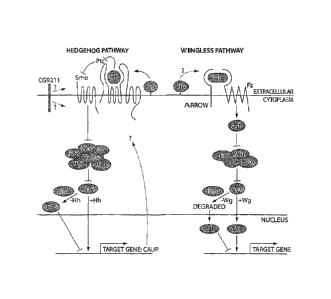
Figure 1 Hedgehog signaling pathway (adapted from Michelson, Sci. STKE,
2003(192): PE30, Jul. 22, 2003).
Figure 2 shows short hairpin siRNA antagonists against human Shh inhibits
Shh expression in HEK-293 cells.
Figure 3 shows short hairpin siRNA is specific against human Shh as
compared to Ihh and Dhh.
Figure 4 depicts gli-1 gene expression in embryonic and adult mouse
lung.
Figure 5 shows the inverse relationship between gli-1 expression and the
expression of markers of lung maturation. Between E13.5 and E16.5,
the expression of gli-1 decreases while the expression of the
maturation marker, surfactant type C (Sp-C), increases.
Figure 6 shows the effect of compound B treatm ent of embryonic mouse
lungs
on gli-1 expression.
- 13 -
CA 02561221 2006-09-25
WO 2005/097207
PCT/US2005/009739
Figure 7 shows compound B treatment increases surfactant type C
production
in embryonic mouse lungs.
Figure 8 shows that type II pneumocytes in compound B-treated lungs
differentiate prematurely, as evidenced by the presence of surfactant
producing lamellated bodies.
Figure 9 shows that treatment of embryonic lung cultures -with compound
B
decreases expression of gli-1
Figure 10 shows that treatment of embryonic lung cultures -with compound
B
increases expression of the maturation marker Sp-C. The induction of
Sp-C observed following treatment is comparable to that observed
following treatment with known lung maturation factor
hydrocortisone.
Figure 11 shows that treatment of embryonic lung cultures with hedgehog
agonists has the opposite effect. Treatment with either sonic
hedgehog or with agonist Z increases gli-1 expression and decreases
Sp-C expression.
Figure 12 illustrates gli-1 expression in breast cancer tissue as
visualized by in
situ hybridization.
Figure 13 shows gli-1 expression in lung cancer visualized by in situ
hybridization
Figure 14 illustrates g/i-1 expression in prostate cancer as visualized
by in situ
hybridization
Figure 15 depicts gli-1 expression in benign prostatic hyperplasia as
visualized
by in situ hybridization
Figure 16 shows: (A) Ptc-lacZ transgene expression in newborn mouse ptc-1
(d11) lacZ bladder epithelium. LacZ expression can be detected in the
proliferating urothelial cells and, more weakly, in adjacent
mesenchymal cells. (B) Gil-1 expression in adult mouse bladder
epithelium. Gli-1 expression can be detected in the proliferating
urothelial cells.
Figure 17 shows the expression of gli-I and shh in normal adult bladder
and in
a commercially available bladder tumor.
- 14 -
CA 02561221 2006-09-25
WO 2005/097207
PCT/US2005/009739
Figure 18 shows the expression of shh and gli-1 in eight commercially
available
bladder cancer cell lines. All eight cell lines examined express genes
involved in hedgehog signaling.
Figure 19 shows the expression of shh, ptc-1, smo, gli-1, gli-2, and gli-
3 in
eight commercially available bladder cancer cell lines, as well as in
fetal brain.
Figure 20 shows a schematic representation of the gli-Luc assay.
Figure 21 shows the results of the gli-Luc assay on bladder cancer cell
co-
cultures. Co-culture of S12 cells with either cell line 5637 or cell line
RT4 results in activation of the reporter gene indicating that these cell
lines can activate hedgehog signaling.
Figure 22 shows that the Shh antibody 5E1 inhibits activation of the
reporter
gene in RT-4/S 12 co-cultures.
Figure 23 and 24 show that administration of the Shh antibody 5E1 inhibits
tu_mor
growth in vivo in a nude mouse bladder cancer model.
Figure 25 shows that administration of the Shh antibody 5E1 decreases
expression of gli-1 in vivo in a nude mouse bladder cancer model.
Figure 26 shows that shh is expressed in prostate cancer samples as
visualized
by in situ hybridization.
Figure 27 shows by Q-RT-PCR the expression of gli-I in normal adult
prostate
and in a prostate adenocarcinoma.
Figure 28 = shows the expression of shh and gli-1 in three prostate cancer
cell
lines in comparison with expression in a normal prostate cell line.
Figure 29 shows that prostate cancer cell lines induce expression of
luciferase
when co-cultured with S12 cells in the gli-Luc in vitro assay.
Figure 30 shows that the antagonizing antibody 5E1 inhibits the induction
of
luciferase in by prostate cancer cells in the gli-Luc in vitro assay.
Figure 31 shows the expression of shh in prostatic epithelium and stroma.
in
human BPH samples.
Figure 32 shows the expression of gli-I in the prostatic stoma of human BPH
samples as measured by radioactive in situ hybridization.
- 15 -
CA 02561221 2006-09-25
WO 2005/097207
PCT/US2005/009739
Figure 33 shows that shh and patched-1 are expressed in a proximo-distal
pattern in normal prostate tissue with the highest levels of gene
expression occurring in the proximo or central region.
Figure 34 shows the expression of shh and gli-1 in BPH samples, and
compares
the levels of gene expression to BCC samples.
Figure 35 shows the expression of shh and gli-1 in BPH cell lines, and
compares the levels of gene expression to that of BCC samples,
normal prostate, and prostate cancer.
Figure 36 shows the expression of shh in a variety of colon, lung,
ovarian, renal
and hepatic human cancer cell lines. Expression of shh is measured
using Q-RT-PCR which demonstrates that shh is expressed, to a
varying degree, in. human cancer cell lines derived from several
diverse tissue types.
Figure 37 shows the expression of shh in a variety of passaged tumors
derived
from colon, lung, breast, melanoma, ovarian, prostate, pancreatic and
renal tissue. Expression of shh is measured using Q-RT-PCR which
demonstrates that .shh is expressed, to a varying degree, in passaged
tumors derived from several diverse tissue types.
Figure 38 shows the expression of hedgehog protein in normal human
stomach,
prostate, spleen, small intestine, large intestine, gall bladder,
appendix and kidney tissue. Hedgehog protein expression was
examined by imm-unohistochemistry using a polyclonal anti-
hedgehog antibody.
Figure 39 shows the expression of hedgehog protein in human tumors
derived
from salivary, esophageal, pancreatic, thyroid, colon, endometrial,
kidney and prostate tissue. Hedgehog protein expression was
examined by immunohistochemistry using a polyclonal anti-
hedgehog antibody.
Figure 40 shows increased expression of hedgehog protein in a sample of
pancreatic tumor in comparison to hedgehog protein expression in
normal pancreatic tissue. Hedgehog protein expression was
- 16 -
CA 02561221 2006-09-25
WO 2005/097207
PCT/US2005/009739
measured by immunohistochemistry using a polyclonal anti-
hedgehog antibody.
Figure 41 shows that the Shh blocking antibody 5E1 decreases tumor size
when
administered to mice injected with a cornbination of the Shh
expressing colon cancer cell line HT-29 and fibroblasts.
Figure 42 shows that the Shh blocking antibody 5E1 decreases tumor size
when
administered to mice injected with a cornbination of the Shh
expressing colon cancer cell line HT-29 and fibroblasts.
Figure 43 shows that delayed administration of the Shh blocking antibody
5E1
decreases tumor size when administered to mice injected with a
combination of the Shh expressing colon cancer cell line HT-29 and
fibroblasts.
Figure 44 shows that delayed administration of the Shh blocking antibody
5E1
decreases tumor size when administered to mice injected with a
combination of the Shh expressing colon cancer cell line HT-29 and
fibroblasts.
Figure 45 shows that administration of the Shh blocking antibody 5E1
induces
apoptosis in HT-29/fibroblast mixed tumors.
Figure 46 shows that delayed administration of the Shh blocking antibody
5E1
decreases tumor size when administered to mice injected with the
Shh expressing colon cancer cell line H1-29.
Figure 47 shows that delayed administration of the Shh blocking antibody
5E1
decreases tumor size when administered to mice injected with the
Shh expressing colon cancer cell line 1111-29.
Figure 48 shows that delayed administration of the Shh blocking antibody
5E1
to mice injected with the Shh expressing colon cancer cell line HT-29
decreases expression of gli-1 mRNA.
Figure 49 shows that administration of the Shh blocking antibody 5E1 to
mice
injected with the hedgehog expressing pancreatic cancer cell line
SW1990 decreases tumor weight.
Figure 50 shows that administration of the Shh blocking antibody 5E1 to
mice
injected with the hedgehog expressing pancreatic cancer cell line
-17-
CA 02561221 2006-09-25
WO 2005/097207
PCT/US2005/009739
SW1990 decreases tumor size, and results in eKtensive domains of
necrosis within said tumors.
Figure 51 shows that administration of the Shh blocking antibody 5E1 to
mice
injected with the hedgehog expressing pancreatic cancer cell line
SW1990 decreases tumor volume.
Figure 52 shows that administration of the Shh blocking antibody 5E1 to
mice
injected with the hedgehog expressing pancreatic cancer cell line CF
PAC decreases tumor weight.
Figure 53 shows that administration of the Shh blocking antibody 5E1 to
mice
injected with the hedgehog expressing pancreatic cancer cell line CF
PAC decreases tumor volume.
Figure 54 shows that administration of the Shh blocking antibody 5E1 to
mice
injected with the non-hedgehog expressing colon cancer cell line
SW480 has no effect on tumor volume.
Figure 55 shows Hedgehog expression in human cancers: (a, d) Hedgehog
immunoreactivity in biopsy material taken from human breast ductal
adenocarcinomas. Note the stronger immunoreactivity present on
cancerous epithelium (arrows) than on the adjacent normal ductal
epithelium (arrowhead) demonstrating elevated Hh levels in
cancerous tissues. (b, e) Hedgehog staining in two forms of ovarian
cancer, including a well differentiated borderline serous
adenocarcinoma (b), and a poorly differentiated adenocarcinoma (e).
(c, f) Hedgehog immunoreactivity on samples of uterine cancer
demonstrating expression on both well differentiated (c), and poorly
differentiated, highly invasive cancers (f).
Detailed Description of the Invention
I. Overview
RNA interference (RNAi) is a phenomenon describing double-stranded
(ds)RNA-dependent gene specific posttranscriptional silencing. Initial
attempts to
harness this phenomenon for experimental manipulation of mammalian cells were
- 18 -
CA 02561221 2006-09-25
WO 2005/097207
PCT/US2005/009739
foiled by a robust and nonspecific antiviral defense mechanism activated in
response
to long dsRNA molecules. Gil et al. Apoptosis 2000, 5:107-114. The field was
significantly advanced upon the demonstration that synthetic duplexes of 21-
nucleotide RNAs could mediate gene-specific RNAi in mammalian cells, without
invoking generic antiviral defense mechanisms. Elbashir et al. Nature 2001,
411:494-498; Caplen et al. Proc Natl Acad Sci 2001, 98:9742-9747. As a result,
small interfering RNAs (siRNAs) have become powerful tools to dissect gene
function. The chemical synthesis of small RNAs is one avenue that has produced
promising results. Numerous groups have also sought the development of DNA-
based vectors capable of generating such siRNA within cells. Several groups
have
attained this goal and published similar strategies that, in general, involve
transcription of short hairpin (sh)RNAs that are efficiently processed to form
siRNAs within cells. Paddison et al. PNAS 2002, 99:1443-1448; Paddison et al.
Genes & Dev 2002, 16:948-958; Sui et al. PNAS 2002, 8:5515-5520; and
Brummelkamp et al. Science 2002, 296:550-553. These reports describe methods
to
generate siRNAs capable of specifically targeting numerous endogenously and
exogenously expressed genes.
The present invention relates to the discovery that signal transduction
pathways regulated by hedgehog, patched (ptc), gli, smoothened, and many other
HH signaling pathway proteins can be inhibited, at least in part, by specific
RNAi
antagonists. Since certain HH signaling proteins positively regulate the
overall HH
signaling, while others negatively regulate the overall HH signaling, these
RNAi
antagonists may either increase or decrease the overall HH signaling in an
affected
cell or tissue / organ. It is, therefore, specifically contemplated that these
RNAi
antagonists which modulate signal transduction activity of hedgehog, ptc,
smoothened, etc. will likewise be capable of changing the role of a cell in
tissue
development from what would otherwise occur.
In preferred embodiments, the cell has a substantially wild-type hedgehog
signaling pathway. It is also contemplated that hedgehog antagonists are
particularly
effective in treating disorders resulting from hyperactivation of the hedgehog
pathway, either as a result of mutations in components of the HH signaling
pathway
or as a result of inappropriate activation of the HH signaling pathway in cell
which
- 19 -
CA 02561221 2011-12-19
does not comprise a mutation/lesion in a component of the HH signaling
pathway.
Therefore, it is desirable to have a method for identifying those cells in
which the
hedgehog pathway is hyperactive such that antagonist treatment may be
efficiently
targeted. One of skill in the art will readily recognize, that RNAi
antagonists of the
present invention can modulate hedgehog signaling at any point in the hedgehog
signaling pathway. That is, an exemplary RNAi modulator can regulate HH
signaling by antagonizing hedgehog itself, or any other HH signaling
components
such as the hedgehog receptor patched. It is contemplated that the RNAi
antagonists
of the present invention can be used to modulate hedgehog signaling in a wild-
type
cell or in a cell comprising a mutation in a component of the hedgehog
signaling
pathway.
Thus, the methods of the present invention include, but are not limited to,
the
use of RNAi antagonists that modulate HH signaling in the regulation of repair
and/or functional performance of a wide range of cells, tissues and organs
having the
phenotype of hedgehog gain-of-function and in tissues with wild-type hedgehog
activity. For instance, the subject method has therapeutic and cosmetic
applications
ranging from regulation of neural tissues, bone and cartilage formation and
repair,
regulation of spermatogenesis, regulation of smooth muscle, regulation of
lung, liver
and tissue of other organs arising from the primitive gut, regulation of
hematopoietic
function, regulation of skin and hair growth, etc. Moreover, the subject
methods can
be performed on cells that are provided in culture (in vitro), or on cells in
a whole
animal (in vivo). See, for example, PCT publications WO 95/18856 and WO
96/17924,=
In another aspect, the present invention provides pharmaceutical preparations
comprising, as an active ingredient, an RNAi antagonist of any one of the HH
signaling components such as described herein, formulated in an amount
sufficient
to inhibit, in vivo, proliferation or other biological consequences of
hedgehog gain-
of-function.
The subject treatments using RNAi antagonists of the Fill pathway
components can be effective for both human and non-human animal cells and
subjects. Animal subjects to which the invention is applicable extend to both
- 20 -
CA 02561221 2006-09-25
WO 2005/097207
PCT/US2005/009739
domestic animals and livestock, raised either as pets or for commercial
purposes.
Examples of such non-human animals include non-human primates, dogs, cats,
cattle, horses, sheep, hogs, goats, mice, rats, rabbits, frogs, fish,
chickens, and the
like.
II. Definitions
For convenience, certain terms employed in the specification, examples, and
appended claims are collected here.
The phrase "aberrant modification or mutation" of a gene refers to such
genetic lesions as, for example, deletions, substitution or addition of
nucleotides to a
gene, as well as gross chromosomal rearrangements of the gene and/or abnormal
methylation of the gene. Likewise, misexpression of a gene refers to aberrant
levels
of transcription of the gene relative to those levels in a normal cell under
similar
conditions, as well as non-wild-type splicing of mRNA transcribed from the
gene.
The term "adenocarcinoma" as used herein refers to a malignant tumor
originating in glandular epithelium.
The term "angiogenesis", as used herein, refers to the formation of blood
vessels. Specifically, angiogenesis is a multistep process in which
endothelial cells
focally degrade and invade through their own basement membrane, migrate
through
interstitial stroma toward an angiogenic stimulus, proliferate proximal to the
migrating tip, organize into blood vessels, and reattach to newly synthesized
basement membrane (see Folkman et al., Adv. Cancer Res., Vol. 43, pp. 175-203
(1985)).
"Basal cell carcinomas" exist in a variety of clinical and histological forms
such as nodular-ulcerative, superficial, pigmented, morphealike,
fibroepithelioma
and nevoid syndrome. Basal cell carcinomas are the most common cutaneous
neoplasms found in humans. The majority of new cases of nonmelanoma skin
cancers fall into this category.
"Benign prostatic hyperplasia", or BPH, is a benign enlargement of the
prostate gland that begins normally after age 50 years probably secondary to
the
effects of male hormones. If significant enlargement occurs, it may pinch off
the
urethra making urination difficult or impossible.
- 21 -
CA 02561221 2006-09-25
WO 2005/097207
PCT/US2005/009739
"Burn wounds" refer to cases where large surface areas of skin have been
removed or lost from an individual due to heat and/or chemical agents.
The term "carcinoma" refers to a malignant new growth made up of
epithelial cells tending to infiltrate surrounding tissues and to give rise to
metastases.
Exemplary carcinomas include: "basal cell carcinoma", which is an epithelial
tumor
of the skin that, while seldom metastasizing, has potentialities for local
invasion and
destruction; "squamous cell carcinoma", which refers to carcinomas arising
from
squamous epithelium and having cuboid cells; "carcinosarcoma", which include
malignant tumors composed of carcinomatous and sarcomatous tissues;
"adenocystic
carcinoma", carcinoma marked by cylinders or bands of hyaline or mucinous
stroma
separated or surrounded by nests or cords of small epithelial cells, occurring
in the
mammary and salivary glands, and mucous glands of the respiratory tract;
"epidermoid carcinoma", which refers to cancerous cells which tend to
differentiate
in the same way as those of the epidermis; i.e., they tend to form prickle
cells and
undergo cornification; "nasopharyngeal carcinoma", which refers to a malignant
tumor arising in the epithelial lining of the space behind the nose; and
"renal cell
carcinoma", which pertains to carcinoma of the renal parenchyma composed of
tubular cells in varying arrangements. Other carcinomatous epithelial growths
are
"papillomas", which refers to benign tumors derived from epithelium and having
a
papillomavirus as a causative agent; and "epidermoidomas", which refers to a
cerebral or meningeal tumor formed by inclusion of ectodermal elements at the
time
of closure of the neural groove.
The "corium" or "dermis" refers to the layer of the skin deep to the
epidermis, consisting of a dense bed of vascular connective tissue, and
containing
the nerves and terminal organs of sensation. The hair roots, and sebaceous and
sweat
glands are structures of the epidermis which are deeply embedded in the
dermis.
"Dental tissue" refers to tissue in the mouth that is similar to epithelial
tissue,
for example gum tissue. The method of the present invention is useful for
treating
periodontal disease.
"Dermal skin ulcers" refer to lesions on the skin caused by superficial loss
of
tissue, usually with inflammation. Dermal skin ulcers that can be treated by
the
method of the present invention include decubitus ulcers, diabetic ulcers,
venous
- 22 -
CA 02561221 2006-09-25
WO 2005/097207
PCT/US2005/009739
stasis ulcers and arterial ulcers. Decubitus wounds refer to chronic ulcers
that result
from pressure applied to areas of the skin for extended periods of time.
Wounds of
this type are often called bedsores or pressure sores. Venous stasis ulcers
result from
the stagnation of blood or other fluids from defective veins. Arterial ulcers
refer to
necrotic skin in the area around arteries having poor blood flow.
The term "ED50" means the dose of a drug that produces 50% of its
maximum response or effect.
An "effective amount" of, e.g., a hedgehog antagonist, with respect to the
subject method of treatment, refers to an amount of the antagonist in a
preparation
which, when applied as part of a desired dosage regimen brings about, e.g., a
change
in the rate of cell proliferation and/or the state of differentiation of a
cell and/or rate
of survival of a cell according to clinically acceptable standards for the
disorder to
be treated or for the cosmetic purpose.
The terms "epithelia", "epithelial" and "epithelium" refer to the cellular
covering of internal and external body surfaces (cutaneous, mucous and
serous),
including the glands and other structures derived therefrom, e.g., corneal,
esophegeal, epidermal, nd hair follicle epithelial cells. Other exemplary
epithelial
tissue includes: olfactory epithelium, which is the pseudostratified
epithelium lining
the olfactory region of the nasal cavity, and containing the receptors for the
sense of
smell; glandular epithelium, which refers to epithelium composed of secreting
cells;
squamous epithelium, which refers to epithelium composed of flattened plate-
like
cells. The term epithelium can also refer to transitional epithelium, like
that which is
characteristically found lining hollow organs that are subject to great
mechanical
change due to contraction and distention, e.g., tissue which represents a
transition
between stratified squamous and columnar epithelium.
The term "epithelialization" refers to healing by the growth of epithelial
tissue over a denuded surface.
The term "epidermal gland" refers to an aggregation of cells associated with
the epidermis and specialized to secrete or excrete materials not related to
their
ordinary metabolic needs. For example, "sebaceous glands" are holocrine glands
in
the corium that secrete an oily substance and sebum. The term "sweat glands"
refers
- 23 -
CA 02561221 2006-09-25
WO 2005/097207
PCT/US2005/009739
to glands that secrete sweat, situated in the corium or subcutaneous tissue,
opening
by a duct on the body surface.
The term "epidermis" refers to the outermost and nonvascular layer of the
skin, derived from the embryonic ectoderm, varying in thickness from 0.07-1.4
mm.
On the pahnar and plantar surfaces it comprises, from within outward, five
layers:
basal layer composed of columnar cells arranged perpendicularly; prickle-cell
or
spinous layer composed of flattened polyhedral cells with short processes or
spines;
granular layer composed of flattened granular cells; clear layer composed of
several
layers of clear, transparent cells in which the nuclei are indistinct or
absent; and
horny layer composed of flattened, cornified non-nucleated cells. In the
epidermis of
the general body surface, the clear layer is usually absent.
"Excisional wounds" include tears, abrasions, cuts, punctures or lacerations
in the epithelial layer of the skin and may extend into the dermal layer and
even into
subcutaneous fat and beyond. Excisional wounds can result from surgical
procedures
or from accidental penetration of the skin.
The "growth state" of a cell refers to the rate of proliferation of the cell
and/or the state of differentiation of the cell. An "altered growth state" is
a growth
state characterized by an abnormal rate of proliferation, e.g., a cell
exhibiting an
increased or decreased rate of proliferation relative to a normal cell.
The term "hair" refers to a threadlike structure, especially the specialized
epidermal structure composed of keratin and developing from a papilla sunk in
the
corium, produced only by mammals and characteristic of that group of animals.
Also, "hair" may refer to the aggregate of such hairs. A "hair follicle"
refers to one
of the tubular-invaginations of the epidermis enclosing the hairs, and from
which the
hairs grow. "Hair follicle epithelial cells" refers to epithelial cells that
surround the
dermal papilla in the hair follicle, e.g., stem cells, outer root sheath
cells, matrix
cells, and inner root sheath cells. Such cells may be normal non-malignant
cells, or
transformed/immortalized cells.
The term "hedgehog" is used to refer generically to any member of the
hedgehog family, including sonic, indian, desert and tiggy winkle. The term
may be
used to indicate protein or gene. The term is also used to describe homolog /
ortholog sequences in different animal species (see below).
-24-
CA 02561221 2006-09-25
WO 2005/097207
PCT/US2005/009739
The terms "hedgehog (HH) signaling pathway", "hedgehog (HH) pathway"
and "hedgehog (HH) signal transduction pathway" are all used to refer to the
chain
of events normally mediated by hedgehog, smoothened, ptc, and gli, among
others,
and resulting in a changes in gene expression and other phenotypic changes
typical
of hedgehog activity. The hedgehog pathway can be activated even in the
absence of
a hedgehog protein by activating a downstream component. For example,
overexpression of smoothened will activate the pathway in the absence of
hedgehog.
Hedgehog, gli and ptc gene expression are indicators of an active hedgehog
signaling pathway.
The term "HH signaling component" refers to gene products that participate
in the HH signaling pathway. An HH signaling component frequently materially
or
substantially affects the transmission of the HH signal in cells / tissues,
typically
resulting in changes in degree of downstream gene expression level and/or
phenotypic changes.
Each HH signaling component, depending on their biological function and
effects on the final outcome of the downstream gene activation / expression,
may be
devided into positive and negative regulators. A positive regulator is a HH
signaling
component that positively affects the transmission of the HH signal, i.e.,
stimulates
'downstream biological events when HH is present. Examples include (but are
not
limited to) those genes listed in Table X below. A negative regulator is a HH
signaling component that negatively affects the transmission of the HH signal,
i.e.,
inhibits downstream biological events when HH is present. Examples include
(but
are not limited to) those genes listed in Table Y below.
The term "hedgehog RNAi antagonist" refers to an RNAi agent that inhibits
the bioactivity of an HH signaling component (such as hedgehog, patched, or
01),
such that it represses the expression of the target HH signaling component.
For
example, certain preferred hedgehog RNAi antagonists can be used to overcome a
ptc loss-of-function and/or a smoothened gain-of-function. Other preferred
RNAi
antagonists can be used to overcome an inappropriate increase in hedgehog
signal
transduction, whether said increase in signal transduction is the result in a
mutation/lesion in a component of the hedgehog signaling pathway (e.g., ptc,
gill,
gli3, snzoothened, etc) or whether said increase in signal transduction occurs
in the
- 25 -
CA 02561221 2011-12-19
context of a cell which does not comprise a mutation/lesion in a component of
the
hedgehog signaling pathway (e.g., a wild-type cell with respect to components
of the
hedgehog signaling pathway). An RNAi antagonist may be directed to a protein
encoded by any of the genes in the hedgehog pathway, including (but not
limited to)
sonic, indian or desert hedgehog, smoothened, ptc-1, ptc-2, gli-1, gli-2, gli-
3, etc. In
most cases, the RNAi antagonist would inhibit the activity of the target
protein by,
for example, decreasing production of a protein encoded by any of the genes in
the
hedgehog pathway, thus either upregulating or downregulating HH signaling.
When
the RNAi antagonist inhibits expression of a target protein that normally
functions
as a positive regulator of the hedgehog signaling pathway, the overall effect
is a
decrease or inhibition of hedgehog signaling. When the RNAi antagonist
inhibits
expression of a target protein that normally functions as a negative regulator
of the
hedgehog signaling pathway, the overla effect is an increase or promotion of
hedgehog signaling.
Moreover, more than one antagonist, including non-RNAi antagonists of the
HH signaling pathway, such as antisense nucleotides, antibodies to HH pathway
proteins, small organic molecules, etc., can be administered. The US patent
7,708,998, describes in detail about various modulators of the HH signalling
pathway. Thus, it is further contemplated that when more than one hedgehog
antagonist is administered, said agents can inhibit hedgehog signalling
through
the same mechanism or through differing mechanisms.
The term "hedgehog gain-of-function" refers to an aberrant modification or
mutation of a ptc gene, hedgehog gene, or smoothened gene, or a decrease (or
loss)
in the level of expression of such a gene, which results in a phenotype which
resembles contacting a cell with a hedgehog protein, e.g., aberrant activation
of a
hedgehog pathway. The gain-of-function may include a loss of the ability of
the ptc
gene product to regulate the level of expression of Ci genes, e.g., Glil ,
G1i2, and
G1i3. The term 'hedgehog gain-of-function' is also used herein to refer to any
similar cellular phenotype (e.g., exhibiting excess proliferation) that occurs
due to an
alteration anywhere in the hedgehog signal transduction pathway, including,
but not
limited to, a modification or mutation of hedgehog itself. For example, a
tumor cell
- 26 -
CA 02561221 2006-09-25
WO 2005/097207
PCT/US2005/009739
with an abnormally high proliferation rate due to activation of the hedgehog
signaling pathway would have a 'hedgehog gain-of-function' phenotype, even if
hedgehog is not mutated in that cell.
As used herein, "immortalized cells" refers to cells that have been altered
via
chemical and/or recombinant means such that the cells have the ability to grow
through an indefinite number of divisions in culture.
"Internal epithelial tissue" refers to tissue inside the body that has
characteristics similar to the epidermal layer in the skin. Examples include
the lining
of the intestine. The method of the present invention is useful for promoting
the
healing of certain internal wounds, for example wounds resulting from surgery.
The term "keratosis" refers to proliferative skin disorder characterized by
hyperplasia of the horny layer of the epidermis. Exemplary keratotic disorders
include keratosis follicularis, keratosis palmaris et plantaris, keratosis
pharyngea,
keratosis pilaris, and actinic keratosis.
"Lamellated bodies" refers to a subcellular structure found in lung cells that
are producing surfactants. Lamellated bodies are thought to be the site of
lung
surfactant biosynthesis. The bodies have a multilayered membranous appearance
in
an electron micrograph.
The term "LD50" means the dose of a drug that is lethal in 50% of test
subjects.
The term "nail" refers to the horny cutaneous plate on the dorsal surface of
the distal end of a finger or toe.
The term "overexpression" as used in reference to gene expression levels
means any level of gene expression in cells of a tissue that is higher than
the normal
level of expression for that tissue. The normal level of expression for a
tissue may be
assessed by measuring gene expression in a healthy portion of that tissue.
The term "patched loss-of-function" refers to an aberrant modification or
mutation of a ptc gene, or a decreased level of expression of the gene, which
results
in a phenotype that resembles contacting a cell with a hedgehog protein, e.g.,
aberrant activation of a hedgehog pathway. The loss-of-function may include a
loss
of the ability of the ptc gene product to regulate the level of expression of
Ci genes,
e.g., Glil, G1i2 and G1i3.
- 27 -
CA 02561221 2006-09-25
WO 2005/097207
PCT/US2005/009739
The term "pharmaceutically acceptable salts" refers to physiologically and
pharmaceutically acceptable salts of the compounds of the invention, i.e.,
salts that
retain the desired biological activity of the parent compound and do not
impart
undesired toxicological effects thereto.
"Standard hybridization conditions" refer to salt and temperature conditions
substantially equivalent to 0.5 SSC to about 5 x SSC and 65 C for both
hybridization and wash. The term "standard hybridization conditions" as used
herein
is therefore an operational definition and encompasses a range of
hybridization
conditions. Nevertheless, for the purposes of this present disclosure "high
stringency" conditions include hybridizing with plaque screen buffer (0.2%
polyvinylpyrrolidone, 0.2% Ficoll 400; 0.2% bovine serum albumin, 50 mM Tris-
HC1 (pH 7.5); 1 M NaCl; 0.1% sodium pyrophosphate; 1% SDS); 10% dextran
sulfate, and 100 ug/ml denatured, sonicated salmon sperm DNA at 65 C for 12-20
hours, and washing with 75 mM NaC1 / 7.5 mM sodium citrate (0.5 x SSC)/1% SDS
at 65 C. "Low stringency" conditions include hybridizing with plaque screen
buffer,
10% dextran sulfate and 110 ug/ml denatured, sonicated salmon sperm DNA at
55 C for 12-20 hours, and washing with 300 mM NaC1 / 30mM sodium citrate (2.0
x SSC)/1% SDS at 55 C. See also Current Protocols in Molecular Biology, John
Wiley & Sons, Inc. New York, Sections 6.3.1-6.3.6, (1989).
A "patient" or "subject" to be treated by the subject method can mean either a
human or non-human animal.
The term "prodrug" is intended to encompass compounds that, under
physiological conditions, are converted into the therapeutically active agents
of the
present invention. A common method for making a prodrug is to include selected
moieties that are hydrolyzed under physiological conditions to reveal the
desired
molecule. In other embodiments, the prodrug is converted by an enzymatic
activity
of the host animal.
As used herein, "proliferating" and "proliferation" refer to cells undergoing
mitosis.
Throughout this application, the term "proliferative skin disorder" refers to
any disease/disorder of the skin marked by unwanted or aberrant proliferation
of
cutaneous tissue. These conditions are typically characterized by epidermal
cell
- 28 -
CA 02561221 2006-09-25
WO 2005/097207
PCT/US2005/009739
proliferation or incomplete cell differentiation, and include, for example, X-
linked
ichthyosis, psoriasis, atopic dermatitis, allergic contact dermatitis,
epidermolytic
hyperkeratosis, and seborrheic dermatitis. For example, epidermodysplasia is a
form
of faulty development of the epidermis. Another example is "epidermolysis",
which
refers to a loosened state of the epidermis with formation of blebs and bullae
either
spontaneously or at the site of trauma.
As used herein, the term "psoriasis" refers to a hyperproliferative skin
disorder that alters the skin's regulatory mechanisms. In particular, lesions
are
formed which involve primary and secondary alterations in epidermal
proliferation,
inflammatory responses of the skin, and an expression of regulatory molecules
such
as lymphokines and inflammatory factors. Psoriatic skin is morphologically
characterized by an increased turnover of epidermal cells, thickened
epidermis,
abnormal keratinization, inflammatory cell infiltrates into the dermis layer
and
polymorphonuclear leukocyte infiltration into the epidermis layer resulting in
an
increase in the basal cell cycle. Additionally, hyperkeratotic and
parakeratotic cells
are present.
The term "skin" refers to the outer protective covering of the body,
consisting of the corium and the epidermis, and is understood to include sweat
and
sebaceous glands, as well as hair follicle structures. Throughout the present
application, the adjective "cutaneous" may be used, and should be understood
to
refer generally to attributes of the skin, as appropriate to the context in
which they
are used.
The term "small cell carcinoma" refers to a type of malignant neoplasm,
commonly of the bronchus. Cells of the tumor have endocrine like
characteristics
and may secrete one or more of a wide range of hormones, especially regulatory
peptides like bombesin.
The term "smoothened gain-of-function" refers to an aberrant modification
or mutation of a snio gene, or an increased level of expression of the gene,
which
re suits in a phenotype that resembles contacting a cell with a hedgehog
protein, e.g.,
aberrant activation of a hedgehog pathway. While not wishing to be bound by
any
particular theory, it is noted that ptc may not signal directly into the cell,
but rather
interact with smoothened, another membrane-bound protein located downstream of
-29-
CA 02561221 2006-09-25
WO 2005/097207
PCT/US2005/009739
plc in hedgehog signaling (Mango et al., (1996) Nature 384: 177-179). The gene
87770 is a segment-polarity gene required for the correct patterning of every
segment
in Drosophila (Alcedo et al., (1996) Cell 86: 221-232). Human homologs of stno
have been identified. See, for example, Stone et al. (1996) Nature 384:129-
134, and
GenBank accession U84401. The smoothened gene encodes an integral membrane
protein with characteristics of heterotrimeric G-protein-coupled receptors;
i.e., 7-
transmembrane regions. This protein shows homology to the Drosophila Frizzled
(Fz) protein, a member of the wingless pathway. It was originally thought that
smo
encodes a receptor of the Hh signal. However, this suggestion was subsequently
disproved, as evidence for ptc being the Hh receptor was obtained. Cells that
express
gin fail to bind Hh, indicating that smo does not interact directly with Hh
(Nusse,
( 1996) Nature 384: 119-120). Rather, the binding of Sonic hedgehog (SHH) to
its
receptor, PTCH, is thought to prevent normal inhibition by PTCH of smoothened
(SMO), a seven-span transmembrane protein.
Recently, it has been reported that activating smoothened mutations occur in
sporadic basal cell carcinoma, Xie et al. (1998) Nature 391: 90-2, and
primitive
neuroectodermal tumors of the central nervous system, Reifenberger et al.
(1998)
Cancer Res 58: 1798-803.
The term "therapeutic index" refers to the therapeutic index of a drug defined
a s LD50/ED50.
As used herein, "transformed cells" refers to cells that have spontaneously
converted to a state of unrestrained growth, i.e., they have acquired the
ability to
grow through an indefinite number of divisions in culture. Transformed cells
may be
characterized by such terms as neoplastic, anaplastic and/or hyperplastic,
with
respect to their loss of growth control.
"Urogenital" refers to the organs and tissues of the urogenital tract, which
includes among other tissues, the prostate, ureter, kidney and bladder. A
"urogenital
cancer" is a cancer of a urogenital tissue.
LII. Exenzplaiy Targets of the Hedgehog Signaling Pathway
Hedgehog, which encodes a secreted signaling molecule, was originally
identified in Drosophila as an essential embryonic patterning gene. Hh family
- 30 -
CA 02561221 2006-09-25
WO 2005/097207
PCT/US2005/009739
members subsequently were discovered in diverse species, including in human,
where they exert a wide range of developmental effects (see, for example, Ho
and
Scott; Curr. Opin. Neurobiol. 12, 57-63, 2002; Ingham and McMahon, Geizes Dev.
15, 3059-3087, 2001). Of further interest, aberrant HH signaling is associated
with a
number of human diseases, including several types of cancer (For review, see
Bale,
Annu. Rev. Genomics Hurn. Genet. 3, 47-65, 2002; Taipale and Beachy, Nature
411,
349-354, 2001). From intensive genetic and biochemical investigations, the
following view of the Hh signaling pathway has emerged (Figure 1) (for review,
see
Nybakken and Perrimon, Curr. Opin. Genet. Dev. 12, 503-511, 2002).
In the absence of FM, the transmembrane receptor, Patched (Ptc), inhibits a
second membrane-bound protein, Smoothened (Smo). This process enables an
intracellular high-molecular-weight protein complex--which includes the
kinesin-
related molecule Costal2 (Cos2), the serine-threonine protein kinase Fused
(Fu), and
the protein Suppressor of fused [Su(fu)]--to promote the proteolytic
processing of
full-length Cubitus interruptus (Cil 55), thereby generating a transcriptional
repressor Ci75. Although not yet proven to interact directly with the
inhibitory
complex, protein kinase A (PKA), glycogen synthase kinase 3 (GSK3), and casein
kinase 1a (CK1 a) also modify Ci to regulate its cleavage. This process also
depends
on Slimly. Binding of Hh to Ptc relieves inhibition of Smo and, by an unknown
mechanism, Smo suppres ses the Ci-proces sing activity of the cytoplasmic
complex.
Unprocessed Ci155 then translocates to the nucleus, where it activates the
expression of specific target genes.
Recently, Lum et al. (Science 299: 2039-2045, 2003) identified several
additional members of th HH signaling pathway. Using both in vitro and in vivo
assays, these authors identified four genes whose products were not previously
= recognized as having specific roles in Hh signaling: CKI a, dally-like
(4),
caupolican (caup), and the predicted gene, CG9211. Among them, CKI a is a
negative regulator, while clip, caup and CG921I are all positive regulators.
All HH signaling pathway genes in various species can be routinely obtained
from public and proprietazy databases, such as GenBank, EMBL, FlyBase, to name
but a few. In certain organisms, such as human and Drosophila, the whole
genome is
sequenced, and sequence comparison programs, such as the BLAST series of
-31-
CA 02561221 2006-09-25
WO 2005/097207
PCT/US2005/009739
programs offered online at the NTCBI website can be used to retrieve the most
updated sequences of any known. HH signal pathway genes. The following tables
list
several representative members of the known HH signaling pathway genes in
various species. It is by no means exhaustive, and should not be viewed as
limiting
in any sense. Rather, it serves as a useful starting point for an exhaustive
search,
which a skilled artisan would be able to perform these searches using routine
biotechniques. Some genes may have several different database entries with
different accession numbers, but are nonetheless same or almost the same in
sequence. Regardless, only one entry for each gene is provided in the tables
below.
The genes are listed as either positive or negative regulators of the HH
signaling pathway. Thus an RNAi antagonist inhibiting a positive regulator
will be
useful to down-regulate the HH signaling, for example, in conditions involving
hyperactivity of HH signaling. In contrast, an RNAi antagonist inhibiting a
negative
regulator will be useful to up-regulate the HH signaling, for example, in
conditions
involving hypoactivity of HH signaling.
Table X. Positive Regulators of HH Signaling
Drosophila
Other Species (Acc. No.)
(Acc. No.)
Hh Human Shh (NI\4_000193); human Ihh (XM_050846); human Dhh
(NM_079735) (NM_021044) .
mouse Shh (N-IVI_009170); rat Shh (NM 017221); cow Shh
(AF144100); house shrew Shh (AB081406); chicken Shh
(L28099); Japanese firebelly newt Shh (D63339); bastard halibut
Shh (AB029748); smaller spotted catshark Shh (AF393835);
Eleutherodactylus coqui Shh (AF113403); Iberian ribbed newt
Shh-related protein (AF003532); Xenopus Shh (L39213);
Takifugu rubripes Shh (AJ507296); Zebrafish Shh (NM_131063);
mouse Ihh (N1\4_010544.); rat Ihh (XM_237298); chicken Ihh
(U58511); Xeriopus banded HH (U26404); zebrafish Twhh
(NM_131199).
-32-
CA 02561221 2006-09-25
WO 2005/097207
PCT/US2005/009739
Smo Human Smo (U84401); rat Smo. (1584402); mouse Smo
(NM 078719) (XM_133018); Xenopus Smo (AF302766); zebrafish Smo
(AF395809); chicken Smo (AF019977).
dip Human homolog (AF030186); mouse homolog (X83577); Rat
(AF317090) homolog (L02896); Zebrafish homolog (AF354754); chicken
homolog (L29089).
CG9211 Human homolog (AY027658); 1VIouse homolog (AF388037); rat
(Protein: homolog (U68726); Xenopus homolog (AF388036); zebrafish
AAF52461; homolog (AF461120).
see AE003615
for nucleotide)
Caup Human homolog (AF124733); "mouse homolog (AF124732);
(X95178) Xenopus homolog (AF338157); chicken homolog (AF091504);
zebrafish homolog (AY017309).
Ci (X54360) Human Gli (NM 005269); hum_an G1i2 (4 variants: NM_030379,
NM_030380, NM_030381, NMI 005270); human Gli3
(NM 000168); human G1i4 (NN4_138465); Mouse Gli
(NM 010296); rat Glil (XM_235221); horse Glil (AF510668);
chicken Glil (U60762); Xenopus Glil (U57454); zebrafish Glil
(NM 178296); mouse G1i2 (XIVI_196215); rat G1i2 (XM_222557);
chicken Gli2 (AF022818); zebrafish G1i2 (AF085746); mouse G1i3
(NM_008130); rat G1i3 (XM_225412); chicken G1i3 (U60763);
common quail G1i3 (AF231112); Xenopus G1i3 (17542461); eastern
newt G1i3 (AF316110); Xenopus G1i4 (U42462).
Fu (Protein Human homolog (AF200815); Mouse homolog (AF195272,
P23647, see AK006827, AF124142); rat hornolog (NM_019232, D49836);
X80468 for rabbit homolog (AF139639); Xenopus homolog (AF057138);
gene) spiny dogfish homolog (AJ223715); chicken homolog
(AF039943); cow homolog (X6 1036); zebrafish homolog
(BC052134).
* Nucleotide sequence accession numbers from the public databases are
listed
in "()."
- 33 -
CA 02561221 2006-09-25
WO 2005/097207
PCT/US2005/009739
Table Y. Negative Regulators of HH Signaling
Drosophila
Other Species (Ace. No.)
(Ace. No.)
Ptc (M28999) Human PTC1 (1559464); human PTC2 (AF091501); mouse
Ptcl (1J46155); rat Ptcl (AF079162); Xenopus Ptcl
(AF302765); chicken Ptcl (U40074); zebrafish Ptcl
(X98883); Japanese firebelly newt Ptcl (AB000848); mouse
Ptc2 (AB010833); chicken Ptc2 (AF409095); Xenopus Ptc2
(AB037688); zebrafish Ptc2 (AJ007742); Japanese firebelly
newt Ptc2 (AB000846)
Cos2 (A1F019250) Human homolog (AY237538); rat homolog (XM_218828);
mouse homolog (XM_133575); Anopheles gambiae str. PEST
homolog (XM_309818).
Su(fu) Human SUFU (NM_016169); mouse hornolog (AJ131692);
(NM_080502) rat homolog (XM_219957); chicken horriolog (AF487888);
Anopheles gambiae str. PEST homolog (D34_321114);
zebrafish homolog (BC045348).
Sgg (X70862) Human GSK3I3 (L33801); mouse GSK3i3 (AF156099); rat
GSK3f3 (X53428); zebrafish GSK3p (A13032265); Xenopus
GSK3r3 (U31862).
Pka-C1 Human PKA-C1 (X07767, M34181, M34182); rat homolog
(AY069425) (X57986); mouse homolog (BC003238); sheep homolog
(AF238979); bovine homolog (X67154); pig homolog
(X05998); rabbit homolog (AF367428;); hamster homolog
(M63311); Xenopus homolog (AJ413219).
CKla (AT069346) Human homolog (X80693); mouse homolog (BC019740); rat
homolog (U77582); chicken homolog (A_I042862); sheep
homolog (AB050945); bovine homolog (AB050944); pig
homolog (F22872).
Slmb (AF032878) Human homolog (AF101784; AF176022); mouse homolog
(AF391190); Xenopus (M98268); chicken (AF113946).
- 34 -
CA 02561221 2006-09-25
WO 2005/097207
PCT/US2005/009739
Nucleotide sequence accession numbers from the public databases are listed
in "()."
Patched was originally identified in Drosophila as a segment polarity gene,
one of a group of developmental genes that affect cell differentiation within
the
individual segments that occur in a homologous series along the anterior-
posterior
axis of the embryo. See Hooper, J.E. et al. (1989) Cell 59:751; and Nakano, Y.
et al.
(1989) Nature 341:508. Patterns of expression of the vertebrate homologue of
patched suggest its involvement in the development of neural tube, skeleton,
limbs,
craniofacial structure, and skin.
Genetic and functional studies demonstrate that patched is part of the
hedgehog signaling cascade, an evolutionarily conserved pathway that regulates
expression of a number of downstream genes. See Perrimon, N. (1995) Cell
80:517;
and Perrimon, N. (1996) Cell 86:513. Patched participates in the constitutive
transcriptional repression of the target genes; its effect is opposed by a
secreted
glycoprotein, encoded by hedgehog, or a vertebrate homologue, which induces
transcriptional activation. Genes under control of this pathway include
members of
the Wnt and TGF-beta families.
Patched proteins possess two large extracellular domains, tvvelve
transmembrane segments, and several cytoplasmic segments. See Hcoper, supra;
Nakano, supra; Johnson, R.L. et al. (1996) Science 272:1668; and Hahn, H. et
al.
(1996) Cell 85:841. The biochemical role of patched in the hedgehog signaling
pathway is unclear. Direct interaction with the hedgehog protein has, however,
been
reported (Chen, Y. et al. (1996) Cell 87:553), and patched may participate in
a
hedgehog receptor complex along with another transmembrane protein encoded by
the smoothened gene. See Perrimon, supra; and Chen, supra.
The human homologue of patched was cloned and mapped to chromosome
9q22.3. See Johnson, supra; and Hahn, supra. This region has been implicated
in
basal cell nevus syndrome (BCNS), which is characterized by developmental
abnormalities including rib and craniofacial alterations, abnormalities of the
hands
and feet, and spina bifida.
- 35 -
CA 02561221 2006-09-25
WO 2005/097207
PCT/US2005/009739
Sporadic tumors also demonstrated a loss of both functional alleles of
patched. Of twelve tumors in which patched mutations were identified with a
single
strand conformational polymorphism screening assay, nine had chromosomal
deletion of the second allele and the other three had inactivating mutations
in both
alleles (Gailani, supra). The alterations did not occur in the corresponding
germ line
DNA.
Most of the identified mutations resulted in premature stop codons or frame
shifts (Lench, N.J., et al., Hum. Genet. 1997 Oct; 100(5-6): 497-502).
Several,
however, were point mutations leading to amino acid substitutions in either
extracellular or cytoplasmic domains. These sites of mutation may indicate
functional importance for interaction with extracellular proteins or with
cytoplasmic
members of the downstream signaling pathway.
The involvement of patched in the inhibition of gene expression and the
occurrence of frequent allelic deletions of patched in BCC support a tumor
suppressor function for this gene. Its role in the regulation of gene families
known to
be involved in cell signaling and intercellular communication provides a
possible
mechanism of tumor suppression.
CKI a is a positive regulator of Ci cleavage, a process that generates its
repressor form (Price and Kalderon, Cell 108, 823-835, 2002, Figure 1). Thus
CK1 a
is a negative regulator of HH signaling. In contrast, dip is a positive Hh
signal
transducer. The latter result is consistent with dip encoding a cell-surface
heparan
sulfate proteoglycan (HSPG) of the glypican class, because such molecules are
known to function as coreceptors for various extracellular ligands (Nybakken
and
Perrimon, Biochim. Biophys. Acta 1573, 280-291, 2002). Lum et al. presented
additional evidence that Dlp acts upstream of or together with Ptc, possibly
by
concentrating free Hh at the cell surface or by presenting Hh to the Ptc
receptor.
Of note, Lum et al. reported that dip inhibitor had no effect in the Wg-
specific cell-culture assay. This result suggests that, in contrast to its
participation in
Hh signaling, Dlp mediates effects of Wg that are not cell-autonomous. This
model
is consistent with prior genetic experiments implicating the involvement of
dip in
regulating the extracellular distribution of Wg. Thus, Dlp appears to play
different
- 36 -
CA 02561221 2006-09-25
WO 2005/097207
PCT/US2005/009739
roles in the Hh and Wg signaling pathways, and can be used as a HH pathway-
specific target.
The gene caup had previously been described as a downstream mediator of
Hh signaling during wing development (Gomez-Skarmeta and Modolell, Genes Dev.
10, 2935-2945, 1996), thus its detection as a positive regulator of the Hh
pathway
was somewhat surprising. Caup, which is a homeodomain transcription factor,
could
conceivably be involved in a positive-feedback loop that amplifies the Hh
signal,
perhaps by activating the expression of proximal positive-signaling components
(Figure 1).
CG9211 is predicted to encode a cell-surface protein having immunoglobulin
domain repeats and fibronectin type III repeats. It is possible that this
factor could
function as a positive Hh regulator by interacting with and modulating the
activity of
other membrane-bound components of the Hh pathway such as Ptc and Smo.
Alternatively, CG9211 could mediate a parallel signaling pathway that
influences
Hh responses (Figure 1).
The identification of new signaling pathway components in Drosophila also
has implications for human disease. For example, the role of CKla in
regulating
basal activity of both Wg and Hh signaling pathways suggests that it could act
as a
tumor suppressor in colon cancer, basal cell carcinoma, rhabdomyosarcoma, or
medulloblastoma. These tumors are associated with inappropriate activity of
one or
the other pathway, except medulloblastoma, which is associated with the
activation
of either (Taipale and Beachy, Nature 411, 349, 2001). In the case of Dlp,
GPC4 and
GPC6 are the most closely related of the six mammalian glypican family members
(De Cat and David, Semin. Cell Dev. Biol. 12, 117, 2001). GPC6 maps to 13q32
(Paine-Saunders et al., Genomics 57, 455, 1999), a human chromosomal locus
whose
deletion (13q32 syndrome) is associated with defects, including
holoprosencephaly
(HPE), anogenital malformations, and an absent thumb (Brown et al., Am. J.
Hum.
Genet. 57, 859, 1995); all of these malformations are consistent with loss of
varying
degrees of Sonic hedgehog signaling (Ramalho-Santos et al., Development 127,
2763, 2000; Chiang et al., Nature 383, 407, 1996). If GPC6 levels are limiting
in
mammalian Hh responsiveness, then loss of GPC6 function may play a role in
13q32
syndrome malformations, possibly alongside other HPE genes in or near this
region
- 37 -
CA 02561221 2006-09-25
WO 2005/097207
PCT/US2005/009739
(Brown et al., Nature Genet. 20, 180, 1998). Finally, mutation of CDO, the
mammalian homolog of CG9211, results in a form of HPE (Cole and Krauss, Curr.
Biol. 13, 411, 2003), consistent with a role for CDO in signaling.
IV. Method for Identifi,ing Additional HH Signaling Pathway Components
The RNAi approach used in Lum et al. (supra) can be extended to cover the
whole genome of any given organism, especially in model organisms such as
worm,
Drosophila, fish, rodents, and human where numerous established cell lines are
readily available. Lum et al. (supra) provide an example of such a large-
scale,
kinase-phosphatase RNAi library screening, using the information provided by
the
sequenced Drosophila genome (Morrison et al., J. Cell Biol. 150, F57, 2000).
As a
result, 4 additional HH signaling components were identified. Such preliminary
in
vitro screen can be done in a high throughput fashion to allow quick screen of
all the
genes within an organism, or at least a specific subset of genes within that
organism,
such as all kinases, etc. (see Lum et al., supra). Results obtained from these
in vitro
screens can be verified in vivo, or in other independent assays to validate
the role of
any identified HH signaling pathway components. These validated components can
then be selected for specific RNAi antagonist screens to achieve the ultimate
goal of
modulating HH signaling, both in vitro and in vivo. Obviously, traditional
genetic,
biochemical means, either alone or in combination, can also be used to
identify
additional HH signaling pathway components.
In theory, any biological process can be examined using this method as long
as a rapid screening procedure can be developed to monitor its function. For
example, fluorescence-based assays of cell proliferation, apoptosis, cell
division,
phagocytosis, protein-protein interactions, cell fusion or virus entry are
amenable to
RNAi studies. In the instant case, the biological functions of the HH
signaling
components are well-studied, and it is within the routine practice of a
skilled artisan
to develop functional assays for any of the HH signaling components. It is
also
feasible to adapt RNAi to study cultured primary cells, where various
differentiation
events could be examined. In addition, it should be possible to devise
screening
schemes in which synthetic phenotypes, genetic modifiers, and particular drug
effects can be analyzed with RNAi methods.
- 38 -
CA 02561221 2006-09-25
WO 2005/097207
PCT/US2005/009739
Until recently, it was not possible to apply RNAi approaches to mammalian
cells because long dsRNAs stimulate an antiviral response involving interferon
and
other intracellular pathways that together cause a generalized inhibition of
protein
synthesis and subsequent apoptosis. However, 21- to 23-nucleotide gene-
specific
dsRNAs effectively inhibit gene function in mammalian cells without
stimulating
the interferon response (Watanabe et al., J. Cell. Biol. 130, 1207, 1995; Song
and
Filmus, Biochim. Biophys. Acta 1573, 241, 2002). These short interfering or
siRNAs
can be synthesized in vitro and transfected directly into cells.
Alternatively,
mammalian cells can be stably transformed with a DNA vector that directs
expression of a hairpin precursor corresponding to the coding region of
interest; the
resulting transcript is subsequently processed into a specific siRNA that
targets the
desired gene. With these advances, and appropriate siRNA libraries, it is
possible to
undertake informative RNAi screens in mammalian cells, including strategies
designed to identify novel drug targets. However, even in the absence of
direct
screening in mammalian cells, information about the HH pathway gathered from
studies conducted in other model organisms such as Drosophila can also be
applied
to the higher eukaryotes, due to the high degree of functional conservation in
this
signaling pathway.
V. Exemplary RNAi antagonists and Synthesis Thereof
RNAi constructs comprise double stranded RNA that can specifically block
expression of a target gene. Accordingly, RNAi constructs can act as
antagonists by
specifically blocking expression of a particular gene. "RNA interference" or
"RNAi"
is a term initially applied to a phenomenon observed in plants and worms where
double-stranded RNA (dsRNA) blocks gene expression in a specific and post-
transcriptional manner. Without being bound by theory, RNAi appears to involve
mRNA degradation; however, the biochemical mechanisms are currently an active
area of research. Despite some uncertainty regarding the mechanism of action,
RNAi
provides a useful method of inhibiting gene expression in vitro or in vivo.
As used herein, the term "dsRNA" refers to siRNA molecules, or other RNA
molecules including a double stranded feature and able to be processed to
siRNA in
cells, such as hairpin RNA moieties.
- 39 -
CA 02561221 2006-09-25
WO 2005/097207
PCT/US2005/009739
The term "loss-of-function," as it refers to genes inhibited by the subject
RNAi method, refers to a diminishment in the level of expression of a gene
when
compared to the level in the absence of RNAi constructs.
As used herein, the phrase "mediates RNAi" refers to (indicates) the ability
to distinguish which RNAs are to be degraded by the RNAi process, e.g.,
degradation occurs in a sequence-specific manner rather than by a sequence-
independent dsRNA response, e.g., a PKR response.
As used herein, the term "RNAi construct" is a generic term used throughout
the specification to include small interfering RNAs (siRNAs), hairpin RNAs,
and
other RNA species which can be cleaved in vivo to form siRNAs. RNAi constructs
herein also include expression vectors (also referred to as RNAi expression
vectors)
capable of giving rise to transcripts which form dsRNAs or hairpin RNAs in
cells,
and/or transcripts which can produce siRNAs in vivo.
"RNAi expression vector" (also referred to herein as a "dsRNA-encoding
plasmid") refers to replicable nucleic acid constructs used to express
(transcribe)
RNA which produces siRNA moieties in the cell in which the construct is
expressed.
Such vectors include a transcriptional unit comprising an assembly of (1)
genetic
element(s) having a regulatory role in gene expression, for example,
promoters,
operators, or enhancers, operatively linked to (2) a "coding" sequence which
is
transcribed to produce a double-stranded RNA (two RNA moieties that anneal in
the
cell to form an siRNA, or a single hairpin RNA which can be processed to an
siRNA), and (3) appropriate transcription initiation and termination
sequences. The
choice of promoter and other regulatory elements generally varies according to
the
intended host cell. In general, expression vectors of utility in recombinant
DNA
techniques are often in the form of "plasmids" which refer to circular double
stranded DNA loops which, in their vector form are not bound to the
chromosome.
In the present specification, "plasmid" and "vector" are used interchangeably
as the
plasmid is the most commonly used form of vector. However, the invention is
intended to include such other forms of expression vectors which serve
equivalent
functions and which become known in the art subsequently hereto.
The RNAi constructs contain a nucleotide sequence that hybridizes under
physiologic conditions of the cell to the nucleotide sequence of at least a
portion of
- 40 -
CA 02561221 2006-09-25
WO 2005/097207
PCT/US2005/009739
the mRNA transcript for the gene to be inhibited (i.e., the "target" gene).
The
double-stranded RNA need only be sufficiently similar to natural RNA that it
has the
ability to mediate RNAi. Thus, the invention has the advantage of being able
to
tolerate sequence variations that might be expected due to genetic mutation,
strain
polymorphism or evolutionary divergence. The number of tolerated nucleotide
mismatches between the target sequence and the RNAi construct sequence is no
more than 1 in 5 basepairs, or 1 in 10 basepairs, or 1 in 20 basepairs, or 1
in 50
basepairs. Mismatches in the center of the siRNA duplex are most critical and
may
essentially abolish cleavage of the target RNA. In contrast, nucleotides at
the 3' end
of the siRNA strand that is complementary to the target RNA do not
significantly
contribute to specificity of the target recognition.
Sequence identity may be optimized by sequence comparison and alignment
algorithms known in the art (see Gribskov and Devereux, Sequence Analysis
Primer,
Stockton Press, 1991, and references cited therein) and calculating the
percent
difference between the nucleotide sequences by, for example, the Smith-
Waterman
algorithm as implemented in the BESTFIT software program using default
parameters (e.g., University of Wisconsin Genetic Computing Group). Greater
than
90% sequence identity, or even 100% sequence identity, between the inhibitory
RNA and the portion of the target gene is preferred. Alternatively, the duplex
region
of the RNA may be defined functionally as a nucleotide sequence that is
capable of
hybridizing with a portion of the target gene transcript (e.g., 400 mM NaC1,
40 mM
PIPES pH 6.4, 1 mM EDTA, 50 C or 70 C hybridization for 12-16 hours;
followed
by washing).
Production of RNAi constructs can be carried out by chemical synthetic
methods or by recombinant nucleic acid techniques. Endogenous RNA polymerase
of the treated cell may mediate transcription in vivo, or cloned RNA
polymerase can
be used for transcription in vitro. The RNAi constructs may include
modifications to
either the phosphate-sugar backbone or the nucleoside, e.g., to reduce
susceptibility
to cellular nucleases, improve bioavailability, improve formulation
characteristics,
and/or change other pharmacokinetic properties. For example, the
phosphodiester
linkages of natural RNA may be modified to include at least one of a nitrogen
or
sulfur heteroatom. Modifications in RNA structure may be tailored to allow
specific
-41-
CA 02561221 2006-09-25
WO 2005/097207
PCT/US2005/009739
genetic inhibition while avoiding a general response to dsRNA. Likewise, bases
may
be modified to block the activity of adenosine deaminase. The RNAi construct
may
be produced enzymatically or by partial/total organic synthesis, any modified
ribonucleotide can be introduced by in vitro enzymatic or organic synthesis.
Methods of chemically modifying RNA molecules can be adapted for
modifying RNAi constructs (see, for example, Heidenreich et al. (1997) Nucleic
Acids Res, 25:776-780; Wilson et al. (1994) J Mol Recog 7:89-98; Chen et al.
(1995)
Nucleic Acids Res 23:2661-2668; Hirschbein et al. (1997) Antisense Nucleic
Acid
Drug Dev 7:55-61). Merely to illustrate, the backbone of an RNAi construct can
be
modified with phosphorothioates, phosphoramidate, phosphodithioates, chimeric
methylphosphonate-phosphodiesters, peptide nucleic acids, 5-propynyl-
pyrimidine
containing oligomers or sugar modifications (e.g., 2'-substituted
ribonucleosides, a-
c onfiguration).
The double-stranded structure may be formed by a single self-
complementary RNA strand or two complementary RNA strands. RNA duplex
formation may be initiated either inside or outside the cell. The RNA may be
introduced in an amount which allows delivery of at least one copy per cell.
Higher
doses (e.g., at least 5, 10, 100, 500 or 1000 copies per cell) of double-
stranded
material may yield more effective inhibition, while lower doses may also be
useful
for specific applications. Inhibition is sequence-specific in that nucleotide
sequences
corresponding to the duplex region of the RNA are targeted for genetic
inhibition.
In certain embodiments, the subject RNAi constructs are "small interfering
RNAs" or "siRNAs." These nucleic acids are around 19-30 nucleotides in length,
and even more preferably 21-23 nucleotides in length, e.g., corresponding in
length
to the fragments generated by nuclease "dicing" of longer double-stranded
RNAs.
The siRNA are double stranded, and may include short overhangs at each end.
Preferably, the overhangs are 1-6 nucleotides in length at the 3' end. It is
known in
the art that the siRNAs can be chemically synthesized, or derived from a
longer
double-stranded RNA or a hairpin RNA. The siRNAs have significant sequence
similarity to a target RNA so that the siRNAs can pair to the target RNA and
result
in sequence-specific degradation of the target RNA through an RNA interference
mechanism_ The siRNAs are understood to recruit nuclease complexes and guide
the
- 42 -
CA 02561221 2006-09-25
WO 2005/097207
PCT/US2005/009739
complexes to the target mRNA by pairing to the specific sequences. As a
result, the
target mRNA is degraded by the nucleases in the protein complex. In a
particular
embodiment, the 21-23 nucleotides siRNA molecules comprise a 3' hydroxyl
group.
The siRNA molecules of the present invention can be obtained using a
number of techniques known to those of skill in the art. For example, the
siRNA can
be chemically synthesized or recombinantly produced using methods known in the
art. For example, short sense and antisense RNA oligomers can be synthesized
and
annealed to form double-stranded RNA structures with 2-nucleotide overhangs at
each end (Caplen, et al. (2001) Proc Natl Acad Sci USA, 98:9742-9747;
Elbashir, et
al. (2001) EMBO J, 20:6877-88). These double-stranded siRNA structures can
then
be directly introduced to cells, either by passive uptake or a delivery system
of
choice, such as described below.
In certain embodiments, the siRNA constructs can be generated by
processing of longer double-stranded RNAs, for example, in the presence of the
enzyme dicer. In one embodiment, the Drosophila in vitro system is used. In
this
embodiment, dsRNA is combined with a soluble extract derived from Drosophila
embryo, thereby producing a combination. The combination is maintained under
conditions in which the dsRNA is processed to RNA molecules of about 21 to
about
23 nucleotides.
The siRNA molecules can be purified using a number of techniques known
to those of skill in the art. For example, gel electrophoresis can be used to
purify
siRNAs. Alternatively, non-denaturing methods, such as non-denaturing column
chromatography, can be used to purify the siRNA. In addition, chromatography
(e.g., size exclusion chromatography), glycerol gradient centrifugation,
affinity
purification with antibody can be used to purify siRNAs.
In certain preferred embodiments, at least one strand of the siRNA molecules
has a 3' overhang from about 1 to about 6 nucleotides in length, though may be
from
2 to 4 nucleotides in length. More preferably, the 3' overhangs are 1-3
nucleotides in
length. In certain embodiments, one strand having a 3' overhang and the other
strand
being blunt-ended or also having an overhang. The length of the overhangs may
be
the same or different for each strand. In order to further enhance the
stability of the
siRNA, the 3 overhangs can be stabilized against degradation. In one
embodiment,
-.43 -
CA 02561221 2006-09-25
WO 2005/097207
PCT/US2005/009739
the RNA is stabilized by including purine nucleotides, such as adenosine or
guanosine nucleotides. Alternatively, substitution of pyrimidine nucleotides
by
modified analogues, e.g., substitution of uridine nucleotide 3' overhangs by
2'-
deoxythymidine is tolerated and does not affect the efficiency of RNAi. The
absence
of a 2' hydroxyl significantly enhances the nuclease resistance of the
overhang in
tissue culture medium and may be beneficial in vivo.
In other embodiments, the RNAi construct is in the form of a long double-
stranded RNA. In certain embodiments, the RNAi construct is at least 25, 50,
100,
200, 300 or 400 bases. In certain embodiments, the RNAi construct is 400-800
bases
in length. The double-stranded RNAs are digested intracellularly, e.g., to
produce
siRNA sequences in the cell. However, use of long double-stranded RNAs in vivo
is
not always practical, presumably because of deleterious effects which may be
caused by the sequence-independent dsRNA response. In such embodiments, the
use
of local delivery systems and/or agents which reduce the effects of interferon
or
PKR are preferred.
In certain embodiments, the RNAi construct is in the form of a hairpin
structure (i.e., hairpin RNA). The hairpin RNAs can be synthesized exogenously
or
can be formed by transcribing from RNA polymerase III promoters in vivo.
Examples of making and using such hairpin RNAs for gene silencing in mammalian
cells are described in, for example, Paddison et al., Genes Dev, 2002, 16:948-
58;
McCaffrey et al., Nature, 2002, 418:38-9; McManus et al., R_NA, 2002, 8:842-
50; Yu
et al., Proc Natl Acad SciUSA, 2002, 99:6047-52). Preferably, such hairpin
RNAs
are engineered in cells or in an animal to ensure continuous and stable
suppression
of a desired gene. It is known in the art that siRNAs can be produced by
processing
a hairpin RNA in the cell.
In yet other embodiments, a plasinid is used to deliver the double-stranded
RNA, e.g., as a transcriptional product. In such embodiments, the plasmid is
designed to include a "coding sequence" for each of the sense and antisense
strands
of the RNAi construct. The coding sequences can be the same sequence, e.g.,
flanked by inverted promoters, or can be two separate sequences each under
transcriptional control of separate promoters. After the coding sequence is
-44 -
CA 02561221 2011-12-19
transcribed, the complementary RNA transcripts base-pair to form the double-
stranded RNA.
PCT application W001/77350 describes an exemplary vector for bi-
directional (or convergent) transcription of a transgene to yield both sense
and
antisense RNA transcripts of the same transgene in a etikaryotic cell.
Accordingly,
in certain embodiments, the present invention provides a recombinant vector
having
the following unique characteristics: it comprises a viral replicon having two
overlapping transcription units arranged in an opposing orientation and
flanking a
transgene for an RNAi construct of interest, wherein the two overlapping
transcription units yield both sense and antisense RNA transcripts from the
same
transgene fragment in a host cell. Also see Tran et al., BMC Biotechnology 3:
21,
2003.
RNAi constructs can comprise either long stretches of double stranded RNA
identical or substantially identical to the target nucleic acid sequence or
short
stretches of double stranded RNA identical to substantially identical to only
a region
of the target nucleic acid sequence. Exemplary methods of making and
delivering
either long or short RNAi constructs can be found, for example, in W001/68836
and
WOO 1/75164.
Exemplary RNAi constructs that specifically recognize a particular gene, or a
particular family of genes can be selected using methodology outlined in
detail
above with respect to the selection of antisense oligonucleotide. Similarly,
methods
of delivery RNAi constructs include the methods for delivery antisense
oligormcleotides outlined in detail above.
In general, it is anticipated that any of the foregoing RNAi antagonists that
decrease the presence or translation of positive, activating HH signaling
proteins,
such as hedgehog, smoothened or gli-1, act as antagonists of HH signaling,
while
RNAi antagonists that decrease the production of negative, inhibitory HH
signaling
proteins, such as patched, will have an agonist effect to 1-111 signaling.
In certain embodiments, the subject RNAi antagonists can be chosen on the
basis of their selectively for the hedgehog pathway. This selectivity can be
for the
hedghog pathway versus other pathways, such as the wingless pathway which
shares certain components with the HH pathway; or for selectivity between
- 45 -
CA 02561221 2006-09-25
WO 2005/097207
PCT/US2005/009739
particular hedgehog pathways using one of several homologs, e.g., ptc-1 v. ptc-
2,
etc.
In particular embodiments, the RNAi antagonist is chosen for use because it
is more selective for one patched isoform over the next, e.g., 1.5-fold, 2-
fold, 3-fold,
5-fold, 10 fold, and more preferably at least 100 or even 1000 fold more
selective for
one patched pathway (ptc-1, ptc-2) over another.
In certain preferred embodiments, the subject inhibitors inhibit hedgehog-
mediated signal transduction with an ED50 of 1 mM or less, more preferably of
1
M or less, and even more preferably of 1 nM or less.
In certain embodiments, an RNAi antagonist which is an antagonist of the
hedgehog pathway is chosen to selectively antagonize hedgehog activity over
protein kinases other than PKA, such as PKC, e.g., the RNAi antagonist
modulates
the activity of the hedgehog pathway at least an order of magnitude more
strongly
than it modulates the activity of another protein kinase, preferably at least
two orders
of magnitude more strongly, even more preferably at least three orders of
magnitude
more strongly. Thus, for example, a preferred inhibitor of the hedgehog
pathway
may inhibit hedgehog activity with a K at least an order of magnitude lower
than its
K for inhibition of PKC, preferably at least two orders of magnitude lower,
even
more preferably at least three orders of magnitude lower. In certain
embodiments,
the Ki for PKA inhibition is less than 10 nM, preferably less than 1 nM, even
more
preferably less than 0.1 nM.
VI. Exemplary Applications of Method and Compositions
Another aspect of the present invention relates to methods of modulating a
differentiated state, survival, and/or proliferation of a cell.
For example, it is contemplated that the subject method could be used to
inhibit angiogenesis. Hedgehog is known to stimulate angiogenesis. Matrigel
plugs
impregnated with hedgehog protein and inserted into mice evince substantial
neovascularization, whereas Matrigel plugs not carrying hedgehog show
comparatively little vascularization. Hedgehog protein is also capable of
increasing
vascalarization of the normally avascular mouse cornea. The ptc-1 gene is
expressed
in normal vascular tissues, including the endothelial cells of the aorta,
vascular
- 46 -
,
CA 02561221 2006-09-25
WO 2005/097207
PCT/US2005/009739
smooth muscle cells, adventitial fibroblasts of the aorta, the coronary
vasculature
and cardiomyocytes of the atria and ventricles. These tissues are also
sensitive to
hedgehog protein. Treatment with exogenous hedgehog causes upregulation of pt-
1
expression. In addition, hedgehog proteins stimulate proliferation of vascular
smooth
muscle cells in vivo. Hedgehog proteins also cause fibroblasts to increase
expression
of angiogenic growth factors such as VEGF, bEGF, Ang-1 and Ang-2. Lastly,
hedgehog proteins are known to stimulate recovery from ischemic injury and
stimulate formation of collateral vessels.
Given that hedgehog promotes angiogenesis, hedgehog antagonists are
expected to act as angiogenesis inhibitors, particularly in situations where
some
level of hedgehog signaling is necessary for angiogenesis.
Angiogenesis is fundamental to many disorders. Persistent, unregulated
angiogenesis occurs in a range of disease states, tumor metastases and
abnormal
growths by endothelial cells. The vasculature created as a result of
angiogenic
processes supports the pathological damage seen in these conditions. The
diverse
pathological states created due to unregulated angiogenesis have been grouped
together as angiogenic dependent or angiogenic associated diseases. Therapies
directed at control of the angiogenic processes could lead to the abrogation
or
mitigation of these diseases.
Diseases caused by, supported by or associated with angiogenesis include
ocular neovascular disease, age-related macular degeneration, diabetic
retinopathy,
retinopathy of prematurity, corneal graft rejection, neovascular glaucoma,
retrolental
fibroplasia, epidemic keratoconjunctivitis, Vitamin A deficiency, contact lens
overwear, atopic keratitis, superior limbic keratitis, pterygium keratitis
sicca,
Sjogren's syndrome, acne rosacea, phylctenulosis, syphilis, Mycobacteria
infections,
lipid degeneration, chemical burns, bacterial ulcers, fungal ulcers, Herpes
simplex
infections, Herpes zoster infections, protozoan infections, Kaposi's sarcoma,
Mooren's ulcer, Terrien's marginal degeneration, mariginal keratolysis,
rheumatoid
arthritis, systemic lupus, polyarteritis, trauma, Wegener's granulomatosis,
sarcoidosis, scleritis, Stevens-Johnson syndrome, pemphigoid radial
keratotomy,
corneal graph rejection, rheumatoid arthritis, osteoarthritis chronic
inflammation
-47 -
CA 02561221 2006-09-25
WO 2005/097207
PCT/US2005/009739
(e.g., ulcerative colitis or Crohn's disease), hemangioma, Osler-Weber-Rendu
disease, and hereditary hemorrhagic telangiectasia.
In addition, angiogenesis plays a critical role in cancer. A tumor cannot
expand without a blood supply to provide nutrients and remove cellular wastes.
Tumors in which angiogenesis is important include solid tumors such as
rhabdomyosarcomas, retinoblastoma, Ewing sarcoma, neuroblastoma, and
osteosarcoma, and benign tumors such as acoustic neuroma, neurofibroma,
trachoma
and pyogenic granulomas. Angiogenic factors have been found associated with
several solid tumors. Prevention of angiogenesis could halt the growth of
these
tumors and the resultant damage to the animal due to the presence of the
tumor.
Angiogenesis is also associated with blood-born tumors such as leukemias, any
of
various acute or chronic neoplastic diseases of the bone marrow in which
unrestrained proliferation of white blood cells occurs, usually accompanied by
anemia, impaired blood clotting, and enlargement of the lymph nodes, liver,
and
spleen. It is believed that angiogenesis plays a role in the abnormalities in
the bone
marrow that give rise to leukemia-like tumors.
In addition to tumor growth, angio genesis is important in metastasis.
Initially, angiogenesis is important in the -vascularization of the tumor
which allows
cancerous cells to enter the blood stream and to circulate throughout the
body. After
the tumor cells have left the primary site, and have settled into the
secondary,
metastasis site, angiogenesis must occur before the new tumor can grow and
expand.
Therefore, prevention of angiogenesis could lead to the prevention of
metastasis of
tumors and possibly contain the neoplastic growth at the primary site.
Angiogenesis is also involved in normal physiological processes such as
reproduction and wound healing. Angiogenesis is an important step in ovulation
and
also in implantation of the blastula after fertilization. Prevention of
angiogenesis
could be used to induce amenorrhea, to block ovulation or to prevent
implantation
by the blastula.
It is anticipated that the invention -will be useful for the treatment and/or
prevention of respiratory distress syndrorne or other disorders resulting from
inappropriate lung surface tension. Respiratory distress syndrome results from
insufficient surfactant in the alveolae of the lungs. The lungs of vertebrates
contain
- 48 -
CA 02561221 2006-09-25
WO 2005/097207
PCT/US2005/009739
surfactant, a complex mixture of lipids and protein that causes surface
tension to rise
during lung inflation and decrease during lung deflation. During lung
deflation,
surfactant decreases such that there are no surface forces that would
otherwise
promote alveolar collapse. Aerated alveoli that have not collapsed during
expiration
permit continuous oxygen and carbon dioxide transport between blood and
alveolar
gas and require much less force to inflate during the subsequent inspiration.
During
inflation, lung surfactant increases surface tension as the alveolar surface
area
increases. A rising surface tension in expanding alveoli opposes over-
inflation in
those airspaces and tends to divert inspired air to less well-aerated alveoli,
thereby
facilitating even lung aeration.
Respiratory distress syndrome is particularly prevalent among premature
infants. Lung surfactant is normally synthesized at a very low rate until the
last six
weeks of fetal life. Human infants born more than six w-eeks before the normal
term
of a pregnancy have a high risk of being born with inadequate amounts of lung
surfactant and inadequate rates of surfactant synthesis. 'The more prematurely
an
infant is born, the more severe the surfactant deficiency is likely to be.
Severe
surfactant deficiency can lead to respiratory failure within a few minutes or
hours of
birth. The surfactant deficiency produces progressive collapse of alveoli
(atelectasis)
because of the decreasing ability of the lung to expand despite maximum
inspiratory
effort. As a result, inadequate amounts of oxygen reach the infant's blood.
RDS can
occur in adults as well, typically as a consequence of failure in surfactant
biosynthesis.
Lung tissue of premature infants shows high activity of the hedgehog
signaling pathway. Inhibition of this pathway using hedgehog antagonists
increases
the formation of latnellated bodies and increases the expression of genes
involved in
surfactant biosynthesis. Lamellar bodies are subcellular structures associated
with
surfactant biosynthesis. For these reasons, treatment of premature infants
with a
hedgehog antagonist should stimulate surfactant biosynthesis and ameliorate
RDS.
In cases where adult RDS is associated with hedgehog pathway activation,
treatment
with hedgehog antagonists should also be effective.
It is further contemplated that the use of hedgeh og antagonists may be
specifically targeted to disorders where the affected tissue and/or cells
evince high
- 49 -
CA 02561221 2006-09-25
WO 2005/097207
PCT/US2005/009739
hedgehog pathway activation. Expression of gli genes is activated by the
hedgehog
signaling pathway, including gli-1, gli-2 and gli-3. gli-1 expression is most
consistently correlated with hedgehog signaling activity across a wide range
of
tissues and disorders, while gli-3 is somewhat less so. The gli genes encode
transcription factors that activate expression of many genes needed to elicit
the full
effects of hedgehog signaling. However, the Gli-3 transcription factor can
also act as
a repressor of hedgehog effector genes, and therefore, expression of gli-3 can
cause
a decreased effect of the hedgehog signaling pathway. Whether Gli-3 acts as a
transcriptional activator or repressor depends on post-translational events,
and
therefore it is expected that methods for detecting the activating form
(versus the
repressing form) of Gli-3 protein would also be a reliable measure of hedgehog
pathway activation. gli-2 gene expression is expected to provide a reliable
marker
for hedgehog pathway activation. The gli-1 gene is strongly expressed in a
wide
array of cancers, hyperplasias and immature lungs, and serves as a marker for
the
relative activation of the hedgehog pathway. In addition, tissues, such as
immature
lung, that have high gli gene expression are strongly affected by hedgehog
inhibitors. Accordingly, it is contemplated that the detection of gli gene
expression
may be used as a powerful predictive tool to identify tissues and disorders
that will
particularly benefit from treatment with a hedgehog antagonist
In preferred embodiments, gli-1 expression levels are detected, either by
direct detection of the transcript or by detection of protein levels or
activity.
Transcripts may be detected using any of a wide range of techniques that
depend
primarily on hybridization of probes to the gli-1 transcripts or to cDN-As
synthesized
therefrom. Well known techniques include Northern blotting, reverse -
transcriptase
PCR and microarray analysis of transcript levels. Methods for detecting Gil
protein
levels include Western blotting, immunoprecipitation, two-dimensional
polyacrylamide gel electrophoresis (2D SDS-PAGE) (preferably compared against
a
standard wherein the position of the Gli proteins has been determined), and
mass
spectroscopy. Mass spectroscopy may be coupled with a series of purification
steps
to allow high-throughput identification of many different protein levels in a
particular sample. Mass spectroscopy and 2D SDS-PAGE can also be used to
identify post-transcriptional modifications to proteins including prote olytic
events,
- 50 -
CA 02561221 2006-09-25
WO 2005/097207
PCT/US2005/009739
ubiquitination, phosphorylation, lipid modification etc. Gil activity rraay
also be
assessed by analyzing binding to substrate DNA or in vitro transcriptional
activation
of target promoters. Gel shift assays, DNA footprinting assays and DNA-protein
crosslinking assays are all methods that may be used to assess the presence of
a
protein capable of binding to Gil binding sites on DNA. ( Mol Med 1999 Jun;
77(6):459-68; Cell 2000 Feb 18;100(4):423-34; Development 2000;1 27(19):4293-
4301)
In certain embodiments, gli transcript levels are measured and diseased or
disordered tissues showing abnormally high gli levels are treated with a
hedgehog
antagonist. In other embodiments, the condition being treated is known to have
a
significant correlation with aberrant activation of the hedgehog pathway, even
though a measurement of gli expression levels is not made in the tissue being
treated. Premature lung tissue, lung cancers (e.g., adenocarcinomas, lbroncho-
alveolar adenocarcinomas, small cell carcinomas), breast cancers (e.g.,
inferior
ductal carcinomas, inferior lobular carcinomas, tubular carcinomas), prostate
cancers
(e.g., adenocarcinomas), and benign prostatic hyperplasias all show strongly
elevated gli-1 expression levels in certain cases. Accordingly, gli-1
expression levels
are a powerful diagnostic device to determine which of these tissues should be
treated with a hedgehog antagonist. In addition, there is substantial
correlative
evidence that cancers of urothelial cells (e.g., bladder cancer, other
uprogenital
cancers) will also have elevated gli-1 levels in certain cases. For example,
it is
known that loss of heterozygosity on chromosome 9q22 is common in bladder
cancers. The ptc-1 gene is located at this position and ptc-1 loss of function
is
probably a partial cause of hyperproliferation, as in many other cancr types.
Accordingly, such cancers would also show high gli expression and --would be
particularly amenable to treatment with a hedgehog antagonist.
Expression ofptc-I and ptc-2 is also activated by the hedgehog signaling
pathway, but these genes are inferior to the gli genes as markers of h.edgehog
pathway activation. In certain tissues only one ofptc-1 or ptc-2 is expressed
although the hedgehog pathway is highly active. For example, in testicular
development, desert hedgehog plays an important role and the hedge-hog pathway
is
activated, but only ptc-2 is expressed. Accordingly, these genes may be
individually
- 51 -
CA 02561221 2006-09-25
WO 2005/097207
PCT/US2005/009739
unreliable as markers for hedgehog pathway activation, although simultaneous
measurement of both genes is contemplated as a more useful indicator for
tissues to
be treated with a hedgehog antagonist.
It is anticipated that any degree of gli overexpression may be useful in
determining that a hedgehog antagonist will be an effective therapeutic. In
preferred
embodiments, gli should be expressed at a level at least twice as high as
normal. In
particularly preferred embodiments, expression is four, six, eight or ten
times as high
as normal.
For instance, it is contemplated by the invention that, in light of the
findings
of an apparently broad involvement of hedgehog, ptc, and smoothened in the
formation of ordered spatial arrangements of differentiated tissues in
vertebrates, the
subject method could be used as part of a process for generating and/or
maintaining
an array of different vertebrate tissue both in vitro and in vivo. The
hedgehog
antagonist, whether inductive or anti-inductive with respect to proliferation
or
differentiation of a given tissue, can be, as appropriate, any of the
preparations
described above.
For example, the present method is applicable to cell culture techniques
wherein it is desirable to reduce the level of hedgehog signaling. In vitro
neuronal
culture systems have proved to be fundamental and indispensable tools for the
study
of neural development, as well as the identification of neurotrophic factors
such as
nerve growth factor (NGF), ciliary trophic factors (CNTF), and brain derived
neurotrophic factor (BDNF). One use of the present method may be in cultures
of
neuronal stem cells, such as in the use of such cultures for the generation of
new
neurons and glia. In such embodiments of the subject method, the cultured
cells can
be contacted with a hedgehog antagonist of the present invention in order to
alter the
rate of proliferation of neuronal stem cells in the culture and/or alter the
rate of
differentiation, or to maintain the integrity of a culture of certain
terminally
differentiated neuronal cells. In an exemplary embodiment, the subject method
can
be used to culture, for example, sensory neurons or, alternatively, motor
neurons.
Such neuronal cultures can be used as convenient assay systems as well as
sources
of implantable cells for therapeutic treatments.
- 52 -
CA 02561221 2006-09-25
WO 2005/097207
PCT/US2005/009739
To further illustrate other uses of the subject hedgehog antagonists, it is
noted that intracerebral grafting has emerged as an additional approach to
central
nervous system therapies. For example, one approach to repairing damaged brain
tissues involves the transplantation of cells from fetal or neonatal animals
into the
adult brain (Dunneft et al. (1987) J Exp Biol 123:265-289; and Freund et al.
(1985) J
Neurosci 5:603-616). Fetal neurons from a variety of brain regions can be
successfully incorporated into the adult brain, and such grafts can alleviate
behavioral defects. For example, movement disorder induced by lesions of
dopaminergic projections to the basal ganglia can be prevented by grafts of
embryonic dopaminergic neurons. Complex cognitive functions that are impaired
after lesions of the neocortex can also be partially restored by grafts of
embryonic
cortical cells. The subject method can be used to regulate the growth state in
the
culture, or where fetal tissue is used, especially neuronal stem cells, can be
used to
regulate the rate of differentiation of the stem cells.
Stem cells useful in the present invention are generally known. For example,
several neural crest cells have been identified, some of which are multipotent
and
likely represent uncommitted neural crest cells, and others of which can
generate
only one type of cell, such as sensory neurons, and likely represent committed
progenitor cells. The role of hedgehog antagonists employed in the present
method
to culture such stem cells can be to regulate differentiation of the
uncommitted
progenitor, or to regulate further restriction of the developmental fate of a
committed
progenitor cell towards becoming a terminally differentiated neuronal cell.
For
example, the present method can be used in vitro to regulate the
differentiation of
neural crest cells into glial cells, schwann cells, chromaffin cells,
cholinergic
sympathetic or parasympathetic neurons, as well as peptidergic and
serotonergic
neurons. The hedgehog antagonists can be used alone, or can be used in
combination
with other neurotrophic factors that act to more particularly enhance a
particular
differentiation fate of the neuronal progenitor cell.
In addition to the implantation of cells cultured in the presence of the
subject
hedgehog antagonists, yet another aspect of the present invention concerns the
therapeutic application of a hedgehog antagonist to regulate the growth state
of
neurons and other neuronal cells in both the central nervous system and the
- 53 -
CA 02561221 2006-09-25
WO 2005/097207
PCT/US2005/009739
peripheral nervous system. The ability of ptc, hedgehog, and smoothened to
regulate
neuronal differentiation during development of the nervous system and also
presumably in the adult state indicates that, in certain instances, the
subject
hedgehog antagonists can be expected to facilitate control of adult neurons
with
regard to maintenance, functional performance, and aging of normal cells;
repair and
regeneration processes in chemically or mechanically lesioned cells; and
treatment
of degeneration in certain pathological conditions. In light of this
understanding, the
present invention specifically contemplates applications of the subject method
to the
treatment protocol of (prevention and/or reduction of the severity of)
neurological
conditions deriving from: (i) acute, subacute, or chronic injury to the
nervous
system, including traumatic injury, chemical injury, vascular injury and
deficits
(such as the ischemia resulting from stroke), together with
infectious/inflammatory
and tumor-induced injury; (ii) aging of the nervous system including
Alzheimer's
disease; (iii) chronic neurodegenerative diseases of the nervous system,
including
Parkinson's disease, Huntington's chorea, amyotrophic lateral sclerosis and
the like,
as well as spinocerebellar degenerations; and (iv) chronic immunological
diseases of
the nervous system or affecting the nervous system, including multiple
sclerosis.
As appropriate, the subject method can also be used in generating nerve
prostheses for the repair of central and peripheral nerve damage. In
particular, where
a crushed or severed axon is intubulated by use of a prosthetic device,
hedgehog
antagonists can be added to the prosthetic device to regulate the rate of
growth and
regeneration of the dendritic processes. Exemplary nerve guidance channels are
described in U.S. patents 5,092,871 and 4,955,892.
In another embodiment, the subject method can be used in the treatment of
neoplastic or hyperplastic transformations such as may occur in the central
nervous
system. For instance, the hedgehog antagonists can be utilized to cause such
transformed cells to become either post-mitotic or apoptotic. The present
method
may, therefore, be used as part of a treatment for, e.g., malignant gliomas,
meningiomas, medulloblastomas, neuroectodermal tumors, and ependymomas.
In a preferred embodiment, the subject method can be used as part of a
treatment regimen for malignant medulloblastoma and other primary CNS
malignant
neuroectodermal tumors.
- 54 -
CA 02561221 2006-09-25
WO 2005/097207
PCT/US2005/009739
In certain embodiments, the subject method is used as part of treatment
program for medulloblastoma. Medulloblastoma, a primary brain tumor, is the
most
common brain tumor in children. A medulloblastoma is a primitive
neuroectodermal
tumor arising in the posterior fossa. They account for approximately 25% of
all
pediatric brain tumors (Miller). Histologically, they are small round cell
tumors
commonly arranged in true rosettes, but may display some differentiation to
astrocytes, ependymal cells or neurons (Rorke; Kleihues). PNET's may arise in
other
areas of the brain including the pineal gland (pineoblastoma) and cerebrum.
Those
arising in the supratentorial region generally fare worse than their PF
counterparts.
Medulloblastoma/PNET's are known to recur anywhere in the CNS after
resection, and can even metastasize to bone. Pretreatment evaluation should
therefore include an examination of the spinal cord to exclude the possibility
of
"dropped metastases". Gadolinium-enhanced MRI has largely replaced myelography
for this purpose, and CSF cytology is obtained postoperatively as a routine
procedure.
In other embodiments, the subject method is used as part of treatment
program for ependymomas. Ependymomas account for approximately 10% of the
pediatric brain tumors in children. Grossly, they are tumors that arise from
the
ependymal lining of the ventricles and microscopically form rosettes, canals,
and
perivascular rosettes. In the CHOP series of 51 children reported with
ependymomas, 3/4 were histologically benign. Approximately 2/3 arose from the
region of the 4th ventricle. One third presented in the supratentorial region.
Age at
presentation peaks between birth and 4 years, as demonstrated by SEER data as
well
as data from CHOP. The median age is about 5 years. Because so many children
with this disease are babies, they often require multimodal therapy.
Yet another aspect of the present invention concerns the observation in the
art that pte, hedgehog, and/or smoothened are involved in morphogenic signals
involved in other vertebrate organogenic pathways in addition to neuronal
differentiation as described above, having apparent roles in other endodermal
patterning, as well as both mesodermal and endodermal differentiation
processes.
Thus, it is contemplated by the invention that compositions comprising
hedgehog
- 55 -
,
CA 02561221 2006-09-25
WO 2005/097207
PCT/US2005/009739
antagonists can also be utilized for both cell culture and therapeutic methods
involving generation and maintenance of non-neuronal tissue.
In one embodiment, the present invention makes use of the discovery that
ptc, hedgehog, and smoothened are apparently involved in controlling the
development of stem cells responsible for formation of the digestive tract,
liver,
lungs, and other organs which derive from the primitive gut. Shh serves as an
inductive signal from the endoderm to the mesoderm, which is critical to gut
morphogenesis. Therefore, for example, hedgehog antagonists of the instant
method
can be employed for regulating the development and maintenance of an
artificial
liver that can have multiple metabolic functions of a normal liver. In an
exemplary
embodiment, the subject method can be used to regulate the proliferation and
differentiation of digestive tube stem cells to form hepatocyte cultures which
can be
used to populate extracellular matrices, or which can be encapsulated in
biocompatible polymers, to form both implantable and extracorporeal artificial
livers.
In another embodiment, therapeutic compositions of hedgehog antagonists
can be utilized in conjunction with transplantation of such artificial livers,
as well as
embryonic liver structures, to regulate uptake of intraperitoneal
implantation,
vascularization, and in vivo differentiation and maintenance of the engrafted
liver
tissue.
In yet another embodiment, the subject method can be employed
therapeutically to regulate such organs after physical, chemical or
pathological
insult. For instance, therapeutic compositions comprising hedgehog antagonists
can
be utilized in liver repair subsequent to a partial hepatectomy.
The generation of the pancreas and small intestine from the embryonic gut
depends on intercellular signaling between the endodermal and mesodermal cells
of
the gut. In particular, the differentiation of intestinal mesoderm into smooth
muscle
has been suggested to depend on signals from adjacent endodermal cells. One
candidate mediator of endodermally derived signals in the embryonic hindgut is
Sonic hedgehog. See, for example, Apelqvist et al. (1997) Curr Biol 7:801-4.
The
Shh gene is expressed throughout the embryonic gut endoderm with the exception
of
the pancreatic bud endoderm, which instead expresses high levels of the
- 56 -
CA 02561221 2006-09-25
WO 2005/097207
PCT/US2005/009739
homeodomain protein Ipfl/Pdxl (insulin promoter factor 1/pancreatic and
duodenal
homeobox 1), an essential regulator of early pancreatic development. Apelqvist
et
al., supra, have examined whether the differential expression of Shh in the
embryonic gut tube controls the differentiation of the surrounding mesoderm
into
specialised mesoderm derivatives of the small intestine and pancreas. To test
this,
they used the promoter of the Ipfl/Pdxl gene to selectively express Shh in the
developing pancreatic epithelium. In Ipfl/Pdxl-Shh transgenic mice, the
pancreatic
mesoderm developed into smooth muscle and interstitial cells of Cajal,
characteristic
of the intestine, rather than into pancreatic mesenchyme and spleen. Also,
pancreatic
explants exposed to Shh underwent a similar program of intestinal
differentiation.
These results provide evidence that the differential expression of
endodermally
derived Shh controls the fate of adjacent mesoderm at different regions of the
gut
tube.
In the context of the present invention, it is contemplated therefore that the
subject hedgehog antagonists can be used to control or regulate the
proliferation
and/or differentiation of pancreatic tissue both in vivo and in vitro.
In another embodiment, hedgehog antagonists are used to generate
endodermal tissue from non-endodermal stem cells including mesenchymal stem
cells and stem cells derived from mesodermal tissues. Exemplary mesodermal
tissues from which stem cells may be isolated include skeletal muscle, cardiac
muscle, kidney, bone, cartilage, and fat.
There are a wide variety of pathological cell proliferative and
differentiative
conditions foi. which the inhibitors of the present invention may provide
therapeutic
benefits, with the general strategy being, for example, the correction of
aberrant
insulin expression, or modulation of differentiation. More generally, however,
the
present invention relates to a method of inducing and/or maintaining a
differentiated
state, enhancing survival and/or affecting proliferation of pancreatic cells,
by
contacting the cells with the subject inhibitors. For instance, it is
contemplated by
the invention that, in light of the apparent involvement of ptc, hedgehog, and
smoothened in the formation of ordered spatial arrangements of pancreatic
tissues,
the subject method could be used as part of a technique to generate and/or
maintain
such tissue both in vitro and in vivo. For instance, modulation of the
function of
- 57 -
CA 02561221 2006-09-25
WO 2005/097207
PCT/US2005/009739
hedgehog can be employed in both cell culture and therapeutic methods
involving
generation and maintenance of 13-cells and possibly also for non-pancreatic
tissue,
such as in controlling the development and maintenance of tissue from the
digestive
tract, spleen, lungs, urogenital organs (e.g., bladder), and other organs
which derive
from the primitive gut.
In an exemplary embodiment, the present method can be used in the
treatment of hyperplastic and neoplastic disorders effecting pancreatic
tissue,
particularly those characterized by aberrant proliferation of pancreatic
cells. For
instance, pancreatic cancers are marked by abnormal proliferation of
pancreatic
cells, which can result in alterations of insulin secretory capacity of the
pancreas.
For instance, certain pancreatic hyperplasias, such as pancreatic carcinomas,
can
result in hypoinsulinemia due to dysfunction of13-cells or decreased islet
cell mass.
Moreover, manipulation of hedgehog signaling properties at different points
may be useful as part of a strategy for reshaping/repairing pancreatic tissue
both in
vivo and in vitro. In one embodiment, the present invention makes use of the
apparent involvement of ptc , hedgehog, and snzoothened in regulating the
development of pancreatic tissue. In general, the subject method can be
employed
therapeutically to regulate the pancreas after physical, chemical or
pathological
insult. In yet another embodiment, the subject method can be applied to cell
culture
techniques, and in particular, may be employed to enhance the initial
generation of
prosthetic pancreatic tissue devices. Manipulation of proliferation and
differentiation
of pancreatic tissue, for example, by altering hedgehog activity, can provide
a means
for more carefully controlling the characteristics of a cultured tissue. In an
exemplary embodiment, the subject method can be used to augment production of
prosthetic devices which require 13-islet cells, such as may be used in the
encapsulation devices described in, for example, the Aebischer et al. U.S.
Patent No.
4,892,538, the Aebischer et al. U.S. Patent No. 5,106,627, the Lim U.S. Patent
No.
4,391,909, and the Sefton U.S. Patent No. 4,353,888. Early progenitor cells to
the
pancreatic islets are multipotential, and apparently coactivate all the islet-
specific
genes from. the time they first appear. As development proceeds, expression of
islet-
specific hormones, such as insulin, becomes restricted to the pattern of
expression
characteristic of mature islet cells. The phenotype of mature islet cells,
however, is
- 58 -
CA 02561221 2006-09-25
WO 2005/097207
PCT/US2005/009739
not stable in culture, as reappearance of embryonal traits in mature 13-cells
can be
observed. By utilizing the subject hedgehog antagonists, the differentiation
path or
proliferative index of the cells can be regulated.
Furthermore, manipulation of the differentiative state of pancreatic tissue
can
be utilized in conjunction with transplantation of artificial pancreas. For
instance,
manipulation of hedgehog function to affect tissue differentiation can be
utilized as a
means of maintaining graft viability.
Bellusci et al. (1 997) Development 124:53 report that Sonic hedgehog
regulates lung mesenchymal cell proliferation in vivo. Accordingly, the
present
method can be used to regulate regeneration of lung tissue, e.g., in the
treatment of
emphysema.
Fujita et al. (1997) Biochein Biophys Res Commun 238:658 reported that
Sonic hedgehog is expressed in human lung squamous carcinoma and
adenocarcinoma cells. The expression of Sonic hedgehog was also detected in
the
human lung squamous carcinoma tissues, but not in the normal lung tissue of
the
same patient. They also observed that Sonic hedgehog stimulates the
incorporation
of BrdU into the carcinoma cells and stimulates their cell growth, while anti-
Shh-N
inhibited their cell growth. These results suggest that a ptc, hedgehog,
and/or
smoothened is involved in the cell growth of such transformed lung tissue and
therefore indicates that the subject method can be used as part of a treatment
of lung
carcinoma and adenocarcinomas, and other proliferative disorders involving the
lung
epithelia.
Many other tumors may, based on evidence such as involvement of the
hedgehog pathway in these tumors, or detected expression of hedgehog or its
receptor in these tissues during development, be affected by treatment with
the
subject compounds. Such tumors include, but are by no means limited to, tumors
related to Gorlin's syndrome (e.g., medulloblastoma, meningioma, etc.), tumors
evidenced in ptc knock-out mice (e.g., hemangioma, rhabdomyosarcoma, etc.),
tumors resulting from gli-1 amplification (e.g., glioblastoma, sarcoma, etc.),
tumors
connected with TRC8, a ptc homolog (e.g., renal carcinoma, thyroid carcinoma,
etc.), Ext-/-related tumors "(e.g., bone cancer, etc.), Shh-induced tumors
(e.g., lung
cancer, chondrosarcomas, etc.), and other tumors (e.g., breast cancer,
urogenital
- 59 -
CA 02561221 2006-09-25
WO 2005/097207
PCT/US2005/009739
cancer (e.g., kidney, bladder, ureter, prostate, etc.), adrenal cancer,
gastrointestinal
cancer (e.g., stomach, intestine, etc.), etc.).
Exemplary forms of cancer which may be treated by the subject methods
include, but are not limited to, prostate cancer, bladder cancer, lung cancer
(including either small cell or non-small cell cancer), colon cancer, kidney
cancer,
liver cancer, breast cancer, cervical cancer, endometrial or other uterine
cancer,
ovarian cancer, testicular cancer, cancer of the penis, cancer of the vagina,
cancer of
the urethra, gall bladder cancer, esophageal cancer, or pancreatic cancer.
Additional
exemplary forms of cancer which may be treated by the subject methods include,
but
are not limited to, cancer of skeletal or smooth muscle, stomach cancer,
cancer of
the small intestine, cancer of the salivary gland, anal cancer, rectal cancer,
thyroid
cancer, parathyroid cancer, pituitary cancer, and nasopharyngeal cancer.
Further
exemplary forms of cancer which can be treated with the hedgehog antagonists
of
the present invention include cancers comprising hedgehog expressing cells.
Still
further exemplary forms of cancer which can be treated with the hedgehog
antagonists of the present invention include cancers comprising gli expressing
cells.
In one embodiment, the cancer is not characterized by a mutation in patched-i.
In still another embodiment of the present invention, compositions
comprising hedgehog antagonists can be used in the in vitro generation of
skeletal
tissue, such as from skeletogenic stern cells, as well as the in vivo
treatment of
skeletal tissue deficiencies. The present invention particularly contemplates
the use
of hedgehog antagonists to regulate the rate of chondrogenesis and/or
osteogenesis.
By "skeletal tissue deficiency", it is meant a deficiency in bone or other
skeletal
connective tissue at any site where it is desired to restore the bone or
connective
tissue, no matter how the deficiency originated, e.g., whether as a result of
surgical
intervention, removal of tumor, ulceration, implant, fracture, or other
traumatic or
degenerative conditions.
For instance, the method of the present invention can be used as part of a
regimen for restoring cartilage function to a connective tissue. Such methods
are
useful in, for example, the repair of defects or lesions in cartilage tissue
which is the
result of degenerative wear such as that which results in arthritis, as well
as other
mechanical derangements which may be caused by trauma to the tissue, such as a
- 60 -
CA 02561221 2006-09-25
WO 2005/097207
PCT/US2005/009739
displacement of tom meniscus tissue, meniscectomy, a laxation of a joint by a
torn
ligament, rnalignment of joints, bone fracture, or by hereditary disease. The
present
reparative rnethod is also useful for remodeling cartilage matrix, such as in
plastic or
reconstructive surgery, as well as periodontal surgery. The present method may
also
be applied to improving a previous reparative procedure, for example,
following
surgical repair of a meniscus, ligament, or cartilage. Furthermore, it may
prevent the
onset or exacerbation of degenerative disease if applied early enough after
trauma.
In one embodiment of the present invention, the subject method comprises
treating the afflicted connective tissue with a therapeutically sufficient
amount of a
hedgehog antagonist, particularly an antagonist selective for Indian hedgehog
signal
transduction, to regulate a cartilage repair response in the connective tissue
by
managing the rate of differentiation and/or proliferation of chondrocytes
embedded
in the tissue. Such connective tissues as articular cartilage, interarticular
cartilage
(menisci), costal cartilage (connecting the true ribs and the sternum),
ligaments, and
tendons are particularly amenable to treatment in reconstructive and/or
regenerative
therapies using the subject method. As used herein, regenerative therapies
include
treatment of degenerative states which have progressed to the point of which
impairment of the tissue is obviously manifest, as well as preventive
treatments of
tissue where degeneration is in its earliest stages or imminent_
In an illustrative embodiment, the subject method can be used as part of a
therapeutic intervention in the treatment of cartilage of a diarthroidal
joint, such as a
knee, an ankle, an elbow, a hip, a wrist, a knuckle of either a finger or toe,
or a
tempomandibular joint. The treatment can be directed to the meniscus of the
joint, to
the articular cartilage of the joint, or both. To further illustrate, the
subject method
can be used to treat a degenerative disorder of a knee, such as which might be
the
result of traumatic injury (e.g., a sports injury or excessive wear) or
osteoarthritis.
The subject antagonists may be administered as an injection into the joint
with, for
instance, an arthroscopic needle. In some instances, the injected agent can be
in the
form of al-lydrogel or other slow release vehicle described above in order to
permit a
more extended and regular contact of the agent with the treated tissue.
The present invention further contemplates the use of the subject method in
the field of cartilage transplantation and prosthetic device therapies.
However,
- 61 -
CA 02561221 2006-09-25
WO 2005/097207
PCT/US2005/009739
problems arise, for instance, because the characteristics of cartilage and
fibrocartilage vary between different tissue: such as between articular,
meniscal
cartilage, ligaments, and tendons, between the two ends of the same ligament
or
tendon, and between the superficial and deep parts of the tissue. The zonal
arrangement of these tissues may reflect a gradual change in mechanical
properties,
and failure occurs when implanted tissue, which has not differentiated under
those
conditions, lacks the ability to appropriately respond. For instance, when
meniscal
cartilage is used to repair anterior cruciate ligaments, the tissue undergoes
a
metaplasia to pure fibrous tissue. By regulating the rate of chondrogenesis,
the
subject method can be used to particularly address this problem, by helping to
adaptively control the implanted cells in the new environment and effectively
resemble hypertrophic chondrocytes of an earlier developmental stage of the
tissue.
In similar fashion, the subject method can be applied to enhancing both the
generation of prosthetic cartilage devices and to their implantation. The need
for
improved treatment has motivated research aimed at creating new cartilage that
is
based on collagen-glycosaminoglycan templates (Stone et al. (1990) Clin Orthop
Relat Red 252:129), isolated chondrocytes (Grande et al. (1989) J Orthop Res
7:208;
and Takigawa et al. (1987) Bone Miner 2:449), and chondrocytes attached to
natural
or synthetic polymers (Walitani et al. (1989) J Bone Jt Surg 71B:74; Vacanti
et al.
(1991) Plast Reconstr Surg 88:753; von Schroeder et al. (1991) J Biomed Mater
Res
25:329; Freed et al. (1993) J Biomed Mater Res 27:11; and the Vacanti et al.
U.S.
Patent No. 5,041,138). For example, chondrocytes can be grown in culture on
biodegradable, biocompatible highly porous scaffolds formed from polymers such
as
polyglycolic acid, polylactic acid, agarose gel, or other polymers that
degrade over
time as function of hydrolysis of the polymer backbone into innocuous
monomers.
The matrices are designed to allow adequate nutrient and gas exchange to the
cells
until engraftment occurs. The cells can be cultured in vitro until adequate
cell
volume and density has developed for the cells to be implanted. One advantage
of
the matrices is that they can be cast or molded into a desired shape on an
individual
basis, so that the final product closely resembles the patient's own ear or
nose (by
way of example), or flexible matrices can be used which allow for manipulation
at
the time of implantation, as in a joint.
- 62 -
CA 02561221 2006-09-25
WO 2005/097207
PCT/US2005/009739
In one embodiment of the subject method, the implants are contacted with a
hedgehog antagonist during certain stages of the culturing process in order to
manage the rate of differentiation of chondrocytes and the formation of
hypertrophic
chrondrocytes in the culture.
In another embodiment, the implanted device is treated with a hedgehog
antagonist in order to actively remodel the implanted matrix and to make it
more
suitable for its intended function. As set out above with respect to tissue
transplants,
the artificial transplants suffer from the same deficiency of not being
derived in a
setting which is comparable to the actual mechanical environment in which the
matrix is implanted. The ability to regulate the chondrocytes in the matrix by
the '
subject method can allow the implant to acquire characteristics similar to the
tissue
for which it is intended to replace.
In yet another embodiment, the subject method is used to enhance
attachment of prosthetic devices. To illustrate, the subject method can be
used in the
implantation of a periodontal prosthesis, wherein the treatment of the
surrounding
connective tissue stimulates formation of periodontal ligament about the
prosthesis.
In still further embodiments, the subject method can be employed as part of a
regimen for the generation of bone (osteogenesis) at a site in the animal
where such
skeletal tissue is deficient. Indian hedgehog is particularly associated with
the
hypertrophic chondrocytes that are ultimately replaced by osteoblasts. For
instance,
administration of a hedgehog antagonist of the present invention can be
employed as
part of a method for regulating the rate of bone loss in a subject. For
example,
preparations comprising hedgehog antagonists can be employed, for example, to
control endochondral ossification in the formation of a "model" for
ossification.
In yet another embodiment of the present invention, a hedgehog antagonist
can be used to regulate spermatogenesis. The hedgehog proteins, particularly
Dhh,
have been shown to be involved in the differentiation and/or proliferation and
maintenance of testicular germ cells. Dhh expression is initiated in Sertoli
cell
precursors shortly after the activation of Sry (testicular determining gene)
and
persists in the testis into the adult. Males are viable but infertile, owing
to a complete
absence of mature sperm. Examination of the developing testis in different
genetic
backgrounds suggests that Dhh regulates both early and late stages of
- 63 -
CA 02561221 2006-09-25
WO 2005/097207
PCT/US2005/009739
spermatogenesis. Bitgood et al. (1996) Curr Biol 6:298. In a preferred
embodiment,
the hedgehog antagonist can be used as a contraceptive. In similar fashion,
hedgehog
antagonists of the subject method are potentially useful for modulating normal
ovarian function.
The subject method also has wide applicability to the treatment or
prophylaxis of disorders afflicting epithelial tissue, as well as in cosmetic
uses. In
general, the method can be characterized as including a step of administering
to an
animal an amount of a hedgehog antagonist effective to alter the growth state
of a
treated epithelial tissue. The mode of administration and dosage regimens will
vary
depending on the epithelial tissue(s) that is to be treated. For example,
topical
formulations will be preferred where the treated tissue is epidermal tissue,
such as
dermal or mucosal tissues.
A method that "promotes the healing of a wound" results in the wound
healing more quickly as a result of the treatment than a similar wound heals
in the
absence of the treatment. "Promotion of wound healing" can also mean that the
method regulates the proliferation and/ox growth of, inter alia,
keratinocytes, or that
the wound heals with less scarring, less -wound contraction, less collagen
deposition
and more superficial surface area. In certain instances, "promotion of wound
healing" can also mean that certain methods of wound healing have improved
success rates, (e.g., the take rates of skin grafts,) when used together with
the
method of the present invention.
Despite significant progress in reconstructive surgical techniques, scarring
can be an important obstacle in regaining normal function and appearance of
healed
skin. This is particularly true when pathc=logic scarring such as keloids or
hypertrophic scars of the hands or face causes functional disability or
physical
deformity. In the severest circumstances, such scarring may precipitate
psychosocial
distress and a life of economic deprivation. Wound repair includes the stages
of
hemostasis, inflammation, proliferation, and remodeling. The proliferative
stage
involves multiplication of fibroblasts and endothelial and epithelial cells.
Through
the use of the subject method, the rate of proliferation of epithelial cells
in and
proximal to the wound can be controlled in order to accelerate closure of the
wound
and/or minimize the formation of scar tissue.
¨ 64 -
CA 02561221 2006-09-25
WO 2005/097207
PCT/US2005/009739
The present treatment can also be effective as part of a therapeutic regimen
for treating oral and paraoral ulcers, e.g., resulting from radiation and/or
chemotherapy. Such ulcers commonly develop within days after chemotherapy or
radiation therapy. These ulcers usually begin as small, painful irregularly
shaped
lesions usually covered by a delicate gray necrotic membrane and surrounded by
inflammatory tissue. In many instances, lack of treatment results in
proliferation of
tissue around the periphery of the lesion on an inflammatory basis. For
instance, the
epithelium bordering the ulcer usually demonstrates proliferative activity,
resulting
in loss of continuity of surface epithelium. These lesions, because of their
size and
loss of epithelial integrity, dispose the body to potential secondary
infection.
Routine ingestion of food and water becomes a very painful event and, if the
ulcers
proliferate throughout the alimentary canal, diarrhea usually is evident with
all its
complicating factors. According to the present invention, a treatment for such
ulcers
that includes application of a hedgehog antagonist can reduce the abnormal
proliferation and differentiation of the affected epithelium, helping to
reduce the
severity of subsequent inflammatory events.
The subject method and compositions can also be used to treat wounds
resulting from dermatological diseases, such as lesions resulting from
autoimmune
disorders such as psoriasis. Atopic dermititis refers to skin trauma resulting
from
allergies associated with an immune response caused by allergens such as
pollens,
foods, dander, insect venoms and plant toxins _
In other embodiments, antiproliferative preparations of hedgehog antagonists
can be used to inhibit lens epithelial cell proliferation to prevent post-
operative
complications of extracapsular cataract extraction. Cataract is an intractable
eye
disease and various studies on a treatment of cataract have been made. But at
present, the treatment of cataract is attained by surgical operations.
Cataract surgery
has been applied for a long time and various operative methods have been
examined.
Extracapsular lens extraction has become the "method of choice for removing
cataracts. The major medical advantages of this technique over intracapsular
extraction are lower incidence of aphakic cystoid macular edema and retinal
detachment. Extracapsular extraction is also required for implantation of
posterior
- 65 -
CA 02561221 2006-09-25
WO 2005/097207
PCT/US2005/009739
chamber-type intraocular lenses, which are now considered to be the lenses of
choice in most cases.
However, a disadvantage of extracapsular cataract e,draction is the high
incidence of posterior lens capsule opacification, often called after-
cataract, which
can occur in up to 50% of cases within three years after surgery. After-
cataract is
caused by proliferation of equatorial and anterior capsule lens epithelial
cells that
remain after extracapsular lens extraction. These cells proliferate to cause
Sommerling rings, and along with fibroblasts, which also deposit and occur on
the
posterior capsule, cause opacification of the posterior capsule, which
interferes with
vision. Prevention of after-cataract would be preferable to treatment. To
inhibit
secondary cataract formation, the subject method provides a. means for
inhibiting
proliferation of the remaining lens epithelial cells. For example, such cells
can be
induced to remain quiescent by instilling a solution containing a hedgehog
antagonist preparation into the anterior chamber of the eye alter lens
removal.
Furthermore, the solution can be osmotically balanced to provide minimal
effective
dosage when instilled into the anterior chamber of the eye, thereby inhibiting
subcapsular epithelial growth with some specificity.
The subject method can also be used in the treatment of corneopathies
marked by corneal epithelial cell proliferation, as for example in ocular
epithelial
disorders such as epithelial downgrowth or squamous cell carcinomas of the
ocular
surface.
Levine et al. (1997) J Neurosci 17:6277 show that hedgehog proteins can
regulate mitogenesis and photoreceptor differentiation in the vertebrate
retina, and
Ihh is a candidate factor from the pigmented epithelium to promote retinal
progenitor proliferation and photoreceptor differentiation. Likewise, Jensen
et al.
(1997) Development 124:363 demonstrated that treatment c.f cultures of
perinatal
mouse retinal cells with the amino-terminal fragment of Sonic hedgehog protein
results in an increase in the proportion of cells that incorporate
bromodeoxyuridine,
in total cell numbers, and in rod photoreceptors, amacrine cells and Muller
glial
cells, suggesting that Sonic hedgehog promotes the proliferation of retinal
precursor
cells. Thus, the subject method can be used in the treatment of proliferative
diseases
of retinal cells and regulate photoreceptor differentiation.
- 66 -
CA 02561221 2006-09-25
WO 2005/097207
PCT/US2005/009739
Yet another aspect of the present invention relates to the use of the subject
method to control hair growth. Hair is basically composed of keratin, a tough
and
insoluble protein; its chief strength lies in its disulfide bond of cys tine.
Each
individual hair comprises a cylindrical shaft and a root, and is contained in
a follicle,
a flask-like depression in the skin. The bottom of the follicle contains a
finger-like
projection termed the papilla, which consists of connective tissue :from which
hair
grows, and through which blood vessels supply the cells with nouxishment. The
shaft is the part that extends outwards from the skin surface, whilst the root
has been
described as the buried part of the hair. The base of the root expands into
the hair
bulb, which rests upon the papilla. Cells from which the hair is produced grow
in the
bulb of the follicle; they are extruded in the form of fibers as the cells
proliferate in
the follicle. Hair "growth" refers to the formation and elongation of the hair
fiber by
the dividing cells.
As is well known in the art, the common hair cycle is divided into three
stages: anagen, catagen and telogen. During the active phase (anagen), the
epidermal
stem cells of the dermal papilla divide rapidly. Daughter cells move upward
and
differentiate to form the concentric layers of the hair itself. The
transitional stage,
catagen, is marked by the cessation of mitosis of the stem cells in the
follicle. The
resting stage is known as telogen, where the hair is retained within the scalp
for
several weeks before an emerging new hair developing below it dislodges the
telogen-phase shaft from its follicle. From this model it has become clear
that the
larger the pool of dividing stem cells that differentiate into hair cclls, the
more hair
growth occurs. Accordingly, methods for increasing or reducing hair growth can
be
carried out by potentiating or inhibiting, respectively, the proliferation of
these stem
cells.
In certain embodiments, the subject method can be employed as a way of
reducing the growth of human hair as opposed to its conventional removal by
cutting, shaving, or depilation. For instance, the present method c an be used
in the
treatment of trichosis characterized by abnormally rapid or dense growth of
hair,
e.g., hypertrichosis. In an exemplary embodiment, hedgehog antagonists can be
used
to manage hirsutism, a disorder marked by abnormal hairiness. The subject
method
can also provide a process for extending the duration of depilation.
- 67 -
CA 02561221 2006-09-25
WO 2005/097207
PCT/US2005/009739
Moreover, because a hedgehog antagonist will often be cytostatic to
epithelial cells, rather than cytotoxic, such agents can be used to protect
hair follicle
cells from cytotoxic agents that require progression into S-phase of the cell-
cycle for
efficacy, e.g., radiation-induced death. Treatment by the subject method can
provid
protection by causing the hair follicle cells to become quiescent, e.g., by
inhibiting
the cells from entering S phase, and thereby preventing the follicle cells
from
undergoing mitotic catastrophe or programmed cell death. For instance,
hedgehog
antagonists can be used for patients undergoing chemo- or radiation-therapies
that
ordinarily result in hair loss. By inhibiting cell-cycle progression during
such
therapies, the subject treatment can protect hair follicle cells from death,
which
might otherwise result from activation of cell death programs. After the
therapy has
concluded, the instant method can also be removed with concomitant relief of
the
inhibition of follicle cell proliferation.
The subject method can also be used in the treatment of folliculitis, such as
folliculitis decalvans, folliculitis ulerythematosa reticulata or keloid
folliculitis. For
example, a cosmetic preparation of a hedgehog antagonist can be applied
topically in
the treatment of pseudofolliculitis, a chronic disorder occurring most often
in the
submandibular region of the neck and associated with shaving, the
characteristic
lesions of which are erythematous papules and pustules containing buried
hairs.
In certain other embodiments, the subject method can be employed as a way
of increasing the growth of human hair. Sato et al. (.1 Clin Invest 104: 855-
864,
October 1999) reported that upregulation of Shh activity in postnatal skin
functions
as a biologic switch that induces resting hair follicles to enter anagen with
consequent hair growth. Sato et al. used an adenovirus vector, AdShh, to
transfer th
murine Shh cDNA to skin of postnatal day 19 C57BL/6 mice. The treated skin
showed increased mRNA expression of Shh, Patched (the Shh receptor), and Glil
(a
transcription factor in the Shh pathway). In mice receiving AdShh, but not in
controls, acceleration into anagen was evident, since hair follicle size and
melanogenesis increased and the hair-specific keratin ghHb-1 and the melanin
synthesis¨related tyrosinase mRNAs accumulated. Finally, C57BL/6 mice showed
marked acceleration of the onset of new hair growth in the region of AdShh
administration to skin 2 weeks after treatment, but not in control
vector¨treated or
- 68 -
CA 02561221 2006-09-25
WO 2005/097207
PCT/US2005/009739
untreated areas. After 6 months, AdShh-treated skin showed normal hair and
normal
skin morphology. Thus, it may be useful in certain situations to stimulate
hair
growth by inhibiting certain negative regulators of the hh pathway (see table
Y
above).
In another aspect of the invention, the subject method can be used to induce
differentiation and/or inhibit proliferation of epithelially derived tissue.
Such forms
of these molecules can provide a basis for differentiation therapy for the
treatment of
hyperplastic and/or neoplastic conditions involving epithelial tissue. For
example,
such preparations can be used for the treatment of cutaneous diseases in which
there
is abnormal proliferation or growth of cells of the skin.
For instance, the pharmaceutical preparations of the invention are intended
for the treatment of hyperplastic epidermal conditions, such as keratosis, as
well as
for the treatment of neoplastic epidermal conditions such as those
characterized by a
high proliferation rate for various skin cancers, as for example squamous cell
carcinoma. The subject method can also be used in the treatment of autoimmune
diseases affecting the skin, in particular, of dermatological diseases
involving
morbid proliferation and/or keratinization of the epidermis, as for example,
caused
by psoriasis or atopic dermatosis.
Many common diseases of the skin, such as psoriasis, squamous cell
carcinoma, keratoacanthoma and actinic keratosis are characterized by
localized
abnormal proliferation and growth. For example, in psoriasis, which is
characterized
by scaly, red, elevated plaques on the skin, the keratinocytes are known to
proliferate much more rapidly than normal and to differentiate less
completely.
In one embodiment, the preparations of the present invention are suitable for
the treatment of dermatological ailments linked to keratinization disorders
causing
abnormal proliferation of skin cells, which disorders may be marked by either
inflammatory or non-inflammatory components. To illustrate, therapeutic
preparations of a hedgehog antagonist, e.g., which promotes quiescence or
differentiation can be used to treat varying forms of psoriasis, be they
cutaneous,
mucosal or ungual. Psoriasis, as described above, is typically characterized
by
epidermal keratinocytes that display marked proliferative activation and
differentiation along a "regenerative" pathway. Treatment with an
antiproliferative
- 69 -
CA 02561221 2006-09-25
WO 2005/097207
PCT/US2005/009739
embodiment of the subject method can be used to reverse the pathological
epidermal
activation and can provide a basis for sustained remission of the disease.
A variety of other keratotic lesions are also candidates for treatment with
the
subject method. Actinic keratoses, for example, are superficial inflammatory
premalignant tumors arising on sun-exposed and irradiated skin. The lesions
are
erythematous to brown with variable scaling. Current therapies include
excisional
and cryosurgery. These treatments are painful, however, and often produce
cosmetically unacceptable scarring. Accordingly, treatment of keratosis, such
as
actinic keratosis, can include application, preferably topical, of a hedgehog
antagonist composition in amounts sufficient to inhibit hyperproliferation of
epidermal/epidermoid cells of the lesion.
Acne represents yet another dermatologic ailment which may be treated by
the subject method. Acne vulgaris, for instance, is a multifactor disease most
commonly occurring in teenagers and young adults, and is characterized by the
appearance of inflammatory and noninflammatory lesions on the face and upper
trunk. The basic defect which gives rise to acne vulgaris is hypercomification
of the
duct of a hyperactive sebaceous gland. Hypercomification blocks the normal
mobility of skin and follicle microorganisms, and in so doing, stimulates the
release
of lipases by Propinobacterium oozes and Staphylococcus epidermidis bacteria
and
Pitrosporum ovule, a yeast. Treatment with an antiproliferative hedgehog
antagonist,
particularly topical preparations, may be useful for preventing the
transitional
features of the ducts, e.g., hypercomification, which lead to lesion
formation. The
subject treatment may further include, for example, antibiotics, retinoids and
antiandrogens.
The present invention also provides a method for treating various forms of
dermatitis. Dermatitis is a descriptive term referring to poorly demarcated
lesions
that are either pruritic, erythematous, scaly, blistered, weeping, fissured or
crusted.
These lesions arise from any of a wide variety of causes. The most common
types of
dermatitis are atopic, contact and diaper dermatitis. For instance, seborrheic
dermatitis is a chronic, usually pruritic, dermatitis with erythema, dry,
moist, or
greasy scaling, and yellow-crusted patches on various areas, especially the
scalp,
with exfoliation of an excessive amount of dry scales. The subject method can
also
- 70 -
CA 02561221 2006-09-25
WO 2005/097207
PCT/US2005/009739
be used in the treatment of stasis dermatitis, an often chronic, usually
eczematous
dermatitis. Actinic dermatitis is dermatitis that due to exposure to actinic
radiation
such as that from the sun, ultraviolet waves, or x- or gamma-radiation.
According to
the present invention, the subject method can be used in the treatment and/or
prevention of certain symptoms of dermatitis caused by unwanted proliferation
of
epithelial cells. Such therapies for these various forms of dermatitis can
also include
topical and systemic corticosteroids, antipruritics, and antibiotics.
Ailments that may be treated by the subject method are disorders specific to
non-humans, such as mange.
In still another embodiment, the subject method can be used in the treatment
of human cancers, such as tumors of epithelial tissues such as the skin. For
example,
hedgehog antagonists can be employed in the subject method as part of a
treatment
for human carcinomas, adenocarcinomas, sarcomas and the like. Exemplary form'
s
of cancer which may be treated by the subject methods include, but are not
limited
to, prostate cancer, bladder cancer, lung cancer (including either small cell
or non-
small cell cancer), colon cancer, kidney cancer, liver cancer, breast cancer,
cervical
cancer, endometrial or other uterine cancer, ovarian cancer, testicular
cancer, cancer
of the penis, cancer of the vagina, cancer of the urethra, gall bladder
cancer,
esophageal cancer, or pancreatic cancer. Additional exemplary forms of cancer
which may be treated by the subject methods include, but are not limited to,
cancer
of skeletal or smooth muscle, stomach cancer, cancer of the small intestine,
cancer
of the salivary gland, anal cancer, rectal cancer, thyroid cancer, parathyroid
cancer,
pituitary cancer, and nasopharyngeal cancer. Further exemplary forms of cancer
which can be treated with the hedgehog antagonists of the present invention
include
cancers comprising hedgehog expressing cells. Still further exemplary forms of
cancer which can be treated with the hedgehog antagonists of the present
invention
include cancers comprising gli expressing cells. In one embodiment, the cancer
is
not characterized by a mutation in patched-1.
In yet another aspect, the subject method can be used in regulating the
activity in a noncanonical Shh pathway that is independent of the Patched-
Smoothened receptor complex and the Gli transcription factors. In a recent
report,
Jarov et al. (Dev. Biol. 261(2): 520-536, 2003) describes that, when Shh was
-71-
CA 02561221 2006-09-25
WO 2005/097207
PCT/US2005/009739
immobilized to the substrate (extracellular matrix) or produced by
neuroepithelial
cells themselves after transfection, neural plate explants failed to disperse
and
instead formed compact structures. Changes in the adhesive capacities of
neuroepithelial cells caused by Shh could be accounted for by inactivation of
surface
1-integrins combined with an increase in N-cadherin-mediated cell adhesion.
This
immobilized-Shh-mediated adhesion does not contradict or interfere with the
previously known (soluble) Shh-mediated inductive, mitogenic, and trophic
functions, since the immobilized Shh promoted differentiation of
neuroepithelial
cells into motor neurons and floor plate cells with the same potency as
soluble Shh.
Jarov et al. also demonstrated that Shh regulation of adhesion properties
during
neural tube morphogenesis is rapid and reversible, and it does not involve the
classical Patched-Smoothened-Gli signaling pathway, and it is independent and
discernible from Shh-mediated cell differentiation. Thus, modifications of the
adhesive properties of neural epithelial cells induced by Shh cannot be
attributed to
its differentiation-promoting effect, but reveal a novel function of Shh in
this tissue
that has never been described before.
Therefore, the methods of the invention may be used to regulate this non-
canonical hedgehog pathway that does not depend on Ptc, Smo, and/or Gli. More
specifically, hedgehog antagonists (such as RNAi inhibitors of Shh) may be
used to
disrupt this function in neuronal or other applicable tissues, preferably at
specific
developmental stages.
In another aspect, the present invention provides pharmaceutical preparations
comprising hedgehog antagonists. The hedgehog antagonists for use in the
subject
method may be conveniently formulated for administration with a biologically
acceptable medium, such as water, buffered saline, polyol (for example,
glycerol,
propylene glycol, liquid polyethylene glycol and the like) or suitable
mixtures
thereof. The optimum concentration of the active ingredient(s) in the chosen
medium can be determined empirically, according to procedures well known to
medicinal chemists. As used herein, "biologically acceptable medium" includes
any
and all solvents, dispersion media, and the like which may be appropriate for
the
desired route of administration of the pharmaceutical preparation. The use of
such
media for pharmaceutically active substances is known in the art. Except
insofar as
- 72 -
CA 02561221 2006-09-25
WO 2005/097207
PCT/US2005/009739
any conventional media or agent is incompatible with the activity of the
hedgehog
antagonist, its use in the pharmaceutical preparation of the invention is
contemplated. Suitable vehicles and their formulation inclusive of other
proteins are
described, for example, in the book Remington 's Pharmaceutical Sciences
(Remington's Pharmaceutical Sciences. Mack Publishing Company, Easton, Pa.,
USA 1985). These vehicles include injectable "deposit formulations".
Pharmaceutical formulations of the present invention can also include
veterinary compositions, e.g., pharmaceutical preparations of the hedgehog
antagonists suitable for veterinary uses, e.g., for the treatment of livestock
or
domestic animals, e.g., dogs.
Methods of introduction may also be provided by rechargeable or
biodegradable devices. Various slow release polymeric devices have been
developed
and tested in vivo in recent years for the controlled delivery of drugs,
including
proteinaceous biopharmaceuticals. A variety of biocompatible polymers
(including
1 5 hydrogels), including both biodegradable and non-degradable polymers,
can be used
to form an implant for the sustained release of a hedgehog antagonist at a
particular
target site.
The preparations of the present invention may be given orally, parenterally,
topically, or rectally. They are, of course, given by forms suitable for each
administration route. For example, they are administered in tablets or capsule
form,
by injection, inhalation, eye lotion, ointment, suppository, controlled
release patch,
etc. administration by injection, infusion or inhalation; topical by lotion or
ointment;
and rectal by suppositories. Oral and topical administrations are preferred.
The phrases "parenteral administration" and "administered parenterally" as
used herein means modes of administration other than enteral and topical
administration, usually by injection, and includes, without limitation,
intravenous,
intramuscular, intraarterial, intrathecal, intracapsular, intraorbital,
intracardiac,
intradermal, intraperitoneal, transtracheal, subcutaneous, subcuticular,
intraarticular,
subcapsular, subarachnoid, intraspinal and intrastemal injection and infusion.
The phrases "systemic administration," "administered systemically,"
"peripheral administration" and "administered peripherally" as used herein
mean the
administration of a compound, drug or other material other than directly into
the
- 73 -
CA 02561221 2006-09-25
WO 2005/097207
PCT/US2005/009739
central nervous system, such that it enters the patient's system and, thus, is
subject to
metabolism and other like processes, for example, subcutaneous administration.
These compounds may be administered to humans and other animals for
therapy by any suitable route of administration, including orally, nasally, as
by, for
example, a spray, rectally, intravaginally, parenterally, intracistemally and
topically,
as by powders, ointments or drops, including buccally and sublingually.
Regardless of the route of administration selected, the RNAi antagonists of
the present invention, which may be used in a suitable hydrated form, and/or
the
'pharmaceutical compositions of the present invention, are formulated into
pharmaceutically acceptable dosage forms such as described below or by other
conventional methods known to those of skill in the art.
Actual dosage levels of the active ingredients in the pharmaceutical
compositions of this invention may be varied so as to obtain an amount of the
active
ingredient that is effective to achieve the desired therapeutic response for a
particular patient, composition, and mode of administration, without being
toxic to
the patient.
The selected dosage level will depend upon a variety of factors including the
activity of the particular compound of the present invention employed, or the
ester,
salt or amide thereof, the route of administration, the time of
administration, the rate
of excretion of the particular compound being employed, the duration of the
treatment, other drugs, compounds and/or materials used in combination with
the
particular hedgehog antagonist employed, the age, sex, weight, condition,
general
health and prior medical history of the patient being treated, and like
factors well
known in the medical arts.
A physician or veterinarian having ordinary skill in the art can readily
determine and prescribe the effective amount of the pharmaceutical composition
required. For example, the physician or veterinarian could start doses of the
compounds of the invention employed in the pharmaceutical composition at
levels
lower than that required in order to achieve the desired therapeutic effect
and
gradually increase the dosage until the desired effect is achieved.
In general, a suitable daily dose of an RNAi antagonist of the invention will
be that amount of the compound that is the lowest dose effective to produce a
- 74 -
CA 02561221 2006-09-25
WO 2005/097207
PCT/US2005/009739
therapeutic effect. Such an effective dose will generally depend upon the
factors
described above. Generally, intravenous, intracerebroventricular and
subcutaneous
doses of the compounds of this invention for a patient will range from about
0.0001
to about 100 mg per kilogram of body weight per day.
If desired, the effective daily dose of the active compound may be
administered as two, three, four, five, six or more sub-doses administered
separately
at appropriate intervals throughout the day, optionally, in unit dosage forms.
The term "treatment" is intended to encompass also prophylaxis, therapy and
cure.
The patient receiving this treatment is any animal in need, including
primates, in particular humans, and other non-human mammals such as equines,
cattle, swine and sheep; and poultry and pets in general.
The RNAi antagonist of the invention can be administered as such or in
admixtures with pharmaceutically acceptable and/or sterile carriers and can
also be
administered in conjunction with other antimicrobial agents such as
penicillins,
cephalosporins, aminoglycosides and glycopeptides. Conjunctive therapy thus
includes sequential, simultaneous and separate administration of the active
compound in a way that the therapeutic effects of the first administered one
is not
entirely disappeared when the subsequent is administered.
Pharmac ogenomics
The ability to rapidly assess gene expression in patients promises to
radically
change the means by which a physician selects an appropriate pharmaceutical
for
treating a particular disease. Gene expression profiles of diseased tissue can
be
obtained and therapeutic measures can be selected based on the gene expression
profile. This methodology is particularly effective when the molecular
mechanism of
action for a given therapeutic is known. In other words, if an anti-tumor
agent acts
by inhibiting a particular oncoprotein, it is desirable to know whether a
particular
cancer expresses that oncogene before attempting to treat the cancer with the
anti-
tumor agent. As expression profiling becomes faster, cheaper and more
reliable,
such information may become a routine part of treatment selection, minimizing
- 75 -
CA 02561221 2006-09-25
WO 2005/097207
PCT/US2005/009739
fruitless treatment protocols and allowing the more rapid application of
appropriate
therapeutics.
In addition, if a pool of patients suffering from a certain type of disorder
can
be segregated into subgroups based on gene expression profiles, drugs can be
re-
tested for their ability to affect these defined subgroups of patients. Thus
drugs that
appeared useless in the patient group as a whole may now be found to be useful
for
patient subgroups. This type of screening may allow the resurrection of failed
compounds, the identification of new compounds and the identification of new
uses
for well-known compounds.
The expression of a particular gene can be assessed in many ways. The level
of gene transcript or the level of encoded protein may be determined. The
presence
of a protein may be determined directly, through methods such as antibody
binding,
mass spectroscopy and two-dimensional gel electrophoresis, or indirectly, by
detecting an activity of the protein, be it a biochemical activity or an
effect on the
levels of another protein or expression of one or more genes.
Methods for measuring levels of gene transcripts are well known in the art
and depend for the most part on hybridization of a single stranded probe to
the
transcript in question (or a cDNA thereof). Such methods include Northern
blotting,
using a labeled probe, or PCR amplification of the cDNA (also known as RT-
PCR).
mRNAs and cDNAs may be labeled according to various methods and hybridized to
an oligonucleotide array. Such arrays may contain ordered probes corresponding
to
one or more genes, and in preferred embodiments, the array contains probes
corresponding to all the genes in the genome of the organism from which the
RNA
was obtained.
A number of methodologies are currently used for the measurement of gene
expression. The most sensitive of these methodologies utilizes the polymerase
chain
reaction (PCR) technique, the details of which are provided in U.S. Pat. No.
4,683,195, U.S. Pat. No. 4,683,202, and U.S. Pat. No. 4,965,188, all to Mullis
et al.,
all of which are specifically incorporated herein by reference. The details of
PCR
technology, thus, are not included herein. Recently, additional technologies
for the
amplification of nucleic acids have been described, most of which are based
upon
isothermal amplification strategies as opposed to the temperature cycling
required
- 76 -
CA 02561221 2011-12-19
for PCR. These strategies include, for example, Strand Displacement
Amplification
(SDA)(U.S. Pat. Nos. 5,455,166 and 5,457,027 both to Walker; Walker et al.
(1992)
PNAS 89:392) and Nucleic Acid Sequence-based Amplification (NASBA)(U.S.
Pat. No. 5,130,238 to Malek et al.; European Patent 525882 to Kievits et al.).
Each of these amplification technologies are similar in that they employ the
use of
short, deoxyribonucleic acid primers to define the region of amplification,
regardless of the enzymes or specific conditions used.
Until recently, RNA amplification required a separate, additional. step and
the use of non-thermostable reverse transcriptase enzymes to generate a cDNA
capable of being amplified by a thermostable DNA polymerase, such as Taq. The
discovery of a recombinant thermostable enzyme (rTth) capable of coupling
reverse
transcription of the RNA with DNA amplification in a single enzyme: single
reaction procedure greatly simplified and enhanced RNA amplification (see,
Myers
& Gelfand (1991) Biochemistry 30:7661-7666; U.S. Pat. No. 5,407,800 to Gelfand
and Meyers).
In gene expression analysis with microarrays, an array of "probe"
oligonucleotides is contacted with a nucleic acid sample of interest, i.e.,
target, such
as polyA mRNA from a particular tissue type. Contact is carried out under
hybridization conditions and unbound nucleic acid is then removed. The
resultant
pattern of hybridized nucleic acid provides information regarding the genetic
profile
of the sample tested. Gene expression analysis finds use in a variety of
applications,
including: the identification of novel expression of genes, the correlation of
gene
expression to a particular phenotype, screening for disease predisposition,
identifying the effect of a particular agent on cellular gene expression, such
as in
toxicity testing; among other applications. Detailed methods for analyzing
transcript
levels are described in the following patents: U.S. Pat. No. 5,082,830 and WO
97/27317.
Other references of interest include: Schena et al., Science (1995) 467-470;
Schena et al., P.N.A.S. U.S.A. (1996) 93: 10614-10616; Pietu et al., Genome
Res.
(June 1996) 6: 492-503; Zhao etal., Gene (Apr. 24, 1995) 156: 207-213; Soares,
Curr. Opin. Biotechnol. (October 1997) 8: 542-546; Raval, J. Pharmacol Toxicol
-77 -
CA 02561221 2006-09-25
WO 2005/097207
PCT/US2005/009739
Methods (November 1994) 32: 125-127; Chalifour et al., Anal. Biochem (Feb. 1,
1 994) 216: 299-304; Stolz & Tuan, Mol. Biotechnol. (December 19960 6: 225-
230;
Flong et al., Bioscience Reports (1982) 2: 907; and McGraw, Anal. Biochem.
(1984)
1-43: 298.
VIII Pharmaceutical Compositions and Formulations
The RNAi constructs of the invention may also be admixed, encapsulated,
conjugated or otherwise associated with other molecules, molecule structures
or
mixtures of compounds, as for example, liposomes, polymers, receptor targeted
molecules, oral, rectal, topical or other formulations, for assisting in
uptake,
distribution and/or absorption. The subject RNAi constructs can be provided in
formulations also including penetration enhancers, carrier compounds and/or
transfection agents.
Representative United States patents that teach the preparation of uptake,
distribution and/or absorption assisting formulations which can be adapted for
delivery of RNAi constructs include, but are not limited to, U.S. 5,108,921;
5,354,844; 5,416,016; 5,459,127; 5,521,291;51543,158; 5,547,932; 5,583,020;
5,591,721; 4,426,330;4,534,899; 5,013,556; 5,108,921;
5,213,804;
5,227,170;5,264,221; 5,356,633; 5,395,619; 5,416,016; 5,417,978;5,462,854;
5,469,854; 5,512,295; 5,527,528; 5,534,259; 5,543,152; 5,556,948; 5,580,575;
and
5,595,756.
While it is possible for a compound of the present invention to be
administered alone, it is preferable to administer the compound as a
pharmaceutical
formulation (composition). The hedgehog antagonists according to the invention
may be formulated for administration in any convenient way for use in human or
veterinary medicine. In certain embodiments, the compound included in the
pharmaceutical preparation may be active itself, or may be a prodrug, e.g.,
capable
of being converted to an active compound in a physiological setting.
Thus, another aspect of the present invention provides pharmaceutically
acceptable compositions comprising a therapeutically effective amount of one
or
more of the compounds described above, formulated together with one or more
- 78 -
CA 02561221 2006-09-25
WO 2005/097207
PCT/US2005/009739
pharmaceutically acceptable carriers (additives) and/or diluents. As described
in
detail below, the pharmaceutical compositions of the present invention may be
specially formulated for administration in solid or liquid form, including
those
adapted for the following: (1) oral administration, for example, drenches
(aqueous or
non-aqueous solutions or suspensions), tablets, boluses, powders, granules,
pastes
for application to the tongue; (2) parenteral administration, for example, by
subcutaneous, intramuscular or intravenous injection as, for example, a
sterile
solution or suspension; (3) topical application, for example, as a cream,
ointment or
spray applied to the skin; or (4) intravaginally or intrarectally, for
example, as a
pessary, cream or foam. However, in certain embodiments the subject compounds
may be simply dissolved or suspended in sterile water. In certain embodiments,
the
pharmaceutical preparation is non-pyrogenic, i.e., does not elevate the body
temperature of a patient.
The phrase "therapeutically effective amount" as used herein means that
amount of a compound, material, or composition comprising a compound of the
present invention which is effective for producing some desired therapeutic
effect by
overcoming a hedgehog gain-of-function phenotype in at least a sub-population
of
cells in an animal and thereby blocking the biological consequences of that
pathway
in the treated cells, at a reasonable benefit/risk ratio applicable to any
medical
treatment.
The phrase "pharmaceutically acceptable" is employed herein to refer to
those compounds, materials, compositions, and/or dosage forms which are,
within
the scope of sound medical judgment, suitable for use in contact with the
tissues of
human beings and animals without excessive toxicity, irritation, allergic
response, or
other problem or complication, commensurate with a reasonable benefit/risk
ratio.
The phrase "pharmaceutically acceptable carrier" as used herein means a
pharmaceutically acceptable material, composition or vehicle, such as a liquid
or
solid filler, diluent, excipient, solvent or encapsulating material, involved
in carrying
or transporting the subject antagonists from one organ, or portion of the
body, to
another organ, or portion of the body. Each carrier must be "acceptable" in
the sense
of being compatible with the other ingredients of the formulation and not
injurious
to the patient. Some examples of materials which can serve as pharmaceutically
-79 -
CA 02561221 2006-09-25
WO 2005/097207
PCT/US2005/009739
acceptable carriers include: (1) sugars, such as lactose, glucose and sucrose;
(2)
starches, such as corn starch and potato starch; (3) cellulose, and its
derivatives, such
as sodium carboxymethyl cellulose, ethyl cellulose and cellulose acetate; (4)
powdered tragacanth; (5) malt; (6) gelatin; (7) talc; (8) excipients, such as
cocoa
butter and suppository waxes; (9) oils, such as peanut oil, cottonseed oil,
safflower
oil, sesame oil, olive oil, corn oil and soybean oil; (10) glycols, such as
propylene
glycol; (11) polyols, such as glycerin., sorbitol, mannitol and polyethylene
glycol;
(12) esters, such as ethyl oleate and ethyl laurate; (13) agar; (14) buffering
agents,
such as magnesium hydroxide and aluminum hydroxide; (15) alginic acid; (16)
pyrogen-free water; (17) isotonic saline; (18) Ringer's solution; (19) ethyl
alcohol;
(20) phosphate buffer solutions; and (21) other non-toxic compatible
substances
employed in pharmaceutical formula.tions.
As set out above, certain embodiments of the present hedgehog antagonists
may contain a basic functional group, such as amino or alkylamino, and are,
thus,
capable of forming pharmaceutically acceptable salts with pharmaceutically
acceptable acids. The term "pharmaceutically acceptable salts" in this
respect, refers
to the relatively non-toxic, inorganic and organic acid addition salts of
compounds
of the present invention. These salts can be prepared in situ during the final
isolation
and purification of the compounds of the invention, or by separately reacting
a
purified compound of the invention in its free base form with a suitable
organic or
inorganic acid, and isolating the salt thus formed. Representative salts
include the
hydrobromide, hydrochloride, sulfate, bisulfate, phosphate, nitrate, acetate,
valerate,
oleate, palmitate, stearate, laurate, benzoate, lactate, phosphate, tosylate,
citrate,
maleate, fumarate, succinate, tartrate, naphthylate, mesylate, glucoheptonate,
lactobionate, and laurylsulphonate salts and the like. (See, for example,
Berge et al.
(1977) "Pharmaceutical Salts", J. Pliarm. Sci. 66:1-19)
The pharmaceutically acceptable salts of the subject compounds include the
conventional nontoxic salts or quaternary ammonium salts of the compounds,
e.g.,
from non-toxic organic or inorganic acids. For example, such conventional
nontoxic
salts include those derived from inorganic acids such as hydrochloride,
hydrobromic, sulfuric, sulfamic, phosphoric, nitric, and the like; and the
salts
prepared from organic acids such as acetic, propionic, succinic, glycolic,
stearic,
- 80 -
CA 02561221 2006-09-25
WO 2005/097207
PCT/US2005/009739
lactic, malic, tartaric, citric, ascorbic, palmitic, maleic, hydroxymaleic,
phenylacetic,
glutamic, benzoic, salicyclic, sulfanilic, 2-acetoxybenzoic, fumaric,
toluenesulfonic,
methanesulfonic, ethane disulfonic, oxalic, isothionic, and the like.
In other cases, the compounds of the present invention may contain one or
more acidic functional groups and, tirus, are capable of forming
pharmaceutically
acceptable salts with pharmaceutically acceptable bases. The term
"pharmaceutically
acceptable salts" in these instances refers to the relatively non-toxic,
inorganic and
organic base addition salts of compounds of the present invention. These salts
can
likewise be prepared in situ during the final isolation and purification of
the
compounds, or by separately reacting the purified compound in its free acid
form
with a suitable base, such as the hydroxide, carbonate or bicarbonate of a
pharmaceutically acceptable metal cation, with ammonia, or with a
pharmaceutically
acceptable organic primary, secondary or tertiary amine. Representative alkali
or
alkaline earth salts include the lithium, sodium, potassium, calcium,
magnesium, and
aluminum salts and the like. Representative organic amines useful for the
formation
of base addition salts include ethylarnine, diethylamine, ethylenediamine,
ethanolamine, diethanolamine, piperazine and the like. (See, for example,
Berge et
al., supra).
Pharmaceutically acceptable base addition salts are formed with metals or
amines, such as alkali and alkaline earth metals or organic amines. Examples
of
metals used as cations are sodium, potassium, magnesium, calcium, and the
like.
Examples of suitable amines are N,NI-dibenzylethylenediamine, chloroprocaine,
choline, diethanolamine, dicyclohexylamine, ethylenediamine, N-
methylglucamine,
and procaine (see, for example, Berge et al., "Pharmaceutical Salts," J. of
Pharma
Sci., 1977, 66,1-19). The base addition salts of said acidic compounds are
prepared
by contacting the free acid form with a sufficient amount of the desired base
to
produce the salt in the conventional manner. The free acid form may be
regenerated
by contacting the salt form with an acid and isolating the free acid in the
conventional manner. The free acid forms differ from their respective salt
forms
somewhat in certain physical properties such as solubility in polar solvents,
but
otherwise the salts are equivalent tc= their respective free acid for purposes
of the
present invention. As used herein, a "pharmaceutical addition salt" includes a
- 81 -
CA 02561221 2006-09-25
WO 2005/097207
PCT/US2005/009739
pharmaceutically acceptable salt of an acid form of one of the components of
the
compositions of the invention. These include organic c.r inorganic acid salts
of the
amines. Preferred acid salts are the hydrochlorides, acetates, salicylates,
nitrates and
phosphates. Other suitable pharmaceutically acceptable salts are well known to
those
skilled in the art and include basic salts of a variety of inorganic and
organic acids.
For siRNA oligonucleotides, preferred examples of pharmaceutically
acceptable salts include but are not limited to (a) salts formed with cations
such as
sodium, potassium, ammonium, magnesium, calcium, polyamines such as spermine
and spermidine, etc.; (b) acid addition salts formed with inorganic acids, for
example
hydrochloric acid, hydrobromic acid, sulfuric acid, phc. sphoric acid, nitric
acid and
the like; (c) salts formed with organic acids such as, for example, acetic
acid, oxalic
acid, tartaric acid, succinic acid, maleic acid, fumaric acid, gluconic acid,
citric acid,
malic acid, ascorbic acid, benzoic acid, tannic acid, palmitic acid, alginic
acid,
polyglutamic acid, naphthalenesulfonic acid, methanesulfonic acid, p-
toluenesulfonic acid, naphthalene disulfonic acid, polygalacturonic acid, and
the
like; and (d) salts formed from elemental anions such as chlorine, bromine,
and
iodine.
Wetting agents, emulsifiers and lubricants, such as sodium lauryl sulfate and
magnesium stearate, as well as coloring agents, release agents, coating
agents,
sweetening, flavoring and perfuming agents, preservatives and antioxidants can
also
be present in the compositions.
Examples of pharmaceutically acceptable antioxidants include: (1) water
soluble antioxidants, such as ascorbic acid, cysteine hydrochloride, sodium
bisulfate,
sodium metabisulfite, sodium sulfite and the like; (2) oil¨soluble
antioxidants, such
as ascorbyl palmitate, butylated hydroxyanisole (BHA), butylated
hydroxytoluene
(BHT), lecithin, propyl gallate, alpha-tocopherol, and the like; and (3) metal
chelating agents, such as citric acid, ethylenediamine tetraacetic acid
(EDTA),
sorbitol, tartaric acid, phosphoric acid, and the like.
Formulations of the present invention include those suitable for oral, nasal,
topical (including buccal and sublingual), rectal, vaginal and/or parenteral
administration. The formulations may conveniently be presented in unit dosage
form
and may be prepared by any methods well known in the art of pharmacy. The
- 82 -
CA 02561221 2006-09-25
WO 2005/097207
PCT/US2005/009739
amount of active ingredient which can be combined with a carrier material to
produce a single dosage form will vary depending upon the ho St being treated,
the
particular mode of administration. The amount of active ingredient that can be
combined with a carrier material to produce a single dosage form will
generally be
that amount of the compound that produces a therapeutic effect. Generally, out
of
one hundred per cent, this amount will range from about 1 per cent to about
ninety-
nine percent of active ingredient, preferably from about 5 per cent to about
70 per
cent, most preferably from about 10 per cent to about 30 per cent.
Methods of preparing these formulations or compositions include the step of
bringing into association a compound of the present invention with the carrier
and,
optionally, one or more accessory ingredients. In general, the formulations
are
prepared by uniformly and intimately bringing into association. a compound of
the
present invention with liquid carriers, or finely divided solid carriers, or
both, and
then, if necessary, shaping the product.
Formulations of the invention suitable for oral administration may be in the
form of capsules, cachets, pills, tablets, lozenges (using a flavored basis,
usually
sucrose and acacia or tragacanth), powders, granules, or as a solution or a
suspension in an aqueous or non-aqueous liquid, or as an oil-in-water or water-
in-oil
liquid emulsion, or as an elixir or syrup, or as pastilles (using an inert
base, such as
gelatin and glycerin, or sucrose and acacia) and/or as mouthwashes and the
like,
each containing a predetermined amount of a compound of the present invention
as
an active ingredient. A compound of the present invention may also be
administered
as a bolus, electuary or paste.
In solid dosage forms of the invention for oral administration (capsules,
tablets, pills, dragees, powders, granules and the like), the active
ingredient is mixed
with one or more pharmaceutically acceptable carriers, such as sodium citrate
or
dicalcium phosphate, and/or any of the following: (1) fillers or extenders,
such as
starches, lactose, sucrose, glucose, mannitol, and/or silicic acid; (2)
binders, such as,
for example, carboxymethylcellulose, alginates, gelatin, polyvinyl
pyrrolidone,
sucrose and/or acacia; (3) humectants, such as glycerol; (4) disintegrating
agents,
such as agar-agar, calcium carbonate, potato or tapioca starch, alginic acid,
certain
silicates, and sodium carbonate; (5) solution retarding agents, such as
paraffin; (6)
- 83 -
CA 02561221 2006-09-25
WO 2005/097207
PCT/US2005/009739
absorption accelerators, such as quaternary ammonium compounds; (7) wetting
agents, such as, for example, cetyl alcohol and glycerol monostearate; (8)
absorbents, such as kaolin and bentonite clay; (9) lubricants, such a talc,
calcium
stearate, magnesium stearate, solid polyethylene glycols, sodium lauryl
sulfate, and
mixtures thereof; and (10) coloring agents. In the case of capsules, tablets
and pills,
the pharmaceutical compositions may also comprise buffering agents. Solid
compositions of a similar type may also be employed as fillers in soft and
hard-filled
gelatin capsules using such excipients as lactose or milk sugars, as well as
high
molecular weight polyethylene glycols and the like.
A tablet may be made by compression or molding, optionally with one or
more accessory ingredients. Compressed tablets may be prepared using binder
(for
example, gelatin or hydroxypropylmethyl cellulose), lubricant, inert diluent,
preservative, disintegrant (for example, sodium starch glycolate or cross-
linked
sodium carboxymethyl cellulose), surface-active or dispersing agent. Molded
tablets
may be made by molding in a suitable machine a mixture of the powdered
compound moistened with an inert liquid diluent.
The tablets, and other solid dosage forms of the pharmaceutical comp:" sitions
of the present invention, such as dragees, capsules, pills and granules, may
optionally be scored or prepared with coatings and shells, such as enteric
coatings
and other coatings well known in the pharmaceutical-formulating art. They may
also
be formulated so as to provide slow or controlled release of the active
ingredient
therein using, for example, hydroxypropylmethyl cellulose in varying
proportions to
provide the desired release profile, other polymer matrices, liposomes and/or
microspheres. They may be sterilized by, for example, filtration through a
bacteria-
retaining filter, or by incorporating sterilizing agents in the form of
sterile solid
compositions that can be dissolved in sterile water, or some other sterile
injectable
medium immediately before use. These compositions may also optionally contain
opacifying agents and may be of a composition that they release the active
ingredient(s) only, or preferentially, in a certain portion of the
gastrointestinal tract,
optionally, in a delayed manner. Examples of embedding compositions that can
be
used include polymeric substances and waxes. The active ingredient can also be
in
- 84 -
CA 02561221 2006-09-25
WO 2005/097207
PCT/US2005/009739
micro-encapsulated form, if appropriate, with one or more of the above-
described
excipients.
Liquid dosage forms for oral administration of the compounds of the
invention include pharmaceutically acceptable emulsions, microemulsions,
solutions, suspensions, syrups and elixirs. In addition to the active
ingredient, the
liquid dosage forms may contain inert diluents commonly used in the art, such
as,
for example, water or other solvents, solubilizing agents and emulsifiers,
such as
ethyl alcohol, isopropyl alcohol, ethyl carbonate, ethyl acetate, benzyl
alcohol,
benzyl benzoate, propylene glycol, 1,3-butylene glycol, oils (in particular,
cottonseed, groundnut, corn, germ, olive, castor and sesame oils), glycerol,
tetrahydrofuryl alcohol, polyethylene glycols and fatty acid esters of
sorbitan, and
mixtures thereof.
Besides inert diluents, the oral compositions can also include adjuvants such
as wetting agents, emulsifying and suspending agents, sweetening, flavoring,
coloring, perfuming and preservative agents.
Suspensions, in addition to the active compounds, may contain suspending
agents as, for example, ethoxylated isostearyl alcohols, polyoxyethylene
sorbitol and
sorbitan esters, microcrystalline cellulose, aluminum metahydroxide,
bentonite,
agar-agar and tragacanth, and mixtures thereof.
It is known that sterols, such as cholesterol, will form complexes with
cyclodextrins. Thus, in preferred embodiments, where the inhibitor is a
steroidal
alkaloid, it may be formulated with cyclodextrins, such as a-, f2.- and y-
cyclodextrin,
dimethyl- f3 cyclodextrin and 2-hydroxypropy113-cyc1odextrin.
Formulations of the pharmaceutical compositions of the invention for rectal
or vaginal administration may be presented as a suppository, which may be
prepared
by mixing one or more compounds of the invention with one or more suitable
nonirritating excipients or carriers comprising, for example, cocoa butter,
polyethylene glycol, a suppository wax or a salicylate, and which is solid at
room
temperature, but liquid at body temperature and, therefore, will melt in the
rectum or
vaginal cavity and release the active hedgehog antagonist.
- 85 -
CA 02561221 2006-09-25
WO 2005/097207
PCT/US2005/009739
Formulations of the present invention which are suitable for vaginal
administration also include pessaries, tampons, creams, gels, pastes, foams or
spray
formulations containing such carriers as are known in the art to be
appropriate.
Dosage forms for the topical or transdermal administration of a compound of
this invention include powders, sprays, ointments, pastes, creams, lotions,
gels,
solutions, patches and inhalants. The active compound may be mixed under
sterile
conditions with a pharmaceutically acceptable carrier, and with any
preservatives,
buffers, or propellants that may be required.
The ointments, pastes, creams and gels may contain, in addition to an active
compound of this invention, excipients, such as animal and vegetable fats,
oils,
waxes, paraffins, starch, tragacanth, cellulose derivatives, polyethylene
glycols,
silicones, bentonites, silicic acid, talc and zinc oxide, or mixtures thereof.
Powders and sprays can contain, in addition to a compound of this invention,
excipients such as lactose, talc, silicic acid, aluminum hydroxide, calcium
silicates
and polyamide powder, or mixtures of these substances. Sprays can additionally
contain customary propellants, such as chlorofluorohydrocarbons and volatile
unsubstituted hydrocarbons, such as butane and propane.
Transdermal patches have the added advantage of providing controlled
delivery of a compound of the present invention to the body. Such dosage forms
can
be made by dissolving or dispersing the hedgehog antagonists in the proper
medium.
Absorption enhancers can also be used to increase the flux of the hedgehog
antagonists across the skin. The rate of such flux can be controlled by either
providing a rate controlling membrane or dispersing the compound in a polymer
matrix or gel.
Another aspect of the invention provides aerosols for the delivery of RNAi
constructs to the respiratory tract. The respiratory tract includes the upper
airways,
including the oropharymc and larynx, followed by the lower airways, which
include
the trachea followed by bifurcations into the bronchi and bronchioli. The
upper and
lower airways are called the conductive airways. The terminal bronchioli then
divide
into respiratory bronchioli which then lead to the ultimate respiratory zone,
the
alveoli, or deep lung.
- 86 -
CA 02561221 2006-09-25
WO 2005/097207
PCT/US2005/009739
Herein, administration by inhalation may be oral and/or nasal. Examples of
pharmaceutical devices for aerosol delivery include metered dose inhalers
(MDIs),
dry powder inhalers (DPIs), and air-jet nebulizers. Exemplary nucleic acid
delivery
systems by inhalation which can be readily adapted for delivery of the subject
RNAi
constructs are described in, for example, U.S. patents 5,756,353; 5,858,784;
and
PCT applications W098/31346; W098/10796; W000/27359; W001/54664;
W002/060412. Other aerosol formulations that may be used for delivering the
double-stranded RNAs are described in U.S. Patents 6,294,153; 6,344,194;
6,071,497, and PCT applications W002/066078; W002/053190; W001/60420;
W000/66206. Further, methods for delivering RNAi constructs can be adapted
from
those used in delivering other oligonucleotides (e.g., an antisense
oligonucleotide)
by inhalation, such as described in Templin et al., Antisense Nucleic Acid
Drug
Dev, 2000, 10:359-68; Sandrasagra et al., Expert Opin Biol Ther, 2001, 1:979-
83;
Sandrasagra et al., Antisense Nucleic Acid Drug Dev, 2002, 12:177-81.
The human lungs can remove or rapidly degrade hydrolytically cleavable
deposited aerosols over periods ranging from minutes to hours. In the upper
airways,
ciliated epithelia contribute to the "mucociliaiy excalator" by which
particles are
swept from the airways toward the mouth. Pavia, D., "LungMucociliary
Clearance,"
in Aerosols and the Lung: Clinical and Experimental Aspects, Clarke, S. W. and
Pavia, D., Eds., Butterworths, London, 1984. In the deep lungs, alveolar
macrophages are capable of phagocytosing particles soon after their
deposition.
Warheit et al. Microscopy Res. Tech., 26: 412-422 (1993); and Brain, J. D.,
"Physiology and Pathophysiology of Pulmonary Macrophages," in The
Reticuloendothelial System, S. M. Reichard and J. Filkins, Eds., Plenum, New.
York., pp. 315-327, 1985. The deep lung, or alveoli, are the primary target of
inhaled therapeutic aerosols for systemic delivery of RNAi constructs.
In preferred embodiments, particularly where systemic dosing with the RNAi
construct is desired, the aerosoled RNAi constructs are formulated as
microparticles.
Microparticles having a diameter of between 0.5 and ten microns can penetrate
the
lungs, passing through most of the natural barriers. A diameter of less than
ten
microns is required to bypass the throat; a diameter of 0.5 microns or greater
is
required to avoid being exhaled.
- 87 -
CA 02561221 2006-09-25
WO 2005/097207
PCT/US2005/009739
Another aspect of the invention relates to coated medical devices. For
instance, in certain embodiments, the subject invention provides a medical
device
having a coating adhered to at least one surface, wherein the coating includes
the
subject polymer matrix and an RNAi construct containing modifications as
disclosed
herein. Optionally the coating further comprises protein noncovalently
associated
with the RNAi construct (or selected to interact with the RNAi construct upon
release from the coating). Such coatings can be applied to surgical implements
such
as screws, plates, washers, sutures, prosthesis anchors, tacks, staples,
electrical
leads, valves, membranes. The devices can be catheters, intraluminal devices,
wires,
implantable vascular access ports, blood storage bags, blood tubing, central
venous
catheters, arterial catheters, vascular grafts, intraaortic balloon pumps,
heart valves,
cardiovascular sutures, artificial hearts, a pacemaker, ventricular assist
pumps,
extracorporeal devices, blood filters, hemodialysis units, hemoperfasion
units,
plasmapheresis units, and filters adapted for deployment in a blood vessel.
In some embodiments according to ,the present invention, monomers for
forming a polymer are combined with an RNAi construct and are mixed to make a
homogeneous dispersion of the RNAi construct in the monomer solution. The
dispersion is then applied to a stent or other device according to a
conventional
coating process, after which the crosslinking process is initiated by a
conventional
initiator, such as UV light. In other embodiments according to the present
invention,
a polymer composition is combined with an RNAi construct to form a dispersion.
The dispersion is then applied to a surface of a medical device and the
polymer is
cross-linked to form a solid coating. In other embodiments according to the
present
invention, a polymer and an RNAi construct are combined with a suitable
solvent to
form a dispersion, which is then applied to a stent in a conventional fashion.
The
solvent is then removed by a conventional process, such as heat evaporation,
with
the result that the polymer and RNAi construct (together forming a sustained-
release
drug delivery system) remain on the stent as a coating. An analogous process
may
be used where the RNAi construct is dissolved in the polymer composition.
Where
the RNAi is to be pre-mixed with a protein, solvents are preferably selected
so as to
preserve the tertiary structure of the protein.
- 88 -
CA 02561221 2006-09-25
WO 2005/097207
PCT/US2005/009739
In some embodiments according to the invention, the system comprises a
polymer that is relatively rigid. In other embodiments, the system comprises a
polymer that is soft and malleable. In still other embodiments, the system
includes a
polymer that has an adhesive character. Hardness, elasticity, adhesive, and
other
characteristics of the polymer are widely variable, depending upon the
particular
final physical form of the system, as discussed in more detail below.
Embodiments of the system according to the present invention take many
different forms. In some embodiments, the system consists of the RNAi
construct
suspended or dispersed in the polymer. In certain other embodiments, the
system
consists of an RNAi construct and a semi solid or gel polymer, which is
adapted to
be injected via a syringe into a body. In other embodiments according to the
present
invention, the system consists of an RNAi construct and a soft flexible
polymer,
which is adapted to be inserted or implanted into a body by a suitable
surgical
method. In still further embodiments according to the present invention, the
system
consists of a hard, solid polymer, which is adapted to be inserted or
implanted into a
body by a suitable surgical method. In further embodiments, the system
comprises a
polymer having the RNAi construct suspended or dispersed therein, wherein the
RNAi construct and polymer mixture forms a coating on a surgical implement,
such
as a screw, stent, pacemaker, etc. In particular embodiments according to the
present invention, the device consists of a hard, solid polymer, which is
shaped in
the form of a surgical implement such as a surgical screw, plate, stent, etc.,
or some
part thereof. In other embodiments according to the present invention, the
system
includes a polymer that is in the form of a suture having the RNAi construct
dispersed or suspended therein.
In some embodiments according to the present invention, provided is a
medical device comprising a substrate having a surface, such as an exterior
surface,
and a coating on the exterior surface. The coating comprises a polymer and an
RNAi construct dispersed in the polymer, wherein the polymer is permeable to
the
RNAi construct or biodegrades to release the RNAi construct. Optionally, the
coating further comprises a protein that associates with the RNAi construct.
In
certain embodiments according to the present invention, the device comprises
an
RNAi construct suspended or dispersed in a suitable polymer, wherein the RNAi
- 89 -
CA 02561221 2006-09-25
WO 2005/097207
PCT/US2005/009739
construct and polymer are coated onto an entire substrate, e.g., a surgical
implement.
Such coating may be accomplished by spray coating or dip coating.
In other embodiments according to the present invention, the device
comprises an RNAi construct and polymer suspension or dispersion, wherein the
polymer is rigid, and forms a constituent part of a device to be inserted or
implanted
into a body. Optionally, the suspension or dispersion further comprises a
polypeptide that non-covalently interacts with the RNAi construct. For
instance, in
particular embodiments according to the present invention, the device is a
surgical
screw, stent, pacemaker, etc. coated with the RNAi construct suspended or
dispersed
in the polymer. In other particular embodiments according to the present
invention,
the polymer in which the RNAi construct is suspended forms a tip or a head, or
part
thereof, of a surgical screw. In other embodiments according to the present
invention, the polymer in which RNAi construct is suspended or dispersed is
coated
onto a surgical implement such as surgical tubing (such as colostomy,
peritoneal
lavage, catheter, and intravenous tubing). In still further embodiments
according to
the present invention, the device is an intravenous needle having the polymer
and
RNAi construct coated thereon.
As discussed above, the coating according to the present invention comprises
a polymer that is bioerodible or non bioerodible. The choice of bioerodible
versus
non-bioerodible polymer is made based upon the intended end use of the system
or
device. In some embodiments according to the present invention, the polymer is
advantageously bioerodible. For instance, where the system is a coating on a
surgically implantable device, such as a screw, stent, pacemaker, etc., the
polymer is
advantageously bioerodible. Other embodiments according to the present
invention
in which the polymer is advantageously bioerodible include devices that are
implantable, inhalable, or injectable suspensions or dispersions of RNAi
construct in
a polymer, wherein the further elements (such as screws or anchors) are not
utilized.
In some embodiments according to the present invention wherein the
polymer is poorly permeable and bioerodible, the rate of bioerosion of the
polymer
is advantageously sufficiently slower than the rate of RNAi construct release
so that
the polymer remains in place for a substantial period of time after the RNAi
- 90 -
CA 02561221 2006-09-25
WO 2005/097207
PCT/US2005/009739
construct has been released, but is eventually bioeroded and resorbed into the
surrounding tissue. For example, where the device is a bioerodible suture
comprising the RNAi construct suspended or dispersed in a bioerodible polymer,
the
rate of bioerosion of the polymer is advantageously slow enough that the RNAi
construct is released in a linear manner over a period of about three to about
14 days,
but the sutures persist for a period of about three weeks to about six months.
Similar
devices according to the present invention include surgical staples comprising
an
RNAi construct suspended or dispersed in a bioerodible polymer.
In other embodiments according to the present invention, the rate of
bioerosion of the polymer is advantageously on the same order as the rate of
RNAi
construct release. For instance, where the system comprises an RNAi construct
suspended or dispersed in a polymer that is coated onto a surgical implement,
such
as an orthopedic screw, a stent, a pacemaker, or a non-bioerodible suture, the
polymer advantageously bioerodes at such a rate that the surface area of the
RNAi
construct that is directly exposed to the surrounding body tissue remains
substantially constant over time.
In other embodiments according to the present invention, the polymer
vehicle is permeable to water in the surrounding tissue, e.g. in blood plasma.
In
such cases, water solution may permeate the polymer, thereby contacting the
RNAi
construct. The rate of dissolution may be governed by a complex set of
variables,
such as the polymer's permeability, the solubility of the RNAi construct, the
pH,
ionic strength, and protein composition, etc. of the physiologic fluid.
In some embodiments according to the present invention, the polymer is non-
bioerodible. Non bioerodible polymers are especially useful where the system
includes a polymer intended to be coated onto, or form a constituent part, of
a
surgical implement that is adapted to be permanently, or semi permanently,
inserted
or implanted into a body. Exemplary devices in which the polymer
advantageously
forms a permanent coating on a surgical implement include an orthopedic screw,
a
stent, a prosthetic joint, an artificial valve, a permanent suture, a
pacemaker, etc.
There are a multiplicity of different stents that may be utilized following
percutaneous transluminal coronary angioplasty. Although any number of stents
- 91 -
CA 02561221 2006-09-25
WO 2005/097207
PCT/US2005/009739
may be utilized in accordance with the present invention, for simplicity, a
limited
number of stents will be described in exemplary embodiments of the present
invention. The skilled artisan will recognize that any number of stents may be
utilized in connection with the present invention. In addition, as stated
above, other
medical devices may be utilized.
A stent is commonly used as a tubular structure left inside the lumen of a
duct to relieve an obstruction. Commonly, stents are inserted into the lumen
in a
non-expanded form and are then expanded autonomously, or with the aid of a
second device in situ. A typical method of expansion occurs through the use of
a
catheter-mounted angioplasty balloon which is inflated within the stenosed
vessel or
body passageway in order to shear and disrupt the obstructions associated with
the
wall components of the vessel and to obtain an enlarged lumen.
The stents of the present invention may be fabricated utilizing any number of
methods. For example, the stent may be fabricated from a hollow or formed
stainless
steel tube that may be machined using lasers, electric discharge milling,
chemical
etching or other means. The stent is inserted into the body and placed at the
desired
site in an unexpanded form. In one exemplary embodiment, expansion may be
effected in a blood vessel by a balloon catheter, where the final diameter of
the stent
is a function of the diameter of the balloon catheter used.
It should be appreciated that a stent in accordance with the present invention
may be embodied in a shape-memory material, including, for example, an
appropriate alloy of nickel and titanium or stainless steel.
Structures formed from stainless steel may be made self-expanding by
configuring the stainless steel in a predetermined manner, for example, by
twisting it
into a braided configuration. In this embodiment after the stent has been
formed it
may be compressed so as to occupy a space sufficiently small as to permit its
insertion in a blood vessel or other tissue by insertion means, wherein the
insertion
means include a suitable catheter, or flexible rod.
On emerging from the catheter, the stent may be configured to expand into
the desired configuration where the expansion is automatic or triggered by a
change
in pressure, temperature or electrical stimulation.
- 92 -
CA 02561221 2006-09-25
WO 2005/097207
PCT/US2005/009739
Regardless of the design of the stent, it is preferable to have the RNAi
construct applied with enough specificity and a sufficient concentration to
provide
an effective dosage in the lesion area. In this regard, the "reservoir size"
in the
coating is preferably sized to adequately apply the RNAi construct at the
desired
location and in the desired amount.
In an alternate exemplary embodiment, the entire inner and outer surface of
the stent may be coated with the RNAi construct in therapeutic dosage amounts.
It
is, however, important to note that the coating techniques may vary depending
on
the RNAi construct and any included protein. Also, the coating techniques may
vary
depending on the material comprising the stent or other intraluminal medical
device.
The intraluminal medical device comprises the sustained release drug
delivery coating. The RNAi construct coating may be applied to the stent via a
conventional coating process, such as impregnating coating, spray coating and
dip
coating.
In one embodiment, an intraluminal medical device comprises an elongate
radially expandable tubular stent having an interior luminal surface and an
opposite
exterior surface extending along a longitudinal stent axis. The stent may
include a
permanent implantable stent, an implantable grafted stent, or a temporary
stent,
wherein the temporary stent is defined as a stent that is expandable inside a
vessel
and is thereafter retractable from the vessel. The stent configuration may
comprise a
coil stent, a memory coil stent, a Nitinol stent, a mesh stent, a scaffold
stent, a sleeve
stent, a permeable stent, a stent having a temperature sensor, a porous stent,
and the
like. The stent may be deployed according to conventional methodology, such as
by
an inflatable balloon catheter, by a self-deployment mechanism (after release
from a
catheter), or by other appropriate means. The elongate radially expandable
tubular
stent may be a grafted stent, wherein the grafted stent is a composite device
having a
stent inside or outside of a graft. The graft may be a vascular graft, such as
an
ePTFE graft, a biological graft, or a woven graft.
The RNAi construct, and any associated molecules, may be incorporated
onto or affixed to the stent in a number of ways. In the exemplary embodiment,
the
RNAi construct is directly incorporated into a polymeric matrix and sprayed
onto
-93-
CA 02561221 2006-09-25
WO 2005/097207
PCT/US2005/009739
the outer surface of the stent. The RNAi construct elutes from the polymeric
matrix
over time and enters the surrounding tissue. The RNAi construct preferably
remains
on the stent for at least three days up to approximately six months, and more
preferably between seven and thirty days.
In certain embodiments, the polymer according to the present invention
comprises any biologically tolerated polymer that is permeable to the RNAi
construct and while having a permeability such that it is not the principal
rate
determining factor in the rate of release of the RNAi construct from the
polymer.
In some embodiments according to the present invention, the polymer is non-
bioerodible. Examples of non-bioerodible polymers useful in the present
invention
include poly(ethylene-co-vinyl acetate) (EVA), polyvinylalcohol and
polyurethanes,
such as polycarbonate-based polyurethanes. hi other embodiments of the present
invention, the polymer is bioerodible. Examples of bioerodible polymers useful
in
the present invention include polyanhydride, polylactic acid, polyglycolic
acid,
polyorthoester, polyalkylcyanoacrylate or derivatives and copolymers thereof.
The
skilled artisan will recognize that the choice of bioerodibility or non-
bioerodibility
of the polymer depends upon the final physical form of the system, as
described in
greater detail below. Other exemplary polymers include polysilicone and
polymers
derived from hyaluronic acid. The skilled artisan will understand that the
polymer
according to the present invention is prepared. under conditions suitable to
impart
permeability such that it is not the principal rate determining factor in the
release of
the RNAi construct from the polymer.
Moreover, suitable polymers include naturally occurring (collagen,
hyaluronic acid, etc.) or synthetic materials that are biologically compatible
with
bodily fluids and mammalian tissues, and essentially insoluble in bodily
fluids with
which the polymer will come in contact. In addition, the suitable polymers
essentially prevent interaction between the RNAi construct dispersed/suspended
in
the polymer and proteinaceous components in the bodily fluid. The use of
rapidly
dissolving polymers or polymers highly soluble in bodily fluid or which permit
interaction between the RNAi construct and endogenous proteinaceous components
are to be avoided in certain instances since dissolution of the polymer or
interaction
- 94 -
CA 02561221 2006-09-25
WO 2005/097207
PCT/US2005/009739
=with proteinaceous components would affect the constancy of drug release. The
selection of polymers may differ where the RNAi construct is pre-associated
with
protein in the coating.
Other suitable polymers include polypropylene, polyester, polyethylene vinyl
acetate (PVA or EVA), polyethylene oxide (PEO), polypropylene oxide,
polycarboxylic acids, polyalkylacrylates, cellulose ethers, silicone, poly(dl-
lactide-
co glycolide), various Eudragits (for example, NE30D, RS PO and RL PO),
polyalkyl-alkyacrylate copolymers, polyester-polyurethane block copolymers,
polyether-polyurethane block copolymers, polydioxanone, poly-(3-
hydroxybutyrate), polylactic acid (PLA), polycaprolactone, polyglycolic acid,
and
PEO-PLA copolymers.
The coating of the present invention may be formed by mixing one or more
suitable monomers and a suitable RNAi construct, then polymerizing the monomer
to form the polymer system. In this way, the RNAi construct, and any
associated
protein, is dissolved or dispersed in the polymer. In other embodiments, the
RNAi
construct, and any associated protein, is mixed into a liquid polymer or
polymer
dispersion and then the polymer is further processed to form the inventive
coating.
Suitable further processing may include crosslinking with suitable
crosslinking
RNAi constructs, further polymerization of the liquid polymer or polymer
dispersion, copolymerization with a suitable monomer, block copolymerization
with
suitable polymer blocks, etc. The further processing traps the RNAi construct
in the
polymer so that the RNAi construct is suspended or dispersed in the polymer
vehicle.
Any number of non-erodible polymers may be utilized in conjunction with
the RNAi construct. Film-forming polymers that can be used for coatings in
this
application can be absorbable or non-absorbable and must be biocompatible to
minimize irritation to the vessel wall. The polymer may be either biostable or
bioabsorbable depending on the desired rate of release or the desired degree
of
polymer stability, but a bioabsorbable polymer may be preferred since, unlike
biostable polymer, it will not be present long after implantation to cause any
adverse, chronic local response. Furthermore, bioabsorbable polymers do not
present
- 95 -
CA 02561221 2011-12-19
the risk that over extended periods of time there could be an adhesion loss
between
the stein and coating caused by the stresses of the biological environment
that could
dislodge the coating and introduce further problems even after the stent is
encapsulated in tissue.
Suitable film-forming bioabsorbable polymers that could be used include
polymers selected from the group consisting of aliphatic polyesters,
poly(amino
acids), copoly(ether-esters), polyalkylenes
oxalates, polyami des,
poly(iminocarbonates), polyorthoesters, polyoxaesters,
polyamido esters,
polyoxaesters containing amido groups, poly(anhydrides), polyphosphazenes,
biomolecules and blends thereof. For the purpose of this invention aliphatic
polyesters include homopolymers and copolymers of lactide (which includes
lactic
acid d-,1- and meso lactide), c¨caprolactone, glycolide (including glycolic
acid),
hydroxybutyrate, hydroxyvalerate, para-dioxanone, trimethylene carbonate (and
its
alkyl derivatives), 1,4-dioxep an-2-one, 1,5-dioxepan-2-one, 6,6-dimethyl- 1,4-
dioxan-2-one and polymer blends thereof. Poly(iminocarbonate) for the purpose
of
this invention include as described by Kemnitzer and Kohn, in the Handbook of
Biodegradable Polymers, edited by Domb, Kost and Wisemen, Hardwood Academic
Press, 1997, pages 251-272. Copoly(ether-esters) for the purpose of this
invention
include those copolyester-ethers described in Journal of Biomaterials
Research, Vol.
22, pages 993-1009, 1988 by Cohn and Younes and Cohn, Polymer Preprints (ACS
Division of Polymer Chemistry) Vol. 30(1), page 498, 1989 (e.g. PEO/PLA).
Polyalkylene oxalates for the purpose of this invention include U.S. Pat. Nos.
4,208,511; 4,141,087; 4,130,639; 4,140,678; 4,105,034; and 4,205,399. =
Polyphosphazenes, co-, ter- and higher order mixed monomer
based polymers made from L-lactide, D,L-lactide, lactic acid, glycolide,
glycolic
acid, para-dioxanone, trimethylenc carbonate and c-caprolactone such as are
described by Allcock in The Encyclopedia of Polymer Science, Vol. 13, pages 31-
41, Wiley Intersciences, John Wiley & Sons, 1988 and by Vandorpe, Schacht,
Dejardin and Lemmouchi in the Handbook of Biodegradable Polymers, edited by
Domb, Kost and Wiseman, Hardwood Academic Press, 1997, pages 161-182.
Polyanhydrides from diacids of the form HOOC¨C6H4-0-(CH2)11,--0-C6H4-0001-1
where in is an integer in the range of
- 96-
CA 02561221 2011-12-19
from 2 to 8 and copolymers thereof with aliphatic alpha-omega diacids of up to
12
carbons. Polyoxaesters polyoxaamides and polyoxaesters containing amines
and/or
amido groups are described in one Of more of the following U.S. Pat. Nos.
5,464,929; 5,595,751; 5,597,579; 5,607,687; 5,618,552; 5,620,698; 5,645,850;
5,648,088; 5,698,213 and 5,700,583. Polyorthoesters such as those described by
Heller in Handbook of Biodegradable Polymers, edited by Domb, Kost and
Wisemen, Hardwood Academic Press, 1997, pages 99-118. Film-forming
polymeric biomolecules for the purpose of this invention include naturally
occurring materials that may be enzymatically degraded in the human body or
are
hydrolytically unstable in the human body such as fibrin, fibrinogen,
collagen,
elastin, and absorbable biocompatible polysaccharides such as chitosan,
starch,
fatty acids (and esters thereof), glucoso-glycans and hyaluronic acid.
Suitable film-forming biostable polymers with relatively low chronic tissue
response, such as polyurethanes, silicones, poly(meth)acrylates, polyesters,
polyalkyl oxides (polyothylene oxide), polyvinyl alcohols, polyethylene
glycols and
polyvinyl pyn-olidone, as well as, hydrogels such as those formed from
crosslinked
polyvinyl pyrrolidinone and polyesters could also be used. Other polymers
could
also be used if they can be dissolved, cured or polymerized on the stent.
These
include polyolefins, polyisobutylene and ethylene-alphaolefin copolymers;
acrylic
polymers (including methacrylate) and copolymers, vinyl halide polymers and
copolymers, such as polyvinyl chloride; polyvinyl ethers, such as polyvinyl
methyl
ether; polyvinylidene halides such as polyvinylidene fluoride and
polyvinylidene
chloride; polyacrylonitrile, polyvinyl ketones; polyvinyl aromatics such, as
polystyrene; polyvinyl esters such as polyvinyl acetate; copolymers of vinyl
monomers with each other and olefins, such as etheylene-methyl methacrylate
copolymers, acrylonitrile-styrene copolymers, ABS resins and ethylene-vinyl
acetate
copolymers; polyamicles,such as Nylon 66 and polycaprolactam; alkyd resins;
polycarbonates; polyoxymethylenes; polyimides; polyethers; epoxy resins,
polyurethanes; rayon; rayon-triacetate, cellulose, cellulose acetate,
cellulose acetate
butyrate; cellophane; cellulose nitrate; cellulose propionate; cellulose
ethers (i.e.
carboxymethyl cellulo se and hydoxyalkyl celluloses); and combinations
thereof.
- 97 -
CA 02561221 2006-09-25
WO 2005/097207
PCT/US2005/009739
Polyamides for the purpose of this application would also include polyamides
of the
form -NH-(CH2)-00- and NH-(CH2),c-NH-00-(CH2)y-CO, wherein n is preferably
an integer in from 6 to 13; x is an integer in the range of form 6 to 12; and
y is an
integer in the range of from 4 to 16. The list provided above is illustrative
but not
limiting.
The polymers used for coatings can be film-forming polymers that have
molecular weight high enough as to not be waxy or tacky. The polymers also
should
adhere to the stent and should not be so readily deformable after deposition
on the
stent as to be able to be displaced by hemodynamic stresses. The polymer's
molecular weight is preferably high enough to provide sufficient toughness so
that
the polymers will not be rubbed off during handling or deployment of the stent
or
crack during expansion of the stent. In certain embodiments, the polymer has a
melting temperature above 40 C, preferably above about 45 C, more preferably
above 50 C and most preferably above 55 C.
Coating may be formulated by mixing orie or more of the therapeutic RNAi
constructs with the coating polymers in a coating mixture. The RNAi construct
may
be present as a liquid, a finely divided solid, or any other appropriate
physical form.
Optionally, the mixture may include one or more proteins that associate with
the
RNAi construct. Optionally, the mixture may include one or more additives,
e.g.,
nontoxic auxiliary substances such as diluents, carriers, excipients,
stabilizers or the
like. Other suitable additives may be formulated with the polymer and RNAi
construct. For example, hydrophilic polymers selected from the previously
described
lists of biocompatible film forming polymers may be added to a biocompatible
hydrophobic coating to modify the release profile (or a hydrophobic polymer
may be
added to a hydrophilic coating to modify the release profile). One example
would be
adding a hydrophilic polymer selected from th group consisting of polyethylene
oxide, polyvinyl pyrrolidone, polyethylene glycol, carboxymethyl cellulose,
hydroxymethyl cellulose and combination thereof to an aliphatic polyester
coating to
modify the release profile. Appropriate relative amounts can be determined by
monitoring the in vitro and/or in vivo release profiles for the therapeutic
RNAi
constructs.
- 98 -
CA 02561221 2006-09-25
WO 2005/097207
PCT/US2005/009739
The thickness of the coating can determine th rate at which the RNAi
construct elutes from the matrix. Essentially, the RNAi construct elutes from
the
matrix by diffusion through the polymer matrix. Polymers are permeable,
thereby
allowing solids, liquids and gases to escape therefrom. The total thickness of
the
polymeric matrix is in the range from about one micron to about twenty microns
or
greater. It is important to note that primer layers and metal surface
treatments may
be utilized before the polymeric matrix is affixed to the medical device. For
example, acid cleaning, alkaline (base) cleaning, salinization and parylene
deposition may be used as part of the overall process descTibed.
To further illustrate, a poly(ethylene-co-vinylacetate), polybutylmethacrylate
and RNAi construct solution may be incorporated into or onto the stent in a
number
of ways. For example, the solution may be sprayed onto the stent or the stent
may
be dipped into the solution. Other methods include spin coating and RF plasma
polymerization. In one exemplary embodiment, the solution is sprayed onto the
stent
and then allowed to dry. In another exemplary embodiment, the solution may be
electrically charged to one polarity and the stent electrically changed to the
opposite
polarity. In this manner, the solution and stent will be attracted to one
another. In
using this type of spraying process, waste may be reduced and more precise
control
over the thickness of the coat may be achieved.
In another exemplary embodiment, the RNAi construct may be incorporated
into a film-forming polyfluoro copolymer comprising aia amount of a first
moiety
selected from the group consisting of polymerize d vinylidenefluoride and
polymerized tetrafluoroethylene, and an amount of a second moiety other than
the
first moiety and which is copolymerized with the first moiety, thereby
producing the
polyfluoro copolymer, the second moiety being capable of providing toughness
or
elastomeric properties to the polyfluoro copolymer, wherein the relative
amounts of
the first moiety and the second moiety are effective to provide the coating
and film
produced therefrom with properties effective for use in treating implantable
medical
devices.
In one embodiment according to the present invention, the exterior surface of
the expandable tubular stent of the intraluminal medical device of the present
- 99 -
CA 02561221 2006-09-25
WO 2005/097207
PCT/US2005/009739
invention comprises a coating according to the present invention. The exterior
surface of a stent having a coating is the tissue-contacting surface and is
biocompatible. The "sustained release RNAi construct delivery system coated
surface" is synonymous with "coated surface", which surface is coated, covered
or
impregnated with a sustained release RNAi construct delivery system according
to
the present invention.
In an alternate embodiment, the interior luminal surface or entire surface
(i.e.
both interior and exterior surfaces) of the elongate radially expandable
tubular stent
of the intraluminal medical device of the present invention has the coated
surface.
The interior luminal surface having the inventive sustained release RNAi
construct
delivery system coating is also the fluid contacting surface, and is
biocompatible and
blood compatible.
In certain embodiments, the polymeric complexes of th subject invention
can be associated with one or more ligands effective to bind to specific cell
surface
proteins or matrix on the target cell, thereby facilitating sequestration of
the complex
to target cells, and in some instances, enhancing uptake of the RNAi construct
by the
cell. Merely to illustrate, examples of ligands suitable for use in targeting
the
supramolecular complexes and liposomes of the present invention to specific
cell
types are listed in the Table below.
Ligand Receptor Cell type
folate folate receptor epithlial carcinomas,
bone marrow stem cells
water soluble vitamins vitamin receptor various cells
pyridoxyl phosphate CD4 CD4 ¨I- lymphocytes
apolipoproteins LDL liver
hepatocytes,
vascular
endothelial
cells
insulin insulin receptor
transferrin transferrin receptor endothelial cells
galactose asialoglycoprotein liver hepatocytes
receptor
- 100 -
CA 02561221 2006-09-25
WO 2005/097207
PCT/US2005/009739
sialyl-Lewisx E, P selectin activated
endothelial
cells
Mac-1 L selectin neutrophils, leukocytes
VEGF Flk-1, 2 tumor epithelial cclls
basic FGF FGF receptor tumor epithelial cclls
EGF EGF receptor epithelial cells
VCAM-1 a4b1 integrin vascular
endothelial
cells
ICAM-1 aLb2 integrin vascular
endothelial
cells
PECAM-1/CD31 avb3 integrin vascular
endothelial
cells,
activated platelets
osteopontinavbi integrin endothelial cells and
,
avb5 integrin smooth muscle cells in
atherosclerotic plaques
RGD sequences avb3 integrin tumor endothelial cells,
vascular smooth r-nuscle
cells
HIV GP 120/41 or CD4 CD4 + lymphocytes
GP120
The present invention also contemplates the derivatization of the subject
polymeric complexes with ligands that promote transcytosis of the complees. To
further illustrate, a polymeric complex can be covalently linked to an
intermalizing
peptide which drives the translocation of the complex across a cell membr-ane
in
order to facilitate intracellular localization of the RNAi construct. In this
regard, the
internalizing peptide, by itself, is capable of crossing a cellular membrane
by, e.g.,
transcytosis, at a relatively high rate. The internalizing peptide is
conjugated, e.g., as
covalent pendant group, to the polymer.
- 101 -
CA 02561221 2006-09-25
WO 2005/097207
PCT/US2005/009739
In one embodiment, the internalizing peptide is derived from the Drosophila
antepennepedia protein, or homologs thereof. The 60 amino acid lcmg
homeodomain of the homeo-protein antepennepedia has been demonstrated to
translocate through biological membranes and can facilitate the translocation
of
heterologous polypeptides to which it is couples. See for example Derossi et
al.
(1994) J Biol Chem 269:10444-10450; and Perez et al. (1992) J Cell Sci 102:7
17-
722. Recently, it has been demonstrated that fragments as small as 16 amino
acids
long of this protein are sufficient to drive internalization. See Derossi et
al. (1996) J
Biol Chem 271:18188-18193. The present invention contemplates a R.N_Ai-
containing polymeric complex that is decorated with at least a portion of the
antepennepedia protein (or homolog thereof) sufficient to increase the
transmembrane transport of the decorated complex, relative to the undecorated
complex, by a statistically significant amount.
Another example of an internalizing peptide is the HIV transactivator (TAT)
protein. This protein appears to be divided into four domains (Kuppuswamy et
al.
(1989) Nucl. Acids Res. 17:3551-3561). Purified TAT protein is taken up by
cells in
tissue culture (Frankel and Pabo, (1989) Cell 55:1189-1193), and peptides,
such as
the fragment corresponding to residues 37 -62 of TAT, are rapidly taken up by
cell
in vitro (Green and Loewenstein, (1989) Cell 55:1179-1188). The highly basic
region mediates internalization and targeting of the internalizing moiety to
the
nucleus (Ruben et al., (1989) J. Virol. 63:1-8). Peptides or analogs that
include a
sequence present in the highly basic region, such as
CFITKALGISYGRKKRRQRRRPPQGS (SEQ ID NO: 1), are conjugated to the
polymer to aid in internalization and targeting those complexes to the
intracellular
milleau.
Another exemplary transcellular polypeptide can be generated to include a
sufficient portion of mastoparan (T. Higashijima et al., (1990) J. Biol. ChQm.
265:14176) to increase the transmembrane transport of the RNAi complexes.
Other suitable internalizing peptides can be generated using all or a portion
of, e.g., a histone, insulin, transferrin, basic albumin, prolactin and
insulin-like
growth factor I (IGF-I), insulin-like growth factor II (IGF-II) or other
growth
- 102 -
CA 02561221 2006-09-25
WO 2005/097207
PCT/US2005/009739
factors. For instance, it has been found that an insulin fragment, showing
affinity for
the insulin receptor on capillary cells, and being less effective than insulin
in blood
sugar reduction, is capable of transmembrane transport by receptor-mediated
transcytosis and can therefore serve as an internalizing peptide for the
subject
transcellular polypeptides. Preferred growth factor-derived internalizing
peptides
include EGF (epidermal growth factor)-derived peptides, such as CMHIESLDSYTC
(SEQ ID NO: 2) and CMYIEALDKYAC (SEQ ID NO: 3); TGF-beta (transforming
growth factor beta)-derived peptides; peptides derived from PDGF (platelet-
derived
growth factor) or PDGF-2; peptides derived from IGF-I (insulin-like growth
factor)
or IGF-II; and FGF (fibroblast growth factor)-derived peptides.
Another class of translocating/internalizing peptides exhibits pH-dependent
membrane binding. For an internalizing peptide that assumes a helical
conformation
at an acidic pH, the internalizing peptide acquires the property of
amphiphilicity,
e.g., it has both hydrophobic and hydrophilic interfaces. More specifically,
within a
pH range of approximately 5.0-5.5, an internalizing peptide forms an alpha-
helical,
amphiphilic structure that facilitates insertion of the moiety into a target
membrane.
An alpha-helix-inducing acidic pH environment may be found, for example, in
the
low pH environment present within cellular endosomes. Such internalizing
peptides
can be used to facilitate transport of RNAi-complexes, taken up by an
endocytic
mechanism, from endosomal compartments to the cytoplasm.
Yet other preferred internalizing peptides include peptides of apo-lipoprotein
A-1 and B; peptide toxins, such as melittin, bombolittin, delta hemolysin and
the
pardaxins; antibiotic peptides, such as alamethicin; peptide hormones, such as
calcitonin, corticotrophin releasing factor, beta endorphin, glucagon,
parathyroid
hormone, pancreatic polypeptide; and peptides corresponding to signal
sequences of
numerous secreted proteins. In addition, exemplary internalizing peptides may
be
modified through attachment of substituents that enhance the alpha-helical
character
of the internalizing peptide at acidic pH.
Yet another class of internalizing peptides suitable for use within the
present
invention include hydrophobic domains that are "hidden" at physiological pH,
but
are exposed in the low pH environment of the target cell endosome. Upon pH-
- 103 -
CA 02561221 2006-09-25
WO 2005/097207
PCT/US2005/009739
induced unfolding and exposure of the hydrophobic domain, the moiety binds to
lipid bilayers and effects translocation of the covalently linked complexes
into the
cell cytoplasm. Such internalizing peptides may be modeled after sequences
identified in, e.g., Pseudomonas exotoxin A, clathrin, or Diphtheria toxin.
Pore-forming proteins or peptides may also serve as internalizing peptides
herein. Pore- forming proteins or peptides may be obtained or derived from,
for
example, C9 complement protein, cytolytic T-cell molecules or NK-cell
molecules.
These moieties are capable of forming ring-like structures in membranes,
thereby
allowing transport of attached complexes through the membrane and into the
cell
interior.
Mere membrane intercalation of an internalizing peptide may be sufficient
for translocation of the RNAi-complexes across cell membranes. However,
translocation may be improved by attaching to the internalizing peptide a
substrate
for intracellular enzymes (i.e., an "accessory peptide"). It is preferred that
an
accessory peptide be attached to a portion(s) of the internalizing peptide
that
protrudes through the cell membrane to the cytoplasmic face. The accessory
peptide
may be advantageously attached to one terminus of a
translocating/internalizing
moiety or anchoring peptide. An accessory moiety of the present invention may
contain one or more amino acid residues. In one embodiment, an accessory
moiety
may provide a substrate for cellular phosphorylation (for instance, the
accessory
peptide may contain a tyrosine residue).
An exemplary accessory moiety in this regard would be a peptide substrate
for N-myristoyl transferase, such as GNAAAARR, SEQ ID NO: 4 (Eubanks et al.,
in: Peptides. Chemistry and Biology, Garland Marshall (ed.), ESCOM, Leiden,
1988, pp. 566-69) In this construct, an internalizing, peptide would be
attached to
the C-terminus of the accessory peptide, since the N-terminal glycine is
critical for
the accessory moiety's activity. This hybrid peptide, attached to a RNAi-
containing
polymer complex, is N-myristylated and further anchored to the target cell
membrane, e.g., it serves to increase the local concentration of the complex
at the
cell membrane.
- 104 -
CA 02561221 2006-09-25
WO 2005/097207
PCT/US2005/009739
Suitable accessory peptides include peptides that are kinase substrates,
peptides that possess a single positive charge, and peptides that contain
sequences
which are glycosylated by membrane-bound glycotransferases. Accessory peptides
that are glycosylated by membrane-bound glycotransferases may include the
sequence x-NLT-x, where "x" may be another peptide, an amino acid, coupling
agent or hydrophobic molecule, for example. When this hydrophobic tripeptide
is
incubated with microsomal vesicles, it crosses vesicular membranes, is
glycosylated
on the luminal side, and is entrapped within the vesicles due to its
hydrophilicity (C.
Hirschberg et al., (1987) Ann. Rev. Biochem. 56:63-87). Accessory peptides
that
contain the sequence x-NLT-x thus will enhance target cell retention of
corresponding complexes.
As described above, the internalizing and accessory peptides can each,
independently, be added to an RNAi construct-containing complex or liposome by
chemical cross-linking or through non-covalent interaction (e.g., use of
streptavidin-
biotin conjugates, His6-Ni interactions, etc). In certain instances,
unstructured
polypeptide linkers can be included between the peptide moieties and the
polymeric
complex or liposome.
It is also contemplates that such internalizing and accessory peptides can be
associated directly with an RNAi construct, such as through a covalent linkage
to a
hydroxyl group on the backbone of the nucleic acid. In certain embodiments,
the
linkage is susceptible to cleavage under physiological conditions, such as by
exposure to esterases, or simple hydrolysis reactions. Such compositions can
be
used alone ("naked RNAi" constructs) or formulated in polymeric complexes or
liposomes.
Ophthalmic formulations, eye ointments, powders, solutions and the like, are
also contemplated as being within the scope of this invention.
Pharmaceutical compositions of this invention suitable for parenteral
administration comprise one or more compounds of the invention in combination
with one or more pharmaceutically acceptable sterile isotonic aqueous or
nonaqueous solutions, dispersions, suspensions or emulsions, or sterile
powders
which may be reconstituted into sterile injectable solutions or dispersions
just prior
- 105 -
CA 02561221 2006-09-25
WO 2005/097207
PCT/US2005/009739
to use, which may contain antioxidants, buffers, bacteriostats, solutes which
render
the formulation isotonic with the blood of the intended recipient or
suspending or
thickening agents.
Examples of suitable aqueous and nonaqueous carriers that may be employed
in the pharmaceutical compositions of the invention include water, ethanol,
polyols
(such as glycerol, propylene glycol, polyethylene glycol, and the like), and
suitable
mixtures thereof, vegetable oils, such as olive oil, and injectable organic
esters, such
as ethyl oleate. Proper fluidity can be maintained, for example, by the use of
coating
materials, such as lecithin, by the maintenance of the required particle size
in the
case of dispersions, and by the use of surfactants.
These compositions may also contain adjuvants such as preservatives,
wetting agents, emulsifying agents and dispersing agents. Prevention of the
action of
microorganisms may be ensured by the inclusion of various antibacterial and
antifungal agents, for example, paraben, chlorobutanol, phenol sorbic acid,
and the
like. It may also be desirable to include isotonic agents, such as sugars,
sodium
chloride, and the like into the compositions. In addition, prolonged
absorption of the
injectable pharmaceutical form may be brought about by the inclusion of agents
that
delay absorption such as aluminum monostearate and gelatin.
In some cases, in order to prolong the effect of a drug, it is desirable to
slow
the absorption of the drug from subcutaneous or intramuscular injection. This
may
be accomplished by the use of a liquid suspension of crystalline or amorphous
material having poor water solubility. The rate of absorption of the drug then
depends upon its rate of dissolution, which, in turn, may depend upon crystal
size
and crystalline form. Alternatively, delayed absorption of a parenterally
administered drug form is accomplished by dissolving or suspending the drug in
an
oil vehicle.
Injectable depot forms are made by forming microencapsule matrices of the
subject compounds in biodegradable polymers such as polylactide-polyglycolide.
Depending on the ratio of drug to polymer, and the nature of the particular
polymer
employed, the rate of drug release can be controlled. Examples of other
biodegradable polymers include poly(orthoesters) and poly(anhydrides). Depot
- 106 -
CA 02561221 2006-09-25
WO 2005/097207
PCT/US2005/009739
injectable formulations are also prepared by entrapping the drug in liposomes
or
microemulsions that are compatible with body tissue.
When the compounds of the present invention are administered as
pharmaceuticals, to humans and animals, they can be given per se or as a
pharmaceutical composition containing, for example, 0.1 to 99.5% (more
preferably,
0.5 to 90%) of active ingredient in combination with a pharmaceutically
acceptable
carrier.
The addition of the active compound of the invention to animal feed is
preferably accomplished by preparing an appropriate feed premix containing the
active compound in an effective amount and incorporating the premix into the
complete ration.
Alternatively, an intermediate concentrate or feed supplement containing the
active ingredient can be blended into the feed. The way in which such feed
premixes
and complete rations can be prepared and administered are described in
reference
books (such as "Applied Animal Nutrition", W.H. Freedman and CO., San
Francisco, U.S.A., 1969 or "Livestock Feeds and Feeding" 0 and B books,
Corvallis, Ore., U.S.A., 1977).
In any of the foregoing embodiments, the invention contemplates that the
pharmaceutical preparations may be non-pyrogenic.
The pharmaceutical preparations for use in the methods of the present
invention may comprises combinations of two or more hedgehog antagonists. For
example, two different HH pathway RNAi antagonists may be combined with a
pharmaceutically acceptable carrier or excipient. The two RNAi antagonists may
act
additively or synergistically. In another example, one or more RNAi
antagonists
may be combined with one or more non-RNAi hedgehog antagonists (e.g., one or
more small organic molecules), and with a pharmaceutically acceptable carrier
or
excipients. Said combination of hedgehog antagonists may act additively or
synergistically.
Examples:
The invention now being generally described, it will be more readily
understood by reference to the following examples, which are included merely
for
- 107 -
CA 02561221 2006-09-25
WO 2005/097207
PCT/US2005/009739
purposes of illustration of certain aspects and embodiments of the present
invention,
and are not intended to limit the invention.
Example 1: Hedgehog, lung development and surfactant production
Respiratory distress syndrome results from insufficient surfactant in the
alveolae of the lungs. The lungs of vertebrates contain surfactant, a complex
mixture
of lipids and protein that causes surface tension to rise during lung
inflation and
decrease during lung deflation. During lung deflation, surfactant decreases
such that
there are no surface forces that would otherwise promote alveolar collapse.
Aerated
alveoli that have not collapsed during expiration permit continuous oxygen and
carbon dioxide transport between blood and alveolar gas and require much less
force
to inflate during the subsequent inspiration. During inflation, lung
surfactant
increases surface tension as the alveolar surface area increases. A rising
surface
tension in expanding alveoli opposes over-inflation in those airspaces and
tends to
divert inspired air to less well-aerated alveoli, thereby facilitating even
lung aeration.
Respiratory distress syndrome is particularly prevalent among premature
infants. Lung surfactant is normally synthesized at a very low rate until the
last six
weeks of fetal life. Human infants born more than six weeks before the normal
term
of a pregnancy have a high risk of being born with inadequate amounts of lung
surfactant and inadequate rates of surfactant synthesis. The more prematurely
an
infant is born, the more severe the surfactant deficiency is likely to be.
Severe
surfactant deficiency can lead to respiratory failure within a few minutes or
hours of
birth. The surfactant deficiency produces progressive collapse of alveoli
(atelectasis)
because of the decreasing ability of the lung to expand despite maximum
inspiratory
effort. As a result, inadequate amounts of oxygen reach the infant's blood.
RDS can
occur in adults as well, typically as a consequence of failure in surfactant
biosynthesis.
The role of the hedgehog signaling pathway in lung maturation and
surfactant production was investigated, with the finding that inhibition of
the
hedgehog signaling pathway stimulated surfactant production.
The expression of a hedgehog-regulated gene, Gli-1, was assessed in
embryonic mouse lung tissue. Gli-1 was strongly expressed in the embryonic
lung,
- 108 -
CA 02561221 2006-09-25
WO 2005/097207
PCT/US2005/009739
however this expression decreases during lung maturation (Figure 4). Note that
the
decline in hedgehog signaling towards the end of embryogenesis correlates with
the
maturation of the distal lung epithelium into respiratory pneumocytes. a
transcription factor indicative of hedgehog signaling, continues to be
expressed in
the conducting, but not respiratory airways in the adult.
METHODS: Sections of paraformaldehyde-fixed, paraffin-embedded tissue
were cleared, re-hydrated, digested with proteinase K, acetylated and
hybridized
with [331]-labeled sonic hedgehog and gli-1 RNA probes over night,
respectively.
After high stringency post-hybridization washes, slides were dipped in photo-
emulsion, incubated for up to three weeks, developed, and imaged using dark
field
illumination. Dark-field signals were filled in with artificial color (red)
and
superimposed with bright-field images.
To further correlate the decrease in gli-1 expression with lung maturation,
expression of gli-1 was compared to expression of the lung maturation marker,
surfactant type C (Sp-C) (Figure 5). This analysis demonstrates that as
expression of
gli-1 decreases between E13.5-E16.5, the expression of Sp-C increases.
METHODS: E13 .5 and E16.5 mouse lung explants were dissected and
analyzed by Quantatative Real-Time PCR (Q-RT-PCR). Briefly, total ribonucleic
acid (RNA) is isolated from the tissue and subjected to reverse transcription
to
generate DNA. This DNA is amplified in a polymerase chain reaction using gene-
specific primers as well as primers for the ubiquitously expressed
housekeeping
gene GAPDH. The two primer sets are labeled with different fluorophores,
allowing
for quantification of both signals in the same reaction tube in a real-time
PCR
machine (TaqMan). When calculating the expression levels of gli-1 and Sp-C,
the
specific signal is normalized to the GAPDH signal, which serves as a measure
of the
total DNA used in the reaction.
As Gli-1 expression is a marker for hedgehog signaling, it appears that the
hedgehog signaling pathway is active in immature lung tissue. Accordingly, it
was
hypothesized that inhibition of the hedgehog signaling pathway would permit
more
rapid lung maturation and, particularly, stimulate surfactant production.
Treatment of embryonic mouse lungs with hedgehog antagonist compound B
downregulates Gli-1 expression (Figure 6). METHODS: E13.5 embryonic mouse
- 109 -
CA 02561221 2006-09-25
WO 2005/097207
PCT/US2005/009739
lungs were dissected. Explants were grown exposed to the air-liquid interface
in
lung explant medium (DMEM based, additives optimized for the culture of mouse
lungs) for 67 hrs. They were then processed for quantitative real-time PCR (Q-
RT-
PCR). Briefly, total ribonucleic acid (RNA) is isolated from the tissue and
subjected
to reverse transcription to generate DNA. 'This DNA is amplified in a
polymerase
chain reaction using gene-specific primers as well as primers for the
ubiquitously
expressed housekeeping gene GAPDH. The two primer sets are labeled with
different fluorophores, allowing for quantification of both signals in the
same
reaction tube in a real-time PCR machine (TaqMan). When calculating the
expression level of gli-1, the specific signal is normalized to the GAPDH
signal,
which serves as a measure of the total DNA used in the reaction.
Compound B treatment increases surfactant type C production in embryonic
mouse lungs (Figure 7). Surfactant production is a measure of lung maturity,
and the
inability to produce surfactant is the primary cause of adult and infant
respiratory
distress syndrome. The increase in surfactant type C production was assessed
by
measuring expression of Sp-C, which encodes a protein critical for the
production of
surfactant.
METHODS: E13.5 old embryonic mouse lungs were dissected. Explants
were grown submerged in lung explant medium (DMEM based, additives optimized
for the culture of mouse lungs) for 50 hrs. They were then processed for Q-RT-
PCR.
Briefly, total ribonucleic acid (RNA) is isolated from the tissue and
subjected to
reverse transcription to generate DNA. This DNA is amplified in a polymerase
chain
reaction using gene-specific primers as well as primers for the ubiquitously
expressed housekeeping gene GAPDH. The two primer sets are labeled with
different fluorophores, allowing for quantification of both signals in the
same
reaction tube in a real-time PCR machine (TaqMan). When calculating the
expression level of Sp-C, the specific signal is normalized to the GAPDH
signal,
which serves as a measure of the total DNA used in the reaction.
Lamellated bodies are subcellular structures found in surfactin-producing
lung cells and are thought to be a site of surfactin production. Type II
pneumocytes
in compound B-treated lungs differentiate prematurely, as evidenced by the
presence
of surfactant producing lamellated bodies. No such structures could be
observed in
- 1 10 -
CA 02561221 2006-09-25
WO 2005/097207
PCT/US2005/009739
the vehicle-treated controls (Figure 8). METHODS: E13.5 old embryonic mouse
lungs were dissected. Explants were grown exposed to the air-liquid interface
in
lung explant medium (DMEM based, additives optimized for the culture of mouse
lungs) for 67 hrs. They were then processed for transmission electron
microscopy
and photographed at a magnification of 62,000.
Figures 9 and 10 show similar results as obtained above upon treatment of
embryonic lung cultures with Compound B (Figure 9-10). The increase in Sp-C
expression observed following Compound B treatment is comparable to that
observed when embryonic lung explants are treated_ with the steroid hormone
hydrocortisone. Steroids are known to increase lung maturation and surfactant
production in animals, including humans.
The specificity of the effects of hedgehog antagonists on lung maturation is
demonstrated by examining the effects of agonists of hedgehog signaling on
lung
maturation. Treatment of embryonic lung cultures with either a lipid modified
sonic
hedgehog or with a hedgehog agonist compound result in increased expression of
gli-1 and decreased expression of Sp-C (Figure 11).
In summary, these results demonstrate that hedgehog inhibitors can stimulate
maturation and surfactin production in immature lung tissue. The hedgehog
signaling pathway is active in immature lung tissues, where surfactins are not
produced in substantial levels, while the hedgehog pathway is relatively
inactive in
the adult respiratory airway, where surfactins are produced. Treatment of
immature
lung tissue with antagonists of the hedgehog signaling pathway causes rapid
maturation and the increased presence of molecular and cytological markers
associated with surfactin production. Opposite results obtained upon the
treatment of
lung explants with hedgehog antagonists and agonists demonstrate the
specificity of
these results.
Example 2: Gli-1 expression in human tumors
Hedgehog pathway activation in human tumors
Hedgehog signaling plays a causative role in the generation of basal cell
carcinoma (BCC). Hedgehog signaling was analyzed to determine whether this
pathway is active in other human tumors, more specifically prostate, lung and
breast
- 111 -
CA 02561221 2006-09-25
WO 2005/097207 PCT/US2005/009739
cancer, as well as benign prostate hyperplasia. Hedgehog proteins are known
proliferative agents for a variety of cell types. Since hedgehogs have a known
proliferative effect on a variety of cell types, hedgehog antagonists may be
valuable
therapeutics for cancers in which high level hedgehog signaling is present.
The question of hedgehog activation in the tumor types was addressed by
conducting radioactive in situ hybridization experiments with gli-1, a known
transcriptional effector gene of hedgehog signaling.
Briefly, sections of paraformaldehyde-fixed, paraffin-embedded tissue were
cleared, re-hydrated, digested with proteinase K, acetylated and hybridized
with
[331-labeled RNA probes over night. After high stringency post-hybridization
washes, slides were dipped in photo-emulsion, incubated for up to three weeks,
developed, and imaged using dark field illumination. Dark-field signals were
filled
in with artificial color (red) and superimposed with bright-field images. Gil-
1
expression was graded on a scale from "-" to "+" through "++-H-". Gli-1
expression
was rated "-" when expression was no higher in hyperproliferative cells than
in other
non-proliferative cells present in the slide. Ratings of "+" through "+-+++"
were
given for increased expression levels, with any cell rated "++" or above
considered
to have substantially increased gli-1 expression. When the signal was not
interpretable, a sample is indicated as "ND".
The data for these experiments are summarized in table 1-4 below. In brief, 8
out of 18 breast cancer samples showed substantially increased gli-1
expression. 7
out of 11 lung cancer samples, 11 of 19 benign prostatic hypertrophy samples
(BPH), and 6 of 15 prostate cancer samples all showed strong gli-1 expression.
Table 1: Results of Gli-1 in situ hybridization in breast cancer tissue
Tissue Diagnosis Sample Number Age/Sex Signal
Breast Inf Ductal Carcinoma 1 93F ND
Breast Inf Ductal Carcinoma 2 37F
Breast Inf Ductal Carcinoma 3 54F
Breast Inf Ductal Carcinoma 4 39F ++
Breast Inf Ductal Carcinoma 5 73F
- 112 -
CA 02561221 2006-09-25
WO 2005/097207 PCT/US2005/009739
Breast Inf Ductal Carcinoma 6 65F I I I +
Breast Inf Ductal Carcinoma 7 58F ND
-
Breast Inf Ductal Carcinoma 8 48F +
Breast Inf Ductal Carcinoma 9 27F ++
Breast Inf Ductal Carcinoma 10 NA +4-1-
Breast Inf Ductal Carcinoma 11 34F +
Breast Inf Lobular Carcinoma 12 46F +
Breast Inf Lobular Carcinoma 13 F -
Breast Inf Lobular Carcinoma 14 56F +
Breast Inf Lobular Carcinoma 15 70F -
Breast Intraductal Carcinoma 16 40F +-H-
Breast Intraductal Carcinoma 17 55F I I I
Breast Medullary Carcinoma 18 NA +
Breast Tubular Carcinoma 19 75F -
Breast Tubular Carcinoma 20 60F -
Table 2: Results of Gli-1 in situ hybridization in lung cancer tissue
Tissue Diagnosis Sample Number Age/Sex Signal
Lung Adenocarcinoma 1 54F I I I -F+
Lung Adenocarcinoma 2 61 M ND
Lung Adenocarcinoma 3 61F ++++
Lung Adenocarcinoma 4 58F +++
Lung Adenocarcinoma 5 77M ND
Lung Adenocarcinoma 6 65M ++
Lung Adenocarcinoma 7 73M ND
Lung Adenocarcinoma 8 69M ND
Lung Adenocarcinoma 9 82M ND
Lung Adenocarcinoma 10 NA -
Lung Adenocarcinoma 11 F ND
Lung Adenocarcinoma 12 56F +
- 113 -
CA 02561221 2006-09-25
WO 2005/097207 PCT/US2005/009739
Lung Broncho-alveolar adenocar 13 70F +
Lung Broncho-alveolar adenocar 14 76F -
Lung Small Cell Carcinoma 15 68M ++
Lung Small Cell Carcinoma 16 61M ND
Lung Small Cell Carcinoma 17 70M 1 i I
++
Lung Small Cell Carcinoma 18 NA ND
Lung SCC 19 60F ND
Lung SCC 20 63M +-H-++
- 114 -
CA 02561221 2006-09-25
WO 2005/097207
PCT/US2005/009739
Table 3: Results of Gli-1 in situ hybridization in benign prostate
hyperplasia
Tissue Diagnosis Sample Number Age/Sex Signal
Prostate BPH 1 65M +
Prostate BPH 2 86M +-H-+
Prostate BPH 3 53M +
Prostate BPH 4 65M +-H-+
Prostate BPH 5 68M -H-
Prostate BPH 6 . 70M
++
Prostate BPH 7 54M -
Prostate BPH 8 m ++
Prostate BPH 9 69M -
Prostate BPH 10 M -
Prostate BPH 11 73M I I I
Prostate BPH 12. 53M I I I
+
Prostate BPH 13 84M -
Prostate BPH 14- 67M -
Prostate BPH 15 66M ++
Prostate BPH 16 69M ++
Prostate BPH 17 72M +4++
Prostate BPH 1S M ++
Prostate BPH 19 60M -
Prostate BPH 20 60M -
- 115-
CA 02561221 2006-09-25
WO 2005/097207
PCT/US2005/009739
Table 4: Results of Gli-1 in situ hybridization in prostate cancer tissue
Tissue Diagnosis Sample Number Age/Sex Signal
Prostate Adenocarcinoma 1 79M +
Prostate Adenocarcinoma 2 72M +
Prostate BPH next to 3 81M ND
Adenocarcinoma
Prostate Adenocarcinoma 4 79M ++
Prostate Adenocarcinoma 5 81M ND
Prostate Adenocarcinoma 6 73M -
Prostate Adenocarcinoma 7 79M ++
Prostate Adenocarcinoma 8 M I I I
Prostate Adenocarcinoma 9 69M ND
Prostate Adenocarcinoma 10 53M I I I
Prostate Adenocarcinoma 11 65M +
Prostate Adenocarcinoma 12 60M ++
Prostate Adenocarcinoma 13 66M ND
Prostate Adenocarcinoma 14 66M +
Prostate Adenocarcinoma 15 92M -
Prostate Adenocarcinoma 16 80M -
Prostate Adenocarcinoma 17 78M ND
,
Prostate Adenocarcinoma 18 85M -
Prostate Adenocarcinoma 19 78M -
Prostate Adenocarcinoma 20 93M I I I
In summary, high level Gli-1 expression, i.e., hedgehog signaling activation,
can be observed in human prostate cancer and benign prostatic hyperplasia,
lung
cancer and breast cancer (Figures 12-15). Hedgehog pathway activation in these
tumor types has never before been described. The presence of an exceptionally
active hedgehog pathway in these proliferating cells strongly suggests a
causal link
between the hedgehog pathway and hyperproliferation in these disorders. It is
- 116 -
CA 02561221 2006-09-25
WO 2005/097207
PCT/US2005/009739
expected that hedgehog antagonists will be effective as antiproliferative
agents in
these cancer types.
Example 3: Bladder cancer
Cytogenetic and mutational data suggest hedgehog activation plays a causative
role in bladder cancer
The cytogenetic and molecular alterations found in bladder cancer are
heterogeneous. In establishing the primary, specific mutations in cancers, it
is often
useful to examine near-diploid cancers, which do not yet have complex,
multiple
chromosome changes accompanied by hyperdiploidy. Gibas et al., found monosomy
of chromosome 9 in 4 out of 9 cases of transitional cell carcinoma of the
bladder
(Gibas et al. (1984) Cancer Research 44:1257-1264). In three of these, the
karyotype
was near diploid, and in one, monosomy 9 was the only abnormality observed.
Therefore, monosomy of chromosome 9 may initiate malignant transformation in a
subgroup of such cancers.
More evidence that this change appears as an early event was presented by
two other group who reported that deletions of chromosome 9 are tho only
genetic
changes present frequently in superficial papillary tumors (Dalbagni et al.
(1993)
Lancet 342: 469-471). In fact, 9q deletions are estimated to occur in
approximately
60-70 percent of bladder tumors (Cairns et al. (1992) Oncogene 8: 1083-1085;
Dalbagni et al., supra). One study reported that deletion of 9q22 occurs in
35% of
informative cases (Simoneau et al. 1999). The hedgehog signaling pathway
component patched-1 is located on 9q22.
LOH of all other chromosomes is infrequent (less than 10%) in low-grade,
non-invasive cancers. Likewise, alteration in bladder-cancer associated
oncogenes
(ERBB2, EGFR) are also rare in superficial, low-grade tumors (Cairns et al.,
supra).
On the basis of these cytogenetic findings, the following model for bladder
carcinogenesis has been proposed: Initiation occurs by deletion of tuanor-
suppressor
genes on chromosome 9, leading to superficial papillary or occasionally flat
tumors,
a few of which may then acquire further mutations (e.g., p53) and progress to
invasion.
- 117 -
CA 02561221 2006-09-25
WO 2005/097207
PCT/US2005/009739
Three groups observed trisomy 7 in a low percentage of bladder cancers
(Sandberg, supra; Berger et al. supra; Smeets et al., supra). Shh, which
according to
our own experiments continues to be expressed in bladder epithelium throughout
adult life, localizes to chromosome 7. Berger et al. also observed deletions
of 1 0q24,
the locus of su(fu) (Berger et al (1986) Cancer Genetics and Cytogenetics 23:
1-24).
Likewise, Smeets et al. suggested that 10q loss may be a primary event in the
development of bladder cancer (Smeets et al. (1987) Cancer Genetics and
Cytogenetics 29: 29-41).
This data suggests mechanisms by which the baseline expression of
hedgehog signaling present in the adult bladder epithelium may be increased,
thus
leading to increased proliferation of urothelial cells. This hypothesis is
supported by
the cytological data, as well as by the finding of McGarvey et al. that
described ptc-
1, sin and gli-3 expression in normal human urothelium and two transitional
cell
carcinoma lines (McGarvey et al. (1998) Oncogene 17: 1167-1172).
Hedgehog signaling was examined in the mouse bladder, and found to be
present in normal bladder. In Ptc-lacZ transgenic newborn mice (ptc-1 (d1 1)
1c2cZ),
LacZ expression can be detected in the proliferating urothelial cells of the
bladder
epithelium, and more weakly, in adjacent mesenchymal cells (Figure 16A).
Additional in situ hybridization analysis of adult mouse bladder indicates
expression
of gli-1 in the bladder epithelium, and specifically in the proliferating
urothelial cells
(Figure 16B).
METHODS: For lacZ staining, ptc-1 (d11) lacZ bladder was harvested_ from
the transgenic newborn mouse pups identified by lacZ detection using tails.
Bladders
were fixed in lacZ fixative, rinsed and stained for lacZ 0/N at 37 C, then
processed
for standard histology. Sections were counter-stained with eosin. For in situ
hybridization, sections of paraformaldehyde-fixed, paraffin-embedded tissue
were
cleared, re-hydrated, digested with proteinase K, acetylated and hybridized
with
[33PFlabeled gli-1 RNA probe over night. After high stringency post-
hybridiation
washes, slides were dipped in photo-emulsion, incubated for up to three weeks,
developed, and imaged using dark field illumination. Dark-field signals were
filled
in with artificial color (red) and superimposed with bright-field images.
- 118-
CA 02561221 2006-09-25
WO 2005/097207
PCT/US2005/009739
Hedgehog signaling in bladder cancer
Hedgehog signaling and hedgehog pathway gene expression was analyzed in
a human bladder cancer, and in several bladder cancer cell lines. Gene
expression in
these tissues was measured using Quantitative Real-Time PCR (Q-RT-PCR). These
results are summarized in Figures 17-19, and demonstrate that hedgehog pathway
genes are expressed in bladder cancer cell lines.
Figure 17 demonstrates that shh expression is increased 12-fold and gli-1
expression is increased 2.5 fold in a bladder tumor sample when compared to
normal
adult bladder. Figure 18 examines shh and gli-1 expression in eight human
bladder
cancer cell lines, and Figure 19 examines expression of shh, ptc-1, Dna, gli-
1, gli-2,
and gli-3 in the same eight human bladder cancer cell lines. These results
indicate
that components of the hedgehog pathway are expressed in eight out of eight
cell
lines examined.
METHODS: Experiment 1 (Figure 17) ¨ evaluation of hedgehog signaling in
a bladder tumor.
For Quantitative Real-Time Polymerase Chain Reaction (Q-RT-PCR)
experiments, commercially available cDNA (Clontech) was amplified using an ABI
Prism 7700 Sequence Detection System (TaqMan) from Perkin Elmer and gene-
specific primers. The housekeeping gene GAPDH was used to normalize RNA
concentration and PCR efficiency, and GAPDH primers were added to the same
reactions. Since probes for both genes are labeled with different
fluorophores, the
specific signal and that of GAPDH can be detected in the same tube. Signal
intensities were calculated using the algorithms provided in Sequence Detector
v1.7,
the software provided by the manufacturer.
Experiment 2 (Figures 18-19) ¨hedgehog signaling in eight bladder cancer
cell lines.
Bladder cancer cell lines were purchased from ATCC (American Type
Culture Collection) and maintained as recommended in the product description.
At
confluency, cells were rinsed and switched to medium containing 1% serum, a
treatment that increases hedgehog signaling. Cells were then grown 2 more
days,
collected in Trizol (GIBCO-BRL) and RNA isolated according to the
manufacturer's protocol. The RNA was then transcribed into first strand cDNA
- 119-
CA 02561221 2006-09-25
WO 2005/097207
PCT/US2005/009739
according to standard protocols, and amplified using an ABI Prism 7700
Sequence
Detection System (TaqMan) from Perkin Elmer and gene-specific primers. The
housekeeping gene GAPDH was used to normalize RNA concentration and PCR
efficiency, and GAPDH primers were added to the same reactions. Since probes
for
both genes are labeled with different fluorophores, the specific signal and
that of
GAPDH can be detected in the same tube. Signal intensities were calculated
using
the algorithms provided in Sequence Detector v1.7 , the software provided by
the
manufacturer.
In vitro assay to examine hedgehog signaling in bladder cancer cell lines
The expression of components of the hedgehog signaling pathway in the
eight bladder cancer cell lines examined suggested that hedgehog signaling is
active
in bladder cancer cells. However the gene expression observed may not be
indicative
of functional signaling. To assess whether functional hedgehog signaling
occurs in
bladder cancer cell lines, a gli-Luc in vitro assay was used. This assay is
summarized schematically in Figure 20. Briefly, 10T 1/2 (S12) fibroblasts
expressing
a luciferase reporter gene responsive to hedgehog serve as an indicator of
hedgehog
signaling. When these cells are contacted with functional hedgehog protein,
the
hedgehog signaling pathway is activated in the S12 cells, and luciferase is
expressed. In the experiments presented here, S12 cells are co-cultured with
bladder
cancer cells. If the bladder cancer cell line secretes functional hedgehog
protein,
luciferase expression will be activated in the adjacent S12 cells.
Figure 21 shows luciferase induction in S12 cells alone, and in S12 cells co-
cultured with three bladder cancer cell lines. Two of the three cell lines
examined
induced expression of luciferase in S12 cells indicating that these bladder
cancer cell
lines secrete functional hedgehog protein.
To confirm the specificity of this activation of hedgehog signaling by bladder
cancer cell lines, S12/RT-4 co-cultures were treated with the Shh blocking
antibody
(5E1). Figure 22 demonstrates that 5E1 treatment of co-cultures inhibits
expression
of luciferase in S12 cells with an IC50 of 85ng/m1 and an IC90 of 500ng/ml. It
should
be noted that this model also provides a means for evaluating the in vitro
efficacy of
other hedgehog antagonists including small molecule and polypeptide
antagonists.
- 120 -
CA 02561221 2006-09-25
WO 2005/097207
PCT/US2005/009739
Hedgehog signaling in an in vivo mouse bladder tumor model
Injection of bladder tumor cells into nude mice induces tumor formation.
Based on the ability of the Shh antibody 5E1 to inhibit hedgehog signaling in
the in
vitro gli-Luc assay described in detail above, the ability of 5E1 to inhibit
bladder cell
tumor growth in vivo was examined. Briefly, nude mice were injected
subcutaneously with 107 RT-4 cells. The mice were divided into two groups and
treated with either 5E1 or with a control IgG antibody. Figures 23 and 24 show
that
treatment with 5E1 significantly decreased the size of the tumor in comparison
to
treatment with the IgG control. It is important to note that due to the
procedure used
in this particular experiment (injection of tumor cells with Matrigel) the
tumors start
out with an average size of 100mm3 due to the Matrigel matrix (= 100 jil
injection
volume). Matrigel is a liquid when kept on wet ice, but solidifies upon
injection.
Thus, the average tumor size in the 5E1 group at the end of the experiment is
roughly equal to that at the beginning of treatment. Results are highly
statistically
significant (Student's t-test: p=0.017). It should be noted that this model
also
provides a means for evaluating the in vivo efficacy of other hedgehog
antagonists
including small molecule and polypeptide antagonists.
In addition to evaluating the effect of 5E1 treatment on tumor size,
expression of gli-1 in both the RT-4 tumors and in the surrounding tissue was
also
evaluated. 5E1 treatment decreased expression of gli-1 in both the RT-4 tumors
and
in adjacent tissue (Figure 25). This finding is significant because the in
vitro
experiments outlined above indicate that these hedgehog-expressing cells can
activate hedgehog signaling in adjacent cell. Given the complex nature of
cancer
progression, it is possible that hedgehog signaling influences cancer both
directly
and indirectly. The indirect effects may include the induction of
proliferative factors,
angiogenic factors, or anti-apoptotic factors, to name a few. The induction of
such
factors may occur within the cancer cells themselves or in adjacent cells.
Thus, the
demonstration that a hedgehog antagonist 5E1 can inhibit hedgehog signaling in
both cancer cells and in surrounding cells has significant implications.
METHODS: Exponentially growing RT-4 cultures were trypsinized, spun
down, and resuspended in a small volume of culture medium. The proportion of
- 121 -
CA 02561221 2006-09-25
WO 2005/097207
PCT/US2005/009739
viable tumor cells was determined by trypan blue exclusion. 107 cells/animal
were
resuspended in 100 1Matrigel (a commercially available preparation of basement
membrane components) and injected subcutaneously in the right side of the
flank of
6-8 week-old athymic male BALB/c nu/nu nude mice. Treatment was begun the day
after injection of the cells. Mice were divided into two groups containing 16
animals/group. The control group (IgG control antibody) and the 5E1-treated
group
were injected 3x/week intraperitoneally with 10mg/kg antibody. Tumors were
measured 2x/week by caliper in 2 dimensions and measurements converted to
tumor
mass using the formula for a prolate ellipsoid (a x b2 x 1/2). As noted above,
in this
particular example the tumors were injected in combination with Matrigel.
Therefore, the tumors have an initial size of 100 mm3 and the inhibition of
tumor
size observed following 5E1 treatment is nearly a complete inhibition of tumor
growth.
Expression of gli-1 was measured using Q-RT-PCR as described throughout
the application.
The inhibition of tumor growth by the hedgehog antagonist 5E1 supports the
utility of the claimed invention. It is expected that antagonism of hedgehog
signaling
using a range of agents would have similar effects in decreasing tumor growth,
and
the efficacy of any candidate compound could be easily assessed using the in
vitro
and in vivo methods described above.
Example 4: Prostate Cancer
Hedgehog signaling plays an important role in normal prostate development.
Sonic hedgehog is required for prostate growth, and expression of Shh is
strongly
correlated with prostate ductal branching (Podlasek et al. (1999)
Developmental
Biology 209: 28-39). Recent evidence supporting the essential role of shh in
proper
prostate branching demonstrates that treatment of embryonic prostate with the
hedgehog antagonist cyclopamine inhibits growth and branching (W. Bushman,
unpublished result). Additionally, the maintenance of low levels of hedgehog
signaling in the adult mouse prostate suggests additional roles for hedgehog
signaling beyond this early role in the initial growth and branching of the
embryonic
prostate.
- 122 -
CA 02561221 2006-09-25
WO 2005/097207
PCT/US2005/009739
Recent studies have examined the correlation between the expression of
components of the hedgehog pathway and prostate cancer. These results show a
correlation between increased expression of shh and/or gli-1 and prostate
cancer.
Additional cytological data supports the idea that mis-regulation of the
hedgehog
pathway plays a role in prostate cancer. Two studies have described deletions
of a
fragment of chromosome 10 containing the Su(fu) locus in prostate cancers
(Carter
et al. (1990) PNAS 87: 8751-8755; Li et al. (1997) Science 275: 1943-1947).
Given
the evidence in the literature suggestive of a role for hedgehog signaling in
prostate
cancer, hedgehog signaling in several prostate cancer cell lines was examined.
Additionally, the ability of hedgehog antagonists to decrease activation of
hedgehog
signaling in prostate tumor cell lines was demonstrated. These results suggest
that,
like in bladder cancer cells, antagonism of hedgehog signaling has utility in
decreasing growth and proliferation of prostate cancer cells.
Hedgehog signaling in prostate cancer
Expression of shh and gli-1 in both human prostate cancer samples and in
commercially available prostate cancer cell lines was examined. Figure 26
shows in
situ hybridization analysis of human prostate cancer samples, and demonstrates
the
abundant expression of shh. Similarly, Figure 27 demonstrates high levels of
gli-1
expression in prostate cancer cells as measured by Q-RT-PCR. Finally, Figure
28
examined expression of both shh and gli-1 by Q-RT-PCR in three commercially
available prostate cancer cell lines. These results indicate hedgehog
signaling occurs
in all three commercially available cell lines.
METHODS: In situ hybridization: Paraformaldehyde-fixed tissue is cryo-
sectioned into 30 .m sections, digested with proteinase K, hybridized
overnight with
digoxigenin-labeled RNA probe. After high stringency post-hybridization
washes,
sections are incubated with an anti-digoxigenin antibody which is labeled with
alkaline phosphatase. The signal is visualized by addition of BM purple, a
commercially available chromagen solution that reacts with the alkaline
phosphatase
to form a purple precipitate.
Prostate cancer cell lines were purchased from ATCC (American Type
Culture Collection) and maintained as recommended in the product description.
At
- 123 -
CA 02561221 2006-09-25
WO 2005/097207
PCT/US2005/009739
confluency, cells were rinsed and switched to medium containing 1% serum, a
treatment that increases hedgehog signaling. Cells were then grown 2 more
days,
collected in Trizol (GIBCO-BRL) and RNA isolated according to the
manufacturer's protocol. The RNA was then transcribed into first strand cDNA
according to standard protocols, and amplified using an ABI Prism 7700
Sequence
Detection System (TaqMan) from Perkin Elmer and gene-specific primers. The
housekeeping gene GAPDH was used to normalize RNA concentration and PCR
efficiency, and GAPDH primers were added to the same reactions. Since probes
for
both genes are labeled with different fluorophores, the specific signal and
that of
GAPDH can be detected in the same tube. Signal intensities were calculated
using
the algorithms provided in Sequence Detector v1.7 , the software provided by
the
manufacturer.
In vitro assay to examine hedgehog signaling in prostate cancer cell lines
The expression of components of the hedgehog signaling pathway in prostate
cancer samples and cell lines suggests that hedgehog signaling is active in
prostate
cancer. However the gene expression observed may not be indicative of
functional
signaling. To assess whether functional hedgehog signaling occurs in prostate
cancer
cell lines, the gli-Luc in vitro assay was employed. This assay was summarized
above, and is represented schematically in Figure 20. Briefly, 10T 1/2 (S12)
fibroblasts expressing a luciferase reporter gene responsive to hedgehog
serves as an
indicator of hedgehog signaling. When these cells' are contacted with
functional
hedgehog protein, the hedgehog signaling pathway is activated in the S12
cells, and
luciferase is expressed. In the experiments presented here, S12 cells are co-
cultured
with prostate cancer cells. If the prostate cancer cell line secretes
functional
hedgehog protein, luciferase expression will be activated in the adjacent S12
cells.
Figure 29 shows no induction of luciferase in S12 cells cultured alone, or in
S12 cells cultured with PZ-HPV-7 (normal) cells. However, luciferase induction
is
observed when S12 cells are cultured with any of three prostate cancer cell
lines:
22Rvl , PC-3, or LNCaP. This result indicates that these prostate cancer cell
lines
secrete functional hedgehog protein.
- 124 -
CA 02561221 2006-09-25
WO 2005/097207
PCT/US2005/009739
To confirm the specificity of this activation of hedgehog signaling by
prostate cancer cell lines, S12/prostate cancer co-cultures were treated with
the Shh
blocking antibody (5E1). Figure 30 demonstrates that 5E1 treatment of co-
cultures
inhibits expression of luciferase in S12 cells.
METHODS: S12 cultures and co-cultures, and luciferase assays were
performed as detailed above.
Example 5: Benign Prostatic Hyperplasia (BPH)
As detailed above, hedgehog signaling appears to have both an important
role in early prostate patterning, and a role in maintenance of the adult
prostate.
Although prostate cancer is one potential affect of misregulation of hedgehog
signaling in the adult prostate, another common condition of the prostate that
seems
to correlate with hedgehog expression is benign prostatic hyperplasia (BPH).
BPH is a disease of the central prostate, and is characterized by increased
smooth muscle around the prostatic urethra. Interestingly, shh is expressed in
a
gradient in the adult prostate with highest expression in the central zone of
the
prostate. Additionally, shh is involved in smooth muscle differentiation in
other
tissues including the gut and lung (Apelqvist et al. (1997) Current Biology 7:
801-
804; Pepicelli et al. (1998) Current Biology 8: 1083-1086). This evidence
identified
hedgehog signaling as a good candidate for involvement in the etiology of BPH.
Finally, transcription of shh is increased by exposure to dihydro-testosterone
(DHT)
(Podlasek et al., supra). This is significant because the concentration of 5-
alpha-
reductase, an enzyme which converts testosterone to DHT, is elevated in BPH
stoma (Wilkin et al. (1980) Acta Endocrinology 94: 284-288). This data
suggests
that mis-regulation of hedgehog signaling may be involved in BPH, and thus
that the
present invention provides utility for the treatment of BPH.
Hedgehog signaling in BPH
Expression of sonic hedgehog and gli-1 expression in human BPH samples
was examined. Figures 31 and 32 show in situ hybridization analysis of human
BPH
samples, and demonstrate that both shh and gli-1 are abundantly expressed in
BPH.
Furthermore, Figure 33 demonstrates that shh is not ubiquitously expressed
- 125 -
CA 02561221 2006-09-25
WO 2005/097207
PCT/US2005/009739
throughout the prostate, but is instead present in a gradient with the highest
level of
both hedgehog and ptc-1 transcripts present in the proximal central zone of
the
prostate.
Additionally, the expression of shh and gli-1 by Q-RT-PCR was analyzed.
Figure 34 shows that both shli and 0-1 are expressed in BPH samples.
Expression
of shh and gli-1 in basal cell carcinoma (BCC) samples is provided for
comparison.
These results demonstrate that gli-1 is expressed in BPH samples at a level
similar to
that found in a cancer type known to be caused by a hedgehog pathway mutation.
Finally, Figure 35 shows the expression of shh and gli-1 in BPH cell lines,
and
compares expression to that observed in BCC, prostate cancer cell lines, and
normal
prostate fibroblasts. Note that gli-1 is expressed at similar levels in both
BPH cell
lines and in BCC samples. These results are suggestive of a role for hedgehog
signaling in BPH and further suggests that antagonism of hedgehog signaling
has
significant utility in the treatment of BPH.
METHODS: In situ hybridization (Figures 31 and 33): Paraformaldehyde-
fixed tissue is cryo-sectioned into 30jum sections, digested with proteinase
K,
hybridized overnight with digoxigenin-labeled RNA probe. After high stringency
post-hybridization washes, sections are incubated with an anti-digoxigenin
antibody
which is labeled with alkaline phosphatase. The signal is visualized by
addition of
BM purple, a commercially available chromagen solution that reacts with the
alkaline phosphatase to form a purple precipitate.
Radioactive In situ hybridization (Figure 32): Briefly, 7mm sections of
paraformaldehyde-fixed, paraffin-embedded tissue containing large basal cell
islands are cleared, re-hydrated, digested with proteinase K, acetylated and
hybridized overnight with 33P- labeled RNA probes. After high stringency post-
hybridization washes, slides were dipped in photo emulsion and incubated in
the
dark for 14 days at 4 C. After developing, slides were counter-stained with
hematoxylin and eosin and imaged using dark-field illumination. Dark-field
images
were converted to red artificial color and superimposed with bright-field
images.Q-
RT-PCR: Samples were collected in Trizol (GIBCO-BRL) and RNA isolated
according to the manufacturer's protocol. The RNA was then transcribed into
first
strand cDNA according to standard protocols, and amplified using an ABI Prism
- 126 -
CA 02561221 2006-09-25
WO 2005/097207
PCT/US2005/009739
7700 Sequence Detection System (TaqMan) from Perkin Elmer and gene-specific
primers. The housekeeping gene GAPDH was used to normalize RNA concentration
and PCR efficiency, and GAPDH primers were added to the same reactions. Since
probes for both genes are labeled with different fluorophores, the specific
signal and
that of GAPDH can be detected in the same tube. Signal intensities were
calculated
using the algorithms provided in Sequence Detector v1.7 , the software
provided by
the manufacturer.
Example 6: Additional analysis of hedgehog expression in normal and
hyperproliferative tissue
To further access the range of tissues in which the methods and compositions
of the present invention may be useful in inhibiting the proliferation,
growth,
differentiation or survival of cells, hedgehog expression was analyzed in a
range of
normal and cancerous human tissues. Expression was examined at both the level
of
hedgehog mRNA using quantitative RT-PCR and at the ievel of hedgehog protein
by immunohistochemistry.
Figure 36 presents Q-RT-PCR analysis of Sonic hedgehog (shh) expression
in a variety of human cancer cell lines. Shh expression was examined in human
colon, lung, ovarian, renal and hepatic cell lines, and these results indicate
that shh is
expressed, at varying concentrations, in cell lines derived from each of these
tissues.
Figure 37 presents Q-RT-PCR analysis of shh expression in passaged colon,
lung, breast, melanoma, ovarian, prostate, pancreatic and renal tumors. The
results
demonstrate that shh is expressed, at varying levels, in passaged tumors
derived
from each of these tissues.
Although the expression of shh RNA in a sample provides evidence that
hedgehog signaling may be active in a cell, further information may be gleaned
by
examing the expression of hedgehog protein in a cell. In order to address this
question, immunohistochemistry using a polyclonal anti-hedgehog primary
antibody
was performed on both normal and cancerous human tissue samples. Figure 38
shows that hedgehog protein is expressed in normal human tissue harvested from
a
variety of sources including the stomach, prostate, spleen, small intestine,
large
intestine, gall bladder, kidney and appendix. It is interesting to note that
hedgehog
- 127 -
CA 02561221 2006-09-25
WO 2005/097207
PCT/US2005/009739
expression is observed in normal adult tissue derived from either the mesoderm
or
endoderm.
Expression of hedgehog protein was additionally observed in human tumors
harvested from a range of tissues. Figures 39 and 40 demonstrate that hedgehog
protein is detectable by immunohistochemistry in tumors derived from salivary
esophageal, pancreatic, thyroid, colon, endometrial, kidney and prostate
tissue.
These results indicate that hedgehog is expressed, at both the mRNA and
protein level, in a wide range of both normal and hyperproliferative tissues.
Further
analysis is needed to ascertain, for a given tissue type, the differences in
the level of
hedgehog expression between normal tissue and hyperproliferative tissue. Such
analysis will help provide a better understanding of the mechanistic role of
increased
hedgehog expression in hyperproliferative conditions including cancer.
METHODS: Q-RT-PCR: Samples were collected in Trizol (GIBCO-BRL)
and RNA isolated according to the manufacturer's protocol. The RNA was then
transcribed into first strand cDNA according to standard protocols, and
amplified
using an ABI Prism 7700 Sequence Detection System (TaqMan) from Perkin Elmer
and gene-specific primers. The housekeeping gene GAPDH was used to normalize
RNA concentration and PCR efficiency, and GAPDH primers were added to the
same reactions. Since probes for both genes are labeled with different
fluorophores,
the specific signal and that of GAPDH can be detected in the same tube. Signal
intensities were calculated using the algorithms provided in Sequence Detector
v1.7
, the software provided by the manufacturer.
Immunohistochemistry: Samples were harvested and processed for
ifnmunohistochemistry using standard methods. Samples were incubated overnight
with a polyclonal anti-hedgehog primary antibody.
Example 7: Antagonism of hedgehog Signaling in Colon Cancer
The growth of tumors is a complex process that requires proliferation,
angiogenesis, the inhibition of cell death, and many other complex.
interactions
between the cancer cells and the surrounding tissue. An additional mechanism
by
which hedgehog signaling may influence tumor growth and progression is through
the induction of factors that enhance proliferation, angiogenesis, and the
inhibition
- 128 -
CA 02561221 2006-09-25
WO 2005/097207
PCT/US2005/009739
of cell death. For example, sonic hedgehog has been shown to induce VEGF in
fibroblasts _ Thus, the use of hedgehog antagonists may prevent hedgehog
signaling
from inducing factors that promote tumor formation, and therefore inhibit
tumor
formation or progression.
Given the complex interplay which likely exists between tumor cells and the
surrounding tissue, we have used two models to analyze the effects of hedgehog
antagonists in inhibiting the proliferation, growth, differentiation and
survival of
hyperproliferative tissues. In the first model, mice are injected with a
combination of
hedgehog expressing cancer cells and fibroblasts, and the effects of hedgehog
antagonists on the growth of this mixed-tumor are examined over time. In the
second model, mice are injected with hedgehog expressing cancer cells which
have
not been previously combined with fibroblast cells. Without wishing to be
bound by
any particular theory, both models appear to recapitulate at least to some
degree the
complex interactions which occur during tumor formation. In the mixed tumor
model, cancer cells and fibroblast cells interact ¨ much like cancer cells and
stromal
cells interact during the development of many forms of cancer. In the second
model
however, it appears that surrounding endogenous cells invade and interact with
the
injected hedgehog expressing cancer cells similarly recapitulating the
interactions
which occur in both the mixed-tumor model and during the development of many
forms of cancer. Accordingly, results obtained using either model help to
address the
use of hedgehog antagonists in inhibiting the proliferation, growth,
differentiation
and survival of hyperproliferative cells.
Model I: Mixed Tumor Model
To help address this model, the ability of the antagonistic hedgehog antibody
5E1 to inhibit tumor growth in mice injected with a combination of hedgehog
expressing colon cancer cells and fibroblasts was investigated. Two
experiments
were performed to assess the effects of 5E1 treatment on tumor size in mice
injected
with hedgehog expressing colon cancer cells. In the first experiment,
treatment with
5E1, or PBS control, was initiated on the same day as injection with the tumor
cells.
The results are summarized in Figures 41 and 42, and demonstrate that
treatment
with 5E1 significantly decreases tumor size, weight, and rate of growth in
- 129 -
CA 02561221 2006-09-25
WO 2005/097207
PCT/US2005/009739
comparison to that of mice treated with PBS (Figures 41 and 42). The
experiment
was performed using two separate colon cancer cell lines with similar affects.
In the second experiment, treatment with 5E1 was delayed until the eleventh
day of tumor growth. The results are summarized in Figures 43 and 44, and
demonstrate that treatment with 5E1 significantly decreases the size and rate
of
growth of the tumor when compared to control mice (Figures 43 and 44). The
experiment was performed using two separate colon cancer cell lines with
similar
affects.
To further understand the mechanism by which administration of a hedgehog
antagonist inhibits the growth of tumors in vivo, TUNEL analysis was performed
on
mixed tumors treated with either 5E1 or with the PBS control. Figure 45
demonstrates that at least a portion of the cells in the HT-29/fibroblast
mixed tumor
die apoptotically following administration of the hedgehog antagonist 5E1.
This
result demonstrates that treatment of these hyperproliferative cells with a
hedgehog
antagonist inhibits the proliferation, growth and survival of the mixed tumor
cells in
vivo, and that at least some of this effect is due to the apoptotic death of
cells in the
mixed tumor following treatment.
These results demonstrate the utility of hedgehog antagonists in the
inhibition of proliferation and growth of cancer cells. Additionally, this
model
provides an in vivo method for easily evaluating the efficacy of candidate
hedgehog
antagonists.
METHODS: Experiment 1. Twenty nude mice were injected subcutaneously
with a combination of 106 HT-29 cells (a Shh expressing colon cancer cell
line) and
106 10T V2 cells (a fibroblast cell line) in a volume of 1000. The mice were
randomized into two groups. Group A was treated with PBS, and group B was
treated with 5E1. The treatments were initiated on the same day as injection
of the
tumor cells. Treatment was administered IP, 3 times/week over a period of
thirty
days, and at a dose of 6rag/kg. Additionally, this experiment was carried out
under
an identical protocol using another Shh expressing colon cancer cell line
(Co1o205)
with similar results.
Experiment 2¨ delayed administration. Twenty nude mice were injected
subcutaneously with a combination of 106 HT-29 cells (a Shh expressing colon
- 130 -
CA 02561221 2006-09-25
WO 2005/097207
PCT/US2005/009739
cancer cell line) and 106 10T 1/2 cells (a fibroblast cell line) in a volume
of 1000.
The mice were randomized into two groups. Grc.up A was treated with PBS, and
group B was treated with 5E1. Treatment was initiated after the tumor had
grown to
day 11. Such tumors had a volume of approximately 90-210 mm3. Treatment was
administered IP, 3 times/week over a period of twenty-nine days (until day 40
of
total tumor growth), and at a dose of 6mg/kg. Additionally, this experiment
was
carried out under an identical protocol using another Shh expressing colon
cancer
cell line (Colo205) with similar results.
Model II
Similar experiments were conducted to assess the efficacy of a hedgehog
antagonist in decreasing the growth, proliferation and survival of tumors
derived
from the transplantation of HT-29 cells alone. Hedgehog expressing HT-29 colon
cancer cells were injected subcutaneously into nude mice as described in
detail
above. Figures 46 and 47 show that delayed administration of the hedgehog
antagonist, 5E1, significantly reduces the growth of such tumors in vivo when
compared to tumors treated with the PBS control. Consistent with these
results,
treatment with 5E1 also significantly reduces the expression of gli-1 in these
tumors
when compared to tumors treated with the PBS control (Figure 48).
The results obtained using the two in vivo models described in detail above
demonstrate that the antagonism of hedgehog signaling can significantly
inhibit the
growth, proliferation, and survival of hedgehog expressing tumors.
METHODS: Nude mice were injected subcutaneously with 106 HT-29 cells
(a Shh expressing colon cancer cell line) in a volume of 100111. The mice were
randomized into two groups. Group A was treated with PBS, and group B was
treated with 5E1. Treatment was initiated after the tumor had grown to day 11.
Treatment was administered IP, 3 times/week over a period of fifty days, and
at a
dose of 6mg/kg. Tumor volumes were measured over time. Additionally,
expression
of gli-1 mRNA was analyzed by Q-RT-PCR in PBS treated versus 5E1 treated
tumors.
Example 8: Antagonism of hedgehog Signaling in Pancreatic Cancer
- 131 -
CA 02561221 2006-09-25
WO 2005/097207
PCT/US2005/009739
We had previously demonstrated that hedgehog mRNA and protein are
expressed in several pancreatic cancer cell lines, as well as in primary human
pancreatic tissue samples. Given the existence of hedgehog expressing
pancreatic
cancer cell lines, we examined the ability of antagonism of hedgehog signaling
to
decrease growth, proliferation, and survival of pancreatic cancel cells in
xenografts
in nude mice. Similar to the results observed with xenografts of hedgehog
expressing bladder, prostate and colon cancer c1.1 lines, administration of a
hedgehog antagonist decrease the size and survival of tumors generated by
xenografts of hedgehog expressing pancreatic cancer cells.
SW1990 Xenograft
SW-1990 is a hedgehog expressing pancreatic ductal adenocarcinoma cell
line. To assess the potential efficacy of administration of hedgehog
antagonists to
treat pancreatic tumors, tumors were generated in nude mice by subcutaneous
injection of SW-1990 cells. In these experiments, SW-1990 cells were injected
in
the absence of fibroblasts. Animals that received the SW-1990 cells were
divided
into two groups, and immediately began receiving treatment with either the
hedgehog blocking antibody 5E1 or PBS. Animals receiving 5E1 received a dose
of
2 mg/kg, intraveneously, once per week.
The effects of treatment with the hedgehog antagonist 5E1 were evaluated by
measuring tumor volume and weight, as well as by visual inspection of the
tumors.
Interestingly, tumor volume was variable due to inflammation, and thus visual
analysis and tumor weight appear to be a more ccurate measure of the effects
of
hedgehog antagonism on these tumors.
Figure 49 demonstrates that administration of the blocking antibody 5E1
results in a significant decrease in the weigh of SW1990 xenograft tumors. The
effects of 5E1 treatment are most dramatically related through visual
inspection of
the tumors. Figure 50 shows that 5E1 treated tumors are smaller than control
tumors,
and that the 5E1 treated tumors contain extensive regions of necrosis.
Although
volume of SW1990 xenograft tumors was variable, owing to inflammation, Figure
51 indicates the overall trend of decreased volume of xenograft tumors
following
administration of the hedgehog antagonist 5E1.
- 132 -
CA 02561221 2006-09-25
WO 2005/097207
PCT/US2005/009739
CF PAC Xenograft
To further confirm the results demonstrating that inhibition of hedgehog
signaling has efficacy in inhibiting growth, proliferation and survival of
hedgehog
expressing pancreatic tumors, similar experiments were conducted with another
hedgehog expressing pancreatic tumor cell line, CF PAC. Like S W1990, CF PAC
is
a hedgehog expressing pancreatic ductal adenocarcinoma cell line. Experiments
were performed using similar methods for generating SW1990 xenografts, and for
testing the efficacy of the hedgehog antagonist 5E1 in said xenografts. The
only
difference in the two experiments is that 5E1 treatment was delayed until
approximately 11 days following administration of CF-PAC cells
The effects of treatment with the hedgehog antagonist 5E1 were evaluated by
measuring tumor volume and weight. Interestingly, tumor volume was variable
due
to inflammation, and thus visual analysis and tumor weight appear to be a more
accurate measure of the effects of hedgehog antagonism on these tumors.
Figure 52 demonstrates that administration of the blocking antibody 5E1
results in a significant decrease in the weight of CF PAC xenograft tumors.
Although the volume of CF PAC xenograft tumors was variable, owing to
inflammation, Figure 53 indicates the overall trend of decreased -volume of
xenograft tumors following administration of the hedgehog antagonist 5E1.
Additional hedgehog expression in human cancers, such as human breast
ductal adenocarcinoma, ovarian cancer, uterine cancer, are shown in Figure 55.
Example 9: Non-hedgehog expressing cancer cell line
Efficacy of antagonism of hedgehog signaling in regulating the growth,
proliferation and survival of hyperproliferative cells was examined using a
cancer
cell line which does not express hedgehog. Without being bound by any
particular
theory, it is possible that the antagonism of hedgehog signaling is most
effective in
regulating cell growth, proliferation and survival in cells in which hedgehog
signaling is already hyper-activated. Such cells would include, for example,
cells
comprising a mutation in a component of the hedgehog signaling pathway wherein
- 133 -
CA 02561221 2006-09-25
WO 2005/097207
PCT/US2005/009739
the mutation results in at least one of gain-of-function of an activator of
hedgehog
signaling or loss-of-function of a repressor of hedgehog signaling (e.g,
patched).
SW-480 is a colon adenocarcinoma cell line which does not express
hedgehog. SW-480 cells were administered subcutaneously to nude mice to
generate
xenografts, as previously described. Approximately seven days after
administration
of the SW-480 cells, treatment with either 5E1 or PBS control was initiated
(delayed
administration). In 5E1 treated animals, administration was at a dose of 2
mgrkg,
intravenously, once per week. Tumor volumes were measured regularly throughout
treatment. Figure 54 demonstrates that administration of 5E1 appears to have
no
effect on tumor volume in SW-480 xenografts.
The results of these experiments further underscore that unregulated
hedgehog signaling can result in hyper-proliferation and/or inappropriate cell
survival. These results demonstrate the utility of inhibition of inappropriate
hedgehog signaling as a method of inhibiting inappropriate cell proliferation,
growth
and survival. Examples of conditions which can be treated by these methods
include,
but are not limited to, various forms of cancer.
Additionally, the observation that hedgehog antagonism is most effective in
regulating cell proliferation, growth and survival in cells which express
hedgehog, or
cells in which the hedgehog signaling pathway is hyperactivated, suggest
diagnostic
methods for predicting which conditions and which patients (e.g., which forms
of
cancer) are most likely to respond to treatment regimens which include a
hedgehog
antagonist.
Example 10: Screens for RNAi Inhibitors of IIH Signaling Components
The foregoing examples present both in vitro and in vivo models for
examining the effects of hedgehog RNAi antagonist on cell proliferation. Th
models provide assays for testing a range of RNAi antagonists for the ability
to
inhibit cell growth and proliferation. Such screens can be used in initial
assays to
identify lead RNAi constructs, and can also be used to evaluate the relative
efficacies of candidate RNAi antagonists.
RNAi antagonistic agents that can be analyzed in this way may interfere with
hedgehog signaling at any point(s) along the signal transduction pathway. For
- 134 -
CA 02561221 2006-09-25
WO 2005/097207
PCT/US2005/009739
example, preferred RNAi antagonists may interact with hedgehog, patched-1, or
smoothened, alone or in combination. Additional preferred agents may interact
with
an intracellular component of the hedgehog pathway including gli-1, or gli-
3.
The in vitro and in vivo methods described above are not specific for the
cancer cell lines explicitly described herein. Any cell type or cell line
could be
similarly tested, and these methods could be easily used to assess the ability
of
hedgehog RNAi antagonists to inhibit tumor growth and proliferation in other
types
of cancer cells. Additionally, the in vitro assay could be employed to analyze
hedgehog signaling and the ability of hedgehog RNAi antagonists to block
hedgehog signaling in other non-cancerous hyperproliferative cell types. For
example, hyperproliferative conditions include many other classes of disorders
including skin maladies such as psoriasis. The effects of candidate hedgehog
RNAi
antagonists on these cell types can be easily assessed using the methods
described
here.
Example ii. siRNA Inhibition of Shh Expression in Cancer Cell Lines
The following experiments demonstrates the effectiveness and specificity of
certain siRNA constructs, such as short haripin siRNA (sh siRNA) transcribed
off
plasmids transfected into target cells.
Five potential siRNA antagonists of the human Shh were designed accordling
to the teaching of the instant specification, and three of the five pairs were
selected
for initial testing. Specifically, for each of the three selected siRNA
antagonist, two
21-base polyribonucleotide (RNA) oligoes were ordered and synthesized as 5'-
phosphorylated, de-salted, de-protected pairs of RNA oligoes (Dharmacon
Research,
Inc., Lafayette, CO). The sequences for the three oligo pairs are:
#1 sense: 5'-P cga gau guc ugc ugc uag ucc (SEQ ID NO: 5)
#1 antisense: 5'-P acu agc agc aga cau cue gcc (SEQ ID NO: 6)
#4 sense: 5'-P cag agu agc ccu aac cgc ucc (SEQ ID NO: 7)
#4 antisense: 5'-P agc ggu uag ggc uac ucu gcc (SEQ ID NO: 8)
#5 sense: 5'-P cgg uca agu cca gcu gaa gcc (SEQ ID NO: 9)
#5 antisense: 5'-P cuu cag cug gac uug acc gcc (SEQ ID NO: 10)
- 135 -
CA 02561221 2006-09-25
WO 2005/097207
PCT/US2005/009739
The RNA oligoes were then dissolved in 10 mM Tris-HC1 (pH 8.0) to a final
concentration of 100 M. For each siRNA antagonist, 10 L of each of the two
RNA oligoes were then mixed and annealed very slowly using a PCR block in ABI
7700 PCR machine.
To test the effectiveness of the three siRNA antagonists, a confluent plate of
HEK-293 cells were splited 1:3 and plated onto a 24-well tissue culture plate.
The
final density of cells was about 14,000 cells/well. Lipofectamine infection
was
carried out according to manufacturer's instruction. Specifically, 0.4 g
phShhFL (a
human Shh-encoding plasmid "pcDNA3.1-hShh") and either 0, 20, 100, or 500
pmole of each annealed Shh siRNA antagonist were mixed in 25 L of serum-free
DMEM (no Pen./Strep.) with 4 1, of "Plus Reagent" for 15 minutes. At the same
time, 1 L of Lipofectamine was mixed in 25 I, of serum-free DMEM (no
Pen./Strep.) with 4 I, of "Plus Reagent" for 15 minutes. At the end of the
incubation, the two mixtures were combined to form complexes for 15 minutes.
For
each well of HEK-293 cells, the medium was changed to 200 ;AL of serum-free
DMEM (no Pen./Strp.), and the formed complexes were then added to the
respective
test wells. After 3 hours of incubation at 37 C in a 5% CO2 tissue culture
incubator,
2504 of DMEM (no Pen./Strep.) with 20% PBS were added to each well. The
plate of cells were then returned to the incubator. After about 68 hours of
incubation,
cells in each well were fixed in 4% PFAJPAS, and stained with 1:200 dilution
of
rabbit anti-Shh antibody and Cy3-labled anti-rabbit secondary antibody. The
results
shown in Figure 2 indicated that siRNA #1 nearly completely inhibits Sbh
expression in transfected cells, even at the lowest amount of siRNA (20
pmoles).
The inhibitory effect of siRNA #4 is less pronounced, although it cannot be
ruled out
that experimental error is not the main reason why this siRNA is less
effective based
on a single experiment. The inhibitory effect of #5 is slightly less effective
than #1.
Therefore, #1 siRNA was used for future experiments.
Next, the specificity of the inhibitory effects #1 Shh siRNA antagonist was
tested against two other related Hh proteins, Ihh and Dhh. Specifically, the
same
HEK-293 cells were splited and seeded to a final density of about 14,000
cells/well
in a 24-well tissue culture plate. The same Lipofectamine transfection as
described
above were carried out for each of the following combinations:
- 136 -
CA 02561221 2006-09-25
WO 2005/097207
PCT/US2005/009739
0.4 iug of pcDNA3.1-hShh + 0, 50, or 250 pmoles of siRNA #1;
0.4 lig of pcDNA3.1-h1hh + 0, 50, or 250 pmoles of siRNA #1;
0.4 1..tg of pcDNA3.1-hDhh + 0, 50, or 250 pmoles of siRNA #1; and,
0.4 .t,g of pcDNA3.1-hShh + 250 pmoles of siRNA #1 reverse strand only
control.
After 2 hours of incubation with each of the complexes, the medium in each
transfection well was replaced with 1 mL of fresh DMEM (no Pen./Strep.) + 10%
FBS, for the purpose of getting healthier cells at the end of the experiment.
After 2
more days of incubation, all cells were fixed as described above, and stained
with H-
160 pan-Rh rabbit polyclonal antibody (Cat. No. sc-9024, Santa Cruz
Biotechnology, CA) and Cy3-labeled anti-rabbit secondary antibody. The results
shown in Figure 3 indicated that #1 siRNA for Shh is very specific for Shh,
and did
not obviously inhibit Ihh or Dhh expression. The experiment also confirms that
50
pmoles of #1 siRNA almost completely inhibited expression of human Shh. In
addition, the negative control, 250 pmoles of reverse-strand-only #1 siRNA was
completely ineffective under the same conditions.
To confirm the reduced Rh mRNA transcription in transfected cells, HEK-
293 cells were transfected in 6-well tissue culture plates (final seeding
density about
100,000 cells/well) using similar methods as described above (result not
shown).
The siRNA antagonist sequence selected by the methods above can then be
used to derive a short hairpin siRNA sequence, which can then be cloned into a
plasmid vector. The plasmid can be stably transfected into a host cell to
establish a
stable cell line. The established stable cell line may constitutively or
inducibly
express siRNA for human Shh, or any other HH signaling components. These
stable
cell lines are very useful for a number of purposes. For example, if the
stable cell
line is based on a well-established cancer cell line such as HT-29, they can
be used
to study the effects of attenuating HH signaling on cancer cell growth. They
are also
useful for in vitro studies, such as expression profiling in co-culture with
HH-
responsive fibroblasts to understand paracrine signaling via HH in cancer. The
stable
cell lines can also be used to evaluate efficiency of other HH inhibitors,
such as the
5E1 antibody, in xenograph animal models using these stable lines.
- 137 -
CA 02561221 2011-12-19
One such plasmid with a derived short hairpin sequence of the #1 siRNA of
human Shh was constructed, and used to stably transfect HEK-293 cells.
Briefly, based on the #1 siRNA sequence of human Shh, the following short
hairpin oligoes were designed:
#1 top strand: 5'-P cga gat gtc tgc tgc tag t tte aag aga act agc age aga cat
etc
g FFTT g (SEQ ID NO: 11);
#1 bottom strand: 5'-P gat cca aaa acg aga tgt ctg ctg cta gtt ctc ttg aaa cta
gca gca gac ate tcg (SEQ ID NO: 12)
The oligoes were dissolved in TE as 100 faM stock. These two oligoes were
then mixed in a 1:1 ratio to make 50 tit of 10 M stock, which was heated to
100 'V
for 5 minutes in a PCR block. The PCR block was turned off to allow
temperature to
drop slowly to 40 C over the course of about 1 hour.
The annealed oligo was subcloned into the mulficloning sites (between Apa I
and Barn HI sites) in pcDNA3.1-U6-hygro(-) vector using standard molecular
biology techniques. This type of vector expresses the insert sequence off its
U6
snRNA promoter for RNA Polymerase III, and the RNA transcript starts precisely
at
the 5'-end "cag" of the top strand, and terminates precisely at the 3'-end
'VITT
sequence (Paddison, Genes and Dev. 16: 948-958, 2002). The resulting single
strand
RNA transcript forms a stem-loop structure, or short hairpin structure, with
the stern
of the hairpin matching the sequence of the #1 siRNA. Similar vectors with
different
mammalian selectable markers, such as Zeomycin and puromycin, are also
available.
- 138 -
DEMANDES OU BREVETS VOLUMINEUX
LA PRESENTE PARTIE DE CETTE DEMANDE OU CE BREVETS
COMPREND PLUS D'UN TOME.
CECI EST LE TOME 1 DE 2
NOTE: Pour les tomes additionels, veillez contacter le Bureau Canadien des
Brevets.
JUMBO APPLICATIONS / PATENTS
THIS SECTION OF THE APPLICATION / PATENT CONTAINS MORE
THAN ONE VOLUME.
THIS IS VOLUME 1 OF 2
NOTE: For additional volumes please contact the Canadian Patent Office.