Note: Descriptions are shown in the official language in which they were submitted.
CA 02561344 2006-09-26
WO 2005/099369 PCT/US2005/011083
METHODS AND PRODUCTS FOR PRODUCING
LATTICES OF EMR-TREATED ISLETS
IN TISSUES, AND USES THEREFOR
RELATED APPLICATIONS
This application claims benefit of priority to U.S. Provisional Application
No. 60!561,052, filed April 9, 2004, U.S. Provisional Application No.
60/614,382,
filed September 29, 2004, and U.S. Provisional Application No. 601641,616,
filed
January 5, 2005; is a continuation-in-part of U.S. Patent Application No.
101465,137,
filed June 19, 2003, which claims benefit of priority to U.S. Provisional
Application
No. 601389,871, filed June 19, 2002; is a continuation-in-part of U.S. Patent
Application
No. 101033,302, filed December 27, 2001, which claims benefit of priority to
U.S.
Provisional Application No. 60/258,855, filed December 28, 2000; and is a
continuation-
in-part of U.S. Patent Application No. 10/080,652, filed February 22, 2002,
which claims
priority to U.S. Provisional Application No. 60/272,745, filed March 2, 2001.
BACKGROUND OF THE INVENTION
Field of the Invention.
The invention relates to the treatment of tissue with electromagnetic
radiation
(EMR) to produce lattices of EMR-treated islets in the tissue. The invention
also relates
to devices and systems for producing lattices of EMR-treated islets in tissue,
and
cosmetic and medical applications of such devices and systems.
Description of the Related Art
Electromagnetic radiation, particularly in the form of laser light, has been
used in
a variety of cosmetic and medical applications, including uses in dermatology,
dentistry,
ophthalmology, gynecology, otorhinolaryngology and internal medicine. For most
dermatological applications, the EMR treatment can be performed with a device
that
delivers the EMR to the surface of the targeted tissues. For applications in
internal
medicine, the EMR treatment is typically performed with a device that works in
combination with an endoscope or catheter to deliver the EMR to internal
surfaces and
_1_
CA 02561344 2006-09-26
WO 2005/099369 PCT/US2005/011083
tissues. As a general matter, the EMR treatment is typically designed to (a)
deliver one
or more particular wavelengths (or a particular continuous range of
wavelengths) of EMR
to a tissue to induce a particular chemical reaction, (b) deliver EMR energy
to a tissue to
cause an increase in temperature, or (c) deliver EMR energy to a tissue to
damage or
destroy cellular or extracellular structures.
Until recently, all photothermal applications of light in medicine have been
based
on one of three approaches. The first approach, known as the principle of
selective
photothermolysis, sets specific requirements for the wavelengths used (which
need to be
absorbed preferentially by chromophores in the target area) and for the
duration of the
optical pulse (which needs to be shorter than characteristic thermal
relaxation time of the
target area). This approach was later extended, and is often called the
extended theory of
selective photothermolysis, to encompass situations in which the target area
and target
chromophore are physically separated. The second approach relies on heat
diffusion
from the target chromophore to the target area. The third approach relies on
absorption
by a chromophore which is substantially uniformly present in the tissue (e.g.,
water). In
this last case, the damage zone can, in principle, be controlled by
manipulating
wavelength, fluence, incident beam size, pulse width, and cooling parameters.
All three
approaches have drawbacks, the most significant of which is the difficulty in
eliminating
unwanted side effects. Usually, primary absorption of optical energy by water
causes
bulk tissue damage.
Examples of typical applications in photodermatology include the treatment of
dyschromia (skin tone) and skin remodeling. The standard approach to treating
dyschromia uses selective absorption of light by melanin in a pigmented lesion
or by
hemoglobin in blood vessels. A number of lasers and spectrally filtered arc-
discharge
lamps have been used for such treatments. Usually, the endpoint of treatment
is the
coagulation of vessels and pigmented lesions. The thermal stress to these
targets causes
vessels to collapse and die, and pigmented lesions to crust over followed by
sloughing-off
of the dead skin. In both cases, the skin tone is improved and, as a side
effect of such
treatment, skin remodeling can occur as the thermal stress to tissues
surrounding the
blood vessels and pigmented lesions can stimulate new collagen production.
These
CA 02561344 2006-09-26
WO 2005/099369 PCT/US2005/011083
treatment applications are generally safe due to the limitation of the damage
to small
structures such as vessels and melanin-containing spots.
One problem with selective photothermolysis is that the wavelength selected
for
the radiation is generally dictated by the absorption characteristics of the
chromophore
and may not be optimal for other purposes. Skin is a scattering medium, but
such
scattering is far more pronounced at some wavelengths than at others.
Unfortunately,
wavelengths preferentially absorbed by melanin, for example, are also
wavelengths at
which substantial scattering occurs. This is also true for the wavelengths
typically
utilized for treating vascular lesions. Photos absorption in skin also varies
over the
optical wavelength band, and some waveler~gths typically used in selective
photothermolysis are wavelengths at which skin is highly absorbent. The fact
that
wavelengths typically utilized for selective photothermolysis are highly
scattered andlor
highly absorbed limits the ability to selectiv-~ly target body components and,
in particular,
limits the depths at which treatments can be effectively and efficiently
performed.
Further, much of the energy applied to a target region is either scattered and
does not
reach the body component undergoing treatment, or is absorbed in overlying or
surrounding tissue. This low efficiency for such treatments means that larger
and more
powerful EMR sources are required in order to achieve a desired therapeutic
result.
However, increasing power generally causes undesired and potentially dangerous
heating
of tissue. Thus, increasing efficacy often decreases safety, and additional
cost and energy
must be utilized to mitigate the effects of thus undesired tissue heating by
surface cooling
or other suitable techniques. Heat management for the more powerful EMR source
is
also a problem, generally requiring expensive and bulky water circulation or
other heat
management mechanisms. A technique which permits efficacious power levels and
minimizes undesired heating is therefore desirable.
Photodermal treatments are further complicated because chromophore
concentrations in a target (e.g., melanin in hair follicles) varies
significantly. from target
to target and from patient to patient, making it difficult to determine
optimal, or even
proper, parameters for effective treatment o~ a given target. High absorption
by certain
types of skin, for example dark skinned individuals or people with very tanned
skin, often
makes certain treatments difficult, or even impossible, to safely perform. A
technique
- 3-
CA 02561344 2006-09-26
WO 2005/099369 PCT/US2005/011083
which permits all types and pigmentations of skin to be safely treated,
preferably with
little or no pain, and preferably using substantially the same parameters, is
therefore
desirable.
Absorption of optical energy by water is widely used in two approaches for
skin
remodeling: ablative skin resurfacing, typically performed with either COa
(10.6w) or
Er:YAG (2.94 p.) lasers, and non-ablative skin remodeling using a combination
of deep
skin heating with light from Nd:YAG (1.34 ~,), Er:glass (1.56,) or diode laser
(1.44,)
and skin surface cooling for selective damage of sub-epidermal tissue.
Nevertheless, in
both cases, a healing response of the body is initiated as a result of the
limited thermal
damage, with the final outcome of new collagen formation and modification of
the
dermal collagen/elastin matrix. These changes manifest themselves in smoothing
out
rhytides and general improvement of skin appearance and texture (often
referred to as
"skin rejuvenation"). The principal difference between the two techniques is
the region
of body where damage is initiated. In the resurfacing approach, the full
thickness of the
epidermis and a portion of upper dermis are ablated and/or coagulated. In the
non-
ablative approach, the zone of coagulation is shifted deeper into the tissue,
with the
epidermis being left intact. In practice, this is achieved by using different
wavelengths:
very shallow-penetrating ones in the ablative techniques (absorption
coefficients of 900
cm 1 and ~ 13000 cm 1 for C02 and Er: YAG wavelengths, respectively) and
deeper-
penetrating ones in the non-ablative modalities (absorption coefficients
between 5 and 25
cm 1). In addition, contact or spray cooling is applied to skin surface in non-
ablative
techniques, providing thermal protection for the epidermis. Resurfacing
techniques have
demonstrated significantly higher clinical efficacy. One drawback, which
severely
limited popularity of this treatment in the recent years, is a prolonged post-
operative
period requiring continuous care. Non-ablative techniques offer considerably
reduced
risk of side effects and are much less demanding on post-operative care.
However,
clinical efficacy of the non-ablative procedure is often unsatisfactory. The
reasons for
such differences in the clinical outcomes of the two procedures are not
completely
understood. However, one possibility is that damage (or lack thereof) to the
epidermis
may be an important factor determining both safety and efficacy outcomes.
Obviously,
destruction of the protective outer epidermal barrier (in particular, the
stratum corneum)
-4-
CA 02561344 2006-09-26
WO 2005/099369 PCT/US2005/011083
in the course of ablative skin resurfacing increases chances of wound
contamination and
potential complications. At the same time, release of growth factors (in
particular, TGF-
a) by epidermal cells have been shown to play a crucial role in the wound
healing process
and, therefore, in the final skin remodeling. Clearly, this process does not
occur if the
epidermis is intact.
SUMMARY OF THE INVENTION
The present invention depends, in part, upon the discovery that, when using
electromagnetic radiation (EMR) to treat tissues, there are substantial
advantages to
producing lattices of EMR-treated islets in the tissue rather than large,
continuous regions
of EMR-treated tissue. The lattices are periodic patterns of islets in one,
two or three
dimensions in which the islets correspond to local maxima of EMR-treatment of
tissue.
The islets are separated from each other by non-treated tissue (or differently-
or less-
treated tissue). The EMR-treatment results in a lattice of EMR-treated islets
which have
been exposed to a particular wavelength or spectrum of EMR, and which is
referred to
herein as a lattice of "optical islets." When the absorption of EMR energy
results in
significant temperature elevation in the EMR-treated islets, the lattice is
referred to herein
as a lattice of "thermal islets." When an amount of energy is absorbed that is
sufficient to
significantly disrupt cellular or intercellular structures, the lattice is
referred to herein as a
lattice of "damage islets." When an amount of energy (usually at a particular
wavelength) sufficient to initiate a certain photochemical reaction is
delivered, the lattice
is referred to herein as a lattice of "photochemical islets." By producing EMR-
treated
islets rather than continuous regions of EMR-treatment, more EMR energy can be
delivered to an islet without producing a thermal islet or damage islet,
and/or the risk of
bulk tissue damage can be lowered.
Thus, in various aspects, the invention provides improved devices and systems
for
producing lattices of EMR-treated islets in tissues, and improved cosmetic and
medical
applications of such devices and systems
In one aspect, the invention provides methods for increasing the permeability
of
the stratum corneum of the skin of a subject to a compound by applying EMR
radiation to
the stratum corneum to produce a lattice of EMR-treated islets. In particular,
the
invention provides methods for increasing the permeability of the stratum
corneum by
-5-
CA 02561344 2006-09-26
WO 2005/099369 PCT/US2005/011083
treating the stratum corneum with an EMR-treatment device that produces a
lattice of
EMR-treated islets the stratum corneum, in which the lattice of EMR-treated
islets is
heated to a temperature sufficient to increase the permeability of the stratum
corneum to
the compound. In some embodiments, the is a therapeutic agent such as a
hormone, a
steroid, a non-steroidal anti-inflammatory drug, an anti-neoplastic agent, an
antihistamine
or an anesthetic agent. In specific embodiments, the therapeutic agent is
insulin,
estrogen, prednisolone, loteprednol, ketorolac, diclofenac, methotrexate, a
histamine H1
antagonists, chlorpheniramine, pyrilamine, mepyramine, emedastine,
levocabastine or
lidocaine. In some embodiments, the compound is a cosmetic agent such as a
pigment,
reflective agent or photoprotectant. In general, the lattice of EMR-treated
islets is heated
to a temperature sufficient to at least partially melt a crystalline lipid
extracellular matrix
in the stratum corneum. In some embodiments, the increase in permeability is
reversible.
In some embodiments, the stratum corneum remains damaged until it is replaced
by new
growth.
In another aspect, the invention provides methods of transdermal delivery of a
compound to a subject by treating a portion of the stratum corneum of the
subject with an
EMR-treatment device that produces a lattice of EMR-treated islets heated to a
temperature sufficient to increase the permeability of the stratum corneum to
the
compound.
In some embodiments, the invention provides methods for increasing the
permeability of the stratum corneum by using an EMR-treatment device that
delivers
EMR energy to endogenous chromophores (e.g., water, lipid, protein) in the
tissue. In
other embodiments, the EMR-treatment device delivers EMR energy to exogenous
EMR-
absorbing particles in contact with the tissue.
In another aspect, the invention provides methods for selectively damaging a
portion of tissue in a subject by applying EMR radiation to produce a lattice
of EMR-
treated islets which absorb an amount of EMR sufficient to damage the tissue
in the
EMR-treated islets but not sufficient to cause bulk tissue damage. In some
embodiments,
the damage is coagulation or denaturation of intracellular or extracellular
proteins in the
EMR-treated islets. In other embodiments, the damage is killing of cells or
ablation of
tissue.
-6-
CA 02561344 2006-09-26
WO 2005/099369 PCT/US2005/011083
In another aspect, the invention provides methods of producing lattices of
damage
islets in a tissue in order to treat various pathological conditions of a
tissue. For example,
in some embodiments, a lattice of damage islets is produced to cause damage to
tissues in
a wart, a callus, a psoriasis plaque, a sebaceous gland (to treat acne), a
sweat gland (to
treat body odor), fat tissue, or cellulite.
In another aspect, the invention provides methods of reducing pigment in the
skin
of a subject by treating a portion of the skin with an EMR-treatment device
that produces
a lattice of EMR-treated islets in at least one volume of tissue containing
the pigment,
whereby the pigment is destroyed without killing cells including the pigment.
In another
aspect, the invention provides methods of reducing pigment in the skin of a
subject by
treating a portion of the skin with an EMR-treatment device that produces a
lattice of
EMR-treated islets in at least one volume of tissue containing the pigment,
whereby cells
including the pigment are destroyed. In any of these embodiments, the. pigment
can be
present in a tattoo, port wine stain, birthmark, or freckle.
In another aspect, the invention provides methods for skin rejuvenation, skin
texturing, hypertrophic scar removal, skin lifting, stretch mark removal, non-
skin-surface
texturing (e.g. lip augmentation), and improved wound and burn healing by
treating a
portion of tissue of a subject with an EMR-treatment device that produces a
lattice of
EMR-treated damage islets in a desired treatment area and thereby activates an
natural
healing and/or repair process which improves the desired tissue
characteristic.
In another aspect, the invention provides methods for photodynamic therapy of
a
subject in need thereof, by treating a portion of tissue of the subject with
an EMR-
treatment device that produces a lattice of EMR-treated islets in a desired
treatment area
and activates a photodynamic agent present in the islets. In some embodiments,
the
photodynamic agent is administered to the subject prior to treatment. In some
embodiments, the photodynamic agent is an antineoplastic agent or a psoralen.
In the various embodiments of the invention, the lattices of EMR-treated
islets
can include a multiplicity of islets in which each islet has a maximum
dimension of 1 p.m
to 30 mm, 1 p,m to 10 p,m, p,m to 100 p,m, 100 pm to 1 mm, 1 mm to 10 mm, or
greater.
In addition, the lattices can have fill factors of 0.01-90%, 0.01-0.1%, 0.1-
1%, 1-10%, 10-
30%, 30-50%, or greater. In addition, the lattices of islets can have minimum
depths
-
CA 02561344 2006-09-26
WO 2005/099369 PCT/US2005/011083
from the surface of a tissue of 0-4 mm, 0-50 p,m, 50-500 p.m, or 500 p,m - 4
mm, as well
as sub-ranges within these.
In the various embodiments of the invention, the lattices of EMR-treated
islets
can be heated to temperatures of 35-40°C, 40-50°C, 50-
100°C, i00-200°C, or greater
than 200°C. In some embodiments, the papillary dernlis is not heated to
a temperature
above 40-43°C to prevent pain. In some embodiments, the upper layers of
the tissue are
cooled to reduce heating of those layers and/or produce subsurface thermal or
damage
islets.
In another series of aspects, the invention provides devices and systems for
practicing the methods of the invention.
This, in one aspect of the invention is an apparatus for performing a
treatment on
a target area of a patient's skin in order to create treatment islets.
According to this
aspect, the apparatus features a housing that defines a target treatment area
on the
patient's skin when placed in proximity to the patient's skin, and an LED or
diode laser
bar mounted within the housing. The LED or diode laser bar can be used to
apply optical
energy to the target area. The LED or diode laser bar includes multiple
emitters of
optical energy for creating treatment islets in the patient's skin. The
emitters can be
spaced apart by varying amounts. In one aspect, the emitters are spaced apart
by about
50 to 900 p.m. The width of the emitters can also vary. In some aspects, the
widths are
about 50 to 150 p,m. In some aspects, the emitters can be within about 50 to
1000
microns of the patient's skin, allowing the emitters to create treatment
islets. The
emitters can emit light in a variety of wavelengths, including, for example,
in the
wavelength range of about 290 to 10,000 nm. The diode laser bar can include
any
number of emitters. Some embodiments use between 10 and 25 emitters. Other
embodiments can include multiple LEDs or diode laser bars in a hand piece to
form a
stack.
The apparatus set forth above can also include a variety of other components,
such as, for example, a cooling element or a heating element attached to the
housing. A
cooling element can be disposed between the diode laser bar and the patient's
skin when
in use to cool the patient's skin. A heating element, on the other hand, can
heat the
patient's skin. In both cases, the element can allow passage of at least a
portion of the
_g_
CA 02561344 2006-09-26
WO 2005/099369 PCT/US2005/011083
optical energy from the LED or diode laser bar. The cooling or heating element
can be
made from, for example, sapphire or diamond.
The apparatus set forth above can also include a motor to move the diode laser
bar
with respect to the housing. The apparatus can include circuitry to vary the
control of the
motor to move the diode laser bar or LED in a direction opposite to a
direction of
movement of the housing across the patient's skin.
The apparatus set forth above can, in some aspects, include a mechanism
coupled
to the emitters for creating treatment islets in the patient's skin. This
mechanism can be,
for example, a lens array. The mechanism can also be a bundle of optical
fibers, wherein
each fiber is connected to at least one emitter.
The apparatus set forth above can be, in some aspects, a hand held device. The
hand held device can be a hand held dermatological device that includes, for
instance,
control switches and a button to activate the diode laser bar or LED. The hand
held
device can be a stand-alone device or can be a device that communicates via an
umbilical
cord with a base unit.
Another aspect of the invention is an apparatus for performing a treatment on
a
target area of a patient's skin by applying optical energy on the target area.
According to
this aspect, the apparatus includes an optical energy source, an applicator
movable to a
position proximate the target area of the patient's skin for applying optical
energy to the
target area, and one or more optical fibers for transmitting optical energy
from the optical
energy source to the applicator. The applicator can include a mechanism for
delivering
optical energy onto the target area in order to create islets of treatment.
The mechanism
can be, for example, a total internal reflection element. The optical energy
source can be
either a coherent or a non-coherent light source.
Another aspect of the invention is a handheld dermatological device. In this
aspect, the device includes a housing capable of being manually manipulated to
position a
head portion of the housing in proximity to a person's skin, and a plurality
of optical
fibers within the housing to couple radiation from a radiation source through
the hand
piece to the person's skin. In this aspect, the optical fibers can be spaced
apart to output
radiation to create treatment islets.
-9-
CA 02561344 2006-09-26
WO 2005/099369 PCT/US2005/011083
Another embodiment of the invention can be an apparatus for treating skin that
includes a speed sensor. In this aspect, the apparatus features a light
emitting assembly
for applying optical energy to the target area of the patient's skin, the
light emitting
assembly including a head portion movable across the target area of the
patient's skin and
an optical energy source for outputting optical energy from the light emitting
assembly.
The source is movably mounted relative to the head, and a sensor determines
the speed of
movement of the head portion across the target area of the patient's skin. The
apparatus
can include circuitry in communication with the sensor for controlling
movement of the
source relative to the head portion based on the speed of movement of the head
portion
across the target area of the patient's skin, such that islets of treatment
are formed on the
target area of the patient's skin. The circuitry, for instance, can control
the movement of
the source such that the source is moved in a direction generally opposite the
direction of
movement of the head portion from a first position in the head portion to a
second
position in the head portion at generally the same speed as the movement of
the head
portion, and when the source reaches the second position, it is returned to
the first
position. The source can, for instance, be mounted on a linear translator in
the head
portion. In some aspects, the sensor can be a capacitive imaging array or an
optical
encoder. The source can be either a coherent or a non-coherent light source.
According to another aspect of the invention, an apparatus for performing a
treatment on a target area of a patient's skin can prevent the passage of
light to the
patient's skin if the apparatus is not in contact with the patient's skin.
Such an apparatus
can feature a light emitting assembly including a non-coherent light source
for applying
optical energy to the target area and a plurality of light directing elements
at an output
end of the light emitting assembly. The light directing elements can be shaped
so that
substantially no light will pass through the output end when the output end is
not in
contact with the patient's skin. Further, the light directing elements can
create treatment
islets in the patient's skin during use. The light directing elements can be,
for example,
selected from a group including an array of pyramids, cones, hemispheres,
grooves, and
prisms.
According to another aspect of the invention, an apparatus for performing a
treatment on a target area of a patient's skin can feature a light emitting
assembly
- 10-
CA 02561344 2006-09-26
WO 2005/099369 PCT/US2005/011083
including a non-coherent light source for applying optical energy to the
target area, and
an element at an output end of the light emitting assembly that includes an
optically
diffusive surface with optically transmissive spots for output light spatial
modulation.
The optically transmissive spots can be one or more of circles, slits,
rectangles, ovals, or
irregular shapes.
Another aspect of the invention is a light emitting assembly for use in
performing
a treatment on a target area of a patient's skin. According to this aspect,
the light
emitting assembly includes a non-coherent light source and a light guide for
transmitting
optical energy from the light source to the target area. The light guide can
include a
bundle of optical fibers, with the bundle of optical fibers creating islets of
treatment on
the patient's skin during use. The fibers can be, for instance, spaced apart
at an output of
the light emitting assembly in order to create the treatment islets. Further,
a micro-lens
can be attached to an output end of the light guide to focus and/or modulate
the light.
The light source can be, for example, a linear flash lamp, an arc lamp, an
incandescent
lamp, or a halogen lamp.
Another aspect of the invention features a light emitting assembly that
includes a
plurality of non-coherent light sources and a plurality of light guides. Each
light guide
can transmit optical energy from a different one of the light sources to the
target area of
the patient's skin. In this aspect, the plurality of light guides provide
light spatial
modulation. The output ends of the light guides can be used to create islets
of treatment
on the patient's skin. In this aspect, the light source can be a linear flash
lamp, an arc
lamp, an incandescent lamp, or a halogen lamp.
Another aspect of the invention is an apparatus for performing a treatment on
a
target area of a patient's skin that includes a light emitting assembly and a
mask. The
light emitting assembly is for applying optical energy from an optical energy
source to
the target area of the patient's skin. The mask is attached to the light
emitting assembly,
and the mask is positioned between the optical energy source and the target
area when the
apparatus is in use. The mask includes one or more dielectric layers with a
plurality of
openings therethrough for passage of optical energy from the optical energy
source to the
target area. The apparatus can therefore create treatment islets in the
patient's skin. In
this aspect, the dielectric layers can have a high reflectance over a spectral
band emitted
-11-
CA 02561344 2006-09-26
WO 2005/099369 PCT/US2005/011083
by the optical energy source. The openings in the mask can have various shapes
or
identical shapes. For instance, the openings can be lines, circles, slits,
rectangles, ovals,
or irregular shapes. In some aspects, the apparatus can include a cooling or a
heating
element for cooling or heating the mask during use. The optical energy can be
over a
wide wavelength band. In one embodiment, infrared light is used. The optical
energy
can be applied with a pulse width of 100 fsec to 1 sec.
In another aspect, a dermatological device can include a housing capable of
being
manually manipulated to position a head portion of the housing in proximity to
a person's
skin, a light path between an energy source and the head portion, and a mirror
with holes
in it. The mirror is within the light path and the holes allow for passage of
optical energy
from the energy source to the target treatment area. Such a device can be used
to create
treatment islets in the person's skin. The energy source can be within the
device or in a
separate unit.
In another aspect of the invention, an apparatus for performing a treatment on
a
target area of a patient's skin includes a light emitting assembly for
applying optical
energy to the target area and an element attached to the light emitting
assembly. The
element is disposed between the light emitting assembly and the target area of
the
patient's skin when the apparatus is in use, and the element includes a
reflective material
to reflect optical energy from the light emitting assembly back to the light
emitting
assembly and openings in the reflective material to allow passage therethrough
of optical
energy from the light emitting assembly.
According to another aspect of the invention, an apparatus can include a skin
lifting implement or vacuum source. According to one aspect, such an apparatus
features
a skin lifting implement to lift and stretch the target area of the skin
beneath the lifting
implement and a light emitting assembly for applying optical energy to the
target area.
During use, the light emitting assembly can be oriented to emit light toward
the patient's
skin in order to treat the patient's skin. The light emitting assembly can, in
one
embodiment, create treatment islets in the patient's skin.
Another aspect of the invention is a method for performing a treatment on a
target
area of a patient's skin beneath a skin fold. According to this aspect, the
method includes
lifting the patient's skin to form a skin fold and applying light beams from
generally
- 12-
CA 02561344 2006-09-26
WO 2005/099369 PCT/US2005/011083
opposite sides of said skin fold such that said light beams intersect at said
target area of
the patient's skin.
Another aspect of the invention is a composition for use in performing a
treatment
on a target area of a patient's skin. The composition can feature a material
applicable
selectively over portions of the target area of a patient's skin. The material
can include
an absorbing exogenous chromophore. Application of optical energy on the
material can
selectively heat the portions of the target area. In one aspect, the
composition can include
a high concentration of the chromophore so that treatment islets are created
in the
patient's skin. The chromophore can be dispersed within the composition so
that only
portions of the composition having the chromophore heat up upon the
application of the
optical energy.
Another aspect of the invention features a substance for use in performing a
treatment on a target area of a patient's skin. The substance features a film
applicable
over the target area of a patient's skin and a composition containing an
absorbing
exogenous chromophore. The composition is selectively affixed to portions of
the film
so that appiication of optical energy on the composition selectively heats the
portions of
the target area adjacent the composition. The chromophore can be carbon, a
metal, an
organic dye, a non-organic pigment, or a fullerene. IN one aspect, the
composition can
be printed using a printing head on the patient's skin. The film can be, for
example, an
optically clear polymer.
Another aspect of the invention is a kit for use in performing a treatment on
a
target area of a patient's skin. The kit can include a material applicable
selectively over
portions of the target area of a patient's skin and a light emitting assembly
for applying
optical energy to the target area of the patient's skin. The material can
include an
absorbing exogenous chromophore. In this aspect, application of optical energy
from the
light emitting assembly on the material heats the exogenous chromophores to
selectively
heat portions of the target area of the patient's skin. In one aspect, the
optical energy has
one or more wavelength bands that match the absorption spectrum of the
absorbing
exogenous chromophore. The material can be, in some aspects, a patch or a
lotion for
application to the patient's skin.
-13-
CA 02561344 2006-09-26
WO 2005/099369 PCT/US2005/011083
Another aspect of the invention is a dermatological device that features a
housing
capable of being manually manipulated to position a head portion of the
housing in
proximity to a person's skin so that the head portion defines a target
treatment area on the
person's skin when in contact with the person's skin. The device also includes
a
substrate having a plurality of absorbing elements, where incident radiation
from an
energy source heats up the absorbing elements so that the absorbing elements
create
treatment islets in the stratum corneum of the person's skin. The substrate
can be, for
instance, a mask that blocks incident radiation in areas of the mask without
the absorbing
elements. The mask can be a contact plate that acts as a cooling plate in some
embodiments. The absorbing elements can be a variety of materials, such as,
for
example, carbon or a metal.
Another aspect of the invention is a dermatological delivery device. According
to
this aspect, the device includes a substrate having a plurality of absorbing
elements and a
composition contained on at least one side of the substrate. Incident
radiation from an
energy source can heat up the absorbing elements so that the absorbing
elements create
treatment islets in the stratum corneum of a person's skin. After removal of
the substrate,
at least a substantial portion of the composition remains on the person's
skin.
Another aspect of the invention is a light emitting assembly for use in
performing
a treatment on a target area of a patient's skin. According to this aspect,
the assembly
can features a solid state laser, a fiber bundle for receiving optical energy
from the laser,
and focusing optics at an output end of the fiber bundle for projecting
optical energy from
each fiber of the fiber bundle onto the target area. The fiber bundle can
spatially
modulate the optical energy from the laser to create islets of treatment on
the patient's
skin.
According to another aspect of the invention, a light emitting assembly for
use in
performing a treatment on a target area of a patient's skin includes a solid
state laser, a
phase mask including a plurality of openings for propagating emission from the
laser, and
focusing optics at an output end of the phase mask to provide light spatial
modulation on
the target area. The light emitting assembly can be used to create islets of
treatment on
the patient's skin_
- 14-
CA 02561344 2006-09-26
WO 2005/099369 PCT/US2005/011083
Another aspect of the invention includes a light emitting assembly for use in
performing a treatment on a target area of a patient's skin. The light
emitting assembly
can include a bundle of fiber lasers and focusing optics at an output end of
the bundle to
focus emission of each laser onto the target area. The bundle of fiber lasers
and focusing
optics can create islets of treatment on the patient's skin.
Another aspect of the invention is an apparatus for performing a treatment on
a
target area of a patient's skin that includes a light emitting assembly and a
plurality of
light directing elements. The light emitting assembly includes a light source
for applying
optical energy to the target area of the patient's skin. The light directing
elements are
positioned at an output end of the light emitting assembly for output light
spatial
modulation and concentration. The optical energy can be applied in a multitude
of sub-
areas, with a substantial portion of the target area between the sub-areas
remaining
unaffected. The light source is selected from a linear flash lamp, an arc
lamp, an
incandescent lamp, or a halogen lamp in one embodiment. In other embodiment,
the light
source can be a solid state laser, a fiber laser, and a dye laser. In one
aspect, the light
directing elements can be a reflector, a mask, or a light duct. In another
aspect, the light
directing elements can be a micro lens array, or an array of pyramids, cones,
hemispheres, grooves, or prisms. In another aspect, the light direction
elements are
focusing optics at an output end of a fiber bundle for projecting optical
energy from each
fiber of the fiber bundle onto the target area.
BRIEF DESCRIPTION OF THE DRAWINGS
The following drawings are illustrative of embodiments of the invention and
are
not meant to limit the scope of the invention as encompassed by the claims.
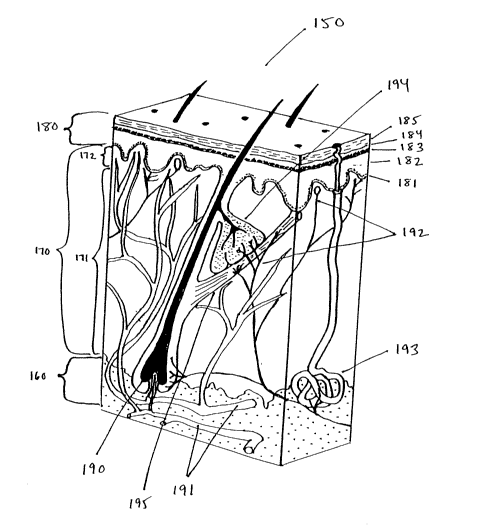
FIG. 1 is a diagram showing an exemplary cross-section of human skin.
FIG. 2 is a schematic diagram showing the layers of skin.
FIGS. 3A and 3B are semi-schematic perspective and side views respectively of
a
section of a patient's skin and of equipment positioned thereon for practicing
one
embodiment.
FIGS. 4A and 4B are top views of various matrix arrays of cylindrical lenses,
some of which are suitable for providing a line focus for a plurality of
target portions.
-15-
CA 02561344 2006-09-26
WO 2005/099369 PCT/US2005/011083
FIG. 5 is a side schematic view of some components that can be used in some
aspects of the invention.
FIG. 6 is a side view of a hand piece that can be used in some aspects of the
invention.
FIG. 7 is a perspective view of another embodiment of the invention.
FIG. 8 is a perspective view of yet another embodiment of the invention.
FIG. 9A is a side view of yet another embodiment of the invention.
FIGS. 9B to 9E are enlarged, side views of the distal end of the embodiment of
FIG. 9A.
FIG. 10A is a side view of yet another embodiment of the invention.
FIGS. i0B and lOC are enlarged, side views of the distal end of the embodiment
of FIG. 10A.
FIG. 11 is a side view of yet another embodiment of the invention.
FIG. 12A is a side view of an embodiment of the invention using a diode laser
bar.
FIG. 12B is a perspective view of a diode laser bar that can be used in the
embodiment of FIG. 12A.
FIG. 12C is a side view of yet another embodiment of the invention, which uses
multiple diode laser bars.
FIG. 12D is a side view of yet another embodiment of the invention, which uses
multiple diode laser bars.
FIG. i2E is a side view of yet another embodiment of the invention, which uses
multiple optical fibers to couple optical energy.
FIG. 13A is a side view of another embodiment of the invention.
FIG. 13B is a perspective view of a light source and optical fiber that can be
used
along with the embodiment of FIG. 13A.
FIG. 13C is a side view of an embodiment of the invention using a fiber
bundle.
FIG. 13D is a bottom view of the embodiment of FIG. 13C.
FIG. 13E is an enlarged, side view of a distal end of one of the embodiments
of
13A-13D.
- 16-
CA 02561344 2006-09-26
WO 2005/099369 PCT/US2005/011083
FIG. 14A is a side view of another embodiment of the invention, which uses a
fiber bundle.
FIG. 14B is a side view of another embodiment of the invention, which uses a
phase mask.
FIG. 14C is a side view of another embodiment of the invention, which uses
multiple laser rods.
FIG. 15 is a bottom view of another embodiment of the invention, which uses
one
or more capacitive imaging arrays.
FIG. 16 is a side view of another embodiment of the invention, which uses a
motor capable of moving a diode laser bar within a hand piece.
FIG. 17 is a top view of one embodiment of a diode laser bar.
FIG. 18 is a side cross-sectional view of the diode laser bar of FIG. 17.
FIGS. 19A-19C are top views of three optical systems involving arrays of
optical
elements suitable for use in delivering radiation in parallel to a plurality
of target
portions.
FIGS. 20A-21D are side views of various lens arrays suitable for delivering
radiation in parallel to a plurality of target portions.
FIGS. 22A-22D are side views of Fresnel lens arrays suitable for delivering
radiation in parallel to a plurality of target portions.
FIGS. 23A-23C are side views of holographic lens arrays suitable for use in
delivering radiation in parallel to a plurality of target portions.
FIGS. 24A-24B are side views of gradient lens arrays suitable for use in
delivering radiation in parallel to a plurality of target portions.
FIGS. 25A-25C are top views of various matrix arrays of cylindrical lenses,
some
of which are suitable for providing a line focus for a plurality of target
portions.
FIGS: 26A-26D are cross-sectional or side views of one layer of a matrix
cylindrical lens system suitable for delivering radiation in parallel to a
plurality of target
portions.
FIGS. 27A, 27B, and 27C are a perspective view and cross-sectional side views,
respectively, of a two layer cylindrical lens array suitable for delivering
radiation in
parallel to a plurality of target portions.
- 17-
CA 02561344 2006-09-26
WO 2005/099369 PCT/US2005/011083
FIGS. 28-31 are side views of various optical objective arrays suitable for
use in
concentrating radiation to one or more target portions.
FIGS. 32A-37 are side views of various deflector systems suitable for use with
the
arrays of FIGS. 10-13 to move to successive target portions.
FIGS. 38 and 39 are side views of two different variable focus optical system
suitable for use in practicing the teachings of this invention.
FIG. 40 is a perspective view of another embodiment of the invention for
creating
treatment islets.
FIGS. 41A and 41B are side views of yet another embodiment of the invention.
FIGS. 42A and 42B are side and top view, respectively, of an embodiment of the
invention having a skin lifting implement, such as a vacuum.
FIG. 43A is a side view of yet another embodiment of the invention.
FIG. 43B is an enlarged, side view of the distal end of the embodiment of FIG.
43A.
FIG. 43C is an enlarged, bottom view of the distal end of the embodiment of
FIG.
43A.
FIG. 44 is a perspective view of another embodiment of the invention for
creating
treatment islets.
FIG. 45 is a perspective shot of two views of another embodiment of the
invention for creating treatment islets.
FIG. 46 is a perspective view of another embodiment of the invention for
creating
treatment islets.
FIG. 47 is a side view of an embodiment of the invention using a film with
active
islets.
FIG. 4.8 is a perspective view of another embodiment of the invention for
creating
treatment islets.
FIGS. 49A to 51B are side views of various embodiments of the invention for
creating treatment islets.
FIG. 52-62 are as described in the examples.
FIG. 63 is the four-layer model of skin used in the computational model
described
in Example 1.
- 18-
CA 02561344 2006-09-26
WO 2005/099369 PCT/US2005/011083
FIG. 64 is the threshold fluence for skin damage at the depths of 0.25 mm ( 1
), 0.5
mm (2), and 0.75 mm (3) in the adiabatic mode as a function of the wavelength.
FIG. 65 is the penetration depth of light inside the type II skin vs. the
wavelength
for a circular beam of diameter 0.1 mm striking the skin through sapphire.
FIG. 66 is the normalized irradiance on the beam axis as a function of skin
depth
for 800 (1), 1060 (2), 1200 (3), 1440 (4), 1560 (5), and 1700 (6) nm
wavelengths.
FIG. 67 is the normalized irradiance on the beam axis as a function of depth
for
1064 nm light focused to skin depths of 0.5 (1), 0.6 (2), 0.7 (3), and 1 (4)
mm.
FIG. 68 is tissue irradiance vs. depth for the collimated beam of diameter 10
mm
(1) and 0.1 mm (2) striking type II skin surface through sapphire at
wavelength 1060 nm.
DETAILED DESCRIPTION
The present invention depends, in part, upon the discovery that, when using
electromagnetic radiation (EMR) to treat tissues, whether for purposes of
photodynamic
therapy, photobleaching, photobiomodulation, photobiostimulation,
photobiosuspension,
thermal stimulation, thermal coagulation, thermal ablation or other
applications, there are
substantial advantages to producing lattices of EMR-treated islets in the
tissue rather than
large, continuous regions of EMR-treated tissue. The EMR-treated tissues can
be any
tissues for which such treatment is useful and appropriate, including but not
limited to
dermal tissues, mucosal tissues (e.g., oral rnucosa, gastrointestinal mucosa),
ophthalmic
tissues (e.g., retinal tissues), vaginal tissue and glandular tissues (e.g.,
prostate tissue).
The lattices are periodic patterns of islets in one, two or three dimensions
in
which the islets correspond to local maxima of EMR-treatment of tissue. The
islets are
separated from each other by non-treated tissue (or differently- or less-
treated tissue).
The EMR-treatment results in a lattice of EMR-treated islets which have been
exposed to
a particular wavelength or spectrum of EMR, and which is referred to herein as
a lattice
of "optical islets." When the absorption of EMR energy results in significant
temperature
elevation in the EMR-treated islets, the lattice is referred to herein as a
lattice of "thermal
islets." When an amount of energy is absorbed that is sufficient to
significantly disrupt
cellular or intercellular structures, the lattice is referred to herein as a
lattice of "damage
islets." When an amount of energy (usually at a particular wavelength)
sufficient to
-19-
CA 02561344 2006-09-26
WO 2005/099369 PCT/US2005/011083
initiate a certain photochemical reaction is delivered, the lattice is
referred to herein as a
lattice of "photochemical islets."
By producing EMR-treated islets rather than continuous regions of EMR-
treatn~ent, untreated regions (or differently- or less-treated regions)
surrounding the islets
can act as thermal energy sinks, reducing the elevation of temperature within
the EMR-
treated islets and/or allowing more EMR energy to be delivered to an islet
without
producing a thermal islet or damage islet and/or lowering the risk of bulk
tissue damage.
Moreover, with respect to damage islets, it should be noted that the
regenerative and
repair responses of the body occur at wound margins (i.e., the boundary
surfaces between
damaged and intact areas) and, therefore, healing of damaged tissues is more
effective
with smaller damage islets, for which the ratio of the wound margin to volume
is greater.
As described more fully below, the percentage of tissue volume which is EMR-
treated versus untreated (or differently- or less-treated) can determine
whether optical
islets become thermal islets, damage islets or photochemical islets. This
percentage is
referred to as the "fill factor", and can be decreased by increasing the
center-to-center
distances) of islets of fixed volume(s), and/or decreasing the ~olume(s) of
islets of fixed
center-to-center distance(s).
Because untreated tissue volumes act as a thermal sink, these volumes can
absorb
energy from treated volumes without themselves becoming thermal or damage
islets.
Thus, a relatively low fill factor can allow for the delivery of high fluence
energy to some
volumes while preventing the development of bulk tissue damage. Finally,
because the
untreated tissue volumes act as a thermal sink, as the fill factor decreases,
the likelihood
of optical islets reaching critical temperatures to produce thermal islets or
damage islets
also decreases (even if the EMR power density and total exposure remain
constant for the
islet areas).
Finally, as described in detail below, the present invention also depends, in
part,
upon the application of discoveries relating to the EMR and thermal energy
absorption,
transfer, and dissipation properties of tissue. Based, in part, upon these
discoveries, the
invention provides improved devices and systems for producing lattices of EMR-
treated
islets in tissues, and improved cosmetic and medical applications of such
devices and
systems in dermatology, dentistry, ophthalmology, gynecology,
otorhinolaryngology and
-20-
CA 02561344 2006-09-26
WO 2005/099369 PCT/US2005/011083
internal medicine in combination with endoscope and catheter techniques.
Although the
devices, systems and methods of the invention are described in detail for
dermatological
applications, they can be used for treatment of any tissue surface or
subsurface areas to
which EMR can be delivered.
References and Definitions.
The patent, scientific and medical publications referred to herein establish
knowledge that was available to those of ordinary skill in the art at the time
the invention
was made. The entire disclosures of the issued U.S. patents, published and
pending
patent applications, and other references cited herein are hereby incorporated
by
reference.
All technical and scientific terms used herein, unless otherwise defined
below, are
intended to have the same meaning as commonly understood by one of ordinary
skill in
the art. References to techniques employed herein are intended to refer to the
techniques
as commonly understood in the art, including variations on those techniques or
substitutions of equivalent or later-developed techniques which would be
apparent to one
of skill in the art. In addition, in order to more clearly and concisely
describe the subject
matter which is the invention, the following definitions are provided for
certain terms
which are used in the specification and appended claims.
Numerical Ranges. As used herein, the recitation of a numerical range for a
variable is intended to convey that the invention may be practiced with the
variable equal
to any of the values within that range. Thus, for a variable which is
inherently discrete,
the variable can be equal to any integer value within the numerical range,
including the
end-points of the range. Similarly, for a variable which is inherently
continuous, the
variable can be equal to any real value within the numerical range, including
the end-
points of the range. As an example, and without limitation, a variable which
is described
as having values between 0 and 2 can take the values 0, 1 or 2 if the variable
is inherently
discrete, and can take the values 0.0, 0.1, 0.01, 0.001, or any other real
values > 0 and S 2
if the variable is inherently continuous. Finally, the variable can take
multiple values in
the range, including any sub-range of values within the cited range_
-21-
CA 02561344 2006-09-26
WO 2005/099369 PCT/US2005/011083
Or. As used herein, unless specifically indicated otherwise, the word "or" is
used
in the inclusive sense of "and/or" and not the exclusive sense of "either/or."
Skin Structure
Although the devices and systems of the invention, and the general methods of
the
invention, can be practiced with many tissues of the body, currently the most
common
applications of EMR-treatment to tissues are in the field of dermatology.
Therefore, the
structure of the skin, including its constituent tissues, is described below
in some detail,
and the remainder of the disclosure will use the skin as an example. In
addition, certain
applications will be described which are uniquely adapted to the skin (e.g.,
tattoo
removal, permeation of the stratum corneum). It should be understood, however,
that the
general methods are applicable to other tissues, and that one of ordinary
skill in the art
can adapt the teachings of the disclosure to other organs and tissues with
merely routine
experimentation.
The skin is the largest organ in the human body, consisting of several layers
of
distinct tissues with distinct properties, and ranges in thickness from
approximately 0.5
mm to approximately 4 mm. Fig. 1 illustrates a typical cross section of skin
150,
showing various layers with differing cellular and intercellular structures.
The skin lies on top of the superficial fascia or subcutaneous tissue 160, a
layer of
fatty tissue that overlies the more densely fibrous fascia.
Above the subcutaneous tissue is the dermis 170, which comprises fibroeiastic
connective tissue, and ranges in thickness from approximately 0.3 mm on the
eyelids to
approximately 3.0 mm on the back. The dermis is highly vascularized and
includes a
variety of sensory nerves with temperature, pressure and pain receptors that
are organized
into small nerve bundles that ascend along with the blood vessels and
lymphatic vessels
to form a network of interlacing nerves beneath the epidermis, i.e., the
superficial nerve
plexus of the papillary dermis. Some of the nerves appear to penetrate the
epidermis for
short distances. The derrnis includes two layers: a reticular layer 171 and a
papillary
layer 172. The reticular layer 171 includes cells in a matrix of dense, coarse
bundles of
collagenous fibers. The papillary layer 172 includes cells in a matrix of
loose
-22-
CA 02561344 2006-09-26
WO 2005/099369 PCT/US2005/011083
collagenous and elastic fibers, with elevations or papillae which project
toward the
epidermis. Cell types in the dermis include fibroblasts, mast cells and
macrophages.
The epidermis 180 comprises the outermost stratified layers of the skin, and
ranges in thickness from approximately 0.05 mm on the eyelids to approximately
1.5 mm
on the palms and soles. The epiderrr~is is avascular and consists largely of
epithelial cells
which mature as they pass from the innermost layer of columnar cells to the
outermost
layer of tile-like squamous cells, with the cells becoming increasingly
flattened and
keratinized as they progress outward. The innermost layer is referred to as
the stratum
basale, basal cell layer, or stratum germinativum 181, and is the only layer
in normal
epidermis in which cell division occurs. The next layer, the stratum spinosum
182,
includes prickle cells and keratinocytes, and begins the production of
keratin. The next
layer, the stratum granulosum 183, is a darker layer with intercellular
granules and
increased keratin production. In thick skin, there is an additional
transitional layer, tl3e
stratum lucidum 184. Finally, the outermost layer is the stratum corneum (SC)
185, a
horny layer of highly keratinized squamous cells.
The cells of the stratum corneum 185 (and the stratum lucidum 184, when
present) are highly keratinized ("horny") and surrounded by an extracellular
matrix
consisting largely of crystalline lipids. As a result, the stratum corneum
forms a hard,
resilient barrier to water transport, and is not permeable to most aqueous or
organic
solvents or solutes. The stratum comeum 185 is about 15 p.m deep on most
anatomic
sites but can be in the ranges of 10-300 p.m (e.g., 20 p,m at the forearm and
50-60 p.m at
the wrist).
Also shown are typical organs and structures within the skin, including a hair
follicle 190, blood vessels 191, nerve fibers 192, a sweat gland 193, a
sebaceous gland
194, and an arrector pili muscle 195_
Normal skin temperature is approximately 29-37°C. When exposed to
temperatures in excess of 40-43°C, tie sensory nerves of the dermis
will transmit a pain
response in most human subjects.
Fig. 2 is a schematic cross-sectional view of a human skin section 150. It
shows
depths into the skin, from the surface in p.m. The stratum corneum 185 and
stratum
lucidum 184 are shown extending to a depth of approximately 15 p.m below the
skin
-23-
CA 02561344 2006-09-26
WO 2005/099369 PCT/US2005/011083
surface. The remaining layers (i.e., layers 181-183) of the epidermis 180
extend from the
stratum lucidumlcorneum 184/185 to the boundary with the dermis 170 at a depth
from
the surface in the range of approximately 50-150 p,m. Also shown are exemplary
shallow
islets 196 affecting the stratum lucidu<m/corneum 184/185, deeper islets 197
affecting the
stratum lucidum/corneum 184/185 and deeper layers of the epidermis 180, and
subsurface islets 198 spanning portions of the deeper epidermis 180 and upper
dermis
170.
The depths shown in Fig. 2 are merely exemplary. Different locations in the
typical human body have different depth profiles for the stratum
corneum/lucidum,
epidermis, and dermis. In addition, as described below. a great variety of
other islet
configurations are possible which are not shown in the figure (e.g., islets
entirely in the
dermis; islets entirely in the subcutaneous tissue; islets spanning the dermis
and
subcutaneous tissue; islets spanning portions of the epidermis, dermis and
subcutaneous
tissue).
Categories of EMR-Treated Islets
The present invention depends, in part, on the creation of a multiplicity of
treated
volumes of the skin which are separated by untreated volumes. The multiplicity
of
volumes can be described as defining a "lattice," and the treated volumes,
because they
are separated by untreated volumes, can be described as "islets" within the
skin.
Depending upon the nature of the treatment, in particular the amount of energy
transfer to
the islets, the degree of heating of the tissue, or the wavelengths) of the
energy, four
different categories of lattices can be produced: lattices of optical islets
(LOI), lattices of
thermal islets (LTI), lattices of damage islets (LDI), and lattices of
photochemical islets
(LPCI). These different categories of EMR-treated islets, devices and systems
for
producing such EMR-treated islets, and cosmetic and medical applications for
such
devices and systems are separately discussed in detail below. As used herein,
the terms
"treatment islet," "islets of treatment," and "EMR-treated islets" are used
interchangeably
to mean any of the categories of islets described below.
-24-
CA 02561344 2006-09-26
WO 2005/099369 PCT/US2005/011083
A. Optical Islets
In accordance with the present invention, EMR-treatment of completely or
partially isolated volumes or islets of tissue produces a lattice of EMR-
treated islets
surrounded by untreated volumes. Although the islets can be treated with any
form of
EMR, they are referred to herein as "optical" islets for convenience, as many
embodiments of the invention include the use of EMR within the ultraviolet,
visible and
infra-red spectrum. Other forms of EMR useful in the invention include
microwave,
radio frequency, low frequency and EMR induced by direct current.
As noted above, when the total energy transfer per unit cross-sectional area
(i.e.,
fluence) or the rate of energy transfer per unit cross-sectional area (i.e.,
flux) becomes
sufficiently high, the tissue of an optical islet will be heated, resulting in
a thermal islet.
If the temperature increase is sufficiently high, the tissue of a thermal
islet will be
damaged, resulting in a damage islet. Thus, although all thermal islets and
damage islets
are also optical islets, not all optical islets are thermal islets or damage
islets. In some
embodiments, as described below, it can be desirable to produce optical islets
without
producing thermal or damage islets. In such embodiments, the fill factor can
be
decreased in order to provide a greater volume of untreated tissue to act as a
thermal sink.
As described in detail in the Examples below, a model of optical islets was
developed which describes the propagation of light into skin taking into
account the skin
type and the characteristics of the light source. The particular approach used
below is
applicable to a wide range of islet dimensions (e.g_ , 10-30,000 p,m in the
lateral plane), is
generally accepted in tissue optics, and is referred ~o as the light transport
theory
(Chandrasekhar (1960), Radiative Transfer (University Press, Oxford); Ishimaru
(1970,
Wave Propagation and Scattering in Random Medla, Volume 1 (Academic Press, New
York); Jacques et al.(1995), in Optical-Thermal Response of Laser-Irradiated
Tissue,
Welch et al.., eds. (Plenum Press, New York), pp. 561-606). Briefly, the skin
is
considered as a multilayer structure with each layer being a turbid medium
where light
undergoes both absorption and multiple scattering_ This approach neglects
macroscopic
coherence effects like diffraction and speckle forn3ation. Several techniques
may be used
to solve the light transport problem in a tissue. So me of them, particularly
the two-flux
and diffusion approximations, break down when the input beam is sufficiently
narrow or
-25-
CA 02561344 2006-09-26
WO 2005/099369 PCT/US2005/011083
is focused into the tissue, and are not suitable for dealing with the islet
formation
problem. The Monte-Carlo technique described below is not subject to such
limitations
and is capable of modeling various tissue structures, spot profiles,
wavelength spectra,
and angular distributions of the incident light (Jacques et al. (1995),
supra).
B. Thermal Islets
In accordance with another aspect of the present invention, EMR-treatment of
isolated volumes or islets of tissue can produce a lattice of thermal islets
with
temperatures elevated relative to those of surrounding untreated volumes.
Thermal islets
result when energy is absorbed by an EMR-treated optical islet significantly
faster than it
is dissipated and, therefore, significant heating occurs.
Heating can result from the absorbance of EMR by water present throughout a
volume of treated tissue, by endogenous chromophores present in selected cells
or
tissues) (e.g., melanin, hemoglobin), by exogenous chromophores within the
tissue (e.g.,
tattoo ink) or, as described below, by exogenous chromophores applied to the
surface of
the tissue.
With respect to skin, in order to avoid causing pain to a subject, the maximal
temperature of the basal membrane, which is adjacent to the nerve terminals of
the
papillary dermis, should not exceed 40-45°C. Assuming no active cooling
of the skin
surface, the temperature rise in the basal membrane, ~T2, can be related to
the
temperature rise in the hyperthermic islets, ~T1, by an approximate formula:
OTZ = fAT 1
where f is the fill-factor of the optical lattice at the skin surface. This
formula indicates
that the temperature in the SC can attain relatively high values without
triggering the pain
response of the body if the fill factor is sufficiently low.
For example, setting OT2 to 12°C and f to ~.3 yields ~T 1 of
40°C. In practice,
the temperature rise AT1 may be limited by other factors, such as, for
example, the
threshold of structural damage to the SC or the desired size of the damage
islets.
The thermal islet model is based, in part, on the time-dependent heat
equation.
Specifically, as described in more detail below, the thermal constants of the
skin layers
-26-
CA 02561344 2006-09-26
WO 2005/099369 PCT/US2005/011083
are obtained from Takata's relations (Takata et al. (1977), in Report SAM TR-
77 38 (San
Antonio, TX: US Air Force School for Aerospace Medicine)) and are functions of
the
volume fraction of water in the corresponding layer. Specific effects
associated with the
bio-heat equation, e.g., the metabolic heat generation and the change of the
blood
perfusion rate while heating the living tissue, can be neglected for EMR
pulses of short
duration (Sekins et al.11990), Thermal Science for Physical Medicine, in
Therapeutic
Pleat and Cold, 4th edition, Lehmann, ed. (Baltimore: Williams ~ Wilkins) pp.
62-112).
In practice, the EMR-heating can dominate strongly over metabolic heating and
heat
transfer by the blood flow. Moreover, the changes in the blood perfusion rate
can occur
with the delay of about 1 min with respect to the variations of the tissue
temperature
(Sekins et al. (1990), in Therapeutic Heat and Cold, 4th edition, Lehmann, ed.
(Baltimore: Williams & Wilkins) pp. 62-112), and do not affect tie islet
formation
dynamics unless tissues are under combined action (with EMR) of simultaneous
physical
factors (e.g., elevated or lowered external pressure, ultrasound, elevated or
lowered skin
surface temperature).
It should be emphasized that a lattice of thermal islets is at time-dependent
phenomenon. If absorptive heating occurs at too great a rate or for too long a
period, heat
will begin to diffuse away from the EMR-treated islets and into the
surrounding untreated
tissue volumes. As this occurs, the thermal islets will spread into the
untreated volumes
and, ultimately, the thermal islets will merge and result in bulk heating. By
using a
sufficiently short pulse width relative to the temperature relaxation time of
the target, it is
possible to avoid merging or overlapping of thermal islets in a lattice.
C. Dama a Ig slets
In accordance With another aspect of the present invention, EMR-treatment of
isolated volumes or islets of tissue can produce a lattice of damage islets
surrounded by
volumes of undamaged tissue (or differently- or less-damaged tissue). Damage
islets
result when the temperature increase of an EMR-treated thermal islet is
sufficient to
result in protein coagulation, thermal injury, photodisruption, pho~oablation,
or water
vaporization. Depending upon the intended use, damage islets with lesser
degrees of
damage (e.g., protein coagulation, thermal injury) or greater degrees of
damage (e.g.,
photodisruption, photoablation, or water vaporization) may be appropriate. As
before,
_ 27 _
CA 02561344 2006-09-26
WO 2005/099369 PCT/US2005/011083
damage can result from the absorbance of EMR by water present throughout a
volume of
treated skin, by endogenous chromophores present in selected cells or tissues)
in the skin
(e.g., melanin, hemoglobin), by exogenous chromophores within the skin (e.g.,
tattoo ink)
or, as described below, by exogenous chromophores applied to the surface of
the skin.
As described in detail below, in some embodiments of the invention, the damage
islets are thermal injuries with coagulation of structural proteins. Such
damage can result
when, for example, the light pulse duration varies from several microseconds
to about 1
sec, but the peak tissue temperature remains below the vaporization threshold
of water in
the tissue (Pearce et al. (1995), in Optical-Thermal Response of Laser-
Irradiated Tissue,
Welch et al., eds. (Plenum Press, New York), pp. 561-606). The degree of
damage is a
function of the tissue temperature and the duration of the thermal pulse and
can be
quantified by any of several damage functions known in the art. In the
description below,
for example, the damage function yielding the Arrhenius damage integral
(Pearce et al.
(1995), in Optical-Thermal Response of Laser-Irradiated Tissue, Welch et al.,
eds_
(Plenum Press, New York), pp. 561-606; Henriques (1947), Arch. Path~~. 43:480-
502) is
employed. Other mechanisms and models of damage islet formation ca.n apply to
embodiments with relatively short and intense pulses, particularly in
connection with
photodisruption, photoablation, and water vaporization.
D. Photochemical Islets
In accordance with another aspect of the present invention, EM~2-treatment of
isolated volumes or islets of tissue can produce a lattice of photochemical
islets
surrounded by volumes of tissue in which a photochemical reaction has not been
induced.
The photochemical reaction can involve endogenous biomolecules or exogenous
molecules. For example, exposure of the skin to certain wavelengths of EMR can
result
in increased melanin production and "tanning" through the activation of
endogenous
biomoiecules and biological pathways. Alternatively, for example, exogenous
molecules
can be administered in photodynamic therapy, and activation of these molecules
by
certain wavelengths of EMR can cause a systemic or localized therapeutic
effect.
_2g_
CA 02561344 2006-09-26
WO 2005/099369 PCT/US2005/011083
Treatment Parameters.
In the practice of the invention, a variety of different treatment parameters
relating to the applied EMR can be controlled and varied according to the
particular
cosmetic or medical application. These parameters include, without limitation,
the
following:
A. The Shape of EMR-Treated Islets.
The optical islets can be formed in any shape which can be produced by the
devices described below, limited only by the ability to control EMR beams
within the
tissue. Thus, depending upon the wavelength(s), temporal characteristics (e_
g.,
continuous versus pulsed delivery), and fluence of the EMR; the geometry,
incidence and
focusing of the EMR beam; and the index of refraction, absorption coefficient,
scattering
coefficient, anisotropy factor (the mean cosine of the scattering angle), and
the
configuration of the tissue layers; and the presence or absence of exogenous
chromophores and other substances, the islets can be variously-shaped volumes
extending from the surface of the skin through one or more layers, or
extending from
beneath the surface of the skin through one or more layers, or within a single
layer. If the
beams are not convergent, such beams will define volumes of substantially
constant
cross-sectional areas in the plane orthogonal to the beam axis (e.g.,
cylinders,
rectanguloids). Alternatively, the beams can be convergent, defining voluniss
of
decreasing cross-sectional area in the plane orthogonal to the central axis of
the beams
(e.g., cones, pyramids). The cross-sectional areas can be regular in shape
(e_g., ellipses,
polygons) or can be arbitrary in shape. In addition, depending upon the
wavelengths)
and fluence of an EMR beam, and the absorption and scattering characteristics
of a tissue
for the wavelength(s), an EMR beam may penetrate to certain depths before
being
initially or completely absorbed or dissipated and, therefore, an EMR-treated
islet may
not extend through the entire depth of the skin but, rather, may extend
between the
surface and a particular depth, or between two depths below the surface.
Generally, the lattice is a periodic structure of islets in one, two, or tree
dimensions. For instance, a two-dimensional (2D) lattice is periodic in two
dimensions
and translation invariant or non-periodic in the third. The type of
periodicity is
characterized by the voxel shape. For example, and without limitation, them
can be
-29-
CA 02561344 2006-09-26
WO 2005/099369 PCT/US2005/011083
layer, square, hexagonal or rectangle lattices. The lattice dimensionality can
be different
from that of an individual islet. A single row of equally spaced infinite
cylinders is an-
example of the 1D lattice of 2D islets (if the cylinders are of finite length
this is the 1D
lattice of 3D islets). The lattice dimensionality is equal to or smaller than
the
dimensionality of its islets (this fact follows from the fact that the lattice
cannot be
periodic in the dimension where its islets are translation invariant). Hence,
there exists a
total of 6 lattice types with each type being an allowed combination of the
islet and lattice
dimensionalities. For certain applications, an "inverted" lattice can be
employed, in
which islets of intact tissue are separated by areas of EMR-treated tissue and
the
treatment area is a continuous cluster of treated tissue with non treated
islands.
Referring to Figure 3A, each of the treated volumes can be a relatively thin
disk,
as shown, a relatively elongated cylinder (e.g., extending from a first depth
to a second
depth), or a substantially linear volume having a length which substantially
exceeds its
width and depth, and which is oriented substantially parallel to the skin
surface. The
orientation of the lines for the islets 214 in a given application need not
all be the same,
and some of the lines may, for example, be at right angles to other lines (see
for example
Figs. 4A and 4B). Lines also can be oriented around a treatment target for
greater
efficacy. For example, the lines can be perpendicular to a vessel or parallel
to a wrinl~le.
Islets 214 can be subsurface volumes, such as spheres, ellipsoids, cubes or
rectanguloLds
of selected thickness. The islets can also be substantially linear or planar
volumes. TL-~e
shapes of the islets are determined by the combined optical parameters of the
beam,
including beam size, amplitude and phase distribution, the duration of
application and., to
a lesser extent, the wavelength.
The parameters for obtaining a particular islet shape can be determined
empirically with only routine experimentation. For example, a 1720 nm laser
operatir~g
with a low conversion beam at approximately 0.005-2 J and a pulse width of 0.5-
2 ms,
can produce a generally cylindrically shaped islet. Alternatively, a 1200 nm
laser
operating with a highly converting beam at approximately 0.5-10 J and a pulse
width of
0.5-3 sec, can produce a generally ellipsoid-shaped islet.
By suitable control of wavelength, focusing, incident beam size at the surface
and
other parameters, the islets, regardless of shape, can extend through a
volume, can be
-30-
CA 02561344 2006-09-26
WO 2005/099369 PCT/US2005/011083
formed in a single thin layer of a volume, or can be staggered such that
adjacent islets are
in different thin layers of volume. Most configurations of a lattice of islets
can be formed
either serially or in simultaneously. Lattices with islets in multiple thin
layers in a
volume can be easily formed serially, for example using a scanner or using
multiple
energy sources having different wavelengths. Islets in the same or varying
depths can be
created, and when viewed from the skin surface, the islets at varying depths
can be either
spatially separated or overlapping.
The geometry of the islets affects the thermal damage in the treatment region.
Since a sphere provides the greatest gradient, and is thus the most spatially
confined, it
provides the most localized biological damage, and can therefore be preferred
for
applications where this is desirable.
B. The Size of EMR-Treated Islets.
The size of the individual islets within the lattices of EMR-treated islets of
the
invention, can vary widely depending upon the intended cosmetic or medical
application.
As discussed more fully below, in some embodiments it is desirable to cause
substantial
tissue damage to destroy a structure or region of tissue (e.g., a sebaceous
gland, hair
follicle, tattooed area) whereas in other embodiments it is desirable to cause
little or no
damage while administering an effective amount of EMR at a specified
wavelength (e.g.,
photodynamic therapy). As noted above with respect to damage islets, however,
the
healing of damaged tissues is more effective with smaller damage islets, for
which the
ratio of the wound margin to volume is greater.
As a general matter, the size of the EMR-treated islets of the present
invention
can range from 1 p,m to 30 mm in any particular dimension. For example, and
without
limitation, a lattice of substantially linear islets can consist of parallel
islets have a length
of approximately 30 mrn and a width of approximately 10 p.m to 1 mm. As
another
example, and without limitation, for substantially cylindrical islets in which
the axis of
the cylinder is orthogonal to the tissue surface, the depth can be
approximately 10 p.m to
4 mm and the diameter can be approximately 10 p,m to 1 mm. For substantially
spherical
or ellipsoidal islets, the diameter or major axis can be, for example, and
without
limitation, approximately 10 p,m to 1 mm. Thus, in some embodiments, the
islets can
have a maximum dimension in the range from 1 p,m to 10 p,m, 10 p,m to 100 p,m,
100 p,m
-31-
CA 02561344 2006-09-26
WO 2005/099369 PCT/US2005/011083
to 1 mm, 1 mm to 10 mm, or 10 mm to 30 mm, as well as all possible ranges
within 1 p.m
to 30 mm.
When considering the size of the optical, thermal, damage or photochemical
islets
of the invention, it is important to note that the boundaries of the islets
may not be clearly
demarcated but, rather, may vary continuously or blend into the untreated
tissue (or
differently- or less-treated tissue). For example, EMR beams are subject to
scattering in
various tissues and, therefore, even beams of coherent light will become
diffuse as they
penetrate through multiple layers of cells or tissues. As a result, optical
and
photochemical islets typically will not have clear boundaries between treated
and
untreated volumes. Similarly, thermal islets typically will exhibit a
temperature gradient
from the center of the islet to its boundaries, and untreated tissue
surrounding the islet
also will exhibit a temperature gradient due to conduction of heat. Finally,
damage islets
can have irregular or indistinct boundaries due to partially damaged cells or
structures or
partially coagulated proteins. As used herein, therefore, the size of an islet
within a
lattice of islets, refers to the size of the islet as defined by the intended
minimum or
threshold amount of EMR energy delivered. As discussed in greater detail
below, this
amount is expressed as the minimum fluence, F~,;I,, and is determined by the
nature of the
cosmetic or medical application. For example, for photodynamic therapy, F~,;n
can be
determined by the minimum fluence necessary to cause the desired photochemical
reaction. Similarly, for increasing the permeability of the stratum corneum,
FI";n can be
determined by the minimum fluence necessary to achieve the desired SC
temperature,
and for destroying tissue, Fn,;" can be determined by the minimum fluence
necessary to
ablate the tissue or vaporize water. In each case, the size of the EMR-treated
islet is
defined by the size of the tissue volume receiving the desired minimum
fluence.
Because of the scattering effects of tissue, the minimum size of an EMR-
treated
islet increases with the targeted depth in the tissue, ranging from several
microns on the
stratum corneum to several millimeters in subcutaneous tissue. For a depth of
approximately 1 mm into a subject's tissue, the minimum diameter or width of
an islet is
estimated to be approximately 100 pm, although much larger islets (e.g., 1-10
mm) are
possible. The size of a damage islet can be either smaller or larger than the
size of the
corresponding optical islet, but is generally larger as greater amounts of EMR
energy are
-32-
CA 02561344 2006-09-26
WO 2005/099369 PCT/US2005/011083
applied to the optical islet due to heat diffusion. For a minimum size islet
at any
particular depth in the skin, the wavelength, beam size, convergence, energy
and pulse
width have to be optimized.
C. The Depth of EMR-Treated Islets.
The EMR-treated islets of the invention can be located at varying points
within a
tissue, including surface and subsurface locations, locations at relatively
limited depths,
and locations spanning substantial depths. The desired depth of the islets
depends upon
the intended cosmetic or medical application, including the location of the
targeted
molecules, cells, tissues or intercellular structures.
For example, optical islets can be induced at varying depths in a tissue or
organ,
depending upon the depth of penetration of the EMR energy, which depends in
part upon
the wavelengths) and beam size. Thus, the islets can be shallow islets that
penetrate
only surface layers of a tissue (e.g., 0-50 p,m), deeper islets that span
several layers of a
tissue (e.g., 50-500 p.m), or very deep, subsurface islets ((e.g., 500 pm - 4
mm). Using
optical energy, depths of up to 25 mm can be achieved using wavelengths of
1,000-1,300
nm. Using microwave and radio frequency EMR, depths of several centimeters can
be
achieved.
For thermal islets or damage islets, subsurface islets can be produced by
targeting
chromophores present only at the desired depth(s), or by cooling upper layers
of a tissue
while delivering EMR. For creating deep thermal or damage islets, long pulse
widths
coupled with surface cooling can be particularly effective.
D. Fill Factor of EMR-Treated Lattices
In a given lattice of EMR-treated islets, the percentage of tissue volume
which is
EMR-treated is referred to as the "fill factor" or f, and can affect whether
optical islets
become thermal islets, damage islets or photochemical islets. The fill factor
is defined by
the volume of the islets with respect to a reference volume that contains all
of the islets.
The fill factor may be uniform for a periodic lattice of uniformly sized EMR-
treated
islets, or it may vary over the treatment area. Non-uniform fill factors can
be created in
situations including, but not limited to, the creation of thermal islets using
topical
application of EMR-absorbing particles in a lotion or suspension (see below).
For such
-33-
CA 02561344 2006-09-26
WO 2005/099369 PCT/US2005/011083
situations, an average fill factor (f~~g) can be calculated by dividing the
volume of all
EMR-treated islets Virsrer by the volume of all tissue vr''ssue in the
treatment area,
V rsret
.~ t
J avg = ~ Vtissue
r t
Generally, the fill factor can be decreased by increasing the center-to-center
distances) of islets of fixed volume(s), andlor decreasing the volumes) of
islets of fixed
center-to-center distance(s). Thus, the calculation of the fill factor will
depend on
volume of an EMR-treated islet as well as on the spacing between the islets.
In a periodic
lattice, where the centers of the nearest islets are separated by a distance
d, the fill factor
will depend on the ratio of the size of the islet to the spacing between the
nearest islets d.
For example, in a lattice of parallel cylindrical islets, the fill factor will
be:
2
where d is the shortest distance between the centers of the nearest islets and
r is the radius
of a cylindrical EMR-treated islet. In a lattice of spherical islets, the fill
factor will be the
ratio of the volume of the spherical islet to the volume of the cube defined
by the
neighboring centers of the islets:
f. - 4~c r
3 ~d~
where d is the shortest distance between the centers of the nearest islets and
r is the radius
of a spherical EMR-treated islet. Similar formulas can be obtained to
calculate fill
factors of lattices of islets of different shapes, such as lines, disks,
ellipsoids,
rectanguloids, or other shapes.
Because untreated tissue volumes act as a thermal sink, these volumes can
absorb
energy from treated volumes without themselves becoming thermal or damage
islets.
Thus, a relatively low fill factor can allow for the delivery of high fluence
energy to some
volumes while preventing the development of bulk tissue damage. Finally,
because the
untreated tissue volumes act as a thermal sink, as the fill factor decreases,
the likelihood
of optical islets reaching critical temperatures to produce thermal islets or
damage islets
- 34 -
CA 02561344 2006-09-26
WO 2005/099369 PCT/US2005/011083
also decreases (even if the EMR power density and total exposure remain
constant for the
islet areas).
The center-to-center spacing of islets is determined by a number of factors,
including the size of the islets and the treatment being performed. Generally,
it is desired
that the spacing between adjacent islets be sufficient to protect the tissues
and facilitate
the healing of any damage thereto, while still permitting the desired
therapeutic effect to
be achieved. In general, the fill factor can vary in the range of 0.1-90%,
with ranges of
0.1-1%, 1-10%, 10-30% and 30-50% for different applications. The interaction
between
the fill factor and the thermal relaxation time of a lattice of EMR-treated
islets is
discussed in detail below. In the case of lattices of thermal islets, it can
be important that
the fill factor be sufficiently low to prevent excessive heating and damage to
islets,
whereas with damage islets it can be important that the fill factor be
sufficiently low to
ensure that there is undamaged tissue around each of the damage islets
sufficient to
prevent bulk tissue damage and to permit the damaged volumes to heal.
Applications of EMR-Treated Islets
EMR-treated islets can be used in a variety of applications in a variety of
different
organs and tissues. For example, EMR treatments can be applied to tissues
including, but
not limited to, skin, mucosal tissues (e.g., oral mucosa, gastrointestinal
mucosa),
ophthalmic tissues (e.g., conjuctiva, cornea, retina), and glandular tissues
(e.g., lacrimal,
prostate glands). As a general matter, the methods can be used to treat
conditions
including, but not limited to, lesions (e.g., sores, ulcers), acne, rosacea,
undesired hair,
undesired blood vessels, hyperplastic growths (e.g., tumors, polyps, benign
prostatic
hyperplasia), hypertrophic growths (e.g., benign prostatic hypertrophy),
neovascularization (e.g., tumor-associated angiogenesis), arterial or venous
malformations (e.g., hemangiomas, nevus flammeus), and undesired pigmentation
(e.g.,
pigmented birthmarks, tattoos).
A. Thermal Islets
In some aspects, the invention provides methods of treating tissues by
creating
lattices of thermal islets. These methods can be used in, for example, methods
of
-35-
CA 02561344 2006-09-26
WO 2005/099369 PCT/US2005/011083
increasing the permeability of the stratum corneum to various agents,
including
therapeutic agents and cosmetic agents, and methods for producing therapeutic
hyperthermia.
1. Reversible Permeation of the Stratum Corneum
In one embodiment, lattices of thermal islets are produced in order to
reversibly
increase the permeability of the stratum corneum by heating islets of tissue
to
temperatures of 35-100°C. The increased permeability results from the
melting of the
extracellular matrix of crystalline lipids that surrounds the cells of the
stratum corneum
and, when present, the stratum lucidum. When this matrix melts (i.e., loses
its crystalline
structure), the SC becomes more permeable to molecules on the surface of the
skin,
allowing some molecules to diffuse inward. When the temperature of the layer
returns to
the normal range (i.e., 29-37°C), the intercellular matrix
recrystallizes, the SC becomes
more impermeable, and any molecules which had diffused below the SC can remain
there, further diffuse into surrounding tissues, or enter the systemic
circulation. Thus, as
used herein, the increased permeability is "reversible" because the lipid
intercellular
matrix recrystallizes. In different embodiments, the increase in permeability
is reversed
within i second to 2 hours after the EMR-treatment is discontinued. Thus, in
some
embodiments, the increase in permeability is reversed within 15 minutes, 30
minutes, 1
hour or 2 hours after the EMR-treatment is discontinued.
In these embodiments, the thermal islets define permeation pathways which can
extend through or mostly through the stratum corneum and stratum lucidum
layers so that
a compound, for example, a cosmetic or therapeutic agent applied to the
exterior surface
of the skin is able to efficiently penetrate the stratum corneum/stratum
lucidum. This
penetration can be superficial and remain just below or within the stratum
corneum, or
can be deeper into the interior layers of the epidermis or dermis and,
possibly, into the
blood stream via the vascularization in the dermis. This enables the
percutaneous
delivery of cosmetic or therapeutic agents locally to the epidermis and
dermis. To the
extent the compound diffuses away from the site of treatment, the local
delivery of the
compound can be greater (e.g., delivery to a joint region). Moreover, to the
extent that
the compound reaches the vasculature of the dermis, delivery can be systemic.
-36-
CA 02561344 2006-09-26
WO 2005/099369 PCT/US2005/011083
In some embodiments, the compound is a therapeutic agent. Examples of
therapeutic agents include, without limitation, a hormone, a steroid, a non-
steroidal anti-
inflammatory drug, an anti-neoplastic agent, an antihistamine and an
anesthetic agent.
Specific examples include, without limitation, hormones such as insulin and
estrogen,
steroids such as prednisolone and loteprednol, non-steroidal anti-inflammatory
drugs
such as ketorolac and diclofenac, anti-neoplastic agents such as methotrexate,
and
antihistamines such as histamine H1 antagonists, chlorpheniramine, pyrilamine,
mepyramine, emedastine, levocabastine and lidocaine.
In other embodiments, the compound is a cosmetic agent. Examples of cosmetic
agents include, without limitation, pigments (including both naturally
occurring and
synthetic chromophores, dyes, colorants or inks) reflective agents {including
light-
scattering compounds), and photoprotectants (including sunscreens). Such
cosmetic
agents can be used to add coloration to the skin, or to mask existing
coloration (e.g.,
birthmarks, pigmented lesions, tattoos) by adding differently colored pigments
or
reflective agents. The invention provides improved methods of applying
cosmetic agents
because (a) the agents are contained within the stratum corneum and will not
be smeared,
or rubbed or washed off, and (b) the agents will remain within the stratum
corneum until
the cells of that layer are replaced through the normal process of outgrowth
from the
stratum basale (e.g., approximately 21-28 days). Thus, a single application of
a cosmetic
agent can last for several weeks, which can be advantageous relative to
cosmetics which
must be applied daily. Conversely, the application of the cosmetic agent is
limited to
several weeks, which can be advantageous relative to tattoos which are usually
permanent unless removed by photobleaching or tissue ablation. In one
embodiment,
pigments for a desired temporary tattoo can be applied to the skin (e.g., by a
film, brush,
printing), the stratum corneum can be EMR-treated to increase permeability,
and the
pigments can diffuse into the skin to create the temporary tattoo. In other
embodiments,
an artificial tan can be created by delivering a colorant or, conversely, a
tan can be
prevented by delivering a sunscreen into the skin.
The increased permeability of the stratum corneum can be made painless or less
painful for a subject by using lattices of thermal islets (or damage islets)
rather than a
continuous area of heating. Because the entire area and thickness of the skin
is not
-37-
CA 02561344 2006-09-26
WO 2005/099369 PCT/US2005/011083
heated, a 40-43°C isotherm can be terminated near the epidermisldermis
boundary
instead of deeper in the dermis. Therefore, nerve endings found in papillary
dermis are
not exposed to the 40-43°C temperatures associated with a pain
response. As a result, the
enhanced permeability paths defined by the thermal islets can be created
without pain
even though the SC has been exposed to temperatures significantly higher than
40-43°C.
A significant (orders of magnitude) increase in permeability of the stratum
corneum occurs when the temperature of the extracellular lipids of the SC is
raised to the
transition temperature, Tm, at which the lipid state changes from the
mesomorphic (liquid
crystal) state to the liquid state (Tm=64°C for rat SC, see Ogiso et
al. (1996), Biochirn.
Biophys. Acta 1301(1-2):97-104). Simple estimates of the required heat flux to
achieve
this temperature, and thereby reversibly melt the lipid layers of the stratum
corneum, can
be made as follows.
For example, the thickness of the SC can be chosen to be d=15 pm, such as can
be
found on the volar forearm, for the purposes of this calculation. The stratum
corneum
(SC) is known to be composed of a mixture of water, lipids and proteins with
the
following approximate weights: W1=20% water, W2=50% lipids, and W3=30%
protein.
The lipids of the SC are composed of the following: ceramides (50%),
cholesterol (28%),
free fatty acids (17%), and cholesterol sulfate (5%). The thermal parameters
of the SC
are determined to be the weighted sum of the corresponding parameters of the
constituents with the appropriate weight factors W i, W2, and W3:
Constituent Weight factorDensity, Specific Thermal
g/cm3 heat, conductivity,
J/(g K) W/(cm IC)
Water 0.2 1 4.18 0.0058
Protein 0.3 1.3 1.55 0.00027
Lipids 0.5 0.31 (fat) 0.975 0.0022
Whole SC 1 p=0.745 c=1.788 x=2.341E-3
(Av )
A typical initial SC temperature is To=30 C. The latent heat of fusion, ~,,
(for
melting) for the SC lipids is assumed to be similar in value to that known for
the lipid
DPPC (dipalmitoylphosphatidylcholine). This parameter is ?v, =14500 J/mol=2
J/gm,
where the molecular weight is 734 gm/mole. Assuming the adiabatic mode
(neglect heat
-38-
CA 02561344 2006-09-26
WO 2005/099369 PCT/US2005/011083
loss) and temperature equilibration among the constituents, the threshold
fluence for
melting the lipid, F, may be evaluated as follows:
F'm =~~Z'n~-Z'o~~c+~.~-p~d
Using the estimates of the parameters above, the value for the required
fluence to
melt the lipids of the SC is Fm = 0.07 J/cm2. This fluence may be achieved in
a variety of
ways as discussed herein. For example, EMR may be absorbed directly and
converted to
heat by one or more of the constituents acting as endogenous chromophores of
the SC, or
EMR may be absorbed by exogenous chromophores on the skin surface (e.g.,
carbon
dots). Note that the relative contribution of energy to actually melt the
lipids is small
(~3%) and that most of the energy is needed to bring the SC from the ambient
temperature, To, to the melting point, Tm.
= 0.033
[(Tm- TO)-c]
The thermal relaxation time, TRT, of the SC is estimated as follows:
x := 2.341x 10 3- Watt rtes := 10 3-s
cm-I~
TRT := dy~ P ~c TRT = 0.64ms
2-x
As an example, a heat flux of ~ 1 kWcni 2 for 70 ps will satisfy this
condition. Note that
if the melting point temperature needs to be maintained for a time exceeding
the TRT,
then the required heat flux must balance the heat loss once the required
temperature is
reached.
The size of the enhanced permeability paths can range from the diameter of an
intercellular lipid space (e.g., 1 wm) or the thickness of a horny cell (e.g.,
0.5 p.m) at one
extreme, to about the SC thickness (e.g., 10-500 p.m). Typically, however, the
enhanced
permeability paths are about ~0 p.m to 1 mm in diameter and less than 50 p,m
in depth to
avoid damage to the viable epidermal layers, as well as to reduce or eliminate
pain and
discomfort. Nonetheless, for some embodiments, thermal islets can extend into
deeper
-39-
CA 02561344 2006-09-26
WO 2005/099369 PCT/US2005/011083
layers of the epidermis and dermis to denature them and stimulate blood
microcirculation
for faster drug absorption in the body. Targeting deeper tissues with higher
temperatures,
however, could necessitate pain control for the patient.
Generally, the spacing of thermal islets should be as dense as possible to
maximize the permeability and thus delivery efficiency. However, if the paths
are too
dense, then the depth-temperature selectivity is impacted. For example, if the
spacing
were zero, then heat would only effectively diffuse downward rather than
radially,
making it difficult to heat the stratum corneum sufficiently to produce
enhanced
permeability paths while preventing injury and pain to the deeper epidermal
and dermal
layers. Thus, generally, the fill factor is less than 30%, but greater than 1
%, although it is
not excluded that higher or lower percentage fill factors can be used for this
application.
2. Thermal Islets in Deep Tissues
In accordance with the present invention, and as more fully described below,
thermal islets can be produced which span from a tissue surface to deeper
layers of the
tissue, or which are present entirely in subsurface layers (see, e.g., Fig. 2,
islet 198).
Such thermal islets can be used for applications such as thermally-enhanced
photobiomodulation, photobiostimulation and photobiosuspension, as well as the
creation
of damage islets, as described below.
C. Damage Islets
In some aspects, the invention provides methods of treating tissues by
creating
lattices of damage islets. These methods can be used in, for example, skin
rejuvenation,
tattoo removal (e.g., killing cells containing ink particles, ablation of
tattoo ink particles),
acne treatment (e.g., damaging or destroying sebaceous glands, killing
bacteria, reducing
inflammation), pigmented lesion treatment, vascular lesion treatment, and
nevus
flammeus ("port wine stain") removal (e.g., reducing pathological
vasculature), among
others. Lattices of damage islets can also be used to increase the
permeability of the
stratum corneum. The time for recovery or healing of such damage islets can be
controlled by changing the size of the damage islets and the fill factor of
the lattice.
1. Tissue Remodeling
In one embodiment, the invention provides methods of tissue remodeling based
on controlled tissue damage.
-40-
CA 02561344 2006-09-26
WO 2005/099369 PCT/US2005/011083
One embodiment of tissue remodeling is skin "rejuvenation," a complex process
involving one or more of (a) reduction in skin dyschromia (i.e., pigment non-
uniformities), (b) reduction in telangiectasia (i.e., vascular malformations),
(c)
improvement in skin texture (e.g., reduction of rhytides and wrinkles, skin
smoothing,
pore size reduction), and (d) improvement in skin tensile properties (e.g.,
increase in
elasticity, lifting, tightening). Techniques used for skin rejuvenation can be
divided into
three broad classes: ablative, non-ablative and fractional (including the
lattices of islets
of the present invention).
In the ablative resurfacing approach, the full thickness of the epidermis and
a
portion of upper dermis are ablated andlor coagulated. The ablative techniques
typically
deliver more pronounced clinical results, but entail considerable post-
operative recovery
time and care, discomfort, and risk of infection. For example, laser skin
resurfacing (e.g.,
using a C02 laser an with absorption coefficient of 900 cni 1, or an Er:YAG
laser with
an absorption coefficient of ~ 13,000 cm 1) requires weeks of recovery time,
followed by
a period of up to several months during which the treated skin is
erythematous.
In the non-ablative approach, the zone of coagulation is shifted deeper into
the
tissue, with the epidermis being left intact (e.g., using lasers with
absorption coefficients
of 5-25 cm I). The non-ablative techniques entail considerably less post-
operative
recovery time and care, discomfort, and risk of infection.
The fractional approach is also non-ablative but, instead of coagulating the
entire
treatment area or damage zone, entails partial or fractional damage of the
treatment area..
That is, a lattice of damage islets is created within the treatment area.
The present invention provides methods of skin rejuvenation in which thermal
and damage islets can be relatively deep in the dermis and hypodermis (e.g.,
depths >
500 p.m from the skin surface). In order to prevent epidermal damage, active
or passive
cooling of the epidermis can be employed.
2. Lifting and Ti~htenin~ Skin .
The creation of lattices of damage islets can result in skin lifting or
tightening as a
result of (a) shrinkage of collagen fibrils subjected to elevated temperatures
(immediate
effect) or (b) coagulation of localized areas in the dermis and hypodermic
(immediate to
short-term effect).
-41 -
CA 02561344 2006-09-26
WO 2005/099369 PCT/US2005/011083
3. Smoothing Skin Texture
The creation of lattices of damage islets can result in smoother skin texture
as a
result of coagulation of localized areas in the dermis and hypodermis
(immediate to
short-term effect). This technique also can be used for texturing tissues or
organs other
than the dermis/epidermis (e.g., lip augmentation).
4. Promoting Collagen Production
The creation of lattices of damage islets can result in the promotion of
collagen
production as a result of the healing response of tissues to thermal stress or
thermal shock
(medium- to long-term effect).
5. Removing Tattoos
The creation of lattices of damage islets can be used to remove tattoos by
killing
the cells containing the tattoo ink particles (typically cells of the upper
dermis). After
these cells are killed, the tattoo ink is cleared away from the tissue site by
normal
scavenging processes. Alternatively, or in addition, lattices of damage islets
can be used
to remove tattoos by selecting the wavelengths) of the EMR treatment to cause
selective
absorption of the EMR energy by the tattoo ink particles. In some embodiments,
the
pulse width of the incident pulse is chosen to match the thermal relaxation
time of the ink
particles. The absorption of the EMR energy by the tattoo ink particles can
cause the
cells to be heated and killed; can cause the ink particles to undergo
photobleaching or be
broken into smaller molecules which are removed by normal processes; or can
otherwise
cause the ink to be destroyed.
6. Increasing Permeability of the Stratum Corneum
The creation of lattices of damage islets can be used in order to increase the
permeability of the stratum corneum by heating islets of tissue to
temperatures higher
than 100°C to create small holes in SC. Thus, in these embodiments, the
EMR treatment
coagulates, ablates, vaporizes, or otherwise damages or removes portions of
the SC,
including the crystalline intercellular lipid structure or cells, to form a
lattice of damage
islets through the SC. This method increases the permeability of the SC for a
longer
period of time than the thermal islet methods described above because the
damaged areas
or holes can remain in the SC until that layer of cells is replaced through
the normal
process of outgrowth from the stratum hassle (e.g., approximately 21-28 days).
-42-
CA 02561344 2006-09-26
WO 2005/099369 PCT/US2005/011083
7. Treatin Acne
The creation of lattices of damage islets can be used to treat acne by
selecting the
wavelengths) of the EMR treatment to cause selective absorption of the EMR
energy by
sebum, or targeting the lattice to sebaceous glands, in order to selectively
damage or
destroy the sebaceous glands. The EMR treatment can also be targeted to
bacteria within
acne sores.
8. Treating Hypertrophic Scars
The creation of lattices of damage islets can be used to treat hypertrophic
scars by
inducing shrinkage and tightening of the scar tissue, and replacement of
abnormal
connective tissue with normal connective tissue.
9. Reducin _~~ Body Odor
The creation of lattices of damage islets can be used to treat body odor by
selectively targeting eccrine glands, thereby reducing the production of
eccrine sweat or
altering its composition.
10. Removing Warts and Calluses
The creation of lattices of damage islets can be used to treat warts and
calluses by
selectively targeting the pathological tissue to kill cells or cause tissue
peeling. The
pathological tissue can be replaced with normal tissue by normal biological
processes.
11. Treating Psoriasis
The creation of lattices of damage islets can be used to treat psoriasis by
using
EMR of appropriate wavelength to selectively target psoriasis plaques, thereby
stopping
or reversing plaque formation. The pathological tissue can be replaced with
normal
tissue by normal biological processes.
12. Improving Wound and Burn Healing
The creation of lattices of damage islets can be used to decrease the time
needed
for the healing of wounds or burns (including frostbite) by increasing the
wound or burn
margin without substantially increasing the volume_
13. Reducing Cellulite or Fat volume
The creation of lattices of damage islets can be used to reduce cellulite by
changing the mechanical stress distribution at the dermis/hypodermis border.
- 43 -
CA 02561344 2006-09-26
WO 2005/099369 PCT/US2005/011083
Alternatively, or in addition, lattices of damage islets can be used to reduce
fat in the
hypodermis (subcutaneous tissue) by heating and damaging fatty cells inside
islets.
14. Decreasing Body Hair
The creation of lattices of damage islets can be used in order to decrease the
amount or presence of body hair by targeting lattices of damage islets to hair
follicles in
the skin. The methods can selectively target melaniri or other chromophores
present in
hair or hair follicles, or may non-selectively target water in the hair
follicle.
15. Ablation or Welding_of Internal Epithelia
The creation of lattices of damage islets can be used in order to damage or
destroy
internal epithelia to treat conditions such a benign prostatic hyperplasia or
hypertrophy,
or restenosis. The methods can also be used to weld tissues together by
creating damage
areas at tissue interfaces.
16. Creation of Identification Patterns
The creation of lattices of damage islets can be used in order to create
identification patterns in tissues which result from the ablation of tissue or
other
structures, or which result from the tissue healing process. For example,
patterns can be
created in hair shafts by "etching" the hair with a lattice of damage islets.
Alternatively,
dermal, epidermal or other epithelial tissues can be patterned using the
healing process to
create defined areas with altered appearances.
D. Photochemical Islets
In some aspects, the invention provides methods of treating tissues by
creating
lattices of photochemical islets. These methods can ire used in, for example,
activating
EMR-dependent biological responses (e.g., melanin production or "tanning") and
photodynamic therapy (e.g., psoralen therapy for vitiligo or
hypopigmentation). For
example, vitiligo, white stretch marks (i.e., striae alba), and hypo-
pigmentation can be
treated by creating photochemical islets which, with or without photodynamic
agents,
increase the production of pigmentation in the treated areas. In particular,
by targeting
the stratum basale, proliferation and differentiation of melanocytes can be
promoted.
-44-
CA 02561344 2006-09-26
WO 2005/099369 PCT/US2005/011083
Products and Methods for Producing Lattices of EMR-Treated Islets
Figure 5 shows a broad overview schematic of an apparatus 100 that can be used
in one embodiment of the invention to produce islets of treatment in the
patient's skin.
For this apparatus 230, optical energy 232 from a suitable energy source 234
passes
through optical device 236, filter 238, cooling mechanisms 240, 242, and
cooling or
heating plate 244, before reaching tissue 246 (i.e_, the subject's skin). Each
of these
components is described in greater detail below. Generally, however, the EMR
from the
energy source 234 is focused by the optical device 236 and shaped by masks,
optics, or
other elements in order to create islets of treatment on the subject's skin.
In some
embodiments of the invention, certain of these components, such as, for
example, filter
328 where a monochromatic energy source is utilized or optics 236, may not
necessarily
be present. In other embodiments, the apparatus may not contact the skin. In
yet another
embodiment, there is no cooling mechanism 4 such that there is only passive
cooling
between the contact plate and the skin.
A suitable optical impedance matching lotion or other suitable substance would
typically be applied between plate 244 and tissue 246 to provide enhanced
optical and
thermal coupling. Tissue 246, as shown in Figure 5, is divided into an upper
region 248,
which, for applications where radiation is applied to the skin surface, would
be the
epidermis and dermis, and a lower region 250, which would be a subdermal
region in the
previous example. Region 250, for instance, can be the hypodermis.
Figure 6 shows a hand held device 260 which can contain the components of
apparatus 230 set forth in connection with Figure 5. In particular, the
housing 264 of
hand held device 260 can contain the energy source 264, optical device 236,
filter 238,
and the cooling mechanism 240 and cooling plate 244 (only cooling plate 244 is
shown in
Figure 6). When in use, optical energy passes through the cooling plate 244 to
contact
the patient's skin. In some embodiments, the housing 264 can also support a
button to
activate the energy source.
The hand held device 260 of Figure 6 also includes a connection 266 for an
umbilical cord or cable connection to a control or base unit (not shown) that
can
communicate through control signals with the hand held device 260. The control
unit can
include, for example, a supply of coolant for the cooling mechanism 244. In
another
- 45 -
CA 02561344 2006-09-26
WO 2005/099369 PCT/US2005/011083
embodiment, the control unit can include power settings and the like for the
energy
source (not shown in Figure 6) within the hand held device 260. In addition,
the control
unit can include a microcomputer and controller to control certain features of
the
invention, as will be described below in greater detail. The cable connecting
the control
unit to the connection 266 of the hand held device 260 can include supply
lines for
coolant and wires for control and power of the hand held device 260. In
another
embodiment, the energy source may be contained in the base unit with the
energy being
delivered to the hand held device through the umbilical cord. For example,
optical
energy may be delivered through an optical fiber in the umbilical cord. In
another
embodiment, all components are contained in the hand held device such that
there is no
base unit.
Figs. 3A and 3B show another schematic representation of a system 208 for
creating islets of treatment. Figures 3A and 3B show a system for delivering
optical
radiation to a treatment volume V located at a depth d in the patient's skin
and having an
area A. Figures 3A and 3B also show treatment or target portions 214 (i.e.,
islets of
treatment) in the patient's skin 200. A portion of a patient's skin 200 is
shown, which
portion includes an epidermis 202 overlying a dermis 204, the junction of the
epidermis
and dermis being referred to as the dermis-epidermis (DE) j unction 206. The
treatment
volume may be at the surface of the patient's skin (i.e., d = O) such that
islets of treatment
are formed in the stratum corneum. In addition, the treatment volume V may be
below
the skin surface in one or more skin layers or the treatment volume may extend
from the
skin surface through one or more skin layers.
The system 208 of Figures 3A and 3B can be incorporated within a hand held
device, such as device 260 depicted in Figure 6. System 2~8 includes an energy
source
210 to produce electromagnetic radiation (EMR). The output from energy source
210 is
applied to an optical system 212, which is preferably in the form of a
delivery head in
contact with the surface of the patient's skin, as shown in Fig. 3B. The
delivery head can
include, for example, a contact plate or cooling element 216 that contacts the
patient's
skin, as is also shown in Figure 6 (with numeral 244). The system 208 can also
include
detectors 216 and controllers 218. The detectors 216 can, for instance, detect
contact
with the skin andlor the speed of movement of the device over the patient's
skin and can,
- 46 -
CA 02561344 2006-09-26
WO 2005/099369 PCT/US2005/011083
for example, image the patient's skin. The controller 21~ can be used, for
example, to
control the pulsing of an EMR source in relation to contact with the skin
and/or the speed
of movement of the hand piece.
Throughout this specification, the terms "head", "hand piece" and "hand held
device" may be used interchangeably.
mach of these components is discussed in greater detail below.
A. Electromagnetic Radiation Sources
The energy source 210 may be any suitable optical energy source, including
coherent and non-coherent sources, able to produce optical energy at a desired
wavelength or a desired wavelength band or in multiple wavelength bands. The
exact
energy source 210, and the exact energy chosen, may be a function of the type
of
treatment to be performed, the tissue to be heated, the depth within the
tissue at which
treatment is desired, and of the absorption of that energy in the desired area
to be treated.
For example, energy source 210 may be a radiant lamp, a halogen lamp, an
incandescent
lamp, an arc lamp, a fluorescent lamp, a light emitting diode, a laser
(including diode and
fiber lasers), the sun, or other suitable optical energy source. In addition,
multiple energy
sources may be used which are identical or different. For example, multiple
laser sources
may be used and they may generate optical energy having the same wavelength or
different wavelengths. As another example, multiple lamp sources may be used
and they
may be filtered to provide the same or different wavelength band or bands. In
addition,
different types of sources may be included in the same device, for example,
mixing both
lasers and lamps.
Energy source 210 may produce electromagnetic radiation, such as near infrared
or visible light radiation over a broad spectrum, over a limited spectrum, or
at a single
wavelength, such as would be produced by a light emitting diode or a laser. In
certain
cases, a narrow spectral source may be preferable, as the wavelengths)
produced by the
energy source may be targeted towards a specific tissue type or may be adapted
for
reaching a selected depth. In other embodiments, a wide spectral source may be
preferable, for example, in systems where the wavelengths) to be applied to
the tissue
may change, for example, by applying different filters, depending on the
application.
Acoustac, RF or other EMF sources may also be employed in suitable
applications.
-47-
CA 02561344 2006-09-26
WO 2005/099369 PCT/US2005/011083
For example, ITV, violet, blue, green, yellow light or infrared radiation
(e.g.,
about 290-600 nm, 1400 - 3000 nm) can be used for treatment of superficial
targets, such
as vascular and pigment lesions, fine wrinkles, skin texture and pores. Blue,
green,
yellow, red and near IR light in a range of about 450 to about 1300 nm can be
used for
treatment of a target at depths up to about 1 millimeter below the skin. Near
infrared
light in a range of about 800 to about 1400 nm, about 1500 to about 1800 nm or
in a
range of about 2050 nm to about 2350 nm can be used for treatment of deeper
targets
(e.g., up to about 3 millimeters beneath the skin surface) -- (See Table 1B).
1. Coherent Light Sources.
The energy saurce 210 can be any variety of a coherent light source, such as a
solid-state laser, dye laser, diode laser, fiber laser, or other coherent
light source. For
example, the energy source 210 can be a neodymium (Nd) laser, such as a Nd:YAG
laser.
In this exemplary embodiment, the energy source 210 includes a neodymium (Nd)
laser
generating radiation having a wavelength around 1064 nm. Such a laser includes
a lasing
medium, e.g., in this embodiment a neodymium YAG laser rod (a YAG host crystal
doped with Nd+3 ions), and associated optics (e.g., mirrors) that are coupled
to the laser
rod to form an optical cavity for generating lasing radiation. In other
embodiments, other
laser sources, such as chromium (Cr), Ytterbium (Yt) or diode lasers, or
broadband
sources, e.g., lamps, can be employed for generating the treatment radiation.
Lasers and other coherent light sources can be used to cover wavelengths
within
the 100 to 100,000 nrn range. Examples of coherent energy sources are solid
state, dye,
fiber, and other types of lasers. For example, a solid state laser with lamp
or diode
pumping can be used. The wavelength generated by such a laser can be in the
range of
400 - 3,500 nm. This range can be extended to 100 - 20,000 nm by using non-
linear
frequency converting. Solid state lasers can provide maximum flexibility with
pulse
width range from ferntoseconds to a continuous wave.
Another example of a coherent source is a dye laser with non-coherent or
coherent pumping, which provide wavelength-tunable light emission. Dye lasers
can
utilize a dye dissolved either in liquid or solid matrices. Typical tunable
wavelength
bands cover 400 - 1200 nm and a laser bandwidth of about 0.1-10 nm. Mixtures
of
different dyes can provide mufti wavelength emission. Dye laser conversion
efficiency is
-48-
CA 02561344 2006-09-26
WO 2005/099369 PCT/US2005/011083
about 0.1-1 % for non-coherent purr~ping and up to about 80 % with coherent
pumping.
Laser emission could be delivered to the treatment site by an optical
waveguide, or, in
other embodiments, a plurality of waveguides or laser media could be pumped by
a
plurality of laser sources (lamps) next to the treatment site. Such dye lasers
can result in
energy exposure up to several hundreds of J/cm2, pulse duration from
picoseconds to tens
of seconds, and a fill factor from about 0.1 % to 90 %.
Another example of a coherent source is a fiber laser. Fiber lasers are active
waveguides a doped core or undoped core (Raman laser), with coherent or non-
coherent
pumping. Rare earth metal ions can be used as the doping material. The core
and
cladding materials can be quartz, glass or ceramic. The core diameter could be
from
microns to hundreds of microns. Pumping light could be launched into the core
through
the core facet or through cladding. The light conversion efficiency of such a
fiber laser
could be up to about 80% and the wavelength range can be from about 1,100 to
3,000
nm. A combination of different rare-earth ions, with or without additional
Raman
conversion, can provide simultaneous generation of different wavelengths,
which could
benefit treatment results. The range can be extended with the help of second
harmonic
generation (SHG) or optical parametric oscillator (0P0) optically connected to
the fiber
laser output. Fiber lasers can be combined into the bundle or can be used as a
single fiber
laser. The optical output can be directed to the target with the help of a
variety of optical
elements described below, or can be elirectly placed in contact with the skin
with or
without a protective/cooling interface window. Such fiber lasers can result in
energy
exposures of up to about several hundreds of J/cm~ and pulse durations from
about
picoseconds to tens of seconds.
Diode lasers can be used for the 400 -100,000 nm range. Since many
photodermatology applications require a high-power light source, the
configurations
described below using diode laser bars can be based upon about 10 -100 W, 1-cm-
long,
cw diode laser bar. Note that other sources (e.g., LEDs and microlasers) can
be
substituted in the configurations described for use with diode laser bars with
suitable
modifications to the optical and mechanical sub-systems.
Other types of lasers (e.g., gas, excimer, etc.) can also be used.
- 49 -
CA 02561344 2006-09-26
WO 2005/099369 PCT/US2005/011083
2. Non-Coherent Light Sources
A variety of non-coherent sources of electromagnetic radiation (e.g., arc
lamps,
incandescence lamps, halogen lamps, light bulbs) can be used in the invention
for the
energy source 210. There are several monochromatic lamps available such as,
for
example, hollow cathode lamps (HCL) and electrodeless discharge lamps (EDL).
HCL
and EDL could generate emission lines from chemical elements. For example,
sodium
emits bright yellow light at 550 nm. The output emission could be concentrated
on the
target with reflectors and concentrators. Energy exposures up to about several
tens of
Jlcm2, pulse durations from about picoseconds to tens of seconds, and fill
factors of about
1 % to 90 % can be achieved.
Linear arc lamps use a plasma of noble gases overheated by pulsed electrical
discharge as a light source. Commonly used gases are xenon, krypton and their
mixtures,
in different proportions. The filling pressure can be from about several torr
to thousands
of torn The lamp envelope for the linear flash lamp can be made from fused
silica, doped
silica or glass, or sapphire. The emission bandwidth is about 180-2,500 nm for
clear
envelope, and could be modified with a proper choice of dopant ions inside the
lamp
envelope, dielectric coatings on the lamp envelope, absorptive filters,
fluorescent
converters, or a suitable combination of these approaches.
In some embodiments, a Xenon-filled linear flash lamp with a trapezoidal
concentrator made from BK7 glass can be used. As set forth in some embodiments
below, the distal end of the optical train can, for example, be an array of
microprisms
attached to the output face of the concentrator. The spectral range of EMR
generated by
such a lamp can be about 300 - 2000 nm, energy exposure can be up to about
1,000
J/cm2, and the pulse duration can be from about 0.lms to 10s.
Incandescent lamps are one of the most common light sources and have an
emission band from 300 to 4,000 nm at a filament temperature of about 2,500 C.
The
output emission can be concentrated on the target with reflectors andlor
concentrators.
Incandescent lamps can achieve energy exposures of up to about several
hundreds of
J/cm2 and pulse durations from about seconds to tens of seconds.
Halogen tungsten lamps utilizio the halogen cycle to extend the lifetime of
the
lamp and permit it to operate at an elevated filament temperature (up to about
3,500 C),
-50-
CA 02561344 2006-09-26
WO 2005/099369 PCT/US2005/011083
which greatly improves optical output. The emission band of such a lamp is in
the range
of about 300 to 3,000 nm. The output emission can be concentrated on the
target with
reflectors and/or concentrators. Such lamps can achieve energy exposures of up
to
thousand of J/cm2 and pulse durations from about 0.2 seconds to continuous
emission.
Light-emitting diodes (LEDs) that emit light in the 290-2,000 nm range can be
used to cover wavelengths not directly accessible by diode lasers.
Referring again to Figures 3A and 3B, the energy source 210 or the optical
system
212 can include any suitable filter able to select, or at feast partially
select, certain
wavelengths or wavelength bands from energy source 210. In certain types of
filters, the
filter may block a specific set of wavelengths. It is also possible that
undesired
wavelengths in the energy from energy source 210 may be wavelength shifted in
ways
known in the art so as to enhance the energy available in the desired
wavelength bands.
Thus, filter may include elements designed to absorb, reflect or alter certain
wavelengths
of electromagnetic radiation. For example, filter may be used to remove
certain types of
wavelengths that are absorbed by surrounding tissues. For instance, dermis,
hypodermic
and epidermis tissues are primarily composed of water, as is much of the rest
of the
human body. By using a filter that selectively remove, wavelengths that excite
water
molecules, the absorption of these wavelengths by the body may be greatly
reduced,
which may contribute to a reduction in the amount of heat generated by light
absorption
in these molecules. Thus, by passing radiation througiz a water-based filter,
those
frequencies of radiation that may excite water molecules will be absorbed in
the water
filter, and will not be transmitted into tissue. Thus, a water-based filter
may be used to
decrease the amount of radiation absorbed in tissue above the treatment region
and
converted into heat. For other treatments, absorption of the radiation by
water may be
desired or required for treatment.
B. Optical S s
Generally, optical system 212 of Figures 3A and 3B functions to receive
radiation
from the source 210 and to focus/concentrate such radiation to one or more
beams 222
directed to a selected one or more treatment or target portions 214 of volume
V, the focus
being both to the depth d and spatially in the area A (see Figure 3B). Some
embodiments
of the invention use such an optical system 212, and other embodiments do not
use an
-51 -
CA 02561344 2006-09-26
WO 2005/099369 PCT/US2005/011083
optical system 212. In some embodiments, the optical system 212 creates one or
more
beams which are not focused or divergent. In embodiments with multiple
sources,
optical system 212 may focus / concentrate the energy from each source into
one or more
beams and each such beam may include only the energy from one source or a
combination of energy from multiple sources.
If an optical system 212 is used, the energy of the applied light can be
concentrated to deliver more energy to target portions 214. Depending on
system
parameters, portions 214 may have various shapes and depths as described
above.
The optical system 212 as shown in Figs. 3A and 3B may focus energy on
portions 214 or a selected subset of portions 214 simultaneously.
Alternatively, the
optical system 212 may contain an optical or mechanical-optical scanner for
moving
radiation focused to depth d to successive portions 214. In another
alternative
embodiment, the optical system 212 may generate an output focused to depth d
and may
be physically moved on the skin surface over volume V, either manually or by a
suitable
two-dimensional or three-dimensional (including depth) positioning mechanism,
to direct
radiation to desired successive portions 214. For the two later embodiments,
the
movement may be directly from portion to portion to be focused on or the
movement
may be in a standard predetermined pattern, for example a grid, spiral or
other pattern,
with the EMR source being fired only when over a desired portion 214.
Where an acoustic, RF or other non-optical EMR source is used as energy source
210, the optical system 212 can be a suitable system for concentrating or
focusing such
EMR, for example a phased array, and the term "optical system" should be
interpreted,
where appropriate, to include such a system.
C. Cooling Elements.
As set forth above, the system 20~ can also include a cooling element 215 to
cool
the surface of the skin 200 over treatment volume V. As shown in Figs. 3A and
3B, a
cooling element 215 can act on the optical system 212 to cool the portion of
this system
in contact with the patient's skin, and thus the portion of the patient's skin
in contact with
such element. In some embodiments of the invention intended for use on the
stratum
corneum, the cooling element 215 might not be used or, alternatively, might
not be
cooled during treatment (e.g., cooling only applied before and/or after
treatment). In
-52-
CA 02561344 2006-09-26
WO 2005/099369 PCT/US2005/011083
some embodiments, cooling can be applied fractionally on a portion of the skin
surface
(cooling islets), for example, between optical islets. In some embodiments,
cooling of
the skin is not required and a cooling element might not be present on the
hand piece. In
other embodiments, cooling may be applied only to the portions of tissue
between the
treatment islets in order to increase contrast.
The cooling element 215 can include a system for cooling the optical system
(and
hence the portion in contact with the skin) as well as a contact plate that
touches the
patient's skin when in use. The contact plate can be, for example, a flat
plate, a series of
conducting pipes, a sheathing blanket, or a series of channels for the passage
of air,
water, oil or other fluids or gases. Mixtures of these substances may also be
used, such as
a mixture of water and methanol. For example, in one embodiment, the cooling
system
can be a water-cooled contact plate. Figure 6, for example, shows a cooling
plate 244
that is in contact with the person's skin when in use. In another embodiment,
the cooling
mechanism may be a series of channels carrying a coolant fluid or a
refrigerant fluid (for
example, a cryogen), which channels are in contact with the patient' s skin
200 or with a
plate of the apparatus 208 that is in contact with the patient's skin. In yet
another
embodiment, the cooling system may comprise a water or refrigerant fluid (for
example
R134A) spray, a cool air spray or air flow across the surface of the patient's
skin 200. In
other embodiments, cooling may be accomplished through chemical reactions (for
example, endothermic reactions), or through electronic cooling, such as
Pettier cooling.
In yet other embodiments, cooling mechanism 215 may have more than one type of
coolant, or cooling mechanism 215 andlor contact plate may be absent, for
example, in
embodiments where the tissue is cooled passively or directly, for example,
through a
cryogenic or other suitable spray. Sensors or other monitoring devices may
also be
embedded in cooling mechanism 215 or other portions of the hand held device,
for
example, to monitor the temperature, or determine the degree of cooling
required by the
patient's skin 200, and may be manually or electronically controlled_
In certain cases, cooling mechanism 215 may be used to maintain the surface
temperature of the patient's skin 200 at its normal temperature, which may be,
for
example, 37 or 32 °C, depending on the type of tissue being heated. In
other
embodiments, cooling mechanism 215 may be used to decrease the temperature of
the
-53-
CA 02561344 2006-09-26
WO 2005/099369 PCT/US2005/011083
surface of the patient's skin 200 to a temperature below the normal
temperature of that
type of tissue. For example, cooling mechanism 215 may be able to decrease the
surface
temperature of tissue to, for example, a range between 25 °C and -5
°C. In other
embodiments, a plate can function as a heating plate in order to heat the
patient's skin.
Some embodiments can include a plate that can be used for cooling and heating.
A contact plate of the cooling element 215 may be made out of a suitable heat
transfer material, and also, where the plate contacts the patient's skin 200,
of a material
having a good optical match with the tissue. Sapphire is an example of a
suitable
material for the contact plate. Where the contact plate has a high degree of
thermal
conductivity, it may allow cooling of the surface of the tissue by cooling
mechanism 215.
In other embodiments, contact plate may be an integral part of cooling
mechanism 215,
or may be absent. In some embodiments of the invention, such as shown in Figs.
3A and
3B, energy from energy source 210 may pass through contact plate. Ire these
configurations, contact plate may be constructed out of materials able to
transmit at least
a portion of energy, for example, glass, sapphire, or a clear plastic. In
addition, the
contact plate may be constructed in such a way as to allow only a ports on of
energy to
pass through contact plate, for example, via a series of holes, passages,
apertures in a
mask, lenses, etc. within the contact plate. In other embodiments of the
invention, energy
may not be directed through the cooling mechanism 215.
In certain embodiments of the invention, various components of system 208 may
require cooling. For example, in the embodiment shown in Figs. 3A and 3B,
energy
source 210, optics 212, and filter may be cooled by a cooling mechanism (not
shown).
The design of cooling mechanism may be a function of the components used in
the
construction of the apparatus. The cooling element 215 for the patient's skin
200 and the
cooling element for the components of the system 208 may be part of the same
system,
separate systems or one or both may be absent. Cooling mechanism for the
components
of the system 208 may be any suitable cooling mechanism known in tt~e art.
Cooling of
the components may be accomplished through convective or conductive cooling.
In some
embodiments, the cooling element can prevent optics 212 from overheating due
absorption of EMR.
-54-
CA 02561344 2006-09-26
WO 2005/099369 PCT/US2005/011083
D. Devices for Producing a Multiplicity of Treated Islets
A number of different devices and structures can be used to spatially modulate
and/or concentrate EMR in order to generate islets of treatment in the skin.
For example,
the devices can. use reflection, refraction, interference, diffraction, and
deflection of
incident light to create treatment islets. A number of these devices are
briefly
summarized below, with a more detailed explanation of the devices in the
remainder of
the specification, and in particular in connection with the section entitled
Devices and
Systems for Producing Islets of Treatment, Example 4. Methods for generating
islets of
treatment, and numerous other devices and methods for creating islets of
treatment are set
forth throughout this specification. In addition, although some devices and
methods for
generating islets of treatment are briefly set forth below, the invention is
not limited to
these particular methods and devices.
Splitting of EMR by reflection of the light can be obtained using specular or
diffuse reflection of the light from surfaces with refractive indices higher
than 1. Splitting
of EMR by refraction can be obtained using refraction on angular- or curved
surfaces.
Diffraction splitting is based on the fact that light can bend around edges.
Deflection
splitting can be achieved when light propagates inside a media with a non-even
distribution of refractive indices.
1. Blocking Portions of the EMR Beam
In some embodiments, a mask can be used to block portio ns of the EMR
generated by the EMR source from reaching the tissue. The mash can contain a
number
of holes, lines, or slits, which function to spatially modulate the ElVIR to
create islets of
treatment. Figures 7 and 8 illustrate two embodiments of the invention in
which the islets
of treatment are formed generally through the use of a mirror containing holes
or other
transmissive portions. Light passes through the holes in the mirror and
strikes the
patient's skin, creating islets of treatment. Light reflected by the mirror
stays in the
optical system through a system of reflectors and may be redirected through
the holes to
improve efficiency. One effective mask is a contact cooling mask (i.e., it
contacts the
skin during treatment) with a high reflection and minimum absorption for
masking light.
2. Focusing, Directing, or Concentrating the EMIZ Beam
-55-
CA 02561344 2006-09-26
WO 2005/099369 PCT/US2005/011083
In some embodiments, spatial modulation and concentration of the E~VIR can be
achieved by shaping an end portion of a light guide with prisms, pyramids,
ones,
grooves, hemispheres, or the like in order to create output light spatial
modulation and
concentration, and therefore to form islets of treatment in a patient's skin.
Far example,
Figures 9A through 10A depict such embodiments. Numerous exemplary types of
imaging optics and/or diffractive optics that can also be used in this
embodir~nent of the
invention are set forth in the section entitled Devices and Systems for
Creation of Islets
(Example 2) below.
In addition, in some embodiments, such as that of Figure l0A-lOC, tine end of
the
light guide can be shaped in order to introduce light total internal
reflection ~'TIR) when
the distal end of the device is in contact with air, while allowing EMR to
pass through
when the distal end is in contact with a lotion or skin surface.
Alternatively, some embodiments can use spatially modulated phase arrays to
introduce phase shifts between different portions of the incident beam. As a
result of
interference between the said portions, amplitude modulation is introduced i~
the output
beam.
3. Arrays of EMR Sources
Instead of splitting the EMR into multiple beams, one can use a plurality of
light
sources or a single light source with a serial or parallel optical multiplexer
to form islets
of treatment in the patient's skin. For example, the embodiment of Figure 13
uses a line
or array of non-coherent EMR sources to create islets of treatment. Other
embodiments
of the invention, such as that shown in Figure 12C, use an array of diode
laser bars in
order to form islets of treatment. Still other embodiments, use a bundle of
optical fibers
to deliver spatially modulated EMR to the patient's skin. Figures 12E, 13B-~,
and 14A
are exemplary embodiments that use a bundle of optical fibers.
4. Pulsing the EMR Source
In some embodiments, the invention can include a sensor for determining the
speed of movement of the hand piece across the target area of the patient's
skin. The
hand piece can further include circuitry in communication with the sensor for
controlling
the optical energy in order to create islets of treatment. The circuitry can
control, for
example, pulsing of the optical energy source based on the speed of movement
of the
-56-
CA 02561344 2006-09-26
WO 2005/099369 PCT/US2005/011083
head portion across the skin in order to create islets of treatment. In
another embodiment,
the circuitry can control movement of the energy source, a scanner or other
mechanism
within the apparatus based on the speed of movement of the head portion across
the skin
in order to expose only certain areas of the skin to the EMR energy as the
head is moved
over the skin in order to create islets of treatment. Figures 15 and 16 are
exemplary
embodiments according to this aspect of the invention.
5. Lattices of Exogenous Chromo hn ores
In other embodiments, spatially selective islets of treatment can be created
by
applying to the skin surface a desired pattern of a topical composition
containing a
preferentially absorbing exogenous chromophore. The chromophore can also be
introduced into the tissue with a needle, for example, a micro needle as used
for tattoos.
In this case, the EMR energy may illuminate the entire skin surface where such
pattern of
topical composition has been applied. Upon application of appropriate EMR, the
chromophores can heat up, thus creating islets of treatment in the skin.
Alternatively, the
EMR energy may be focused on the pattern of topical composition. A variety of
substances can be used as chromophores in the invention including, but not
limited to,
carbon, metals (Au, Ag, Fe, etc.), organic dyes (Methylene Blue, Toluidine
Blue, ~tc.),
non-organic pigments, nanoparticles (such as fullerenes), nanoparticles with a
shell,
carbon fibers, etc. The desired pattern can be random and need not be regular
or pre-
determined. It can vary as a function of the skin condition at the desired
treatment area
and be generated ad hoc.
In some embodiments, the invention provides a film or substrate material ~rith
a
lattice of dots, lines or other shapes, either on the surface of the film or
embedded within
the film, in which the dots, lines or other shapes include a chromophore
appropriate to the
EMR source. The dots, lines or other shapes may be the same or different sizes
aid
different shapes may be included on the film.
The dots, lines or other shapes may be formed from a material that can be
glued,
welded or otherwise attached to the stratum corneum to create islets, and such
attachment
may be sufficient to allow the film to be removed from the skin while leaving
the dots,
lines or other shapes on the skin. For example, the dots, lines or other
shapes may be
formed of an ultraviolet curing compound such that when the film is applied to
the skin
-57-
CA 02561344 2006-09-26
WO 2005/099369 PCT/US2005/011083
and ultraviolet light is applied to the film, the dots, lines or other shapes
are attached to
the skin and the film may be removed prior to EMR energy being applied. In
other cases,
the dots, lines or other shapes may be formed of a suitable phase-changing
material (e.g.,
albumin), which can be used for welding. In other cases, the film is not
removed and the
EMR energy is applied through the film.
In other methods, the dots, lines or other shapes may be manually applied to
the
skin individually or by spraying or other techniques. In other embodiments,
the hand
piece may apply the shapes to the skin prior to applying the EMR energy. As
one
example, the shapes may be contained in a lotion, gel, powder or other topical
composition that is applied to the stein manually prior to using the hand
piece to apply the
EMR energy. Alternatively, the lotion is dispensed by the hand piece onto the
stein prior
to the hand piece delivering EMR energy. As another example, a film containing
the
shapes may be applied to the skin manually or by the hand held device (as for
example a
tape dispenser).
6. Creating thermal lattices using.patterned cooling
Some embodiments can produce thermal (and damage) lattices (or treatment
islets) by employing uniform EMR beams and spatially modulated cooling
devices. The
resulting thermal lattice in such cases will be inverted with respect to the
original cooling
matrix.
E. Controllers and Feedback Systems
Some embodiments of the invention can also include speed sensors, contact
sensors, imaging arrays, and controllers to aid in various functions of
applying EMR to
the patient's skin. System 208 of Fig. 3A includes an optional detector 216,
which may
be, for example, a capacitive imaging array, a CCD camera, a photodetector, or
other
suitable detector for a selected characteristic of the patient's skin. The
output from
detector 216 can be applied to a controller 218, which is typically a suitably
programmed
microprocessor or other such circuitry, but may be special purpose hardware or
a hybrid
of hardware and software. Control 218 can, for example, control the turning on
and
turning off of the light source 210 or other mechanism for exposing the light
to the skin
(e.g., shutter), and control 218 may also control the power profile of the
radiation.
Controller 218 can also be used, for example, to control the focus depth for
the optical
-58-
CA 02561344 2006-09-26
WO 2005/099369 PCT/US2005/011083
system 212 and to control the portion or portions 214 to which radiation is
focused/concentrated at any given time. Finally, controller 218 can be used to
control the
cooling element 215 to control both the skin temperature above the volume V
and the
cooling duration, both for pre-cooling and during irradiation.
F. Creation of Lattices Usin. Non-optical EMR Sources
The lattices of the invention can also be produced using non-optical sources.
For
example, as noted above, microwave, radio frequency and low frequency or DC
EMR
sources can be used as energy sources to create lattices of EMR-treated
islets. In
addition, for treating tissue surfaces, the tissue surface can be directly
contacted with
heating elements in the pattern of the desired lattice.
The following examples illustrate some preferred modes of practicing the
present
invention, but are not intended to limit the scope of the claimed invention.
Alternative
materials and methods may be utilized to obtain similar results.
EXAMPLE 1
Computational and Theoretical Models of Islets and Islet Formation
The optical, thermal and damage islets models described above were analyzed
using computational models. To get a three-dimensional optical islet below the
skin
surface and limited from all sides, the beam can be focused into the skin.
Three
dimensional thermal or damage islets below the skin surface can be produced
using three
dimensional optical islets or using skin surface cooling in combination with
optical
beams with converted, diverged or collimated beams. On the other hand, two-
dimensional and one-dimensional islets below or including the skin surface and
three-
dimensional islets including the skin surface can be obtained using a
collimated beam
incident normal to the skin surface. For this reason, the effects of both
collimated and
focused beams were considered. Furthermore, the procedures emphasized here are
those
where the thermal and damage islets appear due to the light absorption by the
tissue water
rather than by other chromophores (i.e., melanin and hemoglobin). This
mechanism is
characteristic for treatment in the near infrared (NlR) range. As a standard
example, type
II skin per Fitzpatrick's classification (Fitzpatrick (1998), Arch. Der-
fzzatol. 124:869-71)
-59-
CA 02561344 2006-09-26
WO 2005/099369 PCT/US2005/011083
was used and the wavelength of light was assumed to be 800 nm or longer. The
light
pulses were generally assumed to be rectangular.
To handle the periodicity of the islets, periodic boundary conditions for
light and
temperature were applied at the relevant interfaces between the voxels (i.e.,
the
periodically repeated cells that comprise the lattice, where each cell
includes an islet and
a portion of the space surrounding the islet). More precisely, the voxel
interfaces were
considered as the heat insulating surfaces showing perfect light reflection.
This
technique allows evaluation of solutions for light transport and heat
equations within one
voxel only, which can then be propagated periodically to the whole lattice.
A. Computational model of skin.
Skin was approximated by a planar four-layer structure exhibiting cylindrical
symmetry as shown in Fig. 63. The particular layers included into the model
were the
upper layer incorporating the stratum corneum and the 3 upper layers of
epidermis: the
basal layer of epidermis, the reticular dermis with the upper vessel plexus,
and the
dermis.
In the visible and NIR spectral ranges, the absorption coefficient of each
layer
includes contributions from the three basic chromophores: blood, melanin, and
water.
The corresponding expression can be written as:
,~~ =Bk ~k(~),~b(~)+(1-Bk -Wk)',~T(~)+Mx,~M(a.)+W~ ,~W(a,)~ (Al)
where k = 1._.4 is the layer number, M~, , B~ and W~ are the volume fractions
of
melanin, blood and water in the layer (factor Mk is unity for the melanin
containing
layers including the upper and basal layers and Mk is zero for the other
layers), Ck is
the correction factor, ,uab(~,), ,uaM(~,~, ,uaW(~,) and ,ctaT(~,~ are the
absorption
coefficients of blood, melanin, water, and the background tissue absorption,
respectively.
The latter absorption coefficient is suggested to be wavelength independent
and equal to
0.015 mni 1. This value was obtained from the comparison of the measured and
-60-
CA 02561344 2006-09-26
WO 2005/099369 PCT/US2005/011083
calculated spectra of the skin reflection near 800 nm, where the absorption of
the main
three chromophores is very small.
The correction factors are the numbers from zero through unity taking into
account the fact that blood is confined to the vessels rather than being
distributed
homogeneously in the tissue bulk. If the vessel is thick enough, the light
cannot penetrate
to its inner part and, therefore, the interior of the vessel does not work as
an absorber. If
this is the case the correction factor is appreciably smaller than unity.
Conversely, for
very thin vessels the correction factor is close to unity. It follows that the
correction
factor depends on the mean vessel diameter and the blood absorption
coefficient at the
particular wavelength. To evaluate these factors, numeric data from
(Verkruysse et al.
(1997), Physics in Medicine and Biology 42: 51-65) were used.
Several publications address the absorption spectrum of blood (see, e.g.,
Roggan
et al. (1999), Biomedical Optics 4: 36-46; Yaroslavsky et al. (1996), Proc.
SPIE 2678:
314-24; Svaasand et al. (1995), Lasers in Medical Science 10: 55-65). The
generally
accepted relation is:
uab(~.)=(1-H)-,ctaW(~,)+H-(OS-,uaHb02(~.)+(1-OS)-,uaHb(~,)) , (A2)
where H is the hematocrit (i.e. the percentage of blood volume occupied by red
blood
cells), OS is the oxygen saturation, ,uaHb(~,) and ,uaHbOz (~,) are the
wavelength
dependent absorption coefficients of hemoglobin and oxyhemoglobin,
respectively. In
this invention, typical values of 0.4 for the hematocrit and 0.8 for the OS
were used, the
latter being the average value for the venous (0.7) and arterial (0.9) blood.
The
absorption spectra of hemoglobin and oxyhemoglobin, in turn, may be
approximated by
sums of the Gaussian bands. The intensities and widths of the bands can be
found in
(Douven et al. (2000), Proc SPIE 3914: 312-23).
Being the turbid medium, blood affects the scattering coefficient of the layer
where it is present. The effect of blood on the total scattering coefficient
is introduced by
the relation (Douven et al. (2000), Proc SPIE 3914: 312-23):
-61 -
CA 02561344 2006-09-26
WO 2005/099369 PCT/US2005/011083
fix ~~~ = B~ Ck ~b~~~+ ~1- Bx ~' f~T,~ ~~~ ~ (A3)
where the total scattering coefficient of blood is given by
usb(~,)=,us0-H-(1-H)-(1.4-H).~68~ m~~ ~0=X0.72-mrri', (A4)
and the anisotropy factor of the blood scattering is assumed constant over the
visible and
NIR wavelength ranges:
gb = 0.995 . (A5)
The total scattering coefficient of the bloodless tissue, ,usT~ , falls with
the
increase of wavelength. There are several empirical relations reported in the
literature to
describe this dependence (Douven et al. (2000), Proc SPIE 3914: 312-23;
Jacques (1996)
In Advances in Optical Irnagifag and Photon Migration eds. Alfano et al. 2:
364-71).
These relations break down above 1000 nm where the decrease of the scattering
coefficient becomes very slow (Troy et al. (2001), Journal of Biomedical
Optics 6: 167-
176). To cover both the visible and NIR ranges, the expression for the total
scattering
coefficient of the bloodless tissue was rearranged in the following way:
T, ~ - ~0~ - 577~nm~~ ~ < 950 nm,
A6
const(~.), ~,>_ 950 nm,
where ,us0~ are the scattering coefficients at the reference wavelength 577 nm
listed in
Table 1.
-62-
CA 02561344 2006-09-26
WO 2005/099369 PCT/US2005/011083
The expression for the anisotropy of scattering was constructed to include the
contribution from blood in the same manner as expression (A3):
gk(~)= Bk ~k''~b(~.)gb+(1-BkJ',~Tk(a,)' gT(a.) ~ (A7)
~k
where gT(~,) is the anisotropy factor of the bloodless tissue. The latter
factor is an
increasing function of wavelength below 1000 nm and measurements using the
integrated
sphere technique suggest that gT(~,~ does not exceed 0.9 for 1000 nm < ~, <
1900 nm
(Troy et al. (2001), Journal of Biotneclical Optics 6: 167-176). Therefore, to
describe the
wavelength dependence of the anisotropy factor of the bloodless tissue, the
corresponding
expression from (Tsai et al. (1999), Proc. SPIE 3601: 327-334) from above was
limited
at gT(~,~ = 0.9 yielding:
~,-500 nm ,~, < 1125 nm,
0.7645+0.2355 1-exp - ,
gT(~,) = 729.1 nm (A8)
0.9, ~, >-1125 nm.
Melanin is confined entirely to the epidermis with its total concentration
depending on the skin type. In the context of the four-layer model used in
this invention,
there are two layers containing melanin: the upper and basal layers. The
partitioning of
melanin between the two layers depends on the skin type as well. For light
stein melanin
is confined mainly to the basal layer, while for dark skin the distribution of
melanin in the
epidermis is somewhat more homogeneous. The fraction of melanin in the basal
layer
was assumed to be 50% for skin types V and VI and 70% for the other skin types
(Fitzpatrick (1998), Af-eh. Dernzatol. 124: 869-71). The total amount of
melanin is
characterized by the optical density (OD) of the epidermis, that is, the
product of the
melanin absorption coefficient and the epidermal thickness. In the model
described by
this invention, the total OD is the sum of contributions from the upper and
basal layers.
-63-
CA 02561344 2006-09-26
WO 2005/099369 PCT/US2005/011083
Despite the OD variability due to many factors, for instance, tanning, the
typical OD
values listed in Table 1 were used here. These values are pertinent to the
reference
wavelength ~.~, ~ 800 nm .
Melanin OD in the infrared range can be described by the following relation:
OD(800nm)-exp - ~' 800nm ~ ~ ~ ~~ -1000 nm,
182, nm
OD(~,) _ -z.ia (A9)
OD(1000 nm)- ~ , a, > ~,~..
1000 nm
The absorption spectrum of water in the visible and near-IR may be found in
the
literature (Hale et al. (1973), Applied Optics 12: 555-63; Querry et al.
(1978), Applied
Optics 17: 3587-92). The volume fractions of water in the skin layers are
listed in Table
1. The indices of refraction of the layers were assumed to be constant through
the visible
and NIR ranges and are listed in Table 1.
Thermal parameters of the skin layers were evaluated by applying Takata's
relations (Takata et al. (1977) Laser induced thermal damage in skin. Report
SAM-TR-
77-38 Brooks Air Force Base (TX: US Air Force School for Aero-space Medicine))
yielding the density, the specific heat, and the thermal conductivity of a
soft tissue as a
function of the water content. The values obtained in this way are listed in
Table 1
together with the thermal parameters of the sapphire window.
Degrees of damage were quantified by comparing the fractions of the undamaged
and coagulated tissues at a particular site. Let c(t) be the fraction of the
undamaged
tissue at time t, so that c(0) =1. The fraction of the coagulated tissue is
given by 1- c(t).
The kinetic model of the tissue damage yields relation ~t S2(t) = F(T (t)),
where S2(t) ---- ln(c(0)~c(t)), and F(T) is a function of the absolute
temperature (in Kelvin)
called the damage function (Pearce et al. (1995), In Optical-thermal response
of laser-
irradiated tissue eds. Welch et al. (NY and London: Plenum Press) pp. 561-
606). The
damage function used in this invention was (Pearce et al. (1995) In Optical-
thermal
-64-
CA 02561344 2006-09-26
WO 2005/099369 PCT/US2005/011083
response of laser-irradiated tissue eds. Welsh et al. (NY and London: Plenum
Press) pp.
561-606; Henriques (1947), Arclz. Pathol. 43: 480-502,; Henriques et al.
(1947), Am. J.
Pathol_ 23: 531-49; Moritz et al. (1947), Am. J. Pathol. 23:695-720; Wright
(2003), J.
Biomech. Eng. 125: 300-04):
F(T)=A~exp -R T, , (A10)
where R=8.31 Jl(mole~K) is the universal gas constant, A is the rate constant,
and Ea is
the activation energy of the coagulation process. Given the damage function
(A10), the
Arrhenius damage integral was obtained:
r
S2(t) = A ~ ~exp - R Ttt,) dt', (A11)
0
which is a measure of the damage degree (Pearce et al. (1995) In Optical-
thermal
response of laser-irradiated tissue eds. Welsh et al. (NY and London: Plenum
Press) pp.
561-606). The apparent inconvenience in using this measure is that the
Arrhenius
integral tends to infinity when the tissue becomes fully coagulated, i.e.,
ctt) -~ 0 . The
more practical measure of the damage degree used here is the relative fraction
change of
the undamaged tissue: 52~ =~c(0)-c(t)~c(0)=1-exp(-S2). The latter parameter is
always positive and never exceeds unity. Clearly, 52,1 = 0 indicates the
absence of
damage while SZ, =1 means that the tissue is fully coagulated. It is worth
noting that
parameters S2i and S2 are very close to each other when the damage degree is
small as
compared to unity. The parameter values used in the simulations here were:
A=3.11098
s-1 and Ea=6.28-105 J/mole (Pearce et al. (1995) In Qptical-thermal response
of laser-
irradiated tissue eds. Welsh et al. (NY and London: Plenum Press) pp. 561-
606).
-65-
CA 02561344 2006-09-26
WO 2005/099369 PCT/US2005/011083
B. Theoretical model of islet lattice relaxation.
The theory of selective photothermolysis considers the thermal relaxation time
(TRT) of an individual target as the characteristic time required for an
overheated target
to come to the thermal equilibrium with its environment. It is suggested that
the TRT is
d 2~(8 c~) , d Z~(16 a), and d 2(24 a) for the planar (one-dimensional),
cylindrical (two-
dimensional), and spherical (three-dimensional) targets, with d being the
target width
(one-dimensional) or diameter (two or three-dimensional).
This definition can be extended to an islet lattice. Significantly, if the
lattice is
very sparse, i.e., the fill factor is much smaller than 1, the LTRT can be
almost equal to
the TRT of an individual islet. It can be expected, however, that dense
lattices will come
to an equilibrium faster than the sparse ones, as well as that the LTRT will
be determined
predominantly by the dimensionality of the lattice, its fill factor, and the
islet TRT.
A precise definition of LTRT was formulated as follows: let the islets be
heated to
temperature To at time zero with the tissue temperature in between them being
Tb<To. If
no external action occurs, the temperature gradients in the lattice will decay
in time and
the lattice will approach the thermal equilibrium at stationary temperature
Tst = Tb + (T~ -Tb ) ~ f . Since the stationary temperature cannot be reached
for a finite
time, the LTRT can be defined as the time needed for the islets to cool down
to the
intermediate temperature TI = Ts~ + (To - Tst ) - e-1 = Tb + (To -Tb ) .1 + f -
(e -1) g
with a bein
a
the natural logarithm base.
The LTRT is dependent on the lattice fill factor, f, which can be illustrated
by first
considering the particular case of the two-dimensional lattice. Disregarding
the effect of
the precise voxel and islet shapes, it can be assumed that the islet and the
voxel are
infinite cylinders of radii ro and R = ro~~, respectively. Apparently, the
cylindrical
pattern cannot be translated in space to form a lattice. However, it is
unlikely that the
transformation of the actual voxel into the cylinder of the same cross-
sectional area can
change the LTRT appreciably. The significance of this transformation is that
it decreases
the dimensionality of the problem to 1. The time-dependent heat equation
within the
cylindrical voxel was solved mathematically by applying a periodic (symmetry)
boundary
conditions on its outer surface.
-66-
CA 02561344 2006-09-26
WO 2005/099369 PCT/US2005/011083
Therefore, the heat equation, the initial condition, and the boundary
conditions in
the cylindrical frame can be written as follows:
p c a T(r't) - rc' a r a T(r~ t) ~ (A12)
at r ar ar
1, r<_ro,
T(r,0)=To ~ (A13)
O,r>ro,
a T (0, t) = a T (R, t) = 0 , (A14)
at at
where p, c, and xare the density, the specific heat, and the thermal
conductivity of the
tissue. It is suggested that Tb= 0, which does not limit the generality of the
analysis.
Introducing the dimensionless time 2 = t~TRT and the dimensionless coordinate
~ = r~ro
(where TRT = doe ~(16 a) = roe ~(4 ce) is the TRT of the cylindrical islet and
ee = xy(p c) is
the thermal diffusivity) the following equations were obtained:
a z T {~~ z~ = 4~ ~ ~ ~ ~ ~'~~~ ~~ a (A15)
T~~~O~ = To . 1~ ~ < la
(A16)
0,~>1,
a T(0,~)= a T~ f -l,z)=0. (A17)
az az
-67-
CA 02561344 2006-09-26
WO 2005/099369 PCT/US2005/011083
Equations (A15)-(A17) can be solved numerically to evaluate the LTRT, that is
the time
when the temperature at the voxel center reduces to T (0, 2) = T = To ~ 'f ~
1. It is worth
noting that set (A15)-(A17) is linear with respect to temperature and the LTRT
does not
depend on the initial temperature thereof. Consequently, the ratio of the LTRT
to the
islet TRT depends on the lattice fill factor only. Apparently, this
simplification comes
from the assumptions made for reducing the dimensionality of the problem.
C. Lattice temperature relaxation time (LTRT).
To obtain the lattices of the thermal islets (LTI), a corresponding lattice of
optical
islets (LOI) has to be created first. The next step is to make the pulse width
short enough
to avoid overlapping of the adj acent thermal islets. It should be emphasized
that LTI is a
time-dependent structure and the latter requirement implies that the islets
should not
overlap at the time instant when the temperature reaches its maximum.
The limitation on the pulse width may be specified in the context of the
theory of
selective photothermolysis {Anderson et al. (1983), Science 220: 524-26;
Altshuler et al.
(2001), Lasers ira Surgery and Medicine 29: 416-32). In its original
formulation this
theory deals with isolated targets inside tissue. It points out that the
selective heating of a
target is possible if the pulse width is smaller than some time interval
characteristic for
the target and referred to as the temperature relaxation time (TRT). The TRT,
in essence,
is the cooling time of the target, which is the time required by an instantly
heated target to
cool to lle of its initial temperature. This concept is applicable easily to
the individual
islets. It may be pointed out that the TRT of the planar islet (layer of the
tissue, one-
dimensional) is d 2~(8 c~) with d and ~xbeing the target width and the thermal
diffusivity
of the tissue, respectively. For the cylindrical (two-dimensional) and
spherical (three-
dimensional) targets the corresponding relations read: d Z~(16 e~) and d 2~~24
c~) with d
being the islet diameter (Altshuler et al. (2001), Lasers if2 Surgery afad
Medicine 29: 416
- 32). This concept was generalized to periodic lattices of the optical islets
as discussed
below.
It is postulated that the lattice temperature dynamics depends on the relation
between the islet and voxel areas rather than by the precise islet and voxel
shapes. This
-68-
CA 02561344 2006-09-26
WO 2005/099369 PCT/US2005/011083
should be valid if the voxels are not very anisotropic, i.e., long in one
direction and short
in the others. The anisotropic lattices, in turn, may be considered as the
lattices of
smaller dimensionality. In particular, the lattice dimensionality is reduced
from 2 to 1 if
the voxels are very long and narrow rectangles: it is possible to switch from
such
rectangles to the infinitely long stripes of the same width making up a one-
dimensional
lattice.
Thermal dynamics of LTI depends on the method of the LOI introduction into the
skin. First method is a "sequential method" or "sequential LOT". In this case
in every
time instant just one (or several distant) optical islet is being created in
the tissue. Laser
beam scanners can be used to create sequential LOI. Second method is "parallel
method"
or "parallel LOI". In this case, a multitude of optical islets are created in
the tissue
simultaneously during the optical pulse. Thermal interaction between islets in
the
sequential LOI is minimal. For parallel LOI, thermal interaction between
different islets
can be very significant. To evaluate the lattice thermal relaxation time
(LTRT), for
parallel LOI, the same reasoning used to find the TRT of an individual islet
is followed.
The islets are heated instantly to temperature To keeping the space outside
them at the
constant background temperature Tb< To. By letting the islets cool through the
conduction of heat to the surrounding tissue, the lattice will approach
thermal equilibrium
at the stationary temperature
Tsc = Z'b + ~Z'o - Z'b ~' f ~ (A22)
which depends on the fill factor. The LTRT may be defined as the
characteristic cooling
time when the islet temperature (more precisely, the maximum temperature
within the
islet) reaches the intermediate value between the initial and stationary
temperatures:
T'~ =Z'st+~Z'o-Z's~~'~' =Z'b+(Z'o-Tb)_ 1+ f -(e-1) . (A23)
a
-69-
CA 02561344 2006-09-26
WO 2005/099369 PCT/US2005/011083
Using this definition the LTRT of a very sparse lattice equals the TRT of an
individual islet. For such a lattice each islet cools independently on the
others. For
denser lattices, however, the temperature profiles from different islets
overlap causing the
LTRT to decrease. This cooperative effect was studied by evaluating the LTRT
to TRT
ratio as a function of the fill factor for the particular case of the lattice
of the cylindrical
islets, as described herein. The LTRT decreases monotonically with the growth
of the fill
factor. Therefore, the denser is the islet lattice the smaller is the time
while the lattice
relaxes by coming down to the thermal equilibrium with the surrounding tissue.
When
the fill factor approaches unity, the LTRT approaches some limit close but
somewhat
larger than the TRT. The distinction is due to some disagreement between the
definition
of LTRT used here and the conventional definition of TRT. The real temperature
decay
is not exponential due to the heating of the surrounding tissues. Therefore,
the time
necessary for the target to decrease its temperature to 1/e of its initial
value is always
larger than TRT and this time is the actual upper limit of LTRT (the LTRT
approaches
this limit when the fill factor is zero).
As a rough estimate of the dependence of the LTRT to TRT ratio on the fill
factor, a simple relation may be used:
LTRT ~ 1 (A24)
TRT ~3~f
providing a good fit of the numeric data for f > 0.1. Actually, equation
(A2,4) means
that the LTRT is proportional to the time interval, OZl~2 ~ ~x), while the
heat front covers
the distance between the islets A = d~~. If the voxel size is very large
compared to the
islet diameter, the contrast of the thermal lattice may become small before
the heat front
covers distance O. Therefore, equation (A24) overestimates the LTRT
appreciably if
f<0.1.
-70-
CA 02561344 2006-09-26
WO 2005/099369 PCT/US2005/011083
D. Light fluence parameters for islet formation in a tissue.
In order to get isolated islets, the incident fluence has to be bounded from
both
above and below: F~;n < F < F",~ . The meaning of the latter expression is
that the
fluence has to be large enough to provide the desired effect within the islets
but should be
insufficient to cause the same effect in the whole bulk of the tissue.
Practically, the right-
hand-side inequality is sufficient to avoid the bulk effect in all cases while
the left-hand-
side warrants the formation of the islets only if the pulse width is rather
short so that the
relation between the delivered light energy and the attained effect is local.
This means
that the effect depends on the total irradiance at the same point of the
tissue rather than on
the average irradiance over some area. For the longer pulses, however, the
dependence
may become non-local due to the heat and mass transfer within the tissue
(Sekins et al.
(1990) In Therapeutic Heat and Cold, 4-th edition Ed. Lehmann (Baltimore:
Williams &
Wilkins) pp. 62-112). Therefore, the islets may not appear even if the left-
hand-side
inequality holds. F~;n can be found as a fluence needed to heat up tissue in a
islet to the
threshold temperature for the tissue coagulation, T~. . If the pulse width is
short enough to
neglect the heat conduction, the threshold fluence for the protein coagulation
is given by:
Ft"~ = POTS -Ti ~I~a ~ (A18)
where P is the skin density, c is its specific heat, ,c~ is the skin
absorption coefficient, and
T is the initial temperature. The threshold of the bulk damage F",a,~ is the
fluence
needed to heat up tissue, both within the islets and between the islets (bulk
tissue), to the
threshold temperature. Because the volume of this tissue is 1l f times larger
than the
volume occupied by islets:
FI"~ = F",;~ ~ f . (A19)
This formula is based on the assumption that the treatment is safe provided
that
enough intact tissue is left between the islets for assured recovery. A more
conservative
-71-
CA 02561344 2006-09-26
WO 2005/099369 PCT/US2005/011083
assumption is that, in addition to the first criterion, the treatment is safe
until the
temperature in the islets reaches the threshold of thermomechanical effects,
T,t,~ . In this
case
F,;,a,s = F~;a ~ (T~ -T., )~(Ta -T > (A20)
The first criterion predicts a significant safety gap. For example, ford=0.25,
the
islets and spaces between them have equal safety margins, F~ ~F~;n = 4 . The
second
criterion is more restrictive. For skin, T",~ can be determined as the
temperature of
vaporization of water T",~ =100° C . Protein coagulation temperature
for ms range pulse
width is T~ = 67° C and the second criterion yields the safety margin
F,t,~ ~F~;" = 2.1.
A large safety margin is one of the most important features of the lattice
approach. The above estimate of safety is true for periodical (regular)
lattices. If lattice
is irregular, islets can overlap and create large area of damage. It is the
main reason why
later analysis is focused on regular lattices.
Isolated islets are considered before the islet lattices. A typical method of
creating a 3-dimensional (three-dimensional) optical islet is focusing light
inside the skin.
The optical islet of a high contrast may be obtained if the numerical aperture
(NA) of the
input beam is sufficiently large. However, if the NA is too large one may
expect trapping
and waveguide propagation of light in epidermis, which has a higher index of
refraction
than the underlying dermis.
E. Wavelength dependenc~of threshold fiuences.
The threshold fluences for the islet treatments Fa,;~ are always wavelength
dependent. The particular dependence of this kind is illustrated by Fig. 64,
which shows
the spatially confined thermal damage of the type II skin caused by the pulses
of the
collimated light of diameter 0.1 mm striking the skin surface through
sapphire. The pulse
width was assumed to be short enough to neglect the leakage of the heat energy
out of the
treatment site during the pulse (the so-called adiabatic mode). If the islet
is the cylinder
-72-
CA 02561344 2006-09-26
WO 2005/099369 PCT/US2005/011083
of diameter d=0.1 mm, the temperature relaxation time is TRT = d 2(16 c~) ~ 10
ms ,
where ~x 10-3 cm~ s 1 is the thermal diffusivity. The threshold light fluence
was evaluated
incident on the skin, which heated the tissue by 30 °C at the
characteristic depth of 0.25
mm (curve 1), 0.5 mm (curve 2) and 0.75 mm (curve 3), respectively, and
coagulated
tissue up to this depth. The regions of low threshold fluence in Fig. 64
correspond to the
absorption peaks of the tissue water.
The region of the low threshold fluence near 970 nm coincides with the weak
water absorption peak. However, other minima are shifted from the water
absorption
peaks arid this shift is an increasing function of the depth of damage. The
reason for this
is that tl~e low threshold fluence is always a compromise between the strong
absorption
and low attenuation of light in the skin. Minimum threshold of damage for 0.25
mm, 0.5
mm and 0.75 mm depth was observed for 1450 nm, 1410 nm, and 1405 nm,
respectively.
As can be seen in Fig. 64, the threshold fluence depends on depth of tissue
damage. A
behavior of threshold of damage spectrum F~,, (~,) is similar for all depths
with an
exception of the 1400 - 1600 nm range. In this range, damage spectrum F~,,
(~,) has
coinciding minima for 0.25 mm and 0.5 mm depths. For a deeper damage (0.75
mm),
F,,~~~,) has two minima (1405 nm and 1530 nm), which are optimum wavelengths
for
deeper vertical cylinder type damage islets, and one maximum {140 nm).
The important feature of plots 1-3 in Fig. 64 is the steep decrease of the
threshold
fluence towards the long-wavelength side that should be attributed to the
decrease of the
tissue scattering coefficient. Actually, the bulk scattering that causes the
narrow beam to
diverge while propagating into the skin reduces the tissue irradiance. For
wavelengths
longer than, typically, 1200 nm the scattering coefficients of the skin layers
become
relatively small providing the opportunity to create the cylindrical damage
islets of a
perfect shape at rather low fluences. The other issue is the relationship
between the
minima on curves 1-3 and the absorption maxima of the tissue water.
it is instructive to compare the penetration depth spectrum of Fig. 65 with
the
threshold fluence spectrum of Fig. 64. The comparison suggests that deeper-
penetrating
wavelengths may not necessarily be optimal from the viewpoint of maximizing
thermal
impact. Instead, the optimal wavelength for a given depth should be selected
by
-73-
CA 02561344 2006-09-26
WO 2005/099369 PCT/US2005/011083
maximizing the product of irradiance (at the depth of interest) and the
absorption
coefficient. For islet depths up to 0.75 mm it is reasonable to use
wavelengths ranging
from 1200 to 1800 nm, laying outside the strong absorption peaks of water and
providing
relatively low scattering of light in the tissue.
For treatment at superficial (up 0.75 mm) depth collimated beam with diameter
around 0.1 can be effectively used to form LOI. To prevent stratum corneum and
epidermis from damage, wavelengths with high absorption by water (around 1.45,
1.9
wm) can be used to take advantages of the low water content in stratum corneum
and
epidermis vs. dermis. Additionally, selective cooling of stratum corneum and
epidermis
can be employed. For deeper targets in the dermis and hypodermis, large sizes
of optical
islets have to be used.
F. Forrrlation of optical islets using the focusing method.
Fig. 66 illustrates the formation of the three-dimensional optical islets by
the
focusing method. It shows the calculated distribution of the skin irradiance
on the axis of
the uniform beam focused inside the type II skin (Fitzpatrick (1998), Arch.
Dermatol.
124: 869-71) to the depth of 0.5 mm. The beam diameter is 1 mm so that its
numerical
aperture is 1. The skin irradiance was normalized to the input light fluence
at the
skin/sapphire boundary. Curves 1 through 6 were obtained for the specified
wavelengths
using the four-layer skin model described in this invention. Each curve
demonstrates a
sharp peak at ttie focusing depth - the so-called ballistic focus. This peak
broadens due to
multiple scattering of light on the microscopic skin heterogeneities like cell
membranes,
mitochondria, cell nuclei, etc. The ballistic focus itself is formed by a
small portion of
photons reaching the focusing depth without scattering. The contribution of
the ballistic
photons into the total energy balance is very small; however, these photons
are
concentrated in a tiny area around the focusing point forming the sharp peak
of
irradiance. The size of the latter area and, therefore, the height of the
ballistic peak are
determined by the aberrations of the ballistic light due to the macroscopic
changes of the
skin refraction index. The skin model described here uses different refraction
indices for
different layers and postulates the planar layer boundaries. Real layer
boundaries are
curved yielding larger aberrations than the plane boundaries. Therefore, this
model may
-74-
CA 02561344 2006-09-26
WO 2005/099369 PCT/US2005/011083
overestimate the height of the ballistic peak. The other issue is the size of
the mesh
elements used in the Monte-Carlo simulations. Actually, the Monte-Carlo
routine of this
invention evaluates the average irradiance within the voxel rather than the
local
irradiance at a certain point. The size of mesh elements used here was 10 p,m
in both
directions. Smaller elements were not used because the light transport theory
does not
describe the microscopic oscillations of the irradiance and the voxel size has
to be much
larger than the wavelength.
The majority of incident photons undergo multiple scattering and do not
contribute to the ballistic peak itself_ However, light scattering is highly
anisotropic in
the NIR range. This means that the direction of the scattered photon is
strongly
correlated with its initial direction. For this reason, the irradiance
distribution formed by
the scattered light may be somewhat close to that formed by the ballistic
light.
Particularly, a high peak of scattered irradiance may appear around the
focusing point
being much wider than the ballistic peak and involving much more light energy.
The
composite (ballistic plus scattered) peak around the focusing point is called
the
"geometrical focus". The magnitude of the irradiance maximum in the focus
becomes
small if the scattering coefficient is too large for a particular wavelength
or the focusing
is too deep.
G. Relationship between irradiance and focus depth.
The maximum of irradiance around the focusing point decreases gradually with
the increase of the focusing depth. Simultaneously, a wide peak of irradiance
appears
above the focusing point. The latter peak may be called "diffused focus". This
is
illustrated by Fig. 67 where focusing of the 1064 nm light to depths 0.5 (1),
0.6 (2), 0_7
(3), and 1 (4) mm inside the skin through sapphire is analyzed. In the latter
case, the
geometrical focus can hardly be recognized whereas the "diffused" one is
clearly seen.
The irradiance profile inside the skin is determined by the two competing
processes: the geometrical convergence and the divergence through the multiple
scattering of light in the bulk tissue. The scattering coefficient decreases
gradually with
the increase of the wavelength.
-75-
CA 02561344 2006-09-26
WO 2005/099369 PCT/US2005/011083
H. Monte-Carlo simulations of light transport.
The plane or cylindrical optical islets perpendicular to the skin surface may
be
obtained by using a narrow collimated light beam in the skin. A beam is
considered
collimated in the skin if it neither converges nor diverges in a non-
scattering space with
the refractive index matching that of skin at the depth of treatment ~o.
Minimal diameter
of collimated beam can be found from the formula (Yariv (1989) Quantum
Electronics
(NY: John Wiley and Sons)):
d~;~ = 5(zo ~,/~t)'l Z , (A21 )
where ~, is the wavelength. For typical depth zo=1 mm and ~ 1500 nm, d~"=0.1
mm.
The spot profile may be a line (stripe) for the one-dimensional islet and some
limited
shape like circle or square for the; two-dimensional islet. For a circular
optical beam
(wavelength 1200 nm) of diameter 100 p,m striking the skin through sapphire,
the
transverse intensity profile of the beam is flat at small depths and transfers
to a Gaussian
when moving deeper into the skin. Therefore, the optical islet is a cylinder
very sharp at
the top and somewhat blurred at the bottom. It will be demonstrated below that
the weak
irradianee outside the original cylinder may not contribute to the tissue
damage provided
the pulse is short enough. This opens the opportunity of creating the damage
islets of a
very precise cylindrical shape.
I. Effects of beam diameter and wavelen t~ h on~enetration depth.
To evaluate the shape of an islet it is important to account for an effect of
beam
diameter on the penetration depth of light into the skin. The penetration
depth is defined
as the depth into the tissue where the irradiance is ile of the fluence
incident onto the
skin surface. This effect is well studied for beams wider than, typically, 1
mm (Klavuhn
(2000) lllumination geometry: the importance of laser beam spatial
characteristics Laser
hair removal technical note No 2 (Published by Lumenis Inc)). However, if the
beam is
only several tens of micrometers in diameter, which is much smaller than the
diffuse
length of light in the skin, the propagation dynamics may be very different
from that of
-76-
CA 02561344 2006-09-26
WO 2005/099369 PCT/US2005/011083
wider beams. In particular, for such narrow beams the irradiance decreases
monotonically when moving deeper into the skin along the beam axis whereas for
the
wider beams a subsurface irradiance maximum may occur. This is illustrated by
Fig. 68,
where plots 1 and 2 are for wide (diameter 10 mm) and narrow (diameter 0.1 mm)
beams
at wavelength 1060 nm. It should be noted herewith that the total bulk
irradiance in skin
is the sum of the direct and scattered components and the subsurface maximum
is due to
the scattered component only. When the beam diameter decreases the on-axis
irradiance
becomes predominantly due to the direct component and the subsurface maximum
disappears.
Fig. 65 shows the wavelength dependence of the penetration depth for the
uniform circular beam of incident diameter 0.1 mm. The dependence appears to
be rather
flat in contrast to the case of the wide beam (Jacques et al. (1995) In
Optical-thermal
response of laser-irradiated tissue eds. Welch et al. (NY and London: Plenum
Press) pp.
561-606; Jacques (1996) In Advances in Optical Imaging and Photon Migration
eds.
Alfano et al. 2: 364-71; Anderson et etl. (1994), Proc. SPIE MS-102: 29-35).
The
maximum variation of the penetration depth in the specified range is 30-
35°10 only. The
penetration depth is limited by the water absorption and the tissue
scattering. Apparently,
the effect of scattering is stronger for the narrow beams than for the wide
ones. The
tissue scattering becomes smaller with the wavelength rise while the water
absorption
increases. These two effects partially compensate each other and the net
variations of the
penetration depth are rather small.
J. Dynamics of damage development.
The lattices of the damage islets develop from those of the thermal islets
provided
certain restrictions on the pulse width and the light flux are met. The
dynamics of the
damage development is governed by the Arrhenius formula. The relationship
between
the temperature and damage islets is not straightforward. Various tissue sites
may show
the same peak temperature but a different damage degree, depending on the time
the
temperature is maintained above the activation threshold of the coagulation
reaction.
Moreover, if the pulse width is small the temperature islets can become very
sharp at the
end of the pulse. If this is the case, the steep temperature gradients may
cause the islets
to extend and damage the surrounding tissue after the light is off. The effect
of such
77 -
CA 02561344 2006-09-26
WO 2005/099369 PCT/US2005/011083
extension leads to onset of bulk damage when the fill factor increases beyond
the safe
limit.
The LOI technique has several fundamental differences and potential advantages
vs. traditional treatment, which employs uniform optical beams for bulk tissue
heating
and damage. The following conclusions were reached from the computational and
theoretical models of islets and islet formation:
(1) In addition to traditional parameters characterizing light treatment, such
as the
wavelength, the fluence, the pulse width and the spot size, two new important
factors arm
introduced: the fill factor (fractional volume) and the size of islets.
Furthermore, the
resulting therapeutic effect can be influenced by the geometry (shape,
symmetry) and
dimensionality of the lattice and islets. LOI can be introduced at different
depths at the
tissue. For example, in the skin LOI can be localized in stratum corneum,
epidermis,
dermis, or hypodermis. For deep LOI, focusing technique and selective
supe~cial
cooling can be used. A suitable range of wavelengths for the LOI treatment is
the near-
infrared range (900 - 3000 nm~, with water serving as the main target
chromophore.
(2) The main potential advantage of the LOI approach vs. the traditional one
is a
significantly higher safety margin between the threshold of therapeutic effect
and the
threshold of unwanted side effects. The safety margin is defined as FI"~ /
Fm;" , where
F",L" is the threshold of the desired therapeutic effect and F",~ is the
threshold of the
continuous bulk damage. The theoretical upper limit for the safety margin is
1/f, where_f
is the fill factor of the lattice. Practically, the safety margin is
determined by the
expression F",~ = F~;o ~ (T~ -~; )~(T~. -T., ), where T",~ is the temperature
of water
vaporization, Tt,. is the minimal temperature, which still provides the
therapeutic effect.
This margin can be up to 2 times higher than in case of traditional
photothermal
treatment. it should also be en-~phasized that the periodicity of the lattice
is important for
keeping the safety margin stable and for maintaining reproducibility of
results.
(3) The efficacy of the lattice treatment can be increased by minimizing the
size
of the islets and maximizing the fill factor of the lattice. Small-size
spherical or elliptical
islets can be produced by using wavelengths in the 900 to 1800 nm range and
focusing
technique with a high numerical aperture for depth in the skin up to 0.7 mm
with minimal
irradiation of epidermis. The positions of the optical islets correspond to
the locations of
_78_
CA 02561344 2006-09-26
WO 2005/099369 PCT/US2005/011083
ballistic foci. For deeper focusing, the ballistic focus disappears and the
maximal
irradiance stabilizes at ~ 0.5 mm depth (the diffuse focus).
(4) Small size column-like islets can be created in the tissue using
collimated
micro beams. The confocal parameter of such a beam must be longer than the
depth of
column in the tissue. For depths exceeding 0.5 mm, the diameter of the micro
beam is
generally larger than 0.1 mm. In contrast with broad beams, the depth of
penetration of
the micro beams is relatively insensitive to the wavelength in the range 800 -
1800 nm.
However, the threshold fluence for tissue damage depends strongly on the
wavelength.
The minimal threshold fluences can be found in the range between 1380 and 1570
nm.
The depth of the resulting column can be controlled by the fluence. For a
superficial
column with 0.25 to 0.5 mm depth, the minimal threshold fluence can be
achieved in the
1400 -1420 nm wavelength range and the absolute value of this fluence is
between 12
and 80 J/cm2. For a deeper-penetrating column of a 0.75 mm depth, the minimal
threshold fluences are found at 1405 nm (4O0 J/cma) and 1530 nm (570 J/cm2).
In
principle, a LOI can be created at a depth up to several millimeters in
tissue, but in this
case the size of the islets will also grow to several millimeters.
(5) The extent of the optical damage is determined by the size of the optical
islets
and the fluence. A damage islet is collocated with the original optical islet
if the pulse
width is shorter than the thermal relaxation time of the optical islet and the
fluence is
close to the minimal effective fluence. For higher fluences, the damage islets
can grow in
size even after termination of the optical pulse and, as a result, the fill
factors of LTI an
LDI can be higher than the fill factor of the original LOI. Islets of a
lattice can be created
in tissue sequentially using scanner or concurrently using lattice of optical
beams. In the
latter case, the optimal pulse width is shorter than the thermal relaxation
time of the
lattice, approximately given by LTRT= TRT/3, f, where LTRT and TRT are the
thermal
relaxation times of the LOI and a single islet, respectively.
The concept of the lattices of optical islets can be used as a safe yet
effective
treatment modality in dermatology, dentistry, ophthalmology, and other
biomedical
applications where the target of treatment is sufficiently superficial. The
same concept
can be applied for other sources of energy such as microwave, radiofrequency,
ultrasound, and others.
- 79 -
CA 02561344 2006-09-26
WO 2005/099369 PCT/US2005/011083
EXAMPLE 2
Devices and Systems for Creation of Islets
One embodiment of the invention was described above in connection with Figures
3Aand 3B. The following types of lenses and other focusing optics can be used
with
such an embodiment.
Lenses and Other Focusing Elements.
Figs. 19A-27C illustrate various systems for delivering radiation in parallel
to a
plurality of target portions 214. The arrays of these figures are typically
fixed focus
arrays for a particular depth d. This depth may be changed either by using a
different
array having a different focus depth, by selectively changing the position of
the array
relative to the surface of the patient's skin or to target volume V or by
controlling the
amplitude-phase distribution of the incident radiation. Figs. 28-31 show
various optical
lens arrays which may be used in conjunction with the scanning or deflector
systems of
Figs. 32A-37 to move to successive one or more focused portions 214 within
target
volume V. Finally, Figs. 38 and 39 show two different variable focus optical
systems
which may,. for example, be moved mechanically or manually over the patient's
skin to
illuminate successive portions 214 thereon.
A. Focusing, elements
Figs. 19A-C show a focusing element 1 on a substrate 3, the focusing element
having a border which is in a hexagonal pattern (Fig. 19A~, a square pattern
(Fig. 19B),
and a circular or elliptical pattern (Fig. 19C). Standard optical materials
can be used for
these elements. While the hexagonal and square patterns of Figs. 19A and 19B
can
completely fill the working area of the focusing element plate 4, this is not
true for the
element pattern of Fig 19C. Radiation from source 210 would typically be
applied
simultaneously to all of the focusing elements 1; however, the radiation could
also be
applied sequentially to these elements by use of a suitable scanning
mechanism, or could
be scanned in one direction, illuminating/irradiating for example four of the
elements at a
time.
- 80 -
CA 02561344 2006-09-26
WO 2005/099369 PCT/US2005/011083
B. Micro lens s std ems
Figs. 20A and 20B are cross-sectional views of a micro-lens system fused in a
refracting material 8, for example, porous glass. The refractive index for the
material of
lenses 5 must be greater than the refractive index of refracting material 8.
In Fig. B2,
beam 11 initially passes through planar surface 10 of refracting material 8
and is then
refracted both by primary surface 6 and by secondary surface 7 of each micro-
lens 5,
resulting in the beam being focused to a focal point 12. Tha process is
reversed in Fig.
B2A, but the result is the same. In Figs. 20C and 20D, the l ncident beam 11
is refracted
by a primary lens surface 6 formed of the refracting material 8. Surfaces 6
and 7 for the
various arrays can be either spherical or aspherical.
C. Lenses and lens arrays in immersion materials
In Figs. 21A and 21B, the lens pieces 15 are mounted to a substrate and are in
an
immersion material 16. The refraction index of lens pieces 15 are greater than
the
refraction index of immersion material 16. Immersion material 16 can be in a
gas (air),
liquid (water, cryogen spray) or a suitable solid gas and liquid can be used
for cooling of
the skin. The immersion material is generally at the primary and secondary
plane
surfaces, 13 and 14, respectively. The focusing depth can be adjusted by
changing the
refractive index of immersion material. In Fig. 21B, the primary surface 6 and
secondary
surface 7 of each lens piece 15 allows higher quality focusing to be achieved.
For Figs.
21C and 21D, the lens pieces 15 are fixed on a surface of a refracting
material 8, the
embodiment of Fig. 21D providing a deeper focus than that of Fig. 21C, or that
of any of
other arrays shown in Figs. 21B-21D for a given lens 15. T'he lens arrays
shown in Figs.
21B-21D are preferred lens arrays for practicing the teachings of this
invention.
D. Fresnellenses
Figs. 22A-D show Fresnel lens surfaces 17 and 18 formed on a refracting
material
8. Changing the profile of Fresnel lens surface 17 and 18, tie relationship
between the
radius of center 17 and ring 18 of the Fresnel surface, makes it possible to
achieve a
desired quality of focusing. The arrays of Figs. 22C and 22D permit a higher
quality
focusing to be achieved and are other preferred arrays. Surfaces 17 and 18 can
be either
spherical or aspherical.
-81-
CA 02561344 2006-09-26
WO 2005/099369 PCT/US2005/011083
E. Holographic lenses and spatially modulated phase array
In Figs. 23A and 23B, the focusing of an incident beam 11 is achieved by
forming
a holographic lens 19 on a surface of refracting material 8. Holographic
lenses 19 may
be formed on either of the surfaces of refracting material 8 as shown in Figs.
23A and
23B or on both surfaces. Fig. 23C shows that the holographic material 20
substituted for
the refracting material 8 of Figs. 23A and 23B. The holographic lens is formed
in the
volume of material 20.
Techniques other than holography can be used to induce phase variations into
different portions of the incident beam and, thus, provide amplitude
modulation of the
output beams.
F. Gradient lenses
In Figs. 24A and 24B, the focusing elements are formed by gradiient lenses 22
having primary plane surfaces 23 and secondary plane surfaces 24. As shown in
Fig.
24B, such gradient lenses may be sandwiched between a pair of refracting
material plates
8 which provide support, protection and possibly cooling for the lenses.
G. Cylindrical lenses
Figs. 25A, 25B and 25C illustrate various matrix arrays of cylindrical lenses
25.
The relation of the lengths 26 and diameters 27 of the cylindrical lenses 25
can vary as
shown in the figures. The cylindrical lens 25 of Figs. 25B and 25C provide a
line focus
rather than a spot or circle focus as for the arrays previously shown.
Figs. 26A-26D are cross-sectional views of one layer of a matrix cylindrical
lens
system. The incident beam 11 is refracted by cylindrical lenses 25 (Figs. 26A
and 26B)
or half cylinder lenses 29 (Figs. 26C and 26D) and focus to a line focus 28.
In Figs. 26C
and 26D, the cylindrical lenses 29 are in the immersion material 16. Primary
working
optical surface 30 and secondary optical working surface 31, which may be
spherical or
aspherical, allowing high quality focusing to be achieved. As shown in Figs.
25A-26D
the line focuses for adjacent lenses may be oriented in different directions,
the
orientations being at right angles to each other for certain of the lenses i~
these figures.
In Figs. 27A, 27B and 27C, a matrix of focal spots is achieved by passing
incident
beam 11 through two layers of cylindrical lenses 32 and 35. Figs. 27B and 27C
are
_82_
CA 02561344 2006-09-26
WO 2005/099369 PCT/US2005/011083
cross-sections looking in two orthogonal directions at the array shown in Fig.
27A. By
changing the focal distance of primary layer lens 32, having a surface 33, and
secondary
lens 35, having a surface 36, it is possible to achieve a rectangular focal
spot of a desired
size. Primary layer lens 32 and secondary layer lens 35 are mounted in
immersion
material 16. Lenses 32 and 35 may be standard optical fibers or may be
replaced by
cylindrical lenses, which may be spherical or aspherical. Surfaces 34 and 37
can be of
optical quality to minimize edge losses.
Described above optical system can be used with a pulse laser (0.1-100 ms) to
introduce simultaneously into the skin a lattice of optical islets. For
example it can be an
Er:glass laser (1.56 microns wavelength) or a Nd:YAG laser (1.44 microns) with
fiber
delivery and imaging optics to formed uniform beam before focusing elements.
H. One, two, and three-lens objectives
Fig. 28 shows a one-lens objective 43 with a beam splitter 38. The beam 11
incident on angle beam splitter (phase mask) 38 divides and then passes
through the
refracting surfaces 41 and 42 of lens 43 to focus at central point 39 and off
center point
40. Surfaces 41 and 42 can be spherical and/or aspherical. Plate 54 having
optical planar
surfaces 53 and 55 permits a fixed distance to be achieved between optical
surface 55 and
focusing points 39, 40. Angle beam sputter 38 can act as an optical grating
that can split
beam 11 into several beams and provide several focuses.
In Fig. 29, a two lens 43,46 objective provides higher quality focusing and
numerical aperture as a result of optimal positioning of optical surfaces 4
~., 42 and 44.
All of these surfaces can be spherical or aspherical. Optical surface 45 of
lens 46 can be
planar to increase numerical aperture and can be in contact with plate 54.
Plate 54 can
also be a cooling element as previously discussed.
Fig. 30 differs from the previous figures in providing a three-lens objective,
Lenses
43, 46 and 49. Fig. 31 shows a four lens objective system, the optical
surfaces 50 and 51
of lens 52 allowing an increased radius of treatment area {i.e., the distance
between points
39 and 40).
-83-
CA 02561344 2006-09-26
WO 2005/099369 PCT/US2005/011083
I. Mirror-containing optical systems
Figs. 32A, 32B and 32C illustrate three optical systems, which may be utilized
as
scanning front ends to the various objectives shown in Figs. 28-31. In these
figures, the
collimated initial beam 11 impinges on a scanning mirror 62 and is reflected
by this
mirror to surface 41 of the first lens 43 of the objective optics. Scanning
mirr~r 62 is
designed to move optical axis 63 over an angle f. Rotational displacement of a
normal 64
of mirror 62 by an angle f causes the angle of beam 11 to be varied by an
angle 2f_ The
optical position of scanning mirror 62 is in the entrance pupil of the
focusing objective.
To better correlate between the diameter of scanning mirror 62 and the radius
of the
working surface (i.e., the distance between points 39 and 40) and to increase
the focusing
quality, a lens 58 may be inserted before scanning mirror 62 as shown in Fig.
32B
Optical surfaces 56 and 57 of lens 58 can be spherical or aspherical. For
additional
aberration control, a lens 61 may be inserted between lens 58 and mirror 62,
the lens 61
having optical surfaces 59 and 60.
Figs. 33A, 33B and 33C are similar to Figs. 32A, 32B and 32C except that the
light source is a point source or optical fiber 65 rather than collimated beam
11. Seam 66
from point source 65, for example the end of a fiber, is incident on scanning
mirror 62
(Fig. 33A) or on surface 57 of lens 58 (Figs. 33B and 33C).
J. Scanning systems
Figs. 34A and 34B show a two mirror scanning system. In the simpler case
shown in Fig. 34A, scanning mirror 67 rotates over an angle f2 and scanning
mirror 62
rotates over an angle fl. Beam 63 is initially incident on mirror 67 and is
reflected by
mirror 67 to mirror 62, from which it is reflected to surface 41 of optical
lens 43. 3n Fig.
34B, to increase the numerical aperture of the focusing beam, increase work
area on the
skin and decrease aberration between scanning mirrors 62 and 67, an objective
ier~s 106
is inserted between the mirrors. While a simple one-lens objective 106 is
shown in this
figure, more complex objectives may be employed. Objective lens 106 refracts
the beam
from the center of scanning mirror 67 to the center of scanning mirror 62.
In Fig. 35, scanning is performed by scanning lens 70, which is movable in
direction s. When scanning lens 70 is moved to an off center position 73,
optical surface
68 refracts a ray of light along optical axis 71 to direction 72.
- 84 -
CA 02561344 2006-09-26
WO 2005/099369 PCT/US2005/011083
In Fig. 36, scanning is performed by rotating lens 76 to, for example,
position 77.
Surface 74 is planar and surface 75 is selected so that it does not influence
the direction
of refracted optical axis 72. In Fig. 37, scanning is performed by the moving
of point
source or optical fiber 65 in directions.
K. Zoom lens objectives
Figs. 38 and 39 show zoom lens objectives to move the damage islets to
different
depths. In Fig. 38, a first component is made up of a single lens 81 movable
along the
optical axis relative to a second component, which is unmovable and consists
of two
lenses 84 and 87. Lens 84 is used to increase numerical aperture. To increase
numerical
aperture, range of back-focal distance and decrease focal spot size, optical
surfaces 79,
80, 82, 83 and 85 can be aspherical. The relative position of the first and
second
components determines the depth of focal spot 12.
Fig. 39 shows zoom lens objectives with spherical optical surfaces. The first
component is made up of a single lens 90 movable with respect to the second
component
along the optical axis. The second component, which is unmovable, consists of
five
lenses 93, 96, 99, 102, and 105. The radius of curvature of surfaces 88 and 89
are
selected so as to compensate for aberrations of the unmovable second
component. Again,
the depth of focus may be controlled by controlling the distance between the
first and
second components. Either of the lens systems shown in Figs. 38 and 39 may be
mounted so as to be movable either manually or under control of control 218 to
selectively focus on desired portions 214 of target volume V or to non-
selectively focus
on portions of the target volume.
L. Focus Depth.
While as may be seen from Table B 1, depth d for volume V and the focal depth
of
optical system 212 are substantially the same when focusing to shallow depths,
it is
generally necessary in a scattering medium such as skin to focus to a greater
depth,
sometimes a substantially greater depth, in order to achieve a focus at a
deeper depth d.
The reason for this is that scattering prevents a tight focus from being
achieved and
results in the minimum spot size, and thus maximum energy concentration, for
the
focused beam being at a depth substantially above that at which the beam is
focused. The
-85-
CA 02561344 2006-09-26
WO 2005/099369 PCT/US2005/011083
focus depth can be selected to achieve a minimum spot size at the desired
depth d based
on the known characteristics of the skin.
M. Wavelen tg-hh.
Both scattering and absorption are wavelength dependent. Therefore, while for
shallow depths a fairly wide band of wavelengths can be utilized while still
achieving a
focused beam, the deeper the focus depth, the more scattering and absorption
become
factors, and the narrower the band of wavelengths available at which a
reasonable focus
can be achieved. Table B 1 indicates preferred wavelength bands for various
depths,
although acceptable, but less than optimal, results may be possible outside
these bands.
Table Bl
Depth of Wavelength range,NumericaiAperture
damage, nm range
p.m
0-200 290 - 10000 <3
200-300 400-1880 ~z <2
2050-2350
300-500 600--1850 & <2
2150-
2260
500-1000 600-1370 & 1600-1820<1.5
1000-2000 670-1350 & 1650-1780<1
2000-5000 800-1300 ~ <1
N. Pulse Width.
Normally the pulse width of the applied radiation should be less than the
thermal
relaxation time (TRT) of each of the targeted portions or optical islets 214,
since a longer
duration will result in heat migrating beyond the boundaries of these
portions. Since the
portions 214 will generally be relatively small, pulse durations will also be
relatively
short. However, as depth increases, and the spot sizes thus also increase,
maximum pulse
width or duration also increase. The pulse-widths can be longer than the
thermal
relaxation time of the target portion 214 if density of the targets is not too
high, so that
the combined heat from the target areas at any point outside these areas is
well below the
damage threshold for tissue at such point. Generally, thermal diffusion theory
indicates
that pulse width z for a spherical islet should be i<500 D~'!24 and the pulse
width for a
cylindrical islet with a diameter D is z<50 D2/16, where D is the
characteristic size of the
g6 .
CA 02561344 2006-09-26
WO 2005/099369 PCT/US2005/011083
target. Further, the pulse-widths can sometimes be longer than the thermal
relaxation
time of the target portion 214 if density of the targets is not too high, so
that the
combined heat from the target areas at any point outside these areas is well
below the
damage threshold for tissue at such point. Also, as will be discussed later,
with a suitable
cooling regimen, the above limitation may not apply, and pulse durations in
excess of the
thermal relaxation time for a damage portion 214, sometimes substantially in
excess of
TRT, may be utilized.
O. Power.
The required power from the radiation source depends on the desired
therapeutic
effect, increasing with increasing depth and cooling and with decreasing
absorption due
to wavelength. The power also decreases with increasing pulse width.
P. Cooling.
Typically cooler 215 is activated before source 210 to pre-cool the patient's
skin
to a selected temperature below normal skin temperature, for example -
5°C to 10°C, to a
depth of at least DE junction 206, and preferably to depth d to protect the
entire skin
region 220 above volume V. However, in accordance with the teachings of this
invention, if pre-cooling extends for a period sufficient for the patient's
skin to be cooled
to a depth below the volume V, and in particular if cooling continues after
the application
of radiation begins, then heating will occur only in the radiated portions
214, each of
which portions will be surrounded by cooled skin. Therefore, even if the
duration of the
applied radiation exceeds TRT for portions 214, heat from these portions will
be
contained and thermal damage will not occur beyond these portions. Further,
while
nerves may be stimulated in portions 214, the cooling of these nerves outside
of portions
214 will, in addition to permitting tight control of damage volume, also block
pain signals
from being transmitted to the brain, thus permitting treatments to be effected
with greater
patient comfort, and in particular permitting radiation doses to be applied to
effect a
desired treatment which might not otherwise be possible because of the
resulting pain
experienced by the patient. This cooling regimen is an important feature of
this
invention.
_g7_
CA 02561344 2006-09-26
WO 2005/099369 PCT/US2005/011083
O. Numerical Aperture.
Numerical aperture is a function of the angle 9 for the focused radiation beam
222
from optical device 212. It is preferable that this number, and thus the angle
9, be as
large as possible so that the energy at portions 214 in volume V where
radiation is
concentrated is substantially greater than that at other points in volume V
(and in region
220), thereby minimizing damage to tissue in region 220, and in portions of
volume V
other than portions 214, while still achieving the desired therapeutic effect
in the portions
214 of volume V. Higher numerical aperture of the beam increases safety of
epidermis,
but it is limited by scattering and absorption of higher incidence angle
optical rays. As
can be seen from Table B l, the possible numerical aperture decreases as the
focus depth
increases.
EXAMPLE 3
Enhanced-Penetration Channels and Optical Clearance of PigLSkin Ifz Vitro
A lattice of damage islets was created in the stratum corneum of farm pig skin
using a standard flash-arc-lamp system that emits in the 650-1200 nm band
(StarLux RsTM, Palomar Medical Technologies, Burlington, MA) and a damage
islet
mask consisting of carbon particles in a film which was applied to the surface
of the skin.
Furthermore, to determine optical clearance of treated areas of pig skin
specimens, a 40%
solution of glucose in water was applied to the surface of the specimen.
Optical
clearance refers to a change in optical properties of the tissue which makes
it more
transparent in the optical range by reducing light scattering. Permeation of
the skin by
glucose or glycerin increases the optical clearance by reducing the refractive
index
differences between the interstitial solution and the intercellular matrix
proteins collagen
and elastin.
In a first set of experiments, an approximately 4 cm2 farm pig skin specimen
was
glued (LOCTTTE 411 glue) to a rigid transparent substrate and cleaned with an
alcohol
wipe. The dry skin surface was divided into four 1 cm2 areas. A damage islet
mask was
placed on the surface of the specimen and covered with a thin layer of lotion
{LuxLotionTM, Palomar Medical Technologies, Burlington, MA) to imporve optical
coupling to the light source. Two of the four 1 cm' areas of the specimen
received two
_88_
CA 02561344 2006-09-26
WO 2005/099369 PCT/US2005/011083
pulses (duration 20 ms) at 36 J/cm2 using the StarLux Rs hand piece. One 1 cma
area of
the specimen received two pulses (duration 10 ms) at 20 J/cm2. The fourth 1
cm2 area of
the specimen served as a non-treated control. The distances between the
treated areas
were approximately 1 cm. After treatment, loose carbon particles on the
surface were
removed, and the specimen was covered with 40% solution of glucose, and kept
warm
using a hair dryer. The surface of the sample was kept wet by adding fresh
glucose
solution.
Thin blue wires were placed under the test areas of the specimen after the
treatment, and optical clearance of the tissue was assessed by observation of
visual
appearance of blue wires through the specimen.
The skin specimen was photographed before the treatment (Figure 52),
immediately after the treatment, and every 15 min for 75 min after the
treatment. Carbon
particles were removed after the treatment, and the cleaned sample was
photographed.
The lattice of damage islets procedure described above created damage islets
in
the stratum corneum of the farm pig skin specimen that were barely noticeable
(Figure
53). Maximum optical clearance was observed 60 min after the 36 J/ cm2 light
pulses (20
ms). The 36 J/cm2 (20 ms) pulses achieved noticeably better clearance than the
20 J/cm2
(10 ms) pulses. No detectable clearance was observed in the control (non-
treated) area of
specimen. (See Figure 54).
A lattice of thermal damage islets was created in the stratum corneum of the
farm
pig skin specimen in vitro. The thermal damage islets (i.e., enhanced
permeability paths)
allowed for superior permeation of the skin by topically applied glucose as
evidenced by
significantly higher optical clearance than non-treated areas.
In a second set of experiments, an approximately 4 cm2 farm pig skin specimen
was glued to a rigid transparent substrate and cleaned with alcohol wipe, and
a damage
islet mask was placed on the surface of the specimen and covered with a thin
layer of
lotion, as described above. Two adjacent, approximately 1 cm'' areas of
specimen
received one pulse of 36 J/cm2 for 20 ms. Carbon particles were removed after
treatment,
and the specimen was covered with a 40% solution of glucose in water and
maintained at
the room temperature for 1 hr. The specimen was warmed up to approximately
40°C for
2-3 min twice during this period. After one hour, one of the treated areas
received two
-S9-
CA 02561344 2006-09-26
WO 2005/099369 PCT/US2005/011083
additional pulses of 20 ms at 36 Jlcm2. Carbon particles were removed and
specimen was
covered with 40% glucose solution and kept warm using a hair dryer. The
surface of the
sample was kept wet with fresh glucose solution as needed. Approximately two
hours
after treatment, optical clearance was assessed by visual observation and
documented by
photography (see Figures 55 and 56).
The specimen area treated with three pulses (20 ms) at 36 J/cm2 showed total
optical clearance, as compared to no clearance of the non-treated area. The
specimen
area treated with one pulse (20 ms) at 36 J/cma showed only partial optical
clearance, as
compared to no clearance of the non-treated area.
Enhanced Penetration Channels and Optical Clearance of Human Skin In Vivo
A lattice of damage islets was created in the stratum corneum of a.human
subject
in vivo using a flash-arc-lamp system (StarLux RsTM, Palomar Medical
Technologies,
Burlington, MA) and a damage islet mask, as described above. Furthermore, to
determine optical clearance of treated areas of skin specimens, a 40% solution
of glucose
in water was applied to the surface of the specimen.
A tattoo site on a subject's right leg was cleaned with an alcohol wipe and
dried.
The skin area pre-treatment was photographed (Figure 57). A flash-arc-lamp
system
hand piece aperture was covered with a thin layer of lotion (LuxLotionTM,
Palomar
Medical Technologies, Burlington, MA) and laser treatment was applied to the
selected
skin area through the damage islets mask.
A pain tolerance test was performed by applying a series of pulses with
incrementally increasing fluence to a selected skin site. The damage islets
mask was
placed on a dry skin surface and covered with a thin layer of lotion. The pain
tolerance
test was performed at both the tattooed and non-tattooed sites, and the
maximum
tolerated fluences were used for the treatments. Two pulses (10 ms) at 10
J/cma, two
pulses (10 ms) at 18 J/cm2, and two pulses (20 ms) at 24 J/cm2 were tested at
the tattoo
area. Two pulses (20 ms) at 24 J/cm2 , two pulses (20 ms) at 30 J/cm2 and
three pulses
(20 ms) at 36 J/cm' were tested at the tattooed and non-tattooed skin areas.
Two different tattoo sites of skin were treated with two pulses (10 ms) at 18
J/cm~'
two pulses (20 ms) at 24 J/cm'. Three different non-tattooed skin sites were
treated with
two pulses (20 ms) at 30 J/cm', two pulses (20 ms) at 24 J/cm2 and three
pulses (20 ms)
-90-
CA 02561344 2006-09-26
WO 2005/099369 PCT/US2005/011083
at 36 Jlcm2. (See Figure 58). The selected skin sites were cleaned with
alcohol wipes
and photographed after each treatment.
The subject's tattooed skin area was covered with one layer of a dressing
sponge
soaked with a 40% solution of glucose in water and kept warm using a hair
dryer. The
dressing sponge was kept wet by adding fresh glucose solution every 1-2 min,
and was
replaced every 5 min. The treated area was photographed every 15 min for 90
min.
Optical clearance and stratum corneum islets were assessed by visual
observation using
an optical magnifier.
The subject was provided with glycerin cream for treatment of the tested area.
Photos of the treated skin site were taken 6, 9, 24 and 48 hours post
treatment. After 48
hours, the skin area was again covered with one layer of dressing sponge, wet
with 40%
solution of glucose if water, and kept warm by using hair dryer. As before,
the dressing
sponge was kept wet by adding fresh glucose solution every 1-2 min, and was
replaced
every 5 min. The treated area was photographed every 20 min for 60 min.
Optical
clearance and stratum corneum islets were assessed by visual observation using
optical
magnifier.
The lattice of damage islets procedure describe above created noticeable
damage
islets on the stratum corneum of the non-tattooed skin site of the subject
after both two
pulses (20 ms) at 30 J/cm2, and three pulses (20 ms) at 36 J/cm2 (Figures 59A
and 59B).
The tattooed area did not show any notable damage islets 90 min after exposure
(Figures
59C and 59D). No significant optical clearance was observed at any treated
areas at the
90 rnin time point.
At the 6, 9, 24 and 48 hour time points, the lattice of damage islets became
more
detectable. The tattooed skin sites became clearly defined at 6 hours after
exposure
(Figure 60), and the area treated with three pulses (20 ms) at 36 J/cm2
developed edema
(Figure 61 ).
At the 48 hour post-treatment time point, the area treated with three pulses
(20
ms) at 36 J/cm2 was more red (Figure 62). The redness was interpreted as
enhanced
optical clearance due to the application of glycerin cream by the subject, and
increased
visibility of the vasculature of the dermis. Treatment of the skin site with
40% glucose
-91-
CA 02561344 2006-09-26
WO 2005/099369 PCT/US2005/011083
solution 48 hours after the EMR treatment did not cause any further
improvement in
optical clearance.
The lattice of damage islets procedure employing three pulses (20 ms) at 36
J/cm2
for normal skin and two pulses (20) at 24 J/cm~' ms for tattooed skin
demonstrated a good
pain tolerance margin. The method created visually noticeable damage islets in
vavo at
the selected human skin areas, and the damage islets became more defined over
6 hours.
Treatment of damaged islets on human skin in vivo with a glycerin cream of the
site
subjected to three pulses (20 ms) at 36 J/cm2 resulted in optical clearance
manifested by
increased visibility of the dermal vasculature.
EXAMPLE 4
Devices and Systems for Producing Islets of Treatment
A number of different devices and structures can be used to generate islets of
treatment in the skin. Figure 40 illustrates one system for producing the
islets of
treatment on the skin 280. An applicator 282 is provided with a handle so that
its head
284 can be near or in contact with the skin 280 and scanned in a direction 286
over the
skin 280. The applicator 282 can include an islet pattern generator 288 that
produces a
pattern of areas of enhanced permeability in the SC or arrangement 290 of
islets particles
292 on the surface of the skin 280, which when treated with EMR from
applicator 210
produces a pattern of enhanced permeability. In other embodiments, the
generator 288
produces thermal, damage or photochemical islets into the epidermis or dermis.
In one embodiment, the applicator 282 includes a motion detector 294 that
detects
the scanning of the head 284 relative to the skin surface 296. This generated
information
is used by the islet pattern generator 288 to ensure that the desired fill
factor or islet
density and power is produced on the skin surface 296. For example, if the
head 284 is
scanned more quickly, the pattern generator responds by imprinting islets more
quickly.
The following description describes this embodiment of the invention, as well
as other
embodiments, in greater detail. Further, the following sections elaborate on
the types of
EMR sources that can be used with the applicator 282 and on the methods and
structures
that can be used to generate the islets of treatment.
-92-
CA 02561344 2006-09-26
WO 2005/099369 PCT/US2005/011083
A. Hand piece with diode laser bar
Some embodiments of the invention use one or more diode laser bars as the EMR
source. Because many photodermatology applications require a high-power light
source,
a standard 40-W, 1-cm-long, cw diode laser bar can be used in some
embodiments. Any
suitable diode laser bar can be used including, for example, 10-100 W diode
laser bars.
A number of types of diode lasers, such as those set forth above, can be used
within the
scope of the invention. Other sources (e.g., LEDs and diode lasers with SHG)
can be
substituted for the diode laser bar with suitable modifications to the optical
and
mechanical sub-systems.
Figure 12A shows one embodiment of the invention using a diode laser bar.
Many other embodiments can be used within the scope of the invention. In this
embodiment, the hand piece 310 includes a housing 313, a diode laser bar 315,
and a
cooling or heating plate 317. The housing 313 supports the diode laser bar 315
and the
cooling or heating plate 317, and the housing 313 can also support control
features (not
shown), such as a button to fire the diode laser bar 315. The housing 313 can
be made
from any suitable material, including, for example, plastics. The cooling
plate, if used,
can remove heat from the patient's skin. The heating plate, if used, can heat
the patient's
skin. The same plate can be used for heating or cooling, depending on whether
a heat
source or source of cooling is applied to the plate.
The diode laser bar 315 can be, in one embodiment, ten to fifty emitters
(having
widths of 50-to-150 p.m in some embodiments or 100-to-i50 pm in others) that
are
located along a 1-cm long diode bar with spacing of 50 to 900 p,m. In other
embodiments, greater than or less than fifty emitters can be located on the
diode laser bar
315, the emitter spacing, and the length of the diode laser bar 315 can also
vary. In
addition, the width of the emitters can vary. The emitter spacing and the
number of
emitters can be customized during the manufacturing process.
The diode laser bar 315 can be, in one embodiment, twenty-five 100-to-150 p.m
or
50-to-150 pm wide emitters that are located along a 1 cm long diode bar, each
separated
by around 50 to 900 microns in some embodiments, and approximately 500 microns
in
others. Figures 17 and 18 depict top and cross-sectional views, respectively,
of such a
diode laser bar assembly in this embodiment. In this embodiment, twenty-five
emitters
-93-
CA 02561344 2006-09-26
WO 2005/099369 PCT/US2005/011083
702 are located directly beneath the surface plate 704 that is placed in
contact with the
skin during treatment. Two electrodes 706 are located to each side of the
emitters 702.
The bottom of the diode assembly contains a cooling agent 70~ to control the
diode laser
and plate 704 temperatures_
In the embodiment of Figures 17 and 1 S, the divergence of the beam emanating
from the emitters 702 is between 6 and 12 degrees along one axis (the slow
axis) and
between 60 and 90 degrees along the fast axis. The plate 704 may serve as
either a
cooling or a heating surface and also serves to locate the emitters 702 in
close and fixed
proximity to the surface of the tissue to be treated. The distance between the
emitters 702
and the plate 704 can be between about 50 and 1000 micrometers, and more
particularly
between about 100 and 1000 micrometers in some embodiments, in order to
minimize or
prevent distortion effects on the laser beam without using any optics for low
cost and
simplicity of manufacture. During use, the distance between the emitters 702
and the
patient's skin can be between about 50 and 1000 micrometers, and more
particularly 100
and 1000 micrometers in some embodiments. In such embodiments, imaging optics
are
not needed, but other embodiments could include additional optics to image the
emitter
surfaces 702 directly onto the tissue surface. In other embodiments, greater
than or less
than twenty-five emitters can be located on the diode laser bar, and the
length of the
diode laser bar can also vary. In addition, the width of the emitters and
light divergence
can vary. The emitter spacing and the number of emitters can be customized
during the
manufacturing process.
Figure 12B shows a perspective view of one embodiment of a diode laser bar 330
that can be used for the diode laser bar 315 in Figure 12A. The diode laser
bar 330 has
length L of around 1 cm, a width W of around 1 mm, and a thickness T of around
0.0015
mm. The depiction of Figure 12B shows 12 emitters 332, each of which emits
radiation
334 as shown in Figure 12B. The diode laser bar 330 can be placed within the
device
310 of Figure 12A so that the side S of the diode laser bar 315 is oriented as
shown in
Figure 12A. The emitters, therefore, emit radiation downward toward the skin
319 in the
embodiment of Figure 12A.
Referring again to Figure 12A, the plate 317 can be of any type, such as those
set
forth above, in which light from an EMR source can pass through the plate 317.
In one
-94-
CA 02561344 2006-09-26
WO 2005/099369 PCT/US2005/011083
embodiment, the plate 317 can be a thin sapphire plate. In other embodiments,
other
optical materials with good optical transparency and high thermal
conductivityldiffusivity, such as, for example, diamond, can be used for the
plate 317.
The plate 317 can be used to separate the diode laser bar 315 from the
patient's skin 319
during use. In addition, the plate 317 can provide cooling or heating to the
patient's skin,
if desired. The area in which the plate 317 touches the patient's skin can be
referred to as
the treatment window. The diode laser bar 315 can be disposed within the
housing 313
such that the emitters are in close proximity to the plate 317, and therefore
in close
proximity to the patient's skin when in use.
In operation, one way to create islets of treatment is to place the housing
313,
including the diode laser bar 315, in close proximity to the skin, and then
fire the laser.
Wavelengths near 1750 - 2000 nm and in the 1400-1600 nm range can be used for
creating subsurface islets of treatment with minimal effect on the epidermis
due to high
water absorption. Wavelengths in the 290-10,000 can be used in some
embodiments,
while in other wavelengths in the 900-10,000 nm range can be used for creating
surface
and subsurface islets on the skin. Without moving the hand piece across the
skin, a series
of treatment islets along a line can be formed in the skin. Figure 40 shows
one possible
arrangement 290 of islets on the surface of the skin 280 from the use of such
a diode laser
bar, where the diode laser bar 315 is pulsed as it moves over the skin in
direction A of
Figure 12A.
In another embodiment, the user can simply place the hand piece in contact
with
the target skin area and move the hand piece over the skin while the diode
laser is
continuously fired to create a series of lines of treatment. For example,
using the diode
laser bar 330 of Figure 12B, 12 lines of treatment would appear on the skin
(one line for
each emitter).
In another embodiment, an optical fiber can couple to the output of each
emitter
of the diode laser bar. In such an embodiment, the diode laser bar need not be
as close to
the skin during use. The optical fibers can, instead, couple the light from
the emitters to
the plate that will be in close proximity to the skin when in use.
Figure 12C shows another embodiment of the invention, which uses multiple
diode laser bars to create a matrix of islets of treatment. As shown in Fig.
12C, multiple
-95-
CA 02561344 2006-09-26
WO 2005/099369 PCT/US2005/011083
diode laser bars can be arranged to form a stack of bars 325. In Figure 12C,
for example,
the stack of bars 325 includes five diode laser bars. In a similar manner as
set forth
above in connection with Figure 12A, the stack of bars 325 can be mounted in
the
housing 313 of a hand piece H101 with the emitters very close to a cooling
plate 317.
In operation, the hand piece 310 of Figure 12C can be brought close to the
skin
surface 319, such that the cooling plate 317 is in contact with the skin. The
user can
simply move the hand piece over the skin as the diode lasers are pulsed to
create a matrix
of islets of treatment in the skin. The enussion wavelengths of the stacked
bars need not
be identical. In some embodiments, it may be advantageous to mix different
wavelength
bars in the same stack to achieve the desired treatment results. By selecting
bars that
emit at different wavelengths, the depth of penetration can be varied, and
therefore the
islets of treatment spot depth can also be varied. Thus, the lines or spots of
islets of
treatment created by the individual bars can be located at different depths.
During operation, the user of the hand piece 310 of Figure 12A or 12C can
place
the treatment window of the hand piece in contact with a first location on the
skin, fire
the diode lasers in the first location, and then place the hand piece in
contact with a
second location on the skin and repeat firing.
In addition to the embodiments set forth above in which the diode laser bars)
is
located close to the skin surface to create islets of treatment, a variety of
optical systems
can be used to couple light from the diode laser bar to the skin. For example,
with
reference to Figures 12A and 12C, imaging optics can be used to re-image the
emitters
onto the skin surface, which allows space to be incorporated between the diode
laser bar
315 (or the stack of bars 325) and the cooling plate 317. In another
embodiment, a
diffractive optic can be located between the diode laser bar 315 and the
output window
(i.e., the cooling plate 317) to create an arbitrary matrix of treatment
spots. Numerous
exemplary types of imaging optics and/or diffractive optics that can also be
used in this
embodiment of the invention are set forth in the section entitled Devices and
Systems for
Creation of Islets (Example 2) above.
Another embodiment of the invention is depicted in Figure 12D. In this
embodiment, the housing 313 of the hand piece 310 includes a stack 325 of
diode laser
- 96 -
CA 02561344 2006-09-26
WO 2005/099369 PCT/US2005/011083
bars and a plate 317 as in previous embodiments. This embodiment, however,
also
includes four diffractive optical elements 330 disposed between the stack 250
and the
plate 317. In other embodiments, more or fewer than four diffractive optical
elements
330 can be included. The diffractive optical elements 330 can diffract and/or
focus the
energy from the stack 325 to form a pattern of islets of treatment in the skin
319. In one
aspect of the invention, one or more motors 334 is included in the hand piece
310 in order
to move the diffractive optical elements 330. The motor 334 can be any
suitable motor,
including, for example, a linear motor or a piezoelectric motor. In one
embodiment, the
motor 334 can move one or more of the diffractive optical elements 330 in a
horizontal
direction so that those elements 330 are no longer in the optical path,
leaving only one (or
perhaps more) of the diffractive optical elements 334 in the optical path. In
another
embodiment, the motor 334 can move one or more of the diffractive optical
elements 330
in a vertical direction in order to change the focusing of the beams.
In operation, by incorporating more than one diffractive optics 330 in the
hand
piece 310 along with a motor 334 for moving the different diffractive optics
330 between
the stack 325 of diode laser bars and the plate 317, the diffractive optics
330 can be
moved in position between the stack 325 and the cooling plate 317 in order to
focus the
energy into different patterns. Thus, in such an embodiment, the user is able
to choose
from a number of different islets of treatment patterns in the skin through
the use of the
same hand piece 310. In order to use this embodiment of the invention, the
user can
manually place the hand piece 310 on the target area of the skin prior to
firing, similar to
the embodiments described earlier. In other embodiments, the hand piece
aperture need
not tough the skin. In such an embodiment, the hand piece may include a stand
off
mechanism (not shown) for establishing a predetermined distance between the
hand piece
aperture and the skin surface.
Figure 12E shows another embodiment of the invention. In this embodiment,
optical fibers 340 are used to couple light to the output/aperture of the hand
piece 310.
Therefore, the diode laser bar (or diode laser bar stacks or other light
source) can be
located in a base unit or in the hand piece 310 itself. In either case, the
optical fibers
couple the light to the output/aperture of the hand piece 310.
- 97 -
CA 02561344 2006-09-26
WO 2005/099369 PCT/US2005/011083
In the embodiment of Figure 12E, the optical fibers 340 may be bonded to the
treatment window or cooling plate 317 in a matrix arrangement with arbitrary
or regular
spacing between each of the optical fibers 340. Figure 12E depicts five such
optical
fibers 340, although fewer or, more likely, more optical fibers 340 can be
used in other
embodiments. For example, a matrix arrangement of 30 by 10 optical fibers
could be
used in one exemplary embodiment. In the depicted embodiment, the diode laser
bar (or
diode laser bar stacks) is located in the base unit (which is not shown). The
diode laser
bar (or diode laser bar stacks) can also be kept in the hand piece. The use of
optical
fibers 340 allow the bars) to be located at an arbitrary position within the
hand piece 310
or, alternatively, outside the hand piece 310.
As an example of an application of a diode laser bar to create thermal damage
zones in the epidermis of human skin, a diode laser ba.r assembly, as depicted
in Figures
17 and 18, emitting at a wavelength 7i,=1.47 p,m, was constructed and applied
to human
skin ex vivo at room temperature in a stamping mode (that is, in a mode where
the
assembly does not move across the skin during use). The diode bar assembly had
a
sapphire window, which was placed in contact with the skin and the laser was
pulsed for
about 10 ms. The treated skin was then sliced through the center of the laser-
treated
zones to reveal a cross-section of the stratum corneun~, epidermis and dermis.
The
resulting thermal damage channels were approximately 100 p,m in diameter and
125-150
p,m in depth for the 10 mJ per channel treatments.
B. Hand piece with speed sensor
According to one embodiment of the invention, an apparatus can include a light
emitting assembly for applying optical energy to the target area of the
patient's skin, a
sensor for determining the speed of movement of the head portion across the
target area
of the patient's skin, and circuitry in communication with the sensor for
controlling the
optical energy in order to create islets of treatment. The circuitry can
control, for
example, pulsing of the optical energy source based on the speed of movement
of the
head portion across the skin in order to create islets of treatment. In
another embodiment,
the circuitry can control movement of the energy source within the apparatus
based on
- 9~ -
CA 02561344 2006-09-26
WO 2005/099369 PCT/US2005/011083
the speed of movement of the head portion across the skin in order to treat
certain areas
of the skin, while not exposing other areas, in order to create islets of
treatment..
Figure 15 is a bottom view of an embodiment of the invention that includes a
speed sensor for measuring the speed of movement of the hand piece across the
patient's
skin. The embodiment of Figure 15 can be used, for example, in the embodiment
of
Figure 12A. That is, the hand piece 310 of Figure 12A can include a housing
310, a
diode laser bar 315 {or more than one diode laser bars as in Figure 12C), and
a plate 317.
Figure 15 shows a bottom view of a hand piece in vc~hich it is equipped with a
speed
sensor 350, 352.
A number of types of speed sensors can be used to measure the hand piece speed
relative to the skin surface. For example, the speed sensor can be an optical
mouse, a
laser mouse, a wheel/optical encoder, or a capacitive imaging array combined
with a flow
algorithm similar to the one used in an optical mouse. A capacitive imaging
array can be
used to measure both hand piece speed and to create an image of the treated
area.
Capacitive imaging arrays are typically used for thumbprint authentication for
security
purposes. However, a capacitive imaging array can also be used to measure the
hand
piece speed across the skin surface. By acquiring capacitive images of the
skin surface at
a relatively high frame rate (for example, 100-2000 frames per second), a flow
algorithm
can be used to track the motion of certain features within the image and
calculate speed.
In the embodiment of Figure 15, two capacitive imaging arrays 350, 352 are
located on the bottom of the hand piece, with one on each side of the
treatment window
354. The diode laser bar 356 output is directed through the treatment window,
that is,
through a cooling plate or the like. The orientation of the capacitive imaging
arrays 350,
352 can vary in different embodiments of the invention. As the device is
moved, both
capacitive imaging arrays 350, 352 measure the speed of the hand piece across
the
patient's skin. The configuration can include circuitry that is in
communication with the
capacitive imaging arrays 350, 352 to measure the speed and determine an
appropriate
rate for firing the light source (e.g., diode laser) based on that speed. The
circuitry,
therefore, can also be in communication with the laser in order to pulse the
laser at an
appropriate speed. The speed sensor incorporated in the hand piece, therefore,
can
provide feedback to the laser pulse generator. In some embodiments, after an
initial pulse
-99-
CA 02561344 2006-09-26
WO 2005/099369 PCT/US2005/011083
of radiation, the pulsing of the diode laser bar 356 might not be enabled
until the
capacitive imaging arrays 350, 352 sense movement of the hand piece over the
skin. This
circuitry can be located in the hand piece in some embodiments or, in other
embodiments,
in a base unit. When the diode laser bar 356 is enabled for firing by the user
(for example
by depressing a footswitch), a laser pulse generator for the laser fires the
laser at a rate
proportional to the hand piece speed.
In operation, the embodiment described above can be used to create a uniform
matrix of treatment islets by manually moving a hand piece that includes a
single diode
laser bar (or multiple diode laser bars) across the skin surface and pulsing
the laser at a
rate proportional to the hand piece speed. For example, decreasing the time
interval
between laser pulses as the hand piece speed increases can be used to keep a
constant
matrix of lines of islets of treatment on the skin. Similarly, increasing the
time interval
between laser pulses as the hand piece speed decreases can be used to keep a
constant
matriac of lines of islets of treatment on the skin. The treatment head,
including treatment
window or light aperture of the hand piece, can be rotated to vary the spacing
between
islets of treatment in the direction orthogonal to hand piece movement.
In addition to measuring hand piece speed, the capacitive imaging arrays 350,
352
can also image the skin after the line of islets of treatment has been created
in order to
view the treatment results. Acquired images can be viewed in real time during
treatment.
The hand piece can include, for example, a display that shows the treatment
area of the
skin under the cooling plate. Alternatively, the acquired images can be stored
in a
computer for viewing after the treatment is complete. In some embodiments, the
system
can be configured to display images from both sensors, so that the hand piece
can be
moved either forward or backward.
In the configurations discussed above, the diode laser is used at a relatively
low
duty cycle because the laser is turned off in between islets of treatment. In
some
embodiments of the invention, the diode laser can be used more efficiently by
keeping
the diode laser on for a longer time, for example, if the of islets of
treatment are lines
instead of spots. Figure 16 depicts an example of a hand piece 310 in which
the diode
laser bar 315 can be mounted on a miniature linear translator 372 inside the
hand piece.
The hand piece 310 of Figure 16 can be largely the same as the embodiments set
forth
- 100 -
CA 02561344 2006-09-26
WO 2005/099369 PCT/US2005/011083
above. That is, it ca.n include a diode laser bar 315 adjacent a plate 317 in
a hand piece.
This embodiment, however, also include a miniature linear translator 372 that
can move
the diode laser bar 315 in the forward or backward direction within the hand
piece 310.
Other suitable motors, such as, for example, a piezoelectric motor or any type
of linear
motor, can be used instead of the miniature linear translator 372. In
alternative
embodiments, the diode laser bar 315 can be mounted on a cylindrical shaft
that can be
rotated to accomplish the same function as the linear translator 372. A single-
axis
galvanometer-driven mirror can also be used.
In the embodiment of Figure 16, as the hand piece 310 is moved forward (left
in
the Figure), the diode laser bar 315 would be moved backward (right in the
Figure)
within the hand piece at the same speed. After the diode laser bar 315 reaches
the rear of
the hand piece 310 it would be moved to the front of the hand piece, and the
cycle would
be repeated. The spacing between the lines of islets of treatment can be
adjusted by
varying the time required to move from the rear to the front of the hand piece
310. In this
embodiment, for example, a speed sensor can measure the speed of movement of
the
hand piece 310 across the skin. This speed sensor can be similar to those
described
above. Such a speed sensor can be in communication with circuitry that moves
the diode
laser bar 315 (through the motor 372) based on the speed of the hand piece 310
across the
skin. Thus, by appropriately moving the diode laser bar 315 within the hand
piece 310, a
matrix of treatment islets can be created on the patient's skin.
Figures 41A and 41B illustrate another embodiment of the invention that
includes
a speed sensor. In this embodiment, the hand piece 400 includes a non-coherent
EMR
source 404 disposed within the housing 402 of the hand piece 400. The non-
coherent
EMR source 404 can be any of the types set forth above, including, for
elcample, a linear
flash lamp, an arc Iamp, an incandescence lamp, or a halogen lamp. In one
embodiment,
the light source 404 is a Xe-filled linear flash lamp.
The hand piece 400 can also include an optical reflector 406, one or more
optical
filters 405, and a light duct 410 (or concentrator). The optical reflector 530
can serve to
reflect and direct the light into the concentrator 410. The concentrator 41 O
can be made
from glass BK7, and can have a trapezoidal shape. In other embodiments, the
concentrator 410 can be made from different materials and its shape can vary.
The
-101-
CA 02561344 2006-09-26
WO 2005/099369 PCT/US2005/011083
concentrator 410 can be used, for example, for homogenization of the beam. In
some
embodiments, the optical filter 408 might not be used. If used, the filter 408
can serve to
filter out certain wavelengths of light from the EMR source 404. In addition,
the optical
reflector 406 might not be used in some embodiments. In some embodiments, a
cooling
plate (not shown in Figures 41A and 41B) can be attached to the housing 402 or
at the
end of the optical path in order to cool the patient's skin.
The housing 402 can be equipped with a speed sensor 412. This speed sensor
0560 can measure the speed of movement of the housing 402 with respect to the
patient's
skin. In the embodiment of Figures 41A and 41B, the housing 402 of the hand
piece 400
is capable of movement independently from the light source 404 within the
housing 402.
That is, when the housing 402 moves with a speed V with respect to the
patient's skin,
the light source 404 can move within the housing 402 such that the light
source 404
remains fixed with respect to the patient's skin. That is, the speed v of the
light source
404 with respect to the patient's skin is approximately zero, which means that
the light
source 404 would move relative to the housing and within the housing at a
speed of -V.
In this embodiment, the Iight source 404 does not move and is held steady
during
application of radiation i~ order to guarantee the desired energy exposure.
When
treatment of the selected part of skin has been completed, the light source
404 can move
within the housing 402 in order to reach its initial position. That is, the
light source 404
can move forward in a leap-frog manner with a speed v>V (where both v and V
are
measured relative to the patient's skin) for treatment of the next part of
skin. Such a leap-
frog motion is set forth in Figure 41 B.
As set forth above, for synchronization of the speed V of the housing 402 and
the
speed v of the light source 404, the housing 402 is equipped with the speed
sensor 412.
The speed sensor 412 can measure the movement of the housing 402 with respect
to the
patient's skin and then move the light source 404 within the housing 540210 at
an
appropriate speed in order to remain fixed with respect to the patient's skin.
The hand
piece 400 or a base unit associated with the hand piece 400 can include
circuitry that
receives the speed of movement of the housing 402 and then sends a signal to a
motor
that moves the light source 404 within the housing 402 at an appropriate
speed. The hand
- 102 -
CA 02561344 2006-09-26
WO 2005/099369 PCT/US2005/011083
piece 400, therefore, can include a linear motor or linear translator, such as
those set forth
above, to move the light source 404 within the housing 402.
The description above indicates that the light source 404 is moveable within
the
housing 402. The reflector 406, the filter 408, and the concentrator 412, if
used, can be
connected to the light source 404 in some embodiments in a manner so that
these
components move within the housing 402 along with the light source 404. Figure
41B
depicts an embodiment in which these components move along with the light
source 404.
In some embodiments using a Xe-filled linear flash lamp, the spectral range of
the
EMR is 300 - 3000 nm, the energy exposure up to 1000 J/cma, the pulse duration
is from
about O.lms to 10s, and the fill factor is about 1% to 90 %.
Another embodiment of the invention involves the use of imaging optics to
image
the patient's skin and use that information to determine medication
application rates,
application of EMR, or the like in order to optimize performance. For
instance, some
medical or cosmetic skin treatments require that the medication application
rate be
accurately measured and its effect be analyzed in real time. The skin surface
imaging
system can detect the size of reversible or irreversible holes created with
techniques
proposed in this specification for creating treatment islets in the stratum
corneum. For
this purpose, a capacitive imaging array can be used in combination with an
image
enhancing lotion and a specially optimized navigation / image processing
algorithm to
measure and control the application rate.
The use of a capacitive imaging array is set forth above in connection with
Figure
15. Such capacitive image arrays can be used, for example, within the
applicator 282 of
Figure 40 according to this embodiment of the invention. As set forth above,
in addition
to measuring hand piece speed, the capacitive imaging arrays 350, 352 (Figure
15) can
also image the skin. Acquired images can be viewed in real time during
treatment via a
display window of the device.
One example of a suitable capacitive sensor for this embodiment of the
invention
is a sensor having an array of 8 image-sensing rows by 212 image-sensing
columns. Due
to inherent limitations of capacitive array technology, a typical capacitive
array sensor is
capable of processing about 2000 images per second. To allow for processing
skin
images in real time, an orientation of the sensor can be selected to aid in
functionality. In
- 103 -
CA 02561344 2006-09-26
WO 2005/099369 PCT/US2005/011083
one embodiment, for instance, the images are acquired and processed along the
columns.
This allows for accurate measurement of velocity up to about 200 mmls.
For the sensor to function reliably and accurately, the skin surface can be
treated
with an appropriate lotion. The selection of the lotion can be important to
the light-based
skin treatment and navigation sensor operation. The lotion should be optically
transparent to the selected wavelength, provide image enhancement to a sensor,
and
function as a friction reduction lubricant.
Circuitry containing a processing algorithm or the like can be in
communication
with the capacitive image sensor. The capacitive sensor and its associated
processing
algorithm are capable of determining a type of lotion and its effect on the
skin surface.
This can be performed in real time by sequentially analyzing the image
spectral
characteristics. The processing algorithm can also perform sensor calibration,
image
contrast enhancement, and filtering, as well as processing and control of
images of the
skin surface with navigation code to aid in various applications.
Real time acquired images can be used for statistical analysis of a marker
concentration in a lotion. The markers are put in a lotion to function as
identifiers of a
treatment area. The marker can be a chron-~ophore itself (i.e., a chromophore
that heats
up upon application of irradiation) or it can be a chemical that indicates the
presence of
the chromophore or medication in the lotion. As one example, the marker emits
or
reflects light proportional to the incident light to indicate the
concentration of a
chromophore or medication in the lotion_ The capacitive sensor, therefore, can
function
to determine whether the marker concentration of a given lotion is at an
appropriate level.
The circuitry can, for instance, send a signal to the user of the
concentration of the
marker. Alternatively, the circuitry can determine if the marker concentration
meets a
preselected set point concentration level for a certain marker. If the set
point is not met,
the circuitry can communication to the user to let the user know that more (or
perhaps
less) lotion may be needed on the patient's skin. Selected markers with the
right lotion
pH level can also be used as an eye safety enhancement feature for light
treatment on
human body.
The sensor can also function as a contact sensor. This allows for real time
determination of immediate contact of a hand piece with the patient's skin.
The
- 104 -
CA 02561344 2006-09-26
WO 2005/099369 PCT/US2005/011083
combination of hardware and software allows this determination within one
image frame.
The algorithm measures in real time a skin contact and navigation parameters
(position,
velocity and acceleration) along the x-axis and y-axis. This enhances the
safety of light
treatment on human skin by allowing for the control of the velocity and the
quality of
skin contact. The quality of contact can be a function of lotion type and
pressure applied
to the treatment device.
The capacitive sensor along with image processing and special lotion can be
used
for detecting a skin imperfection and measuring its size in real time. The
resolution of
the sensor will depend on pixel size, image processing and the sub pixel
sampling.
The capacitive sensor and image processing allow for determination of whether
the device is operating on biological skin or some form of other surface. It
is possible
under proper sampling conditions to extract the type of skin the device is
moving across.
This is accomplished by comparing real time processed images to a stored
pattern or
calculated set of parameters. In addition, the combination of the capacitive
sensor and
image pattern recognition, navigation algorithm, and special lotion, can be
used to
determine the presence of skin hair and provide statistical information about
the density
and size of the hair.
The capacitive sensor with a combination of two types of lotion, a calibrated
skin
penetration lotion and image enhancing lotion, can determine the effect of
skin
rejuvenation on skin over a large area. This analysis can be performed in real
time by
treating the skin with two lotions and then moving the: capacitive sensor over
the skin
area of interest. The real time algorithm determines ttie effective area of
treatment and
the enhancement factor above the norm.
C. Mirror with holes
Figures 7 and 8 illustrate embodiments of the invention in which the islets of
treatment are formed generally through the use of a mirror containing holes or
other
transmissive portions. Light passes through the holes in the mirror and
strikes the
patient's skin, creating islets of treatment. Light reflected by the mirror
stays in the
optical system and through a system of reflectors is re-reflected back toward
the mirror
which again allows light to pass through the holes. In this manner, the use of
a mirror
-105-
CA 02561344 2006-09-26
WO 2005/099369 PCT/US2005/011083
containing holes can be more efficient than the use of a mask with holes,
where the mask
absorbs rather than reflects light.
In the embodiment of Figure 7, the patterned optical radiation to form the
islets of
treatment is generated by a specially designed laser system 420 and an output
mirror 422.
The laser system 420 and output mirror 422 can be contained in, for instance,
a hand
piece. In other embodiments, the laser system 420 can be contained in a base
unit and the
light passing through the holes in the mirror can be transported to the hand
piece aperture
through a coherent fiber optic cable. In still other embodiments, tie laser
can be mounted
in the hand piece and microbeams from the laser can be directed to the skin
with an
optical system. In the illustrated embodiment, the laser system 420 comprises
a pump
source 426, which optically or electrically pumps the gain medium 428 or
active laser
medium. The gain medium 428 is in a laser cavity defined by rear mirror 430
and output
mirror 422. Any type of EMR source, including coherent and non-coherent
sources, can
be used in this embodiment instead of the particular laser system 420 shown in
Figure 7.
According to one aspect of the invention, the output mirror 422 includes
highly
reflective portions 432 that provide feedback (or reflection) into tLze laser
cavity. The
output mirror 422 also includes highly transmissive portions 434, which
function to
produce multiple beams of light that irradiate the surface 438 of the
patient's skin 440.
Figure 7 depicts the highly transmissive portions 434 as being circular
shapes, although
other shapes, including, for example, rectangles, lines, or ovals, can also be
used. The
transmissive portions 434 can, in some embodiments, be holes in the mirror. In
other
examples, the transmissive portions 434 include partially transparent micro
mirrors,
uncoated openings, or openings in the mirror 422 with an antireflection
coating. In these
embodiments, the rest of the output mirror 422 is a solid mirror or an
uncoated surface.
In one implementation, the output mirror 422 functions as a diffractive mufti-
spot
sieve mirror. Such an output mirror 422 can also serve as a terminal or
contact
component of the optical system 420 to the surface 438 of the skin 440. In
other
embodiments, the output mirror 422 can be made from any reflective material.
Because of the higher refractive index of the illuminated tissue of the skin
440,
divergence of the beams is reduced when it is coupled into the skin 440. In
other
embodiments, one or more optical elements (not shown) can be added to the
mirror 422
- 106 -
CA 02561344 2006-09-26
WO 2005/099369 PCT/US2005/011083
in order to image a sieve pattern of the output mirror 422 onto the surface of
the skin 440.
In this latter example, the output mirror 422 is usually held away from the
skin surface
438 by a distance dictated by the imaging optical elements.
Proper choice of the laser cavity length L, operational wavelength ~, of the
source
426, the gain g of the laser media 428, dimensions or diameter D of the
transmissive
portions 434 (i.e., if circular) in the output mirror 422, and the output
coupler (if used)
can help to produce output beams 436 with optimal properties for creating
islets of
treatment. For example, when D2/4~,L < 1, effective output beam diameter is
made
considerably smaller than D, achieving a size close to the system's wavelength
~, of
operation. This regime can be used to produce any type of treatment islets.
Typically, the operational wavelength ranges from about 0.29 p,m to 100 pm and
the incident fluence is in the range from 1 mJ/cma to 100 J/cma. The effective
heating
pulse width can be in the range of less than 100 times the thermal relaxation
time of a
patterned compound (e.g., from 100 fsec to lsec).
In other embodiments, the chromophore layer is not used. instead the
wavelength
of light is selected to directly create the pathways.
In one example, the spectrum of the light is in the range of ar around the
absorption peaks for water. These include, for example, 970 nm, L 200 nm, 1470
nm,
1900 nm, 2940 nm, and/or any wavelength > 1800 nm. In other examples, the
spectrum
is tuned close to the absorption peaks for lipids, such as 0.92 p.m, L .2 p,m,
1.7 p,m, andJor
2.3 pm, and wavelengths like 3.4 p,m, and longer or absorption peaks for
proteins, such as
keratin, or other endogenous tissue chromophores contained in the SC.
The wavelength can also be selected from the range in which this absorption
coefficient is higher than 1 cm 1, such as higher than about 10 cm 1 _
Typically, the
wavelength ranges from about 0.29 p,m to 100 p,m and the incident fluence is
in the range
from 1 mJ/cm2 to 1000 Jlcm2. The effective heating pulse width is preferably
less than
100 x thermal relaxation time of the targeted chromophores (e.g., f-rom 100
fsec to 1 sec).
The embodiment of Figure 7 can be used to create islets of treatment in the
stratum corneum. Controlling permeability of the stratum corneurn can also be
accomplished by absorption, scattering, or refractive coupling. Slc~n or
topical cooling
can be applied to prevent SC damage between the pathways and to. control their
size.
- 107 -
CA 02561344 2006-09-26
WO 2005/099369 PCT/US2005/011083
Figure 8 depicts a second embodiment of a hand piece 450 that uses a mirror in
order to reflect portions of EMR, while allowing certain patterns of the EMR
to pass
through holes in order to create islets of treatment. The embodiment of Figure
8 ir~cludes
a light source 452 and, in some embodiments, beam-shaping optics 454 and a
wavcguide
456. These components can be in a hand piece 450, such as those hand pieces
set forth
above. In other embodiments, the light source 452 can be in a base unit
outside of the
hand piece 450. The light source 452 can be a laser, a flashlamp, a halogen
lamp, an
LED, or another coherent or thermal source. In short, the light source 452 can
be any
type of EMR source as set forth above. The beam-shaping optics 454 can be
reflective or
refractive and can serve to direct EMR downward toward the output of the hand
piece.
The beam-shaping optics 454 can generally be disposed above and to the sides
of the
light source 452. The waveguide 456 can be used, for example, for
homogenization of
the beam 458.
The hand piece 150 of the embodiment of Figure 8 can also include an output
window 460 near the optical output from the hand piece 450. The output window -
460
can be coated with a generally non-transparent coating. The coating can be,
for instance,
a reflective coating, such as a mufti-layer dielectric coating. Such a
dielectric coating can
be selected to have a high reflectance over a spectral band defined by the EMR
source
452. If selected to be highly reflective, such a dielectric coating will not
absorb a Iarge
amount of light causing it to heat up. In addition, the window with the
dielectric dating
can be cooled if necessary for heat removal from the skin. Such a dielectric
coating can
be fabricated by vacuum deposition of one or, more likely, multiple dielectric
layers. In
some embodiments, the output window 460 can be made from a lattice of
microler~ses
that serves to provide spatial modulation of the power density in the lattice
of optical
islets.
The coating of the output window 460 can have a number of openings (or 1-~oles
or
transmissive portions) 462, which reshape the output beam into a plurality of
bean~lets
464. The openings 464 can be coated with anti-reflective coatings, or can
contain Fresnel
or refractive lenses for angular beam shaping. The openings 464 do not
necessarily have
to be of circular shape, as depicted in Figure 8. The shape of the openings
464 carp be
- 108 -
CA 02561344 2006-09-26
WO 2005/099369 PCT/US2005/011083
adjusted depending on the skin condition to be treated. For example, the
openings 464
can be circular, slits, rectangles, ovals, lines, or irregular shapes. In some
embodiments,
the shape of the openings 464 can be changed on demand (adaptively) depending
on
underlying skin conditions by using, for example, an electro-optical or thermo-
optical
effect.
The device can contain a cooling implement 466 to provide active contact
cooling
to the treatment area. The cooling implement 466 can be, for example, a
sapphire cooling
plate that is cooled by a water manifold or the like built into the hand
piece, as set forth
above. In addition, any other type of cooling implement 466, such as those set
forth
above, can be used.
The device of the embodiment of Figure 8 can also include a device for
monitoring the temperature of the waveguide 456 and/or the patient's skin 470.
The
temperature monitoring can be done, for example, using an optical device. In
such an
embodiment, a separate optical source 472 can be used to shine a probing beam
474 onto
the output window 460. The reflected light is then detected with a detector
476. When
the refractive indices of the layers in the mufti-layer dielectric coating (or
mirror or
output window 460) change as a result of temperature change, the reflection
coefficient
of the coating changes as well. Thus, a temperature change can be deduced from
the
reflection measurements. A section 478 of the output window 460 can be
optically
separated from the skin 470 in order to reduce background parasitic signal
from the skin
470 in measuring the temperature of the output window 460. The optical source
472 and
the detector 476 can be built into the hand piece.
In some embodiments, the openings 462 in the output window 460 can be coated
with phase-changing material, which changes its transparency as a result of
temperature
change. That is, the transparency of the openings 462 decreases when the
temperature
increases. Thus, overheating of skin 470 can be prevented by blocking the
beamlets 474
with the decreased transparency of the openings 416.
In operation, the output window 460 is brought into contact with the treatment
area 470 (i.e., the patient's skin). The light source 452 is then fired to
output radiation
from the hand piece. The openings 462 in the output window 462 form islets of
treatment on the patient's skin 470.
- 109 -
CA 02561344 2006-09-26
WO 2005/099369 PCT/US2005/011083
The device of Figure 8 can be used either in the stamping modes or the sliding
modes. A stamping mode is a mode in which the device is placed on the skin and
the
radiation source is activated while the device remains stationary on the skin.
In the
sliding mode, the device can be moved over the skin while in contact with the
skin. In
the stamping modes, the resulting temperature in the skin (and, possibly, the
damage
profile) is completely determined by the geometry of the openings and the
illumination/cooling parameters. In the sliding modes, an additional degree of
control is
available by varying the velocity of scanning.
The device of Figure 8 can have an optical coating (i.e., on the treatment
window
460) to provide light spatial modulation. Some embodiments can use technology
similar
to a gradient mirror, which is a mirror with variable transmission over its
radius. An
embodiment including a plurality of gradient mirrors could be beneficial for
enhancement
of parameters of the light source (such as the effect of photon recycling) and
system
cooling capabilities (very thin coating thickness).
In some embodiment, the coating, (such as, for example, a multilayer
dielectric
high reflective coating with lattice of transparent zones) can be coated
directly on the
contact cooling surface of a sapphire chilled hock. In such an embodiment, the
entire
sapphire block can be used as a cooling area, but the irradiated area is
limited by the area
of the transparent zones. Such an embodiment can be effective for a
combination of LOI
treatment with skin upper layer protection for deep dermal or fat treatments.
In another embodiment, where a laser source is used, the laser itself can have
an
output that is not uniform. For example, in such an embodiment, the laser
itself can be
surrounded by a reflector, which can be a high reflector. The reflector
surrounding the
laser, and in particular at the output end of the laser, can have areas that
are less reflective
than other areas. That is, the reflector in such an embodiment does not have
uniform
reflectivity. These areas can result in increased radiation output from the
laser source in
discrete areas (or holes). Thus, the reflector or mirror surrounding the laser
can itself
generated spatially modulated light as an output. The laser source can
therefore be
housed in a hand piece that has the laser source output close to the output
from the hand
piece. The hand piece can therefore be brought into close proximity to the
skin and fired
to create treatment islets.
- 110 -
CA 02561344 2006-09-26
WO 2005/099369 PCT/US2005/011083
D. Skin lifting_implement
Another embodiment of the invention is illustrated in Figure 42A. In this
embodiment, a hand piece contains two light-emitting assemblies 520 that are
positioned
at an angle to each other. Each light-emitting assembly 520 includes a light
source 501, a
beam-shaping implement 502, and an output window 503. The light source 501 can
be
any variety of EMR source as set forth above. The beam-shaping implement 502
can be
a device to reflect and focus EMR from the light source 501. The output window
503 can
be a contact plate for the patient's skin that is similar to those contact or
cooling plates set
forth above.
The skin-lifting implement 508 is used to create a skin fold of the treatment
skin
area 505. The skin-lifting implement 508 can be, for example, a vacuum
implement.
Parameters of the illumination (wavelength spectrum, power, cooling, etc.) can
be
selected in such a way that beamlets 506 of EMR create an area of sufficient
irradiance
only in one or more limited spatial zones 507 where the beamlets 506
intersect. Thus, the
dimensions of the damage zone (or areas with islets of treatment) can be
controlled with
high precision. The device of Fig. 42A can contain masks 504 with coatings or
reflective
surface in the output windows 503 similar to those set forth above in
connection with
Figures 7 and 8.
In one embodiment, the mask 504 of each assembly 520 can slide with respect to
the corresponding window 503. For example, with reference to Figure 42B, the
mask
504 is movable within the window 503 so that, for example, the mask stays
fixed with
respect to the patient's skin for a brief period of time when the hand piece
moves over the
skin. The mask 504, therefore, can slide within the hand piece at a rate
proportional to
the speed of movement of the hand piece over the patient's skin in a manner as
set forth
above_ Thus, the mutual positions of the beamlets 506 and, therefore, the
zones of
overlapping beamlets 506, can be controlled with even greater precision to
create islets of
treatment in the patient's skin. After a brief period of time in which the
mask 504
remains fixed with respect to the patient's skin, the mask 504 leap-frogs in
position
within the output window 503 in order to treat a different area of the
patient's skin.
Like the device of Figure 8, the device of Figure 42A can be used either in
the
stamping modes or the sliding modes.
-111-
CA 02561344 2006-09-26
WO 2005/099369 PCT/US2005/011083
Another implementation can be a vacuum chamber surrounding the treatment
area. That is, a vacuum change can surround the distal tip of a hand piece
(i.e., the
portion in contact with the patient's skin). Such an implementation can be
beneficial in
increasing the density of treatment islets. The vacuum chamber can laterally
stretch the
skin and keep it stretched and in contact with distal tip during treatment.
After releasing
of the skin from the vacuum change the skin will reform back to its initial
size with
significantly denser islets.
The use of such a vacuum changer surrounding the hand piece distal tip can
also
increase blood circulation, which can benefit treatment of conditions where
hemoglobin
is a chromophore. A further increase of the vacuum force can bring the skin
into direct
contact with the tip of the hand piece and in the contact area internal blood
pressure will
be relived and blood circulation will decrease. If the chamber design allows
skin to
stretch laterally outside the tip area, further compression of blood vessels
will increase
skin transparency to certain wavelengths of light and will increase light
penetration
depth. Another advance of this concept is that a lower temperature and a lower
energy
level can be used for stretched skin in order to denature the skin. In
addition, stretched
skin can result in a lower scattering level and better penetration for light.
E. Hand pieces with non-coherent light sources to form islets of treatment
Figure 9A shows another embodiment of the invention. In this embodiment, the
invention is a hand piece 540 that includes an EMR source 542 and a distal end
544
shaped in a manner to create output light spatial modulation and
concentration, and
therefore to form islets of treatment in a patient's skin. The EMR source 542
can, in
some embodiments, be any of the types of non-coherent sources set forth above,
including, for example, a linear flash lamp, an arc lamp, an incandescence
lamp, or a
halogen lamp. In one embodiment, the light source 542 is a Xe-filled linear
flash lamp.
The hand piece 540 can also include an optical reflector 546, one or more
optical
filters 548, and a light duct 550 (or concentrator). The optical reflector 546
can serve to
reflect and direct the light into the concentrator 550. The concentrator 550
can be made
from BK7 glass, and can have a trapezoidal shape. In other embodiments, the
concentrator 550 can be made from different materials and its shape can vary.
The
concentrator 550 can be used, for example, for homogenization of the beam. In
some
- 112 -
CA 02561344 2006-09-26
WO 2005/099369 PCT/US2005/011083
embodiments, the optical filter 548 might not be used. If used, the filter 548
can serve to
filter out certain wavelengths of light from the EMR source 542. In addition,
the optical
reflector 546 might not be used in some embodiments. In some embodiments, a
cooling
plate (not shown in Figures 9A-E) can be attached to the housing of the hand
piece or at
the end of the optical path in order to cool the patient's skin.
The distal end 544 of the concentrator 550 can include an array shaped in a
manner to create output light spatial modulation and concentration, and
therefore to form
islets of treatment in a patient's skin. For example, the distal end 544 can
include an
array of pyramids (Fig. 9B), cones (Fig. 9C), hemispheres (Fig. 9D), grooves
(Fig. 9E),
prisms, or other structures for output light spatial modulation and
concentration. The
distal end, therefore, can be made from any type of array, such as micro
prisms, that
create output modulation and concentration to produce islets of treatment.
In the exemplary embodiment of Figures 9A-E using a Xe-filled linear flash
lamp,
the spectral range of electromagnetic radiation is about 300 - 3000 nm, the
energy
exposure is up to about 1000 J/cm2, the laser pulse duration is from about
lops to 10s,
and the fill factor is from about 1 % to 90%.
Figure 43A shows another embodiment of the invention. In this embodiment, the
invention is a hand piece 540 that includes many of the same elements as in
the
embodiment of Figure 9A. That is, the embodiment of Figure 43A can include an
EMR
source 542, an optical reflector 546, one or more optical filters 548, a light
duct 550 (or
concentrator), and a cooling plate (not pictured). Each of these components
can be
similar to or the same as the components set forth above in connection with
Figure 9A.
In the embodiment of Figure 43A, the distal end 544 of the concentrator 550
can
be made as an optically diffusive surface with clear (polished) spots for
output light
spatial modulation. For example, with reference to Figure 43B, which shows a
side and a
top view of the distal end 544, the distal end 544 can include a scattering
film 560 with
circular openings 570. The scattering film 560 with circular openings 570 can
create
output modulation to produce islets of treatment on the patient's skin. In
particular, the
openings 570 (which can be clear, polished spots) can allow for the passage of
EMR in
order to create the islets of treatment.
- 113 -
CA 02561344 2006-09-26
WO 2005/099369 PCT/US2005/011083
Figure 13A shows another embodiment of the invention. In this embodiment, the
invention is a hand piece 540 that includes many of the same elements as in
the
embodiment of Figures 9A and 43A. That is, the embodiment of Figure 13A can
include
an EMR source 542, an optical reflector 546, one or more optical filters 548,
a light duct
550 (or concentrator), and a cooling plate (not pictured). Each of these
components can
be similar to or the same as the components set forth above in connection with
Figure 9A.
In the embodiment of Figure 13A, the light guide 550 can be made from a bundle
of optical fibers 580 doped with ions of rear earth metals. For example, the
light guide
550 can be made from a bundle of Er3+ doped fiber. The active ions inside the
light guide
core 582 can act as fluorescent (or super fluorescent) converters to provide
desired spatial
modulation and spectrum conversion. Thus, the light guide 550 in the
embodiment of
Figure 13A can create spatial modulation of the EMR in order to create islets
of
treatment.
Figures 13B, 13C, and 13D show embodiments in which the optical fibers 580 are
wrapped around the EMR source 542 in order to couple light into the optical
fibers 580.
As shown in Figure 13C, each individual fiber or group of fibers 580 can have
its output
directed to the skin. Figure 13D shows a bottom view of the output from the
hand piece.
As shown in Figure 13D, the fibers 580 can have an output distribution that is
spatially
modulated in order to create islets of treatment.
Figure 13E shows another embodiment that uses the same general structure as
the
embodiments of Figures 13A, 13B, and 13C. In the embodiment of Figure 13E, the
output of the fiber bundle 580 (i.e., the bundle of Figures 13B-D) can have a
distal end
that is made from an array of micro lenses 586 attached to the output face of
the light
guide. The array of micro lenses 586 can serve to focus and concentrate the
output from
the fiber bundle 580 in order to create islets of damage.
Figures 11 shows another embodiment of the invention. In this embodiment, the
invention includes a hand piece 600 with multiple sets of EMR sources 604,
reflectors
602, filters 606, and light guides 608. The output of each light guide can
also be a
cooling plate. Each of these components can be similar to or the same as the
components
set forth above in connection with Figure 9A. In this embodiment, the spacing
between
the individual EMR sources (emitters) can provide the desired light spatial
modulation in
- 114-
CA 02561344 2006-09-26
WO 2005/099369 PCT/US2005/011083
order to form islets of treatment. Figure 11 shows four sets of EMR sources
604 and
associated components. In other embodiments, however, more than or less than
four sets
of EMR sources 604 can be used. In addition, an array of EMR sources can be
used in
some embodiments. For instance, such an array could be 4 by 6, for a total
array of 24
EMR sources.
F. Hand piece with total internal reflection
Figures 10A-lOC show another embodiment of the invention in which the output
EMR from the hand piece is totally internally reflected when the hand piece is
not in
contact with a patient's skin. When the hand piece is in contact with a
patient's skin, the
output EMR is spatially modulated in order to create islets of treatment in
the patient's
skin.
In the embodiment of Figures 10A-10C, the invention is a hand piece 540 that
includes many of the same components as in the embodiment of Figures 9A-E.
That is,
the embodiment of Figures 10A-10C can include an EMR source 542, an optical
reflector
546, one or more optical filters 54~, a light duct 550 (or concentrator), and
a cooling plate
(not pictured). Each of these components can be similar to or the same as the
components set forth above in connection with Figure 9A.
The total internal reflection in the embodiment of Figures l0A-lOC is caused
by
the shape of the distal end 544 of the light duct 550. The distal end 550 can
be an array
of prisms, pyramids, hemispheres, cones, etc..., such as set forth in Figures
lOB and IOC.
The array of elements have dimensions and shapes that introduce light total
internal
reflection (TIR) when the distal end 544 is in a contact with air, as shown in
Figure IOB.
In contrast, the distal end 544 does not cause TIR (it frustrates TIR) when
the distal end
544 is in a contact with a lotion or skin surface, as shown in Figure 10C.
Further, when
the distal end 544 is in a contact with a lotion or skin surface, this leads
to light spatial
modulation and concentration of the EMR in a contact area of the patient's
skin, causing
islets of treatment.
In the exemplary embodiment of Figure l0A-lOC using a Xe-filled linear flash
lamps, the spectral range of electromagnetic radiation is about 300 - 3000 nm,
the energy
exposure is up to about 1000 JIcm2, the laser pulse duration is from about
O.lms to 10
seconds, and the fill factor is from about 1 °~o to 90%.
- 115 -
CA 02561344 2006-09-26
WO 2005/099369 PCT/US2005/011083
The embodiments of Figures 10A, 10B, and lOC depict the use of a non-coherent
light source in a hand piece. However, a mechanism can also be used to cause
TIR in an
embodiment using a coherent light source, such as, for example, a solid state
laser or a
diode laser bar. Referring to the embodiments of Figures 12A-E, 15 and 16, the
light
from the diode laser bar 315 (in Fig. 12A) can also be coupled to the skin via
a total
internal reflection (TIR) prism. Since the diode laser bar 315 might not be
located in
close proximity to the skin surface, an optical system might be required to re-
image the
emitters onto the skin. Thus, a distal end with prisms or the like can be used
to re-image
the emitters onto the skin. In one embodiment, a TIR prism can be used. When
the TIR
prism is not in contact with patient's skin, light from the diode laser bar
would be
internally reflected and no light would be emitted from the hand piece.
However, when
the patient's skin is coated with an index-matching lotion and the skin is
brought into
contact with the hand piece (and, in particular, the prism), light is coupled
into the skin.
Thus, in a manner similar to that described above for non-coherent light
sources, TIR
reflection prisms or arrays can also be used in embodiments using coherent
light sources.
This feature can be important for eye and skin safety.
G. Solid state laser embodiments
Figures 14A, 14B, and 14C show additional embodiments of the invention.
Figure 14A shows an embodiment in which the apparatus includes a laser source
620,
focusing optics (e.g., a lens) 622, and a fiber bundle 624. The laser source
620 can be
any suitable source for this application, for example, a solid state laser, a
fiber laser, a
diode laser, or a dye laser. In one embodiment, the laser source 620 can be an
active rod
made from garnet doped with rare earth ions. The laser source 620 can be
housed in a
hand piece or in a separate base unit.
In the exemplary embodiment as in Figure 14A, the laser source 620 is
surrounded by a reflector 626 (which can be a high reflector HR) and an output
coupler
628 (0C). In other embodiments, the reflector 626 and the coupler 628 are not
used.
Various types and geometries of reflectors can be used for reflector 626. The
fiber
bundle 624 is located optically downstream from the lens 622, so that the
optical lens 622
directs and focuses light into the fiber bundle 624.
- 116 -
CA 02561344 2006-09-26
WO 2005/099369 PCT/US2005/011083
In one embodiment, an optical element 630, such as a lens array, can be used
to
direct and output the EMR from the fiber bundle 624 in order to focus the EMR
onto the
patient's skin 632. The optical element 630 can be any suitable element or an
array of
elements (such as lenses or micro lenses) for focusing EMR. In the embodiment
of
Figure 14A, the optical element 630 is a micro lens array. In other
embodiments, an
optical element 630 might not be used. In such an embodiment, the outputs of
the fibers
in the fiber bundle 624 can be connected to one side of a treatment window
(such as a
cooling plate of the apparatus), where the other side of the treatment window
is in contact
with the patient's skin 632.
In operation, the laser source 620 generates EMR and the reflector 626
reflects
some of it back toward the output coupler 628. The EMR then passes through the
output
coupler 628 to the optical lens 622, which directs and focuses the EMR into
the fiber
bundle 624. The micro lens array 630 at the end of the fiber bundle 624
focuses the EMR
onto the patient's skin 632.
Figure 14S shows another embodiment of the invention. In this embodiment, the
apparatus includes a laser source 620 and a phase mask 640. The laser source
620 can be
any type of laser source and can be housed in a hand piece or in a separate
base unit, such
as in the embodiment of Figure 14A. In one embodiment, the laser source 620
can be an
active rod made from garnet doped with rare earth ions. Also like the
embodiment of
Figure 14A, the laser source 620 can be surrounded by a reflector 626 (which
can be a
high reflector HR) and can output EMR into an output coupler 628 (0C).
The embodiment of the invention of Figure 14B includes a phase mask 640 that
is
located between the output coupler 628 and an optical element 642. The phase
mask 640
can include a set of apertures that spatially modulate the EMR. Various types
of phase
masks can be used in order to spatially modulate the EMR in order to form
islets of
treatment on the patient's skin 632. The optical element 642 can be any
suitable element
or an array of elements (such as lenses or micro lenses) that focuses the EMR
radiation
onto the patient's skin 632. In embodiment of Figure 14B, the optical element
642 is a
lens.
In operation, the laser source 620 generates EMR and the reflector 626
reflects
some of it back toward the output coupler 628. The EMR then passes through the
output
- 117 -
CA 02561344 2006-09-26
WO 2005/099369 PCT/US2005/011083
coupler 628 to the phase mask 640, which spatially modulates the radiation.
The optical
element 642, which is optically downstream from the phase mask 640 so that it
receives
output EMR from the phase mask 640, generates an image of the apertures on the
patient's skin.
Figure 14C shows another embodiment of the invention. In this embodiment, the
apparatus includes multiple laser sources 650 and optics to focus the EMR onto
the
patient's skin 632.. The multiple laser sources 650 can be any suitable
sources for this
application, for example, diode lasers or fiber lasers. For example, the laser
sources 650
can be a bundle of active rods made from garnet doped with rare earth ions.
The laser
sources 650 can optionally be surrounded by a reflector and/or an output
coupler, similar
to the embodiments of Figures 14A and 14B.
In the embodiment of Figure 14C, an optical element 642 can be used for
focusing the EMR onto the patient's skin 632. Any suitable element or an array
of
elements (such as lenses or micro lenses) can be used for the optical element
642. The
optical element, for example, can be a lens 642.
In operation, the bundle of lasers 650 generate EMR. The EMR is spatially
modulated by spacing apart the laser sources 650 as shown in Figure 14C. The
EMR that
is output from the laser sources 650, therefore, is spatially modulated. This
EMR passes
through the output coupler 628 to the optical element 642, which focuses the
EMR onto
the patient's skin 632 to form islets of treatment.
In the exemplary embodiment of Figures 14A, 14B, and 14C, which each use a
garnet laser rod doped with rare earth ions, the spectral range of
electromagnetic radiation
is about 400 - 3000 nm, the energy exposure is up to about 1000 J/cm2, the
laser pulse
duration is from about lops to 10s, and the fill factor is from about 1% to
90%.
H. Consumer-Oriented Products and Methods
In another aspect, the invention can involve creating many zones of increased
permeability in the SC without causing irreversible structural damage, or
minimizing
such damage, to the tissue. Reversible permeability is achieved by creating
permeability
of a topical in the SC for a limited time. Generally, this limited time
corresponds to the
application of EMR energy. After application of the EMR energy, the SC closes.
Alternatively, permeability may remain for a period of time after application
of the EMR
- 118 -
CA 02561344 2006-09-26
WO 2005/099369 PCT/US2005/011083
energy. The time for permeability should be achieved in a limited time to
prevent risk of
infection. Using the principles of the present invention, such treatment can
be made safe
and painless, and thus can be practiced, for example, by members of general
public, i.e.,
individuals with no special training_ One such use is for enhancing the
delivery of topical
cosmetic compositions or pharmaceutical agents during in-home application.
Figure 44 is a schematic of a hand piece 670 according to this embodiment of
the
invention. In one example, the hand piece 670 emits a pattern 672 of beamlets
674 that
irradiate the surface 676 of the skin 680. It creates thermal zones, e.g.,
moderate
hyperthermia, in the skin to thereby create temporary permeability paths 682.
The
temporary permeability paths 682 can be created by inducing a series of phase
transitions
in the intercellular lipids connecting corneocytes of the stratum corneum
layer 684.
Lipids in the SC start to melt at about 35 C and completely melt at about 85
C. The hand
piece 670 can also include a vibrator for skin massaging and/or an ultrasound
or
iontophoresis enhancer of permeability.
The hand piece 670 in one example uses an internal array of waveguides or an
array of light emitting diodes (LED) or laser diodes to create the beamlet
pattern 672.
Suitable examples of LEDs or laser diodes are set forth above in connection
with other
embodiments. For example, a one-dimensional array of diode lasers or a stack
of light
emitting diode bars can be used. Numerous other types of EMR sources can also
be used
in this embodiment. In some embodiments, hand piece 670 can include multiple
light
sources for topical photo activation inside skin. In some embodiments, the
wavelength of
light is selected so that the skin is not damaged, but the SC become permeable
for a
limited period of time.
For controlled heating of the SC, endogenous or exogenous chromophores can
used. For endogenous chromophores, water, lipids or proteins can be used. In
one
example, the spectrum of the light is in the range of or around the absorption
peaks for
water. These include, for example, 970 nm, 1200 nm, 1470 nm, 1900 nm, 2940 nm,
and/or any wavelength > 1800 nm_ In other examples, the spectrum is tuned
close to the
absorption peaks for lipids, such as 0.92 p,m, 1.2 p,m, 1.7 p,m, and/or 2.3
p,m, and
wavelengths like 3.4 p.m, and longer for absorption peaks for proteins, such
as keratin, or
other endogenous tissue chromophores contained in the SC.
- 119 -
CA 02561344 2006-09-26
WO 2005/099369 PCT/US2005/011083
As a result of the phase transitions, balance between solid and liquid phases
of
lipids shifts towards the latter. This, in turn, leads to the development of
enhanced
permeability paths (not pictured) through the SC. Molecules, molecular
complexes, or
particles of a topical composition 694 may be discharged from (or through) an
applicator
688 of the hand piece 670 or applied directly to the skin and penetrate
through the paths
682 into the epidermis and dermis due to enhanced diffusion. The topical
composition
can be applied to the skin before, during, or after EMR treatment
corresponding to the
time that the SC has enhanced permeability.
In some embodiments, the bottom plate 690 of the hand piece 670 is cooled to
increase skin safety and comfort as well as to accelerate restoration of the
normal
permeability of the SC after having delivered the composition. In other
embodiments,
the plate 690 is heated to facilitate the process of the pathway creation.
Additional
topical compound can be used after treatment to accelerate healing of SC after
treatment.
In some embodiments, the thermal regimen can be reversed. For example, the
hand piece 670 can create zones of hypothermia at the skin surface 676 in
order to initiate
the process of "freezing" of lipids in the SC. As a result, the lipid
component shrinks and
paths of facilitated percolation can be created. The formula above still
holds, with
minimal allowable temperature at the basal membrane approximately 15-18 C. The
plate
in such an embodiment can be heated for better skin protection and speedy
restoration of
the permeability.
This concept can be used for temporal delivery of cosmetic compounds into the
skin, preferable into the epidermis. The compound can be removed from the skin
with
the growth of the epidermis. In addition, the compound can used for skin
whitening or
darkening, better scattering, and tattooing.
EXAMPLE 5
Thermal Permeation of the Stratum Comeum
Lattices of thermal islets can be used to increase the permeability of the
stratum
corneum layer in a variety of ways, and to varying degrees, in accordance with
the
invention.
- 120 -
CA 02561344 2006-09-26
WO 2005/099369 PCT/US2005/011083
At temperatures in the range of 35-40°C, the outer most layers of the
skin are
subject to "soft hyperthermia" which is sufficient to increase the diffusion
of some
compounds into and through the stratum corneurn and stratum lucidum. The
permeability increases with "moderate hyperther-mia" at temperatures in the
range of 40-
50°C. These temperatures are sufficient to initiate a phase change
which partially melts
or liquefies the typically crystalline lipid intercellular matrix of the
stratum corneum and
stratum lucidum. Generally, changes induced by this moderate heating, however,
are
reversible. After the heat source is removed, the lipid intercellular matrix
recrystallizes
with little or no permanent change. At temperature in the range of 50-
100°C, the skin is
subjected to "strong hyperthermia" which causes modification of the structure
of the
stratum corneum and stratum lucidum that is only partially reversible. By
85°C, lipid
intercellular matrix is completely liquefied. the Heating the stratum corneum
to
temperatures of 100-200°C causes evaporation of water and induces
irreversible
disruption of the stratum corneum to form micro gaps, but does not remove the
stratum
corneum. Rapid heating of the stratum corneum to temperatures greater than
200°C
causes denaturation of the proteins of horny cells and vaporization of the
lipids or water
of the stratum corneum structure. The resulting pressure waves from the
vaporization can
create holes in the stratum corneum.
Moderate and strong hyperthermia typically induce a pain response in a
subject.
Generally, the sensory nerves in the papillary dermis serve to sense and
transmit heat,
pain, and other noxious sensations. When exposed to temperatures in excess of
40-43°C,
these sensory nerves will transmit a pain response in most subjects. Thus,
moderate and
strong hyperthermia typically require at least local anesthesia if applied
uniformly or
continuously on the skin surface. The local anesthesia can be achieved, for
example,
either by using topical formulations (e.g., lidocaine, LMX4TM, Ferndale
Laboratories,
Inc., Ferndale, MI) or by pre-cooling the treatment area in order to decrease
the
sensitivity of the skin.
1. EMR-absorbing Particles
In some embodiments, the invention provides a film with a lattice of EMR-
absorbing particles in the form of dots, lines or other shapes, either on the
surface of the
film or embedded within the film. The EMR-absorbing particle arrangement can
be
-121-
CA 02561344 2006-09-26
WO 2005/099369 PCT/US2005/011083
random, or can have a regular pattern, such as a grid structure. For example,
the material
of the film can be a transparent, temperature-stable, preferably flexible
composition with
low thermal conductivity, such as an optically clear polymer; whereas the
material of the
EMR-absorbing particles is a substance, such as carbon, ink, or metal, which
is
appropriate to the EMR source. The EMR-absorbing particles can be spheres with
diameters of 1-1000 p,m, typically 50-500 p,m. The spheres can be packed into
the film
with a fill factor of about 1-100 °!o. For higher fill factors, such as
about 50-100%, the
film plays the additional role of protecting the skin from light. The size of
a resulting
thermal islet on the skin can be smaller or larger than an EMR-absorbing
particle
depending on particle temperature, degree of contact of the particle with the
skin, and the
presence of other substances (e.g., oil, lotion, vaseline) with appropriate
thermal
properties at the particlelskin interface that may help to conduct heat away
or keep the
heat of the particle confined to the particle/skin interface.
In some embodiments, the film can include additional waveguides on top of the
EMR-absorbing particles. In certain embodiments, the waveguides can be cone-
shaped.
The purpose of the waveguides is to provide additional concentration of EMR
energy into
the islets. This can be achieved, for example, by using a transparent material
with a
refractive index higher than that of the film, and utilizing the phenomenon of
the total
internal reflection (TIR).
In another aspect of the invention, the film or the EMR-absorbing particles of
the
film can be impregnated with a cosmetic or therapeutic agent to be delivered
through the
stratum corneum. In these embodiments, the EMR-absorbing particles contain
cavities
which are filled with the agent intended for delivery, and have openings
oriented towards
the skin surface. Initially, the openings are closed by plugs to prevent
leakage of the
agent. When EMR energy is applied, the EMR-absorbing particles are heated and
produce thermal islets with increased permeability in the skin. The material
of the plugs
is selected such that it is melted by the temperature increase, allowing the
release of the
agent to the thermal islets. In addition, in some embodiments, the contents of
the
particles can expand and form a series of jet-like streams directed toward the
skin.
In one specific embodiment, a film with a pattern of carbon dots is employed.
The carbon dots can be embedded in the film, or can be transferred from the
film onto the
- 122 -
CA 02561344 2006-09-26
WO 2005/099369 PCT/US2005/011083
skin and the film removed. For example, the carbon dots can be transferred by
a first
laser pulse, and then the dots on the skin can be irradiated by a second laser
pulse or by
irradiation from another source.
In some embodiments, the plurality of EMR-absorbing particles is exposed to
EMR in the form of a uniform incident optical beam. Such a beam can be
generated by,
for example, a laser or flash lamp. The exposed particles absorb the radiation
and release
it as heat into the underlying areas of the stratum comeum, increasing the
permeability of
the stratum corneum and creating enhanced permeability paths for delivery of
the agent.
The wavelengths) of EMR used for exposure of the EMR-absorbing particles can
be important. For example, the wavelengths) can be in the range of
approximately 290
nm to approximately 1000 p,m. Generally, the wavelengths) can be poorly
absorbed in
the body, particularly the skin, but well absorbed by the EMR-absorbing
particles. The
ratio of the absorption coefficient of the EMR-absorbing particles to the
absorption
coefficient of skin should be greater than 1. Thus, when irradiated, the EMR-
absorbing
particles will be preferentially heated and will transfer heat to the stratum
corneum layer
of the underlying skin. In contrast, EMR that does n~t strike the particles
will not be
absorbed efficiently by the skin and, in addition, the resulting heat will be
distributed
over a large depth profile within the skin, resulting in only diffuse heating,
avoiding
significant local heating and damage to the skin or other structures.
In some embodiments, the incident fluence is in the range of 1 mJlcm2 to 1000
J/cm2. If highly absorbing particles are used, typically 1 mJl cma is required
per 20°C of
heating of the stratum corneum.
In some embodiments, the incident radiation can be applied in a pulsed fashion
to
minimize damage to the epidermis and dermis. The effective heating pulse width
should
be less than 100 times the thermal relaxation time of the islets. Thus, pulse
widths are
typically in the range of 100 femto seconds to 1 second, depending on the
islet size that is
selected.
In addition to the use of films, as set forth above, the invention can be
practiced
by providing a topical composition that includes EMR-absorbing particles
(e.g.,
chromophores) in a liquid carrier, such as a solution, suspension, cream or
lotion. The
topical composition can be applied to the skin, resulting in a random
distribution of the
- 123 -
CA 02561344 2006-09-26
WO 2005/099369 PCT/US2005/011083
EMR-absorbing particles on the surface. The density of the EMR-absorbing
particles on
the surface can be controlled by varying the concentration of the EMR-
absorbing
particles in the topical composition, or by varying the amount of the topical
composition
which is applied. Upon application of the EMR source, the EMR-absorbing
particles can
warm up, thus selectively producing thermal islets of treatment. Any of the
variety of
materials set forth above for EMR-absorbing particles can be used in the
topical
composition.
In another embodiment, a spatially selective pattern of EMR-treated islets can
be
created by applying to the skin surface a desired pattern of a topical
composition
containing a preferentially absorbing exogenous chromophore. First, a desired
pattern of
the composition is applied to the selected skin treatment area using a
printing head
mounted on a scanner. Next, an EMR delivery system delivers a beam of
radiation to the
treatment area, thus preferentially heating the composition. The resulting
heat diffusion
from the patterned chromophores of the composition yields a corresponding
pattern of
thermal islets. The dimensions of a thermal islet can, far example, vary
between 1 p,m
and 3 mm, and the distance between the islets can, for example, vary between 1
and 1000
times their dimensions.
In another embodiment, instead of applying the topical composition directly to
the
skin surface, the composition can be applied first to an EMR-transparent film.
Then, the
film can be applied to the skin surface, and the radiation can be delivered
through the
film. The spectral composition of the incident radiation should match the
absorption
spectrum of the chromophore. Any of a variety of substances can be used as
chromophores in this embodiment including, but not limited to, carbon, metals
(e.g., Au,
Ag, Fe), organic dyes (e.g., methylene blue, toluidine blue, trypan blue), non-
organic
pigments, and fullerenes. Fluences of the radiation can, for example, range
from 0.1 to
1000 Jlcm2, and pulse width can, for example, range from 1 ps to 10 sec. The
desired
pattern need not be regular or pre-determined. It can vary as a function of
the skin
condition at the desired treatment area or be generated ud hoc by the
physician or
technician.
In another embodiment, all of the features described with respect to a film
can be
implemented at the distal end of a light source which is contacted to the
skin.
- 124 -
CA 02561344 2006-09-26
WO 2005/099369 PCT/US2005/011083
In another embodiment, the hand piece of an EMR source can be scanned along
the skin surface. A tracking/imaging device (e.g., digital camera or
capacitance array) in
the hand piece can detect, segment, and follow target volumes (e.g., pigmented
lesions or
vascular abnormalities). An EMR-absorbing particle (e.g., chromophore)
dispenser in the
hand piece can dispense the EMR-absorbing particles according to the tracking
information, following projection of the target on the skin surface. The EMR
source can
then irradiate the EMR-absorbing particles dispensed in the treatment area.
Another embodiment is a dermatological delivery device that includes a
substrate.
According to this embodiment, the substrate has a plurality of absorbing
elements, such
as those set forth above, and a composition contained on at least one side of
the substrate.
Incident radiation from an energy source can heat up the absorbing elements so
that the
absorbing elements create treatment islets in the stratum corneum of a
person's skin.
After removal of the substrate, at least a substantial portion of the
composition remains
on the person's skin. That is, the composition, which can be cosmetic,
therapeutic, or
medical, can be attached to or disposed within or on the substrate in a manner
so that at
least some meaningful portion of the composition remains on the skin when the
substrate
is removed.
When the goal of treatment is to facilitate penetration of a cosmetic or
therapeutic
agcnt, the tracking/imaging device can be replaced with a dispenser for the
agent.
2. Exothermic Compounds
In other embodiments, a film is employed which includes particles of an
exothermic compound, and the particles are held in close proximity to or
deposited onto
the skin surface.
In some embodiments, small volumes of the exothermic compound 780 are
attached to or embedded in a film 782 or other carrier, as shown in Figure 47.
Application of this film 782 to the surface of the skin 784 holds the compound
in a heat
conductive relationship with the stein 784. In certain embodiments, light or
electrical
discharge (as shown originating from light source 788) is used to ignite
(initiate) a
reaction of the exothermic compound, which leads to a controlled release of
the chemical
encrgy into the underlying stratum corneum. For example, a mixture of a light-
absorbing
chromophore (e.g., carbon) with an exothermic reagent (e.g., nitroglycerin)
can be used.
- 125 -
CA 02561344 2006-09-26
WO 2005/099369 PCT/US2005/011083
The chromophore absorbs the energy, and releases it as heat that ignites the
exothermic
reagent.
3. Patterned Radiation
In other embodiments, a continuous or mostly continuous coating of an EMR-
absorbing compound is applied to the skin. For example, carbon paper, dye
solution, or a
thin layer of an EMR-absorbing lotion can be used. The EMR source must have a
spectrum that matches the absorption peaks of the EMR-absorbing compound. For
example, if water is used as the EMR-absorbing compound, the spectrum can
include
wavelengths of approximately 1.45, 1.9 and > 2_3 p,m.
The continuous coating is then exposed to a pattern of EMR. The EMR pattern
can be produced using a source of uniform radiation, such as a laser or flash
lamp, and an
amplitude or phase mask or other delivery system for producing optical islets
or beamlets
of the pattern. Alternatively, the beamlets can be produced through multiple
sources,
such as multiple diode laser emitters or fiber bundles, for example. The
beamlets locally
heat the EMR-absorbing compound (e.g., chromophore) coating, which then
creates
thermal islets.
In another example, an interference pattern (e.g., Moire pattern) is created
by a
source at the skin surface. The patterns are designed such that the intensity
at the nodes,
or regions of constructive interference, exceeds a threshold for creating the
permeability
paths through the stratum corneum whereas the intensity between the nodes
remains
below the threshold.
In a particular embodiment, the patterned radiation can be a periodic lattice.
The
parameters of the patterned radiation are controlled by selecting the geometry
of the
incident beam, source settings, and properties of the EMR-absorbing compound,
as well
as its concentration.
EXAMPLE 6
Rapid Acne Treatment Device
Another embodiment of the invention is shown in Figures 49A-B, 50, and 51A-B.
The purpose of the device of this embodiment is rapid reduction of volume and
redness
of inflammatory acne lesions (single lesion treatment). For example, the
reduction in
-126-
CA 02561344 2006-09-26
WO 2005/099369 PCT/US2005/011083
redness and inflammation may occur within about 8-12 hrs. Although these
embodiments are described for use in acne treatment, there are other possible
uses of
these embodiments as well.
A. Acne treatment device with bulk output
Figures 49A, 49B, and 50 show one en-~bodiment of the acne treatment device.
In
this embodiment, the primary role of the light 842 is to facilitate delivery
of a topical
medication through the stratum corneum without seriously compromising the skin
barrier
function. Optionally, the light can also provide an additional benefit in
mitigating the
acne, independent of the topical medication. The system in this embodiment
includes an
applicator 840 to deliver light and a patch 844, which can contain a topical
medication
and which can also heat upon exposure to light, facilitating penetration of
the stratum
corneum. This medication can be result in vascular contraction, an anti-
inflammatory
effect, and reduction of bacterial. It can also be medication with a PDT
effect.
In this embodiment, the device is a pulsed-light system, implemented as a hand-
held cordless applicator 840 and a charger 850_ In this embodiment, the
applicator 840
can be a stand-alone device. In other embodiments, the applicator 840 can be
attached to
a base unit through an umbilical cord. The applicator 840 includes a
rechargeable battery
846 that stores energy sufficient for a number of optical pulses, such as, for
example, up
to 15 optical pulses. A charger contact plate 852 on the applicator 840
engages with the
charger 850 in order to recharge the rechargeable battery 846 (see Figure
49B). The
applicator 840 can also include a power supply 854.
The applicator 840 can also include a spring 856 and a contact plate 858,
which
together form a spring-loaded contact plate. The spring-loaded contact plate
can ensure a
controlled mechanical pressure of the contact plate 858 on the patient's skin
860. In
addition, the spring-loaded contact plate can form a system that enables light
output from
the applicator 840 only when the plate 858 is in contact with patient's skin
860. For
example, a sensor or the like can be included in the applicator 840 to sense
when the
contact plate 858 is in contact with the skin 860, and the applicator 840 can
disable the
light source of the applicator 840 when the contact plate 858 is not in
contact with the
patient's skin 860. The contact plate 858 can be made from a transparent
material such
-127-
CA 02561344 2006-09-26
WO 2005/099369 PCT/US2005/011083
as, for example, sapphire. The contact plate 858 can have other features
similar to other
contact plates described in this specification (such as, for example, cooling
features).
In the embodiment of Figures 49A, 49B, and 50, the applicator 840 includes an
EMR source 862 and optionally, a reflector 864 and a filter 866. The reflector
864 and
filter 866, if used, can be the same as or similar to those set forth in the
embodiments
above. The EMR source 862 can be, for example, a Xe-flashlamp-based device, as
shown in Figure 49A. Other EMR sources 862 can be used in other embodiments.
The
applicator 840 can also include controls 868 to control the fluence of the
light, the
filtering of the light through filter 866, and other functions.
The system also includes a patch 844. The role of the patch 844 is two-fold.
First, it serves as a container for the topical composition 870 for
application through the
skin 860. Second, it can feature a highly absorptive optical pattern 872,
realized either as
a net or as a set of separate "islands" (such as dots). Figure 50 shows an
enlarged patch
844 (the applicator 840 is not to scale in Figure 50). Referring to Figure 50,
the patch
844 contains a topical medication 870, an adhesive ring 874, an external
occlusive film
876, a pattern of optical absorbers 872, and a protective film 878. The
topical medication
870 can be any compound, composition, or medicine intended for delivery
through the
skin 860. For instance, it can be a compound to treat acne.
The pattern of optical absorbers 872 can be made out of inert and
biocompatible
material to ensure a high absorption of light energy. For example, the optical
absorbers
872 can be made from carbon powder. Numerous other types of optical absorbers
872
can also be used in place of or in addition to carbon powder. The pattern of
the optical
absorbers 872 can vary in different embodiments. In some embodiments, an
organized
matrix arrangement can be used, while in other embodiments, a less organized
or random
arrangement of optical absorbers 872 can be used.
The adhesive ring 874 is formed at the bottom of the patch 180 and is used for
securing the patch 844 to the skin 860. The adhesive ring 874 can be shaped as
a ring
with an opening in the middle, although other geometries can also be used. The
opening
can prevent the adhesive ring 874 from interfering with the operation of the
patch 844.
That is, the opening will contact the skin 860 and not the adhesive ring 874,
preventing
the adhesive ring 874 from obstructing in the functionality of the patch 844.
The
- 128 -
CA 02561344 2006-09-26
WO 2005/099369 PCT/US2005/011083
adhesive ring 874 can be made from any adhesive material. In addition,
although this
application uses the term adhesive ring, any device for attaching the patch
844 to the skin
surface 860 can be used in place of the adhesive ring 874. The protective film
878 covers
the bottom of the patch 844 prior to use and is intended to be removed before
application.
The protective film 878 serves to keep the adhesive ring 874 fresh prior to
use and to
protect the rest of the patch 180 from contamination. The occlusive film 876
is generally
transparent to light 880 and serves to protect the top of the patch 844.
In operation, the patch 844 is brought into contact with the skin 860. In an
embodiment for treating acne, the patch 844 can be placed over a portion of
skin 860
with an acne lesion 861. The user can then use the applicator 840 to deliver
pulses of
light 880 to the patch 844. When a pulse of light 880 shines on the patch 844,
the optical
absorbers 872 absorb the light energy, which results in a rapid temperature
elevation.
Since the optical absorbers 872 contact the skin surface 860, some of the
thermal energy
will be conducted to the stratum corneum, creating a corresponding pattern of
enhanced
permeability channels in the stratum corneum. Thus, penetration of the topical
medication 870 into the skin 860 is accelerated, enabling faster effect of the
medication.
The patch 844 is then left on the skin 860 for a short period of time, for
example, up to
about two hours. Parameters of the light/patch system are selected in such a
way so that
no irreversible damage is caused to the stratum corneum; that is, so that
integrity of the
skin barrier is restored in a short time. The expected benefit is a more rapid
improvement
in the appearance of the acne lesion or other application.
B Acne treatment device with spatially modulated output
Figures 51A and 51B show a second embodiment of a treatment device for acne
(or possibly other conditions). The device is similar to that described above,
but the
pattern of optical absorbers is created on the output plate of the device,
rather than on a
separate patch. In this embodiment, no patch is needed, and the topical
composition can
be applied directly to the skin 860.
Referring to Figure 51 A, the applicator 840 of this embodiment can include
many
of the same components as the embodiment of Figures 49 and 50. For example,
the
applicator 840 can include a rechargeable battery 846, a charge contact plate
852, a
power supply 854, an EMR source 862, a reflector 864, a filter 866, and a
contact plate
- 129 -
CA 02561344 2006-09-26
WO 2005/099369 PCT/US2005/011083
858 and spring 856. The charge contact plate 852 provides for engagement of
the
applicator 840 with a charger 850 so that the applicator 840 can be recharged.
Each of
these components can be the same or similar to those set forth above.
In the embodiment of Figure 51A, the contact plate 858 contains a pattern of
optical absorbers 886. In this embodiment, the pattern of optical absorbers
886 is robust
enough to withstand multiple treatments. The optical absorbers 886 can be made
from
any suitable absorbing material, such as, for example, carbon powder. The
pattern of
optical absorbers 886 can be integrated in the contact plate 858 so that if
the optical
absorbers 886 are heated up, this heat can warm the skin 860.
In operation, the user can apply a topical compound 884 or medication to the
skin
860 over an acne lesion 861. The user can place the contact plate 858 of the
applicator
840 in contact with the patient's skin 860 and then use the applicator 840 to
deliver
pulses of light 880 to the optical absorbers 886. The optical absorbers 886
heat up,
creating a pattern of enhanced permeability channels in the stratum corneum.
Alternatively, the topical compound 884 can be applied after creation of the
pattern of
enhanced permeability channels in the stratum corneum.
In an embodiment using a flash lamp, the technical specifications of the
treatment
devices of Figures 49 through 51 can be as summarized in Table El below. These
embodiments can be used for a number of applications, including skin diseases
and
cosmetic treatments.
Table E1
S ecificati~n Symbol Value Units
Incident Fluence Finc 1 - 25 J/cm
Wavelength Range (of ~,~n, a,~,aX400 - 2000 nm
EMR
source)
S of Size (of optical SS 1- 50 dia. mm
absorbers)
Pulse width (of EMR source)PW 1 - 1000 ms
Lifetime Tlife 10-10000 ulses
Number of Lamps (of EMR #lamps 1-10 #
source)
Pulse Period (of EMR T 1 - 10 sec
source)
Islandlmesh Diameter ID 10-100 um
Pattern pitch PP 100-5000 ~ um
- 130 -
CA 02561344 2006-09-26
WO 2005/099369 PCT/US2005/011083
EXAMPLE 7
Treatment of Deep Layer of Tissue
The present invention provides means for creating non-uniform (modulated)
temperature profiles {MTP) deep in the dermis and in hypodermis (typically, at
depths
exceeding 500 pm). In some embodiments, such profiles result in formation of a
pattern
{lattice) of islets of damage (LID). Active or passive cooling is applied to
epidermal
surface in order to prevent epidermal damage. Thus, the technique of the
present
invention combines advantageous features of non-ablative and fractional
techniques.
Creation of MTPs leads to improvements in skin structure and texture via the
following
mechanisms {the list is not eacclusive):
1. Lifting and tightening of skin as a result of shrinkage of collagen fibrils
subjected to elevated temperature (immediate effect).
2. Lifting and tightening of skin as a result of coagulation of localized
areas in the
dermis and hypodermic (immediate to short-term effect).
3. Improvement in skin texture as a result of coagulation of localized areas
in the
dermis and hypodermis (immediate to short-term effect).
4. Promotion of collagen production due to healing response to thermal stress
and/or thermal shock (medium- to long-term effect).
A number of other local and systemic pathologies can be treated with the
technique:
1. Acne. By selecting the wavelength of the optical radiation to promote
preferential absorption of the optical energy by sebum and/or organizing the
pattern to
target preferentially sebaceous glands, selective destruction of the glands
can be
achieved.
2. Hype~rophic scars. By inducing tightening and shrinkage in the scar tissue,
transformation of the abnormal connective tissue to normal one can be
initiated.
3. Odor reduction. B y selectively targeting eccrine glands, production of
eccrine
sweat can be reduced, and its composition can be changed.
4. Non-skin-surface texturing. The technique can be used for organ
augmentation
(e.g., lips).
- 131 -
CA 02561344 2006-09-26
WO 2005/099369 PCT/US2005/011083
5. Cellulite. By changing mechanical stress distribution at the
dermis/hypodermis
border, the appearance of cellulite can be improved.
It appears that the angular profile in skin is neither Gaussian nor
Lambertian. In
fact it is close to uniform. In further considerations we used the Gaussian
angular profile
90 deg in full width (1/e2 level). The transverse intensity profile was
assumed to be flat.
A source with the blackbody spectrum at 3000 K as halogen lamp, the skin is of
type II were simulated. The heat production at the specified depth is
normalized to the
input light flux so that the resultant value is expressed in llcm. The pass
bands are 0.9 -
1.3; 0.9 - 1.5; 0.9 -1.8 l.~,m. The depths in tissue are 2 and 3 mm.
Therefore, we have 6
variants.
Damage profiles.
To evaluate the damage profiles the following model was used: The
monochromatic light strikes the skin of type II through sapphire plate 5 mm in
thickness.
The initial plate temperature is 0 C. The plate surface opposite to the skin
is held at fixed
temperature 0 C. The light is monochromatic. There are 3 steps: precooling,
light
treatment, and post cooling. The sapphire plate with dielectric mirror type
coating with
transparent holes is held in contact with the skin all the time. The
irradiance distribution
is evaluated using the MC routine, then, the irradiance data are used to
evaluate the
temperature and damage dynamics. The beam is 7 mm in diameter and the full
angle of
divergence is 90 deg in the skin.
Under the reasonable choice of the input fluence the damage zone is 1- 6 mm in
diameter that is smaller than the beam diameter. For the i0 s treatment time
the depth of
the damage zone is 2-2_8 mm (1064 nm), about 2 mm (1270 nm), about 1.5 mm
(1700
nm), 1.1 - 1.2 mm (1560 nm) depending on the treatment time. (The larger is
the
treatment time the deeper is the damage zone). The characteristics of the
damage zone
are almost independent on the precooling and post cooling times. When using
collimated
beam instead of divergent one the light flux may be slightly decreased,
however, the
location and the shape of the damage zone does not change appreciably. The
damage
zone is almost spherical for 1064 and 1270 nm and becomes squeezed in the
vertical
direction for 1700 and 1560 nm. It appears that the distance between the spots
has to
exceed the spot diameter by at least 1.5 mm.
- 132 -
CA 02561344 2006-09-26
WO 2005/099369 PCT/US2005/011083
Experimental Results
A tungsten halogen lamp-based device with appropriate filters provides output
radiation between 800 nm and 3.0 mm at adjustable fluences and pulse widths
from 1 to
15 J/cm~. This device also has a cooled sapphire window interface through
which the
radiation is applied that contacts with the sample tissue. The beam diameter
is fixed at 8
mm. Full thickness, farm pig skin is prepared and placed on a heated pad to
provide
approximate temperatures of 35 degrees C at the bottom layer (fat and sub-
dermis) with a
surface temperature approximately 30 degrees C. The sapphire window is cooled
to
approximately 10 degrees C via water cooling lines and a chiller. In one
experiment, the
device is place in contact with the pig skin for a prescribed precooling
period prior to
turning on the lamp for treatment.
Figures 51 (a-c) demonstrate skin tightening without epidermis damage. A
single
treatment exposure is then applied in succession to each of the upper-left
four squares
(Fig. 51b) followed by a treatment to the lower-left four squares (Fig. 51c).
There is a clear distortion of the skin surface (seen by the distortion of the
grid
lines) that suggests shrinkage as a result of the treatment. LDH staining
reveals the extent
of thermal damage to the tissue in Figure 52. The damaged zones span 4-5 mm
and are 1
mm in thickness just below the epidermal layer. Note that the epidermis is not
damaged
by the treatment.
Equivalents.
While this invention has been particularly shown and described with references
to
preferred embodiments thereof, it will be understood by those skilled in the
art that
various changes in form and details may be made therein without departing from
the
spirit and scope of the invention as defined by the appended claims. Those
skilled in the
art will recognize, or be able to ascertain using no more than routine
experimentation,
many equivalents to the specific embodiments of the invention described
specifically
herein. Such equivalents are intended to be encompassed in the scope of the
appended
claims.
- 133 -