Note: Descriptions are shown in the official language in which they were submitted.
CA 02562065 2014-12-23
. ,
CA 2562065
CARBOCYCLIC AND HETEROCYCLIC POLYSULFIDES USEFUL IN THE
TREATMENT OF CANCER AND OTHER PROLIFERATIVE DISORDERS
Technical Field
[0001] The invention relates to organosulfur compounds and methods of using
thereof
Background Art
[0002] Cancer remains one of the most important unmet medical challenges to
mankind. A
number of options for treating tumors are available, including surgery,
radiation,
chemotherapy, or any combination of these approaches. Among these,
chemotherapy is widely
used for all types of cancers, in particular for those inoperable or with
metastatic
characteristics. Despite a variety of chemotherapeutic compounds being used in
clinics,
chemotherapy is generally not curative, but only delays disease progression.
Commonly,
tumors and their metastasis become refractory to chemotherapy, as the tumor
cells develop the
ability of multidrug resistance. In some cases, the tumors are inherently
resistant to some
classes of chemotherapeutic agents. In other cases, the acquired resistance
against
chemotherapeutic agents is developed during the chemotherapeutic intervention.
Thus, there
remain significant limitations to the efficacy of available chemotherapeutic
compounds in
treating different classes of tumors. Furthermore, many cytotoxic and
cytostatic agents used
for chemotherapeutic treatment of tumors have severe side effects, resulting
in termination of
the chemotherapy in some patients. Thus, there remains a need for new
chemotherapeutic
agents.
[0003] Dibenzyl trisulfide (DBTS) is a biologically active polysulfide
secondary metabolite
that was isolated from the sub-tropical shrub, Petiveria alliacea L.
(Phytolaccaceae). It has
been reported that DBTS has immunomodulatory activities ("Immunomodulatory
activities of
Petiveria alliacea.", by Williams, L. A. D., Gardner, T. L., Fletcher, C. K.,
Naravane, A.,
Gibbs, N. and Fleischhacker, R. Phytother. Res., 1997, 11, 251-253; "A
sulfonic anhydride
derivative from dibenzyl trisulphide with agro-chemical activities", by
Williams, L. A. D.,
Vasquez, E., Klaiber, I., Kraus, W. and Rosner, H. Chemosphere, 2003,51, 701-
706). In
investigating the cellular and molecular mechanisms of DBTS for its
immunomodulatory
1
CA 02562065 2014-12-23
,
CA 2562065
activity, Rosner and co-workers reported that DBTS preferentially binds to an
aromatic region
of bovine serum albumin and attenuates the dephosphorylation of tyrosyl
residues of MAP
kinase (erkl/erk2) in SH-SY5Y neuroblastoma cells (in "Disassembly of
microtubules and
inhibition of neurite outgrowth, neuroblastoma cell proliferation, and MAP
kinase tyrosine
dephosphorylation by dibenzyl trisulphide", by Rosner, H., Williams, L. A. D.,
Jung, A. and
Kraus, W. Biochim. Biophy. Acta, 2001, 1540, 166-177). In addition, they
reported that
DBTS causes a reversible disassembly of microtubules and did not affect actin
dynamics in
SH-SY5Y neuroblastoma cells and in Wistar 38 human lung fibroblasts.
Furthermore, they
reported that DBTS also inhibits neuroblastoma cell proliferation and neurite
outgrowth from
spinal cord explants.
[0004] In a different study, Mata-Greenwood and co-workers tested the
antiproliferative
and differentiating activity of a large set of extracts derived from various
plants ("Discovery of
novel inducers of cellular differentiation using HL-60 promyeolocytic cells",
by Mata-
Greenwood, E., Ito A., Westernburg, H., Cui, B., Mehta, R. G., Kinghorn, A. D.
and Pezzuto, J.
M. Anticancer Res. 2001, 21, 1763-1770). They reported that the lipophilic
extract of the roots
of Petiveria alliacea L., and the active fraction from the lipophilic extract
showed
antiproliferative and differentiating activity in HL-60 promyelocytic cells.
From the active
fraction of the lipophilic extract, they isolated two active organosulfur
compounds, i.e., 2-
[(phenylmethyDdithio]ethanol and dibenzyl trisulfide. They reported that these
two
organosulfur compounds induced monocyte-like differentiation and strong
cytotoxicity.
Furthermore, they reported that none of these two isolates demonstrated
antiproliferative
activity in HL-60 cells.
Summary
[0005] The present disclosure relates to organosulfur compounds,
pharmaceutical
compositions, and methods of using thereof. More particularly, the present
disclosure relates to
substituted di-, tri-, tetra- and penta-sulfide compounds, including
pharmaceutically acceptable
salts and partially oxidized sulfone derivatives thereof. Compounds as
described herein exhibit
anti-tumor, anticancer, anti-inflammation, anti-infectious, and/or
antiproliferation activity. The
present disclosure also relates to methods of making and formulating
organosulfur compounds.
2
CA 02562065 2014-12-23
CA 2562065
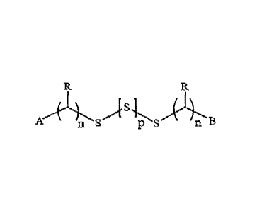
[0006] The present disclosure provides compounds having formula
AA B
)s1
n S PS n B (1\
I) or -m
(2)
wherein A and B are the same or different, and are independently an optionally
substituted aryl, heteroaryl, or a 5-14 membered ring which may be monocyclic
or multicyclic
and optionally containing a heteroatom;
each S is optionally in the form of an oxide;
S1 and S2 are independently S, SO or SO2;
each R is H, halogen, carboxyl, cyano, amino, amido, an amino acid, an
inorganic
substituent, SRI, OR' or RI, wherein each R1 is alkyl, alkenyl, alkynyl, aryl,
heteroaryl, a
carbocyclic ring or a heterocyclic ring, each of which is optionally
substituted and may contain
a heteroatom;
m, n and p are independently 0-3;
or a compound having formula (3) or (4):
).S,L
A n s
P SAB (3) Or
S)'S s B (4)
wherein A, B, R, S, n and p are as defined above;
or a compound having formula (5):
A
\1/1
\A=
n s p S
(5)
wherein A, B, S, n and p are as defined above; and
3
CA 02562065 2014-12-23
CA 2562065
Z is (CRI2)q or (CR1=CR1)q* wherein q is 0-3 and the * represents that C=C may
be
replaced with alkynyl, 0, S, NR; or Z is an optionally substituted aryl,
heteroaryl or
heterocyclic ring;
wherein A and B together may form a cyclic ring system;
and a pharmaceutically acceptable salt, ester, prodrug or metabolite thereof;
provided said compound is not dibenzyltrisulfide, di(p-
chlorobenzyl)trisulfide,
(p-chlorobenzyl)benzyltrisulfide, di(p-nitrobenzyl)trisulfide, di(3-pheny1-2-
propeny1)-trisulfide,
diphenyltrisulfide, or di(p-t-butylphenyl)trisulfide.
[0007] In the above formula 1-5, each Z may be
, r-sel/WR
a a a'
I
fvt.rt,i
or
wherein each W is independently a bond, CR, N, NR, S, or 0;
each R is as defined above.
[0008] In the above formula 1-5, each R may be H, halo, OR1, SR1, CO2R1,
CONR12, CO,
CN, CF3, OCF3, NO2, NRIRI, ()CORI; or R is C1_10 alkyl, C3-10 cyclic alkyl, C2-
10 alkenyl, C2_10
alkynyl, an aryl, heteroaryl, a carbocyclic ring or a heterocyclic ring, each
of which may
contain a heteroatom.
[0009] In the above formula 1-5, each A and B may be benzene, pyridine,
pyridazine,
pyrimidine, pyrazine, triazine, isoxazole, isothiazole, oxadiazole,
[1,2,4]oxadiazole, triazole,
thiadiazole, pyrazole, imidazole, thiazole, oxazole, benzoxazole, pyrrole,
furan, thiophene
indolizine, indole, isoindole, indoline, benzofuran, benzothiophene, indazole,
benzimidazole,
benzthiazole, purine, quinoxaline, quinoline, isoquinoline, cinnoline,
phthalazine, quinazoline,
quinoxaline, naphthyridine, pteridine, acridine, phenazine, phenothiazine,
indene, naphthalene,
benzoxadiazol, or benzo[1,2,5]-oxadiazole.
4
CA 02562065 2014-12-23
CA 2562065
[0010] In another aspect, each A and B are independently
_sr).
r
;
tv W.
W , t =
> I R4
X \iµ w =
R2 X
R3
R5
R6
< ;
I ; X
W ' I
X
õtrvkila
W W RI
W
9
; t
R6
W
R2 or R3
where X and W are independently S, 0, NR7, CR7;
or one W in a 6-membered monocyclic or bicyclic ring may be a bond; and
each RI, R2, R3, R4, R5, R6, R7 is H, halogen, carboxyl, cyano, amino, amino
acid, amido, an
inorganic substituent, SRI, OR1 or R1, wherein each R1 is alkyl, alkenyl,
alkynyl, aryl, heteroaryl, a
carbocyclic ring or a heterocyclic ring, each of which is optionally
substituted and may contain a
heteroatom. For example, each RI, R2, R3, R4, R5, R6, R7 may be H, halo, OR1,
SRI, CO2R1,
CONR12, C=0, CN, CF3, OCF3, NO2, NRIRI, OCORI; or each RI, R2, R3, R-4, R59
R69 R7 is C1-10
alkyl, C3-10 cyclic alkyl, C2-10 alkenyl, C2-10 alkynyl, an aryl, heteroaryl,
a carbocyclic ring or a
heterocyclic ring, each of which may contain a heteroatom.
[0011] Examples of aryl, heteroaryl, or heterocyclic ring include but are not
limited to
piperazine, piperidine, morpholine, thiomorpholine, phenyl, furanyl,
thiophenyl, pyridinyl,
pyrimidinyl, pyrazinyl, triazinyl, quinoxalinyl, thiazolyl, oxazolyl,
imidazolyl, quinolinyl,
naphthalenyl, pyridazinyl, pyrazolopyrimidinyl, benzoimidazolyl,
benzothiazolyl,
benzene-thiophene, pyrazolyl, pyrrolyl, indolyl, isoindolyl, quinolizinyl,
quinolinyl, isoquinolinyl,
or quinazolinyl, each of which is optionally substituted with a heteroatom
selected from 0, N, S
CA 02562065 2014-12-23
CA 2562065
and halo; or substituted with C1_10 alkyl, C3_10 cyclic alkyl, C2_10 alkenyl,
C2_10 alkynyl, aryl, or
heterocycle, each optionally containing a heteroatom.
[0012] In the above formula 1-5, each S may be a mono-oxide or a di-oxide.
[0013] In another aspect, the compound has the formula (6)
(CH2) s, (CH2)n
R (6)
and each n is 1-3; and
R is H, halo, alkyl or halogenated alkyl.
[0014] In yet another aspect, the compound has the formula (7)
z(C (CHA
Ar Ar (7)
wherein Ar is an optionally substituted thiophene, benzothiophene, pyridine or
pyrazine.
[0015] Examples of compounds having formula 1-5 include but are not limited to
di(fluorobenzyl)trisulfide, di(o-chlorobenzyl)trisulfide,
di(methylbenzyl)trisulfide,
di(trifluoromethylbenzyl)trisulfide, di(2-phenylethyl)trisulfide, di(2-
thiophen-yl-methyl)trisulfide,
di(4-pyridin-yl-ethyl)trisulfide, di(2-pyrimidin-yl-ethyl)trisulfide, or
di(3-benzothiophen-yl-methyl)trisulfide. In particular examples, the compound
is
di(p-fluorobenzyl)trisulfide, di(m-methylbenzyl)trisulfide, or di-(p-
methylbenzyl)trisulfide.
[0016] The present disclosure also provides methods for making a composition
comprising a
compound having formula 1-5 as described above, and also provides compositions
prepared
according to such methods. In one aspect, such a method comprises: a)
dissolving such a
compound in a water-soluble organic solvent, a non-ionic solvent, a water-
soluble lipid, a
cyclodextrin, a vitamin, a fatty acid, a fatty acid ester, a phospholipid, or
a combination thereof, to
provide a solution; and b) adding saline or a buffer containing 1-10%
carbohydrate solution. The
organic solvent may be polyethylene glycol (PEG), an alcohol, N-methyl-2-
pyrrolidone, /V,N-
dimethylformamide, N,N-dimethylacetamide, dimethyl sulfoxide, or a combination
thereof.
[0017] In the above process, the non-ionic surfactant may be
polyoxyethyleneglycerol-
triricinoleat 35, PEG-succinate, polysorbate 20, polysorbate 80, polyethylene
glycol 660 12-
hydroxystearate, sorbitan monooleate, poloxamer, ethoxylated persic oil,
capryl-caproyl macrogol-
6
CA 02562065 2014-12-23
. .
CA 2562065
8-glyceride, glycerol ester,PEG 6 caprylic glyceride, glycerin, glycol-
polysorbate, or a combination
thereof. Particular examples of non-ionic surfacts are polyethylene glycol
modified
CREMOPHOR (polyoxyethyleneglyceroltriricinoleat 35), CREMOPHOR EL,
hydrogenated
CREMOPHOR RH40, hydrogenated CREMOPHOR RH60, SOLUTOL HS (polyethylene
glycol 660 12-hydroxystearate), LABRAFIL (ethoxylated persic oil), LABRASOL
(capryl-
caproyl macrogo1-8-glyceride), GELUCIRE (glycerol ester), and SOFTIGEN (PEG
6 caprylic
glyceride).
[0018] In the above process, the lipid may be a vegetable oil, a triglyceride,
a plant oil, or a
combination thereof. For example, the lipid may be castor oil, polyoxyl castor
oil, corn oil, olive
oil, cottonseed oil, peanut oil, peppermint oil, safflower oil, sesame oil,
soybean oil, hydrogenated
vegetable oil, hydrogenated soybean oil, a triglyceride of coconut oil, palm
seed oil, and
hydrogenated forms thereof, or a combination thereof.
[0019] In the above process, the vitamin may be tocopherol; and the fatty acid
and fatty acid
ester may be oleic acid, a monoglyceride, diglyceride, a mono- or di-fatty
acid ester of PEG, or a
combination thereof.
[0020] In the above process, the cyclodextrin may be alpha-cyclodextrin, beta-
cyclodextrin,
hydroxypropyl-beta-cyclodextrin, or sulfobutyl ether-beta-cyclodextrin. The
phospholipid may be
soy phosphatidylcholine, or distearoyl phosphatidylglycerol, and hydrogenated
forms thereof, or a
combination thereof. Furthermore, the carbohydrate in the above process may
comprise dextrose.
[0021] The present disclosure also provides methods for preparing a compound
of formula 1-2
as described above, comprising: a) contacting N-trimethylsilyl imidazole with
sulfur dichloride in a
halogenated solvent to provide diimidazolylsulfide; and b) contacting said
diimidazolylsulfide with
mercaptan. In one example, the halogenated solvent is dichloromethane.
[0022] In one aspect, N-trimethylsilyl imidazole in hexane is contacted with
sulfur dichloride
in dichloromethane. In another aspect, sulfur dichloride as a neat compound is
contacted with N-
trimethylsily1 imidazole in hexane and dichloromethane. In yet another aspect,
the methods further
comprise recrystallizing the trisulfide. In one example, the trisulfide is
recrystallized in n-hexanes,
hexanes, heptane, petroleum ether or a combination thereof.
[0023] The present disclosure also provides a pharmaceutical composition
comprising a
compound having formula 1-5 as described above, and a pharmaceutically
acceptable excipient.
Such compounds and pharmaceutical compositions thereof may be used for
ameliorating or treating
neuroblastoma. Thus, the present disclosure also provides methods for
ameliorating or treating
7
CA 02562065 2014-12-23
CA 2562065
neuroblastoma, comprising administering to a system or a subject in need
thereof an effective
amount of a compound of formula 1-5 or a pharmaceutical composition thereof
and optionally with
an antiproliferative agent, whereby said neuroblastoma is ameliorated or
treated.
[0024] The present disclosure also provides methods for ameliorating or
treating a condition
comprising administering to a subject or a system in need thereof a compound
of formula 1-5 or a
pharmaceutical composition thereof, wherein said compound may be
dibenzyltrisulfide,
di(p-chlorobenzyl)trisulfide, (p-chlorobenzyl)benzyltrisulfide, di(p-
nitrobenzyl)trisulfide,
di(3-phenyl-2-propeny1)-trisulfide, diphenyltrisulfide, or di(p-t-
butylphenyl)trisulfide. The subject
may be a human or an animal such as a mammal. The system may be a cell or
tissue, or other
systems where compounds may be administered in vitro.
[0025] The present disclosure also provides methods for treating or
ameliorating a cell
proliferative disorder other than neuroblastoma, comprising administering to a
system or a subject
in need thereof an effective amount of a compound of formula 1-5 or a
pharmaceutical composition
thereof and optionally with an antiproliferative agent, whereby said cell
proliferative disorder in
said system or subject is ameliorated or treated. The present disclosure also
provides methods for
reducing or inhibiting cell proliferation or for inducing cell death. The
present disclosure further
provides methods for inducing apoptosis. In particular examples, a compound
used in such
methods is dibenzyltrisulfide, di(p-fluorobenzyl)trisulfide, di(p-
methylbenzyl)trisulfide or di(m-
methylbenzyl)trisulfide, and optionally with an antiproliferative agent.
[0026] In one aspect, cell proliferation is reduced, or said cell death is
induced. The cell
proliferative disorder may be a tumor or a cancer including but not limited to
leukemia, lymphoma,
lung cancer, colon cancer, CNS cancer, melanoma, ovarian cancer, renal cancer,
prostate cancer,
breast cancer, head-neck cancer, pancreatic cancer, or renal cancer. In
another aspect, cell
apoptosis is induced. In another aspect, tubulin assemly or disassembly is
disrupted, or G2/M
progression of the cell cycle, cell mitosis, or a combination thereof, is
inhibited. In yet another
aspect, endothelial cell proliferation, angiogenesis, or a combination
thereof, is inhibited.
[0027] The present disclosure also provides methods for ameliorating or
treating restenosis,
comprising administering to a subject in need thereof an effective amount of a
compound of
formula 1-5 or a pharmaceutical composition thereof, whereby restenosis in
said subject is
ameliorated or treated. The restenosis may be associated with neointimal
hyperplasia. The
compound may be administered via oral or parental administration, or via a
stent. The present
8
CA 02562065 2014-12-23
CA 2562065
disclosure also provides a pharmaceutical composition for the treatment of a
cell proliferative
disorder, comprising a compound of formula 1-5, and a pharmaceutically
acceptable excipient.
[0028] Various embodiments of the claimed invention relate to a compound
having the
formula:
A An s S s
n ¨ (0
wherein A and B are the same, and are each: (a) a phenyl ring substituted with
F, Br, I, or
halogenated alkyl; or (b) a heterocyclic ring that is: thiophene, pyridine,
pyrazine, or
benzothiophene, wherein said heterocyclic ring is unsubstituted or substituted
with halo,
halogenated alkyl, OCF3, OMe, t-Butyl, or CH3; each R is H; each n is 1 or 2;
and p is 1; or a
pharmaceutically acceptable salt thereof. The compound may be di(p-
fluorobenzyptrisulfide,
di(m-trifluoromethylbenzyl)trisulfide, di(2-thiophen-yl-methyl)trisulfide,
di(4-pyridin-yl-
ethyptrisulfide, di(2-pyrimidin-yl-ethyl)trisulfide, or di(3-benzothiophen-yl-
methyptrisulfide. Also
claimed are compositions comprising such a compound or pharmaceutically
acceptable salt thereof
as well as a method for preparing such a compound comprising: a) contacting N-
trimethylsilyl
imidazole with sulfur dichloride in a halogenated solvent to provide
diimidazolylsulfide; and b)
contacting said diimidazolylsulfide with a thiol, wherein the thiol has the
formula A(CH2)nSH,
wherein n is 1 or 2, and A is as defined as above for A and B. Such a compound
or salt thereof may
be for use in reducing cell proliferation and may be for use in treatment of a
cancer or in
ameliorating or treating restenosis, as described herein.
9
CA 02562065 2014-12-23
CA 2562065
Brief Description of the Drawings
[0029] Figures 1A¨C show the responses of H460 cells (non-small cell lung
cancer line) to
different concentrations of DBTS, colcemid, and paclitaxel, respectively, as
determined on Real-
Time Electronic Sensing System (RT-CES system).
[0030] Figure 2 shows the responses of MV522 cells (lung cancer cell line) to
different
concentrations of dibenzyl trisulfide (DBTS), as determined on RT-CES system.
[0031] Figure 3 shows responses of MCF-7 cells (breast cancer cell line) to
different
concentrations of dibenzyl trisulfide (DBTS), as determined on RT-CES system.
[0032] Figure 4 shows responses of A549 cells (lung cancer cell line) to
different
concentrations of dibenzyl trisulfide (DBTS), as determined on RT-CES system.
[0033] Figure 5 shows responses of PC3 cells (prostate cancer cell line) to
different
concentrations of dibenzyl trisulfide (DBTS) (Figure 6A) and 5-fluorouracil
(Figure 6B), as
determined on RT-CES system.
[0034] Figure 6 shows responses of A431 cells (epidermoid cancer cell line) to
different
concentrations of dibenzyl trisulfide (DBTS), as determined on RT-CES system.
[0035] Figure 7 shows responses of HT1080 cells (fibrosarcoma cell line) to
different
concentrations of dibenzyl trisulfide (DBTS), as determined on RT-CES system.
[0036] Figure 8 shows responses of MDA-231 cells (breast cancer cell line) to
different
concentrations of dibenzyl trisulfide (DBTS), as determined on RT-CES system.
[0037] Figure 9 shows responses of HT-29 cells (colon cancer cell line) to
different
concentrations of dibenzyl trisulfide (DBTS), as determined on RT-CES system.
[0038] Figure 10 shows responses of HC-2998 cells (colon cancer cell line) to
different
concentrations of dibenzyl trisulfide (DBTS), as determined on RT-CES system.
[0039] Figure 11 shows responses of OVCAR4 cells (ovarian cancer cell line) to
different
concentrations of dibenzyl trisulfide (DBTS), as determined on RT-CES system.
[0040] Figure 12 shows responses of A2780 cells (colon cancer cell line) to
different
concentrations of dibenzyl trisulfide (DBTS), as determined on RT-CES system.
9a
CA 02562065 2006-10-03
WO 2005/112933 PCT/US2005/013474
[0041] Figure 13 shows responses of HepG2 cells (human hepatoma cell line) to
different
concentrations of dibenzyl trisulfide (DBTS), as determined on RT-CES system.
[0042] Figure 14 shows mouse sarcoma S180 tumors (planted into mice by
subcutaneous
implanting) treated with dibenzyl trisulfide (DBTS).
[0043] Figure 15 shows mouse Lewis lung cancer (planted into mice by
subcutaneous
implanting) treated with dibenzyl trisulfide (DBTS).
[0044] Figure 16 shows Bcap-37 human breast tumors that were xenograft-
transplanted in
immunodeficient nude mice by subcutaneous seeding and were treated with
compound
ACEA100108.
[0045] Figure 17 shows the dynamic change in tumor size in the in vivo
antitumor efficacy
test of compound ACEA100108 on Bcap-37 human breast cancer that was xenograft
transplanted in immunodeficient nude mice by subcutaneous implanting.
[0046] Figure 18 shows the dynamic change in body weight of carrier mice in
the in vivo
antitumor efficacy test of compound ACEA100108 (100108) on Bcap-37 human
breast cancer
that was xenograft- transplanted in immunodeficient nude mice by subcutaneous
implanting.
[0047] Figure 19 shows HCT-8 human colon tumors that were xenograft-
transplanted in
immunodeficient nude mice by subcutaneous seeding and were treated with
compound
ACEA100108.
[0048] Figure 20 shows the dynamic change in tumor size in the in vivo
antitumor efficacy
test of compound ACEA100108 on HCT-8 human colon cancer that was xenograft-
transplanted
in immunodeficient nude mice by subcutaneous implanting.
[0049] Figure 21 shows the dynamic change in body weight of carrier mice in
the in vivo
antitumor efficacy test of compound ACEA100108 (100108) on HCT-8 human colon
cancer that
was xenograft-transplanted in immunodeficient nude mice by subcutaneous
implanting.
[0050] Figure 22 shows ao10/17 human ovarian tumors that were xenograft-
transplanted in
immunodeficient nude mice by subcutaneous seeding and were treated with
compound
ACEA100108.
[0051] Figure 23 shows the dynamic change in tumor size in the in vivo
antitumor efficacy
test of compound ACEA100108 on ao10/17 human ovarian cancer that was xenograft
transplanted in immunodeficient nude mice by subcutaneous implanting.
[0052] Figure 24 shows the dynamic change in body weight of carrier mice in
the in vivo
antitumor efficacy test of compound ACEA100108 (100108) on ao10/17 human
ovarian cancer
that was xenograft-transplanted in immunodeficient nude mice by subcutaneous
implanting.
CA 02562065 2006-10-03
WO 2005/112933 PCT/US2005/013474
[0053] Figure 25 shows Bcap-37 human breast tumors that were xenograft-
transplanted in
immunodeficient nude mice by subcutaneous implanting and were treated with
compound
ACEA100108.
[0054] Figure 26 shows the responses of various cell lines to ACEA100108, as
determined
on RT-CES system.
[0055] Figure 27 shows the responses of HT1080 cell to different derivatives
of DBTS, as
determined on RT-CES system.
[0056] Figure 28 shows the images of microtubules in control COS cells that
were not
treated with any drugs.
[0057] Figure 29 shows the images of microtubules in COS cells treated with
different
concentrations of paclitaxel for 4 hours.
[0058] Figure 30 shows the images of microtubules in COS cells treated with
different
concentrations of paclitaxel for 24 hours.
[0059] Figure 31 shows the images of microtubules in COS cells treated with
different
concentrations of vinblastine for 4 hours.
[0060] Figure 32 shows the images of microtubules in COS cells treated with
different
concentrations of vinblastine for 24 hours.
[0061] Figure 33 shows the images of microtubules in COS cells treated with
different
concentrations of DBTS for 4 hours.
[0062] Figure 34 shows the images of microtubules in COS cells treated with
different
concentrations of DBTS for 24 hours.
[0063] Figure 35 shows the images of microtubules in COS cells treated with
different
concentrations of ACEA100108 for 4 hours.
[0064] Figure 36 shows the images of microtubules in COS cells treated with
different
concentrations of ACEA100108 for 24 hours.
[0065] Figure 37 shows the images of microtubules in COS cells treated with
different
concentrations of ACEA100116 for 4 hours.
[0066] Figure 38 shows the images of microtubules in COS cells treated with
different
concentrations of ACEA100116 for 24 hours.
[0067] Figure 39a shows the result of the in vitro microtubule assembly assays
using pure
tubulin (MAP-free) and DBTS.
[0068] Figure 39b shows the electron microscopic images of microtubules
assembled in
vitro in the absence of any drug.
11
CA 02562065 2014-12-23
CA 2562065
[0069] Figure 39c shows the electron microscopic images of microtubules
assembled in
vitro in the presence of 3 uM DBTS.
[0070] Figure 40 shows the result of the in vitro microtubule assembly assays
using pure
tubulin (MAP-free) and ACEA100108.
[0071] Figure 41 shows the result of the in vitro microtubule assembly assays
using pure
tubulin (MAP-free) and ACEA100116.
[0072] Figure 42 shows the fluorescent microscope images of 6-CFDA (top panel)
and
Annexin V (bottom panel) staining of A549 human lung cancer cells treated with
treated with
1 uM ACEA100108, 50 nM paclitaxel, 10 nM vinblastine or DMSO for 24 hrs.
[0073] Figure 43 show the cell cycle distribution of A549 human lung cancer
cells after
they were treated with 25 uM ACEA100108, 7.8 nM paclitaxel, or DMSO for 24
hrs, as
analyzed on a flow cytometry.
Detailed Description - Modes of Carrying Out the Invention
[0074] For clarity of disclosure, and not by way of limitation, the detailed
description of the
invention is divided into the subsections that follow.
A. Definition
[0075] Unless defined otherwise, all technical and scientific terms used
herein have the
same meaning as is commonly understood by one of ordinary skill in the art to
which this
invention belongs. If a definition set forth in this section is contrary to or
otherwise
inconsistent with a definition set forth in the patents, applications,
published applications and
other publications referred to herein, the definition set forth in this
section prevails.
[0076] As used herein, "a" or "an" means "at least one" or "one or more".
[0077] The term "alkyl" as used herein refers to saturated hydrocarbon groups
in a straight,
branched, or cyclic configuration and particularly contemplated alkyl groups
include lower
alkyl groups (i.e., those having ten or less carbon atoms). Exemplary alkyl
groups are methyl,
ethyl, propyl, isopropyl, butyl, sec-butyl, tertiary butyl, pentyl, isopentyl,
hexyl, etc. The term
"alkenyl" as used herein refers to an alkyl as defined above and having at
least one double
bond. Thus, particularly contemplated alkenyl groups include straight,
branched, or cyclic
alkenyl groups having two to ten carbon atoms (e.g., ethenyl, propenyl,
butenyl, pentenyl, etc.).
12
CA 02562065 2006-10-03
WO 2005/112933 PCT/US2005/013474
Similarly, the term "alkynyl" as used herein refers to an alkyl or alkenyl as
defined above and
having at least one triple bond. Especially contemplated alkynyls include
straight, branched, or
cyclic alkynes having two to ten total carbon atoms (e.g., ethpyl, propynyl,
butynyl, etc.) .
[0078] The term "cycloalkyl" as used herein refers to a cyclic alkane (i.e.,
in which a chain
of carbon atoms of a hydrocarbon forms a ring), preferably including three to
eight carbon
atoms. Thus, exemplary cycloalkanes include cyclopropyl, cyclobutyl,
cyclopentyl, cyclohexyl,
cycloheptyl, and cyclooctyl. Cycloalkyls also include one or two double bonds,
which form the
"cycloalkenyl" groups. Cycloalkyl groups are also further substituted by
alkyl, alkenyl, alkynyl,
halo and other general groups.
[0079] The term "aryl" or "aromatic moiety" as used herein refers to an
aromatic ring
system, which may further include one or more non-carbon atoms. Thus,
contemplated aryl
groups include (e.g., phenyl, naphthyl, etc.) and pyridyl. Further
contemplated aryl groups may
be fused (i.e., covalently bound with 2 atoms on the first aromatic ring) with
one or two 5- or 6-
membered aryl or heterocyclic group, and are thus termed "fused aryl" or
"fused aromatic".
[0080] As also used herein, the terms "heterocycle", "cycloheteroalkyl", and
"heterocyclic
moieties" are used interchangeably herein and refer to any compound in which a
plurality of
atoms form a ring via a plurality of covalent bonds, wherein the ring includes
at least one atom
other than a carbon atom. Particularly contemplated heterocyclic bases include
5- and 6-
membered rings with nitrogen, sulfur, or oxygen as the non-carbon atom (e.g.,
imidazole,
pyrrole, triazole, dihydro pyrimidine, indole, pyridine, thiazole, tetrazole
etc.). Further
contemplated heterocycles may be fused (i.e., covalently bound with two atoms
on the first
heterocyclic ring) to one or two ring or heterocycle, and are thus termed
"fused heterocycle" or
"fused heterocyclic base" or "fused heterocyclic moieties" as used herein.
[0081] The term "alkoxy" as used herein refers to straight or branched alkyl
connecting to an
oxygen atom called alkoxides, wherein the hydrocarbon portion may have any
number of carbon
atoms, may further include a double or triple bond and may include one or two
oxygen, sulfur or
nitrogen atoms in the alkyl chains. For example, suitable alkoxy groups
include methoxy,
ethoxy, propyloxy, isopropoxy, methoxyethoxy, etc. Similarly, the term
"alkylthio" refers to
straight or branched chain alkylsulfides, wherein the hydrocarbon portion may
have any number
of carbon atoms, may further include a double or triple bond and may include
one or two
oxygen, sulfur or nitrogen atoms in the alkyl chains. For example,
contemplated alkylthio
groups include methylthio, ethylthio, isopropylthio, methoxyethylthio, etc.
13
CA 02562065 2006-10-03
WO 2005/112933 PCT/US2005/013474
[0082] Likewise, the term "alkylamino" refers to straight or branched
alkylamines, wherein
the amino nitrogen "N" can be substituted by one or two alkyls and the
hydrocarbon portion may
have any number of carbon atoms and may further include a double or triple
bond. Furthermore,
the hydrogen of the alkylamino may be substituted with another alkyl group.
Therefore,
exemplary alkylamino groups include methylamino, dimethylamino, ethylamino,
diethylamino,
etc.
[0083] The term "aryloxy" as used herein refers to an aryl group connecting to
an oxygen
atom, wherein the aryl group may be further substituted. For example suitable
aryloxy groups
include phenyloxy, etc. Similarly, the term "arylthio" as used herein refers
to an aryl group
connecting to a sulfur atom, wherein the aryl group may be further
substituted. For example
suitable arylthio groups include phenylthio, etc.
[0084] The term "halogen" as used herein refers to fluorine, chlorine, bromine
and iodine.
[0085] The term "amino acid" as used herein refers to substituted natural and
unnatural
amino acid with D- or L- configuration or the mixture in which amino and acid
groups are used
to derivatize the contemplated compounds.
[0086] It should further be recognized that all of the above-defined groups
may further be
substituted with one or more substituents, which may in turn be substituted as
well. For
example, an "alkyl" as used herein encompasses alkyls substituted with a
heteroatom.
[0087] The term "substituted" as used herein refers to a replacement of an
atom or chemical
group (e.g., H, NH2, or OH) with a functional group, and particularly
contemplated functional
groups include nucleophilic groups (e.g., -NH2, -OH, -SH, -NC, etc.),
electrophilic groups (e.g.,
C(0)0R, C(X)OH, etc.), polar groups (e.g., -OH), non-polar groups (e.g.,
heterocycle, aryl,
alkyl, alkenyl, alkynyl, etc.), ionic groups (e.g., -NH3), and halogens (e.g.,
-F, -Cl), NHCOR,
NHCONH2, OCH2COOH, OCH2CONH2, OCH2CONHR, NHCH2COOH, NHCH2CON112,
NHSO2R, OCH2-heterocycles, PO3H, SO3H, amino acids, and various combinations
known in
the art. Moreover, the term "substituted" also includes multiple degrees of
substitution, and
where multiple substituents are disclosed or claimed, the substituted compound
can be
independently substituted by one or more of the disclosed or claimed
substituent moieties.
[0088] The term "organ sulfur derivative" as used herein refers to an organic
compound
containing two or more "S" atoms. The term "disulfide", "trisulfide",
"tetrasulfide" or
pentasulfide" as used herein refers to a moiety where two, three, four, or
five sulfur atoms
connect in a linear chain (-S-S-S-), where one or two or three of them may be
further oxidized
into S=0 or SO2, and where the di-, tri-, tetra- and penta-sulfide derivatives
are substituted with
14
CA 02562065 2006-10-03
WO 2005/112933 PCT/US2005/013474
two functional, aryl, alkenyl, heterocyclic groups or substituents at the two
ends of the di-, tri-,
tetra- and penta-sulfide (R-S-(S)0_3-S-R). Two or more trisulfide (-S-S-S-)
moieties may be
connected together by an aromatic or linear chain, which also refers to
"trisulfide" or organo
sulfide. One or two trisulfide or organo sulfide moieties may be connected
together to form
cyclic ring systems.
B. Substituted Organo Sulfur Derivatives and Pharmaceutical
Compositions
Thereof
[0089] The present invention compounds having formula
AA s2"---AB
II µLp S B (1) or - (2)
wherein A and B are the same or different, and are independently an optionally
substituted aryl, heteroaryl, or a 5-14 membered ring which may be monocyclic
or multicyclic
and optionally containing a heteroatom;
each S is optionally in the form of an oxide;
S1 and S2 are independently S, SO or SO2;
each R is H, halogen, carboxyl, cyano, amino, amido, an amino acid, an
inorganic
substituent, SR', OR' or le, wherein each Rl is alkyl, alkenyl, alkynyl, aryl,
heteroaryl, a
carbocyclic ring or a heterocyclic ring, each of which is optionally
substituted and may contain a
heteroatom;
m, n and p are independently 0-3;
or a compound having formula (3) or (4):
A S
n s p S n B
(3) or
),SL
A n s p S B
(4)
wherein A, B, R, S, n and p are as defined above;
or a compound having formula (5):
CA 02562065 2006-10-03
WO 2005/112933 PCT/US2005/013474
S S A
ns
j,s,L
n s p S
(5)
wherein A, B, S, n and p are as defined above; and
Z is (CR12)q or (CR1=CR1)q* wherein q is 0-3 and the * represents that C=C may
be
replaced with alkynyl, 0, S, NR; or Z is an optionally substituted aryl,
heteroaryl or heterocyclic
ring;
wherein A and B together may form a cyclic ring system;
and a pharmaceutically acceptable salt, ester, prodrug or metabolite thereof;
provided said compound is not dibenzyltrisulfide, di(p-
chlorobenzyl)trisulfide,
(p-chlorobenzypbenzyltrisulfide, di(p-nitrobenzyl)trisulfide, di(3-pheny1-2-
propeny1)-trisulfide,
diphenyltrisulfide, or di(p-t-butylphenyl)trisulfide.
[0090] In other embodiments, each R in the above formula 1-5 may be a non-
interfering
substituent. In general, a "noninterfering substituent" is a substituent whose
presence does not
destroy the ability of a compound to behave as a therapeutic agent. For
example, a non-
interfering substituent may improve potency and PK properties. In another
example, the non-
interfering substituent may reduce toxicity. Suitable noninterfering
substituents include halo,
nitro, carboxyl, alkyl, alkenyl, alkynyl, aryl, arylalkyl, arylalkenyl,
alkoxy, alkylthio,
arylalkynyl, heterocycles, amino acids, each of which may further be
substituted with one or
more non-interfering sub stituents. Noninterfering sub stituents may also
include COOR, SR,
OR, wherein R is also a non-interfering substituent, as defined above.
[0091] In the above formula 1-5, A and B may independently be
16
CA 02562065 2006-10-03
WO 2005/112933 PCT/US2005/013474
c:-/-W¨ -=''':''''-'''''''Ri
c-r ---.::--- \
,
1 ,
X --/
W<-
X
W R2 W
R3
W nr\sso
i
WR5
)--T-W- R6
\..XW
. - = ,,/ .. s \ .. ._< ' µi 1 ' % ' %, , I X I .,
i ( ,I
= , 1 1
W I I 1 1
W
v s / = , ..,' i ... . . . . . . . . . . = . , õ ......v . .
..:.......W
Vs .... I m s.. .>........\ / X
W W \X '
W W
I
'VW%)
Xv......W
W <=-IN.-
I I i I
W.,..:: - ''= - - - ''''
W W R2
R6 W W R2 or R3
where X and W are independently S, 0, NR7, CR7;
or one W in a 6-membered monocyclic or bicyclic ring may be a bond; and
each RI, R2, R3, R4, R5, R6, R7 is as previously defined.
[0092] In other embodiments, each R1, R2, R3, R4, R5, R6, R7 may be a polar or
non-polar
substituent. In other examples, each RI, R2, R3, R4, R5, R6, R7 may be a
nucleophilic or
electrophilic non-interfering sub stituent.
[0093] The present invention also encompasses compounds having formula 1-5, as
well as
their salts and prodrugs. Such salts, for example, may be formed from a
positively charged
substitute group (e.g. an amino group on A and / or B) on a compound and a
pharmaceutically
suitable anion. Suitable anions include, but not limited to, chloride,
bromide, iodide, sulfate,
nitrate, phosphate, citrate, methanesulfonate, trifluoroacetate, maleate, and
acetate.
Pharmaceutically acceptable salts may also be formed from a negatively charged
substituted
group (e.g., carboxylate group on A and / or B) on a compound and a cation.
Non-limiting
examples of suitable cations are sodium ion, potassium ion, magnesium ion,
calcium ion, and a
organic ammonium ion such as teteramethylammonium ion, tetrabutylammonium ion,
and other
organic cations.
[0094] The trisulfides may be synthesized following procedures as illustrated
in Scheme 1.
For example, the aromatic or heterocyclic methylene halides (X = I or Br or
Cl) are reacted with
17
CA 02562065 2006-10-03
WO 2005/112933 PCT/US2005/013474
thiourea. The resulted isothiouronium halides are treated with sodium
hydroxide to provide the
corresponding thiol derivatives (Furniss, B. S.; Hannaford, A. J.; Rogers, V.;
Smith, P. W. G.;
Tatchell, A. R. Vogel's Textbook of Practical Organic Chemistry, Longman Group
Limited,
London, 1978, pp 582-583).
Synthesis of thiol derivatives
1) thiourea
Arsx _________________________________________ ArSH
2) NaOH
X = Br or Cl
Method A: Synthesis of symmetric trisulfides
SC12 2 RSH
2 R-S-S-S-R
¨SiMe3 rt 0 - 25 C
diimmidazolylsulfide 60-90%
Method B: Synthesis of unsymmetric or symmetric trisulfides
-78C R'H
RSH + SC12 [
R-S-S-Cl] R-S-S-S-R'
Et3N
70-90%
Scheme 1. Synthetic methods for the symmetric and unsymmetric trisulfides
[0095] The symmetric trisulfide derivatives may be synthesized using Method A.
In
Method A, N-trimethylsilylimidazole is reacted with sulfur dichloride. The
resulting di-
imidazolylsulfide is then reacted with thiol to give the corresponding
trisulfides. Method B can
be used to synthesize symmetric and asymmetric trisulfides. In Method B, the
first thiol is
reacted with sulfur dichloride quantitatively at low temperature. The
resulting intermediate
thiosulfenyl chloride is then reacted with the second thiol to provide the
desired asymmetric or
symmetric trisulfide, depending on the thiol used in the second step.
[0096] The representative aromatic methylene thiols 1-6 (Scheme 2) may be
synthesized
using the similar procedure as described in Vogel's Practical Organic
Chemistry, pp 582-583. In
addition, symmetric trisulfide derivatives 7-32 (Scheme 2) were synthesized by
Method A
similar to the reported procedure (Banerji, A.; Kalena, G. P. Tetrahedron
Letters 1980, 21,
3003-3004). For example, sulfur dichloride (14 mmol) in anhydrous hexanes or
dichloromethane was added to a stirred solution of N-trimethylsilylimidazole
(28 mmol) in
hexanes at room temperature. After stirring for 30 minutes, the reaction
mixture was cooled to 0
18
CA 02562065 2006-10-03
WO 2005/112933 PCT/US2005/013474
C, and a solution of designated thiol (28 mmol) in anhydrous hexanes was added
dropwise for a
period of 30 minutes. The reaction mixture was stirred for 30 minutes, and the
precipitated
imidazole by-product was filtered off. The filtrate was washed with water and
brine, and dried
over anhydrous sodium sulfate. The solvent was evaporated, and the residue is
purified by flash
chromatography on a silica gel column using hexanes-ethyl acetate 100: 1 to
20: 1 as eluents to
provide desired trisulfides 7-32 in 60-90% yields. The aromatic trisulfides 33-
39 were
synthesized by the similar procedure in 30-70% yields.
[9100] Di(p-fluorobenzyl)trisulfide (8). Trisulfide 8 was synthesized in 77%
yield. The
white crystalline was obtained by chromatographic purification followed by
recrystallization
from hexanes. Silica gel TLC Rf= 0.46 (40:1 hexanes-ethyl acetate). 1H NMR
(499.1 MHz,
CDC13) 8 4.00 (s, 4H), 7.01 (t, 4H, J = 8.8 Hz), 7.27 (dd, 411, J= 8.8, 5.4
Hz); 13C NMR (125.7
MHz, CDC13) 5 42.4, 115.6, 115.8, 131.2, 131,3, 132.4, 162.5 (C-F, J= 250 Hz);
19F NMR
(376.5 MHz, CDC13) 8 -114.2; ES MS nilz 337 / 338 (M + Na); Anal. Cakd. for
C14H12F2S3: C,
53.48; H, 3.85; S, 30.59. Found: C, 53.16; H, 4.22; S, 30.24.
[0101] Di(p-ehlorobenzyl)trisulfide (9). Trisulfide 9 was synthesized in 90%
yield. The
white crystalline was obtained by chromatographic purification followed by
recrystallization
from hexanes. Silica gel TLC Rf= 0.45 (40:1 hexanes-ethyl acetate). 1H NMR
(499.1 MHz,
CDC13) 63.98 (s, 4H), 7.22 (d, 4H, J= 8.4 Hz), 7.29 (d, 414, J= 8.4 Hz).
19
CA 02562065 2006-10-03
WO 2005/112933 PCT/US2005/013474
.......--,.., 1) thiourea
Arl X _______________ .., Arl SH 1 - 6
X = Br or Cl 2) NaOH
N,
SH
. '. SH SH
N N
1 2 3
.
411 /NI SH e . SH
0-N S S
4 5 6
2 Ari SFI
______________________________________________________ Ar1S- 1
S Ar
N --- --=.\ SC12 N\ -r-"=N / 0 - 25 C 7-32
2
--1-----:,-/NSiMe3 rt cs,./NN \)
\ 2 Ar2¨SH Ar2 ,S Ar2
diimmidazolylsulfide ________________________________ ). S -S
0 - 25 C 33 - 39
Art =
00 0
= cF3 *I Pi 0 ,..CF 0
e õcH, 0
F CI
7 8 9 10 11 12 13
N
Y) I.N
)
N 0 S ......; ,..-
N N
S
14 15 16 17 18 19 20
,N N am N
\/0
---.N I IP 11 \ 11,- --(i'S 0 1.I
0c.,_,
N-0 Br
..,, ,3
S
21 22 23 24 25 26
N0
0111 0 CH 3 0
ci. 1 0 Me
2 lel Bu-t CI ,., N
Me
27 28 29 30 31 Me 32
Ar2 =
N N N 110
CH3 )a
ilk 0/ ____04F Nr)
i
36
0
37
N F 35 ...,
33 34 N----z\
/ *
---- j
N S
38 39
Scheme 2. Synthetic the symmetric trisulfides by Method A
[0102] Di(m-trifluoromethylbenzyl)trisulfide (12). Trisulfide 12 was
synthesized in 99%
yield. The white crystalline was obtained by chromatographic purification
followed by
recrystallization from hexanes. Silica gel TLC Rf= 0.33 (40:1 hexanes-ethyl
acetate). 1H NMR
(499.1 MHz, CDC13) 8 4.04 (s, 4H), 7.41-7.49 (m, 4H), 7.51-7.58 (m, 411).
CA 02562065 2006-10-03
WO 2005/112933 PCT/US2005/013474
[0103] Di(benzo[B]thiophen-3-yl-methane)trisulfide (22). Trisulfide 22 was
synthesized
in 45% yield. The white solid was obtained by chromatographic purification.
Silica gel TLC Rf=
0.45 (40:1 hexanes-ethyl acetate). 1H NMR (499.1 MHz, CDC13) 5 3.74 (s, 4H),
7.01 (s, 2H),
7.34-7.45 (m, 4H), 7.75 (d, 2H, J= 7.4 Hz), 7.85 (dd, 2H, J= 7.8, 1.1 Hz). ES
MS nilz 391 (M
+H), 413 (M + Na) .
[0104] Di(p-bromobenzyptrisulfide (25). Trisulfide 25 was synthesized in 84%
yield. The
white crystalline was obtained by chromatographic purification followed by
recrystallization
from hexanes. Silica gel TLC Rf= 0.55 (40:1 hexanes-ethyl acetate). 1H NMR
(499.1 MHz,
CDC13) 8 3.96 (s, 411), 7.17 (d, 4H, J= 8.3 Hz), 7.45 (d, 4H, J= 8.3 Hz).
[0105] Di(p-methylbenzyl)trisulfide (26). Trisulfide 26 was synthesized in 99%
yield. The
white crystalline was obtained by chromatographic purification followed by
recrystallization
from hexanes. Silica gel TLC Rf= 0.66 (40:1 hexanes-ethyl acetate). 1H NMR
(499.1 MHz,
CDC13) 5 2.33 (s, 6H), 4.01 (s, 4H), 7.14 (d, 4H, J= 8.0 Hz), 7.21 (d, 4H, J=
8.0 Hz).
[0106] Dis(p-t-butylbenzyl)trisulfide (28). Trisulfide 28 was synthesized in
96% yield.
The white crystalline was obtained by chromatographic purification followed by
recrystallization from hexanes. Silica gel TLC Rf= 0.50 (40:1 hexanes-ethyl
acetate). 1H NMR
(499.1 MHz, CDC13) 8 1.30 (s, 18H), 4.02 (s, 4H), 7.25 (d, 4H, J= 8.3 Hz),
7.35 (d, 411, J=-
8.3 Hz).
[0107] Di(o-chlorobenzyl)trisulfide (30). Trisulfide 30 was synthesized in 77%
yield. The
white crystalline was obtained by chromatographic purification followed by
recrystallization
from hexanes. Silica gel TLC Rf= 0.44 (40:1 hexanes-ethyl acetate). 1H NMR
(499.1 MHz,
CDC13) 6 4.17 (s, 4H), 7.23-7.28 (m, 4H), 7.35-7.43 (m, 4H).
21
CA 02562065 2006-10-03
WO 2005/112933 PCT/US2005/013474
TySH
+ SC12
/*/
S
-78 C Et3N ,,/^-SH S'.---- SArl
Arl_________Ar- y,,
X 41-62
NCI
X =
,..-S
40 Ar.2 ((S S-Ar2
SH
X/ 63 - 68
Arl = 40)
Oki 41) F CI .CF3 140] /CH3 140 CF3
I. 0 0
41 42 43 44 45 46
1.1 4111
Br Oio cH3
1401 / 001
0
CI CH3
47 48 48 50 51 52
N
0 )
S
r\O 0 S r\ki\i N)
53 54 55 56 57 58
N
*
0 /NO
\
S
N N-0
S
59 60 61 62
Ar2 =li
CH3 N
F F aor\i 0/
N F - /
c /
. N....., N
N
....___< 1101 ---(\
SN 0 N)
66 67 68
Scheme 3. Synthetic the symmetric trisulfides by Method B
[0108] Di(2,4,6-trimethylbenzyDtrisulfide (32). Trisulfide 32 was synthesized
in 59%
yield. The white crystalline was obtained by chromatographic purification
followed by
recrystallization from hexanes. Silica gel TLC Rf= 0.65 (40:1 hexanes-ethyl
acetate). 1H NMR
(499.1 MHz, CDC13) 5 2.27 (s, 6H), 2.42 (s, 12 H), 4.23 (s, 4H), 6.87 (s, 4H).
[0109] Di(p-methoxyphenyDtrisulfide (33). Trisulfide 33 was synthesized in 98%
yield.
The white crystalline was obtained by chromatographic purification followed by
22
CA 02562065 2006-10-03
WO 2005/112933 PCT/US2005/013474
recrystallization from hexanes. Silica gel TLC Rf= 0.32 (20:1 hexanes-ethyl
acetate). 1H NMR
(499.1 MHz, CDC13) 5 3.80 (s, 4H), 6.81 (d, 4H, J= 8.8 Hz), 7.47 (d, 4H, J=
8.8 Hz).
[0110] Di(4-trifluoromethylpyridin-2-yl)trisulfide (34). Trisulfide 34 was
synthesized in
53% yield. The white crystalline was obtained by chromatographic purification
followed by
recrystallization from hexanes. Silica gel TLC Rf= 0.61 (10:1 hexanes-ethyl
acetate). 1H NMR
(499.1 MHz, CDC13) 5 7.70 (d, 4H, J= 8.4 Hz), 7.84 (dd, 4H, J= 8.4, 2.4 Hz),
8.73 (s, 2H).
[0111] The asymmetric trisulfide derivatives listed in Tables 1-8 may be
synthesized
following similar procedures as for compounds 41-68, using the corresponding
thiol.
s
la s'sNsNn la la
'0-1 ----o ---0 "gr N
'0
-__-.-.o. iiir
S
di Ss-( N 6 S sr) --.0 0 s¨s,
Nj '0 nW N. ,.-- ..
'N 40
s,
6 s¨s, . 0 s¨s,,,;\_,--s -- 0 s -s , .
N. -0/ \ -1
---"0 W &
N IW
a Ss a S's A ---N, r Srt\I
I S ) "0 ..
NI_
0 ,s,
s s . 0/CH3
----0 0 ---0 10 Ss---(1SNX)
S - F s N IW S 1
& \ / la F S s---(* i )a0 S
N
'0 IW N F ----0 Mr 0
S /
Table 1. Various disubstituted trisulfide derivatives.
23
CA 02562065 2006-10-03
WO 2005/112933 PCT/US2005/013474
s s
0
0
o S
F F
F N j
,..S.N. S
0 S S''Y 0 S"--- '''s--------y 0 SSS 1 \
AI
F F N .
'N".-... N -0
F
S
0
S'''. s / 0 S
1101 NI
0 S s 0 N
F,
F W õP
M
F S
N
0s____(*) CH3
S
S 0 S 41
0/
S .
F N--/
F 0 F
NI:-....'1
____________________ F
Nn
0 s,s.s 0 ss___ 1 0, s s N....,0
--( __ )-4./ F
F N F
S /
S N
F F
,...S7
0 S s--(0 11110 Table 2. Various disubstituted trisulfide
derivatives.
F
_...S S
.....-S,,
F S- ....-S----y.-","- N
F soi s s,N0 110 N.k..õ,..11 F-4,
100
F-) 0 F.--)
F 0 F 0 0 S
F
_.....S
51 ,S N , S..õ...S,...
Z....I,* N
F 1101 S" Sr"NrOi F 0 S-
11'> F
N
* F'L S.
F 0
S N=-\
FEla
F 10 S"--- *S-------yr F 1110/ S*---Sq 0 S--
--ss . 0
51., N . F-il,. .---( /
N-7 F
F-.21,..
F 0 'N".1. F 0 F
S N ..,
....,S,.... ......kaol
F /1101 Sss-0, ------ F r r 0 S' S.¨(/
F-4...
N F ' 7-. S N"... F.21,,F 0 S S N
S /
F 0 F 0
F
F
\1
0 s--ss_4 10
F-.2L 0
F Table 3.
Various disubstituted trisulfide derivatives.
24
CA 02562065 2006-10-03
WO 2005/112933 PCT/US2005/013474
F F F
F
F * S.- S'N()F (1110 s..--'s=-=,s --
-N
-s/-*--.),..-1;
F
0 W \O
Nj $ ---
....N/
F
F
F F
F I.1 S SV)0 F F CH3
N ...,. F 0 F 0 SsS . 0/
N--/
F
F
F
s_.õ..,S
=,.., F F F
F 0 sini ..õ...S..,õ F
N.....I........õ
F 0 S
S \ / F F 0 S----'s (/ N¨
,
.N.-- N F Srµ(
F
F
F
410 Ni
F 10 S--- s.¨< F õ.õ,.S ....e_L. N
I
0 F 0 S S '.--..."0
S /
0 SsN)
0 S SsNn
sS---""
0 ====,,,õ S
0 S
s-(-"N ===.,, S
S"..- N Ah ,N \ 0
N:...,.....,)
0 S"..... N's
sy)
N ,,,N,..- VI ---N/
N= \ F
0 ....,. =-'S \ ,0 S / HC 3 * ,
s s.õ....S...õ
--i / =...õ
S"---- ....'S * 0
NJ 10 N F
N ".... 1
N...........
0
S----'..'e
Ss ¨
N-(NI =0
S S N
1101 0 /
S
Table 4. Various disubstituted trisulfide derivatives.
(N * s....-S,,,s,,,,,) (N 0 s..----ss.-Nf) N
S"---y-N
S NO--1 S \S---1 S *
N..,.....t.)
<N 0 S
(N i
, 4
,..,.
<
N s../s\s N
S'--- s N 0
<S* S I 110 <s 1101 s .
N
S
.......s.,., ao. /CH3
N s.--"'s....,s 4111._,,N \o z/N 0 F N
< S"..--S ¨
<
11101 S S 0
S \ /
F
-....õ / N i F S
S * N \S
NN S N........... N
1101
,.....S .A.....).,,o
< s.---'s',..,
S----1 N
S-
0 * c *
Ie * S S N
S /
S S
Table 5. Various disubstituted trisulfide derivatives.
CA 02562065 2006-10-03
WO 2005/112933
PCT/US2005/013474
s
,NO s s N\O 1N____ 0 s.õ----S,Ns...õ--Na ,N s
' s----- 1 \
O
0 I "
N.-0
S S
----- ...,
/1\1 0 szY) dN 01 s s---yislN ..----S,,s
0 d
N.........,,. il \N.--- / 110
S
IN, 0 s....--S,...
0 S .----- N ,S,....s N 7 s,s4)¨
411 ...¨ N, 1 o
N
)
\N--
N
S
FCH3
IN...... 0 s.----S.,s/"'-rN
/N,...... SS ¨ 7 -4 SsNs . 01
0 . 0 0
N--- N F
N
N, S N 0 I
01 0 S'--- ....".
S---- o/N-"" 0 SsS¨<
/Ni=-=""Th.
IN..... 0
/
\W.-. 0 \N"-- Stsr") 0
\ N--- S
/
Table 6. Various disubstituted trisulfide derivatives.
=
26
CA 02562065 2006-10-03
WO 2005/112933 PCT/US2005/013474
s
s
sSV ,0 Crs VN\'0rYs s (/)
S
i N N--.0
N ''
-,N 0
S S N
S .-..õ.....,..,õ..-I N
S S S /CH3
rYS Sy) ryS s 1 lip ryS s . 0
....õ4õ.õ,..N N -..N
N
S
N---- \
ryS----eSs---( / rySN--< j4F F Sss¨<NDC
\,N N----/ ..,..,.....:õ.., N N F ...,,,,N S
N
r I
rySs--<N 01 (ySssN /
Il'r--S'-- s------'11--
-N i( )
L IL
.s.N 0
.....õ N S k,,,,N I'l /--0
NSSS =
,S, ..,,y...... N
lel N S'" s-----.\ n
IL.,õN
N., N S' s'/'--.'r
IL...1.N S .
-' V
_,....S N N-_-",õ
NS5 s---< I
IL ...:..,..* N N 11..õ,.....,......N S---''-e
1 N I 111
S
I.
NS-.----Ss NS .
" ..5 __ < N
' ''.-.
----µ . N N F U0
S /
HC3
NS-- s 41 0
L...i,..., N
Table 7. Various disubstituted trisulfide derivatives.
,
27
CA 02562065 2006-10-03
WO 2005/112933
PCT/US2005/013474
, ..õ-S
- s eN\17-3 N..,.., õ....S.
41 S 0 411 S S eN() 41 S
S el
.N. 41
SM
N.
'1\1-'
SN j/
NyNs S
---- -...,
41 S I lip
S 41 S S-0-4.- F *
N ' F
Ny--.. ......s_ ./CH3 N
eio
N
________________________ 11 11
rS-----SS---(0 0 411Nr..."---S
s /
Table 8. Various disubstituted trisulfide derivatives.
[0112] The di-substituted(trisulfide) derivatives listed in Schemes 4 and 5
may be
synthesized by similar procedures (Method B). For example, a solution of 1,3-
benzenedimethanethiol or 2-butene-1,4-dithiol (10 mmol) and anhydrous pyridine
(20 mmol) in
30 mL of diethyl ether is added dropwise over a period of 30 minutes to a cold
(-78 C) stirred
solution of sulfur dichloride (20 mmol) in 80 mL of anhydrous diethyl ether.
The reaction
mixture is stirred for 30 minutes. The corresponding second thiol (20 mmol)
and anhydrous
pyridine (20 mmol) in 40 mL of diethyl ether is added dropwise over a period
of 30 minutes at -
78 C, and the reaction mixture is further stirred for an additional 30
minutes. The reaction
mixture is washed with water (2 times), 1 N sodium hydroxide solution (2
times), and then water
(2 times) until pH is neutral. The organic phase is dried over CaCl2 or
anhydrous sodium sulfate,
filtered and concentrated. The residue is passed through a short pad of silica
gel using hexanes-
ethyl acetate as eluent to provide di-substituted trisulfides in 40-90%
yields.
SH S'SNCI S---SNs,....õ
Arl
41/ -78 C
+ SC12 ¨0^ II
Et3N Arl 'SH 41
SH¨ S--....sa _
S
S
Ari =
0F 0 0 0 / =
CI OCH3 S
'Br I.1 c
H3
N
Scheme 4. Synthesis of bis(trisulfide) derivativces
28
CA 02562065 2006-10-03
WO 2005/112933 PCT/US2005/013474
IBr
SH SC12 AriSH ISSArl
r
C r
Br
BH B.....
.s._B,
- \--Arl
Arl =
40 1.1
N 140
Br OCH3 F
40 0
CH3 0 ci
Scheme 5. Synthesis of bis(trisulfide) derivativces
SnC12. H20 ),S,L
1-3
,,,----,,
Ar Br _______________________ i=
.,,..,,
S S Ar
CuC12 Ar
Scheme 6. Synthesis of tri-, tetra- and pentasulfide derivatives
[0113] The trisulfide derivatives may be synthesized by the methods described
above or by
the approach illustrated in Scheme 6. The tetra- and penta-sulfide derivatives
are synthesized by
the similar strategy based on the reported procedure (Sinha, P.; Jundu, A.;
Roy, S.; Prabhakar,
S.; Vairamani, M.; Sankar, A. R.; Kunwar, A. C. Organometallics 2001, 20, 157-
162).
o o 0
II KOH II RSH II
S S S S
...,...- -...... -----)...-
Ar II CI Ar Al" I IlIS-
K+ R
HS
0 0 0
sulfenic sulfonic thioanhydride
0
II
i
0
/S*`=
HS II R
0
0 0 ll
ll II S R
AriS S Arlls
lliR 0
0 0
thiosulfonate
disulfonic thioanhydride
R is aromatic, heteroclic or aliphatic group
Scheme 7. Synthesis of sulfenic sulfonic thioanhydride, thisulfonate, and
disulfonic thioanhydride derivatives
29
CA 02562065 2006-10-03
WO 2005/112933 PCT/US2005/013474
[0114] The symmetric or asymmetric sulfenic sulfonic thioanhydride derivatives
(Scheme 7)
can be synthesized based on the reported procedures (Karpp, D. N.; Gleason, J.
G.; Ash, D. K. J.
Org. Chem. 1971, 36, 322-326; and Harpp, D. N.; Ash, D. K.; Smith, R. A. J.
Org. Chem. 1979,
44, 4135-4140).
[0115] The present invention also provides pharmaceutical compositions
comprising an
effective amount of a compound having formula 1-5 optionally with an
antiproliferative agent,
and a pharmaceutically acceptable excipient. As used herein, an "effective
amount" refers to the
amount of the compound which is required to confer a therapeutic effect on the
treated subject.
The effective amount or dose will vary as recognized by those skilled in the
art, depending on
the types of tumors treated, route of administration, and possible co-
administration with other
therapeutic treatments such as use of other anti-tumor agents or radiation
therapy.
[0116] As used herein, the term "antiproliferative agent" refers to a
therapeutic agent that
may be used for treating or ameliorating a cell proliferative disorder such as
tumors or cancer.
Examples of antiproliferative agents include but are not limited to an
antineoplastic agent, an
alkylating agent, a plant alkaloid, an antimicrobial agent, a sulfonamide, an
antiviral agent, a
platinum agent, and other anticancer agents known in the art. Particular
examples of
antiproliferative agents include but are not limited to cisplatin,
carboplatin, busulphan,
methotrexate, daunorubicin, doxorubicin, cyclophosphamide, mephalan,
vincristine, vinblastine,
chlorambucil, paclitaxel, gemcitabine, and others known in the art. (See e.g.,
Goodman &
Gilman's, The Pharmacological Basis of Therapeutics (9th Ed) (Goodman, et al.,
eds.)
(McGraw-Hill) (1996); and 1999 Physician's Desk Reference (1998)).
[0117] Any suitable formulation of the compounds described herein may be
prepared. In
cases where compounds are sufficiently basic or acidic to form stable nontoxic
acid or base salts,
administration of the compounds as salts may be appropriate. Examples of
pharmaceutically
acceptable salts are organic acid addition salts formed with acids that form a
physiological
acceptable anion, for example, tosylate, methanesulfonate, acetate, citrate,
malonate, tartarate,
succinate, benzoate, ascorbate, a-ketoglutarate, and a-glycerophosphate.
Suitable inorganic salts
may also be formed, including hydrochloride, sulfate, nitrate, bicarbonate,
and carbonate salts.
Pharmaceutically acceptable salts are obtained using standard procedures well
known in the art,
for example, by a sufficiently basic compound such as an amine with a suitable
acid, affording a
physiologically acceptable anion. Alkali metal (e.g., sodium, potassium or
lithium) or alkaline
earth metal (e.g., calcium) salts of carboxylic acids also are made.
CA 02562065 2014-12-23
,
CA 2562065
[0118] The compounds having formula 1-5 as described herein are generally
soluble in organic
solvents such as chloroform, dichloromethane, ethyl acetate, ethanol,
methanol, isopropanol,
acetonitrile, glycerol, N,N-dimethylformamide, /V,N-dimetheylaceatmide,
dimethylsulfoxide, etc.
In one embodiment, formulations are prepared by admixing a compound having
formula 1-5 with a
pharmaceutically acceptable carrier. In one aspect, the formulation may be
prepared using a
method comprising: a) dissolving such a compound in a water-soluble organic
solvent, a non-ionic
solvent, a water-soluble lipid, a cyclodextrin, a vitamin such as tocopherol,
a fatty acid, a fatty acid
ester, a phospholipid, or a combination thereof, to provide a solution; and b)
adding saline our a
buffer containing 1-10% carbohydrate solution. In one example, the
carbohydrate comprises
dextrose. The pharmaceutical compositions obtained using the present methods
are stable and
useful for animal and clinical applications.
[0119] Illustrative examples of water soluble organic solvents for use in the
present methods
include and are not limited to polyethylene glycol (PEG), alcohols,
acetonitrile, N-methy1-2-
pyrrolidone, N,N-dimethylformamide, /V,N-dimethylacetamide, dimethyl
sulfoxide, or a
combination thereof. Examples of alcohols include but are not limited to
methanol, ethanol,
isopropanol, glycerol, or propylene glycol.
[0120] Illustrative examples of water soluble non-ionic surfactants for use in
the present
methods include but are not limited to polyoxyethyleneglycerol-triricinoleat
35, PEG-succinate,
polysorbate 20, polysorbate 80, polyethylene glycol 660 12-hydroxystearate,
sorbitan monooleate,
poloxamer, ethoxylated persic oil, capryl-caproyl macrogo1-8-glyceride,
glycerol ester,PEG 6
caprylic glyceride, glycerin, glycol-polysorbate, or a combination thereof.
Particular examples of
non-ionic surfacts are polyethylene glycol modified CREMOPHOR
(polyoxyethyleneglyceroltriricinoleat 35), CREMOPHOR EL, hydrogenated
CREMOPHOR
RH40, hydrogenated CREMOPHOR RH60, SOLUTOL HS (polyethylene glycol 660 12-
hydroxystearate), LABRAFIL (ethoxylated persic oil), LABRASOL (capryl-
caproyl macrogo1-8-
glyceride), GELUCIRE (glycerol ester), and SOFTIGEN (PEG 6 caprylic
glyceride).
[0121] Illustrative examples of water soluble lipids for use in the present
methods include but
are not limited to vegetable oils, triglycerides, plant oils, or a combination
thereof. Examples of
lipid oils include but are not limited to castor oil, polyoxyl castor oil,
corn oil, olive oil, cottonseed
oil, peanut oil, peppermint oil, safflower oil, sesame oil, soybean oil,
31
CA 02562065 2006-10-03
WO 2005/112933 PCT/US2005/013474
hydrogenated vegetable oil, hydrogenated soybean oil, a triglyceride of
coconut oil, palm seed
oil, and hydrogenated forms thereof, or a combination thereof.
[0122] Illustrative examples of fatty acids and fatty acid esters for use in
the present methods
include but are not limited to oleic acid, monoglycerides, diglycerides, a
mono- or di-fatty acid
ester of PEG, or a combination thereof.
[0123] Illustrative examples of cyclodextrins for use in the present methods
include but are
not limited to alpha-cyclodextrin, beta-cyclodextrin, hydroxypropyl-beta-
cyclodextrin, or
sulfobutyl ether-beta-cyclodextrin.
[0124] Illustrative examples of phospholipids for use in the present methods
include but are
not limited to soy phosphatidylcholine, or distearoyl phosphatidylglyceiol,
and hydrogenated
forms thereof, or a combination thereof
[0125] One of ordinary skill in the art may modify the formulations within the
teachings of
the specification to provide numerous formulations for a particular route of
administration. In
particular, the compounds may be modified to render them more soluble in water
or other
vehicle. It is also well within the ordinary skill of the art to modify the
route of administration
and dosage regimen of a particular compound in order to manage the
pharmacokinetics of the
present compounds for maximum beneficial effect in a patient.
C. Methods of Using Substituted Organo Sulfur Derivatives and
Pharmaceutical Compositions Thereof
[0126] The compounds as described herein may be used as cytotoxic and/or
cytostatic agents
in treating cancers or other types of proliferative disease. These compounds
may function
through any type of action mechanisms. For example, the compounds may inhibit
G2/M
progression of the cell cycle, which might eventually induce apoptosis in
tumor cells (see, e.g.,
Weung, et al. Biochim. Biophys. Res. Comm. 1997, 263, 398-404). Some compounds
may
disrupt tubulin assembly, and other compounds may disrupt tubulin disassembly,
which may
inhibit cell mitosis and induce cell apoptosis (see, e.g., Panda, et al. Proc.
Natl. Acad. Sci. USA,
1997, 94, 10560-10564). The compounds may also inhibit endothelial cell
proliferation and
angiogenesis effect (see, e.g., Witte, et al. Cancer Metastasis Rev. 1998, 17,
155-161).
[0127] The present invention also provides pharmaceutical compositions for the
treatment of
a cell proliferative disorder, comprising any compound having formula 1-5,
including but not
limited to dibenzyltrisulfide, di(p-chlorobenzyl)trisulfide, (p-
chlorobenzypbenzyltrisulfide,
32
CA 02562065 2006-10-03
WO 2005/112933 PCT/US2005/013474
di(p-nitrobenzyl)trisulfide, di(3-phenyl-2-propeny1)-trisulfide,
diphenyltrisulfide,
or di(p-t-butylphenyl)trisulfide.
[0128] To practice the method of the present invention, compounds having
formula 1-5 and
pharmaceutical compositions thereof may be administered orally, parenterally,
by inhalation
spray, topically, rectally, nasally, buccally, vaginally, via an implanted
reservoir, or other drug
administration methods. The term "parenteral" as used herein includes
subcutaneous,
intracutaneous, intravenous, intramuscular, intraarticular, intraarterial,
intrasynovial, intrasternal,
intrathecal, intralesional and intracranial injection or infusion techniques.
[0129] A sterile injectable composition, such as a sterile injectable aqueous
or oleaginous
suspension, may be formulated according to techniques known in the art using
suitable
dispersing or wetting agents and suspending agents. The sterile injectable
preparation may also
be a sterile injectable solution or suspension in a non-toxic parenterally
acceptable diluent or
solvent. Among the acceptable vehicles and solvents that may be employed
include mannitol,
water, Ringer's solution and isotonic sodium chloride solution. In addition,
sterile, fixed oils are
conventionally employed as a solvent or suspending medium (e.g., synthetic
mono- or
diglycerides). Fatty acids, such as oleic acid and its glyceride derivatives,
are useful in the
preparation of injectables, as are pharmaceutically acceptable oils, such as
olive oil or castor oil,
especially in their polyoxyethylated versions. These oil solutions or
suspensions can also
contain a long-chain alcohol diluent or dispersant, or carboxymethyl cellulose
or similar
dispersing agents. Various emulsifying agents or bioavailability enhancers
which are commonly
used in the manufacture of pharmaceutically acceptable solid, liquid, or other
dosage forms can
also be used for the purpose of formulation.
[0130] A composition for oral administration may be any orally acceptable
dosage form
including, but not limited to, tablets, capsules, emulsions and aqueous
suspensions, dispersions
and solutions. In the case of tablets for oral use, commonly used carriers
include lactose and
corn starch. Lubricating agents, such as magnesium stearate, can also be
added. For oral
administration in a capsule form, useful diluents include lactose and dried
corn starch. When
aqueous suspensions or emulsions are administered orally, the active
ingredient can be
suspended or dissolved in an oily phase combined with emulsifying or
suspending agents. If
needed, certain sweetening, flavoring, or coloring agents can be added. A
nasal aerosol or
inhalation compositions can be prepared according to techniques well-known in
the art of
pharmaceutical formulation and can be prepared as solutions in, for example
saline, employing
33
CA 02562065 2013-08-01
suitable preservatives (for example, benzyl alcohol), absorption promoters to
enhance
bioavailability, and/or other solubilizing or dispersing agents known in the
art.
[0131] In addition, the compounds having formula 1-5 may be administered alone
or in
combination with other anticancer agents for the treatment of various cancers
or conditions.
Combination therapies according to the present invention comprise the
administration of at least
one compound of the present invention or a functional derivative thereof and
at least one other
pharmaceutically active ingredient. The active ingredient(s) and
pharmaceutically active agents
may be administered separately or together. The amounts of the active
ingredient(s) and
pharmaceutically active agent(s) and the relative timings of administration
will be selected in
order to achieve the desired combined therapeutic effect.
[01321 In one embodiment, the present invention is directed to a method of
treating or
ameliorating a cancer of a tissue or organ, including but not limited to
leukemia, lymphoma,
lung cancer, colon cancer, CNS cancer, melanoma, ovarian cancer, renal cancer,
prostate cancer,
breast cancer, pancreatic cancer, renal cancer, and other types of
proliferative disease
comprising administering a therapeutically effective amount of a compound
having formula 1-5.
[0133] In another embodiment, the present invention is directed to a method of
treatment of
restenosis after coronary stenting for patients with coronary artery diseases
with a compound
having formula 1-5, such as dibenzyl trisulfide and other substituted
trisulfide derivatives. One
of the main causes of restenosis after coronary stenting for patients with
coronary artery disease
is neointimal hyperplasia which may result from the proliferation and
migration of smooth-
muscle cells and extracellular matrix productions (see, for example,
"Pathology of acute and
chronic coronary stenting in humans", by Farb, A., Sangiorgi, G., Certer, A.
J., et al.
Circulation, 1999, 99, 44-52). Compounds that have anti-proliferation
capability may have an
effect in reducing the risk of clinical and angiographic restenosis when such
compounds are
delivered with a suitable means (see, for example, "A polymer-based,
paclitaxel-eluting stent in
patients with coronary artery disease", by Stone, G. W., Ellis, S. G., Cox, D.
A, et al. New Engl.
J. Med., 2004, 350, 221-231). Thus, dibenzyl trisulfide and other compounds
having formula 1-
may also be useful in inhibiting proliferation of the cells involved in
neointimal hyperplasia
and thus reducing the incidence of neointimal hyperplasia and restenosis.
[0134] Various methods may be used to effectively deliver compounds having
formula 1-5
to their target, such as cells. For example, a composition comprising dibenzyl
trisulfide, or a
another compound having formula 1-5 may be administered orally, parenterally,
or via an
implanted reservoir. In other examples, the approaches described in the
following papers
=
34
CA 02562065 2013-08-01
, may also be used: "A polymer-based, paclitaxel-eluting stent in
patients with coronary artery disease", by Stone, G. W., Ellis, S. G., Cox,
D.A. et al. New Engl.
J. Med. 2004; 350, 221-231; -A randomized comparison of a sirolimus-eluting
stent with a
standard stent for coronary revascularization", by Morice, M.-C., Serruys, P.
W., Sousa, J. E., et
al. New Engl. J. Med. 2002, 346, 1773-1780; "Sirolimus-eluting stents versus
standard stents in
patients with stenosis in a native coronary artery", by Moses, J. W., Leon, M.
B., Popma, J. J., et
al, New Engl. J. Med. 2003, 349, 1315-1323.
[0135] The anticancer efficacy of dibenzyl trisulfide and substituted organo
sulfur analogues
described above may be preliminarily screened in vitro using a penal of cancer
cell lines by
standard endpoint assay formats (see below for the detailed description), or
by real time
electronic cell sensing (RT-CES) system, which provides dynamic cell response
information
after exposing to an anticancer agent. Several endpoint cell-based screening
assay formats for
anticancer agent discovery and validation may be used. For example, National
Cancer Institute
(NCI) provides an endpoint cytotoxicity assay system using a panel of 60
cancer cell lines,
which can be used for a large scale of cell-based screening of anticancer
agents. (See, e.g.,
Monks, A., et al. J Natl. Cancer Inst. 1991, 83, 757-766; Alley, M. C., et al.
Cancer Res. 1988,
48, 589-601; Shoemaker, R. H., et al. Proc. Clin. Biol. Res. 1988, 276, 265-
286; and Stinson, et
al. Proc. Am. Asso. Cancer Res. 1989, 30, 613).
[0136] In this screening method, cell suspension that is diluted to a desired
cell
concentration is added into wells of a 96-well microliter plate so that each
well is having
solution about 100 microliters with cell number between thousands (for
example, 5000) and tens
of thousands (for example, 40,000). The number of cells added to individual
wells depends on
cell type, cell size, cell growth characteristics. Cells in the plate are
incubated at 37 C,
saturated humidity and 5% CO2 atmosphere in a standard cell culture incubator
for about 24 hrs.
Compounds of interest are prepared into test solutions with serial diluted
concentrations. In one
example, the dilution factor in the serial diluted solutions is 10-fold (or 2-
, 3-, 4-fold) and five
(or six to ten) different concentrations with a ratio of highest concentration
to lowest
concentration of 10,000. Other dilution factors and other various
concentrations may also be
used. Typically, the highest concentration of the test compound is 104 M.
About 100
microliters of test solutions are added into each well at 24 hours after
initial cell seeding into
wells. Test solutions of each compound concentration are added into at least
two wells for
replicating purpose. The test compound may be dissolved in an organic solvent
such as DMSO,
CA 02562065 2006-10-03
WO 2005/112933 PCT/US2005/013474
and the 100 microliter test solutions may be a mixture of aqueous solution
with the organic
solvent-based solution or suspension.
[0137] After compound addition, cells are then incubated with the compound for
additional
48 hours at 37 C in 5% CO2 atmosphere and saturated humidity. The cells can
then be assayed
for their viable cell numbers by various assays, for example, the
sulforhodamine B assay (as
described by Rubinstein, L.V., et al. J. NatL Cancer Inst. 1990, 82, 1113-
1118; and Skehan, P.,
et al. J. NatL Cancer Inst. 1990, 82, 1107-1112). A plate reader is then used
to read the optical
densities and an IC50 value, the concentration of drug that causes 50% growth
inhibition, (or
GI50 value to emphasize the correction for the cells counted at time zero), is
derived based on the
dose response curves. Thus, GI50 values are used to measure the growth
inhibitory power of the
test compound. See Boyd, et al in Cytotoxic Anticancer Drugs: Models and
Concepts for Drug
Discovery and Development; Vleriote, F. A., Corbett T. H., Baker L. H. (Eds.);
Kluwer
Academic: Hingham, Mass., 1992, pp 11-34.
[0138] In another assay format, a test compound is assayed for its
cytotoxicity and/or
cytostatic effect on certain cancer cell types, using endpoint assay methods.
Cells in the NCI
cancer cell panel may be used. Cells after a pre-incubation for certain length
of time (for
example, 8 hrs or 24 hrs) are incubated with a test compound at serially-
diluted concentrations
(for example, five 10-fold dilutions) for 24 hrs and/or 48 hrs, and/or other
specific length of
time. The dose dependent cytotoxicity and/or cytostatic effects of test
compounds can then be
tested and evaluated using the 3-(4,5-dimethylthiazol-2-y1)-2,5-
diphenyltetrazolium bromide
(MTT) assay method, as described by, for example, Boyd (In Principle of
Practice of Oncology,
Devita, J. T., Hellman, S. and Rosenberg S. A. (Eds), 1989, Vol, 3, PPO
Update, No. 10).
[0139] Another in vitro assay may be used to evaluate the effect of compounds
in arresting
the cell cycle progression. More specifically, a test compound is added to
cells of certain cell
lines in a concentration-dependent manner. After cells are incubated for
certain specific length
of time, cells are stained using propidium iodide and are used for flow
cytometric assessment.
The cell populations of sub-GO/G1, GO/G1, S and G2/M phases are determined.
All above in
vitro assays are cell-based, single-time point (or multiple-time points using
multiple plates) end-
point assays.
[0140] Test compounds may also be screened using a novel in vitro cell-based
screening
assay system based on the electronic measurement of cell-substrate or cell-
electrode
impedances. In contrast to all the endpoint assay systems, the cell-based
screening assay
system allows for real time monitoring dynamic response of cancer cells to
anticancer agents
36
CA 02562065 2013-08-01
without labeling cells. This system can also be used for a large scale of in
vitro cell-based high
throughput screening of anticancer agents. The approach features in the
integration of molecular
and cell biology with microelectronics and is based on the electronic
detection of biological
assay process.
[0141] The details of this cell electronic sensing technology, called real-
time cell electronic
sensing (RT-CESTm) and associated devices, systems and methods of use are
described in
PCT application number
PCT/US03/22557, filed on July 18, 2003; PCT application number PCT/US03/22537,
filed on
July 18, 2003; PCT application number PCT/1JS04/37696, filed on November 12,
2004; PCT
application number PCT/US05/04481, filed on February 9,2005; United States
patent
application number 10/705,447, filed on November 10, 2003; United States
patent application
number 10/705,615, filed on November 10, 2003; United States patent
application number
10/987,732, filed on November 12, 2004; United States patent application
number 11/055,639,
filed on February 9, 2005.
= [01421 For measurement of cell-substrate or cell-electrode impedance
using RT-CES
technology, microelectrodes having appropriate geometries are fabricated onto
the bottom
surfaces of microtiter plate or similar device, facing into the wells. Cells
are introduced into the
wells of the devices, and make contact to and attach to the electrode
surfaces. The presence,
absence or change of properties of cells affects the electronic and ionic
passage on the electrode
sensor surfaces. Measuring the impedance between or among electrodes provides
important
37
CA 02562065 2006-10-03
WO 2005/112933 PCT/US2005/013474
information about biological status of cells present on the sensors. When
there are changes to
the biological status of the cells analogue, electronic readout signals are
measured automatically
and in real time, and are converted to digital signals for processing and
analysis. In a RT-CES
system, a cell index is automatically derived and provided based on measured
electrode
impedance values. The cell index obtained for a given well reflects: 1) how
many cells are
attached to the electrode surfaces in this well; 2) how well cells are
attached to the electrode
surfaces in this well. Thus, the more the cells of same type in similar
physiological conditions
attach the electrode surfaces, the larger the cell index. And, the better the
cells attach to the
electrode surfaces (e.g., the cells spread-out more to have larger contact
areas, or the cells attach
tighter to electrode surfaces), the larger the cell index.
[0143] Through the use of the RT-CES system, dibenzyl trisulfide has been
shown to inhibit
proliferation of a variety of cancer types. Dibenzyl trisulfide has not
previously been found
using standard endpoint assays. Negative conclusions that dibenzyl trisulfide
has no
antiproliferation activity were made by the previous researchers ("Discovery
of novel inducers
of cellular differentiation using HL-60 promyelocytic cells", Mata-Greenwood,
E., Ito, A.,
Westernburg, H., Cui, B., Mehta, R. G., Kinghorn, A. D. and Pezzuto, J. M.
Anticancer Res.
2001, 21, 1763-1770).
[0144] To evaluate the anticancer efficacy and to predict possible mechanisms
of the
anticancer action of the dibenzyl trisulfide, ten anticancer compounds were
tested with known
mechanisms of action side by side with dibenzyl trisulfide utilizing a panel
of 12 cancer cell
lines. The time-dependent, cell responsive patterns of dibenzyl trisulfide (at
certain
concentrations) were somewhat similar to those of paclitaxel, vinblastine and
colceimid (at
certain concentrations). Thus, dibenzyl trisulfide may have mechanisms of
anticancer action
similar to those of paclitaxel, vinblastine, and colceimid. Dibenzyl
trisulfide may act on cancer
cells through other mechanisms of action, different from those of paclitaxel,
vinblastine and
colceimid. It is also possible that dibenzyl trisulfide act on cancer cells
through multiple
mechanisms of action, including the mechanism of action similar to those of
pacliotaxel,
vinblastine and colceimid.
[0145] In addition to the in vitro cell models and assay formats, anti-tumor
activity of
compounds can be further assessed and evaluated by in vivo animal models with
transplanted
cancer. Most in vivo models are mouse models.
38
CA 02562065 2006-10-03
WO 2005/112933 PCT/US2005/013474
In vitro cell-based screening using real-time cell electronic sensing (RT-CES)
system
[0146] The RT-CES system comprises three components, an electronic sensor
analyzer, a
device station and 16X or 96X microtiter plate devices. Micro electrode sensor
array was
fabricated on glass slides with lithographical microfabrication methods and
the electrode-
containing slides are assembled to plastic trays to form electrode-containing
wells. Each 16X
(or 96X) microtiter plate device used in RT-CES system comprises up to 16 (or
96) such
electrode-containing wells. The device station receives the 16X or 96X
microtiter plate devices
and is capable of electronically switching any one of the wells to the sensor
analyzer for
impedance measurement. In operation, the devices with cells cultured in the
wells are placed
into a device station that is located inside an incubator. Electrical cables
connect the device
station to the sensor analyzer. Under the RT-CES software control, the sensor
analyzer can
automatically select wells to be measured and continuously conduct impedance
measurements.
The impedance data from the analyzer is transferred to a computer, analyzed
and processed by
the integrated software.
[0147] Impedance measured between electrodes in an individual well depends on
electrode
geometry, ionic concentration in the well and whether there are cells attached
to the electrodes.
In the absence of the cells, electrode impedance is mainly determined by the
ion environment
both at the electrode/solution interface and in the bulk solution. In the
presence of the cells,
cells attached to the electrode sensor surfaces will alter the local ionic
environment at the
electrode/solution interface, leading to an increase in the impedance. The
more cells there are
on the electrodes, the larger the increase in cell-electrode impedance.
Furthermore, the
impedance change also depends on cell morphology and the extent to which cells
attach to the
electrodes.
[0148] To quantify cell status based on the measured cell-electrode impedance,
a parameter
termed Cell Index is derived, according to
C/ = max R11(j1)
1=1,...,N Rb (f)
where Rb(f) and Rõ11(f) are the frequency dependent electrode resistances (a
component of
impedance) without cells or with cell present, respectively. N is the number
of the frequency
points at which the impedance is measured. Thus, Cell Index is a quantitative
measure of the
status of the cells in an electrode-containing well. Under the same
physiological conditions,
more cells attached on to the electrodes leads to larger Rõ11(f) value,
leading to a larger value
39
CA 02562065 2006-10-03
WO 2005/112933 PCT/US2005/013474
for Cell Index. Furthermore, for the same number of cells present in the well,
a change in the
cell status such as morphology will lead to a change in the Cell Index. For
example, an increase
in cell adhesion or cell spreading leads to larger cell-electrode contact area
which will lead to an
increase in Rea (f) and thus a larger value for Cell Index. The Cell Index may
also be
calculated using a formula different from the one described here. Other
methods for calculating
the Cell Index based on impedance measurement can be found in PCT application
number
PCT/US04/37696, fined on November 12, 2004, PCT application number
PCT/US05/04481,
filed on February 9, 2005, US patent application number 10/987,732, filed on
November 12,
2004, and US patent application number 11/055,639, filed on February 9, 2005.
[0149] Different types of human cancer cells, including NCI-H460 (non-small
cell lung
cancer cells), MV522 SW (non-small cell lung cancer cells), MCF7 (breast
cancer cells), A549
(non-small cell lung cancer cells), PC3 (prostate cancer cells), A431
(epidermoid cancer cells),
HT1080 (fibrosarcoma cells), MDA.MB2321 (breast cancer cells), HT29 (colon
cancer cells),
HCC2998 (colon cancer cells), OVCAR4 (ovarian cancer cells), A2780 (ovarian
cancer cells)
and HepG2 (human hepatosarcoma) with different numbers (4000 to 20,000 per
well) were
seeded into 16X or 96X microtiter device and monitored by RT-CESTm system. The
cells were
allowed to grow for about 24 hours prior to the addition of dibenzyl
trisulfide dissolved in
DMSO solution (final DMSO concentration: 0.2%; final dibenzyl trisulfide
concentration:
between 1.5625 tiM and 100 M). The cell-electrode impedance was continuously
measured
and the corresponding, time dependent cell-index values were derived and
recorded.
[0150] Figures 1-5, 6A, and 7-12 show the time-dependent cell index for a
number of cell
lines prior to and after addition of dibenzyl trisulfide at various
concentrations. As shown in the
Figures, dibenzyl trisulfide exhibited inhibitory effect on the proliferation
of a number of cancer
cell lines. The susceptibility to dibenzyl trisulfide differs among the cancer
cell types. For some
cancer cell types, a low dosage of dibenzyl trisulfide is sufficient to
significantly inhibit cancer
cell proliferation, whilst for other cancer cell types, a higher dosage is
needed to achieve similar
inhibition degree.
[0151] In one example, Figures 1B and 1C show the time-dependent cell index
for H460
(non-small cell lung cancer cell line) cells prior to and after addition of
colcemid and paclitaxel
at various concentrations. As shown in Figures 1B and 1C, colcemid and
paclitaxel exhibited
inhibitory ability against the proliferation of A431 cells at concentrations
studied. Furthermore,
these figures indicate that after compound addition (colcemid or paclitaxel),
the cell indices for
H460 cells first decreased with time and then increased, showing that H460
cells had complex
CA 02562065 2006-10-03
WO 2005/112933 PCT/US2005/013474
kinetic responses to either colcemid and paclitaxel. It is noteworthy that
cell index curves
shown in Figure 1A for H460 cells under the influence of dibenzyl trisulfide
(DBTS) at
concentration of 25 p.IVI and above are somewhat similar to the curves in
Figures 1B and 1C, i.e.,
after addition of DBTS (25 NI and above), the cell indices for H460 cells
also first decreased
with time and then increased.
[0152] In another example, Figure 6B shows the time-dependent cell index for
A431
(epidermoid cancer cell line) cells prior to and after addition of 5-
flourouracil at various
concentrations. As shown in Figure 6B, 5-flourouracil exhibited inhibitory
ability against the
proliferation of A431 cells at concentrations of 12.5 M and above. The time
dependent cell
index curves in Figure 6B are significantly different from those in Figure 6A.
[0153] In another example, Figure 13 shows the cell index data of HepaG-2 cell
lines under
the influence of dibenzyl trisulfide. As shown in Figure 13, dibenzyl
trisulfide did not
demonstrate anti-proliferation ability on HepaG-2 cells.
In vivo Screening for Anticancer Activity
[0154] To evaluate the in vivo anticancer efficacy of the test compounds
including DBTS
and ACEA100108 (a derivative of DBTS, see Table 33), various mouse models were
used,
including the mouse sarcoma S180 model, the mouse Lewis lung cancer model,
P388
lymphocytic leukemia model, and three human tumor xenograft models in
immunodeficient
nude mice: Bcap-37 human breast cancer, HCT-8 human colon cancer, ao12/17
human ovarian
cancer. Details of the in vivo anticancer efficacy of the test compounds are
provided below.
Assessment of Acute Toxicity of DBTS and Compound ACEA100108
[0155] To evaluate the in vivo acute, intravenous toxicity of DBTS and
ACEA100108
(a derivative of DBTS, see Table 33), the experiments were performed in non-
tumor bearing,
normal Kunming mice by monitoring the acute response of mice to a single dose
of DBTS or
ACEA100108 via intravenous injection (i.v.). The number of death for the
treated mice was
monitored and recorded. LD50 values for these compounds were calculated.
Details of the study
are provided below.
[0156] The following examples are offered to illustrate but not to limit the
invention.
41
CA 02562065 2006-10-03
WO 2005/112933 PCT/US2005/013474
EXAMPLE 1
Anticancer Activity of DBTS Against Mouse Sarcoma S180 and
Mouse Lewis Lung Cancer
[0157] To evaluate the in vivo anticancer efficacy of the test compounds, two
mouse
transplanted tumor models were used for the in vivo evaluation: the mouse
sarcoma S180 model
and the mouse Lewis lung cancer model. Experimental mice were maintained in
the
Pharmacology Lab of Shanghai Pharmaceutical Industry Institute. The mouse
source and
specifications are as follows. The mice were C57BL/6 and Kunming strains,
provided by
Academic Sinica, Experimental Animal Center, and certification number:
Academic Sinica
Experimental Animal Certificate, No. 5. The mouse weight is between 18-20 g.
Both male and
female mice were used. However, for each experiment, animals of same sex were
used. The
number of animals tested were as follows: 30 mice for the test compound group,
including 10
for the high dose group, 10 for the middle dose group and 10 for the low dose
group; 10 mice
were for the positive compound group; 20 mice for the negative control group,
including 10
mice for the Normal Saline group and 10 mice for the solvent only group. The
high, middle and
low doses of DBTS are, 50, 25 and 12.5 mg/kg/d, respectively.
[0158] Test controls. For the negative control, two groups were set up: the
solvent only
control group and normal saline control group. In the solvent only control
group, each mouse
was administered intravenously with the solvent only having the same volume
and same
concentration (10% for the sarcoma S180 model and 5% for the Lewis lung cancer
model) as
those used for high dose DBTS test, once a day, and for 7 or 10 consecutive
days. In the normal
saline group, each mouse was administered with 0.5m1 of normal saline, once
per day and for 7
or 10 consecutive days. For the positive control group, the anticancer
compound,
cyclophosphamide (CTX) was administered intraperitoneally at 30 mg/kg, once
per day and for
7 or 10 consecutive days.
[0159] Preparation and Administration of Test Compounds. Test compound
solutions for
evaluating anti-tumor efficiency cancer models were prepared as follows. In
the mouse sarcoma
S180 mouse model, 200 mg of DBTS was dissolved in 10 mL of castor oil (in
polyoxyethlated
version) first, and then mixed with 90 mL of normal saline. The final DBTS
concentration in the
solution is 0.2%, and the final solvent concentration is 10%. Each mouse was
administered
intravenously with the compound solution of 0.5 mL (high dose), 0.3 mL (middle
dose) and 0.15
mL (low dose), respectively.
42
CA 02562065 2006-10-03
WO 2005/112933 PCT/US2005/013474
[0160] In the mouse Lewis lung cancer model, 200 mg of DBTS was dissolved in 5
mL
castor oil (in polyoxyethlated version). Each time before use, this solution
was diluted with
normal saline to achieve final DBTS concentration of 0.2% (high dose), 0.1%
(middle dose) and
0.05% (low dose) respectively. In this case, each mouse (about 20 g in weight)
was
administered intravenously with 0.5 mL of the compound solution of a given
compound
concentration. The intravenous injection speed was about 0.5 mL / 0.5 mm.
[0161] The dosages and administration of test compounds are within the
knowledge of those
commonly skilled in pharmacology. For example, the test compounds may be
administered by
intravenous injection with a test compound solution twice per day and for 7
consecutive days.
Alternatively, the test compounds may be administered by intravenous injection
with a test
compound solution once per day and for 10 consecutive days.
[0162] Preparation of Tumor Cells for Transplantation and Determination of
Compound
Efficacy. To prepare the tumor cells, the fast grown tumors were first removed
from the
transplanted tumor mice (the sarcoma S180 model or the Lewis lung cancer
model), the tumor
tissues were dissected, and the tumor cell suspensions were prepared from the
dissected tissues
at the concentration of 2-4 x 10⁷ tumor cellshnl. 0.2 mL of the tumor cell
suspension (between 4
and 8 million tumor cells) was then transplanted back into an experimental
mouse by
subcutaneous injection. Twenty four hours after the transplantation, mice were
administered
intravenously with a given dose of DBTS, with normal saline, or solvent only
which served as
the negative control, or with 50 mg/kg CTX intraperitoneally which served as
the positive
control. Two weeks after the transplantation, mice were sacrificed and the
transplanted tumors
were removed from the experimental mice. Each removed solid tumor was
weighted, and the
tumor inhibition rate in the DBTS-treated groups and in the CTX-treated group
was calculated
according to the formula:
Tumor inhibition rate % = (average weight of tumor in the negative control
group ¨
average weight of tumor in the compound treated group)/average weight of tumor
in the
negative control group X 100 (2)
[0163] For the mouse sarcoma S180 model, the S180 cells were subcutaneously
transplanted
at approximately 5 million cells per mouse. After 24 hours of the
transplantation, each mouse in
the test group was administered intravenously with dibenzyl trisulfide at 50,
25, or 12.5 mg/kg
respectively per day and for 7 or 10 consecutive days. For the positive
control group, each
mouse was administered with cyclophosphamide (Cytoxan, CTX) at 50 mg/kg
intraperitoneally
per day and for 7 consecutive days. For the negative control group, each mouse
was
43
CA 02562065 2006-10-03
WO 2005/112933 PCT/US2005/013474
administered intravenously either with normal saline, or with the solvent for
dibenzyl trisulfide
at the same concentration as that in the test group per day and for
consecutive 7 days. For each
group, 10 mice were used.
[0164] For the mouse Lewis lung cancer model, the Lewis lung cancer cells were
subcutaneously transplanted at approximately 5 million cells per mouse. After
24 hours of the
transplantation, each mouse in the test group was administered intravenously
with dibenzyl
trisulfide at 50, 25, or 12.5 mg/kg per day and for 10 consecutive days. For
the positive control
group, each mouse was administered with CTX at 50 mg/kg intraperitoneally per
day and for 10
consecutive days. For the negative control group, each mouse was administered
intravenously
either with normal saline, or with the solvent for dibenzyl trisulfide at the
same concentration as
that in the test group per day and for consecutive 10 days. For each group, 10
mice were used.
[0165] Results. In the mouse sarcoma S180 model, DBTS showed an average tumor
inhibition rate of 63.30%, 54.68% and 48.69% for the 50, 25 and 12.5 mg/kg
dosage groups
respectively (relative to the normal saline control). The detailed results are
shown in Table 9 and
Figure 14, describing an in vivo efficacy study of 0.2 % DBTS in the mouse
sarcoma S180
model. In Figure 14, the seven rows (1-7, respectively) represent results from
the following
administered compounds (iv x 7qd): 1) negative control; 2) normal saline; 3)
DBTS (25 ml/kg);
4) DBTS (15 ml/kg); 5) DBTS (7.5 ml/kg); 6) solvent control (15 ml/kg) and 7)
positive control
CTX (30 mg/kg).
[0166] It was observed that right after the intravenous injection of DBTS,
mice exhibited
transient abnormal reactions including jumping, fast breathing, and lying down
followed by
reduced activities. Such reactions typically lasted 10-15 minutes. The same
abnormal reactions
were also seen in the mice intravenously injected with only solvent.
Therefore, the injection
speed and the high concentration of the solvent other than DBTS may result in
the transient
abnormal reactions in the mice.
[0167] In the Lewis lung cancer model, DBTS showed an average tumor inhibition
rate of
67.05%, 51.34% and 45.21% for the 50, 25 and 12.5 mg/kg dosage groups
respectively (relative
to the normal saline control). The detailed results are summarized in Table 10
and Figure 15,
describing an efficacy study of 0.2 % DBTS on mouse Lewis lung cancer. In
Figure 15, the
seven rows (1-7, respectively) represent results from the following
administered compounds: 1)
negative control; 2) normal saline; 3) DBTS (25 ml/kg); 4) DBTS (15 ml/kg); 5)
DBTS (7.5
ml/kg); 6) solvent control (15 ml/kg) and 7) positive control CTX (30 mg/kg)..
DBTS and the
solvent control were administered iv x 10 qd; the positive control was
administered ip x 7qd. In
44
CA 02562065 2006-10-03
WO 2005/112933 PCT/US2005/013474
contrast to the mice used for the mouse sarcoma S180 experiment, the mice
intravenously
injected with either DBTS or solvent in this experiment showed much minor
transient abnormal
reactions.
[0168] By using the solvent only as the negative control, the average in vivo
tumor inhibition
rates of DTBS for the S180 sarcoma are 50.25%, 38.58% and 30.46% in 50,25 and
12.5 mg/kg
dosage groups respectively, as shown in Table 11. For the Lewis lung cancer
model, the
average in vivo tumor inhibition rates of DBTS are 62.28%, 44.30% and 37.38%
in the 50, 25
and 12.5 mg/kg dosage groups respectively, as shown in Table 12.
[0169] The results generated from two mouse transplanted tumor models
demonstrate the
specific inhibition of transplanted tumor growth in the mice administered
intravenously with
DBTS. When intravenously administered with a high dose of DBTS (50 mg/kg/d,
and for 7 or
consecutive days), a tumor inhibition rate of 65% was achieved in either mouse
transplanted
tumor model, by using the normal saline as the negative control. The solvent
used to prepare
DBTS solution showed a weak inhibitory effect on the tumor growth in the mouse
transplanted
tumor models, and may also cause transient abnormal reactions in mice after
intravenous
injection.
Table 9.
In vivo antittunor efficacy of DBTS in the mouse sarcoma S180 model
(subcutaneously transplanted sarcoma)
Animal weight
Dosage Administration Animal number Tumor weight
Inhibition
Sample (0
(mg/kg/d) method (beginning/end) beginning/end (g) X +/- SD
rate (%)
DBTS 50 iv X 7 qd 10/10 19.5/22.9 0.9810.20+ 63.30
DBTS 25 iv X 7 qd 10/10 19.4/23.8 1.2110.14+ 54.68
DBTS 12.5 iv X 7 qd 10/10 19.4/24.5 1.3710.12+ 48.69
Positive
control (CTX) 30 ip X 7 qd 10/10 19.6/20.3 0.2210.11+ 91.76
Negative Normal
iv X 7 qd 10/10 19.3/24.8 2.6710.15
control saline
p <0.01, as compared with the negative control.
Table 10.
The in vivo antitumor efficacy of DBTS in the mouse Lewis cancer model
(subcutaneously transplanted tumor)
Sample Dosage Administration Animal number
Animal Tumor Inhibition
(mg/kg/d) method (beginning/end) weight (g) weight (g)
rate
beginning/end X +/- SD (%)
CA 02562065 2006-10-03
WO 2005/112933 PCT/US2005/013474
DBTS 50 iv X 10 qd 10/10 18.9/22.9
0.8610.14+ 67.05
DBTS 25 iv X 10 qd 10/10 19.3/23.5
1.2710.22+ 51.34
DBTS 12.5 iv X 10 qd 10/10 19.0/23.9
1.4310.18+ 45.21
Positive 30 ip X 10 qd 10/10 19.1/20.2
0.32310.14+ 87.62
control
(CTX)
Negative Normal iv X 10 qd 20/20 19.2/24.9
2.6110.25
control saline -
-I-: p <0.01, as compared with the negative control.
Table 11.
The in vivo antitumor efficacy of DBTS in the mouse sarcoma S180 model
(subcutaneously transplanted tumor)
Sample Dosage Administration Animal number
Animal Tumor Inhibition
(mg/kg/d) method (beginning/end) weight (g)
weight (g) rate
beginning/end X +/- SD
(%)
DBTS 50 ivX 7 qd 10/10 19.5/22.9
0.9810.20+ 50.25
DBTS 25 ivX 7 qd 10/10 19.4/23.8
1.2110.14+ 38.58
DBTS 12.5 iv X 7qd 10/10 19.4/24.5
1.3710.12+ 30.46
Positive 30 ip X 7qd 10/10 19.6/20.3
0.2210.11+ 88.83
control
(CTX)
Negative 10% solvent iv X 7qd 10/10 19.3/24.7
1.9710.18
control
+: p < 0.01, as compared with the solvent (10%) only negative control.
46
CA 02562065 2006-10-03
WO 2005/112933 PCT/US2005/013474
Table 12.
The in vivo antitumor efficacy of DBTS in the mouse Lewis cancer model
(subcutaneously transplanted tumor)
Sample Dosage Administration Animal number
Animal Tumor Inhibition
(mg/kg/d) method (beginning/end) weight
weight (g) rate
(g) X +/- SD (%)
beginning/end
DBTS 50 iv X 10 qd 10/10 18.9/22.9 0.86
0.14+ 62.28
DBTS 25 iv X 10 qd 10/10 19.3/23.5
1.270.22+ 44.30
DBTS 12.5 iv X 10 qd 10/10 19.0/23.9 1.43
0.18+ 37.28
Positive 30 ip X 7 qd 10/10 19.1/20.2 0.323
0.14+ 85.83
control
(CTX)
Negative 5% solvents iv X 7 qd 20/20 19.1/24.3 2.28
0.25
control
p < 0.01, as compared with 5% solvent only negative control.
EXAMPLE 2
Anticancer Activity of DBTS on Mouse Lewis Lung Cancer
[0170] This study evaluates the in vivo anticancer efficacy of dibenzyl
trisulfide (DBTS) in
the mouse Lewis lung cancer model as in Example 1. The experimental mice were
maintained
in the Pharmacology Lab of Shanghai Pharmaceutical Industry Institute. The
mice for
experiments were C57BL/6 strain, provided by Academic Sinica, Experimental
Animal Center,
certification number: SCXK (Shanghai) 2003-0003. The mouse weight was between
18 and 20
g. Only female mice were used. The numbers of animals tested were as follows:
10 for each
dose group, 10 for positive control group and 20 for negative control group
(10 for physiological
control group and 10 for solvent-control group).
[0171] Test control. For the negative control, two groups were set up: the
solvent only
control group and normal saline control group. In the solvent only control
group, each mouse
was administered intravenously with the solvent only having the same volume
and same
concentration (5% solvent in normal saline) as those used in a high dose DBTS
test, once a day,
for 7-10 consecutive days. In the normal saline group, each mouse was
administered with
0.5 ml of normal saline, once a day, for 10 consecutive days. For positive
control group, an
anticancer compound, cyclophosphamide (Cytoxan, CTX, for intraperitoneal use)
was
administered intraperitoneally at 30 mg/kg, once a day for 7 consecutive days.
In addition, as a
47
CA 02562065 2006-10-03
WO 2005/112933 PCT/US2005/013474
reference group, an anticancer compound, Taxol, was administered intravenously
at 15, 10 and
7.5 mg/kg, once a day for 5 consecutive days.
[0172] Preparation and Administration of Test Compounds. 400 mg of DBTS was
dissolved
in 10 mL of castor oil (solvent) to have a DBTS concentration 40 mg/ml in the
solvent. Each
time before use, this solution was diluted in normal saline to achieve desired
DBTS
concentrations. Normal saline was added to dilute DBTS solution to desired
concentrations of
0.2% (high dose), 0.1% (middle dose) and 0.05% (low dose) respectively. Each
mouse was
administered intravenously with the compound solution of 0.5 mL at a
controlled injection speed
of 0.5 m1/0.5 mm. 24 hrs after the tumor transplantation, intravenous
injections of compound
solutions into carrier mice were performed once a day, for consecutive 7 or 10
days.
[0173] Preparation of Tumor cells for Transplantation and Determination of
Compound
Efficacy. To prepare the tumor cells, the fast growing tumors were first
removed from the
transplanted tumor mice, the tumor tissues were dissected and tumor cell
suspensions were
prepared in normal saline to have a concentration of 2-4 x 10⁷ cells/ml. 0.2
ml of cell
suspension was subcutaneously injected into the axillary region of each mouse.
Twenty four
hours after the transplantation, mice were administered with a given doses of
DBTS, with
normal saline, or solvent only which serves as the negative control, or with
30 mg/kg CTX
intraperitoneally which served as the positive control. About two weeks after
transplantation,
mice were sacrificed and the transplanted tumors were removed from
experimental mice. Each
removed solid tumor was weighed; the tumor inhibition rate in each dosage
group was
calculated according to equation (2) in Example 1 (Anticancer Activity of DBTS
Against Mouse
Sarcoma S180 and Mouse Lewis Lung Cancer).
[0174] For the mouse Lewis lung cancer model, the Lewis lung cancer cells were
subcutaneously transplanted at approximately 6 million cells per mouse. After
24 hours of the
transplantation, each mouse in the test group was administered intravenously
with dibenzyl
trisulfide at 50, 25, or 12.5 mg/kg per day and for 10 consecutive days. For
the positive control
group, each mouse was administered with CTX at 30 mg/kg intraperitoneally per
day and for 7
consecutive days. For the negative control group, each mouse was administered
intravenously
either with normal saline, or with the solvent for dibenzyl trisulfide at the
same concentration as
= that in the test group per day and for consecutive 10 or 7 days. For each
group, 10 mice were
used. For Taxol reference group, each mouse in the test group was administered
intravenously
with Taxol at 15, 10 or 7.5 mg/kg per day and for 5 consecutive days.
48
CA 02562065 2006-10-03
WO 2005/112933 PCT/US2005/013474
[0175] Results. In the Lewis lung cancer model, DBTS showed an average tumor
inhibition
rate of 65.77%, 51.61% and 43.10% for the 50, 25 and 12.5 mg/kg dosage groups
respectively
(relative to the normal saline control). The detailed results are shown in
Table 13. By using the
solvent only as the negative control, the corresponding tumor inhibition rates
are 61.02%,
46.94% and 35.10%, respectively (Table 14). It was observed that right after
the intravenous
injection of DBTS, mice exhibited transient abnormal reactions including
jumping, fast
breathing, and lying down followed by reduced activities. Such reactions
typically lasted 10-15
minutes. The same abnormal reactions were also seen in the mice intravenously
injected with
only solvent.
[0176] In the reference test, Taxol showed an average tumor inhibition rate of
48.94%, 36.97
and 30.28% for the 15, 10 and 7.5 mg/kg dosage groups respectively (relative
to the normal
saline control). The detailed results are shown in Table 15.
[0177] The result generated in the mouse Lewis lung cancer model demonstrates
the specific
inhibition of transplanted tumor growth in the mice administered intravenously
with DBTS.
When intravenously administered with a high dose of DBTS (50 mg/kg/d, and for
10
consecutive days), a tumor inhibition rate of 65% was achieved in the mouse
transplanted tumor
model, by using the normal saline as the negative control. Such data have been
shown to be
reproducible. The solvent used to prepare DBTS solution showed a weak
inhibitory effect on
the tumor growth in the mouse transplanted tumor models, and may also cause
transient
abnormal reactions in mice after intravenous injection.
Table 13.
In vivo antitumor efficacy of DBTS in the mouse Lewis cancer model
(subcutaneously transplanted tumor).
Dosage Administration Animal No. Animal
Tumor weight (g) Tumor
Sample weight (g)
Inhibition
(mg/kg/d) method beginning/end
beginning/end X SD
rate (%)
***
DBTS 50 ivx 10qd 10/10 21.0/23.4 0.9550.20
65.77
***
DBTS 25 ivx10qd 10/10 21.2/23.7 1.350.10 51.61
DBTS 12.5 ivx 10qd 10/10 20.9/24.1 1.590.16***
43.01
Positive
Control (CTX) 30 ipx7qd 10/10 21.1/22.3
0.2580.09*** 90.75
Negative Normal
ivx 10qd 20/20 21.3/26.0 2.790.30
Control saline
as compared with the negative control.
49
CA 02562065 2006-10-03
WO 2005/112933 PCT/US2005/013474
Table 14.
In vivo antitumor efficacy of DBTS in the mouse Lewis cancer model
(subcutaneously transplanted tumor).
Tumor weight
Animal
Tumor
Dosage Administration Animal Number (g)
weight (g) inhibition
Sample
mg/kg/d method Beginning/end ¨
beginning/end X SD
rate %
DBTS 50 ivx10qd
10/10 21.0/23.4 0.95510.20*** 61.02
***
DBTS 25 ivx10qd 10/10 21.2/23.7 1.3510.10 46.94
***
DBTS 12.5 ivx 10qd 10/10 20.9/24.1 1.5910.16 35.10
Negative
5% solvent ivx7qd 10/10 21.3/26.0 2.7910.30
Control
***1<0.01, as compared with the 5% solvent only negative control
Table 15.
In vivo antitumor efficacy of Taxol in the mouse Lewis cancer model
(subcutaneously transplanted tumor). Data is used as reference here.
Animal weight Tumor weight
Tumor
Animal
Dosage Administration (g) (g)
Inhibition
Sample number
(mg/kg/d) method
beginning/end ¨
rate
beginning/end X SD %
***
Taxol 15 ivx5qd 8/8 18.9/19.3 1.4510.14
48.94
***
Taxol 10 ivx5qd 8/8 18.7/21.7 1.7910.09
36.97
Taxol 7.5 ivx5qd 8/8 18.5/22.9 1.9810.14
30.28
Negative Normal
ivx5qd 16/16 18.6/24.9 2.8410.31
Control saline
***P<0.01, as compared with the negative control
Note: Taxol is often used as positive control for anticancer efficacy test.
The dosage is 10
mg/kg/d, iv x 7qd.
EXAMPLE 3
In vivo Anticancer Activity of ACEA100108 on Lewis Lung Cancer and P388
Lymphocytic Leukemia in Mice, and on Bcap-37 Human Breast Cancer and HCT-8
Human Colon Cancer in Nude Mice
[0178] To evaluate the in vivo anticancer efficacy of compound ACEA100108 (a
DBTS
derivative, see Table 33), mouse models with transplanted cancer were used,
including Lewis
lung cancer model and P388 lymphocytic leukemia model, and two human tumor
xenograft
models in immunodeficient nude mice: Bcap-37 human breast cancer and HCT-8
human colon
cancer. All the mouse models are maintained in the Pharmacology Lab of
Shanghai
CA 02562065 2006-10-03
WO 2005/112933 PCT/US2005/013474
Pharmaceutical Industry Institute. For human tumor xenograft models, cancer
cells were passed
twice in vivo before being transplanted into the nude mice for the study.
Cultured human cancer
cells in flask were first xenograft-transplanted in immunodeficient nude mice.
After the cancer
cells grew to a tumor of certain sizes in the nude mice, the tumor was removed
form the nude
mice and tumor tissues were dissected. The cell suspensions were prepared from
the dissected
tumor tissue and transplanted back to immunodeficient nude mice again (i.e.
the second passage
of cancer cells in human cancer xenograft-transplanted model). After the
cancer cells grew to
certain size, the tumor was removed from nude mice and the tumor tissues were
dissected. The
cell suspensions were prepared from dissected tissues and were used for the
study of human
cancer xenograft models described here.
[0179] The mice for experiments were C57BL/6, DBF1 and BALB/c nude mice
strains,
provided by Academic Sinica, Experimental Animal Center, certification number:
SOCK
(Shanghai) 2003-0003. The mouse weight was between 18 and 22 g. Both male and
female
mice were used. However, for each experiment, animals of same sex were used.
For the mouse
transplanted tumor model, the numbers of animals tested were as follows: 10
for each dose
group, 10 for positive control group and 20 for negative control group. For
human tumor
xenograft model, the numbers of animals tested were as follows: 6 for each
dose group, 6 for
positive control group and 12 for negative control group.
[0180] Test control. For negative control, each mouse was administered
intravenously with
the solvent only having the same volume and same concentration as those used
in high dose
ACEA100108 test, once a day, for 7 consecutive days. For positive control
group, an anticancer
compound, Taxol was administered intravenously at 10 mg/kg, once a day for 7
consecutive
days. In a reference group, DBTS was administered intravenously at 50 mg/kg,
once a day for 7
consecutive days.
[0181] Preparation and Administration of Test Compounds. Compound ACEA100108
was
dissolved in hydrogenated castor oil (solvent) to have a compound ACEA100108
concentration
of 20 mg/ml in the solvent. Each time before use, this solution was diluted in
normal saline to
achieve desired ACEA100108 concentrations. Each mouse (about 20 g in weight)
was
administered intravenously with the compound solution of 0.5 mL at a
controlled injection speed
of 0.5 m1/0.5 min. 24 hrs after the tumor transplantation, intravenous
injections of compound
solutions into carrier mice were performed once a day, for consecutive 7 or 10
days. Different
dosages of compound ACEA100108 between 100 and 6.25 mg/kg were used in the
study.
51
CA 02562065 2006-10-03
WO 2005/112933 PCT/US2005/013474
[0182] Preparation of Tumor cells for Transplantation and Determination of
Compound
Efficacy. To prepare the cancer cells for mouse Lewis lung cancer model, human
breast cancer
xenograft model and human colon cancer xenograft model, the fast growing
tumors were first
removed from the transplanted tumor mice. The tumor tissues were dissected and
tumor cell
suspensions were prepared in normal saline to have a concentration of 2-4 x
107 cells/ml.
0.2 ml of cell suspension was subcutaneously injected into the axillary region
(right-side) of
each mouse. Twenty four hours after the transplantation, mice were
administered with a given
dose of ACEA100108, or with solvent only which serves as the negative control,
or with 10
mg/kg Taxol which served as positive control, or with 50 mg/kg DBTS which
served as a
reference test. Between two and four weeks after transplantation, mice were
sacrificed and the
transplanted tumors were removed from experimental mice. Each removed solid
tumor was
weighed; the tumor inhibition rate in each dosage group was calculated
according to equation (2)
in Example 1.
[0183] For human tumor xenograft model, all used materials, including animal
food, animal
cage, supporting materials and apparatus contacted by animals, were high-
pressure sterilized.
Nude mice were maintained in laminar flow shelves under SPF condition. After
tumor
transplantation, mouse weight and tumor size in each compound dosage group
were dynamically
monitored and plotted. The tumor size was determined by measuring the major
axis (a) and
minor axis (b) of the tumor, and tumor volume was calculated according to the
formula
Tumor volume = a X b2 /2 (3)
[0184] To prepare cancer cells for the P388 murine lymphocytic leukemia model,
ascites of
a P388 leukemia-bearing mouse were removed under sterile condition. The
ascites were diluted
in normal saline (1:6 for ascites to normal saline) to prepare cell
suspension. 0.2 mL, of the cell
suspension was then injected intraperitoneally. Twenty four hours after
transplanting the cancer
cells into mice, mice were administered with given doses of a given dose of
ACEA100108, or
with solvent only which serves as the negative control, or with 10 mg/kg Taxol
and with 2
mg/kg MMC (mitomycin C) which served as positive controls, or with 50 mg/kg
DBTS which
served as a reference test. The life span of carrier mice was determined
within 30 days. The life
span ratio comparing to the negative control group of the carrier mice in each
compound
treatment group was calculated according to the formula:
Life span ratio % = average life span for the compound treatment group /
average life
span for the negative control group X 100% (4)
52
CA 02562065 2006-10-03
WO 2005/112933 PCT/US2005/013474
[0185] Results. In the Lewis lung cancer model, ACEA100108 showed the average
of in
vivo tumor inhibition rates of 60.15%, 55.35% and 34.32%, respectively, in 100
(administered
only 5 times because of toxicity), 25 and 6.25 mg/kg dosage groups (relative
to the solvent-only
control). In the same experiment, DBTS showed the average in vivo fumor
inhibition rates of
63.10% and 57.93%, respectively, in 100 (administered only 5 times because of
toxicity) and 25
mg/kg dosage groups, and Taxol showed an in vivo tumor inhibition rate of
43.91% for the
routine administration dosage of 10 mg/kg. The results are summarized in Table
16.
[0186] In murine lymphocytic leukemia model, the average increase in life span
of mice
treated with compound ACEA100108 were 106.18%, 107.22% and 109.28%,
respectively, in
50, 25 and 12.5 mg/kg dosage groups. In the same experiment, the average
increase in life span
of mice was 109.28% for the mice being treated with DBTS compound at a dosage
of 50 mg/kg,
and the average increase in life span of mice treated with 10 mg/kg Taxol
compound was
109.28%. The details are provided in Table 17.
[0187] In Bcap-37 human breast cancer xenograft model in nude mice, ACEA100108
showed the average in vivo tumor inhibition rates of 64.13%, 56.10% and
31.40%, respectively,
in 50, 25 and 8 mg/kg dosage groups. In the same experiment, DBTS showed the
average in
vivo tumor inhibition rate of 66.98% for a 50 mg/kg dosage and Taxol showed an
average in
vivo tumor inhibition rate of 48.84% for the routine administration dosage of
10 mg/kg. The
details are provided in Table 18 and Figure 16, describing an efficacy study
of DBTS and ACEA
100108 on Bcap-37 human breast cancer xenograft-transplanted in nude mice. In
Figure 16, the
seven rows (1-7, respectively) represent results from the following
administered compounds: 1)
negative control; 2) solvent; 3) ACEA 100108 (50 mg/kg); 4) ACEA 100108 (20
mg/kg); 5)
ACEA 100108 (8 mg/kg); 6) DBTS (50 mg/kg); and 7) positive control (taxol, 10
mg/kg). The
test compounds and controls were administered iv x 7qd. The dynamic changes of
-tumor size
are summarized in Table 19 and Figure 17. The dynamic change of body weight of
carrier mice
results are summarized in Table 20 and Figure 18.
[0188] In HCT-8 human lung cancer xenograft model in nude mice, ACEA100108
showed
the average in vivo tumor inhibition rates of 45.62%, 28.10% and 15.03%,
respectively, in 50, 25
and 8 mg/kg dosage groups. In the same experiment, DBTS showed the average in
vivo tumor
inhibition rate of 46.08% for a 50 mg/kg dosage and Taxol showed an average in
vivo tumor
inhibition rate of 33.33% for the routine administration dosage of 10 mg/kg.
The details are
provided in Table 21 and Figure 19, describing an efficacy study of DBTS and
ACEA 100108
on HCT-8 human colon cancer xenograft transplanted in nude mice. In Figure 19,
the seven
53
CA 02562065 2006-10-03
WO 2005/112933 PCT/US2005/013474
rows (1-7, respectively) represent results from the following administered
compounds: 1)
negative control; 2) solvent; 3) ACEA 100108 (50 mg/kg); 4) ACEA 100108 (20
mg/kg); 5)
ACEA 100108 (8 mg/kg); 6) DBTS (50 mg/kg); and 7) positive control (taxol, 10
mg/kg). The
test compounds and controls were administered iv x 7qd. The dynamic changes of
tumor size
are summarized in Table 22 and Figure 20. The dynamic change of body weight of
carrier mice
results are summarized in Table 23 and Figure 21.
[0189] Based on the results from the in vivo evaluation of two mouse tumor
models and two
humor tumor xenograft models, ACEA100108 may be effectively administered at 50
mg/kg and
iv X 7qd. In addition, the anticancer effect of ACEA100108 on mouse Lewis lung
cancer model
and Bcap-37 human breast cancer model is stronger than its effect on HCT-8
human colon
cancer model. However, ACEA100108 did not exhibit anticancer effect on P388
mouse
leukemia model. Furthermore, for the same dosage and same drug-administration
procedure, the
anticancer effect for above models of compound ACEA100108 is comparable with
that of
DBTS, and is better than that of Taxol under routine treatment dosage
conditions.
Table 16.
The in vivo antitumor efficacy of compound ACEA100108 in the mouse Lewis
cancer
model by subcutaneous seeding.
Dosage Administrati Animal No. Animal weight Tumor weight (g)
Inhibition
Sample (g) ¨
mg/kg/d on method beginning/end
rate (%)
beginning/end X 1SD
ACEA100108 100 ivx7qd 10/8 20.6/23.4
1.0810.17*** 60.15
***
ACEA100108 25 ivx7qd 10/10 20.1/24.2
1.2110.22 55.35
***
ACEA100108 6.25 ivx7qd 10/10 20.3/24.4
1.7810.24 34.32
***
ACEA100101 100 ivx7qd 10/6 20.7/23.1
1.0010.15 63.10
***
ACEA100101 25 ivx7qd 10/10 20.5/23.6
1.1410.17 57.93
Positive control ***
ivx7qd 10/10 20.4/23.8 43.91
(Taxol) 1.5210.15
Negative control Solvent ivx7qd 20/20 20.3/24.7
2.7110.26
***P < 0.01, as compared with the negative control group
54
CA 02562065 2006-10-03
WO 2005/112933 PCT/US2005/013474
Table 17.
The in vivo antitumor efficacy of compound ACEA100108 in
the murine P388 lympholytic leukemia model (transplanted by injection of
cancer cells into the peritoneal cavity of host mice).
animal No. Beginning Average life span Life
span
dosage administratio
Sample mg/kg/d n method beginning/en animal weight ¨
ratio
d (g) X SD %
ACEA100108 50 ivx7qd 10/0 20.4 10.3 0.95
106.18
ACEA100108 25 ivx7qd 10/0 20.4 10.411.17
107.22
ACEA100108 12.5 ivx7qd 10/0 20.1 10.6 0.84
109.28
ACEA100101 50 ivx7qd 10/0 20.7 10.6 1.26
109.28
Positive control
ivx7qd 10/0 20.2 10.5 1.18 108.25
(Taxol)
Positive control ***
2 ivx7qd 10/1 20.6 186.59
(MMC) 18.1 0.15
Negative control Solvent ivx7qd 20/0 20.0 9.7 0.66
***p <0.01, as compared with negative control group.
Note: In general, a compound is regarded as having antitumor efficacy when the
life span
ratio of carrier mice in the treatment group is more than 125%.
Table 18.
The in vivo antitumor efficacy of compound ACEA100108 on
Bcap-37 human breast cancer that was xenograft-transplanted in
immunodeficient nude mice by subcutaneous implanting.
animal tumor weight tumor TV
animal weight Tumor
dosage Administration No. (g) inhibition Volume (g)
inhibition
Sample mg/kg/d method beginning ¨ rate
Tv. (cm3\ rate%
beginning/end
'ng/end
/end -inni X SD % ' T/C
***
ACEA100108 50 ivx7qd 6/6 18.1/23.0 0.617 0.09 64.13
0.310 17.67
***
ACEA100108 20 ivx7qd 6/6 17.6/23.0 0.755 0.09 56.10
0.488 27.82
***
ACEA100108 8 ivx7qd 6/6 18.0/23.0 1.18 0.23 31.40
0.985 56.15
***
ACEA100101 50 ivx7qd 6/6 18.0/22.8 0.5680.07 66.98
0.196 11.17
Positive
***
control 10 ivx7qd 6/6 18.1/23.3 0.8810.17 48.84
0.685 39.05
Taxol
Negative Solve
ivx7qd 12/12 18.0/23.5 1.72 0.19
1.754
Control nt
***P<0.01, as compared with negative control group.
CA 02562065 2006-10-03
WO 2005/112933 PCT/US2005/013474
Table 19.
The dynamic change in tumor size in the in vivo antitumor efficacy test of
compound
ACEA100108 on Bcap-37 human breast cancer that was xenograft-transplanted in
immunodeficient nude mice by subcutaneous implanting.
Administration
Sample Dosage method Tumor volume (cm3)
mg/kg Days after tumor transplantation
7 days 14d 21d 24d
Negative
Control Solvent ivx7 qd 0.01 10/12
ft 0.254+ 0.06 0.858+ 0.06 1.754+ 0.37
Taxol 10mg/kg ivx7 qd 0.01 4/6 if 0.065+
0.02 0.249+ 0.07 0.685+ 0.14
ACEA100108 50mg/kg ivx7 qd 0.01 3/6 ft 0.023+ 0.02
0.112+ 0.03 0.31 0.05
ACEA100108 20mg/kg ivx7 qd 0.01 5/6 tt 0.049+ 0.03
0.167+ 0.03 0.488+ 0.07
ACEA100108 8mg/kg ivx7 qd 0.01 4/6 ft 0.068+ 0.02 0.214+ 0.04
0.985+ 0.4
ACEA100101 50mg/kg ivx7 qd 0.01 2/6 ft 0.014+ 0.01
0.079+ 0.01 0.196+ 0.02
10/12 if: it means that out of total 12 mice, 10 had tumor size sufficiently
large when one
touches these mice, one can feel tumor in each mouse.
Table 20.
The dynamic change in body weight of carrier mice in the in vivo antitumor
efficacy test of
compound ACEA100108 on Bcap-37 human breast cancer that was xenograft-
transplanted in immunodeficient nude mice by subcutaneous implanting.
administration
Samples dosage method Body weight of mice (g)
(mg/kg/d) Oday 7 day 14d 21 d 24 d
Negative
Control Solvent ivx7 qd 1810.9 20.2 0.9 21.911.1
23.2 1.2 23.5 0.9
Taxol 10 ivx7 qd 18.1 1.1 19.7 1 21.7 1 22.8
1.2 23.3+1.2
ACEA100108 50 ivx7 qd 18.1 1.1 18.8 0.8 20.5 1 22.2
1.2 23+1.2
ACEA100108 20 ivx7 qd 17.6 0.8 19.7 1 20.8+1 22.7
1.2 23 1.2
ACEA100108 8 ivx7 qd 18 0.6 19.5 1 21.5 1 22.3
1.6 23 1.5
ACEA100101 50 ivx7 qd 1811 18.7 0.8 20.3+0.8 22
0.6 22.8 1.6
=
56
CA 02562065 2006-10-03
WO 2005/112933
PCT/US2005/013474
- Table 21.
The in vivo antitumor efficacy of compound ACEA100108
on HCT-8 human colon cancer that was xenograft-transplanted in
immunodeficient nude mice by subcutaneous implanting.
Animal weight tumor weight (g) tumor
Tumor TV
dosage administration Animal No.
Sample (g) - inhibition volume inhibition
(mg/kg/d) method beginning/end
beginning/end X SD rate % TV
(cm3) rate% TIC
ACEA100108 50 ivx7qd 6/6
18.3/20.3 0.832+0.10*** 45.62 0.525 33.63
ACEA100108 20 ivx7qd 6/6 18.5/22.5
1.1010.23 28.10 0.654 41.89
ACEA100108 8 ivx7qd 6/6
18.8/22.5 1.3010.23 15.03 0.870 55.73
***
ACEA100101 50 ivx7qd 6/6 18.2/23.0 0.825+0.07
46.08 0.502 32.17
Positive
control 10 ivx7qd
6/6 18.7/22.58 1.02 0.11*** 33.33 0.694 44.45
Taxol
Negative
Solvent ivx7qd 12/12 18.8/23.9 1.53+0.23 1.561
control
***P<0.01, as compared with negative control.
Table 22.
The dynamic change in tumor size in the in vivo antitumor efficacy test of
compound
ACEA100108 on HCT-8 human colon cancer that was xenograft-transplanted in
immunodeficient nude mice by subcutaneous implanting.
Tumor volume
Sample Dosage Administration (cm3)
(mg/kg/d) method Days after transplantation
7 day 14d 21d 25d
Negative
control solvent ivx7 qd 0.01 12/12tt
0.253+0.07 0.91110.2 1.561+ 0.26
Taxol 10 ivx7qd 0.01 6/6ft
0.116+0.03 0.308 0.06 0.694 0.15
ACEA100108 50 ivx7 qd 0.01 6/6ff 0.112+0.02
0.236+0.02 0.525 0.14
ACEA100108 20 ivx7 qd 0.01 6/6tt 0.122+0.04
0.317+0.05 0.654+ 0.09
ACEA100108 8 ivx7 qd 0.01 6/611 0.166+0.05 0.379+0.04
0.87 0.15
ACEA100101 50 ivx7 qd 0.01 4/6ft 0.031+0.02
0.204+0.03 0.502+ 0.18
4/6 tt: it means that out of total 6 mice, 4 had tumor size sufficiently large
when one
touched these mice, one could feel the tumor.
Table 23.
The dynamic change in body weight of carrier mice in the hi vivo antitumor
efficacy test of
compound ACEA100108 on HCT-8 human colon cancer that was xenograft-
transplanted
in immunodeficient nude mice by subcutaneous implanting.
Dosage administration
Sample (mg/kg/d) method Mouse Body weight
(g)
0 Day 7d 14d 21d 25d
Negative
control Solvent ivx7qd 18.8 1
20.5+ 0.7 21.8+ 1.1 22.4 0.9 22.9 1.1
Taxol 10 ivx7qd 18.7 1
19.8 1.2 21.7 1.2 22.2+ 0.8 22.5 1.8
57
CA 02562065 2006-10-03
WO 2005/112933 PCT/US2005/013474
ACEA100108 50 ivx7 qd 18.3+ 1 17.7
1.2 17.8 2.3 19.5 1.6 20.3 1
ACEA100108 20 ivx7 qd 18.5 1 19.5 1 20.7 1
21.81 1.2 22.5 1
ACEA100108 8 ivx7 qd 18.8
0.8 19.7 0.8 21.3+ 1.2 21.8 1.2 22.5 1
ACEA100101 50 ivx7 qd 18.21 1.2 18.8 1 19.511
20.710.8 2311.3
EXAMPLE 4
In vivo Anticancer Activity of ACEA100108 on ao10/17 Human Ovarian Cancer in
Nude Mice
[0190] To evaluate the in vivo anticancer efficacy of compound ACEA100108, an
ao10/17
human ovarian cancer xenograft model in immunodeficient nude mice was used.
The cell line
and mice were maintained in the Pharmacology Lab of Shanghai Pharmaceutical
Industry
Institute. For the ao10/17 human ovarian cancer xenograft models, cancer cells
were passed
twice in vivo before being transplanted into the nude mice for the study. In
another word,
cultured human ovarian cancer aol 0/17 cells in flask were first xenograft-
transplanted in
immunodeficient nude mice. After the cancer cells grew to a tumor of certain
sizes in the nude
mice, the tumor was removed form the nude mice and tumor tissues were
dissected. The cell
suspensions were prepared from the dissected tumor tissue and transplanted
back to
immunodeficient nude mice again (i.e. the second passage of cancer cells in
human cancer
xenograft-transplanted model). After the cancer cells grew to certain size,
the tumor was
removed from nude mice and the tumor tissues were dissected. The cell
suspensions were
prepared from dissected tissues and were used for the study of human cancer
xenograft models
described here.
[0191] The mice for experiments were C57BL/6, DBF1 and BALB/c (nude mice)
strains,
provided by Academic Sinica, Experimental Animal Center, certification number:
SOU(
(Shanghai) 2003-0003. The mouse weight was between 18 and 22 g. Only female
mice were
used in this study. For human tumor xenograft model, the numbers of animals
tested were as
follows: 6 for each dose group, 6 for positive control group and 12 for
negative control (solvent
only) group. The high, middle and low doses of ACEA100108 were 50, 25 and 8
mg/kg/d,
respectively.
[0192] Test control. For negative control, each mouse was administered
intravenously with
the solvent only having the same volume and same concentration as those used
in high dose
ACEA100108 test, once a day, for 7 consecutive days. For positive control
group, an anticancer
compound, Taxol was administered intravenously at 10 mg/kg, once a day for 7
consecutive
58
CA 02562065 2006-10-03
WO 2005/112933 PCT/US2005/013474
days. In a reference group, DBTS was administered intravenously at 50 mg/kg,
once a day for 7
consecutive days.
[0193] Preparation and Administration of Test Compounds. Compound ACEA100108
was
dissolved in hydrogenated castor oil (solvent) to have a compound ACEA100108
concentration
of 20 mg/ml in the solvent. Each time before use, this solution was diluted in
normal saline to
achieve desired ACEA100108 concentrations. Each mouse (about 20 g in weight)
was
administered intravenously with the compound solution of 0.5 mL at a
controlled injection speed
of 0.5 m1/0.5 mm. 24 hrs after the tumor transplantation, intravenous
injections of compound
solutions into carrier mice were performed once a day, for consecutive 7 days.
The high, middle
and low dose of compound ACEA100108 was 50, 20 and 8 mg/kg, respectively.
[0194] Preparation of Tumor Cells for Transplantation and Determination of
Compound
Efficacy. To prepare the cancer cells for human ovarian cancer xenograft
model, the fast
growing tumors were first removed from the transplanted tumor mice. The tumor
tissues were
grounded in normal saline (1:6 for tumor volume to saline volume) and tumor
cell suspensions
were prepared in the normal saline. 0.2 ml of cell suspension was
subcutaneously injected into
the axillary region (right-side) of each mouse. Twenty four hours after the
transplantation, mice
were administered with a given dose of ACEA100108, or with solvent only which
serves as the
negative control, or with 10 mg/kg Taxol which served as positive control, or
with 50 mg/kg
DBTS which served as a reference test. Between two and four weeks after
transplantation, mice
were sacrificed and the transplanted tumors were removed from experimental
mice. Each
removed solid tumor was weighed; the tumor inhibition rate in each dosage
group was
calculated according to equation (2) in Example 1.
[0195] For the human ovarian cancer xenograft model, all used materials,
including animal
food, animal cage, supporting materials and apparatus contacted by animals,
were high-pressure
sterilized. Nude mice were maintained in laminar flow shelves under SPF
condition. After
tumor transplantation, mouse weight and tumor size in each compound dosage
group were
dynamically monitored and plotted. The tumor size was determined by measuring
the major
axis (a) and minor axis (b) of the tumor, and tumor volume was calculated
according to the
equation (3) in Example 3.
[0196] Results. In ao10/17 human ovarian cancer xenograft model in nude mice,
ACEA100108 showed the average in vivo tumor inhibition rates of 53.40%, 46.67%
and
33.19%, respectively, in 50, 25 and 8 mg/kg dosage groups. In the same
experiment, DBTS
showed the average in vivo tumor inhibition rate of 57.30% for a 50 mg/kg
dosage and Taxol
59
CA 02562065 2006-10-03
WO 2005/112933 PCT/US2005/013474
showed an average in vivo tumor inhibition rate of 45.39% for the routine
administration dosage
of 10 mg/kg. The details are provided in Table 24 and Figures 22, describing
an efficacy study
of DBTS and ACEA 100108 on ao10/17 human ovarian cancer xenograft-transplanted
in nude
mide. In Figure 22, the seven rows (1-7, respectively) represent results from
the following
administered compounds: 1) negative control; 2) solvent; 3) ACEA 100108 (50
mg/kg); 4)
ACEA 100108 (20 mg/kg); 5) ACEA 100108 (8 mg/kg); 6) DBTS (50 mg/kg); and 7)
positive
control (taxol, 10 mg/kg). The test compounds and controls were administered
iv at 7qd.
[0197] The dynamic changes of tumor size are summarized in Table 25 and Figure
23. The
dynamic change of body weight of carrier mice results are summarized in Table
26 and Figure
24. For the same dosage and same drug-administration procedure, the anticancer
effect of
compound ACEA100108 in ao10/17 human ovarian cancer models is comparable with
that of
compound ACEA100101, and is better than that of Taxol under regular treatment
dosage
conditions.
Table 24.
The in vivo antitumor efficacy of compound ACEA100108 on ao10/17 human ovarian
cancer xenograft transplanted in immunodeficient nude mice
(subcutaneously transplanted tumor).
Bod y wei g ht Tumor
Weight Tumor
Dosage Administration Animal No. (g)
Inhibition
Sample (g)
mg/kg/d Method Beginning/endRate
Beginning/end X SD
***
ACEA100108 50 ivx7qd 6/6 17.2/21.3 0.65710.13
53.40
ACEA100108 20 ivx7qd 6/6 17.2/22.0 0.75210.12***
46.67
***
ACEA100108 8 ivx7qd 6/6 17.7/22.2 0.94210.14
33.19
ACEA100101 50 ivx7qd 6/6 17.3/21.3 0.60210.10***
57.30
Positive Control 0.77 ***
10.12
ivx7qd 6/6 17.8/22.5 45.39
(Taxol)
Negative Control Solvent ivx7qd 12/12 17.8/23.0
1.4110.17
***P<0.01, as compared with negative control.
CA 02562065 2006-10-03
WO 2005/112933
PCT/US2005/013474
Table 25.
The dynamic change in tumor size in the in vivo antitumor efficacy test of
compound
ACEA100108 on ao10/17 human ovarian cancer that was xenograft-transplanted in
immunodeficient nude mice (subcutaneously transplanted tumor).
Administrat
Sample Dosage ion Tumor volume (cm3)
mg/kg method Days after tumor transplantation
7D 14d 21d 24d
Negative
Control Solvent ivx7 qd 0.01+ 7/12ff 0.213+ 0.03
0.985+ 0.03 1.648+ 0.22
Taxol 10 ivx7 qd 0.01+ 2/611 0.033+ 0.02 0.196* 0.03
0.349+ 0.08
ACEA100108 50 ivx7 qd 0.01+ 1/6ff 0.01+ 6/6ff 0.148* 0.02
0.316+ 0.06
ACEA100108 20 ivx7 qd 0.01+ 2/6tt 0.03+ 0.02 0.206* 0.03
0.402+ 0.1
ACEA100108 8 ivx7 qd 0.01+ 3/611 0.048+ 0.03 0.249+ 0.05
0.89+ 0.39
ACEA100101 50 ivx7 qd 0.01 2/6ff 0.01+ 6/6ff 0.129+ 0.01
0.225+ 0.04
7/12 if: it means that out of total 12 mice, 7 had tumor size sufficiently
large when one
touched the mouse, one could feel the tumor.
Table 26.
The dynamic change in body weight of carrier mice in the in vivo antitumor
efficacy test of
compound ACEA100108 on ao10/17 human ovarian cancer that was xenograft-
transplanted in immunodeficient nude mice by subcutaneous implanting.
Dosage Administration
Sample (mg/kg) method Body weight of mice (g)
0 d 7d 14d 21d 24d
Negative
Control Solvent ivx7 qd 17.8 1.1 20.31 0.8 20.9
0.9 21.910.9 23.0 1
Taxol 10 ivx7 qd 17.8 1.2 19 0.9 20.211.2
21.010.9 22.5 1
ACEA100108 50 ivx7 qd 17.21 0.8 16.7 0.8 17.811.5 19.3 1
21.3 1.2
ACEA100108 20 ivx7 qd 17.21 1.2 18.31 0.8 19.8 0.8 20.511
22.0 1.4
ACEA100108 8 ivx7 qd 17.7 0.5 19 0.6 20.5 1 21.2 0.8
22.2 0.8
ACEA100101 50 ivx7 qd 17.3 1 17.31 1.4 18.8 1.2 19.511
21.3 1.2
EXAMPLE 5
In vivo Anticancer Activity of ACEA100108 on Bcap-37 Human Breast Cancer in
Nude Mice
[01981 To evaluate the in vivo anticancer efficacy of compound ACEA100108,
Bcap-37
human breast cancer xenograft model in immunodeficient nude mice was used. The
cell line
and mouse model are maintained in the Pharmacology Lab of Shanghai
Pharmaceutical Industry
Institute. For the Bcap-37 human breast cancer xenograft models, cancer cells
were passed
twice in vivo before being transplanted into the nude mice for the study. In
another word,
cultured human breast cancer Bcap-37 cells in flask were first xenograft-
transplanted in
immunodeficient nude mice. After the breast cancer cells grew to a tumor of
certain sizes in the
61
CA 02562065 2006-10-03
WO 2005/112933 PCT/US2005/013474
nude mice, the tumor was removed form the nude mice and tumor tissues were
dissected. The
cell suspensions were prepared from the dissected tumor tissue and
transplanted back to
immunodeficient nude mice again (i.e. the second passage of cancer cells in
human cancer
xenograft-transplanted model). After the cancer cells grew to certain size,
the tumor was
removed from nude mice and the tumor tissues were dissected. The cell
suspensions were
prepared from dissected tissues and were used for the study of human cancer
xenograft models
_
described here.
[0199] The mice for experiments were BALB/c (nude mice) strains, provided by
Academic
Sinica, Experimental Animal Center, certification number: SOCK (Shanghai) 2003-
0003. The
mouse weight was between 18 and 22 g. Only female mice were used in this
study. For human
tumor xenograft model, the numbers of animals tested were as follows: 6 for
each dose group, 6
for positive control group and 12 for negative control (solvent only) group.
The high, middle
i
and low doses of ACEA100108 were 50, 25 and 8 mg/kg/d, respectively.
[0200] Test control. For negative control, each mouse was administered
intravenously with
the solvent only having the same volume and same concentration as those used
in high dose
ACEA100108 test, once a day, for 7 consecutive days. For positive control
group, an anticancer
compound, Taxol was administered intravenously at 10 mg/kg, once a day for 7
consecutive
days.
[0201] Preparation and Administration of Test Compounds. Compound ACEA100108
was
dissolved in hydrogenated castor oil (solvent) to have a ACEA100108
concentration of 20
mg/ml in the solvent. Each time before use, this solution was diluted in
normal saline to achieve
desired ACEA100108 concentrations. Each mouse (about 20 g in weight) was
administered
intravenously with the compound solution of 0.5 mL at a controlled injection
speed of 0.5 m1/0.5
min. Seven days after the tumor transplantation, the transplanted tumors grew
to size
sufficiently large that could be felt by hands when one touched the animal.
From that time on,
intravenous injections of compound solutions into carrier mice were performed
once a day, for
consecutive 7 or 10 days. The high, middle and low dose of ACEA100108 was 50,
20 and 8
mg/kg, respectively.
[0202] Preparation of Tumor cells for Transplantation and Determination of
Compound
Efficacy. To prepare the cancer cells for human breast cancer xenograft model,
the fast growing
tumors were first removed from the transplanted tumor mice. The tumor tissues
were grounded
in normal saline (1:6 for tumor volume to saline volume) and tumor cell
suspensions were
prepared in the normal saline having cell concentration of 2-4 x 10⁷ cells/ml.
0.2 ml of cell
62
CA 02562065 2006-10-03
WO 2005/112933 PCT/US2005/013474
suspension was subcutaneously injected into the axillary region (right-side)
of each mouse.
About seven days after the transplantation, tumors in the mice grew
sufficiently large so that
tumor could be felt by hands when one touched the animals. From that time on,
mice were
administered with a given dose of ACEA100108, or with solvent only which
serves as the
negative control, or with 10 mg/kg Taxol which served as positive control.
Between three and
four weeks after transplantation, mice were sacrificed and the transplanted
tumors were removed
from experimental mice. Each removed solid tumor was weighed; the tumor
inhibition rate in
each dosage group was calculated according to equation (2) in Example 1. Based
on the tumor
volume, another parameter, namely, tumor volume inhibition rate was also
calculated, according
to
TIC (%) = average volume of tumor in the compound treated group / average
weight of
tumor in the negative control group X 100% (5)
[0203] For the human breast cancer xenograft model, all used materials,
including animal
food, animal cage, supporting materials and apparatus contacted by animals,
were high-pressure
sterilized. Nude mice were maintained in laminar flow shelves under SPF
condition. After
tumor transplantation, mouse weight and tumor size in each compound dosage
group were
dynamically monitored and recorded. The tumors size was determined by
measuring the major
axis (a) and minor axis (b) of the tumor, and tumor volume was calculated
according to the
equation (3) in Example 3.
[0204] Results. In Bcap-37 human breast cancer xenograft model in nude mice,
ACEA100108 showed the average in vivo tumor inhibition rates of 52.24%, 47.31%
and
28.21%, respectively, in 50, 20 and 8 mg/kg dosage groups when the compound
was
administered according to iv X 7qd procedure. Furthermore, it showed the
average in vivo
tumor inhibition rates of 56.92% for 50 mg/kg dosage when the compound was
administered
according to 10 x qd procedure. In the same experiment, Taxol showed an
average in vivo
tumor inhibition rate of 44.33% for the routine administration dosage of 10
mg/kg. The details
are provided in Table 27 and Figure 25, describing an efficacy study of ACEA
100108 on Bcap-
37 human breat cancer xenograft-transplanted in nude mice. In Figure 25, the
seven rows (1-7,
respectively) represent results from the following administered compounds: 1)
negative control;
2) solvent; 3) ACEA 100108 (50 mg/kg); 4) ACEA 100108 (20 mg/kg); 5) ACEA
100108 (8
mg/kg); 6) ACEA 100108 (50 mg/kg); and 7) positive control (taxol, 10 mg/kg).
The test
compounds and controls were administered iv at 7qd, except for ACEA 1001008 at
50 mg/kg,
which was administered iv x 10qd. The result of tumor volume inhibition rates
are shown in
63
CA 02562065 2006-10-03
WO 2005/112933 PCT/US2005/013474
Table 28. The dynamic changes of tumor size are summarized in Table 26. The
dynamic
change of body weight of carrier mice results are summarized in Table 27.
[0205] In the Bcap-37 human breast cancer xenogaft model in nude mice,
ACEA100108
showed a tumor inhibition rate above 50% for a compound administration
procedure in which
compound was administered after the tumor grew to sufficient large so that the
tumor could be
felt by hands. Furthermore, when dosing times of the compound in the nude mice
increased,
there was no apparent increased toxic effect to mice, while there was
increased tumor inhibition.
In addition, the middle dosage of ACEA100108 administered here into nude mice
showed a
better anticancer efficacy than that of the routine treatment dosage of Taxol.
Table 27.
The in vivo antitumor efficacy of compound ACEA100108 on Bcap-37 human breast
cancer xenograft transplanted in immunodeficient nude mice (subcutaneously
transplanted tumor). (Based on tumor weight).
Tumor
Body weight Tumor Weight
Inhibition
Dosage Administration Animal No. (g)
Sample (g)
Rate
mg/kg/d Method Beginning/end ¨
Beginning/end X 1SD C-
T/C
%
***
ACEA100108 50 ivx7qd 6/6 18.2/22.8
0.74510.10 52.24
***
ACEA100108 20 ivx7qd 6/6 18.8/24.3
0.82210.12 47.31
***
ACEA100108 8 ivx7qd 6/6 18.5/24.0
1.1210.18 28.21
***
ACEA100108 50 ivx10qd 6/6 18.8/21.2
0.67210.10 56.92
Positive Control 0921007***
ivx7qd 6/6 18.8/24.3 41.03
(Taxol) ..
Negative Control Solvent ivx7qd 12/12 18.6/24.8
1.5610.14
***P<0.01, as compared with negative control.
64
CA 02562065 2006-10-03
WO 2005/112933 PCT/US2005/013474
Table 28.
The in vivo antitumor efficacy of compound ACEA100108 on Bcap-37 human breast
cancer xenograft transplanted in immunodeficient nude mice (subcutaneously
transplanted tumor). (Based on tumor volume)
Tumor
Dosage Administration Animal No. Body weight Tumor Volume
Volume Sample (g) (g) Inhibition
mg/kg/d Method Beginning/end -
Beginning/end X +SD T/C
%
***
ACEA100108 50 ivx7qd 6/6 18.2/22.8
0.485 0.06 26.91
***
ACEA100108 20 ivx7qd 6/6 18.8/24.3 0.740
0.18 41.06
***
ACEA100108 8 ivx7qd 6/6 18.5/24.0
0.962 0.23 53.38
***
ACEA100108 50 ivx10qd 6/6 18.8/21.2
0.28010.04 15.53
Positive Control ***
ivx7qd 6/6 18.8/24.344.33
(Taxol) 0.799 0.23
Negative Control Solvent ivx7qd 12/12 18.6/24.8
1.80210.43
as compared with negative control.
Table 29.
The dynamic change in tumor size in the in vivo antitumor efficacy test of
compound
ACEA100108 on Bcap-37 human breast cancer that was xenograft-transplanted in
immunodeficient nude mice (subcutaneously transplanted tumor).
Administrat
Sample Dosage ion Tumor volume (cm3)
mg/kg method Days after tumor transplantation
7d 14d 21d 24d
Negative
Control Solvent ivx7 qd 0.01 12/12ff 0.282* 0.07 0.962*
0.25 1.802* 0.43
Taxol 10 ivx7 qd 0.01 6/6ff 0.103* 0.02 0.283*
0.05 0.799* 0.23
ACEA100108 50 ivx7 qd 0.01 6/6ff 0.049* 0.02 0.1691
0.03 0.485* 0.06
ACEA100108 20 ivx7 qd 0.01 6/6ff 0.087* 0.01 0.23 0.04
0.74* 0.18
ACEA100108 8 ivx7 qd 0.01 6/6ff 0.107* 0.02 0.27* 0.03
0.962* 0.23
ACEA100108 50 ivx10 qd 0.01 6/6ff 0.048* 0.03 0.114*
0.02 0.28 0.04
12/12 tt: it means that out of total 12 mice, all had tumor size sufficiently
large when one
touched the mouse, one could feel the tumor.
,
CA 02562065 2006-10-03
WO 2005/112933 PCT/US2005/013474
Table 30.
The dynamic change in body weight of carrier mice in the in vivo antitumor
efficacy test
of compound ACEA100108 on Bcap-37 human breast cancer that was xenograft-
transplanted in immunodeficient nude mice by subcutaneous implanting.
Dosage Administration
Sample (mg/kg) method Body weight of mice (g)
0 d 7d 14d 21d 24d
Negative
Control Solvent ivx7 qd 18.6 0.9 21.1* 1.2 21.9
1.2 23.5+1.2 24.8 1.1
Taxol 10 ivx7 qd 18.8 1 20.5* 1 21.5 1.6 23.0
1.4 24.3*0.8
ACEA100108 50 ivx7 qd 18.2+ 1.2 19.3* 1 20.7 1.2 21.8*1.5
22.8 1.5
ACEA100108 20 ivx7 qd 18.8 1.2 19.5* 1 21.2 1.5 22.3 1.6
24.3+0.8
ACEA100108 8 ivx7 qd 18.5 1 19.8 1.5 21.7 1.4 23.2
1.5 24*1.4
ACEA100108 50 ivx10 qd 18.8+ 1 19.7* 1 20.3 1.4 21.5*1.8
21.2 1.2
EXAMPLE 6
Acute Toxicity Test of DBTS and Compound ACEA100108 :
Determination of the Intravenous Injection LD50 in Mice
[0206] The experiments to test DBTS and ACEA100108 acute toxicity were
performed in
mice. The test mice were randomly divided into six groups (five dosing groups
and one control
group). Each group contained 20 Kunming strain mice, and among them, 50% were
male and
50% were female. After administration of a single dose of DBTS or ACEA100108
via
intravenous injection (i.v.), the acute response to DBTS or ACEA100108
compound, and the
death of the treated mice during the first two weeks were monitored and
recorded. The LD50
value was calculated using the Bliss method. The mouse single i.v. dose LD50
value of DBTS
was 258.53 mg/kg (234.96 to 284.46 mg/kg), and the mouse single i.v. dose LD50
value of
ACEA100108 was 316 mg/kg (284.26-351.28 mg/kg).
[0207] Materials and Method. The test chemical compound were DBTS and
ACEA100108,
which were dissolved into hydrogenated castor oil in the pre-warmed water bath
and made as a
20 mg/ml solution. The solution was further diluted to desired experiment
concentrations with
the normal saline. The administration volume was 0.5 ml i.v. per mouse and the
injection speed
was 0.5m1/0.5 mm.
[0208] The experimental mice were Kunming strain, provided by the Experimental
Animal
Department, Shanghai Pharmaceutical Industry Institute. The certificate number
of the facility
was Animal Facility Certification Number 107. The average weight of the mice
was 18-20
gram. Each test group contained 20 Kunming strain mice, and among them, 10
mice were male
and 10 mice were female. Five experimental doses were used, which were 400
mg/kg, 320
66
CA 02562065 2006-10-03
WO 2005/112933 PCT/US2005/013474
mg/kg, 256 mg/kg, 204.8mg/kg and 163.8 mg/kg. The mice in the control group
were only given
the same volume of the solvent, which were diluted hydrogenated castor oil.
All the testing mice
were given a single intravenous injection of DBTS, ACEA100108, or the solvent
that served as
the control at the injection speed of 0.5 m1/0.5 min. The acute response to
DBTS, ACEA100108
or the solvent immediately after the administration, as well as weight change,
and the death
within the first two weeks of the administration were monitored and recorded.
The intravenous
injection LD50 values in mice were calculated using the Bliss method.
[0209] Result. Immediately after intravenous injection, mice showed behavioral
abnormalities, which included jumping, running, convulsion, and shortness of
breath
(accelerated respiration). At high dose groups, some mice died of convulsive
seizure within 3
mm after the injection. The death occurred within one hour of the
administration and the peak
was at the 12th hour of the administration. No pathological abnormality in the
organs of the dead
mice was found by autopsy. The survival mice showed no severe toxic symptoms
except early
reduced activities and loose hair, which were gradually recovered, and there
was no delayed
toxic manifestations seen within the 14 day following up monitoring. Although
survival mice
were healthy and behaved normal, the mice showed weight loss to some degree.
Based
experimental data, the mouse single i.v. dose LD50 value of DBTS was 258.53
mg/kg (234.96 to
284.56 mg/kg), and the mouse single i.v. dose LD50 value of ACEA100108 was 316
mg/kg
(284.26-351.28 mg/kg). There was no significant difference in LD50 values
between male mice
and female mice (p value > 0.05). The acute toxicity results for DBTS and
ACEA100108 were
summarized in Tables 31 and 32. To evaluate the possible toxic effect of the
solvent on the
mice, the mice in the control group were administered with the same volume of
the solvent. The
mice given the solvent showed early abnormal manifestations and weight loss to
a degree less
than the mice dosed with DBTS or ACEA100108. This suggests that the acute
toxic effects seen
in the dosing mice are related to DBTS or ACEA100108.
67
CA 02562065 2006-10-03
WO 2005/112933 PCT/US2005/013474
Table 31.
Acute toxicity in the Kunming mice given a single intravenous injection dose
of DBTS. ,
Distribution of dead animals on Percentage LD
Average animal
5o
Sex
Dosage Animal number each day after the single of dead (95%CL)
weight (g)
Mg/kg intravenous injection
animals Beginning End
1 2 3 4 5 6 7 8 9 10---14 % g/kg
400 10 10 0 0 0 0 0 0 0 0 0----0 100
261.08 20.1 --
320 10 7 11 0 0 0 0 0 0 0----0 90
(230.3-295.9) 20.1 25.0
256 10 2 11 0 0 0 0 0 0 0----0 40
20.4 26.3
Male 204.8 10 0 0 1 0 0 0 0 0 0 0----0 10
19.9 26.6
163.8 10 0 0 0 0 0 0 0 0 0 0----0 0
20.0 26.9
400 10 10 0 0 0 0 0 0 0 0 0----0 100
256 19.8 --
320 10 5 2 1 0 0 0 0 0 0 0 -0 80
(221.7 -295.5) 20.6 24.5
256 10 3 1 0 1 0 0 0 0 0 0----0 50
19.9 24.2
Female
204.8 10 0 1 0 1 0 0 0 0 0 0----0 20 20.5
24.3
163.8 10 0 0 0 0 0 0 0 0 0 0----0 0
19.9 24.5
400 20 20 0 0 0 0 0 0 0 0 0 -0 100
258.53
50% 320 20 12 3 2 0 0 0 0 0 0 0----0 85
(234.9 -284.4)
Male, 256 20 5 2 1 1 0 0 0 0 0 0----0 45
50% 204.8 20 0 1 1 1 0 0 0 0 0 0----0 15
Female 163.8 20 0 0 0 0 0 0 0 0 0 0----0 0
Table 32.
Acute toxicity in the Kunming mice given a single intravenous injection dose
of
ACEA100108.
Distribution of dead animals on each Percentage Average animal
LDso
Sex
Dosage Animal day after the single intravenous
of dead (95%CL) weight
Mk/kg number injection animals
Beginning End
1 2 3 4 5 6 7 8 9 10---14 % g/kg
_____________________________________________________________________________
..
500 10 10 0 0 0 0 0 0 0 0 0-0 100 319.3
20.3 --
400 10 3 3 0 1 00 000 0----0 70 (271.9
,-375.0) 19.9 26.0
320 10 0 3 1 0 0 0 0 0 0 0----0 40
20.0 26.7
Male 256 10 020 1 0 0 0 0 0 0----0 30
20.2 26.3
204.8 10 0 0 1 0 0 0 0 0 0 0----0 10
20.3 26.3
163.8 10 0 0 0 0 0 0 0 0 0 0----0 0
20.4 27.0
500 10 10 0 0 0 0 0 0 0 0 0-0 100 313.2
19.6 --
400 10 5 2 0 1 0 0 0 0 0 0 -0 80 (272.7 -
359.7) 19.9 23.5
320 10 2 2 1 0 0 0 0 0 0 0----0 50
19.9 24.6
Female 256 = 10 020 1 0 0 0 0 0 0----0 30
20.1 24.6
204.8 10 0 0 0 0 0 0 0 0 0 0----0 0
20.5 24.3
163.8 10 0 0 0 0 0 0 0 0 0 0----0 0
19.6 24.2
500 20 20 0 0 0 0 0 0 0 0 0 -0 100 316
50% 400 20 8 5 02 0 0 0 0 0 -0 75 (284.2 -
351.2)
male, 320 20 2 5 2 0 0 0 0 0 0 ---0 45
50% 256 20 0 4 0 2 0 0 0 0 0 0----0 30
female 204.8 20 0 0 1 0 0 0 0 0 0 0----0 5
163.8 20 0 0 0 0 0 0 0 0 0 0----0 0
68
CA 02562065 2006-10-03
WO 2005/112933 PCT/US2005/013474
EXAMPLE 7
Inhibition of Cell Proliferation by DBTS, Colcemid and Paclitaxel
[0210] H460 cells (human lung cancer cells) were seeded into wells of 16X or
96X
microtiter plate devices (electronic plates, i.e., the plates comprise
microelectrode sensor arrays
in the wells of the plate) with an initial seeding density of 8000 cells per
well and were pre-
incubated in incubator under standard cell culture conditions for about 22
hrs. Dibenzyl
trisulfide (DBTS), colcemil and paclitaxel at different concentrations in DMSO
were added into
wells following the incubation period. The cell status was monitored prior to
and after the
compound addition using RT-CES system. The cell indexes of different wells
were between 1.7
and 1.9 for DBTS and colcemid solutions just before the compound addition, and
between 1.4
and 1.9 for paclitaxel. Figures 1A¨C show the normalized cell index as a
function of time prior
to and after the compound addition. The cell index was normalized against the
cell index values
at a time point just after compound addition (about 23 hrs after cell
seeding).
EXAMPLE 8
Inhibition of Cell Proliferation by DBTS in MV522 Cells
[0211] MV522 cells (human lung cancer cells) were seeded into wells of 16X or
96X
microtiter plate devices (electronic plates, i.e., the plates comprise
microelectrode sensor arrays
in the wells of the plate) with an initial seeding density of 10,000 cells per
well and were pre-
incubated in incubator under standard cell culture conditions for about 22
hrs. Dibenzyl
trisulfide solutions in DMS0 were added into wells following the incubation
period. The cell
status was monitored prior to and after the compound addition using RT-CES
system. The cell
indexes of different wells were between 1.0 and 1.6 just before the compound
addition. Figure 2
shows the normalized cell index as a function of time prior to and after the
compound addition.
The cell index was normalized against the cell index values at a time point
just after compound
addition (about 23 his after cell seeding).
EXAMPLE 9
Inhibition of Cell Proliferation by Dibenzyl Trisulfide in MCF-7 Cells
[0212] MCF-7 cells (human breast cancer cells) were seeded into wells of 16X
or 96X
microtiter plate devices (electronic plates, i.e., the plates comprise
microelectrode sensor arrays
in the wells of the plate)with an initial seeding density of 10,000 cells per
well and were pre-
incubated in incubator under standard cell culture conditions for about 44
his. Dibenzyl
69
CA 02562065 2006-10-03
WO 2005/112933 PCT/US2005/013474
trisulfide solution in DMSO was added into wells following the incubation
period. The cell
status was monitored prior to and after the compound addition using RT-CES
system. The cell
indexes of different wells were between 1.2 and 1.5 just before the compound
addition. Figure 3
shows the normalized cell index as a function of time prior to and after the
compound addition.
The cell index was normalized against the cell index values at a time point
just after compound
addition (about 44.5 his after cell seeding).
EXAMPLE 10
Inhibition of Cell Proliferation by Dibenzyl Trisulfide in A549 Cells
[0213] A549 cells (human lung cancer cells) were seeded into wells of 16X or
96X
microtiter plate devices (electronic plates, i.e., the plates comprise micro
electrode sensor arrays
in the wells of the plate) with an initial seeding density of 8,000 cells per
well and were pre-
incubated in incubator under standard cell culture conditions for about 17
his. Dibenzyl
trisulfide solution in DMSO was added into wells following the incubation
period. The cell
status was monitored prior to and after the compound addition using RT-CES
system. The cell
indexes of different wells were between 0.72 and 1.26 just before the compound
addition.
Figure 4 shows the normalized cell index as a function of time prior to and
after the compound
addition. The cell index was normalized against the cell index values at a
time point just after
compound addition (about 18 his after cell seeding).
EXAMPLE 11
Inhibition of Cell Proliferation by Dibenzyl Trisulfide in PC3 Cells
[0214] PC3 cells (human prostate cancer cells) were seeded into wells of 16X
or 96X
microtiter plate devices (electronic plates, i.e., the plates comprise micro
electrode sensor arrays
in the wells of the plate) with an initial seeding density of 10,000 cells per
well and were pre-
incubated in incubator under standard cell culture conditions for about 22.5
his. Dibenzyl
trisulfide solution in DMSO was added into wells following the incubation
period. The cell
status was monitored prior to and after the compound addition using RT-CES
system. The cell
indexes of different wells were between 0.34 and 0.54 just before the compound
addition.
Figure 5 shows the normalized cell index as a function of time prior to and
after the compound
addition. The cell index was normalized against the cell index values at a
time point just after
compound addition (about 23.5 his after cell seeding).
CA 02562065 2006-10-03
WO 2005/112933 PCT/US2005/013474
EXAMPLE 12
Inhibition of Cell Proliferation by DBTS and 5-Fluorouracil in A431 Cells
[0215] A431 cells (human epidermoid cancer cells) were seeded into wells of
microtiter
plate devices (electronic plates, i.e., the plates comprise microelectrode
sensor arrays in the
wells of the plate) with an initial seeding density of 10,000 cells per well
and were pre-incubated
in incubator under standard cell culture conditions for about 22.3 hrs.
Various concentrations of
DBTS and 5-fluorouracil solutions were added into wells following the
incubation period. The
cell status was monitored prior to and after the compound addition using RT-
CES system. The
cell indexes of different wells of DBTS were between 0.6 and 1.2 for DBTS, and
between 0.6
and 1.2 for 5-fluorouracil just before the compound addition. Figures 6A¨B
show the
normalized cell index as a function of time prior to and after the compound
addition. The cell
index was normalized against the cell index values at a time point just after
compound addition
(22.6 hrs after cell seeding).
EXAMPLE 13
Inhibition of Cell Proliferation by DBTS in HT1080 Cells
10216] HT1080 cells (human fibrosarcoma cells) were seeded into wells of 16X
or 96X
microtiter plate devices (electronic plates, i.e., the plates comprise
microelectrode sensor arrays
in the wells of the plate) with an initial seeding density of 4,000 cells per
well and were pre-
incubated in incubator under standard cell culture conditions for about 18.6
hrs. Dibenzyl
trisulfide solution in DMSO was added into wells following the incubation
period. The cell
status was monitored prior to and after the compound addition using RT-CES
system. The cell
indexes of different wells were between 0.72 and 1.45 just before the compound
addition.
Figure 7 shows the normalized cell index as a function of time prior to and
after the compound
addition. The cell index was normalized against the cell index values at a
time point just after
compound addition (about 20 hrs after cell seeding).
EXAMPLE 14
Inhibition of Cell Proliferation by DBTS in MDA-231 Cells
[0217] MDA-231 cells (human breast cancer cells) were seeded into wells of 16X
or 96X
microtiter plate devices (electronic plates, i.e., the plates comprise
microelectrode sensor arrays
in the wells of the plate) with an initial seeding density of 5,000 cells per
well and were pre-
incubated in incubator under standard cell culture conditions for about 18.7
hrs. Dibenzyl
71
CA 02562065 2006-10-03
WO 2005/112933 PCT/US2005/013474
trisulfide solution in DMSO was added into wells following the incubation
period. The cell
status was monitored prior to and alter the compound addition using RT-CES
system. The cell
indexes of different wells were between 0.65 and 0.82 just before the compound
addition.
Figure 8 shows the normalized cell index as a function of time prior to and
after the compound
addition. The cell index was normalized against the cell index values at a
time point just after
compound addition (about 19.6 hrs after cell seeding).
EXAMPLE 15
Inhibition of Cell Proliferation by DBTS in HT-29 Cells
[0218] HT-29 cells (human colon cancer cells) were seeded into wells of 16X or
96X
microtiter plate devices (electronic plates, i.e., the plates comprise micro
electrode sensor arrays
in the wells of the plate) with an initial seeding density of 10,000 cells per
well and were pre-
incubated in incubator under standard cell culture conditions for about 25
hrs. Dibenzyl
trisulfide solution in DMSO was added into wells following the incubation
period. The cell
status was monitored prior to and after the compound addition using RT-CES
system. The cell
indexes of different wells were between 0.95 and 1.13 just before the compound
addition.
Figure 9 shows the normalized cell index as a function of time prior to and
after the compound
addition (about 26 hrs after cell seeding). The cell index was normalized
against the cell index
values at a time point just prior to compound addition.
EXAMPLE 16
Inhibition of Cell Proliferation by DBTS in HC-2998 Cells
[0219] HC-2998 cells (human colon cancer cells) were seeded into wells of 16X
or 96X
microtiter plate devices (electronic plates, i.e., the plates comprise
microelectrode sensor arrays
in the wells of the plate) with an initial seeding density of 10,000 cells per
well and were pre-
incubated in incubator under standard cell culture conditions for about 24.7
hrs. Dibenzyl
trisulfide solution in DMSO was added into wells following the incubation
period. The cell
status was monitored prior to and after the compound addition using RT-CES
system. The cell
indexes of different wells were between 0.33 and 0.68 just before the compound
addition.
Figure 10 shows the normalized cell index as a function of time prior to and
after the compound
addition. The cell index was normalized against the cell index values at a
time point just after
compound addition (about 25.7 hrs after cell seeding).
72
CA 02562065 2006-10-03
WO 2005/112933 PCT/US2005/013474
EXAMPLE 17
Inhibition of Cell Proliferation by DBTS in OVCAR4 Cells
[0220] OVCAR4 cells (human ovarian cancer cells) were seeded into wells of 16X
or 96X
microtiter plate devices (electronic plates, i.e., the plates comprise micro
electrode sensor arrays
in the wells of the plate) with an initial seeding density of 10,000 cells per
well and were pre-
incubated in incubator under standard cell culture conditions for about 27
hrs. Dibenzyl
trisulfide solution in DMSO was added into wells following the incubation
period. The cell
status was monitored prior to and after the compound addition using RT-CES
system. The cell
indexes of different wells were between 1.4 and 1.7 just before the compound
addition. Figure
11 shows the normalized cell index as a function of time prior to and after
the compound
addition. The cell index was normalized against the cell index values at a
time point just after
compound addition (about 28 hrs after cell seeding).
EXAMPLE 18
Inhibition of Cell Proliferation by DBTS in A2780 Cells
[0221] A2780 cells (human colon cancer cells) were seeded into wells of 16X or
96X
microtiter plate devices (electronic plates, i.e., the plates comprise micro
electrode sensor arrays
in the wells of the plate) with an initial seeding density of 20,000 cells per
well and were pre-
incubated in incubator under standard cell culture conditions for about 16.4
hrs. Dibenzyl
trisulfide solution in DMSO was added into wells following the incubation
period. The cell
status was monitored prior to and after the compound addition using RT-CES
system. The cell
indexes of different wells were between 2.2 and 3.7 just before the compound
addition. Figure
12 shows the normalized cell index as a function of time prior to and after
the compound
addition. The cell index was normalized against the cell index values at a
time point just after
compound addition (about 17.5 hrs after cell seeding).
EXAMPLE 19
Response of HepGZ Cells to DBTS
[0222] HcpG2 cells (human hepatosarcoma cells) were seeded into wells of 16X
or 96X
microtiter plate devices (electronic plates, i.e., the plates comprise micro
electrode sensor arrays
in the wells of the plate) with an initial seeding density of 15,000 cells per
well and were pre-
incubated in incubator under standard cell culture conditions for about 22
hrs. Dibenzyl
trisulfide solution in DMSO was added into wells following the incubation
period. The cell
73
CA 02562065 2006-10-03
WO 2005/112933 PCT/US2005/013474
status was monitored prior to and after the compound addition using RT-CES
system. The cell
index was between 0.7 and 0.97 just before the compound addition. Figure 13
shows the
normalized cell index as a function of time prior to and after the compound
addition. The cell
index was normalized against the cell index values at a time point just after
compound addition
(about 22.7 hrs after cell seeding). From the cell index data shown here, it
appears that dibenzyl
trisulfide exhibits no inhibition effect on HepG2 cell proliferation and no
cytotoxic effect on the
HepG2 cells within the exposing dose range.
EXAMPLE 20
Inhibition of Cancer Cell Proliferation by DBTS and Its Derivatives
[0223] The anticancer potency of DBTS and its derivatives were tested in 8
different types
of human cancer cell lines using the RT-CES system and MTT assay. The 8 cancer
cell lines
were HT1080 (the human fibrosarcoma cell line), H460 (human non small cell
lung cancer cell
line), OVCAR4 (the human ovarian cancer cell line), MCF7 (human breast cancer
cell line)
MDA-MB231 (M231, the human breast cancer cell line) A2780 (the human colon
cancer cell
line) Jurkat (the human T cell leukemia cell line). The test DBTS derivatives
include
ACEA100107, ACEA100108, ACEA100109, ACEA100111, ACEA100115, ACEA100116,
ACEA100117, ACEA100118, ACEA100119, and ACEA100120. ACEA100129 was also tested
in HT1080, HELA and MCF7 cells, having an IC50 value of 0.82 pM, 0.42 p,M and
2.3 pM,
respectively. The chemical structures of the derivatives are shown in Tables
33 and 34.
[0224] For the assay performed on the RT-CES system, the cells were seeded
into the 16X
or 96X microliter plate devices (electronic plates, i.e., the plates comprise
microelectrode sensor
arrays in the wells of the plate) at the seeding density ranging from 5000
cells/well to 15,000
cells/well. The cells were incubated at 5% CO2 and 37 C for overnight till
the cell indices
reached the growth phase where the cell index was between 0.8 and 1.2.
Serially diluted
compounds were then added to the cells followed by dynamic monitoring of the
effect of the
compounds on the cell proliferation and cytotoxicity. The time-dependent IC50
values for each
derivative were calculated based the dose responses of cell index value at
different time points
after compound treatment. The IC50 values shown in Table 35 corresponds to the
time points at
which compound showed the maximum inhibition after the treatment.
[0225] For the MTT assay, the cells were seeded into the regular 96x well
plates at the cell
seeding density ranging from 5000 cells/well to 15,000 cells/well. The cells
were incubated at
5% CO2 and 37 C for overnight. The derivatives were serially diluted and
added to the cells.
74
CA 02562065 2006-10-03
WO 2005/112933 PCT/US2005/013474
The treatment was terminated after 48 hours of incubation by adding MTT
staining reagent.
After 4 hours, the staining was stopped by the stop buffer and then the
colorimetric measurement
was carried out on a microtiter plate reader at dual wave length, 650 nm and
550 nm. The ICso
values for tested derivatives were calculated using the colorimetric readouts
and listed in Table
36.
Table 33.
.'"`"--
DBTS Derivative
DBTS (ACEA100101)
ACEA100108 p-F
ACEA100118 p-Cl
ACEA100115 o-Cl
ACEA100116 m-Me
ACEA100117 m-CF3
ACEA100129 p-Me
Table 34.
Ar
DBTS Derivative Name Ar
ACEA100111
ACEA100107 *
ACEA100109
ACEA100120 N
N
ACEA100119
CA 02562065 2006-10-03
WO 2005/112933
PCT/US2005/013474
Table 35.
1050 values (uM) of DBTS and its derivatives in 7 cancer cell lines using
the RT-CES system.
Cell line
ACEA100107 ACEA100108 ACEA100109 ACEA100111 ACEA100115
HT1080 5.3 1.3 9.4 3.6 2.2
OVCAR4 5.1 2 10.5 12.2 0.5
M231 4.8 3.05 19.1 17.5 1.06
=
A2780 2.65 0.5 6.25 1.3 0.75
H460 20 9.2 42.2 33.5 23.2
MCF7 5.6 2.15 8.8 6.25 7.8
HepG2 >50 >50 >50 >50 >50
Cell line ACEA100116 ACEA100117 ACEA100118 AECA100119 ACEA100120 DBTS
HT1080 1.9 27.5 1.2 36 9 2.2
OVCAR4 0.6 33.5 2.25 34.8 11.5 1.75
M231 1.06 41 2.3 >50 16 2.4
A2780 0.75 11.8 0.7 17.4 4.4 0.4
H460 12.5 >50 9.6 31.5 18.2 11.1
MCF7 2.75 48.8 4.4 31.6 11.9 6.6
HepG2 >50 >50 >50 >50 >50 >50
Table 36.
IC50 values (uM) of DBTS and its derivatives in 8 cell lines using the MTT
assay.
ACEA100107 ACEA100108 ACEA100109 ACEA100111 ACEA100115
Jurkat 1.2 0.35 4.7 2.6 0.51
M231 6.6 4.4 >50 1.6 3.1
HT1080 5.3 19 20.8 34 12.4
A2780 1.05 4.7 6.3 5.35 1.5
MCF-7 >50 >50 >50 >50 >50
OVCAR4 >50 >50 >50 >50 >50
H460 27 10.25 27.4 12.5 11.75
HepG2 >50 >50 >50 >50 >50
ACEA100116 ACEA100117 ACEA100118 ACEA100119 ACEA100120 ACEA100101
Jurkat 0.3 8 0.5 >25 7.3 0.35
M231 0.65 14.9 1.2 >50 9.7 2.4
HT1080 2.2 47.5 1.15 39.3 9 2.55
A2780 0.8 11.7 0.8 >50 15.2 1.2
MCF-7 >50 >50 >50 >50 >50 >50
OVCAR4 >50 >50 >50 >50 >50 >50
H460 5.4 >50 7.95 >50 20.8 14.2
HepG2 >50 >50 >50 >50 >50 >50
76
CA 02562065 2006-10-03
WO 2005/112933 PCT/US2005/013474
EXAMPLE 21
Kinetic inhibition of cancer cell proliferation by ACEA100108
[0226] The anticancer potency of a DBTS derivative, ACEA100108 was tested in 7
cancer
cell lines on the RT-CEA system. The cell lines were HT1080, H460, OVCAR4,
MCF7, MDA-
MB231, HepG2, and A2780. The cancer cells were seeded into 16X or 96X
microtiter plate
devices (i.e. electronic plates) containing wells at cell seeding density
ranging from 5000
cells/well to 15000 cells/well, and the seeded cells were then incubated at 37
C, 5% CO2. The
cancer cell growth was monitored in real time on the RT-CES system till the
cells reached the
growth phase, which takes approximately 20 hours. Cells were then treated with
ACEA100108
which were serially diluted at the concentrations ranging from 50 uM to 0.38
uM. The inhibition
of the cancer proliferation of ACEA100108 and cytotoxicity responses of
various cell lines to
ACEA100108 were monitored on the RT-CES system in real time. The kinetic
curves of the
cell-compound interaction was then recorded and shown in the Figure 26. The
cell index curves
were normalized against the cell index values at a time point just after
compound addition
(approximately 18-24 hrs after cell seeding).
EXAMPLE 22
Kinetic inhibition of cancer cell proliferation by the DBTS derivatives
[0227] The kinetic inhibition of proliferation of HT1080 cancer cells and
cytotoxicity effects
of the DBTS derivatives on HT1080 cancer cells were measured on the RT-CES
system. The
DBTS derivatives are ACEA100107, ACEA100109, ACEA100111, ACEA100114,
ACEA100115, ACEA100116, ACEA100117, ACEA100118, ACEA100119, and ACEA100120.
The HT1080 cells (human fibrosarcoma) were seeded into the wells of 16X or 96X
microtiter
plate devices (electronic plates) at the seeding density of 5000 cells/well.
After 20 hour
incubation at 5% CO2 and 37 C till the cells reached the growth phase, the
serially diluted
DBTS-derivatives at the concentration ranging from 50 uM to 0.38 uM were added
to the cells,
and the cell response to the DBTS derivatives was monitored and recorded in
real time for 48
hours on the RT-CES system. Figure 27 shows the kinetic curves of interactions
between cells
and DBTS-derivatives at different concentrations. The cell index curves were
normalized
against the cell index values at a time point just after compound addition
(approximately 18-24
hrs after cell seeding).
77
CA 02562065 2006-10-03
WO 2005/112933 PCT/US2005/013474
EXAMPLE 23
Suppression of Microtubule Dynamics by DBTS and Its Derivative Compounds
ACEA100108 and ACEA100116 Overview.
[0228] Microtubules are important in numerous cellular processes, including
mitosis when
the duplicated chromosomes are separated into two identical sets before
cleavage of the cell into
two daughter cells. The key role of microtubules and their dynamics in mitosis
and cell division
make microtubules an important target for anticancer drugs. In cells during
interphase,
microtubules exchange their tubulin with soluble tubulin in the cytoplasmic
pool with half times
of ¨3 minutes to several hours. With the onset of mitosis, the interphase
microtubule network
disassembles and is replaced by a population of highly dynamic microtubules
which forms the
mitotic spindle and moves the chromosomes. Mitotic spindle microtubules are 20-
50 times more
dynamic than microtubules in interphase cells, and some spindle microtubules
exchange their
tubulin with tubulin in the soluble pool with half-times as rapid as 15
seconds.
[0229] The dynamics of mitotic spindle microtubules are exquisitely sensitive
to modulation
by regulators and to disruption by microtubule-active drugs. Microtubule-
targeted drugs can
alter microtubule polymerization and dynamics in a wide variety of ways. The
mechanisms of
action of three ACEA compounds designated as DBTS, ACEA100108, and ACEA100116
with
respect to (1) the ability to influence the microtubule network in cultured
cells, (2) the ability to
influence microtubule assembly in vitro and (3) the ability to influence
microtubule dynamics in
vitro, are described below.
Methods
[0230] Cell Culture and Immunocytochemistry. COS cells were grown in DMEM
media
supplemented with non-essential amino acids, 10% FBS, antibiotic-antimycotic
(Gibco BRL) at
37 C and 5.5% CO2. For immunofluorescence microscopy, cells were plated on
polylysine
coated coverslips and treated with various concentrations of the three ACEA
compounds,
paclitaxel or vinblastine for either 4 or 24 hours (see individual figures for
concentrations used
in any given experiment). Cells were then rinsed once with warm PBS, fixed
with cold
methanol, rinsed again in PBS and blocked overnight at 4 C in PBT (PBS, 1%
BSA, 0.5%
Triton X-100). All subsequent stains and washes were done in PBT at room
temperature unless
stated otherwise. Cells were stained with the anti-tubulin mouse antibody DM-1
at 1:1000 for 1
hour, washed four times for 15 minutes each and then treated with Cy-3
conjugated goat anti
mouse antibody at 1:100 for 1 hour in the dark. Next, samples were washed four
times for 15
78
CA 02562065 2006-10-03
WO 2005/112933 PCT/US2005/013474
minutes each in PBT in the dark followed by a final 15 minute wash in PBS in
the dark. Samples
were then viewed by laser scanning confocal microscopy.
[0231] Microtubule Assembly Assays. Microtubule seeds were synthesized by
incubating
purified bovine brain tubulin with 1 mM GTP, 10% glycerol and 10% DMSO at 35
C. for 30
minutes, followed by shearing by passing the assembled microtubules 6 times
through a 27
gauge needle. Microtubule assembly was assayed by adding 27.5 ul of
microtubule seeds to
spectrophotometer cuvettes (maintained at 35 C.) containing 247.5 ul purified
bovine brain
tubulin in a PEM-100 buffer (100 mM Pipes pH = 6.8, 1 mM EDTA, 1 mM MgSO4)
supplemented with 1 mM GTP (and drug where applicable) and monitoring the
0D400 for 2
hours. Since the compounds are dissolved in DMSO and DMSO can have a
significant effect on
microtubule assembly, DMSO was added to all cuvettes so as to equal the
largest volume of
drug added to reactions. It should be noted that the initial velocity of the
microtubule assembly
reactions is so fast that one can not always catch the initial rise on the
light scattering profile
because it is occurring while samples are being prepared. However, all samples
start at the same
optical density, since they are identical with the exception of the drug being
introduced.
[0232] Tubulin Purification and Microtubule Dynamics Assays. Tubulin was
purified, as
described in the literature ("Kinetic stabilization of microtubule dynamic
instability in vitro by
vinblastine", Toso, R. J., Jordan, M. A., Farrell, K. W., Matsumoto, B. and
Wilson, L.,
Biochemistry, 1993, 32, 1285-1293). Briefly, microtubule-associated protein-
rich bovine brain
microtubule protein was prepared by three cycles of assembly and disassembly.
Tubulin was
purified from other microtubule proteins by elution through a Whatman P-11
phosphocellulose
column equilibrated in PEM50 (50 mM Pipes, 1 mM MgSO4, 1 mM EGTA, 0.1 mM GTP).
Purified tubulin (>99% pure) was drop-frozen in liquid nitrogen and stored at -
70 C. Purified
tubulin (15 jiM tubulin dimer) was polymerized at the ends of sea urchin
(Strongylocentrotus
purpuratus) axonemal seeds at 37 C in the presence or absence of ACEA 01, 08
or 16 in
PMEM buffer (87 mM Pipes, 36 mM MES, 1.4 mM MgC12, 1 mM EDTA, pH 6.8) and 2 mM
GTP. The dynamics of individual microtubules were recorded at 37 C using
differential
interference contrast enhanced video microscopy. The ends were designated as
plus or minus on
the basis of the growth rate, the number of microtubules that grew at opposite
ends of the seeds,
and the relative lengths of the microtubules (Panda, D., Goode, B. L.,
Feinstein, S. C. and
Wilson, L., Kinetic stabilization of microtubule dynamics at steady state by
tau and microtubule-
binding domains of tau, Biochemistry, 1995, 34, 11117-11127; Walker, R. A.,
O'Brien, E. T.,
Pryer, N. K., Soboeiro, M. F., Voter, W. A., Erickson, H. P. and Salmon, E.
D., Dynamic
79
CA 02562065 2006-10-03
WO 2005/112933 PCT/US2005/013474
instability of individual microtubules analyzed by video light microscopy:
rate constants and
transition frequencies, J. Cell Biol. 1988, 107, 1437-1448). Plus ends were
analyzed for 10
minutes per slide during the steady-state phase of polymerization (-45 min
after initiation of
polymerization). Life histories of individual microtubules were collected as
described by Panda
et al. 1995 (Panda, D., Goode, B. L., Feinstein, S. C. and Wilson, L., Kinetic
stabilization of
microtubule dynamics at steady state by tau and microtubule-binding domains of
tau,
Biochemistry, 1995, 34 11117-11127.) with modifications. Data points were
collected at 1-3 s
intervals.
[0233] A microtubule was considered to be growing or shortening if it
increased or
decreased in length at a rate >0.5 gm/min. microtubules exhibiting growth
rates of <0.5 gm/min
over a period greater than 30 s were considered to be in an attenuated state.
Average rates,
lengths and durations are the averages of independent events. The catastrophe
frequency was
calculated by dividing the number of shortening events by the total time of
growth and
attenuation tracked, and rescue frequency was calculated by dividing the
number of rescue
events by the total time of shortening tracked. To control for experimental
error, each condition
was filmed over multiple days using at least two distinct tubulin/GTP mixtures
(2-3 slides each).
No gross variation in microtubule dynamics was observed between mixtures or
slides of a given
condition. The concentration of drug used in dynamic instability assays was
chosen by initially
observing microtubules stabilized with half the concentration used in
microtubule assembly
assays. If most microtubules on a slide were stable, the concentration of drug
would be reduced
until any given microtubule tracked would have at least two growth or
shortening events in the
span of 10 minutes.
[0234] Figures 28-38 show the effect of DBTS and organosulfur compounds ACEA
100108
and ACEA 100116 on microtubule network in cultured cells. Figure 28 shows
images of
microtubules in control cells exposed to no drugs. The microtubule networks
appear as
expected. Figure 29 shows images of microtubules in cells exposed to taxol for
4 hours.
Microtubules appear bundled in some locations; with increasing concentration,
bundling appears
more extensive but the microtubules often appear shorter than in the control
cells. Figure 30
shows images of microtubules in cells exposed to taxol for 24 hours. With
increasing dosage,
microtubule abnormalities increase. As this figure shows, there is increased
bundling and the
short microtubules persist. Additionally, major cellular abnormalities become
apparent.
[0235] Figure 31 shows images of microtubules in cells exposed to vinblastine
for 4 hours.
With increasing dosage, the microtubule network begins to fall apart and the
microtubules
CA 02562065 2006-10-03
WO 2005/112933 PCT/US2005/013474
become much shorter. Figure 32 shows images of microtubules in cells exposed
to vinblastine
for 24 hours. As this figure shows, major cell abnormalities are widespread in
the microtubulue
network.
[0236] Figure 33 shows images of microtubules in cells exposed to DBTS for 4
hours. The
microtubule network is completely disrupted; only very short microtubules
exist and the overall
level of tubulin in microtubules appears to be significantly reduced. This
effect could be
quantitated biochemically by non-ionic detergent extraction and
immunoblotting. Figure 34
shows images of microtubules in cells exposed to DBTS for 24 hours. At the
lowest dosage,
there are some microtubules present and the cells appear to have partially
recovered when
compared to cells exposed to the drug for only 4 hours; no cells are viable
after treatment for 24
hours with either 6 uM or 18 uM of DBTS.
[0237] Figure 35 shows images of microtubules in cells exposed to ACEA100108
for 4
hours. Similar to DBTS, the microtubule network is markedly altered at all
concentrations
tested. Microtubules are very short and the overall level of microtubule
content appears to be
reduced. At the highest concentration, cells often round up. Figure 36 shows
images of
microtubules in cells exposed to ACEA100108 for 24 hours. At both 1 uM and 3
uM, the cells
seem to have made somewhat of a recovery between 4 and 24 hours. The
microtubule networks
in both cases appear relatively normal. However, at 9 uM, the microtubules
appear short and the
network is abnormal.
[0238] Figure 37 shows images of microtubules in cells exposed to ACEA100116
for 4
hours. Remnants of the microtubule network remain at 1 uM, but at the two
higher
concentrations, microtubules appear very short and abnormal. Cells are not
elongated but rather
appear to round up in a dose-dependent manner. Figure 38 shows images of
microtubules in
,
cells exposed to ACEA100116 for 24 hours. Cells treated with only 1 uM
ACEA100116 appear
relatively normal; essentially all cells treated with 3 uM or 9 uM were dead
after 24 hours of
exposure to ACEA100116.
[0239] Figures 39-41 show the effect of DBTS and organosulfur compounds ACEA
100108
and ACEA 100116 on microtubule assembly in vitro. As shown in Figure 39a, all
doses of
DBTS inhibit the extent of microtubule assembly significantly. The effect is
especially
prominent at 9 uM. Microtubule structure of both the control (Figure 39b) and
drug treated
(Figure 39c) samples were visualized by electron microscopy. As shown in
Figure 40, lower
dosages of ACEA100108 had minimal effects upon microtubule assembly. In
contrast, 27 uM
ACEA100108 had a marked inhibitory effect upon the extent of microtubule
assembly.
81
,
CA 02562065 2006-10-03
WO 2005/112933 PCT/US2005/013474
[0240] As shown in Figure 41, ACEA100116 is very different that DBTS and
ACEA100108. Whereas the other two drugs inhibit microtubule assembly,
ACEA100116
promotes microtubule assembly. This is apparent at both 9 uM and 27 uM. This
plot also
exhibits a common, but not well understood, phenomena known as "overshooting"
in which the
light scattering pattern does not plateau but rather steadily declines.
Nonetheless, it is clear that
ACEA100116 promotes rather than inhibits microtubule assembly in vitro.
[0241] Furthermore, DBTS, ACEA 100108 and ACEA 100116 were shown to influence
microtubule behavior in vitro. As seen in Table 37, all three drugs altered
the pattern of
microtubule dynamics. DBTS did not affect the microtubule growth rate but did
increase the
average duration of growth events and consequently the average length grown in
a growth event.
It also increased the percentage of time spent growing. The average length of
shortening events
was also reduced.
[0242] ACEA100108 also increased the duration of growth events and the average
length of
growth events; it also had a strong effect upon the length of shortening
events; this effect was
even more pronounced than that of DBTS and ACEA100116 exhibited significantly
different
effects than either of the other two drugs. ACEA100116 increased the growth
rate but had little
effect upon the length of growing events. It had no effect upon the rate of
shortening, but had a
strong effect upon the length of shortening events. While the cell imaging
data can not
distinguish between the drugs binding to tubulin or microtubule associated
proteins, the in vitro
microtubule assembly and in vitro microtubule dynamics assays both used only
purified, MAP-
free tubulin. These observations demonstrate that all three drugs interact
directly with tubulin.
82
CA 02562065 2006-10-03
WO 2005/112933
PCT/US2005/013474
Table 37.
DBTS and its derivative compounds ACEA100108 and ACEA100116 suppress
microtubule dynamics.
Tubulin alone control DBTS .1uM
ACEA100108 .2uM ACEA 100116 .07uM
Growth Rate + SEM (um/min) 1.44 + 0.1 1.55 + 0.1 1.59
+ 0.1 1.79* + 0.1
Length of Excursion (urn) 2.34 + 0.2 3.34 + 0.3* 3.24
+ 0.3* 2.15 + 0.2
Duration of Event (min) 1.63 + 0.2 2.21 + 0.3 2.04 + 0.3 1.2
+ 0.2
% time spent in growth phase 31 45 30 25
Shortening Rate + SEM (um/min) 44.90 12.6 35.40 + 6.0 35.8
+ 4.3 42.0 8.3
Length of Excursion (urn) 10.54 + 0.5 6.02 +-0.3* 3.79
+ 0.3* 4.55 + 0.2*
Duration of Event (min) 0.23 0.06-- 0.17 + 0.04 0.11
0.02 0.11 0.02
% time spent in shortening phase
% time spent in attenuation phase 66 52 69 73
Mean duration of attenuation + SEM 2.63 + 0.3 2.15 + 0.3 3.74
+ 0.7 2.22 + 0.2
Frequency of transitions + SD (events/mm)
Catastrophes 0.12 + 0.03 0.17 + 0.04 0.09
+ 0.03 0.18 + 0.04
Rescues 4.3 + 1.1 5.3 + 1.3 9.4 + 3.0 8.7 +
2.1
Total 0.46 + 0.06 0.50 + 0.07 0.32
+ 0.06 0.61 + 0.08
Dynamicity (um/min) 1.73 1.72 0.83 1.25
As of 3/31/2005 *= p<0.05 or less
EXAMPLE 24
ACEA100108 Induces Apoptosis in Cancer Cells
[0243] To test if ACEA100108 compound induces apoptosis in cancer cells, the
A549
human lung cancer cells were treated with luM ACEA100108 and 50 nM paclitaxel
or 10 nM
vinblastine. Paclitaxel and vinblastine, the two suppressors of microtubule
dynamics were used
as the positive control. A549 cells were seeded in chamber slides at a density
of 10,000
cells/well and 18 hours later were treated with the indicated concentrations
of the anti-mitotic
compounds ACEA100108, paclitaxel and vinblastine. The cells were incubated
with the drugs
for 24 hours and then washed 2X with PBS and 3X with binding buffer (10 mM
HEPES, pH 7.5,
140 mM NaCl, 2.5 mM CaC12). The Cells were stained with lug/mL Annexin V-Cy3
conjugate
(Red, staining the cells that are starting apoptotic process) and 500 uM 6-
CFDA (Green,
staining the viable cells) in 1X binding buffer for 20 minutes. The cells were
gently washed 3X
in lx binding buffer, mounted, viewed under immunofluorescent microscope and
imaged using
an attached CCD camera. Note that live cells show staining only with 6-CFDA
(green), while
necrotic cells will stain only with Annexin V-Cy3 (red). Cells starting the
apoptotic process will
stain both with AnnexinV-Cy3 and 6-CFDA.
[0244] As shown in Figure 42, the cells treated with ACEA100108, paclitaxel,
and
vinblastine showed strong staining of Annexin V, while the control cells which
were only
83
CA 02562065 2006-10-03
WO 2005/112933 PCT/US2005/013474
treated with DMSO showed no Annexin V staining. This indicates that ACEA100108
induces
apoptosis in A549 human lung cancer cells.
EXAMPLE 25
ACEA100108 Induces G2/M Cell-cycle Arrest in Cancer Cells
[0245] Microtubules are extremely important in the process of mitosis, during
which the
duplicated chromosomes of a cell are separated into two identical sets before
cleavage of the cell
into two daughter cells. Compounds which target microtubules such as
paclitaxel, and
vinblastine suppress the microtubule dynamics and block the process of
mitosis. As
consequence, cells will be arrested at G2/M phase. To test if ACEA100108
influences the
process of mitosis in cancer cell dividing, A549 human lung cancer cells were
treated with 25
uM ACEA100108 and 7.8 nM paclitaxel, and the cell-cycle effects of the
compounds were
detected by flow cytometry.
[0246] In briefly, A549 cells were seeded at a density of 500,000 cells in 60
mm tissue
culture dishes. Approximately 18 hours later the cells were treated with the
indicated
concentrations of anti-mitotic compounds and allowed to further incubate for
24 hours. The cells
were washed in PBS, trypsinized, counted and fixed in ice-cold 70 % methanol
and stored at 4
C. The cells were washed with PBS, stained with propidium iodide and kept on
ice until flow
cytometry analysis. As shown in Figure 43, the cell population at G2/M phase
increased
significantly in cells treated with both ACEA100108 and paclitaxel, compared
with the cells
treated with DMSO only.
Example 26
Large scale synthesis of Di(p-chlorobenzyl)trisulfide (9)
[0247] N-Trimethylsilylimidazole (10.67 mL, 97%, d = 0.956, actual weight =
9.89 g, 70.54
mmol) was dissolved in 70 mL of anhydrous hexanes in a dry 250-mL round-bottom
flask. To
this stirred solution was added slowly (40-50 min) sulfur dichloride solution
in dichloromethane
(35.3 mL, 1.0 M, 35.3 mmol) at room temperature under a nitrogen atmosphere.
The white
precipitate was formed. The reaction mixture was stirred for 50 min, and then
cooled to 0 C
under a nitrogen atmosphere. A solution of 4-chlorobenzyl mercaptan (9.5 mL,
96%, actual
weight = 11.19 g, 70.53 mmol) in 50 mL of anhydrous hexanes was added dropwise
under
stirring and nitrogen atmosphere for 40-50 min. The resulting reaction mixture
was stirred at 0
C for 1 hour, and then at room temperature for 3 hours. The white to pale
yellow solid was
84
CA 02562065 2006-10-03
WO 2005/112933 PCT/US2005/013474
filtered off through a pad of Celite and washed with small amount of hexanes.
The filtrate was
washed with water (200 mL, 100 mL) and then saturated aqueous sodium chloride
solution (200
mL). The organic phase was dried over anhydrous sodium sulfate. The drying
agent was filtered
off, and the filtrate was concentrated under reduced pressure.
[0248] The white solid residue was purified by flash chromatography on a
silica gel column
using hexanes¨ethyl acetate (60:1) as an eluent. The fractions were monitored
with silica gel
TLC using hexanes ¨ ethyl acetate (40:1) as a developing solvent (Rf= 0.45).
The desired
fractions were collected, and the solvent was evaporated. The resulting white
solid product was
re-crystallized from hexanes to give 11.06 g (90%) desired product 9 as white
needle crystalline.
1H NMR (499.1 MHz, CDC13) 6 3.98 (s, 4H), 7.23 (d, 4H, J = 8.4 Hz), 7.30 (d,
4H, J = 8.4 Hz);
ES MS m/z 345 (M¨ 1)".
Example 27
Large scale synthesis of Di(p-fluorobenzyl)trisulfide (8)
[0249] N-Trimethylsilylimidazole (21.42 mL, 97%, d = 0.956, actual weight =
19.86 g,
141.6 mmol) was dissolved in 140 mL of anhydrous hexanes in a dry 500-mL round-
bottom
flask. To this stirred solution was added slowly (40-50 min) sulfur dichloride
solution in
dichloromethane (70.8 mL, 1.0 M, 70.8 mmol) at room temperature under a
nitrogen
atmosphere. The white precipitate was formed. The reaction mixture was stirred
for 50 min, and
then cooled to 0 C under a nitrogen atmosphere. A solution of 4-fluorobenzyl
mercaptan (18.04
mL, 20.86 g, 96%, actual weight = 20.0 g, 140.8 mmol) in 100 mL of anhydrous
hexanes was
added dropwise under stirring and nitrogen atmosphere for 40-50 mm. The
resulting reaction
mixture was stirred at 0 C for 1 hour, and then at room temperature for 3
hours. The white to
pale yellow solid was filtered off through a pad of Celite and washed with
small amount of
hexanes. The filtrate was washed with water (400 mL, 300 mL) and then
saturated aqueous
sodium chloride solution (400 mL). The organic phase was dried over anhydrous
sodium sulfate.
The drying agent was filtered off, and the filtrate was concentrated under
reduced pressure.
[0250] The white solid residue was purified by flash chromatography on a
silica gel column
using hexanes¨ethyl acetate (60:1) as an eluent. The fractions were monitored
with silica gel
TLC using hexanes¨ethyl acetate (40:1) as a developing solvent (Rf= 0.46). The
desired
fractions were collected, and the solvent was evaporated. The resulting white
solid product was
re-crystallized from hexanes to give 14.7 g (67%) desired product as white
needle crystalline.
The mother liquor was concentrated. Further re-crystallization provided 10-15%
more
CA 02562065 2006-10-03
WO 2005/112933 PCT/US2005/013474
crystalline product. in. p. 61.5-62.1 C; UV-VIS 2 = 218 nm (co, 63700), X, =
283 nm (co,
12000); 1H NMR (499.1 MHz, CDC13) 64.00 (s, 4H), 7.01 (t, 4H, J= 8.8 Hz), 7.27
(dd, 4H, J=
8.8, 5.4 Hz); 13C NMR (125.7 MHz, CDC13) 5 42.4, 115.6, 115.8, 131.2, 131,3,
132.4, 162.5 (C-
F, J= 250 Hz); 19F NMR (376.5 MHz, CDC13) 8 -114.2; ES MS m/z 337 / 338 (M +
Na); Anal.
Calcd. for Ci4H12F253: C, 53.48; H, 3.85; S, 30.59. Found: C, 53.16; H, 4.22;
S, 30.24.
Example 28
Large scale synthesis of di(p-fluorobenzyl)trisulfide (8) using pure sulfur
dichloride
[0251] N-Trimethylsilylimidazole (226.6 mL, 97%, d = 0.956, actual weight =
205.7 g,
1467mmo1) was dissolved in 1200 mL of anhydrous hexane and 560 mL of anhydrous
dichloromethane (dried with molecular sieves type 3A) in a dry 3000-mL three-
necked flask. To
this stirred solution was added slowly (40-50 min) pure sulfur dichloride
(55.9 mL, 90.63 g, 880
mmol, 0.6 eq) at room temperature under a nitrogen atmosphere. The reaction
took place
immediately with precipitate. The reaction mixture was stirred for 50 min, and
then cooled to 0
C under a nitrogen atmosphere. A solution of 4-fluorobenzyl mercaptan (176 mL,
96%, actual
weight = 200.17 g, 1408 mmol) in 250 mL of anhydrous dichloromethane and 100
mL of
anhydrous hexane was added dropwise under stirring and nitrogen atmosphere for
40-50 min.
The resulting reaction mixture was stirred at 0 C for 1 hour, and then at
room temperature for 3
hours. The reaction was monitored with TLC using hexane-ethyl acetate (40:1)
as a development
solvent, and the result indicated that the reaction was normal and completed.
The white to pale
yellow solid was filtered off through a pad of Celite and washed with small
amount of hexane.
The filtrate was washed twice with water (1000 mL x 2) and then once with
saturated aqueous
sodium chloride solution (1000 inL). The organic phase was dried over
anhydrous sodium
sulfate. The drying agent was filtered off, and the filtrate was concentrated
under reduced
pressure. The crude product was purified by flash chromatography on a silica
gel column (8 x 36
cm) using petroleum ether (60-90 C fraction)¨ethyl acetate (80:1, 60:1, 40:1
and then 20:1) as
gradient eluents. The fractions were monitored with silica gel TLC using n-
hexane¨ethyl acetate
(40:1) as a developing solvent (Rf= 0.46). The desired fractions were
collected, and the solvent
was evaporated. The resulting white solid product was re-crystallized from
1000 mL of hexane
to give 131.0 g of the desired product 8 as white needle crystalline in 59.2%
yield (T yield
221.16 g). m. p. 61.5-62.1 C; UV-VIS 2 = 218 nm (co, 63700), X, = 283 nm (co,
12000); 1H
NMR (499.1 MHz, CDC13) 6 4.00 (s, 4H), 7.01 (t, 4H, J= 8.8 Hz), 7.27 (dd, 4H,
J= 8.8, 5.4
Hz); 13C NMR (125.7 MHz, CDC13) 8 42.4, 115.6, 115.8, 131.2, 131,3, 132.4,
162.5 (C-F, J=
86
CA 02562065 2012-05-15
250 Hz); 1-9F NMR (376.5 MHz, CDC13) 8 -114.2; ES MS rn/z 337 /338 (M + Na);
Anal. Cakd.
for C141-112F2S3: C, 53.48; H, 3.85; S, 30.59. Found: C, 53.16; H, 4.22;
5,30.24.
[02521 The asymmetric trisulfides 41-68 (Scheme 3) can be synthesized by
Method B
similar to the reported procedure (Derbesy, G.;11alpp, D: N. Tetrahedron
Letters, 1994, 35,
5381.-5384). For example, a solution of phenylthiol (C6H5CH2SH) (10 mmol) and
anhydrous
pyridine (10 mmol) in 25 mL of diethyl ether is added drop-wise over a period
of 30 minutes to a
cold (-78 C) stirred solution of sulfur dichloride (10 mmol) in 50 m.L of
anhydrous diethyl
ether. The reaction mixture is stirred for 30 minutes. The corresponding
second thiol (10 mmol)
and anhydrous pyridine (10 mmol) in 25 mL of diethyl ether is added dropwise
over .a period of
30 minutes at -78 C, and the reaction mixture is further stirred for an
additional 30 minutes. The
reaction mixture is washed with water (2 times), 1 N sodium hydroxide solution
(2 times), and
then water (2 times) until pH is neutral. The organic phase is dried over
CaC12, or anhydrous
sodium sulfate, filtered and concentrated. The residue is passed through a
short pad of silica gel
using hexanes-ethyl acetate as eluent to provide high purity products 41-68 in
40-400% yields.
[0253] It is understood that the foregoing detailed description and
accompanying examples
are merely illustrative, and are not to be taken as limitations 'upon the
scope of the invention. =
Various changes and modifications to the disclosed embodiments will be
apparent to those
skilled in the art. Such changes and modifications, include without limitation
those relating to,
the chemical structures, substituents, derivatives, intermediates, syntheses,
formulations and/or
methods of use of the invention,. =
=
=
87