Note: Descriptions are shown in the official language in which they were submitted.
CA 02562415 2011-06-06.
WO 2005/012606
PCT/US2004/011199
-1-
CONCENTRATED AQUEOUS SILK FIBROIN SOLUTIONS FREE OF ORGANIC SOLVENTS AND
USES THEREOF
FIELD OF THE INVENTION
[003] The present invention relates generally to methods for preparation of
concentrated aqueous silk fibroin solutions and to the use of these solutions
in the
production of silk fibroin materials such as, fibers, films, sponge-like
porous foams, 3-
dimensional scaffolds, and hydrogels. In particular, an all-aqueous means for
preparation
of silk fibroin solutions is described.
BACKGROUND OF THE INVENTION
[004] Silk is a well described natural fiber produced by the silkworm, Bombyx
mori,
which has been used traditionally in the form of threads in textiles for
thousands of years.
This silk contains a fibrous protein termed fibroin (both heavy and light
chains) that form
the thread core, and glue-like proteins termed sericin that surround the
fibroin fibers to
cement them together. The fibroin is a highly insoluble protein containing up
to 90% of the
amino acids glycine, alanine and serine leading to 0-pleated sheet formation
in the fibers
(Asakura, et al., Encylopedia of Agricultural Science, Amtzen, C. J., Ritter,
E. M. Eds.;
Academic Press: New York, NY, 1994; Vol. 4, pp 1-11).
CA 02562415 2012-02-02
¨2-.
[005] The unique mechanical properties of reprocessed silk such as fibroin and
its
biocompatibility make the silk fibers especially attractive for use in
biotechnologieal
materials and medical applications. Silk provides an important set of material
options for
biomaterials and tissue engineering because of the impressive mechanical
properties, =
biocompatibility and biodegradability (Altman, G. H., et al., Biomaterials
2003, 24, 401-
416; Cappello, J., et al., J. Contra Release 1998, 53, 105-117; Foo, C. W. P.,
et al., Adv.
Drug Deliver. Rev. 2002, 54, 1131-1143; Dinerman, A. A., et al., J. Control.
Release 2002,
82, 277-287; Megeed, Z., et al., Adv. Drug Deliver. Rev. 2002, 54, 1075-1091;
Perini, P., et
al., ./. Mater. Sci-Mater. M. 2001, 12, 849-853; Altman, G. H., et al.,
Biomaterials 2002, 23,
4131-4141; Panilaitis, B., et al., Biomaterials 2003, 24, 3079-3085). For
example, 3-
dimensional porous silk scaffolds have been described for use in tissue
engineering (Meinel -
et al., Ann Biomed Eng. 2004 Jan; 32(1):112-22; Nazarov, R., et al.,
Biomacromolecules,
2007, 5(3) : 718-726.
Further, regenerated silk fibroin films have been explored as oxygen- alic1
drug-
permeable membranes, supports for enzyme immobilization, and substrates for
cell culture
(Minoura, N., et al., Polymer 1990, 31, 265-269;
Tsukada, M., et al., Polym. ScL Part B Polym. Physics 1994, 32, 961-
968). In addition, silk hydrogels have found numerous applications in tissue
engineering,
as well as in drug delivery (Megeed et al., Pharm Res. 2002 Jul; 19(7):954-9;
Dinerman et
al., J Control Release. 2002 Aug 21;82(2-3):277-87).
[006] However, in order to prepare silk based materials described above,
chemical
agents or organic solvents, such as hexafluoroisopropanol (HFIP), have been
used for
cross-linking or for the processing (Li, M., et al., J. AppL Poly. ScL 2001,
79, 2192-2199;
Min, S., et al., Sent Gakkaishi 1997, 54, 85-92; Naza.rov, IL, et al.,
Biomacromolecules in
press). For example, HFIP is used to optimize solubility of the silk and
methanol is used to
induce an amorphous to 13-sheet conformation transition in the fibroin, in
order to generate
water-stable silk structures.
[Q07) The use of organic solvents in the preparation of silk fibroin materials
represents
a significant drawback, as organic solvents pose biocompatibility problems
when the
processed materials are exposed to cells in vitro or in vivo. Organic solvents
can also
change the properties of fibroin material. For example, the immersion of silk
fibroin films
in organic solvents such as methanol causes dehydration of the hydrated or
swollen
structure, leading to crystallization and thus, loss of solubility in water.
Further, with
respect to tissue engineering scaffolds, the use of organic solvents can
render the silk
CA 02562415 2006-10-10
WO 2005/012606 PCT/US2004/011199
¨ 3 ¨
material to be less degradable. Thus, there is a need in the art for the
development of silk
based materials that can be formed in the absence of chemical cross-linking
and/or organic
solvents.
SUMMARY OF THE INVENTION
[008] The present invention provides for concentrated aqueous silk fibroin
solutions
and an all-aqueous mode for preparation of concentrated aqueous fibroin
solutions that
avoids the use of organic solvents or harsh chemicals. The invention further
provides for
the use of these solutions in production of materials, e.g., fibers, films,
foams, meshes,
scaffolds and hydrogels.
[009] In one embodiment, an aqueous silk fibroin solution is provided that
has a
fibroin concentration of at least 10 wt % and wherein said solution is free of
organic
solvents. Also provided for are aqueous silk fibroin solutions wherein the
fibroin
concentration is at least 15 wt %, at least 20 wt %, at least 25 wt %, or at
least 30 wt %, If
desired, the solution can be combined with a biocompatible polymer before
processing.
[0010] The fibroin of the aqueous silk fibroin solution can be obtained
from a solution
containing a dissolved silkworm silk, e.g. from Bombyx mori, a dissolved
spider silk, e.g.
from Nephila clavipes, or from a solution containing a genetically engineered
silk.
[0011] In one embodiment of the invention, the aqueous silk fibroin
solutions described
herein, further comprise a therapeutic agent. Therapeutic agents include, for
example,
proteins, peptides, nucleic acids and small molecule drugs.
[0012] In another embodiment, a method for the production of a concentrated
aqueous
fibroin solution is provided. The method comprises preparing an aqueous silk
fibroin
solution and dialyzing the solution against a hygroscopic polymer for a
sufficient time to
result in an aqueous fibroin solution of at least 10 wt %.
[0013] Hygroscopic polymers useful in the method of the present invention,
include, for
example, polyethylene glycol, amylase, or sericin. Preferably, the hygroscopic
polymer is a
polyethylene glycol (PEG) with a molecular weight of 8,000 to 10,000 g/mol.
Most
preferably, the PEG has a concentration of 25-50%.
[0014] In one embodiment, a method for the production of a fiber is provided.
The
method comprises processing the concentrated aqueous silk fibroin solution to
form a fiber.
Processing includes, for example electrospinning or wet spinning.
Alternatively, a fiber can
be pulled directly from the solution. If desired, the fiber can be treated
with methanol,
preferably by immersion, after processing. The fiber is then preferably washed
with water.
CA 02562415 2006-10-10
WO 2005/012606 PCT/US2004/011199
¨ 4 ¨
[0015] A composition comprising a fiber that is produced by the method of the
present
invention and a therapeutic agent is also provided.
[0016] In another embodiment, a method of producing a silk foam is provided.
The
method comprises processing the concentrated aqueous silk solution of the
invention to
produce a foam. Processing methods include, for example, salt leaching, gas
foaming,
micropatterning, or by contacting solution with a salt particle. The salt is
preferably
monovalent, e.g. NaCl, KC1, KF1, or NaBr. Alternatively, divalent salts, e.g.
CaCl2, MgSO4,
or MgCl2, may also be used.
[0017] A composition comprising a foam produced by the method of the present
invention and a therapeutic agent is also provided.
[0018] In another embodiment, a method of producing a film is provided. The
method
casting the concentrated aqueous salt solution to form a film. In certain
embodiments, it is
useful to contact the film with water vapor. In addition, the film can be
stretched mono-
axially and bi-axially.
[0019] A composition comprising a film that is produced by the method of the
present
invention and a therapeutic agent is also provided.
[0020] In another embodiment, a method of producing a silk hydrogel is
provided. The
method comprises inducing a sol-gel transition in the concentrated aqueous
silk solution of
the invention.
[0021] The sol-gel transition can be induced by an increase in the silk
fibroin
concentration, an increase in temperature, a decrease in pH, an increase in
the concentration
of salt (e.g. KC1, NaC1, or CaC12.), or by addition of a polymer (e.g.
polyethylene oxide
(PEO).
[0022] A composition comprising a silk hydrogel that is produced by the method
of the
present invention and a therapeutic agent is also provided.
BRIEF DESCRIPTION OF THE DRAWINGS
[0023] The accompanying drawings, which are incorporated in and constitute a
part of
this specification, illustrate embodiments of the invention and, together with
the
description, serve to explain the objects, advantages, and principles of the
invention.
[0024] Figure 1 illustrates one embodiment of the method of the present
invention to
make highly concentrated regenerated silk fibroin solution.
[0025] Figure 2 illustrates one embodiment of the method of the present
invention for
the preparation of porous silk fibroin scaffolds.
CA 02562415 2006-10-10
WO 2005/012606 PCT/US2004/011199
¨ 5 ¨
[0026] Figures 3a and 3b show (FIG. 3a) X-ray diffraction and (FIG. 3b) FTIR
spectrum of a silk fibroin scaffold prepared by the water-based method
described in
Example II.
[0027] Figures 4a and 4b shown the mass of scaffolds remaining over time when
prepared from (FIG. 4a) 4 or 8 wt% silk fibroin with NaCl of 850-1000 pm
diameter
particle size, and (FIG. 4b) of the scaffolds prepared with 6 wt% prepared
with various
particle sizes of NaCl.
[0028] Figures 5a and 5b illustrate one embodiment of the present invention
for silk
film preparation including (FIG, 5a) water treatment and (FIG. 5b) stretching.
[0029] Figure 6 shows the concentration of silk fibroin solution (filled
symbol) and gel
(open symbol) prepared by dialysis against PEG solutions (circle; 25 wt%,
rectangle; 15
wt%, triangle; 10 wt%) at room temperature. Values are average standard
derivation of 3
samples.
[0030] Figure 7 shows the gelation time of silk fibroin aqueous solutions
at various
temperatures (pH 6.5-6.8, without ions). Values are average standard
derivation of 7
samples.
[0031] Figures 8a, 8b, and 8c show the gelation time of silk fibroin
aqueous solutions
with different Ca2+ (pH 5.6-5.9) and K+ (pH 6.2-6.4) concentrations at (FIG
8a) room
temperature, (FIG 8b) 37 C and (FIG 8c) 37 C. Values are average standard
derivation of
7 samples.
[0032] Figure 9 shows the gelation time of silk fibroin aqueous solutions at
various pHs
(4 wt% silk fibroin; without ions; room temperature). Values are average
standard
derivation of 7 samples.
[0033] Figure 10 shows the gelation time of silk fibroin aqueous solutions
at various
PEO contents (4 wt% silk fibroin; pH 6.1-6.4; without ions; room temperature).
Values are
average standard derivation of 7 samples.
[0034] Figures 11 a and llb show the X-ray profiles of (FIG. 11a) freeze-
dried silk
fibroin solutions and (FIG 1 lb) hydrogels prepared from silk fibroin aqueous
solution at
60 C.
[0035] Figures 12a, 12b, and 12c show the compressive strength (FIG. 12a),
compressive modulus (FIG. 12b) and strain at failure (FIG. 12c) of hydrogels
prepared
from silk fibroin aqueous solutions at various temperatures. **: Hydrogel
prepared at 60 C
CA 02562415 2006-10-10
WO 2005/012606 PCT/US2004/011199
¨ 6 ¨
with the silk fibroin concentration of 16 wt% was not crushed under the
conditions used in
the study. Values are average standard derivation of 5 samples.
DETAILED DESCRIPTION OF THE INVENTION
[0036] Methods for preparation of concentrated aqueous silk fibroin
solutions in the
absence of organic solvents or harsh chemicals are described. The process
comprises
forming a solution comprising silk fibroin. Preferably, the solution is in an
aqueous salt,
such as lithium bromide. The solution is then dialyzed against a hygroscopic
polymer for a
sufficient time to result in an aqueous silk fibroin solution of between 10¨
30 wt % or
greater. A preferred hyproscopic polymer is polyethylene glycol (PEG).
[0037] We have discovered that increasing the viscosity of the aqueous silk
fibroin
solution to at least 10 wt % allows for the formation of fibers by
electrospinning, for the
formation of porous 3-dimensional tissue engineering scaffolds, and for other
applications,
e.g., formation of foams and films, while avoiding the use of organic solvents
that can pose
problems when the processed materials are exposed to cells in vitro or in
vivo. Dialysis of
the solution against a hygroscopic polymer is also sufficient to control water
content in the
formation of silk hydrogels.
[0038] As used herein, the term "fibroin" includes silkworm fibroin and insect
or spider
silk protein (Lucas et al., Adv. Protein Chem 13: 107-242 (1958)). Preferably,
fibroin is
obtained from a solution containing a dissolved silkworm silk or spider silk.
The silkworm
silk protein is obtained, for example, from Bombyx mori, and the spider silk
is obtained
from Nephila clavipes. In the alternative, the silk proteins suitable for use
in the present
invention can be obtained from a solution containing a genetically engineered
silk, such as
from bacteria, yeast, mammalian cells, transgenic animals or transgenic
plants. See, for
example, WO 97/08315 and US Patent 5,245,012.
[0039] The silk fibroin solution to be concentrated can be prepared by any
conventional
method known to one skilled in the art. For example, B. mori cocoons are
boiled for about
30 minutes in an aqueous solution. Preferably, the aqueous solution is about
0.02M
Na2CO3. The cocoons are rinsed, for example, with water to extract the sericin
proteins and
the extracted silk is dissolved in an aqueous salt solution. Salts useful for
this purpose
include, lithium bromide, lithium thiocyanate, calcium nitrate or other
chemicals capable of
solubilizing silk. Preferably, the extracted silk is dissolved in about 9-12 M
LiBr solution.
The salt is consequently removed using, for example, dialysis.
CA 02562415 2011-06-06
WO 2005/012606 PCT/US2004/011199
¨ 7 ¨
[0040] The solution is then concentrated using, for example, dialysis against
a
hygroscopic polymer, for example, PEG, a polyethylene oxide, amylose or
sericin.
[0041] Preferably, the PEG is of a molecular weight of 8,000-10,000 g/mol and
has a
= concentration of 25 ¨ 5O%. A slicie-a-lyzer*dialysis cassette (Pierce, MW
CO 3500) is
preferably used. However, any dialysis system may be used. The dialysis is for
a time
period sufficient to result in a final concentration of aqueous silk solution
between 10 ¨
30%. In most cases dialysis for 2¨ 12 hours is sufficient.
[0042] . The concentrated aqueous solution of the present invention can be
processed
into hydrogels, foams, films, threads, fibers, meshes, and scaffolds using
processes known
in the art. See, e.g., Altman, et al., Biornaterials 24:401, 2003.
[0043] Biocompatible polymers can be added to the silk solution to generate
composite
matrices in the process of the present invention.
[0044] Biocompatible polymers useful in the present invention include, for
example,
polyethylene oxide (PEO) (US 6,302,848), polyethylene glycol (PEG) (US
6,395,734),
collagen (US 6,127,143), fibronectin (US 5,263,992), keratin (US 6,379,690),
polyaspartic
acid (US 5,015,476), polylysine (US 4,806,355), alginate (US 6,372,244),
chitosan (US
6,310,188), chitin (US 5,093,489), hyaluronic acid (US 387,413), pectin (US
6,325,810),
polycaprolactone (US 6,337,198), polylactic acid (US 6,267,776), polyglycolic
acid (US
5,576,881), polyhydroxyalkanoates (US 6,245,537), dextrans (US 5,902,800), and
polyanhydrides (US 5,270,419). Two or more biocompatible polymers can be used.
[0045] Silk films can be produced by preparing the concentrated aqueous silk
fibroin
solution and casting the solution. In one embodiment, the film is contacted
with water or
water vapor, in the absence of alcohol. The film can then be drawn or
stretched mono-
axially or biaxially. See, for example, Figures 5a and 5b. The stretching of a
silk blend film
induces molecular alignment of the film and thereby improves the.mechanical
properties of
the film.
[0046] In one embodiment, the film comprises from about 50 to about 99.99 part
by
volume aqueous silk protein solution and from about 0.01 to about 50 part by
volume
biocompatible polymer e.g., polyethylene oxide (PEO). Preferably, the
resulting silk blend
film is from about 60 to about 240 inn thick, however, thicker samples can
easily be formed
by using larger volumes or by depositing multiple layers.
[0047] Foams may be made from methods known in the art, including, for
example,
freeze ¨ drying and gas foaming in which water is the solvent or nitrogen or
other gas is the
blowing agent, respectively. Alternately the foam is made by contacting the
silk fibroin
*Trade mark
CA 02562415 2011-06-06
WO 2005/012606 PCT/US2004/011199
¨ 8 --
solution with granular salt. The pore size of foams can be controlled, for
example by
adjusting the concentration of silk fibroin and the particle size of a
granular salt (for
example, the preferred diameter of the salt particle is between about 50
microns and about
1000 microns). The salts can be monovalent or divalent. Preferred salts are
monovalent,
such as NaCl and KC1. Divalent salts, such as CaC12 can also be used.
Contacting the
concentrated silk fibroin solution with salt is sufficient to induce a
conformational change
of the amorphous silk to a I3-sheet structure that is insoluble in the
solution. After
formation of the foam, the excess salt is then extracted, for example, by
immersing in
water. The resultant porous foam can then be dried and the foam can be used,
for example,
as a cell scaffold in biomedical application. See, Figure 2.
[0048] In one embodiment, the foam is a micropattemed foam. Micropattemed
foams
can be prepared using, for example, the method set forth in U.S. Patent
6,423,252e
The method comprises .c,ontacting
the concentrated silk solution of the present invention with a surface of a
mold, the mold
comprising on at least one surface thereof a three-dimensional negative
configuration of a
predetermined micropattem to be disposed on and integral with at least one
surface of the
foam, lyophilizing the solution while in contact with the micropatterned
surface of the
mold, thereby providing a lyophilized, micropattemed foam, and removing the
lyophilized,
micropattemed foam from the mold. Foams prepared according this method
comprise a
predetermined and designed micropattern on at least one surface, which pattern
is effective ,
to facilitate tissue repair, ingrowth or regeneration. =
[0049] Fibers may be produced using, for example, wet spinning or
electrospinning.
Alternatively, as the concentrated solution has a gel-like consistency, a
fiber can be pulled
directly from the solution.
[0050] Electrospirming can be performed by any means known in the art (see,
for
example, US 6,110,590). Preferably, a steel capillary tube with a 1.0 mm
internal diameter
tip is mounted on an adjustable, electrically insulated stand. Preferably, the
capillary tube
is maintained at a high electric potential and mounted in the parallel plate
geometry. The
capillary tube is preferably connected to a syringe filled with silk solution.
Preferably, a
constant volume flow rate is maintained using a syringe pump, set to keep the
solution at
the tip of the tube without dripping. The electric potential, solution flow
rate, and the
distance between the capillary tip and the collection screen are adjusted so
that a stable jet
CA 02562415 2011-06-06
WO 2005/012606 PCT/US2004/011199
¨ 9 ¨
is obtained. Dry or wet fibers are collected by varying the distance between
the capillary
tip and the collection screen.
[0051] A collection scfeen suitable for collecting silk fibers can be a
wire mesh, a
polymeric mesh, or a water bath.. Alternatively and preferably, the collection
screen is an
aluminum foil. The aluminum foil can be coated with Teflon fluidto make
peeling off the
silk fibers easier. One skilled in the art will be able to readily select
other means of
collecting the fiber solution as it travels through the electric field. As is
described in more
detail in the Examples section below, the electric potential difference
between the capillary
tip and the aluminum foil counter electrode is, preferably, gradually
increased to about 12
kV, however, one skilled in the art should be able=to adjust the electric
potential to achieve
suitable jet stream..
[0052] The present invention additionally provides a non-woven network of
fibers
comprising a fiber of the present invention. The fiber may also be formed into
yams and
fabrics including for example, woven or weaved fabrics.
[0053] The fibroin silk solution of the present invention may also be
coated onto
various shaped articles including biomedical devices (e.g. stents), and silk
or other fibers,
including fragments of such fibers.
[0054] Silk hydrogels can be prepared by methods known in the art, and as
exemplified
herein. The sol-gel transition of the concentrated silk fibroin solution can
be modified by
changes in silk fibroin concentration, temperature, salt concentrations (e.g.
CaCl2, NaCl,
and KC1), pH, hydrophilic polymers, and the like. Before the sol-gel
transition, the
concentrated aqueous silk solution can be placed in a mold or form. The
resulting hydrogel
can then be cut into any shape, using, for example a laser.
[0055] The materials produced using the present invention, e.g., hydrogels,
fibers,
films, foams, or meshes, may be used in a variety of medical applications such
as a drug
(e.g, small molecule, protein, or nucleic acid) delivery device, including
controlled release
systems, wound closure systems, including vascular wound repair devices,
hemostatic
dressings, patches and glues, sutures, and in tissue engineering applications,
such as, for
example, scaffolds for tissue regeneration, ligament prosthetic devices and in
products for
long-term or bio-degradable implantation into the human body. Films may also
be used for
a wide range of materials science and engineering needs, such as controlled
drug release
systems, coatings, composites or as stand alone materials.
[0056] Additionally, these biomaterials can be used for organ repair
replacement or
regeneration strategies that may benefit from these unique scaffolds,
including but are not
*Trade mark
CA 02562415 2012-10-12
- 10 ¨
limited to, spine disc, cranial tissue, dura, nerve tissue, liver, pancreas,
kidney, bladder,
spleen, cardiac muscle, skeletal muscle, tendons, ligaments and breast
tissues.
[0057] In another embodiment of the present invention, silk biomaterials can
contain
therapeutic agents. To form these materials, the silk solution is mixed with a
therapeutic
agent prior to forming the material or loaded into the material after it is
formed. The variety
of different therapeutic agents that can be used in conjunction with the
biomaterials of the
present invention is vast and includes small molecules, proteins, peptides and
nucleic acids.
In general, therapeutic agents which may be administered via the invention
include, without
limitation: antiinfectives such as antibiotics and antiviral agents;
chemotherapeutic agents
(i.e. anticancer agents); anti-rejection agents; analgesics and analgesic
combinations; anti-
inflammatory agents; hormones such as steroids; growth factors (bone
morphogenic
proteins (i.e. BMP's 1-7), bone morphogenic-like proteins (i.e. GFD-5, GFD-7
and GFD-8),
epidermal growth factor (EGF), fibroblast growth factor (i.e. FGF 1-9),
platelet derived
growth factor (PDGF), insulin like growth factor (IGF-I and IGF-II),
transformmg growth
factors (i.e. TGF-13-III), vascular endothelial growth factor (VEGF)); anti-
angiogenic
proteins such as endostatin, and other naturally derived or genetically
engineered proteins,
polysaccharides, glycoproteins, or lipoproteins. Growth factors are described
in The
Cellular and Molecular Basis of Bone Formation and Repair by Vicki Rosen and
R. Scott
Thies, published by R. G. Landes Company.
Additionally, the silk biomaterials of the present invention can be used to
deliver any type
of molecular compound, such as, pharmacological materials, vitamins,
sedatives, steroids,
hypnotics, antibiotics, chemotherapeutic agents, prostaglandins, and
radiopharmaceuticals.
The delivery system of the present invention is suitable for delivery the
above materials and
others including but not limited to proteins, peptides, nucleotides,
carbohydrates, simple
sugars, cells, genes, anti-thrombotics, anti-metabolics, growth factor
inhibitor, growth
promoters, anticoagulants, antimitotics, fibrinolytics, anti-inflammatory
steroids, and
monoclonal antibodies.
[0058] Silk biomaterials containing bioactive materials may be formulated by
mixing
one or more therapeutic agents with the polymer used to make the material.
Alternatively, a
therapeutic agent could be coated on to the material preferably with a
pharmaceutically
acceptable carrier. Any pharmaceutical carrier can be used that does not
dissolve the silk
material. The therapeutic agents, may be present as a liquid, a finely divided
solid, or any
other appropriate physical form.
CA 02562415 2006-10-10
WO 2005/012606 PCT/US2004/011199
- 11 ¨
[0059] The biomaterials described herein can be further modified after
fabrication. For
example, the scaffolds can be coated with additives, such as bioactive
substances that
function as receptors or chemoattractors for a desired population of cells.
The coating can
be applied through absorption or chemical bonding.
[0060] Additives suitable for use with the present invention includes
biologically or
pharmaceutically active compounds. Examples of biologically active compounds
include,
but are not limited to: cell attachment mediators, such as collagen, elastin,
fibronectin,
vitronectin, laminin, proteoglycans, or peptides containing known integrin
binding domains
e.g. "RGD" integrin binding sequence, or variations thereof, that are known to
affect
cellular attachment (Schaffner P & Dard 2003 Cell Mol Life Sci. Jan;60(1):119-
32; Hersel
U. et al. 2003 Biomaterials. Nov;24(24):4385-415); biologically active
ligands; and
substances that enhance or exclude particular varieties of cellular or tissue
ingrowth. For
example, the steps of cellular repopulation of a 3-dimensional scaffold matrix
preferably
are conducted in the presence of growth factors effective to promote
proliferation of the
cultured cells employed to repopulate the matrix. Agents that promote
proliferation will be
dependent on the cell type employed. For example, when fibroblast cells are
employed, a
growth factor for use herein may be fibroblast growth factor (FGF), most
preferably basic
fibroblast growth factor (bFGF) (Human Recombinant bFGF, UPSTATE
Biotechnology,
Inc.). Other examples of additive agents that enhance proliferation or
differentiation
include, but are not limited to, osteoinductive substances, such as bone
morphogenic
proteins (BMP); cytokines, growth factors such as epidermal growth factor
(EGF), platelet-
derived growth factor (PDGF), insulin-like growth factor (IGF-I and II) TGF-
I3, and the
like. As used herein, the term additive also encompasses antibodies, DNA, RNA,
modified
RNA/protein composites, glycogens or other sugars, and alcohols.
[0061] The biomaterials can be shaped into articles for tissue engineering
and tissue
guided regeneration applications, including reconstructive surgery. The
structure of the
scaffold allows generous cellular ingrowth, eliminating the need for cellular
preseeding.
The scaffolds may also be molded to form external scaffolding for the support
of in vitro
culturing of cells for the creation of external support organs.
[0062] The scaffold functions to mimic the extracellular matrices (ECM) of the
body.
The scaffold serves as both a physical support and an adhesive substrate for
isolated cells
during in vitro culture and subsequent implantation. As the transplanted cell
populations
grow and the cells function normally, they begin to secrete their own ECM
support.
CA 02562415 2006-10-10
WO 2005/012606 PCT/US2004/011199
¨ 12 ¨
[0063] In the reconstruction of structural tissues like cartilage and bone,
tissue shape is
integral to function, requiring the molding of the scaffold into articles of
varying thickness
and shape. Any crevices, apertures or refinements desired in the three-
dimensional
structure can be created by removing portions of the matrix with scissors, a
scalpel, a laser
beam or any other cutting instrument. Scaffold applications include the
regeneration of
tissues such as nervous, musculoskeletal, cartilaginous, tendenous, hepatic,
pancreatic,
ocular, integumenary, arteriovenous, urinary or any other tissue forming solid
or hollow
organs.
[0064] The scaffold may also be used in transplantation as a matrix for
dissociated
cells, e.g., chondrocytes or hepatocytes, to create a three-dimensional tissue
or organ.
Tissues or organs can be produced by methods of the present invention for any
species.
[0065] A number of different cell types or combinations thereof may be
employed in
the present invention, depending upon the intended function of the tissue
enginepred
construct being produced. These cell types include, but are not limited to:
smooth muscle
cells, skeletal muscle cells, cardiac muscle cells, epithelial cells,
endothelial cells, urothelial
cells, fibroblasts, myoblasts, chondrocytes, chondroblasts, osteoblasts,
osteoclasts,
keratinocytes, hepatocytes, bile duct cells, pancreatic islet cells, thyroid,
parathyroid,
adrenal, hypothalamic, pituitary, ovarian, testicular, salivary gland cells,
adipocytes, and
precursor cells. For example, smooth muscle cells and endothelial cells may be
employed
for muscular, tubular constructs, e.g., constructs intended as vascular,
esophageal,
intestinal, rectal, or ureteral constructs; chondrocytes may be employed in
cartilaginous
constructs; cardiac muscle cells may be employed in heart constructs;
hepatocytes and bile
duct cells may be employed in liver constructs; epithelial, endothelial,
fibroblast, and nerve
cells may be employed in constructs intended to function as replacements or
enhancements
for any of the wide variety of tissue types that contain these cells. In
general, any cells may
be employed that are found in the natural tissue to which the construct is
intended to
correspond. In addition, progenitor cells, such as myoblasts or stem cells,
may be
employed to produce their corresponding differentiated cell types. In some
instances it may
be preferred to use neonatal cells or tumor cells.
[0066] Cells can be obtained from donors (allogenic) or from recipients
(autologous).
Cells can also be of established cell culture lines, or even cells that have
undergone genetic=
engineering. Pieces of tissue can also be used, which may provide a number of
different
cell types in the same structure.
CA 02562415 2006-10-10
WO 2005/012606 PCT/US2004/011199
¨ 13 ¨
[0067] Appropriate growth conditions for mammalian cells are well known in the
art
(Freshney, R.I. (2000) Culture of Animal Cells, a Manual of Basic Technique.
Hoboken NJ,
John Wiley & Sons; Lanza et al. Principles of Tissue Engineering, Academic
Press; 2nd
edition May 15, 2000; and Lanza & Atala, Methods of Tissue Engineering
Academic Press;
1st edition October 2001). Cell culture media generally include essential
nutrients and,
optionally, additional elements such as growth factors, salts, minerals,
vitamins, etc., that
may be selected according to the cell type(s) being cultured. Particular
ingredients may be
selected to enhance cell growth, differentiation, secretion of specific
proteins, etc. In
general, standard growth media include Dulbecco's Modified Eagle Medium, low
glucose
(DMEM), with 110 mg/L pyruvate and glutamine, supplemented with 10-20% fetal
bovine
serum (FBS) or calf serum and 100 11/m1 penicillin are appropriate as are
various other
standard media well known to those in the art. Growth conditions will vary
dependent on
the type of mammalian cells in use and tissue desired.
[0068] In one embodiment, methods are provided for producing bone or cartilage
tissue
in vitro comprising culturing multipotent cells on a porous silk fibroin
scaffold under
conditions appropriate for inducing bone or cartilage formation. Suitable
conditions for the
generation of bone and cartilage are well known to those skilled in the art.
For example,
conditions for the growth of cartilage tissue often comprise nonessential
amino acids,
ascorbic acid-2-phosphate, dexamethasone, insulin, and TGF-13l. In one
preferred
embodiment, the nonessential amino acids are present at a concentration of 0.1
mM,
ascorbic acid-2-phosphate is present at a concentration of 50 ug/ml,
dexamethasone is
present at a concentration of lOnM, insulin is present at a concentration of 5
ug/ml and
TGF-P1 is present at a concentration of 5 ng/ml. Suitable conditions for the
growth of bone
often include ascorbic acid-2-phosphate, dexamethasone, P-glycerolphoasphate
and
BMP-2. In a preferred embodiment, ascorbic acid-2-phosphate is present at a
concentration
of 50 ug/ml, dexamethasone is present at a concentration of lOnM, P-
glycerolphoasphate is
present at a concentration of 7 mM and BMP-2 is present at a concentration of
1 ug/ml.
[0069] In general, the length of the growth period will depend on the
particular tissue
engineered construct being produced. The growth period can be continued until
the
construct has attained desired properties, e.g., until the construct has
reached a particular
thickness, size, strength, composition of proteinaceous components, and/or a
particular cell
density. Methods for assessing these parameters are known to those skilled in
the art.
CA 02562415 2011-06-06
WO 2005/012606 PCT/US2004/011199
= ¨ 14 ¨
=
[0070] Following a first growth period the construct can be seeded with a
second
population of cells, which may comprise cells of the same type as used in the
first seeding
or cells of a different type. The construct can then be maintained for a
second growth period
which may be different in length from the first growth period and may employ
different
growth conditions. Multiple rounds of cell seeding with intervening growth
periods may be
employed.
[0071] In one preferred embodiment, tissues and organs are generated for
humans. In
other embodiments, tissues and organs are generated for animals such as, dogs,
cats, horses,
monkeys, or any other mammal.
[0072] The cells are obtained from any suitable donor, either human or animal,
or from
- the subject into which they are to be implanted. As used herein, the term
"host" or
"subject" includes mammalian species, including, but not limited to, humans,
monkeys,
dogs, cows, horses, pigs, sheep, goats, cats, mice, rabbits, rats.
[0073] The cells that are used for methods of the present invention, should be
derived
from a source that is compatible with the intended recipient. The cells are
dissociated using=
standard techniques and seeded onto and into the scaffold. In vitro culturing
optionally
may be performed prior to implantation. Alternatively, the scaffold is
implanted into the
subject, allowed to vascularize, then cells are injected into the scaffold.
Methods and
reagents for culturing cells in vitro and implantation of a tissue scaffold
are known to those
skilled in the art.
[0074] Cells can be seeded within the matrix either pre- or post matrix
formation,
depending on the method of matrix formation. Uniform. seeding is preferable.
In theory,
the number of cells seeded does not limit the final tissue produced, however
optimal
seeding may increase the rate of generation. The number of seeded cells can be
optimized
using dynamic seeding (Vunjak-Novakovic et al. Biotechnology Progress 1998,
14(2) :
193-202; Radisic et al. Biotechnology and Bioengineering 2003, 82(4) : 403-
414).
[0075] It is another aspect of the invention that the 3-dimensional porous
silk scaffold,
described herein, can itself be implanted in vivo and serve as tissue
substitute (e.g. to
substitute for bone or cartilage). Such implants, would require no seeding of
cells, but
contain an addition e.g., RGD, that attracts cells.
[0076] In one embodiment, silk matrix scaffolds are seeded with multipotent
cells in the
presence of media that induces either bone or cartilage formation. Suitable
media for the
=
production of cartilage and bone are well known to those skilled in the art.
CA 02562415 2006-10-10
WO 2005/012606 PCT/US2004/011199
¨ 15 ¨
[0077] As used herein, "multipotent" cells have the ability to
differentiate into more
than one cell type in response to distinct differentiation signals. Examples
of multipotent
cells include, but are not liinited to, bone marrow stromal cells (BMSC) and
adult or
embryonic stem cells. In a preferred embodiment BMSCs are used. BMSCs are
multipotential cells of the bone marrow which can proliferate in an
undifferentiated state
and with the appropriate extrinsic signals, differentiate into cells of
mesenchymal lineage,
such as cartilage, bone, or fat (Friedenstein, A.J. 1976. Int Rev Cytol 47:327-
359;
Friedenstein et al. 1987. Cell Tissue Kinet 20:263-272; Caplan, A.I. 1994.
Clin Plast Surg
21:429-435; Mackay et al. 1998. Tissue Eng 4:415-428; Herzog et al. Blood.
2003 Nov
15;102(10):3483-93. Epub 2003 Jul 31).
[0078] The formation of cartilaginous tissue or bone can be monitored by
assays well
known to those in the art including, but not limited to, histology,
immunohistochemistry,
and confocal or scanning electron microscopy (Holy et al., J. Biomed. Mater.
Res (2003)
65A:447-453).
[0079] Using silk based scaffolds, organized tissue with a predetermined form
and
structure can be produced either in vitro or in vivo. For example, tissue that
is produced ex
vivo is functional from the start and can be used as an in vivo implant.
Alternatively, the
silk based structure can be seeded with cells capable of forming either bone
or cartilage and
then implanted as to promote growth in vivo. Thus, the scaffolds can be
designed to form
tissue with a "customized fit" that is specifically designed for implantation
in a particular
patient. For example, cartilaginous tissue or bone tissue produced by methods
of the present
invention can be used to replace large cartilage or bone defects found in
musculoskeletal
disorders and degenerative diseases such as osteoarthritis or rheumatism.
Engineered bone
and cartilage are also suitable for spine and joint replacements such as,
elbow, knee, hip or
finger joints or can be used in osteochondral implants.
[0080] All biomaterials of the present intention may be sterilized using
conventional
sterilization process such as radiation based sterilization (i.e. gamma-ray),
chemical based
sterilization (ethylene oxide), autoclaving, or other appropriate procedures.
Preferably the
sterilization process will be with ethylene oxide at a temperature between 52 -
55 C. for a
time of 8 hours or less. After sterilization the biomaterials may be packaged
in an
appropriate sterilize moisture resistant package for shipment and use in
hospitals and other
health care facilities.
[0081] Unless otherwise defined, all technical and scientific terms used
herein have the
same meaning as commonly understood by one of ordinary skill in the art.
Although
CA 02562415 2011-06-06
WO 2005/012606 PCT/US2004/011199
- 16 --
methods and materials similar or equivalent to those described herein can be
used in the =
practice or testing of the invention, the preferred methods aricl materials
are described .
below. =
In addition, the materials, methods and examples are
illustrative only and not intended to be limiting. In case of conflict, the
present
specification, including definitions, controls.
[0082] The invention will be further characterized by the following examples
which are
intended to be exemplary of the invention.
EXAMPLES
Example I
Preparation of Pure Silk Fibers From Water from Regenerated Silk Solution
by Electrospinning
Methods
Preparation of a regenerated B. mori silk fibroin solution
[0083] B. mori silk fibroin was prepared as follows as a. modification of our
earlier
procedure (Sofia, et al., Journal of Biomedical Materials Research 2001, 54,
139-148).
Cocoons were boiled for 30 min in an aqueous solution of 0.02 M Na2CO3, then
rinsed
thoroughly with water to extract the glue-like sericin. proteins. The
extracted silk was then
dissolved in 9.3 M LiBr solution at room temperature yielding a 20% (w/v)
solution. This
solution was dialyzed in water using a Slide-a-Lyzer dialysis cassette
(Pierce, MWCO
2000) for 48 hours. The final concentration of aqueous silk solution was 8.0
wt%, which
was determined by weighing the remaining solid after drying.
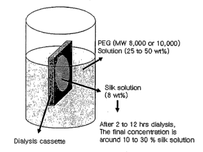
[0084] This solution was concentrated further by exposure to an aqueous
polyethylene
glycol (PEG) (MW 8,000 to 10,000) solution (25-50 wt%) on the outside of a
Slide-a-Lyzer
dialysis cassette (Pierce, MWCO 3500) for 2 to 12 lu.s by osmotic pressure
(Figure 1). The
final concentration of aqueous silk solution could be formed to between 10-30
wt% or
greater.
Electrospinning
[0085] In order to increase the viscosity of aqueous silk solution above 8 wt%
for
spinning, the solution was concentrated using the PEG solution method as
described above.
This was required since the viscosity and surface tension of the pure silk
solution (8 wt%)
CA 02562415 2006-10-10
WO 2005/012606 PCT/US2004/011199
¨ 17 ¨
was not high enough to maintain a stable drop at the end of the capillary tip.
The increase of
silk solutions generated a viscosity and surface tension suitable for
electrospinning. With
the new more concentrated pure silk solutions (10-30%), direct spinning is now
feasible.
The distance between tile tip and the collector was 10-15 cm and flow rate of
the fluid was
0.01 to 0.05 nil/min. As the potential difference between the capillary tip
and the aluminum
foil counter electrode was gradually increased 30 kV (E=2-3 kV/cm), the drop
at the end of
the capillary tip elongated from a hemispherical shape into a cone shape. The
morphology
and diameters of the electrospun fibers were examined using SEM. Silk/PEO
blend solution
produced microsize fibers with diameters 1.5 lam to 251.1m. The morphology of
fiber
surface and fracture surface in liquid nitrogen was well matched with native
silk fiber.
Example II
Preparation of Silk Fibroin Scaffolds
[0086] Porous three-dimensional scaffolds were prepared from silk fibroin
aqueous
solutions by salt-leaching. By adjusting the concentration of silk fibroin and
the particle
size of granular NaCl, the morphological and functional properties of the
scaffolds could be
controlled. The scaffolds had highly homogeneous and interconnected pores and
showed
pore sizes ranging from 470 to 940 um depending on the mode of preparation.
The
scaffolds had porosities >90%. The compressive strength and modulus of
scaffolds was up
to 320 10 KPa and 3330 500 K.Pa, respectively. The scaffolds were fully
degraded by
protease during 21 days. These new silk-based 3-D matrices provide useful
properties as
biomaterial matrices for tissue engineering due to the all-aqueous mode of
preparation,
control of pore size, connectivity of pores, degradability and useful
mechanical features.
Methods
Preparation of silk fibroin aqueous solution
[0087] Cocoons of B. mori were boiled for 20 mm in an aqueous solution of 0.02
M
Na2CO3, and then rinsed thoroughly with distilled water to extract the glue-
like sericin
proteins and wax. The extracted silk fibroin was then dissolved in 9.3 M LiBr
solution at
60 C for 4 hrs, yielding a 20 w/v % solution. This solution was dialyzed in
distilled water
using a Slide-a-Lyzer dialysis cassette (MWCO 3500, Pierce) for 2 days. The
final
concentration of silk fibroin aqueous solution was ca. 8 w/v%, which was
determined by
weighing the remaining solid after drying. To prepare concentrated silk
fibroin solution, 10
ml of 8 w/v % silk fibroin solution was dialyzed against 1 liter of 25 wt%
polyethylene
CA 02562415 2006-10-10
WO 2005/012606
PCT/US2004/011199
¨ 18 ¨
glycol (PEG, 10,000 g/mol) solution at room temperature by using Slide-a-Lyzer
dialysis
cassettes (MWCO 3500). After the required time, the concentrated silk fibroin
solution was
slowly collected by syringe to avoid excessive shearing and the concentration
was
determined. Silk fibroin aqueous solutions with concentration less than 8 wt%
were
prepared by diluting with distilled water. All solutions were stored at 7 C
before use to
avoid premature precipitation. Silk fibroin films prepared from 8 w/v %
solutions were
evaluated to verify the removal of Li + ion by XPS; no residual Li + ion was
detected.
Preparation of silk fibroin scaffolds
[0088] Four grams of granular NaC1 (particle size; 300 ¨ 1180 um) were added
to 2 ml
of silk fibroin aqueous solution (4-10 wt%) in disk-shaped Teflon containers
(Figure 2a).
The container was covered and left at room temperature. After 24 hrs, the
container was
immersed in water and the NaC1 was extracted for 2 days. The porous silk
fibroin scaffolds
formed in this process were stored in water at 7 C before, use.
X-ray diffraction
[0089] X-ray diffraction of freeze-dried samples of the scaffold were obtained
with Ni-
filterd Cu-Kot radiation (A, = 0.15418 urn) from a Rigaku RU-200BH rotating-
anode X-ray
generator operating at 40 kV and 40 mA. X-ray diffraction patterns were
recorded with a
point collimated beam and a imaging plate (Fuji Film BAS-IP SR 127) in an
evacuated
camera. The camera length was calibrated with NaF (d=0.23166 urn).
FTIR spectroscopy
[0090] Approximately 1 mg of freeze-dried sample was pressed into a pellet
with 200
mg of potassium bromide and Fourier transform infrared (FTIR) spectrum was
recorded
with an accumulation of 64 scans and a resolution of 4 cm-1 by Nicolet Magna
860.
Scanning Electron Microscopy (SEM)
[0091] Silk
scaffolds were cut into sections in distilled water using a razor blade and
then freeze-dried. Samples were sputter coated with gold. The morphology of
scaffolds was
observed with a LEO Gemini 982 Field Emission Gun SEM. Pore size was obtained
using
ImageJ software developed at the US National Institutes of Health.
CA 02562415 2011-06-06
WO 2005/012606 PCT/US2004/011199
¨ 19 ¨
Porosity
[0092] The density and porosity of the silk scaffolds were measured by liquid
-
displacement (Zhang, RN., et al.,' J. Biomed. Mater. Res. 1999, 44, 446-455).
Hexane was
used as the displacement liquid as it permeates through silk scaffolds without
swelling or
shrinking the matrix.. The silk scaffold (dry weight, W) was immersed in a
known volume
(V1) of hexane in a graduated cylinder for 5 min. The total volume of hexane
and the
hexane-impregnated scaffold was recorded as V2. The hexane-impregnated
scaffold was
then removed from the cylinder and the residual hexane volume was recorded as
V3. The
total volume of the scaffold was:
(V2 -.V1) + (V1.- V3) 7 V2 - V3.
V2 - V1 is the volume of the polymer scaffold and VI - V3 is the volume of
hexane within the scaffold. The porosity of the scaffold (a) was obtained by:
(%) = (V1-V3) / (V2-V3) x 100
Swelling properties
[0093] Silk fibroin scaffolds were immersed in distilled water at room
temperature for
24 hrs. After excess water was removed, the wet weight of the scaffold (Ws)
was
determined. Samples were then dried in an oven at 65 C under vacuum overnight
and the
dry weight of scaffolds (Wd) was determined. The swelling ratio of the
scaffold and the
water content in the scaffold were calculated as follows:
Swelling ratio = (Ws - Wd)/Wd
Water uptake (%) = [(Ws - Wd)/ Ws] x 100
Mechanical properties
[0094] Resistance to mechanical compression of the scaffolds (12 mm diameter,
10 mm
height, disks) were performed on an Instron 8511 equipped with a 0.1 ICI load
cell at room
temperature. The crosshead speed was 10 mm/min. The compression tests were
conducted
conventionally as an open-sided/confined method. Four samples were evaluated
for each
composition. Cylinder-shaped samples measuring 12 mm in diameter and 10 mm in
height
were used, according to a modification based on the ASTM method F451-95. The
compressive stress and strain were graphed and the average compressive
strength as well as
the compressive modulus and standard deviation determined. The elastic modulus
was
defined by the slope of the initial linear section of the stress-strain curve.
The compressive
strength was determined by drawing a line parallel to this, starting at 1%
strain. The point at
*Trade mark
CA 02562415 2006-10-10
WO 2005/012606 PCT/US2004/011199
¨ 20 ¨
which this line crossed the stress-strain curve was defined as the compressive
strength of
the foam (Thomson RC et al., Biomaterials 1998, 19;1935-1943).
In vitro enzymatic degradation
[0095] The degradation of the silk fibroin scaffolds was evaluated using
protease XIV
(EC 3.4.24.31, Sigma-Aldrich) with an activity of 5.6 U/mg. Samples (12 mm
diameter, 5
mm height) were immersed in 5 ml of phosphate buffer saline (pH 7.4)
containing protease
(1 U) at 37 C. After the specific time samples were washed with phosphate
buffer saline
and distilled water, and freeze-dried. The enzyme solution was replaced with
newly
prepared solution every 24 hrs. For controls, samples were immersed in
phosphate buffer
saline without enzyme.
Results and Discussion õ.
Preparation of water-based scaffolds
[0096] Porous silk fibroin scaffolds were prepared using a salt-leaching
method that has
been previously used in the preparation of porous scaffolds from other
polymers such as
collagen and polylactic acid. The pore size and the porosity of the scaffolds
were regulated
by the addition of granular NaC1 with particle sizes of diameter 300 to 1180
!um to the silk
fibroin aqueous solution. In this process, some of the surface of the NaC1
particles
dissolved in the silk fibroin aqueous solution, while most of the salt was
retained as solid
particles because of saturation of the solution. The silk fibroin aqueous
solutions formed
into hydrogels in the mixture after ¨24 hrs, which resulted in the formation
of water-stable
porous matrices. Table 1 shows the silk fibroin concentrations and particle
sizes of NaC1
used in the study. With an increase in silk fibroin concentration, matrices
were
homogeneously formed through the use of larger particle sizes of the NaCl.
When NaCl
with particle sizes of 500 to 600 tAm were added to 8 wt% silk fibroin
solution, the surface
of the silk fibroin aqueous solutions rapidly formed a hydrogel.
CA 02562415 2006-10-10
WO 2005/012606 PCT/US2004/011199
¨ 21 ¨
Table 1. Preparation of scaffolds from various silk fibroin concentrations and
particle
sizes of NaCl.
Silk fibroin concentration (w/v %)
Particle size of sodium 4 6 8 10
chloride (gm)
1000 1180
850 ¨ 1000 0 0 0El
710 850
600 ¨ 700 0 0El
500 ¨ 600
425 ¨ 500
300 ¨ 425
degree of homogeneity: o>D>x
[0097] In coricentrated salt solutions, solvating forces are significantly
altered from
those in dilute electrolyte solutions because salt ions change the structure
of the
intervening water (Curtis RA et al., Biophys Chem 2002, 98:249-265). The
effect of
concentrated salt solutions with chloride ion, such as NaCl, KC1, CaCl2 and
MgCl2, on
silk fibroin was determined at salt concentrations up to 3 M at room
temperature. When a
drop of silk fibroin solution (8 wt%) was added to concentrated salt solutions
of 3 M, silk
hydrogels formed immediately in the NaCl and KC1 solutions but not in the
CaC12 and
MgC12 solutions. Ions are Classified as kosmotropic or chaotropic, based on
their size and
charge (Grigsby JJ et al., Biophys Chem 2001, 91:231-243). Ions with high
charge density
such as Ca2+ and Mg2+ are highly kosmotropic, and ions with low charge density
such as
K+ are chaotropic. Na+ is weakly kosmotropic and a- is weakly chaotropic.
Kosmotropic
ions bind adjacent water molecules more strongly than chaotropic ions. In
addition,
kosmotropic ions strongly interact with oppositely charged residues on the
protein surface
due to their high charge density. At low salt concentration, the solution
contains a
sufficient number of water molecules to hydrate both the protein surface and
the ions. At
higher salt concentrations, more water molecules are needed to hydrate the
increasing
number of ions. Therefore water molecules are easily removed from the proteins
as
concentrations of salt solutions increase.
[0098] From the primary sequence of the silkworm silk fibroin heavy chain,
seven
internal hydrophobic blocks and seven much smaller internal hydrophilic
blocks, with
two large hydrophilic blocks at the chain ends are present (Zhou, C. Z., et
al., Nucleic
Acids Res. 2000, 28, 2413-2419). The percentage of hydrophobic residues in
silk fibroin
is 79% (Braun, F. N., et al., Int. J. Biol. Macromol. 2003, 32, 59-65) and the
repetitive
CA 02562415 2011-06-06
- 22 -
sequence in these hydrophobic blocks consists of GAGAGS (SEQ ID NO: 4)
peptides
that dominate the 13-sheet structure that forms the crystalline regions in
silk fibroin fibers
and films (Mita, K., et al., J. Mol. Evol. 1994, 38, 583-592).
[0099] Since protein solubility typically decreases as salt concentration
rises,
interactions between proteins become favored (Curtis, R.A., et al., Biophys.
Chem. 2002,
98, 249-265). It is well known that the hydrophobic interactions between non-
polar
residues increase with addition of salt, leading to the salting-out effect
(Robinson, D.R., et
al., J. Am. Chem. Soc. 1965, 87, 2470-2479). The behavior of the fibroin in
the salt
system described may be related to the role of the salt ions in extracting
water that would
otherwise coat the hydrophobic fibroin domains, promoting chain-chain
interactions
leading to the new more stable structure. These hydrophobic interactions
induce protein
folding, resulting in [3-sheet formation (Li, G.Y., et al., Biochem. 2001,
268, 6600-6606).
[00100] Alginate or glass beads were examined to further clarify the ion
effects on
hydrogelation of silk fibroin (8 wt%). While gelation time of silk fibroin
with glass beads
showed a similar result as that observed over 30 days with silk fibroin in a
previous study
(Kim UJ et at, Biomacromolecules, 2004, 5(3) : 786-792, the gelatin time of
the
silk fibroin solution '
with alginate beads was ¨2 times faster due, presumably due to the removal of
water
molecules from the proteins associated with the swelling of the alginate
beads. Compared
with the gelation time (24 hrs) of silk fibroin in saturated NaCl solution,
salt ions strongly
induced protein-protein interactions.
Structural analysis
[00101] Structural changes in the silk fibroin were determined by X-ray
diffraction and
FTIR (Figure 3). X-ray diffraction of silk fibroin scaffolds showed a distinct
peak at 20.8
and a minor peak and 24.6'. These peaks were almost the same as those of the
f3-sheet
crystalline structure (silk II) of native silk fibroin (Asalcura, T., et al.,
Macromolecules
1985, 18, 1841-1845). The results indicate a f3-crystalline spacing distances
of 4.3 and 3.6
A according to the 20.8 and 24.6 reflections, respectively. FTIR spectra of
silk fibroin
scaffolds showed characteristic peaks of silk II at 1701 cm-I and 1623 cm-I
(amide I)
(Asakura, T., et al., Macromolecules 1985, 18, 1841-1845). Silk fibroin in
aqueous
solution at neutral pH exhibited a random coil conformation. From the results
of the X-
ray diffraction and FTIR analyses, the formation of silk fibroin scaffolds
from these
solutions induced a conformational transition from random coil to n-sheet.
CA 02562415 2006-10-10
WO 2005/012606
PCT/US2004/011199
¨ 23 ¨
Morphology
[00102] SEM images of freeze-dried scaffolds prepared from various silk
fibroin
concentrations and variotis sized particles of NaC1 showed highly
interconnected porous
structures and the pore distribution was homogeneous in the whole scaffolds
except for a
thin layer formed on the top surface of the scaffolds, the air-water
interface. The scaffolds
showed rough pore surfaces highly interconnected by a number of smaller pores.
Globular-like structures, 1-3 pm in diameter, were observed on the surfaces of
the pores.
With an increase in silk fibroin concentration, the pore walls were thicker.
Table 2 shows
actual pore sizes in the scaffolds, ranging from 350 to 920 Rm.
Table 2. Measured pore sizes (Rm) of silk fibroin scaffolds.
Silk fibroin concentration (w/v %)
Particle size of sodium 4 6 8 10
chloride (p.m)
1000 1180 940 50 930+40 920+50 920 50
850 ¨ 1000 760 30 750 1 50 750 20
710 ¨ 850 650 1 30 650 50 640 30
600 ¨ 700 570 30 550 30
500 ¨ 600 470 1 30
Values are average standard derivation (N=20).
[00103] The actual pore sizes in the scaffolds were 80-90 % smaller than the
particle
size of NaC1 used in the process. The pore sizes in scaffolds prepared with
the same
particle size of NaC1, regardless of the concentration of silk fibroin used,
resulted in
similar sized pores.
Porosity and swelling properties
[00104] Silk fibroin scaffolds with >90% porosity were formed and
porosities
increased with a decrease in pore size and silk fibroin concentrations (Table
3). These
values were similar as those (84-98%) of HFIP-derived silk scaffolds prepared
by salt
leaching or gas forming (Nazarov R, et al., Bionzacromolecules, in press).
Swelling ratio
and water uptake of the scaffolds are shown in Tables 4 and 5.
CA 02562415 2006-10-10
WO 2005/012606
PCT/US2004/011199
- 24 -
Table 3. Porosity (%) of silk fibroin scaffolds
Silk fibroin concentration (w/v %)
Particle size of sodium 4 6 8 10
chloride (.un)
1000 1180 95 1.8 93 0.7 92 1.3 85 1.5
850 - 1000 95 1.5 95 1 0.2 94 1 0.2
710 850 97 0.4 96 1 1.6 95 1.5
600 - 700 971 1.6 97 1 0.6
500 - 600 97 0.5
Values are average standard derivation (N=3).
Table 4. Swelling ratio of silk fibroin scaffolds.
Silk fibroin concentration (w/v %)
Particle size of sodium 4 6 8 10
chloride (pm)
1000 1180 55.3 1 3.8 36.1 1 0.1 23.6 1.2 19.2
4.3
850 - 1000 50.0 0.2 29.8 0.6 21.5 1.9
710 850 48.6 1 2.0 28.9 1.5 19.8 0.2
600 - 700 46.8 1 2.6 28.4 1 2.7
500 - 600 47.6 2.1
Values are average standard derivation (N=3).
Table 5. Water uptake (%) of silk fibroin scaffolds.
Silk fibroin concentration (w/v %)
Particle size of sodium 4 6 8 10
chloride (p.m)
1000 1180 98.2 0.1 97.3 1 0.1 95.9 0.2 94.9
1.0
850 - 1000 98.0 0.1 96.8 1 0.1 95.2 0.1
710 850 98.0 1 0.1 96.7 1 0.2 95.6 1 0.4
600 - 700 97.9 1 0.1 96.6 1 0.3
500 - 600 97.9 0.1
Values are average standard derivation (N=3).
[00105] Swelling ratio decreased gradually with a decrease in pore size.
However,
swelling ratio decreased significantly with an increase in silk fibroin
concentration due to
the decrease in porosity. The swelling ratio of the scaffold prepared from 8
wt% silk
fibroin was -8 times lower than that of collagen scaffolds, due to the
differences in the
hydrophilicities of proteins (Ma L. et al., Biomaterials 2003, 24:4833-4841).
The value
was similar to polylactic acid scaffolds (Maquet V. et al., Biomaterials 2004,
25:4185-
4194). Water uptake of the scaffolds in distilled water was >93% during 24
hrs. The
high water-binding ability of the scaffolds can be attributed to the highly
porous structure
of the protein network.
CA 025 62415 200 6-1 0-1 0
WO 2005/012606 PCT/US2004/011199
¨ 25 ¨
Mechanical properties
[00106] The scaffolds exhibited a ductile and sponge-like behavior with
different
stiffness depending on the concentration of silk fibroin used in the process.
An elastic
region was observed at initial strain followed by a peak stress. Table 6 shows
the
mechanical properties of the silk fibroin scaffolds. The compressive strength
and modulus
of the scaffolds increased with an increase in silk fibroin concentration.
Table 6. Mechanical properties of silk fibroin scaffolds
Silk fibroin concentration (w/v %)
Particle 4 6 8 10
size of
sodium Compressive Compressive Compressive Compressive Compressive
Compressiv,e Compressive Compressive
chloride modulus stress modulus stress modulus
stress modulus
(gm) stress (kPa) (kPa) (kPa) (kPa) (kPa) (kPa) (kPa)
(kPa)
1000 11 3 70 5 4914 560150 100 10 1300 40 320
1 10 3330 500
1180
850¨ 11 1 8015 5418 620 40 125 10 1530 1 190
1000
710¨ 12 1 100 1 5 58 1 3 670 1 30 140 15 1940 240
850
600¨ 13 3 115 1 10 60 1 5 770 1 50
700
500¨ 13 *3 130 10
600
Values are average standard derivation (N=4).
[00107] The improvement in mechanical properties was attributed to the
increase in
polymer concentration accompanied with the increase in the wall thickness of
the pores.
At the same silk fibroin concentration, scaffolds prepared with smaller
particle sizes of
NaC1 showed higher compressive strength and modulus due to the decreased pore
size. It
is considered that the increased pore wall sites induced by the decreased pore
size
provided more paths to distribute the applied stress. The increased pore sites
may
functioned as a barrier, such as crack disipation, to reduce crack
propagation. In addition,
it has been reported that a more uniform pore distribution improved the
mechanical
properties of polymer matrices. Therefore, stress applied to porous materials
is
concentrated at the pore interface, and if the pore distribution is not
uniform, polymer
matrices typically deform at a lower stress (Harris LD. Et al., J Biomed Mater
Res 1998,
42:396-402). For example, in our recent studies (Nazarov R. et al.,
Biomacromolecules,
in press). three-dimensional silk fibroin scaffolds were developed using a
salt leaching
method with HFIP. While these scaffolds had smaller pore size and utilized a
higher
concentration of silk fibroin in processing, the compressive strengths (30 to
250 KPa) of
CA 02562415 2006-10-10
WO 2005/012606
PCT/US2004/011199
¨ 26 --
the HFIP-derived silk scaffolds (17 wt% silk in HFIP) prepared by salt
leaching were
similar to those found for the aqueous-derived (8-10 wt% silk in water) silk
scaffolds in
the present study. However, the compressive modulus of aqueous-derived silk
scaffolds
was 3-4 times higher (100 to 790 kPa) for the HFIP-derived silk scaffolds.
Enzymatic degradation
[00108] Figure 4a shows the mass of the scaffolds over time prepared from 4 to
8 wt%
silk fibroin with NaCl of 850-1000 gm diameter particle sizes during a
degradation period
of 21 days. The scaffolds in phosphate buffer without protease showed no
degradation
within 21 days. The scaffolds prepared with 4 wt% fibroin rapidly degraded and
the mass
remaining was only 2% after 10 days. The scaffolds prepared from 6 and 8 wt %
fibroin
gradually degraded with time and the mass was reduced to 30 and 20%,
respectively, after
21 days. Figure 4b shows the mass of the scaffolds remaining when
prepared=ftom 6 wt%
silk fibroin with various particle sizes of NaCl. The degradation patterns
suggest that pore
size did not correlate with degradation rate, on the nature of the initial
concentration of
fibroin.
Conclusions
[00109] Porous silk fibroin scaffolds were prepared directly from silk fibroin
aqueous
solutions by a salt leaching method, in the complete absence of any organic
solvents or
chemical crosslinking. The formation of the scaffolds included a structural
transition from
random coil top-sheet. This transition provides a mechanistic basis for the
transition, as
the salt may promote water loss from the hydrophobic domains leading to
enhanced
chain-chain interactions and thus 13-sheet formation. Functional and
morphological
properties of the scaffolds were controlled by the concentration of the silk
fibroin solution
used in the process and the particle size of NaCl.
Example III
Preparation of Silk Hydrogels
[00110] Control
of silk fibroin concentration in aqueous solutions via osmotic stress
was studied to assess relationships to gel formation and structural,
morphological and
functional (mechanical) changes associated with this process. Environmental
factors
potentially important in the in vivo processing of aqueous silk fibroin were
also studied to
CA 02562415 2006-10-10
WO 2005/012606 PCT/US2004/011199
¨ 27 ¨
determine their contributions to this process. Gelation of silk fibroin
aqueous solutions
was affected by temperature, Ca2+, pH and polyethylene oxide (PEO). Gelation
time
decreased with increase in protein concentration, decrease in pH, increase in
temperature,
addition of Ca2+ and With the addition of PEO. No change of gelation time was
observed
with the addition of K. Upon gelation, a random coil structure of the silk
fibroin was
transformed into 13-sheet structure. Hydrogels with fibroin concentrations >4
weight
percent exhibited network and sponge-like structures based on scanning
electron
microscopy. Pore sizes of the freeze-dried hydrogels were smaller as the silk
fibroin
concentration or gelation temperature were increased. Freeze-dried hydrogels
formed in
the presence of Ca2+ exhibited larger pores as the concentration of this ion
was increased.
Mechanical compressive strength and modulus of the hydrogels increased with
increase
in protein concentration and gelation temperature.
Methods
Preparation of silk fibroin aqueous solution
[00111] Cocoons of Bombyx mori, kindly provided by M. Tsukada (Institute of
Sericulture, Tsukuba, Japan) and M. Goldsmith (U. Rhode Island), were boiled
for 20 min
in an aqueous solution of 0.02 M Na2CO3, and then rinsed thoroughly with
distilled water
to extract the glue-like sericin proteins and wax. The extracted silk fibroin
was then
dissolved in 9.3 M LiBr solution at 60 C for 4 hrs, yielding a 20 w/v%
solution. This
solution was dialyzed in distilled water using a Slide-a-Lyzer dialysis
cassette (MWCO
3500, Pierce) for 2 days. The final concentration of silk fibroin aqueous
solution was ca.
8 wt%, which was determined by weighing the remaining solid after drying. Silk
fibroin
film prepared from 8 wt% solutions was evaluated to verify the removal of Li +
ion by
XPS; no residual Li+ ion was detected.
Preparation of concentrated silk fibroin solution by osmotic stress
[00112] Silk fibroin aqueous solution (8 wt%, 10 ml) was dialyzed against
10-25 wt%
polyethylene glycol (PEG, 10,000 g/mol) solution at room temperature by using
Slide-a-
Lyzer dialysis cassettes (MWCO 3500). The volume ratio of PEG to silk fibroin
solution
was 100:1. By osmotic stress, water molecules in the silk fibroin solution
moved into
PEG solution through the dialysis membrane (Parsegian, V. A., et al., Methods
in
Enzymology, Packer, L., Ed.; Academic Press: 1986; Vol. 127, p 400). After the
required
CA 02562415 2006-10-10
WO 2005/012606 PCT/US2004/011199
¨ 28 ¨
time, concentrated silk fibroin solution was slowly collected by syringe to
avoid
excessive shearing and the concentration was determined. Silk fibroin aqueous
solutions
with concentration less than 8 wt% were prepared by diluting 8 wt% solutions
with
distilled water. All solutions were stored at 7 C before use.
Sol-Gel Transitions
[00113] A 0.5 ml of silk fibroin aqueous solution was placed in 2.5 ml flat-
bottomed
vials (diameter: 10 mm). The vials were sealed and kept at room temperature,
37 C and
60 C. Gelation time was determined when the sample had developed an opaque
white
color and did not fall from an inverted vial within 30 sec. To investigate the
effect of ions
and ion concentration on the process, CaC12 or KC1 solutions were added into
the silk
fibroin aqueous solution to generate a final salt concentration of 2.5 to 30
mM. The pH of
the silk fibroin solution was adjusted with HC1 or NaOH solution. For the
preppration of
silk fibroin-poly(ethylene) oxide (PEO, 900,000 g/mol) solution, the required
amount of
PEO solution (5 wt%) was added to silk fibroin solution with mild stirring for
5 minutes.
The blend ratios of silk fibroin/PEO were 100/0, 95/5, 90/10, 80/20 and 70/30
(w/w).
Wide angle X-ray scattering (WAXS)
[00114] X-ray profiles were recorded for freeze-dried silk fibroin
solutions and
hydrogels using a Brucker D8 X-ray Diffi-actometer at 40 kV and 20 mA, with Ni-
filtered
Cu-Ka radiation.
Scanning Electron Microscopy (SEM)
[00115] Silk fibroin solutions and hydrogels were frozen at ¨80 C and then
lyophilized. The samples were fractured in liquid nitrogen and examined using
a LEO
Gemini 982 Field Emission Gun SEM. To check for artifactual morphological
changes
due to freeze-drying, an alternative preparation used Karnovsky's fixative at
room
temperature for 4 hrs. Hydrogels with and without fixative treatment showed
little
morphological change upon freeze-drying. Pore size was obtained by using
ImageJ
software developed at the US National Institutes of Health.
CA 02562415 2006-10-10
WO 2005/012606 PCT/US2004/011199
¨ 29 ¨
Mechanical properties
[00116] Compression tests of hydrogels were performed on an Instron 8511
equipped
with a 2.5 kN load cell at room temperature. A crosshead speed was 10 mm/min.
The
cross-section of samples was 12 mm in diameter and 5 mm in height. The
compression
test was achieved conventionally as an open-sided method. The compression
limit was
98% strain to protect the load cell. Five samples were evaluated for each
composition.
Results
Concentrated Silk Fibroin Solutions
[00117] Silk fibroin aqueous solution with an initial concentration of 8
wt% was
dialyzed against 10-25 wt% PEG solution at room temperature. Silk fibroin
aqueous
solution was concentrated over time by osmotic stress and concentrations of
ca. 21 wt%
were obtained after 9 hrs dialysis against 25 wt% PEG solution (Figure 6).
Longer
dialysis times were required to generate higher concentrations of silk fibroin
aqueous
solution, when lower concentrations of PEG solutions were used. Silk fibroin
gels, 23-33
wt%, were spontaneously generated in the dialysis cassettes during the
concentration
process. These gels were transparent even after drying at room temperature and
at 60 C.
Gelation of silk fibroin aqueous solution
[00118] The influence of temperature, Ca2+ and K+ concentrations, pH and PEO
concentration was investigated on the gelation of silk fibroin aqueous
solutions. Figure 7
illustrates the gelation time of silk fibroin aqueous solution (pH 6.5-6.8) at
various
temperatures. The gelation time of silk fibroin aqueous solution decreased
with increase
in fibroin content and temperature. Concurrently, a conformational change from
random
coil to p-sheet structure was observed and the formation of f3-sheet structure
in the
hydrogels was confirmed by X-ray diffraction as described later. Figure 8
shows the
gelation time of silk fibroin aqueous solution with different Ca2+ and K+
concentrations.
The pHs of silk fibroin solutions with Ca2+ and K+ ions were 5.6-5.9 and 6.2-
6.4,
respectively. Ca2+ resulted in shorter gelation times, whereas there was no
change in
gelation time with the addition of K+ at any temperature. These results with
regenerated
silkworm fibroin differ from prior studies in which K+ ions added to solutions
of spider
silk influenced aggregation and precipitation of the protein, whereas there
was no
rheological change after addition of Ca2+ ions. Figure 9 shows the gelation
time of silk
CA 02562415 2006-10-10
WO 2005/012606
PCT/US2004/011199
fibroin aqueous solution (4 wt%) at different pHs. Gelation time decreased
significantly
with a decrease in pH. This behavior is similar to that observed for the silk
from the
spider, Araneus diadematus, which gels at pH 5.5, but behaves as a viscous
liquid at pH
7.4 (Vollrath, F., et al., Proc. R. Soc. London B, 1998, 265, 817-820); Figure
10 shows
the gelation time of silk fibroin aqueous solution (4 wt%) with different
polyethylene
oxide (PEO) contents. By adding PEO solution, the pH decreased slightly to the
range
6.1-6.4. The gelation time was significantly reduced with the addition of only
5% PEO,
whereas there was no difference in gelation time when the concentration was
above 5%.
Structural analysis of hydrogels
[00119]
Structural changes in the silk fibroin were determined by X-ray diffraction.
Figure 11 shows X-ray profiles of freeze-dried silk fibroin solutions and
hydrogels
prepared from silk fibroin aqueous solutions. When silk fibroin solutions
were, frozen at
low temperature, below the glass transition (-34 ¨ -20 C), the structure was
not
significantly changed (Li, M., et al., J. Appl. Polym. Sci. 2001, 79, 2185-
2191). The
freeze-dried silk fibroin samples exhibited a broad peak at around 20
regardless of the
silk fibroin concentration, indicating an amorphous structure. Silk fibroin in
aqueous
solution at neutral pH exhibited a random coil conformation. (Magoshi, J., et
al.,
Polymeric Materials Encyclopedia; Salamone, J. C., Ed.; CRC Press: NewYork,
1996;
Vol. 1, p. 667; Magoshi, J., et al., Polymeric Materials Encyclopedia;
Salamone, J. C.,
Ed.; CRC Press: NewYork, 1996; Vol. 1, p. 667). All hydrogels prepared from
silk
fibroin solutions showed a distinct peak at 20.6 and two minor peaks at
around 9 and
24 . These peaks were almost the same as those of the I3-sheet crystalline
structure of silk
fibroin. (Ayub, Z. H., et al., Biosci. Biotech. Biochem. 1993, 57, 1910-1912;
Asakura, T.,
et al., Macromolecules 1985, 18, 1841-1845). These peaks indicate I3-
crystalline spacing
distances of 9.7, 4.3 and 3.7 A according to 9 , 20.6 and 24 , respectively.
From the
results of X-ray diffraction, the gelation of silk fibroin solutions induced a
conformational
transition from random coil to I3-sheet as previously reported. (Ayub, Z. H.,
et al., Biosci.
Biotech. Biochem. 1993, 57, 1910-1912; Hanawa, T., et al., Chem. Pharm. Bull.
1995, 43,
284-288; Kang, G. D., et al., Marcromol. Rapid Commun. 2000, 21,788-.791).
CA 02562415 2006-10-10
WO 2005/012606 PCT/US2004/011199
¨ 31 ¨
Morphology offreeze-dried hydrogels
[00120] Morphological features of silk fibroin solutions and hydrogels were
observed
by SEM after freeze-drying at ¨80 C. Freeze-dried silk fibroin solutions of 4-
12 wt%
showed leaf-like morphologies. Freeze-dried silk fibroin solutions of 16 wt%
and 20 wt%
exhibited network and sponge-like structures with pore sizes of 5.014.2 tun
and 4.7 4.0
1.1,m, respectively. By SEM imagery it was determined that freeze-dried
hydrogels
prepared from 4 wt% silk fibroin solution showed leaf-like morphologies and
interconnected pores regardless of temperature and at higher fibroin
concentrations than 4
wt% sponge-like structures were observed. The pore sizes of freeze-dried
hydrogels
(<1.1. 0.8 wn) were smaller than those observed for the freeze-dried silk
fibroin solution
samples. Pore sizes in the freeze-dried hydrogels decreased with increase in
silk fibroin
concentration, and pore sizes decreased as temperature increased at the same
silk fibroin
concentration. The 4 wt% freeze-dried hydrogels with Ca2+ ions showed network
and
sponge-like structures, whereas the 4 wt% freeze-dried hydrogels with K+ ions
had a leaf-
like morphology. In freeze-dried hydrogels with fibroin concentrations >4 wt%,
pore
sizes of freeze-dried hydrogels with Ca2+ were larger than those of freeze-
dried hydrogels
prepared from silk fibroin aqueous solutions without Ca2+ ions. Interestingly,
pore size
was larger in freeze-dried hydrogels with the same silk fibroin concentration
with an
increase in Ca2+ concentrations. In contrast to freeze-dried hydrogels with
Ca2+, pore
sizes of freeze-dried hydrogels with K+ showed sizes similar to those of
freeze-dried
hydrogels prepared from silk fibroin aqueous solutions. These results imply
that Ca2+ was
more effective in inducing interactions among the silk fibroin chains than the
K+. This
result is also consistent with the earlier data wherein Ca2+ resulted in
shorter gelation
times than K+.
Mechanical properties of hydrogels
[00121] The compressive strength and modulus of hydrogels prepared from silk
fibroin
aqueous solutions increased with an increase in silk fibroin concentration
(Figure 12a and
= 12b). The improvement in mechanical properties was attributed to the
increase in polymer
concentration accompanied with the decrease in pore size. At the same silk
fibroin
concentration, hydrogels prepared at higher temperature showed higher
compressive
strengths and moduli due to the decreased pore size. Hydrogels with 4-8 wt%
fibroin
showed less than 55% strain, while hydrogels with 12-16 wt% fibroin revealed
larger
CA 02562415 2006-10-10
WO 2005/012606 PCT/US2004/011199
¨ 32 ¨
strains ranging from 75% to 96% (Figure 12c). The effect of pore size was
considered
since the smaller pore size distributes stress in the hydrogel more evenly to
resist stress
concentration. The smaller pore size and increased number of pores also
function as a
barrier against crack propagation.
Discussion
[00122] Gelation occurs due to the formation of inter- and intramolecular
interactions
among the protein chains, including hydrophobic interactions and hydrogen
bonds (Ayub,
Z. H., et al., Biosci. Biotech. Biochem. 1993, 57, 1910-1912; Hanawa, T., et
al., Chem.
Pharm. Bull. 1995, 43, 284-288; Kang, G. D., et al., Marcromol. Rapid Commun.
2000,
21, 788-.791). With an increase in fibroin content and temperature,
interactions among the ,
fibroin chains increases. Silk fibroin molecules are thereby able to interact
more readily,
leading to physical crosslinks.
[00123] The concentration of Ca2+ ion in the silkworm (Bombyx mori) increases
from 5
mM to 15 mM as silk progresses toward the spinneret, while K+ ion is present
at 5-8
mM.3 Several calcium salts are known to dissolve silk fibroin because of
strong
interactions with the fibroin (Ajisawa, A. J. Seric. Sci. Jpn. 1998, 67, 91-
94; Ha, S.W., et
al., Biomacromolecules 2003, 4, 488-496). Rheological measurements of dilute
solutions
of silk fibroin from Bombyx mori revealed that the protein chains tend to form
clusters by
ionic interaction between C00- ions of amino acid side chains in the fibroin
and divalent
ions such as Ca2+ or Mg2+ (Ochi, A., et al., Biomacromolecules 2002, 3, 1187-
1196).
Through these interactions, the pH of silk fibroin solutions with Ca2+ ions
was
significantly lower than that of silk fibroin solutions in the absence of
these ions, whereas
the addition of monovalent ions such as K+ showed only a slight decrease of
pH. With
lower pH, repulsion among silk fibroin molecules decreases and interactions
among the
chains is easier, resulting in stronger potential for the formation of n-sheet
structure
through hydrophobic interactions. A pH near the isoelectric point (p/= 3.8-
3.9) (Ayub,
Z. H., et al., Biosci. Biotech. Biochem. 1993, 57, 1910-1912; Kang, G. D., et
al.,
Marcromol. Rapid COM1721.117. 2000, 21, 788-791) of silk fibroin accelerated
the sol-gel
transition of silk fibroin aqueous solutions in a fashion similar to other
proteins that
aggregate near their isoelectric points.
[00124] These outcomes may reflect subtle differences in how different silk
proteins
from different organisms utilize physiologically relevant ions to facilitate
sol-gel
CA 02562415 2006-10-10
WO 2005/012606 PCT/US2004/011199
¨ 33 ¨
transitions. Divalent ions may induce aggregation of silk fibroin molecules by
ionic
interactions with negatively charged amino acids present particularly near the
chain ends
of the heavy chain fibroin. The lack of response to different concentrations
of Ca2+ may
suggest a broad window of response physiologically or perhaps a role for
combinations of
ions to fully control this process in vivo or in vitro. Additional studies
will be required to
elucidate these relationships, particularly when considered in concert with
observations
on domain mapping of silks related to processing environments (Bini, E., et
al., J. MoL
Biol. 2004, 335, 27-40).
[00125] The movement of water from the silk fibroin molecules to the
hydrophilic
PEO facilitates inter- and intramolecular interactions among the protein
molecules and
the subsequent formation of the I3-sheet structure. This transition is evident
with silk
based on our recent mechanistic understanding of the process (Jin, H. J., et
al., Nature
2003, 424, 1057-1061). These transitions can be induced by direct addition of
PEO into
the fibroin aqueous solutions, or via separation from the aqueous solutions
across a
dialysis membrane (with PEG). Thus, direct contact between the protein and the
PEO is
not required, only the facilitation of water transport from the protein to the
PEO/PEG to
drive the sol-gel transition.
Conclusions
[00126] From the primary sequence, silkworm silk fibroin heavy chain is
composed of
seven internal hydrophobic blocks and seven much smaller internal hydrophilic
blocks,
with two large hydrophilic blocks at the chain ends (Zhou, C. Z., et al.,
Nucleic Acids Res.
2000, 28, 2413-2419; Jin, H. J., et al., Nature 2003, 424, 1057-1061). The
percentage of
hydrophobic residues in silk fibroin is 79% (Braun, F. N., et al., Int. J.
Biol. MacromoL
2003, 32, 59-65) and the repetitive sequence in hydrophobic residues consists
of
GAGAGS peptides that dominate the 13-sheet structure forming crystalline
regions in silk
fibroin fibers and films (Mita, K., et al., ./. Mol. EvoL 1994, 38, 583-592).
The formation
of these I3-sheets results in insolubility in water (Valluzzi, R., et al., J.
Phys. Chem. B
1999, 103,11382-11392). Hydrophobic regions of silk fibroin in aqueous
solution
assemble physically by hydrophobic interactions and eventually organize into
hydrogels
(Jin, H. J., et al., Nature 2003, 424, 1057-1061). Silk fibroin concentration,
temperature,
Ca2+, pH and PEO affected the gelation of the silk fibroin aqueous solutions.
With
increase in fibroin content and temperature, physical crosslinking among silk
fibroin
CA 02562415 2006-10-10
WO 2005/012606 PCT/US2004/011199
¨ 34 ¨
molecules formed more easily. Ca2+ ions accelerated these interactions,
presumably
through the hydrophilic blocks at the chain ends. The decrease in pH and the
addition of a
hydrophilic polymer decreased repulsion between silk fibroin molecules and
promoted
the desorption of water from the protein, resulting in shorter gelation times.
Upon gelling,
a conformational transition from random coil to I3-sheet structure was induced
and
promoted the insolubility and stability of silk fibroin hydrogels in water.
Silk fibroin
hydrogels had network and sponge-like structures. Pore size was smaller with
increased
silk fibroin concentration and gelation temperature. Freeze-dried hydrogels
showed larger
pore sizes with increases in Ca2+ concentrations than freeze-dried hydrogels
prepared
from silk fibroin aqueous solutions at the same fibroin content. The
compressive strength
and modulus of hydrogels prepared from silk fibroin aqueous solution without
ions .
increased with increase in protein concentration and gelation temperature.
[00127] Hydrogels from natural polymers, such as collagen, hyaluronate,
fibrin,
alginate and chotosan, have found numerous applications in tissue engineering
as well as
in drug delivery. However, they generally offer a limited range of mechanical
properties
(Lee, K. Y., et al., Chem. Rev. 2001, 101, 1869-1879). In contrast, silk
fibroin provides an
important set of material options in the fields of controlled release,
biornaterials and
scaffolds for tissue engineering because of combination with impressive
mechanical
properties, biocompatibility, biodegradability and cell interaction (Altman,
G. H., et al.,
Biomaterials 2003, 24, 401-416; Cappello, J., et al., I. Control. Release
1998, 53, 105-
117; Foo, C. W. P., et al., Adv. Drug Deliver. Rev. 2002, 54, 1131-1143;
Dinerman, A.
A., et al., J. Control. Reledse 2002, 82, 277-287; Megeed, Z., et al., Adv.
Drug Deliver.
Rev. 2002, 54, 1075-1091; Petrini, P., et al., J. Mater. Sci-Mater. M. 2001,
12, 849-853;
Altman, G. H., et al., Biomaterials 2002, 23, 4131-4141; Panilaitis, B., et
al.,
Biomaterials 2003, 24, 3079-3085).
Example IV
Bone regeneration using three-dimensional aqueous-derived silk scaffolds
[00128] We have examined the bone regeneration of human bon marrow stem cells
on
three-dimensional silk scaffolds from aqueous silk solutions of the invention.
To study
the ability of the silk scaffolds to support the growth and differentiation of
the bone
marrow stem cells, we have used the silk scaffolds without any decoration.
CA 02562415 2011-06-06
WO 2005/012606 PCT/US2004/011199
¨ 35 ¨
Methods
Materials
[00129] Bovine serum, Dulbecco's Modified Eagle Medium (DMEM), Minimal
essential medium-a modification (aMEM), basic Fibroblast growth factor (bFGF),
Penicillin-Streptomycin (Pen-Strep), Fungizone, non essential amino acids,
trypsin were
from Gibco (Carlsbad, CA). Ascorbic acid phosphate, Histopaque-'61077,
dexamethasone,
and f3-glycerolphosphate were from Sigma (St. Lois, MO). All other substances
were of
analytical or pharmaceutical grade and obtained from Sigma. Silkworm cocoons
were
= kindly supplied by M. Tsukada (Institute of Sericulture, Tsukuba, Japan)
and Marion
Goldsmith (University of Rhode Island, Cranston, M).
Preparation of scaffolds
[00130] Aqueous-derived silk scaffolds were prepared by adding 4 g of granular
NaCl
("particle size; 1000-4180 um) into 2 ml of 8 wt% silk fibroin solution in
disk-shaped
Teflon containers. The container was covered and left at room temperature.
After 24 hrs,
the container was immersed in water and the NaC1 was extracted for 2 days.
HFIP-
derived silk scaffolds were prepared by adding 4 g of granular NaCl (particle
size;
850-100 um) into 2 ml of 8 wt% silk fibroin in HFIP. The containers were
covered to
reduce of HFIP and to provide the sufficient time for more homogeneous
distribution of
the solution. The solvent was evaporated at room temperature for 3 days. After
the
composite of silk/porogen was treated in methanol for 30 mm to induce p¨sheet
structure
and insolubility in aqueous solution, the composite was immersed in water to
remove
NaC1 for 2 days. The porous silk scaffolds were air-dried.
Human bone marrow stem cell isolation and expansion
[00131] Total bone marrow (25 cm3, Clonetics, Santa Rosa, CA.) was diluted
in 100
ml of isolation medium (5% FBS in RPMI 1640 medium). Cells were separated by
density gradient centrifugation. Briefly, 20 ml aliquots of bone marrow
suspension were
overlaid onto a poly-sucrose gradient (1,077 g/cm3, Histopaque, Sigma, St.
Louis, MO)
and centrifuged at 800xg for 30 min at room temperature. The cell layer was
carefully
removed, washed in 10 ml isolation media, pelleted and contaminating red blood
cells
* .
were lysed in 5 ml of Pure-Gene Lysis solution. Cells were pelleted and
suspended in
*Trade mark
CA 02562415 2011-06-06
WO 2005/012606 PCT/US2004/011199
¨ 36 ¨
expansion medium (DMEM, 10% FBS, 1 ng/ml bFGF, 100U/m1 penicillin, 100pg/m1
streptomycin, 0.254m1 fungizone, nonessential amino acid) and seeded in 75 cm2
flasks
at a density of 5 x 10⁴ cells/cm2. The adherent cells were allowed to reach
approximately
80% confluence (12-17 days for the first passage). Cells were trypsinized,
replated and
passage 2 (P2) cells (80% confluence after 6-8 days), were used for the
experiments.
=
In vitro culture
[00132] For examination of cell growth and differentiation in vitro on silk
scaffolds,
BMSC (5 x 10⁵ cells/scaffold, passage 2) was seeded onto prewetted (a-MEM,
overnight)
silk scaffolds. After 24h, the medium was removed and cultures were maintained
in =
individual wells of 6-well plates. Osteogenic media were a-MEM supplemented
with =
10% FBS, nonessential amino acid, 50i.i.g/m1 ascorbic acid-2-phosphate, 10 nM
dexamethasone, and 7 mMr3-glycerolphosphate in the presence of penicillin ind
streptomycin and fungizone. Cultures were maintained' at 37 C in a humidified
incubator
supplemented with 5% CO2. Half of the medium was changed every 2-3 days.
=
=
Biochemical analysis and histology
[00133] Scaffolds were cultured for 2 and 4 weeks in osteogenic media and
processed
for biochemical analysis and histology. For DNA analysis, 3-4 scaffolds per
group and
time point were disintegrated. DNA content (n=3-4) was measured using the
PicoGreen
assay (Molecular Probes, Eugene, OR), according to the protocol of the
manufacturer.
Samples were measured flurometrically at an excitation wavelength of 480 urn
and an
emission wavelength of 528 nm. For total calcium content, samples (n=4) were
extracted
twice with 0.5 ml 5% trichloroacetic acid. Calcium content was determined by a
colorimetric assay using o-cresolphthalein complexone (Sigma, St. Louis, MO).
The
calcium complex was measured spectrophotometrically at 575 urn. Alkaline
phosphatase
activity was measured using a biochemical assay from Sigma (St. Louis, MO),
based on
conversion of p-nitrophenyl phosphate to p-nitrophenol, which was measured
spectrophotometrically at 405 nrn.
RNA isolation, real-time-reverse transcription polymerase chain reaction (real
time RT-
PCR)
*Trade mark
CA 02562415 2011-06-06
WO 2005/012606 PCT/US2004/011199
¨ 37 ¨
[00134] Fresh scaffolds (n=3-4 per group) were transferred into 2 ml plastic
tubes and
1.0 MI Trizol*was added. Scaffolds were disintegrated using steel balls and a
Microbeater.
Tubes were centrifuged t 12000g for 10 minutes and the supernatant was
transferred to a
,
new tube. Chloroform (200 111) was added to the solution and incubated for 5
minutes at
room temperature. Tubes were again centrifuged at 12000g for 15 minutes and
the upper
aqueous phase was transferred to a new tube. One volume of 70% ethanol (v/v)
was
added and applied to an RNeasy mini spin column (Quiagen, Hilden, Germany).
The
RNA was washed and eluted according to the manufacturer's protocol. The RNA
samples
were reverse transcribed into cDNA using oligo (dT)-selection according to the
manufacturer's protocol (Superscript Preamplification System, Life
Technologies,
(3aithersburg, MD). Collagen type I, Collagen type II, Alkaline phosphatase,
bone .
sialoprotein and Osteopontin gene expression were quantified using the ABI
Prism 7000
Real Time PCR system (Applied Biosystems, Foster City, CA). PCR reaction
conditions
were 2 min at 50 C, 10 min at 95 C, and then 50 cycles at 95 C for 15s, and 1
min at
60 C. The expression data were normalized to the expression of the
housekeeping gene,
glyceraldehyde-3-phosphate-dehydrogenase (GAPDH). The GAPDH probe was labelled
at the 5' end with fluorescent dye VIC and with the quencher dye TAMRA at the
3' end.
Primer sequences for the human GAPDH gene were: Forward primer 5'-ATG GGG AAG
GTG AAG GTC G-3' (SEG ID NO: 1), reverse primer 5'-TAA AAG CCC TOG TGA
CC-3' (SEQ ID NO: 2), probe 5'-CGC CCA ATA CGA CCA AAT CCG TFG AC-
3'(SEQ ID NO: 3). Primers and probes for alkaline phosphatase, bone
sialoprotein (BSP),
osteopontin and were purchased from Applied Biosciences (Assay on Demand #, Hs
00240993 ml (ALP), Hs 00173720 ml (BSP), Hs 00167093 ml (osteopontin)).
= Western blotting analysis
[00135] For total protein extraction, cells were lysed in RIPA buffer [50 mM
Tris-HC1,
pH 8.0, 150 mM NaC1, 1% Nonidet P-40 (NP-40), 0.2% SDS, 5mM NaF] containing
protease inhibitors and phosphatase inhibitors. Protein content was measured
by the
Bradfod method. Proteins were resolved by 3-8% SDS-PAGE and transferred to
membranes. Blots were probed with the primary antibody for 12h at 4 C, washed,
and
incubated with the appropriate peroxidases labed secondary antibodies for lh
at room
temperature. Protein bands were revealed by ECL (Annersham-Pharmacia, )
*Trade mark
CA 02562415 2006-10-10
WO 2005/012606 PCT/US2004/011199
¨ 38 ¨
Scanning electron microscopy (SEM) analysis
[00136] The polymeric surfaces prior to and after cell attachment were
examined by
scanning electron microscopy (SEM). Matrices were fixed using Karnovsky's
fixative for
24h, and washed three times in CMPBS to remove residual fixative. The samples
were
then dried using a graded series of ethanol (50 ¨ 100%) at 15 mm intervals.
After drying,
the samples were sputter coated with gold and examined with a LEO Gemini 982
Field
Emission Gun SEM.
Histological evaluation
[00137] After fixation with 4% phosphate-buffered formaldehyde for at least 24
hours,
specimens were embedded within paraffin and sectioned (4 m). Using standard
histochemical techniques, serial sections were stained with hematoxylin and
eosin and
alcian blue.
Results
SEM analysis
[00138] Characterization of the 3D-silk scaffolds was determined by assessment
of
structurally by SEM for the analysis of pore size distribution and surface
topography.
SEM analysis showed the water-silk scaffold had the interconnected porous
networks
with an average pore size 920 50 pm. Pore surfaces had an appearance of a
rough
structure with nonhomogenous micropores. However, HFIP-silk scaffold had the
poor
interconnected porous networks with an average pore size 900 40 pm and showed
the
smooth surface structure. BMSCs (passage 2) were seeded in water-silk sponge
and
HFIP-silk sponge. More cells adhered to the water-silk sponge than to the HFIP-
silk
sponge. Water-silk sponge facilitated cell seeding. And BMSCs were
homogeneously
distributed throughout the water-silk sponge. In contrast, the distribution of
BMSCs on
HFIP-silk sponge was not homogenous. BMSCs growth was observed on water-silk
sponge. SEM confirmed extensive growth of BMSCs on water-silk sponge, followed
by
growth for up to 4 weeks.
Gross examination
[00139] The cell-scaffold constructs were cultured in osteogenic media under a
5%
CO2 atmosphere at 37 C. Constructs were cultured for up to 28 days in 6 well
plates. The
CA 02562415 2006-10-10
WO 2005/012606 PCT/US2004/011199
¨ 39 ¨
BMSCs-water-silk constructs were became rounded after culturing but BMSCs-HFIP-
silk
constructs were originally flat and did not change after culturing. The tissue
formed in the
water-silk scaffold was Whitish, hard to the touch and with surgical forceps.
But the
BMSCs seeded in the HFIP-silk scaffold did not form whitish tissue. No
significant
difference was noted 2- and 4-week specimens on gross examination. And
homogenous
cellular distribution of BMSCs within the water-silk scaffold was
qualitatively
demonstrated by the uniformity of matrix staining (indicative of MTT
conversion by
viable cells) on the surface and throughout the center of the scaffold. But
HFIP-silk
scaffold displayed intense staining along the surface of construct and regions
of weak
staining within the interior of the construct.
Biochemical analysis
[00140] The porosity of the 3D matrices was ca 92% in both water- and HFIP-
silk. The
compressive strength and modulus water-silk scaffolds were 100 I 10 kPa and
1300 40
kPa. Those of HFIP-scaffolds were 50 5 kPa and 210 60 kPa.
[00141] The total number of cells cultured on the scaffolds was quantified
using a
DNA assay over the time course of the study. For water-silk scaffolds seeded
with cells
suspended in medium, there was an increase from 51,000 12,000 cells after
initial
seedings to 150,000 12,000 cells after 28 days of culture. HFIP-silk
scaffolds seeded
with cells suspended in medium did not show significant proliferation from the
8,000
3,400 cells after the initial cell seeding to 32,000 11,000 cells after 28
days of culture.
[00142] Alkaline phosphatase (ALPase) activity, an indicator of the
osteoprogenitor
cell's commitment to the osteoblastic phenotype, was measured on a per-
scaffold. For
water-silk scaffold, there was a significant increase in ALPase activity after
28 days in
culture (9.7 0.3 mmol/scaffold) compared to 1 day (0.4 0.01
mmol/scaffold). After 28
days of culture for HFIP-silk scaffold, 2.9 0.12 mmol per scaffold was
detected.
[00143] The total calcium content of each sample was measured on a per-
scaffold.
Significant calcium deposition (10.5 0.65 lig/scaffold)was found after 28
days of
culture in osteogenic media for water-silk scaffold. After 28 days of culture
for HFIP-silk
scaffold, there was 1.4 0.1 g of Ca2+ per scaffold.
CA 02562415 2006-10-10
WO 2005/012606 PCT/US2004/011199
¨40 ¨
Expression of osteogenic differentiation associated genes
[00144] To characterize the bone-like tissue produced by BMSC, the expression
of
several osteogenic differentiation and condrogenic differentiation marker
genes were
quantified using real-time RT-PCR assays. The genes analyzed included the
osteogenic
differentiation markers collagen type I (Col I), alkaline phosphatase (ALP),
osteopontin
(OP), bone sialoprotein (BSP), and the condrogenic differentiation marker
collagen type
II (Col II). The transcription level (normalized to GAPDH within the linear
range of
amplification) differences between scaffold types were significant. Col I,
ALP, and OP
transcription levels increased in water-silk scaffold when compared with HFIP-
silk
scaffold. After 28 days of culture, gene expression of Col I, ALP and BSP was
significantly increased by 190%, 1100% and 10500%, respectively, in water-silk
scaffolds when compared with after 1 days of culture. However, OP and Col II
expression
was significantly decreased. BSP expression was regulated similarly in water-
silk
scaffolds and HFIP-silk scaffolds. The differences between scaffold types were
not
statistically significant.
Expression of osteogenic differentiation associated proteins
[00145] In 3-D water-silk scaffold culture condition, human bone marrow stem
cells
expressed the osteoblast markers. In comparison to HFIP-silk constructs, the
expression
of Col I showed significant increase of protein levels under water-silk
culture condition
after 2 weeks. However the expression of Col I was decreased after 4 weeks
culture under
the both conditions. After 28 days of culture, the expression of OP was
increased in
water-silk constructs. The protein showed two bands, of which that at the
highest
molecular weight was assumed to be highly glycosilated, sulfated or
phosphorylated
(Singh et al., JBiol Chem, 1990, 65:18696-18701). The other protein of bone,
BSP was
expressed in cells cultured both in water-silk scaffolds and in HFIP-silk
scaffolds.
However its expression was increased in HFIP-silk constructs after 28 days of
culture.
[00146] We also analyzed the expression of matrix metalloproteinase 13 (MMP13)
and
Col II. MMP13 was expressed in only water-silk constructs. And the Col II was
downregulated in both culture conditions after 4 weeks.
Histological examination
[00147] Histological examination of these specimens using hematoxylin and
eosin =
stains revealed that the percentage of osteoblast-like cells in their cuboidal
or columnar
CA 02562415 2006-10-10
WO 2005/012606 PCT/US2004/011199
¨ 41 ¨
morphology increased with an increase in the culture period in water-silk
constructs.
After 14 days of culture, almost all pores were filled with connective tissue,
fibroblasts
and cuboidal osteoblast-Iike cells. After 28 days, the pores were filled with
extracellular
matrix, osteoblast-like cells and few cells with fibroblast-like morphology.
However, the
histological sections of HFIP-silk scaffolds demonstrated that there was a
sparse
distribution of cells, with their majority forming a cell layer near the
scaffold's surface.
After 28 days of culture, the majority of cells within HFIP-silk constructs
displayed a flat
fibroblastic morphology.
[00148] After culture in osteogenic media, extracellular matrices of
proteoglycan by
alcian blue stains revealed that proteoglycans were detected in water-silk
constructs. No
proteoglycn was histologically detected in HFIP-silk constructs.
Discussion
[00149] The silk protein-based matrix scaffolds are of current interest for
bone tissue
engineering. These scaffolds exhibit higher mechanical properties than the
other common
biodegradable synthetic and natural polymers such as PGA-PLA copolymers and
collagen. HFIP have been used to prepare porous silk fibroin materials.
Although HFIP-
silk scaffolds are known for their unique mechanical properties, these natural
polymers
lack cell-recognition signals, which results in insufficient cell adhesion. To
overcome this
problem, a number of approaches have been developed, including: surface
modification
with arginine-glycine-aspartic acid (RGD), and hybrid with naturally derived
biodegradable polymers.
[00150] Cell adhesion is known as an important cellular process because it
directly
influences cell growth and differentiation. In the present example, used the
silk scaffolds
without any modification. We observed sufficient cell adhesion in water-silk
scaffolds.
More cells adhered to the water-silk scaffolds than to the HFIP-silk
scaffolds. Variations
in surface texture, or microtopography can affect the cellular response. It
was found that a
higher percentage of cells attached to the rougher surface. SEM analysis and
histological
analysis showed our water-silk scaffold had the rough structure with
homogenous pores.
However, HFIP-silk scaffold had the smooth surface structure.
[00151] With respect to the microstructures of the porous scaffolds, high
porosity
(>90%) and interconnected pore network are desirable. In addition, the
preferred pore size
CA 02562415 2006-10-10
WO 2005/012606 PCT/US2004/011199
¨ 42 --
is generally in the range of 50-500 ti,m to permit the ingrowth of cells and
regeneration of
tissue (Katoh K. et al., Biomaterials 2004, 25: 4255-4262; Thomson R, et al.,
In
Principles of Tissue Engineering; Lanza R, Langer R and Vacanti J, eds.
Academic Press:
San Diego, pp. 251-262, 2000). The mitigation of nutrient transport
limitations, external
to three-dimensional cell/polymer constructs, influences the proliferation,
differentiation,
and expression of osteoblastic markers of MSCs seeded on three-dimensional
scaffolds
(Sikavitsas VI. Et al., J Biomed Mater Res 62: 136-148). The structural
characterization
showed that the pore size and the porosity of the water-silk sponge were
controlled by the
size of the NaC1 particulates. We prepared the water-silk sponge with the
regulated pore
size (920 50 pm), which had more than 90% of the porosity. Furthermore, the
pores were
opened to the outside, interconnected and were uniformly distributed
throughout the
sponges.
[00152] Porous silk scaffolds were seeded with human BMSCs, and BMSCs,-silk
constructs were cultured for an extended period of 28 days in two model silk
scaffolds,
(water-silk scaffolds and HFIP-silk scaffolds). The BMSCs seeded in water-silk
scaffolds
demonstrated accelerated proliferation during the first 2 weeks of culture,
and the
strongest ALP activity and the highest calcium deposition at the end of the
culture period.
[00153] The onset of skeletogenesis, whether during fetal development or
adult repair,
begins with the condensation of mesenchymal stem cells. Shortly after the
condensation
stage, cells in the central region of the aggregation begin to adopt a
cartilaginous
phenotype (Ferguson C. et al., Mech. Dev. 1999, 87: 57-66). The expression of
Col II
showed this event in our silk scaffolds. Our investigation of Col II showed
that despite the
gene expression of Col II in early stage, differentiated BMSCs cultured in
water-silk
scaffolds maintained a differentiated phenotype to the end of the culture
period. In early
stage, we observed Col II expression expression in HFIP-silk constructs, too.
But the Col
II gene expression and protein expression were significantly reduced.
[00154] Chondrocytes progress from a proliferative to a hypertrophic state.
The
majority of hypertrophic chondrocytes are ultimately fated to undergo
programmed cell
death, which is accompanied by remodeling of the extracellular matrix (ECM)
and the
subsequent deposition of new bone (Gerber H. et al., Nat. Med. 1999, 5: 623-
628).
MMP13 regulates remodeling of the hypertrophic cartilage matrix. The
expression of
MMP 13 observed in water-silk constructs, but not in HFIP-silk constructs,
indicating that
the cells in water-silk scaffold were mature and hypertrophic chondrocytes.
CA 02562415 2011-06-06
WO 2005/012606 PCT/US2004/011199
¨43 ¨
[00155] The switch from a cartilage template to a bone during endochondral
bone
formation is not a mere switch of cell phenotypes. The cartilaginous ECM is
then
replaced by the bone ECM. Proteoglycan synthesis, the expression of ALP, and
the
expression of type I collagen, but very little type II collagen, were detected
in water-silk
constructs. Although the expression of ALP and type I collagen, the markers of
osteoblastic differentiation, were significantly increased in water-silk
constructs, the other
protein of bone, BSP thought to comprise 8-12% of the total noncollagenous
protein, was
expressed similarly in Water-silk scaffolds and HFIP-silk scaffolds.
Coexpression of type
I and type II collagen demonstrated in a study (Nakagawa T. et al., Oral
Diseases 2003,
9: 255-263) showing that chondrocytes change their phenotype to produce the
bone-like
matrix and remain within the endochondral bone.
[00156] One of the osteoblast markers, OP, appears to be highly expressed at
two
stages of osteogenesis: at an early, proliferative stage and at a later stage,
subsequent to
the initial formation of mineralized bone matrix (Yae, KL. et al., J. Bone
Miner. Res.
1994, 9: 231-240). Early in the culture, the expression of OP was upregurated
in water-
silk constructs. These studies point to the usefulness of water-silk scaffolds
in the initial
formation of bone tissue.
Although type I collagen constitutes the largest portion (90%) of the organic
matrix in
bone, it is not unique to this tissue. Proteoglycan, or at least their
component
glycosaminoglaycan chains, have long been recognized as small but significant
components of the mineralized bone matrix (Fisher LW. et al., J. Biol. Chem.
1982, 258:
6588-6594; Fedarko NS. Et al., J. Biol. Chem. 1990, 265: 12200-12209). After
the culture
in water-silk scaffolds, staining of sections with alcian blue stain clearly
demonstrated the
presence of proteoglycan in the ECM. Proteoglycan can be found in the
cartilage. The
origin and tissue specificity of these porteoglycans have not been determined.
In our
study, the proteoglycan was detected after 14 days of culture in water-silk
constructs, but
not in HFIP-silk constructs.
CA 02562415 2006-10-10
43a
SEQUENCE LISTING
<110> TUFTS UNIVERSITY
<120> CONCENTRATED AQUEOUS SILK FIBROIN SOLUTION
<130> 13297-80CA
<140> PCT/US04/11199
<141> 2004-04-12
<150> 60/551,186
<151> 2004-03-08
<150> 60/461,716
<151> 2003-04-10
<160> 4
<170> PatentIn Ver. 3.2
<210> 1
<211> 19
<212> DNA
<213> Artificial Sequence
<220>
<223> Description of Artificial Sequence: Synthetic
primer
<400> 1
atggggaagg tgaaggtcg 19
<210> 2
<211> 17
<212> DNA
<213> Artificial Sequence
<220>
<223> Description of Artificial Sequence: Synthetic
primer
<400> 2
taaaagccct ggtgacc 17
<210> 3
<211> 26
<212> DNA
<213> Artificial Sequence
<220>
<223> Description of Artificial Sequence: Synthetic
probe
CA 02562415 2006-10-10
43b
<400> 3
cgcccaatac gaccaaatcc gttgac 26
<210> 4
<211> 6
<212> PRT
<213> Artificial Sequence
<220>
<223> Description of Artificial Sequence: Synthetic
peptide
<400> 4
Gly Ala Gly Ala Gly Ser
1 5