Note: Descriptions are shown in the official language in which they were submitted.
CA 02562580 2006-10-05
DRUG-ELUTING TISSUE MARKER
BACKGROUND OF THE INVENTION
Field of the Invention
This invention relates generally to a marker for identifying a site in a
tissue mass
and particularly to a marker that can elute a drug. In another aspect, the
invention relates
to a method for introducing a drug-eluting marker into a tissue mass.
Description of the Related Art
Tissue markers are implanted in tissue at a site of interest. The markers tend
to be
very small and biocompatible. They are most commonly implanted after a biopsy
is
performed to mark the location in case a further procedure at the site is
needed. The
markers are made of a material that can be imaged using an imaging technique
such as
magnetic resonance imaging, ultrasonography, or mammography.
Tissue at the site of implantation can be susceptible to related medical
conditions
such as infection or rejection of the marker, for example, because of the
exposure of the
tissue during the implantation procedure. There can also be damage to the
tissue leading
to and at the implantation site. The related medical conditions and tissue
damage can
require a separate treatment in addition to the implantation of the marker.
Some
treatments require additional puncturing of the tissue to reach the
implantation site for the
application of drugs to address the condition or damage.
It is desirable to minimize the need to repuncture the tissue to treat a
condition or
damage at the implantation site.
SUMMARY OF THE INVENTION
According to the present invention, a tissue marker comprises a marker portion
configured to mark a site within a tissue mass, a drug-eluting portion
comprising a drug,
and a body formed at least in part by the marker portion and the drug eluting
portion and
configured for implantation into the tissue mass.
-1-
CA 02562580 2006-10-05
The body can have a maximum dimension of 10 mm. The site can comprise a
lesion and the body can be configured to mark a lesion having an effective
size of at least
2mm.
At least one of the marker portion and the drug eluting portion can be
imageable.
The marker portion can be imageable.
The marker can comprises a structure that resists migration of the marker from
the
site. The structure can be an anchor.
The marker portion can comprise a substrate and the drug-eluting portion can
comprise a coating on the substrate.
The drug-eluting portion can be embedded in the marker portion. The marker
portion can be bioabsorbable. The marker portion is configured to dissolve
within the
tissue mass at a predetermined rate and release the drug in a timed release
fashion.
The marker portion can at least partially encapsulate the drug-eluting
portion. The
marker portion can comprise at least one opening that allows the drug to pass
out of the
marker portion. The marker portion can comprise a spring.
The body can comprise pores and the drug can be contained within the pores to
form the drug-eluting portion.
The site can be one of a lesion and a biopsy site.
The drug can be one of an anti-inflammatory, antiplatelet, anticoagulant,
antifebrin,
antithrombin, cytostatic, antiproliferative, antibiotic, antimicrobial,
antioxidant,
antiallergic, antitumor, chemotherapeutic, antineoplastic, antimitotic,
thrombolytic,
fibrinolytic, vasodilator, antiviral, antihypertensive, antisecretory,
immunosuppressive,
growth factor, growth factor antagonist; antipolymerase, photodynamic therapy,
antibody
targeted therapy, prodrug, sex hormone, free radical scavengers,
radiotherapeutic,
radiopaque, radiolabelled, peptides, proteins, and enzyme substance, and any
combination
thereof.
According to another aspect of the invention, an imaging marker comprises a
marker portion and a drug-eluting portion comprising a drug, with at least one
of the
marker portion and the drug-eluting portion being imageable.
The marker portion can comprise an imageable substrate and the drug-eluting
portion can comprise a coating containing the drug applied to the substrate.
-2-
CA 02562580 2012-12-28
The marker portion can comprise a substrate of bioabsorbable material
containing the drug.
The marker portion can comprise a substrate that at least partially
encapsulates the drug.
According to yet another aspect of the invention, a method for marking a site
within a tissue mass comprises placing a marker into the tissue mass and
eluting a
drug from the marker.
The placing step can comprise placing the marker at a lesion within the tissue
mass. The placing step can comprise placing the marker at the site of a biopsy
within the tissue mass.
The method can further comprise locating the marker after the placing step.
The locating step can comprise one of palpating the tissue mass and imaging
the
tissue mass. The method can further comprise conducting a medical procedure at
the site after the locating step. The method can further comprising locating
the site
using an imaging system prior to the placing step. The placing step can
comprise
using an imaging system to embed the marker in a tissue mass.
According to another aspect, the present invention relates to an imaging
marker comprising: a marker portion configured to mark a site within a soft
tissue
mass and a drug-eluting portion comprising a drug, with at least one of the
marker
portion and the drug-eluting portion being imageable; and a body formed at
least in
part by the marker portion and the drug-eluting portion, with the body
configured for
implantation into the soft tissue mass, wherein the marker portion comprises a
structure that resists migration of the marker from the site; and wherein the
imaging
marker is suitable to be localized by palpation of the soft tissue mass.
According to another aspect, the present invention relates to the use of the
imaging marker as defined herein for marking a site within a soft tissue mass.
- 3 -
CA 02562580 2012-12-28
According to another aspect, the present invention relates to a tissue marker,
comprising: a marker portion configured to mark a site within a soft tissue
mass, the
marker portion having a length extending along a centerline of the marker
portion
between a first end and a second end and having a substantially continuous
wall
along the length, the substantially continuous wall bounding a hollow
interior; and a
drug-eluting portion comprising a drug, wherein the drug-eluting portion is
disposed
within the hollow interior; wherein the length of the marker portion is
greater than an
average diameter of the hollow interior and wherein the substantially
continuous wall
includes at least one opening adapted to allow the drug to pass out of the
hollow
interior.
According to another aspect, the present invention relates to an imaging
marker comprising a marker portion and a drug-eluting portion comprising a
drug,
with the marker portion, the drug-eluting portion, or both, being imageable,
the
marker portion having a length extending along a centerline of the marker
portion
between a first end and a second end and having a substantially continuous
wall
along the length, the substantially continuous wall bounding a hollow interior
and at
least partially encapsulating the drug eluting portion within the hollow
interior, and
wherein the length of the marker portion is greater than an average diameter
of the
hollow interior and the substantially continuous wall includes at least one
opening
adapted to allow the drug to pass out of the hollow interior.
According to another aspect, the present invention relates to use of a marker
for marking a site within a soft tissue mass, wherein the marker has a length
extending along a centerline of the marker between a first end and a second
end
and has a substantially continuous wall along the length, the substantially
continuous wall bounding a hollow interior and at least partially
encapsulating a drug
within the hollow interior, and wherein the length of the marker is greater
than an
average diameter of the hollow interior and the substantially continuous wall
includes
at least one opening adapted to allow the drug to pass out of the hollow
interior; and
eluting the drug from the marker and wherein the marker is suitable for
placement
into a soft tissue mass.
- 3a -
CA 02562580 2012-12-28
According to another aspect, the present invention relates to a tissue marker,
comprising: a marker portion configured to mark a site within a soft tissue
mass, the
marker portion being formed as a body having open ends, and having a
substantially
continuous wall bounding a hollow interior; and a drug-eluting portion
comprising a
drug, wherein the drug-eluting portion is disposed within the hollow interior
of the
body, and wherein the open ends facilitate a passing of the drug out of the
hollow
interior of the body to a region outside the body; and wherein the body is in
the form
of a tube having a plurality of openings that extend through the wall, and
wherein the
plurality of openings facilitate the passing of the drug out of the hollow
interior of the
tube to the region outside the tube.
According to another aspect, the present invention relates to a tissue marking
apparatus, comprising: a marker introducer that includes a cannula and a
stylet, the
cannula having a lumen and a marker exit port, and the stylet being slidably
received
in the lumen, the stylet having a distal end; and a tissue marker configured
to be
received in the lumen distal to the distal end of the stylet, the tissue
marker
including: a marker portion configured to mark a site within a soft tissue
mass, the
marker portion having a length extending along a centerline of the marker
portion
between a first end and a second end and having a substantially continuous
wall
along the length, the substantially continuous wall bounding a hollow
interior; and a
drug-eluting portion comprising a drug, wherein the substantially continuous
wall that
bounds the hollow interior includes at least one opening configured to allow
the drug
to pass out of the hollow interior.
According to another aspect, the present invention relates to a tissue marking
apparatus, comprising: a marker introducer that includes a cannula and a
stylet, the
cannula having a lumen and a marker exit port, and the stylet being slidably
received
in the lumen, the stylet having a distal end; and an imaging marker configured
to be
received in the lumen distal to the distal end of the stylet, the imaging
marker
including a marker portion and a drug-eluting portion, with at least one of
the marker
portion and the drug-eluting portion being imageable, the marker portion
having a
length extending along a centerline of the marker portion between a first end
and a
- 3b -
CA 02562580 2012-12-28
second end and having a substantially continuous wall along the length, the
substantially continuous wall bounding a hollow interior and at least
partially
encapsulating the drug eluting portion within the hollow interior, and wherein
the
length of the marker portion is greater than an average diameter of the hollow
interior and the substantially continuous wall includes at least one opening
adapted
to allow the drug to pass out of the hollow interior.
According to another aspect, the present invention relates a tissue marking
apparatus, comprising: a marker introducer that includes a cannula and a
stylet, the
cannula having a lumen and a marker exit port, and the stylet being slidably
received
in the lumen, the stylet having a distal end; and a tissue marker configured
to be
received in the lumen distal to the distal end of the stylet, the tissue
marker
including: a marker portion configured to mark a site within a soft tissue
mass, the
marker portion being formed as a body having open ends, and having a
substantially
continuous wall bounding a hollow interior; and a drug-eluting portion
comprising a
drug, wherein the drug-eluting portion is disposed within the hollow interior
of the
body, and wherein the open ends facilitate a passing of the drug out of the
hollow
interior of the body to a region outside the body; and wherein the body is in
the form
of a tube having a plurality of openings that extend through the wall, and
wherein the
plurality of openings facilitate the passing of the drug out of the hollow
interior of the
tube to the region outside the tube.
BRIEF DESCRIPTION OF THE DRAWINGS
In the drawings:
FIG. 1 is a plan view of an introducer used to place a drug-eluting tissue
marker at a location in a tissue mass in accordance with the invention;
FIG. 2 is an enlarged sectional view of area II of FIG. 1 illustrating the
position
of a drug-eluting marker within the introducer prior to ejection;
FIG. 3 is an assembly view of are III of FIG. 4 illustrating the arrangement
of a
handle, a plunger, a cannula, and a stylet of the introducer;
- 3c -
CA 02562580 2012-12-28
FIG. 4 is a sectional view taken along line 4-4 of FIG. 1 and illustrating the
introducer in a ready condition;
FIG. 5 is a sectional view taken along line 4-4 of FIG. 1 and illustrating the
introducer in a discharged condition;
FIG. 6 is a partially broken away perspective view, greatly enlarged, of a
first
embodiment of the drug-eluting marker according to the invention;
- 3d -
CA 02562580 2006-10-05
FIG. 7 is an enlarged view of a second embodiment of the drug-eluting marker
according to the invention;
FIG. 8 is an enlarged view of a third embodiment of the drug-eluting marker
according to the invention; and
FIG. 9 is an enlarged view of an alternate design for the third embodiment of
the
drug-eluting marker according to the invention.
DESCRIPTION OF THE PREFERRED EMBODIMENT
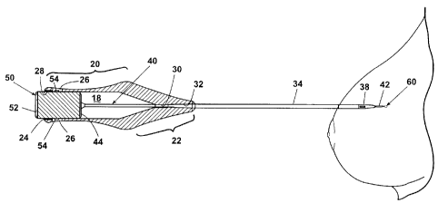
FIGS. 1-4 illustrate a marking apparatus 10 according to the invention, which
is
capable of the percutaneous placement of a drug-eluting tissue marker 60 at a
site within in
a tissue mass according to the invention. Exemplary tissue masses include
breast tissue,
skin tissue, muscle tissue, the tissue of glands, such as the prostate, and
the tissue of
organs such as the lungs, kidney, and liver. For purposes of this application,
these types of
tissue masses are referred to as soft tissue, which expressly excludes
vascular structures.
In the following example, the marking apparatus 10 can be used to place a
marker
60 at the location of a tissue biopsy or a lesion. The marking apparatus 10
comprises an
introducer 12 and a drug-eluting marker 60 (FIG. 2) contained within the
introducer 12.
The drug-eluting marker 12 marks a site in a tissue mass. It preferably does
not perform a
prosthetic function, such as structurally or functionally replacing a part of
the tissue mass.
The introducer 12 includes a handle 16 having a hollow interior 18. The handle
16
comprises a grip portion 20 from which extends a tapered nose portion 22. The
grip
portion 20 defines a rear opening 24 that provides access to the hollow
interior 18. A pair
of detents 26 are formed in the grip portion 20 near the rear opening 24.
Channels 28 are
formed on the interior surface of the grip portion 20 and extend from the rear
opening 24
to the detents 26.
The nose portion 22 comprises a guide passage 30 extending from the tip of the
nose portion 22 to the hollow interior 18 of the handle 16. The guide passage
30 decreases
in diameter inwardly from the tip of the nose portion to form a cannula seat
32.
Alternatively, the diameter of the guide passage 30 may be substantially equal
to or
slightly smaller than the outer diameter of a cannula 34, which in any case is
press-fit
-4-
CA 02562580 2013-07-15
within the cannula seat 32. As is customary, the cannula is formed with a
hollow
interior 36 and a sharpened tip 38.
A stylet 40 comprising a shaft 42 and a base 44 is received within the
hollow interior 18 of the handle 16 in a manner such that the shaft 42 extends
through the guide passage 30 and into the cannula interior 36 and the stylet
base lies within the hollow interior 18.
A plunger 50 comprises a cylindrical body 52 from which extends a pair of
catches 54 at diametrically opposed positions. The cylindrical body 52 is
preferably
sized so that it is slidably received within the rear opening 24 of the handle
16
where it is so oriented with respect to the handle that the catches 54 are
aligned
with the guide channels 28.
It should be noted that the marking apparatus 10 is just one example of an
apparatus for implanting the marker 60. Many other delivery systems and
devices
can also be used to implant the drug-eluting marker 60. For example the marker
60 can be implanted in the tissue through a flexible sheath introduced through
a
vacuum assisted biopsy (VAB) device.
In operation, the introducer 12 begins in the ready condition shown in FIG.
4. In this condition, the stylet shaft is received within the cannula but does
not
extend to the cannula tip 38 thereby forming a marker recess 46 within the
cannula 34, the drug-eluting marker 60 is disposed within the marker recess
46,
and the plunger 50 is in a position relative to the handle 20 in which the
catches
54 are outside the handle 16; that is, the catches 54 are not received within
the
detents 26. However, the plunger 50 is so oriented with respect to the handle
16
that the catches 54 are aligned with the guide channels 28.
With the introducer in the ready condition, the cannula is positioned so that
its tip is at or near the location of a tissue mass where a biopsy has been
taken.
Preferably, the cannula tip is positioned by using an imaging system. The
cannula
tip 38 can be designed for enhanced visibility using common imaging systems,
- 5 -
CA 02562580 2013-07-15
such as CAT scan, ultrasonography and mammography. Suitable cannula tips are
disclosed in U.S. Patent No. 5,490,521, issued February 13, 1996 to R. E.
Davis
and G. L. McLellan. Ultrasound enhancement technology is also disclosed in
U.S.
Patent No. 4,401,124, issued August 30, 1983 to J. F. Guess, D. R. Dietz, and
C.
F. Hollinger; and U.S. Patent No. 4,582,061, issued April 15, 1986 to F. J.
Fry.
- 5a -
CA 02562580 2006-10-05
Once the cannula is positioned at the desired location, the plunger 50 is
moved
from its first or ready condition as illustrated in FIGS. 1 to 4 to a second
or discharged
condition as illustrated in FIG. 5 in which the catches 54 are received within
the detents 26
to lock the plunger 50 in the discharged condition and the stylet shaft
extends beyond the
cannula tip 38. The catches 50 and detents combine to function as a latch for
locking the
plunger in the discharged condition. As the plunger 50 is moved from the ready
condition
to the discharged condition, the plunger 50 drives the stylet base 44 forward
to advance the
stylet shaft 42 within the cannula interior 36. As the stylet shaft 42 is
advanced, the drug-
eluting marker 60 is ejected from the marker recess 46 through the cannula tip
38 and into
the tissue at the biopsy location.
The cannula 34 is illustrated with an opening formed in the cannula tip 38
communicating with the hollow interior 36; however the cannula 34 can
optionally have
an opening in a side wall of the cannula 34 that communicates with the hollow
interior 36
and a closed tip 38, such that the drug-eluting marker 60 is ejected from the
marker recess
46 through the side wall opening.
The drug-eluting tissue marker 60 is preferably readily imaged using
contemporary
imaging techniques. For example, the marker 60 can be made of material
suitable to be
imaged using X-ray, mammography, ultrasound, fluoroscopy, computed tomography,
magnetic resonance imaging (MRI), computerized axial tomography (CAT) scan,
Doppler,
radiation detector, and any possible combination thereof. Optionally, the
marker can be
detectable by palpation of the overlying tissue or visualization of the marker
using a
colored material that is different that the surrounding tissue and blood.
Regardless of the
means, the marker 60 should be readily locatable within the tissue mass.
Additionally, the
marker 60 should not migrate within the tissue from the position in which it
is initially
placed. The marker 60 should be precisely and accurately locatable at the
site, in case an
additional medical procedure is needed. Returning to the biopsy example, if a
tissue
sample taken from a biopsy site is determined to be malignant or the pathology
is
inconclusive, an additional medical procedure at the site is necessary, in
which case it is
important to be able to locate the biopsy site.
The size and shape of the marker 60 can vary according to the application in
which
it is used. For example, lesions can have an effective size ranging from 2mm
to several
-6-
CA 02562580 2006-10-05
centimeters, and thus the size and shape of the marker 60 can be configured
according to
the lesion size. As used herein, the term lesion has its normal meaning and
expressly
includes one or more micro-calcifications. The size of marker 60 has a
practical upper
limit that is related to the following factors. At some point the lesion will
be large enough
that it is palpable even after it is biopsied. For immediate relocating, such
a lesion will not
require the use of a marker as a surgeon or other practitioner will be able to
find the lesion
by palpitation. However, it still may be desirable to place a marker in
anticipation of the
lesion being reduced by future treatment, such as radiation. The marker should
be large
enough for easy location using the imaging system of choice, but should not be
so big that
it will obscure or interfere with imaging the lesion site. For these larger
but not easily
palpable lesions, a larger marker can be used without interfering with the
imaging of the
lesion site. For current imaging systems the useful upper size limit is about
10 mm for the
maximum dimension, whereas the lower size limit is about 1 mm for the minimum
dimension. By way of example, and without limitation, the marker 60 can have a
length of
about 3mm and a width of about 1.5 mm.
FIGS. 6-9 illustrate four possible embodiments of the drug-eluting tissue
marker
60; however, it is understood that the configuration of the marker 60 is not
limiting to the
invention and many other configurations of the marker 60 are possible without
departing
from the scope of the invention. For example, the marker 60 can have a hollow
interior for
enhanced imaging characteristics. Examples of other shapes are shown in U.S.
Patents
6,575,991; 6,371,904; 6,261,243; 6,228,055; and 6,056,700.
A first embodiment of the marker 60 is shown in FIG. 6 having a bight portion
66
from which extend legs 67, which terminate in tips 68. The tips 68 extend in
opposite
directions to function as anchors for the marker 60 and help prevent the
marker 60 from
migrating from the site. The marker 60 further comprises a substrate 62
(partially shown)
of underlying material that is coated with a drug 64. As used herein, the term
"drug" can
include any therapeutic agent, pharmacologic agent, or other substance used
for the
diagnosis, treatment, or prevention of a disease or as a component of a
medication. A
coating of the drug 64 can be deposited on the substrate surface using any
suitable
deposition technique. Alternately, the drug 64 can be embedded in a suitable
material,
such as a polymer, for timed release of the drug 64. At least a portion of the
marker 60 is
-7-
CA 02562580 2006-10-05
locatable from the exterior of the patient, through palpitation or through use
of an imaging
system.
The substrate 62 can be any number of materials that are suitably
biocompatible
and appropriate for the type of drug deposited on the substrate surface. The
underlying
material can be imageable to perform the marking function. Examples of
materials for the
substrate 62 include: stainless steel, tantalum, titanium, nitinol, gold,
platinum, inconel,
iridium, rhodium, silver, tungsten, or another biocompatible metal, or alloys
of any of
these; carbon or carbon fiber; cellulose acetate, cellulose nitrate, silicone,
polyethylene
teraphthalate, polyurethane, polyamide, polyester, polyorthoester,
polyanhydride, polyether
sulfone, polycarbonate, polypropylene, high molecular weight polyethylene,
polytetrafluoroethylene, or another biocompatible polymeric material, or
mixtures or
copolymers thereof; polylactic acid, polyglycolic acid or copolymers thereof,
a
polyanhydride, polycaprolactone, polyhydroxy-butyrate valerate or another
biodegradable
polymer, or mixtures or copolymers of these; a protein, an extracellular
matrix component,
collagen, fibrin or another biologic agent; or a mixture thereof.
Some of these materials cannot be imaged using one of the aforementioned
imaging techniques. In this case, an imageable material can be added to the
drug coating
64 to enhance the radiopaque, mammographic, echogenic, etc. characteristics of
the
marker, allowing the marker to be observed with a corresponding imaging
technique. For
example, a contrast material such as iodine can be added to the coating to
make the marker
imageable during a CAT scan. Alternately, a separate coating can be deposited
on the
marker 60 in addition to the drug coating 64 to make the marker imageable.
Examples of the drug 64 include: anti-inflammatory, antiplatelet,
anticoagulant,
antifebrin, antithrombin, cytostatic, antiproliferative, antibiotic,
antimicrobial, antioxidant,
and antiallergic substances; antitumor and/or chemotherapeutic agents,
including
antineoplastic and antimitotic agents; thrombolytics; fibrinolytics,
vasodilators; antiviral,
antihypertensive, antisecretory, and immunosuppressive agents; growth factors
and growth
factor antagonists; antipolymerases; photodynamic therapy and antibody
targeted therapy
agents; prodrugs; sex hormones; free radical scavengers; radiotherapeutic,
radiopaque, and
radiolabelled agents; and peptides, proteins, and enzymes. Further, any
combination of
-8-
CA 02562580 2006-10-05
drugs, therapeutic agents, or other substances can be used to coat the marker
of the present
invention.
Examples of such anti-inflammatory substances include estradiol, aspirin,
ibuprofen, and naproxen.
Examples of such antiplatelets, anticoagulants, antifibrin, and antithrombins
include sodium heparin, low molecular weight heparins, heparinoids, hirudin,
argatroban,
forskolin, vapiprost, prostacyclin, prostacyclin analogues, dextran, D-phe-pro-
arg-
chloromethylketone (synthetic antithrombin), Dipyridamole, glycoprotein
Hb/IIIa platelet
membrane receptor antagonist antibody, recombinant hirudin, thrombin
inhibitors such as
Angiomax.TM. (Biogen, Inc., Cambridge, Mass.), glycoprotein IIbIIIa
inhibitors,
ticlopidine, clopidigrel, warfarin (e.g. Coumadin0 from Bristol-Myers Squibb
Co.,
Stamford, Conn.), aspirin, and hirulog.
Examples of such cytostatic or antiproliferative agents include, actinomycin D
as
well as derivatives and analogs thereof (manufactured by Sigma-Aldrich,
Milwaukee,
Wis.; or COSMEGEN0 available from Merck & Co., Inc., Whitehouse Station,
N.J.),
angiopeptin, mitomycin, angiotensin converting enzyme inhibitors such as
captopril (e.g.,
Capoten0 and Capozide from Bristol-Myers Squibb Co., Stamford, Conn.),
cilazapril or
lisinopril (e.g., Prinivil and Prinzide0 from Merck & Co., Inc., Whitehouse
Station,
N.J.), calcium channel blockers (such as nifedipine), colchicines, fibroblast
growth factor
(FGF) antagonists, fish oil (omega 3-fatty acid), histamine antagonists,
lovastatin (an
inhibitor of HMB-CoA reductase, a cholesterol lowering drug, brand name
Mevacor
from Merck & Co., Inc., Whitehouse Station, N.J.), monoclonal antibodies (such
as those
specific for Platelet-Derived Growth Factor (PDGF) receptors), nitroprusside,
phosphodiesterase inhibitors, prostaglandin inhibitors, suramin, serotonin
blockers,
steroids, thioprotease inhibitors, triazolopyrimidine (a PDGF antagonist), and
nitric oxide.
Examples of such antiallergic substances include permirolast potassium, and
tranilast.
Examples of such antitumor and/or chemotherapeutic agents include 2-
chlorodeoxyadenosine (also known as cladribine), 6-thioguanine (also known as
2-amino-
6-mercaptopurine), 13-cis-retinoic acid (also known as isoretinoin),
aldesleukin (also
known as interleukin-2), alemtuzumab, alitretinoin, all-trans retinoic acid
(also known as
-9-
CA 02562580 2006-10-05
tretinoin), alpha interferon, altretamine (also known as hexamethylmelamine),
amifostine,
aminoglutethimide, anagrelide, anastrozole, arsenic trioxide, asparaginase,
azacitidine,
azathioprine, BCG (Bacillus Calmette-Guerin), bevacizumab, bexarotene,
bicalutamide,
bleomycin, bortezomib, busulfan, capecitabine, carboplatin, carmustine,
cetuximab,
chlorambucil, cisplatin, cyclophosphamide, cytarabine (also known as
arabinosylcytosine),
cytarabine liposomal, dacarbazine, dactinomycin, daunorubicin liposomal,
darbepoetin
alfa, daunorubicin, decitabine, denileukin diftitox, dexamethasone (also known
as
dexamethasone sodium phosphate and dexamethasone acetate), dexrazoxane,
docetaxel
(e.g., Taxotere from Aventis S.A., Frankfurt, Germany), doxorubicin,
doxorubicin
hydrochloride (e.g., Adriamycin from Pharmacia & Upjohn, Peapack, N.J.),
doxorubicin
liposomal, epirubicin, epoetin alfa, estramustine, erlotinib, etoposide,
exemestane,
filgrastim, fluorouracil (also known as 5-fluorouracil), floxuridine,
fludarabine,
fluoxymesterone, flutamide, fulvestrant, gefitinib, gemcitabine, gemtuzumab,
goserelin,
ozogamicin, hydrocortisone (also known as cortisone), hydroxyurea, ibritumomab
(also
known as ibritumomab tiuxetan), idarubicin, ifosfamide, imatinib mesylate,
irinotecan,
lenalidomide, letrozole, leucovorin, leuprolide, lomustine, mechlorethamine
(also known
as nitrogen mustard, mustine, and mechlorethamine hydrochloride), megestrol
(also known
as megestrol acetate), melphalan, mercaptopurine (also known as 6-
mercaptopurine),
methotrexate (also known as amethopterin and methotrexate sodium),
methylprednisolone,
mesna, mitomycin (e.g., Mutamycin0 from Bristol-Myers Squibb Co., Stamford,
Conn.),
mitoxantrone, nelarabine, nilutamide, octreotide (also known as octreotide
acetate),
oprevelkin, oxaliplatin, paclitaxel (e.g., TAXOL by Bristol-Myers Squibb Co.,
Stamford,
Conn.), paclitaxel protein-bound, pamidronate, peg interferon, pegaspargase,
pegfilgrastim, pemetrexed, pentostatin, prednisolone, prednisone,
procarbazine, raloxifene,
rituximab, sargramostim, sorafenib, streptozocin, sunitinib, tamoxifen,
temozolomide,
teniposide, thalidomide, thiotepa (also known as thiophosphoamide), topotecan,
toremifene, tositumomab (also known as iodine I ¨ 131), trastuzumab,
vinblastine,
vincristine, vinorelbine, and zoledronic acid.
Examples of such thrombolytics and/or fibrinolytics include tissue plasminogen
activator (tPA), recombinant tPA, urokinase, streptokinase, tenecteplase,
alteplase (e.g.
Activase from Genentech, Inc., San Francisco, CA), lysatec, antistreplase
(e.g.
-10-
CA 02562580 2006-10-05
Eminaseg from Wulfing Pharma Gmbh, Germany), reteplase (e.g. Retavaseg from
Centocor, Inc., Malvern, PA), hannahpep (Indian King Cobra venom), ancrod
(Malayan pit
viper venom), and matrix metalloproteinases, such as collagenase.
A second embodiment of the drug-eluting marker is shown in FIG. 7 in which
like
elements are designated with the same number bearing a prime (') symbol. The
marker 60'
can be comprised of a material 70 containing any of the drugs listed above. In
this
embodiment, the marker is made of a bioabsorbable material that will dissolve
within the
body at a predetermined rate, thus releasing the drug in a timed release
fashion. Examples
of such bioabsorbable materials are collagen, regenerated cellulose, synthetic
polymers,
and synthetic proteins.
A third embodiment of the drug-eluting marker is shown in FIG. 8 in which like
elements are designated with the same number bearing a double-prime (")
symbol. The
marker 60" encapsulates, either fully or partially, the drug 64". For
illustrative purposes,
the marker 60" is shown as a hollow tube 72, made of any of the materials
listed above,
that encapsulates the drug 64". Multiple pores 76 extend fully through the
tube 72 to allow
the drug 64" to pass through the exterior of the marker 60". The drug 64" can
also pass
through the open ends of the tube 72. The tube 72 can alternately be solid
with pores 76,
extending only partially through the tube, designed to hold the drug 64".
In an alternate design of the third embodiment shown in FIG. 9, where like
elements are designated with the same number bearing a triple-prime ("1)
symbol, the
marker 60" can embody a spring-like configuration 82 that fully or partially
encapsulates
the drug 64". The marker 60" can further be made of a bioabsorbable material,
as
discussed above, that either fully or partially encapsulated the drug 64".
As in the foregoing example, the marking apparatus 10 can be used to place a
marker at the location of a tissue biopsy. However, the drug-eluting marker 60
can be
used even if a biopsy has not been done. The drug-eluting marker 60 can be
used as part
of a treatment regimen whereby the drug 64 is delivered at a specific site
within the body
where the marker 60 is placed. For example, the marker 60 can be placed at a
lesion
detected using an imagining technique to mark the site prior to chemotherapy.
The
imageability of the marker 60 allows the lesion to be tracked during
chemotherapy since
-11-
CA 02562580 2006-10-05
the lesion often disappears during treatment. The drug-eluting capability of
the marker 60
allows a suitable drug 64 to be delivered to the lesion as part of the
treatment.
While the invention has been specifically described in connection with certain
specific embodiments thereof, it is to be understood that this is by way of
illustration and
not of limitation, and the scope of the appended claims should be construed as
broadly as
the prior art will permit.
-12-