Note: Descriptions are shown in the official language in which they were submitted.
CA 02563024 2006-09-29
WO 2005/094880
PCT/CA2005/000472
- 1 -
METHOD FOR TREATING AUTOIMMUNE
DISEASES WITH ANTIBODIES
=
TECHNICAL FIELD
This application relates to the treatment of autoimmune
diseases using antibodies.
More preferably, the present
invention relates to treatment of autoimmune diseases with
soluble antigen-specific antibodies.
BACKGROUND OF THE INVENTION
Immune thrombocytopenic purpura (ITP) is an autoimmune
disease characterised by platelet clearance mediated by
pathogenic anti-platelet antibodies. It has been previously
suggested that this platelet clearance is mediated by Fcy
receptor (FcyR)-bearing macrophages in the reticuloendothelial
system (RES). While intravenous immunoglobulin (IVIg) is
widely used in the treatment of ITP as well as in a wide
variety of chronic autoimmune and inflammatory diseases, its
mechanism of action is not yet fully elucidated. Possible
mechanisms of action include inhibition of RES function, anti-
idiotype antibodies and immunomodulation..In murine models of
ITP, it has been demonstrated that IVIg ameliorates ITP by a
mechanism completely dependent upon the expression of the
inhibitory FcyRIIB. In humans, there is also evidence that
IVIg increases the level of expression of FcyRIIB. In
addition, it has been previously reported that the clinical
effects of IVIg as well as monoclonal mimetics of IVIg both
ameliorate murine ITP in a manner that correlates with RES
blockade.
This 'competitive' RES blockade has long been
considered to be the primary mechanism whereby IVIg
ameliorates ITP.
CA 02563024 2006-09-29 PracA .2005/
000 47
IjkAla L2tioz)
n6
= - 2 -
The present study was undertaken to investigate if
antibodies to soluble antigens could inhibit autoimmune
diseases.
SUMMARY OF THE INVENTION
According to the present invention, a novel method for
treating an autoimmune disease is provided.
Furthermore, a
novel mechanism of action has been established in accordance
with the present invention for antibody-based treatment
regimes for autoimmune disease, including, but not limited to
anti-CD44 and soluble antigen specific antibody treatment
regimes.
In one embodiment of the invention there is provided a
method for treating autoimmune diseases in a mammal which
method comprises administering to the mammal an effective
amount of at least one antibody specific for a soluble
antigen.
Different types of autoimmune diseases can be treated by
the method of the present invention. According to the present
invention, an autoimmune disease includes, but is not limited
to Immune thrombocytopenia, Immune cytopenia, Idiopathic
thrombocytopenic purpura (ITP), Neuropathy, Chronic
inflammatory demyelinating polyneuropathy (CIDP), Guillain-
Barre syndrome (GBS), Kawasaki's disease, Dermatomyositis,
SLE, Myasthenia gravis, Post-transfusion purpura, Rheumatoid
arthritis, Inflammatory arthritis, Eaton-Lambert
syndrome, toxic epidermal necrolysis, and polymyositis.
In one embodiment, the treatment can be effected for a
time and under conditions sufficient to inhibit platelet
clearance, thereby treating or ameliorating an autoimmune
disease such as immune thrombocytopenic purpura (ITP), for
_ _ _ _
_
example. In a further embodiment, inflammatory arthritis can
CA 02563024 2006-09-29
WO 2005/094880
PCT/CA2005/000472
- 3 -
be prevented or ameliorated by the administration of
antibodies to a soluble antigen in accordance with the present
invention.
The soluble antigen can either be an endogenous or a .
foreign antigen. By foreign antigen it is meant an antigen
that is not normally produced by the same individual or
species.
The antigen can be a non-functional/inert antigen.
In an other embodiment the binding of the antibody to the
antigen does not compromise the function of the antigen.
In an aspect of the invention the soluble foreign
antigen and the antibody can be incubated together to form
antibody-antigen complexes prior to administering the
complexes to the mammal.
In another aspect of the invention, the endogenous
soluble antigen can be obtained from the mammal and incubated
with the antibody to form antibody-antigen complexes, the
complexes being subsequently administered to the mammal.
Alterntively, a soluble antigen may be injected into a mammal
having a pre-existing antibody of interest specific to the
soluble antigen, e.g. a mammal who has been previously
immunised to tetanus toxin (any # of years earlier) may be
administered an injection of soluble tetanus toxin according
=
to an alternate embodiment of the present invention.
The antibody can be administered intravenously,
interperitoneally, intradermally,
intramuscularly,
subcutaneously, orally or rectally.
In another embodiment of the invention, the soluble
antigen can be associated with blood cells and the resulting
antigen-cell complexes can be targeted by antibodies for
inhibiting platelet clearance and thereby treating
thrombocytopenia.
CA 02563024 2013-09-09
- 4 -
In another embodiment, an autoimmune disease treatment
regime is provided to mediate a cellular response in dendritic
cells, such as leukocytes, such that platelet clearance is
slowed and/or inhibited, thereby treating or ameliorating an
autoimmune disease.
In another aspect of the invention there is provided
pharmaceutical compositions for treating autoimmune diseases
such as arthritis and thrombocytopenia, comprising an
effective amount of at least one antibody specific for a
soluble antigen and/or for a soluble antigen associated with a
blood cell.
In yet another aspect of the invention, an antibody to
a soluble antigen may be used in the manufacture of a
medicament for the treatment of an autoimmune disease.
In yet another aspect of the invention, we demonstrate
herein that antibodies to soluble antigens can ameliorate ITP
in an FcyRIIB-dependent manner. Antibody directed to the cell-
associated antigen inhibited ITP in an FcyRIIB-independent
manner. Taken together, these data demonstrate that IgG
antibodies reactive with either a soluble or insoluble antigen
can mimic the effects of IVIg. In addition, the mechanisms of
action of these moieties are quite different: antibody reacted
with soluble antigen may utilize the same pathway used by
IVIg, i.e. an FcyRIIB-dependent pathway, whereas antibody
reacted with a cell-associated antigen may work by additional
and/or other mechanisms of action, and possibly by competitive
RES inhibition.
According to one aspect of the present invention, there
is provided use of at least one of an IgG antibody and a
soluble foreign protein substantially soluble in vivo
CA 02563024 2014-07-17
- 4a -
complementary to the at least one IgG antibody for the
treatment of immune thrombocytopenia in a mammal, without
invoking the biological function of the soluble foreign
protein, wherein the IgG antibody and the soluble foreign
protein form an antibody-antigen complex in the mammal and
wherein the IgG antibody and the soluble foreign protein
individually fail to treat immune thrombocytopenia.
According to another aspect of the present invention, there is
provided use of at least one of an IgG antibody and a soluble
foreign protein substantially soluble in vivo complementary to
the IgG antibody for the inhibition of platelet clearance in a
mammal, without invoking the biological function of the
soluble foreign protein, wherein the IgG antibody and the
soluble foreign protein form an antibody-antigen complex in
the mammal and wherein the IgG antibody and the soluble
foreign protein individually fail to inhibit platelet
clearance in the mammal.
According to still another aspect of the present invention,
there is provided a pharmaceutical composition for the
treatment of immune thrombocytopenia in a mammal, without
invoking the biological function of a soluble foreign protein,
the pharmaceutical composition comprising (i) at least one of
an IgG antibody and the soluble foreign protein substantially
soluble in vivo complementary to the IgG antibody and (ii) a
pharmaceutically acceptable carrier, wherein the IgG antibody
and the soluble foreign protein individually fail to treat
immune thrombocytopenia.
According to yet another aspect of the present invention,
there is provided use of at least one of an IgG antibody and a
complementary soluble protein for the treatment of immune
thrombocytopenia in a mammal, wherein:
CA 02563024 2014-07-17
- 4b -
- the IgG antibody and the complementary soluble protein
form an antibody-protein complex in the mammal;
- the IgG antibody and the complementary soluble protein
individually fail to treat immune thrombocytopenia; and
- the complementary soluble protein is selected from the
group consisting of ovalbumin, albumin and transferrin.
According to a further aspect of the present invention, there
is provided use of at least one of an IgG antibody and a
complementary soluble protein for inhibiting platelet
clearance in a mammal, wherein:
- the IgG antibody and the complementary soluble protein
form an antibody-protein complex in the mammal;
- the IgG antibody and the complementary soluble protein
individually fail to inhibit platelet clearance; and
- the complementary soluble protein is selected from the
group consisting of ovalbumin, albumin and transferrin.
According to yet a further aspect of the present invention,
there is provided a pharmaceutical composition for the
treatment of immune thrombocytopenia for the inhibition of
platelet clearance in a mammal, the pharmaceutical composition
comprising (i) at least one of an IgG antibody and a
complementary soluble protein and (ii) a pharmaceutically
acceptable carrier, wherein:
- the IgG antibody and the complementary soluble protein
form an antibody-protein complex in the mammal;
- the IgG antibody and the complementary soluble protein
individually fail to treat immune thrombocytopenia or
inhibit platelet clearance; and
CA 02563024 2014-07-17
- 4c -
- the complementary soluble protein is selected from the
group consisting of ovalbumin, albumin and transferrin.
BRIEF DESCRIPTION OF THE DRAWINGS
Further features and advantages of the present invention will
become apparent from the following detailed
CA 02563024 2006-09-29
WO 2005/094880
PCT/CA2005/000472
- 5 -
description, taken in combination with the appended drawings,
in which:
Figs. lA and 1B illustrate the association of OVA on
the surface of RBCs.
Figs. 2A and 2B illustrates inhibition of
thrombocytopenia by treating OVA-coupled RBCs with OVA-
specific IgG.
Figs. 3A, 3B and 3C illustrates amelioration of
thrombocytopenia with antibodies reactive with soluble OVA (in
combination with soluble OVA) ameliorate immune
thrombocytopenia.
Figs. 4A and 4B illustrates inhibition of RES function
by antibodies reactive with soluble OVA (Fig. 4A) or OVA-RBCs
(Fig. 4B).
Fig. 5 illustrates that antibodies reactive with
soluble OVA or OVA-RBCs both ameliorate immune
thrombocytopenia independent of complement activity.
Figs. 6A and 6B illustrate that FcyRIIB expression is
required for reversal of immune thrombocytopenia by soluble
OVA in the presence of anti-OVA.
Figs. 7A and 7B illustrate that FcyRIIB expression is
not required for reversal of immune thrombocytopenia by cell-
associated OVA in the presence of anti-OVA.
Figs. 8A and 83 illustrate that antibodies to
endogenous soluble antigens ameliorate
immune
thrombocytopenia.
CA 02563024 2006-09-29
WO 2005/094880
PCT/CA2005/000472
- 6 -
Fig. 9 illustrates that antibodies to albumin and
=
transferrin require the expression of Fc7RIIB to ameliorate
immune thrombocytopenia.
Fig. 10 A and 10B illustrate that antibodies to albumin
ameliorate K/BxN serum-induced inflammatory arthritis.
Fig. 11 illustrates IMCP-like effects shown by IVIg and
anti-CD44 treatment regimes.
=
Fig. 12 illustrates IMCP-like effects as shown by IVIg
and soluble antigen-specific antibody treatment regimes.
Fig. 13 illustrates IVIg-treated leukocytes showing
therapeutic potential in the absence of FCgammaRIIB
expression.
DETAILED DESCRIPTION OF THE PREFERRED EMBODIMENT
In this description by soluble antigen it is meant a
molecule that can be incorporated and circulated in the blood
stream. Examples of soluble antigens comprise but are not
limited to: proteins, glycoproteins, lipids, glycolipids,
peptides, nucleic acids, synthetic molecules or complexes or
aggregates thereof.
By endogenous antigen it is meant antigens that occur
naturally in a mammal and by foreign (or exogenous) antigen it
is meant an antigen that is not normally produced by the same
individual or species.
According to one embodiment of the present invention,
antibodies to soluble antigens were tested for their ability
to ameliorate autoimmune diseases. In one example, the
amelioration of thrombocytopenia was tested. To address this
question, a murine model of ITP was used to determine whether
IgG specific to a soluble prototype antigen could prevent
07/07/2006 12:32 FAX 613 230 6706 .ociLyy RENAULT r VI
ytteff
arr 4rleVOte"tr 4
CA 02563024 2006-09-29
0 7
JULY 2006 0 7 = 7 .06
-7-.
thrombocytopenia. Mice injected With soluble ovalbumin (OVA)
or OVA conjugated to RBCs (OVA-RBC) in the presence of anti-
OVA, were both significantly protected from immune
thrombocytopenia.
Both of these therapeutic regimes functioned
independent of complement activity and both regimes also
blocked reticuloendothelial function as assessed by clearance
rates of fluorescent sensitized syngeneic RBCs. Soluble OVA or .
anti-OVA alone did not have any direct effect on immune
thrombocytopenia in mice. It was found that OVA-RBC + anti-OVA
ameliorated immune thrombocytopenia in normal mice and .
FcyRIIB-/- mice, while soluble OVA + anti-OVA was ineffective
in FcyRIIH-/- mice. In addition, IgG specific for murine
albumin and specific for transferrin also effectively
inhibited IT?. Thus, ' IgG antibodies directed to soluble
antigens can inhibit or reverse immune thrombocytopenia in an
FcyRIIB-dependent manner, whereas antibodies directed to a
cell-associated antigen function independent of FcyRIIB
expression.
Materials and Mathods:
Reagents:
The monoclonal antibody specific for integrin aim (rat
IgG, clone MWReg 30) was purchased from BD Pharmigen
(Mississauga, ON, Canada). Monoclonal murine anti-OVA (IgGif
clone OVA-14), rabbit polyclonal anti-OVA, 1-ethy1-3-(3-
dimethylamino-propyl) carbodiimide (EDAC), OVA (grade V), and
PKH26 red fluorescent cell linker kit were purchased from
Sigma (Oakville, ON, Canada). IVIG was Gamimune,10% from Bayer
(Elkhart, IN). Cobra Venom Factor (CVF), FITC-conjugated
F(ab')2 anti-rabbit IgG, and control rabbit IgG, were purchased
from Cedarlane Laboratories Ltd (Hornby, ON, Canada). Rabbit
anti-mouse 'albumin (IgG fraction), and rabbit anti-mouse
transferrin (IgG fraction), were purchased from Research
_
AMENDED SHEET
CA 02563024 2006-09-29
WO 2005/094880
PCT/CA2005/000472
- 8 -
Diagnostics (Flanders, NJ). Hemolysin (anti-SRBC rabbit serum)
was supplied by Colorado Serum company (Denver, CO).
Microdispenser tubes (250 1) for blood collection were from
VWR, (Mississauga, ON)
Mice:
Female CD1 mice (6-10 wks of age) were purchased from
Charles River Laboratories (Montreal, PQ, Canada). C57BL/6 and
FcyRIIB-/- (B6;129S4-Fcgr2bbniRaVJ) mice were purchased from the
Jackson Laboratory (Bar Harbor, ME). All mice were housed in
the St. Michael's Hospital Research Vivarium.
Induction and treatment of immune thrombocytopenia:
Mice were rendered thrombocytopenic by intraperitoneal
injection of 2 pg anti-platelet (anti-integrin amp) antibody
in 200 pl phosphate buffered saline (PBS), pH 7.2. ITP was
induced by two protocols:
For experiments where the therapeutic intervention
preceded the induction of immune thrombocytopenia (e.g. Figs
2, 3, 5), mice were first injected intravenously with the
indicated therapeutic preparation (eg OVA-RBC sensitized with
anti-OVA IgG), followed at 24 h by a single injection of anti-
platelet antibody. Mice were bled for platelet enumeration
after a further 24 h.
For experiments where the induction of immune
thrombocytopenia preceded the therapeutic intervention (e.g.
Figs 6-8), mice were injected daily (days 0-3) with anti-
platelet antibody and then injected intravenously with the
indicated therapeutic preparation (eg OVA-RBC sensitized with
anti-OVA IgG) on day 2. Mice were bled daily and platelets
counted as described below.
CA 02563024 2006-09-29
WO 2005/094880
PCT/CA2005/000472
- 9 -
In experiments where we wished to avoid the possibility
of the formation of "pre-formed" immune complexes, mice were
injected intraperitoneally with soluble OVA only followed 4
hours later by OVA-specific antibody via the intravenous
route. Mice injected with anti-albumin or anti-transferrin
alone received 1 mg of antibody in a volume of 200 ul on day
2. For all IVIg treatments, mice were injected
intraperitoneally with 0.5 ml of 10 % IVIG (roughly equivalent
to 2 g/kg body weight). Platelets were counted as follows:
Mouse blood (45 pL) was collected via saphenous vein bleeding
into microdispenser tubes preloaded with 5 pl of 1% EDTA in
PBS. Then, 50 pl of this mouse blood was diluted in 450 pl of
1% EDTA/PBS (1:10) and then further diluted to a final
dilution of 1:12,000 in 1% ethylenediaminetetraacetic acid
(EDTA)/PBS. Platelets were enumerated on a flow rate-
calibrated FACScan flow cytometer (Becton Dickinson, San Jose,
CA) using forward scatter (FCS) versus side scatter (SSC) to
gate platelets as previously described.
Preparation of OVA-specific antibody pre-incubated with
soluble OVA:
1 mg OVA was dissolved in 300 pl PBS and was incubated
with the indicated dose (Fig. 3A, 3B x-axes) of OVA-specific
antibody (rabbit polyclonal or mouse monoclonal) for 1 hr at
37 C. The solution was then injected intravenously in a 300p1
volume. In separate experiments the OVA and antibody solution
was incubated as above for 1 hour at 37 C and macromolecular
immune complexes removed by centrifugation at 16,000xg at 4 C
for 1 h followed by filtration of the resulting supernatant
fluid using a 0.2 pm filter (Filtropur S plus 0.2, Sarstedt,
Montreal, PQ). The pellet was resuspended in 300 pl PBS and
intravenously injected as above.
Preparation of OVA-coupled RBCs:
CA 02563024 2006-0-29
WO 2005/094880
PCT/CA2005/000472
- 10 -
OVA was coupled to RBCs as follows: RBCs were
resuspended at 2.5x10⁸/mL in 5 mg/mL OVA in saline and 1.9
mg/mL 1-ethyl-3-(3-dimethylamino-propyl) carbodiimide (EDAC)
was added. Following a 1 hr incubation at 4 C, the cells were
washed once with a 2 mg/mL solution of OVA in PBS followed by
one wash in PBS. To confirm the presence of OVA on RBCs, OVA
coupled RBCs were incubated with 17 pg/mL rabbit polyclonal
anti-OVA, washed, and then incubated with 8 pg/mL FITC
conjugated F(ab')2 anti-rabbit IgG. Cells were washed,
resuspended in PBS, and analyzed by flow cytometry.
Reticuloendothelial system (RES) blockade:
RES blockade was assessed as follows: Whole blood (2
ml, diluted with 1/5 volume 1% EDTA in PBS) from unmanipulated
SCID mice was pooled and centrifuged at 2,000 x g for 15 min
to obtain 1 ml of packed erythrocytes. These packed
erythrocytes were resuspended in 4 ml of PBS and incubated
with 8 pg of anti-TER-119 antibody at 22 C for 30 min. The
resulting opsonized erythrocytes were then washed twice with
PBS and labeled with a fluorescent marker (PKH26 Kit, Sigma,
St. Louis MO) according to the manufacturer's directions.
Briefly, the opsonized erythrocytes were resuspended in 3 ml
of PKH26 'diluent C' and mixed with another 4 ml of 'diluent -
C' containing 10 pl of the 'PKH26 linker'. After a 5 minute
incubation with constant swirling, the mixture was incubated
for 5 minutes with an equal volume of PBS containing 1% bovine
serum albumin. The erythrocytes were washed 5 times and
resuspended in 2 ml PBS. Mice were then injected via the tail
vein with 200 pl of these labeled cells. All mice were bled
via the tail vein at 3 min, 10 min, 30 min, 120 min, and 960
min post injection and the the total number of erythrocytes,
as well as the percent of PKH26-fluorescent erythrocytes, were
counted by flow cytometry. The percentage of labeled
CA 02563024 2006-09-29
WO 2005/094880
PCT/CA2005/000472
- 11 -
erythrocytes at the 3 min time point was considered to be
100%.
Complement depletion:
Complement depleted mice were prepared by
intraperitoneal injection of 5U of Cobra Venom Factor (CVF) in
200 pl phosphate-buffered saline pH 7.2 followed by a second
injection of CVF after 4h. Complement depletion was confirmed
by the complement hemolytic activity assay Briefly, sheep RBCs
(SRBC) were washed in PBS and resuspended at 1x10⁸/mL.
Hemolysin (anti-SRBC rabbit serum) was diluted 1:50 and
incubated with these sheep RBCs at 37 C for 30 min, washed in
PBS and the cells incubated with a 1:10 dilution of mouse sera
from control vs. CVF-treated mice at 37 C for 30 min. The
mixture was then diluted with PBS, centrifuged at 1000 xg for
5 min. Complement activity from the sera was assessed as
follows: SRBC were resuspended in PBS at 1 x 10⁸/mL. One mL of
this was incubated with 1 mL of a 1/50 dilution of anti-SRBC
antibody ('Hemolysin', Colorado serum, Denver, CO) and
incubated for 30 min at 37 C. Cells were washed in PBS, and
adjusted to 1 x 10⁸/mL in PBS. Twenty mL of these cells were
added to 20 W. mouse serum from experimental mice in a 96 well
flat bottom tissue culture plate for 30 min at 37 C. The plate
was then centrifuged at 1,000 x g for 5 min, the supernatant
was transferred to a new 96 well plate and the absorbance was
read at 540 nm. Calculate percent hemolysis: 100 x
(0D540sample-OD540blank)/(0D540max-OD540blank). Calculate
50%
lysis by plotting the log of serum dilution against log
(%lysis/(100-%lysis)).
Statistical analysis:
Data was analyzed using the Student's t test, except
data in Fig. 8, which was analyzed by one-way ANOVA. The level
of significance was set at P< 0.05.
CA 02563024 2006-09-29
WO 2005/094880
PCT/CA2005/000472
- 12 -
Results
Antibodies reactive with a cell-associated antigen can inhibit
immune thrombocytopenia:
OVA-coupled murine RBCs (OVA-RBC) were assessed for
reactivity with mouse (Fig. 1A) and rabbit (Fig. 1B) antibody
specific to OVA by flow cytometry to ensure successful
coupling of the OVA-RBCs. Figs. 1A and 18 illustrate the
association of OVA on the surface of RBCs wherein OVA coupled
RBCs are prepared with 1-ethyl-3-(3-dimethylamino-propyl)
carbodiimide (EDAC) (Sigma Oakville, ON). OVA was coupled to
RBCs as follows: RBCs were resuspended at 2.5x10⁸/mL in 5
mg/mL OVA in saline and 1.9 mg/mL EDAC was added. Following a
1 hr incubation at 4 C, the cells were washed once with a 2
mg/mL solution of OVA in phosphate buffered saline (PBS), pH
7.2 followed by one wash in PBS. The OVA coupled RBCs were
stained with rabbit (Fig. 1A) or mouse (Fig. 18) polyclonal
anti-OVA IgG (solid histogram), control rabbit (Fig. 1A) or
mouse (Fig. 18) IgG (solid line), followed by the appropriate
FITC conjugated secondary antibody (dashed line, secondary
antibody only) and wherein the x axis shows relative
fluorescence intensity; y-axis represents cell number.
The monoclonal anti-OVA antibody employed in this study
did react with OVA (as assessed by ELISA), but did not react
with OVA-RBCs suggesting that the epitope recognized on OVA
may be masked upon coupling with RBCs. Thus monoclonal anti-
OVA was only used in treatments involving soluble OVA.
CD1 mice were injected intravenously with 1x10⁸ OVA-RBCs
pre-incubated with nothing, OVA specific antibodies or an
appropriate control IgG, 50 mg IVIg (roughly equivalent to
2g/kg body weightin a 25g mouse), or were left untreated.
After 24 hours, all mice received anti-platelet antibody and
all mice were bled for platelet enumeration after a further 24
h. Mice that received anti-platelet antibody alone became
CA 02563024 2006-09-29
WO 2005/094880
PCT/CA2005/000472
- 13 -
thrombocytopenic (Figure 2, shaded horizontal bar), compared
to unmanipulated control mice (Figure 2, dashed line). Figs.
2A and 2B illustrates inhibition of thrombocytopenia by
treating OVA-coupled RBCs with OVA-specific IgG; CD1 mice were
pre-injected intravenously with 1x10⁸ OVA-coupled RBCs pre-
incubated with either rabbit (A) or mouse (B) OVA-specific
polyclonal IgG, control IgG, or anti-OVA antibody, as
indicated on the x axis. Mice in the IVIG groups received 50
mg IVIG. All mice (except 'Normal') received anti-platelet
antibody one day later. Mice were bled for platelet
enumeration after a further 24 h. Normal: The dashed line
denotes the mean platelet count of non-injected mice; ITP: The
horizontal bar denotes the mean platelet count ( 1 SEM) of
mice injected with anti-platelet antibody only. The x-axis
indicates treatment; y-axis denotes platelet count; n=9 mice
for each data point. *** P < 0.001 vs. ITP mice. Data are
represented as mean .SEM.
Mice treated with OVA-RBCs pre-incubated with either 50
pg rabbit polyclonal anti-OVA (Figure 2A, 'OVA-RBC + anti-
OVA') or 50 pg murine polyclonal anti-OVA (Figure 2B, 'OVA-RBC
+ anti-OVA') were significantly protected from the development
of immune thrombocytopenia compared with mice receiving OVA-
RBCs alone (OVA-RBC) or OVA-RBC + control IgG (OVA-RBC +
control IgG). The effectiveness of the IgG coated OVA-RBCs was
comparable to that of IVIg (Figure 2A&B).
Antibodies reactive with a soluble antigen can inhibit immune
thrombocytopenia:
CD1 mice were injected intravenously with 1 mg soluble
OVA that had been pre-incubated with serial dilutions of the
indicated amount of rabbit polyclonal anti-OVA (Figure 3A) or
the indicated amount of murine monoclonal anti-OVA antibody
(Figure 3B) one day prior to injection of anti-platelet
antibody. Both of these therapeutic preparations ameliorated
CA 02563024 2006-0-29
WO 2005/094880
PCT/CA2005/000472
- 14 -
immune thrombocytopenia (polyclonal anti-OVA at dosages of 1.0
or 0.5 mg/mouse, monoclonal at dosages of 50 or 10 ug/mouse).
CD1 mice were pre-injected intravenously with 1 mg OVA pre-
incubated with the dose of rabbit polyclonal anti-OVA (A), or
mouse monoclonal anti-OVA (B), as indicated on the x axis.
Mice in the IVIG groups received 50 mg IVIG. The induction of
thrombocytopenia and platelet counting were as in Figure 2.
Panel C: the OVA/polyclonal anti-OVA solution was centrifuged
and the supernatant fluid filtered using a 0.2 um filter to
remove macromolecular immune complexes. The pellet was
resuspended in PBS. Mice were injected with the therapeutic
preparations indicated on the x axis. The induction of
thrombocytopenia, platelet counting, and axis legends are as
in Fig 2. The number of mice for data point were n=15 (A, B),
n=4 (C). *** P < 0.001 vs. ITP mice. Data are represented as
mean SEM.
It is of interest to note that OVA incubated with 50 ug
monoclonal anti-OVA was essentially as successful at
inhibiting ITP as was a standard dose of IVIg (Fig 3B). Mice
treated with soluble OVA alone (Figure 3A&B, 0.0 mg/mouse) or
OVA + control IgG (data not shown) were not significantly
protected from the development of immune thrombocytopenia. OVA
by itself did not affect the platelet count at any dose tested
(0.1 mg, 1 mg, 5 mg and 10 mg, data not shown). Similarly all
of the anti-OVA antibodies, in the absence of OVA, did not
inhibit immune thrombocytopenia (data not shown).
To determine if the OVA + anti-OVA preparation
ameliorated immune thrombocytopenia due to the formation of
large macromolecular immune complexes, we subjected the OVA +
polyclonal anti-OVA preparation (lmg:lmg) to centrifugation at
16,800 xg for 1 hr. at 4 C and the resulting supernatant was
then filtered through a 0.2 uM filter (Filtropur S plus 0.2,
CA 02563024 2006-09-29
WO 2005/094880
PCT/CA2005/000472
- 15 -
Sarstedt, Montreal, PQ). Pretreatment of mice with the
filtered supernatant, but not the dissolved pellet(the pellet
was dissolved by resuspending the pellet in PBS, pH 7.2, back
to the original volume), prior to injection of anti-platelet
antibody protected mice from thrombocytopenia (Figure 3C),
suggesting that the "active" fraction was soluble and less
than 0.2 uM in size.
Antibodies reactive with soluble and a cell-associated soluble
antigen both block RES function:
To assess whether the therapeutic regimes under study
inhibited RES function, we employed a variation of the classic
RES blockade assay, analysing the clearance rate of
fluorescently labelled syngeneic RBCs sensitised with a murine
RBC-specific antibody (anti-TER-119). Mice were subjected to
the indicated therapeutic treatments, and their ability to
clear these intravenously injected labelled RBCs over time was
analysed (Fig 4). For the soluble antigen studies, mice were
injected with nothing, IVIg, OVA-anti-OVA, or control IgG
alone for 24 h followed by sensitized fluorescent RBCs (Figure
4A). At the indicated times post sensitized-fluorescent-RBC
injection, blood was sampled to assess the RBC clearance rate
as a measure of RES function. Only IVIg and OVA-anti-OVA
blocked sensitized RBC clearance. Similar results were
obtained with murine anti-OVA in combination with soluble OVA
(data not shown).
For the cell-associated antigen studies, mice were
injected with nothing, IVIg, anti-OVA sensitized OVA-RBCs, or
OVA-RBCs alone for 24 h followed by sensitized fluorescent
RBCs (Figure 4B). Only IVIg and anti-OVA sensitized OVA-RBCs
blocked sensitized-fluorescent-RBC clearance.
In accordance with Figures 4A and 4B, mice were either
not pre-treated (0), pre-treated with IVIG (0), pre-treated
CA 02563024 2006-09-29
WO 2005/094880
PCT/CA2005/000472
- 16 -
with 1 mg OVA pre-incubated with 1 mg rabbit anti-OVA (n), or
pre-treated with 1 mg control IgG + 1 mg OVA (), followed 24
hours later by intravenous injection with fluorescently
labeled TER-119-opsonized syngeneic RBCs, prepared as follows:
Whole blood (2 ml, diluted with 1/5 volume 1% EDTA in PBS)
from unmanipulated mice was pooled and centrifuged at 2,000 x
g for 15 min to obtain 1 ml of packed erythrocytes. These
packed erythrocytes were resuspended in 4 ml of PBS and
incubated with 8 pg of anti-TER-119 antibody at 22 C for 30
min. The resulting opsonized erythrocytes were then washed
twice with PBS and labeled with a fluorescent marker (PKH26
Kit, Sigma, St. Louis MO) as follows: Briefly, the opsonized
erythrocytes were resuspended in 3 ml of PKH26 'diluent C' and
mixed with another 4 ml of 'diluent C' containing 10 pl of the
'PKH26 linker'. After a 5 minute incubation with constant
swirling, the mixture was incubated for 5 minutes with an
equal volume of PBS containing 1% bovine serum albumin. The
erythrocytes were washed 5 times and resuspended in 2 ml PBS.
Mice were then injected via the tail vein with 200 pl of these
labeled cells. All mice were bled via the tail vein at 3 min,
10 min, 30 min, 120 min, and 960 min post injection and the
total number of erythrocytes, as well as the percent of PKH26-
fluorescent erythrocytes, were counted by flow cytometry. The
percentage of labeled erythrocytes at the 3 min time point was
considered to be 100%.
Blood samples were taken at the times indicated on the
x axis and the percentage of fluorescent RBCs in the
circulation assessed by flow cytometry (Fig. 4B), mice were
either not pre-treated (0), pre-treated with IVIG (0), pre-
treated with anti-OVA sensitized OVA-RBCs (n.), or pre-treated
with OVA-RBCs only (-w) followed 24 hours later with
intravenous injection of fluorescently labelled TER-119-
opsonized autologous RBCs.
CA 02563024 2006-09-29
WO 2005/094880
PCT/CA2005/000472
- 17 -
Antibodies reactive with soluble or cell-associated soluble
antigen inhibit ITR independent of complement activity:
To determine if complement was a contributing factor to
the above therapies, mice were depleted of Complement using
cobra factor venom (CVF) as described above in [46]. CVF
successfully depleted complement from the treated mice as
assessed in a hemolytic activity assay on day 3 post CVF-
treatment (data not shown). Complement depleted mice developed
thrombocytopenia to the same extent as normal mice (Figure 5,
column set 2). Complement depleted and normal mice both
responded to the protective effects of OVA + anti-OVA and OVA-
RBC + anti-OVA (column sets 4 and 5 respectively) to the same
extent. However, complement depleted mice responded to IVIg
treatment with significantly higher platelet counts compared
with normal mice.
As shown in Figure 5, antibodies reactive with soluble
OVA or OVA-RBCs both ameliorate immune thrombocytopenia
independent of complement activity wherein mice were injected
with CVF to deplete complement or were left untreated. After
24 hours, mice were treated with the therapeutic preparations
indicated on the x axis, the induction of thrombocytopenia and
platelet counting were as in Fig 2, control: mice receiving no
therapeutic pre-treatment; Nil: mice treated with anti-
platelet antibody only; 'OVA + anti-OVA': mice pre-treated
with OVA + anti-OVA, followed 24 hr later by injection of
anti-platelet antibody. 'OVA-RBC + anti-OVA': mice pre-treated
with OVA-RBC + anti-OVA, followed 24 hr later by injection of
anti-platelet antibody.
FcyRIIB expression is required for protection with antibodies
reactive with soluble, but not a cell-associated antigen:
Wild type and FcyRIIB-/- mice were injected daily with
anti-platelet antibody (T) to induce stable thrombocytopenia
(Fig 6). Mice were then treated with IVIg, OVA + anti-OVA, or
CA 02563024 2006-09-29
WO 2005/094880
PCT/CA2005/000472
- 18 -
control IgG + OVA on day 2. Treatment of mice with 2 g/kg IVIg
as well as OVA + anti-OVA successfully reversed immune
thrombocytopenia in wild type (Figure 6A), but neither
ameliorated ITP in FcyRIIB-/- mice (Figure 6B). Mice treated
with control IgG + OVA displayed no increase in platelet
counts.
Figs. 6A and 6B illustrate that FcyRIIB expression is
required for reversal of immune thrombocytopenia by soluble
OVA in the presence of anti-OVA wherein wild type mice (Fig.
6A) or mice genetically deficient for FcyRIIB (FcyRIIB-/- )
mice (Fig. 6B) were injected with anti-platelet antibody on
days 0 through 3 denoted by the arrow (T), on day 2 (MI') mice
were injected intraperitoneally with IVIG (0), or
intravenously with OVA + anti-OVA antibody (n), or non-
specific IgG + OVA () and mice were bled daily for platelet
counts (x109/L).
In sharp contrast to the results in Fig 6, ITP was
successfully reversed in normal mice (Figure 7A) and FcyRIIB
mice (Figure 7B) that were therapeutically treated with OVA-
RBCs + anti-OVA. As expected, treatment of mice with OVA-RBCs
alone did not increase platelet counts in thrombocytopenic
mice. Figs. 7A and 7B illustrate that FcyRIIB expression is
not required for reversal of immune thrombocytopenia by cell-
associated OVA in the presence of anti-OVA wherein wild type
mice (Fig. 7A) or FcyRIIB-/- mice (Fig. 7B) were injected with
anti-platelet antibody on days 0 through 3 (T), on day 2 ()
= mice were injected intraperitoneally with IVIG (0), or
intravenously with anti-OVA sensitized OVA-RBCs (n), or OVA-
RBCs alone.
CA 02563024 2006-09-29
WO 2005/094880
PCT/CA2005/000472
- 19 -
Preformation of immune complexes are not necessary for
reversal of ITP:
To determine if it is necessary to incubate antigen and
antibody before injection to ameliorate the thrombocytopenia
in our model, mice were pre-injected with either 1 mg or 10 mg
of soluble OVA followed by 1 mg anti-OVA after 4h. Significant
reversal of ITP was achieved with OVA specific IgG in mice
that were previously treated with either lmg or 10 mg of OVA
(Figure 8A).
To determine if antibody to endogenous soluble antigens
can also inhibit immune thrombocytopenia, thrombocytopenic
mice were treated with 1 mg polyclonal anti-mouse albumin or 1
mg anti-mouse transferrin antibody on day 2. Both of these
antibodies, but not control IgG, significantly ameliorated the
immune thrombocytopenia (Figure 8B). As illustrated in Figs.
8A and 8B, antibodies to endogenous soluble antigens
ameliorate immune thrombocytopenia wherein (Fig. 8A) mice were
treated with IVIG only (0), 10 mg OVA (A), or 1 mg OVA (0),
followed four hours later by 1 mg OVA-specific IgG (1') on day
2 and wherein thrombocytopenia and platelet counting were as
in Fig 6 and wherein (Fig. 8B) mice were treated with IVIG
(0), 1 mg anti-mouse. albumin antibody (A), 1 .mg anti-mouse
transferrin antibody (0), or control IgG (").
In contrast, anti-mouse albumin and anti-mouse
transferrin antibodies failed therapeutically in FcyRIIB-/-
mice, and did not reverse immune thrombocytopenia (Figure 9).
Here,
antibodies to albumin and transferrin require the
expression of FcyRIIB to ameliorate immune thrombocytopenia.
FcyRIIB-/- mice were injected with 2 g anti-platelet antibody
on days 0 through 3 denoted by the arrow (T). On day 2 ()
mice were injected intraperitoneally with 50 mg IVIg (0), or
CA 02563024 2006-09-29
WO 2005/094880
PCT/CA2005/000472
- 20 -
intravenously with 1 mg anti-albumin antibody (A), or 1 mg
anti-transferrin antibody (0). Mice were bled daily for
platelet counting; n=3 mice for each group. Data are presented
as mean SEM.
In another embodiment of the invention antibodies to
soluble antigens were used to treat or ameliorate inflammatory
arthritis.
Material and methods
K/BxN Serum-induced arthritis and arthritis scoring:
For induction of arthritis, mice were given a single
intraperitoneal injection of 600 pl of diluted serum (diluted
to 50% strength with PBS) as previously described by Akilesh
et al (Akilesh, S., Petkova, S., Sproule, T.J., Shaffer, D.J.,
Christianson, G.J., and Roopenian, D. 2004. The MHC class I-
like Fc receptor promotes humorally mediated autoimmune
disease. J Clin Invest 113:1328-1333.). An additional control
group of mice were injected with only PBS instead of K/BxN
serum. Ankle width was measured laterally across the joint
with a caliper (Samona International, Canada). Arthritis was
also clinically scored daily by an independent blinded
observer. Each paw was scored as follows: 0, [unaffected], 1
[slight swelling], 2 [moderate swelling], 3 [severe swelling
involving the entire paw (foot, digits, ankle)], and the
overall score was calculated as the sum of individual scores
for each paw as described by de Fougerolles et al (de
Fougerolles, A.R., Sprague, A.G., Nickerson-Nutter, C.L., Chi-
Rosso, G., Rennert, P.D., Gardner, H., Gotwals, P.J., Lobb,
R.R., and Koteliansky, V.E. 2000. Regulation of inflammation
by collagen-binding integrins alphalbetal and alpha2betal in
models of hypersensitivity and arthritis. J Clin Invest
105:721-729.). Mice injected with anti-albumin or the IgG
control received 1 mg of IgG intravenously in 200 pl PBS four
CA 02563024 2006-09-29
WO 2005/094880
PCT/CA2005/000472
- 21 -
hours prior to the induction of arthritis.
Mice injected
with IVIg received 50 mg of IVIg by an intraperitoneal
injection four hours prior to the induction of arthritis.
IgG reactive with a soluble antigen can ameliorate arthritis:
To further evaluate the therapeutic role of antibodies
directed to a soluble antigens in the K/BxN serum-induced
arthritis model, C57BL/6 mice were injected with 50 mg IVIg, 1
mg anti-albumin, 1 mg non-immune IgG, or nothing 4 hours prior
to receiving K/BxN serum. An additional control group of mice
were injected with only PBS in place of the K/BxN serum. Mice
that received K/BxN serum alone, or K/BxN serum + non-immune
IgG, developed joint swelling (Figure 10A and B). As shown in
Figures 10A & B, antibodies to albumin ameliorate K/BxN serum-
induced inflammatory arthritis. (A) Ankle width and (B)
overall arthritis score following K/BxN serum-induced
arthritis.
C57BL/6 mice were injected on day 0 with K/BxN
serum (0), IVIg + K/BxN serum (p), anti-albumin + K/BxN serum
(A), Non-immune IgG + K/BxN serum (41), or treated with only
PBS in place of K/BxN serum (V). Date represented as the mean
SEM; n=3 mice for each group.
IVIg and the anti-albumin treatment significantly
ameliorated the arthritis as assessed by ankle width
measurements as well as by clinical score as compared to mice
that received K/BxN serum or K/BxN serum plus treatment with
non-immune IgG (Figure 10A and B).
Mechanism of Action:
Our further investigation has also revealed surprising
evidence for the mechanism of action of the treatment regimes
as herein disclosed. In particular, we have established that
antibody treatment regimes such as IVIg, a monoclonal antibody
to CD44 antigen and anti-soluble immune complex antibodies (in
the presence of the antigen) work to ameliorate autoimmune
CA 02563024 2006-09-29
WO 2005/094880
PCT/CA2005/000472
- 22 -
disease via an antibody-mediated cellular programming
mechanism, otherwise herein referred to as IMCP, of non-B and
non-T cell leukocytes. In particular, we show that IVIg,
monoclonal antibody to CD44 antigen and anti-soluble immune
complex antibodies (in the presence of the antigen) can bind
to leukocytes in vitro and upon transfer in vivo, can
ameliorate ITP, for example. More specifically, IVIg, a
monoclonal antibody to the CD44 antigen, and anti-soluble
immune complex antibodies (in the presence of the antigen)
ameliorate autoimmune disease by interacting with a non-B cell
non-T cell leukocyte which then, upon transfer to a host with
an autoimmune disease, ameliorates disease activity. We have
found that the leukocyte which mediates these clinical effects
co-purifies with cells, including a subset of intestinal
epithelial lymphocytes and a subset of activated T-cells,
expressing the CD11c cell surface antigen, a surface marker
expressed on most dendritic cells [data not shown]. Thus, a
novel mechanism of action for IVIg and IVIg-like treatment
regimes for autoimmune disease is herein provided.
Furthermore, a common linking factor is established in
that the expression of FCgamma RIIB inihibitory receptor on
cells is shown in the treatment regimes for anti-CD44 and
antibodies directed to a soluble antigens, as has been
previously established for IVIg. Thus, providing evidence that
a common mode of action is the basis for the treatment regimes
of the present invention.
Having established a common
mechanism of action with IVIg, anti-CD44 antibody, we believe
that an antibody for a soluble antigen, in accordance with the
present invention, will have a similar therapeutic effect as
IVIg or anti-CD44 antibody, in the treatment and/or
amelioration of a plurality of autoimmune diseases.
Accordingly, the embodiments of the present invention may be
CA 02563024 2006-09-29
WO 2005/094880
PCT/CA2005/000472
- 23 -
extended to provide beneficial treatment regimes for the
prevention and/or treatment of other autoimmune diseases.
Materials and Methods
Mice:
CD1 mice (female 6-10 wk of age) and severe combined
immune deficient (SCID) virgin mice (female 6 to 8 weeks of
age) were purchased from Charles River Laboratories (Montreal,
PQ, Canada). C57BL/6, BALB/c, and FcyRIIB-/- mice were (female
8 to 12 weeks of age) were from the Jackson Laboratory (Bar
Harbor, ME).
Reagents:
The monoclonal antibody specific for integrin aIIb (rat
IgGIK, clone MWReg 30) was purchased from BD Pharmingen
(Mississauga, ON). Bovine serum albumin (BSA) was purchased
from Sigma (Oakville, ON, Canada). The IVIg (Gamimune N, 10%)
was from Bayer (Elkhart, IN). To neutralize the pH of the IVIg
(in some experiments), both IVIg and BSA were dialysed against
phosphate buffered saline (PBS) (pH 7.2) in 1:200 ratio for 18
hours at 4 C using 12-14 kDa cutoff dialysis tubing (Spectrum
Laboratories Inc, Rancho Dominguez, CA) under sterile
conditions. Microdispenser tubes (250 L) for blood collection
were from VWR. Complete RPMI-1640 was RPMI-1640 medium
(Sigma, Oakville, ON, Canada) supplemented with 10% heat-
inactivated fetal calf serum, 80 g/ml streptomycin sulphate,
0.2 g/ml amphotericin B, 80 U/ml penicillin G and1.6 mM L-
glutamine.
IVIg-Mediated Cellular Programming (IMCP):
Preparation of IMCP blood:
Blood (400 1, or as otherwise indicated) was collected
in sterile PBS containing 1% EDTA (PBS/EDTA), washed and the
CA 02563024 2006-09-29
WO 2005/094880
PCT/CA2005/000472
- 24 -
cell pellets resuspended in 25 mg/ml of IVIg or BSA in
PBS/EDTA. After, incubation for 20 min (or as otherwise
indicated) at 37 C in a shaking incubator, the cells were
washed 2x in Ca++ and Mg ++ free PBS, resuspended in saline and
immediately injected back into the original mice. For
preparation of WBC-reduced blood cells, the collected blood
was first centrifuged at 900 xg for 5 min at 4 C, the plasma
and buffy coat fractions were discarded. The cell pellets were
washed 3x in PBS and resuspended in 25 mg/ml of IVIg or BSA as
described above.
Preparation of IMCP splenic cells:
Spleens from normal mice were removed, mechanically
disrupted in 5 ml of complete RPMI-1640 medium, and then
filtered through 70- m nylon mesh strainer. Erythrocytes were
lysed using 0.15 M NH4C1, 10 mM KHCO3, 0.1 mM Na2 EDTA (ACK)
lysis buffer and washed 2x in RPMI-1640. The cells
(1.4x10⁶/m1) were incubated with 18 mg/ml dialyzed IVIg (IMCP)
or BSA (IMCP-control), or the indicated concentration (x/ml)
of anti-CD44 (Antibody clone KM-114 or IM7), or with 1 mg of
ovalbumin that was pretreated with 50 ug monoclonal anti-
ovalbumin (Clone OVA-14, antibody subclass IgGl, From Sigma),
or 1 mg of ovalbumin that was pretreated with 50 ug normal
mouse IgG (Catalogue # 10400, from Caltag) for 30 min at 370C
in RPMI-1640. The cells were then washed 2x in RPMI-1640,
resuspended to 5x10⁶/m1 and injected (200 1) into the tail
vein of recipient mice.
Fixation:
Pre-fixed cells: splenic leukocytes (2.5 x10⁶/m1) were
fixed in 1% paraformaldehyde in PBS for 10 minutes, washed 2x
in PBS and then incubated with IVIg or BSA for 30 min as
described above.
CA 02563024 2006-09-29
WO 2005/094880
PCT/CA2005/000472
- 25 -
Post-fixed cells: splenic leukocytes were first incubated
with IVIg or BSA for 30 min as described above, washed 2x in
PBS and then fixed in 1% paraformaldehyde in PBS. The cells
were then washed 2x in PBS, resuspended at 5x10⁶/m1 and
injected (200 1) into the tail vein of recipient mice.
Radiation:
Splenic leukocytes (5x10⁶/m1) were irradiated (2500 rads)
using cell irradiator ( y source, Cs-137) and then incubated
with IVIg or BSA as described above.
Induction and treatment of ITP:
For the administration of IVIg, BSA, or IMPC-cells, mice
were first injected intraperitoneally with 50 mg of IVIg, BSA
(-equivalent to 2 g/kg body weight), IMPC cells, or control-
IMCP cells. After 24 hrs, mice were rendered thrombocytopenic
by the intraperitoneal injection of 2 g anti-CD41 (anti-
integrin aIIb) antibody in 200 L PBS. Twenty-four hours
later, mice were bled by the saphenous vein and the platelets
were counted on a flow rate-calibrated FACScan flow cytometer
(Becton Dickinson) as previously described in detail (Br. J.
Haematol. 115:679-686, 2001; Blood.101: 708-3713, 2003).
T cell purification:
T cells were purified from spleens by magnetic separation
using a T cell negative selection kit (StemCell Technologies,
Vancouver, BC) according to manufacturer's instructions.
Briefly, splenocytes were prepared in Ca++ and Mg++ free PBS
containing 2% heat-inactivated fetal calf serum and 5% normal
rat serum at 108 nucleated cells/mL. Splenocytes were then
incubated with T cell negative selection cocktail (containing
antibodies to CD11b, CD45R, Ly-6G(Gr-1), TER 119) at 20 pl/mL,
followed by biotin selection cocktail at 100 pl/mL, and
magnetic nanoparticles at 100 pl/mL. All incubations were done
CA 02563024 2006-0-29
WO 2005/094880
PCT/CA2005/000472
- 26 -
for 15 min at 4oC. The recovered cells were stained with anti-
CD3-FITC (10 pg/mL) and anti-CD19-PE (4 pg/mL) for 30 min at
400, washed, and analyzed by a FACScan flow cytometer. The
recovered cells were routinely >90% CD3+ and <1% CD19+.
B cell purification:
B cells were purified from the spleen by magnetic
separation using a B cell negative selection kit (StemCell
Technologies, Vancouver, BC) according to manufacturer's
instructions. Briefly, splenocytes were prepared in Ca++ and
Mg++ free PBS containing 2% heat-inactivated fetal calf serum
and 5% normal rat serum at 108 nucleated cells/mL. Splenocytes
were then incubated with mouse FcR blocker (anti-CD16/32) at
10 pl/mL, B cell negative selection cocktail (containing
antibodies to CD4, 0D8, CD11b, Ly-6G(Gr-1), TER 119) at 20
pl/mL, followed by biotin selection cocktail at 100 pl/mL, and
magnetic nanoparticles at 100 pl/mL. All incubations were done
for 15 min at 4oC. The recovered cells were stained with anti-
CD3-FITC (10 pg/mL) and anti-CD19-PE (4 pg/mL) for 30 min at
4oC, washed, and analyzed by FACScan flow cytometer. The
recovered cells were routinely >80% CD19+ and 10 % CD3+.
Results
We found that leukocytes can be treated with IVIg in
vitro, washed free of unbound IVIg, and when as little as 106
of these cells are injected into a mouse, an IVIg-like effect
is observed (ie. rapid reversal of the autoimmune disease
symptom, in ITP, thrombocytopenia). This effect is
specifically observed with blood or splenic leukocytes, but
not red blood cells. The leukocytes must also be biologically
active (ie y irradiated or paraformaldehyde fixed leukocytes
do not work) indicating that simple passive transfer of the
IVIg is not the mode of action. B and T cells are not required
for this clinical effect of IVIg. Thus, we have strong
PCTICA j,' UUM4 7 7
CA 02563024 2006-09-29
n 0 JANUARY 9006 6
I n 6
- 27 -
experimental evidence that the antibody-based treatment
regimes of the present invention, induce a priming event in
innate leukocytes which endows leukocytes with the ability to
ameliorate or inhibit autoimmune disease, specifically in ITP,
thrombocytopenia, or in inflammatory arthritis, joint
inflammation. We call this effect "IVIg-mediated cellular
programming" (IMCP).
This term is intended to more broadly
refer to an antibody-mediated cellular programming effect,
however for simplicity reference is made to the IVIg example,
and hence IMCP is used throughout without prejudice.
It is
not intended to restrict the effect to only IVIg treatment
regimes.
A monoclonal antibody (anti-CD44) is also demonstrated to
inhibit immune thrombocytopenia by the same mechanism (ie. an
IMCP-like effect in Figure 11. Here, anti-CD44 + leukocytes
were incubated for 30 min, unbound anti-CD44 was washed off,
leukocytes were then injected into ITP mice, and an
amelioration of thrombocytopenia resulted. Mice in the first
column (Nil) were uninjected. Mice in the second column (ITP)
were treated with anti-platelet antibody (aCD41) only. On Day
1, mice in the third and fourth column (IMCP) were injected
intravenously with splenic leukocytes (106/mouse) that went
through the IMCP process with IVIg or anti-CD44 for 30 min.
On Day 2 mice in columns (second to fourth) were injected with
2 pg anti-platelet antibody. On Day 3, all mice were bled for
platelet enumeration as described (Blood 105:1546-1548, 2005).
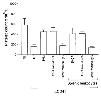
Figure 12 illustrates an antibody-mediated cellular
programming effect, herein referred to as IMCP, as mentioned
above, at work in splenic leukocytes incubated with monoclonal
anit-OVA, thus establishing a basis for the mode of action of
the treatment regimes of the present invention.
As
illustrated, anti-ovalbumin T- ovaItumin _________________ +
__________________ leukauyte-s ate
AM
vumik cvijipt
e
CA 02563024 2006-09-29
Low:14
o Atli!) A r: Y
NG II! 0 01 06
- 28 -
incubated for 30 min, unbound anti-ovalbumin and ovalbumin are
washed off, and leukocytes are injected into ITP mice to
provide ameliorating effect against thrombocytopenia in vivo.
According to Figure 12, mice in the first column (Nil) were
uninjected. Mice in the second column (ITP) were treated with
anti-platelet antibody (aCD41) only. On Day 1, mice in the
third column (IVIg) were injected with 50 mg/ml of dialyzed
IVIg. Mice in the fourth column were injected (i.v.) with 1 mg
OVA that had been pre-incubated with 50 g of monoclonal anti-
OVA (IgGl, clone OVA-14 Sigma). Mice in the fifth column were
treated as in fourth column except with control mouse IgG
(mouse IgG, Cat# 10400, Caltag) in place of monoclonal anti-
OVA. Mice in the sixth column (IMCP) were injected
intravenously with splenic leukocytes (106/mouse) that went
through the IMCP process with dialyzed IVIg for 30 min. Mice
in the seventh column were treated with splenic leukocytes
(106/mouse) that went through IMCP process with 1 mg OVA that
had been pre-incubated with 50 g of monoclonal anti-OVA for
30 min. Mice in the eigth column were treated as in seventh
column except With control mouse IgG in place of monoclonal-
anti-OVA. On Day 2, mice in columns (second to eigth) were
injected with 2 lig anti-platelet antibody. On Day 3, all mice
were bled for platelet enumeration as described (Blood
102:558-560, 2003).
IVIg, anti-CD44 (KM-114), and antibody to soluble
antigens (in the presence of the soluble antigen) cannot
ameliorate thrombocytopenia in mice which are genetically
deficient in the inhibitory Fcy receptor (FcyRIIB)
Interestingly, however, we show here that these same
antibodies can, all ameliorate thrombocytopenia when they are
pre-incubated with leukocytes isolated from mice that are
F-GyR-1-1-11¨(FcyRIZE-/-and the_ FcyRIIB-/-
AME1DED SHEET
PC I Us amp
Lt 14 r e
CA 02563024 2006-09-29
601 iiNHPRY 200
ft,
- 29 -
leukocytes are injected into wild type mice. Thus, the IMCP
effect as herein reported can work where leukocytes do not
express an FcgammaRIIB receptor. Although, FcgammaRIIB
receptor expression was required in the recipient in order to
achieve IMCP.
In the reverse of this experiment (where the
leukocytes are from FcyRIIB+/+ mice and the recipient mice are
FcyRIIB-/-), again, IVIg, anti-CD44, and anti-soluble antigen
(+ the antigen) all cannot ameliorate the thrombocytopenia
(Figure 13).
As shown in Figure 13, mice in the 1st column
(Nil-BL/6) are uninjected C57BL/6 mice. Mice in the 2nd column
(0D41-BL/6) were C57BL/6 mice treated with anti-platelet
antibody (aCD41) only. Mice in the 8th column (Nil-RIB) were
uninjected FcyRIIB-/- mice. Mice in the 9th column (CD41-RIIB)
were FcyRIIB-/- mice treated with anti-platelet antibody
(aCD41) only. On
Day 1, mice in the 3rd column (IVIG-BL/6)
were injected with 50 mg/ml IVIg. Mice in the fourth column
(IVIG-BL/6) were C57BL/6 mice injected intravenously with
splenic leukocytes (106/mouse) from C57BL/6 mice that went
through the IMCP process with IVIg for 30 min. Mice in the 5th
column (IVIG-RIIB) were FcyRIIB-/- mice injected intravenously
with splenic leukocytes (106/mouse) from C57BL/6 mice that went
through the IMCP process with IVIg for 30 min. Mice in the 6th
column (BSA-RIIB) were FcyRIIB-/- mice injected intravenously
with splenic leukocytes (106/mouse) from C57BL/6 mice that went
through the IMCP process with BSA for 30 min. Mice in the 7th
column (BSA-BL/6) were C57BL/6 mice injected intravenously
with splenic leukocytes (106/mouse) from C57BL/6 mice that went
through the IMCP process with BSA for 30 min. Mice in the 10th
column (IVIG-RIIB) were injected with 50 mg/ml IVIg. Mice in
the llth column (IVIG-BL/6) were C57BL/6 mice injected
intravenously with splenic leukocytes (106/mouse) from FcyRIIB-
/-
mice that went through the IMCP process with IVIg for 30
min. Mice in the 12th column (IVIG-RIIB) were FcyRIIB-/- mice-
AMENDED SH
CA 02563024 2006-09-29
WO 2005/094880
PCT/CA2005/000472
- 30 -
injected intravenously with splenic leukocytes (106/mouse) from
FcyRIIB-/- mice that went through the IMCP process with IVIg for
30 min. Mice in the 13th column (BSA-RIIB) were FcyRIIB-/- mice
injected intravenously with splenic leukocytes (106/mouse) from
FcyRIIB-/- mice that went through the IMCP process with BSA for
30 min. Mice in the 14th column (BSA-RIIB) were C57BL/6 mice
injected intravenously with splenic leukocytes (106/mouse) from
FcyRIIB-/- mice that went through the IMCP process with BSA for
30 min. On Day 2, mice in columns (2nd to 7th and 9th to 14th,
inclusive) were injected with 2 jig anti-platelet antibody. On
Day 3, all mice were bled for platelet enumeration as
described in Blood 102:558-560, 2003 with the exception that
mice were bled by the saphenous vein in accordance with this
embodiment of the present invention.
We therefore conclude that IVIg, anti-CD44, and anti-
soluble antigen (in the presence of the antigen) do not
function by binding to the FcyRIIB on the leukocyte but do all
function by a highly related mechanism, which we refer to as
an IVIg-mediated cellular programming mechanism, or IMCP.
Furthermore, the cellular programming mechanism (IMCP) of the
present invention establishes an underlying mode of action for
antibody-based treatment regimes of the present invention that
appears to be more accurate than the previously reported RES
blockade mechanism.
DISCUSSION
We have observed that antibodies to soluble antigens
ameliorated both murine ITP as well as arthritis. Since the
immunological mechanisms involved in both of these diseases is
very different, i.e. phagocytosis of opsonized platelets in
the spleen vs. joint destruction, our data demonstrate that
the therapeutic effects of the anti-soluble-antigen regime
work to ameliorate autoimmune disease, in general.
In
CA 02563024 2006-09-29
WO 2005/094880
PCT/CA2005/000472
- 31 -
addition to the effectiveness of this treatment regime in both
ITP and arthritis treatment, we have also established an
underlying mechanism of action for the anti-soluble-antigen
regime that is common to that of IVIg (the standard therapy
for a multitude of automimmune diseases) and anti-CD44
antibody. That is, an antibody-mediated cellular programming
effect, as illustrated with pre-incubated leukocytes. Thus,
further supporting the potential of an anti-soluble-antigen
. treatment regime of the present invention in the treatment of
a plurality of autoimmune diseases.
The above described antibodies and antibody-antigen and
antibody-antigen-cell complexes can be incorporated in
pharmaceutical compositions to be injected in the mammal. Such
compositions may also comprise a pharmaceutically acceptable
carrier as would be known in the art.
The compositions can be injected in the mammal by
several routes of administration comprising intravenously,
interperitoneally, intradermally,
intramuscularly,
subcutaneously, orally or rectally.
It will be appreciated by persons skilled in the art
that other antigens and antibodies could also be used
according to the above described method to achieve similar
results. It will also be appreciated that the method and
composition could be applied to mammals, other than mice and
rabbits, such as humans.
The embodiment(s) of the invention described above
is(are) intended to be exemplary only. The scope of the
invention is therefore intended to be limited solely by the
scope of the appended claims.