Note: Descriptions are shown in the official language in which they were submitted.
CA 02563080 2011-07-26
MONOCLONAL ANTIBODIES TO HEPATOCYTE GROWTH FACTOR
FIELD OF THE INVENTION
[0002] The present invention relates generally to the combination of
monoclonal antibody
(mAb) and recombinant DNA technologies for developing novel biologics, and
more
particularly, for example, to the production of monoclonal antibodies that
bind to and
neutralize Hepatocyte Growth Factor.
BACKGROUND OF THE INVENTION
[0003] Human Hepatocyte Growth Factor (HGF) is a multifunctional heterodimeric
polypeptide produced by mesenchymal cells. HGF has been shown to stimulate
angiogenesis,
morphogenesis and motogenesis, as well as the growth and scattering of various
cell types
(Bussolino et at., J. Cell. Biol. 119: 629, 1992; Zamegar and Michalopoulos,
J. Cell. Biol.
129:1177, 1995; Matsumoto et al., Ciba. Found. Symp. 212:198, 1997; Birchmeier
and
Gherardi, Trends Cell. Biol. 8:404, 1998; Xin et al. Am. J. Pathol. 158:1111,
2001). The
pleiotropic activities of HGF are mediated through its receptor, a
transmembrane tyrosine
ldnase encoded by the proto-oncogene cMet. In addition to regulating a variety
of normal
cellular functions, HGF and its receptor c-Met have been shown to be involved
in the
initiation, invasion and metastasis of tumors (Jeffers et at., I. Mol. Med.
74:505, 1996;
Comoglio and Trusolino, J. Clin. Invest. 109:857, 2002). HGF/cMet are
coexpressed, often
over-expressed, on various human solid tumors including tumors derived from
lung, colon,
rectum, stomach, kidney, ovary, skin, multiple myeloma and thyroid tissue
(Prat et at., Int. J.
Cancer 49:323, 1991; Chan et al., Oncogene 2:593, 1988; Weidner et at., Am. J.
Respir. Cell.
Mol. Biol. 8:229, 1993; Derksen et at., Blood 99:1405, 2002). HGF acts as an
autocrine
1
CA 02563080 2011-07-26
(Rong et al., Proc. Natl. Acad. Sci. USA 91:4731, 1994; Koochekpour et al.,
Cancer Res.
57:5391, 1997) and paracrine growth factor (Weidner et al., Am. J. Respir.
Cell. Mol. Biol.
8:229, 1993) and anti-apoptotic regulator (Gao et al., J. Biol. Chem.
276:47257, 2001) for
these tumors.
[0004] HGF is a 102 kDa protein with sequence and structural similarity to
plasminogen
and other enzymes of blood coagulation (Nakamura et al., Nature 342:440, 1989;
Weidner et
al., Am. J. Respir. Cell. Mol. Biol. 8:229, 1993,
Fig. 1). Human HGF is synthesized as a 728 amino acid precursor (preproHGF),
which undergoes intracellular cleavage to an inactive, single chain form
(proHGF)
(Nakamura et al., Nature 342:440, 1989; Rosen et al.,J. Cell. Biol. 127:1783,
1994). Upon
extracellular secretion, proHGF is cleaved to yield the biologically active
disulfide-linked
heterodimeric molecule composed of an a-subunit and 3-subunit (Nakamura et
al., Nature
342:440, 1989; Naldini et al., EMBO J. 11:4825, 1992). The a-subunit contains
440 residues
(69 kDa with glycosylation), consisting of the N-terminal hairpin domain and
four kringle
domains. The 0-subunit contains 234 residues (34 kDa) and has a serine
protease-like
domain, which lacks proteolytic activity. Cleavage of HGF is required for
receptor
activation, but not for receptor binding (Hartmann et al., Proc. Natl. Acad.
Sci. USA
89:11574, 1992; Lokker etal., J. Biol. Chem. 268:17145, 1992). HGF contains 4
putative N-
glycosylation sites, 1 in the a-subunit and 3 in the 0-subunit. HGF has 2
unique cell specific
binding sites: a high affinity (Kd = 2 x 10⁻¹ M) binding site for the cMet
receptor and a low
affinity (Kd = 10 M) binding site for heparin sulfate proteoglycans (HSPG),
which are
present on the cell surface and extracellular matrix (Naldini et al., Oncogene
6:501, 1991;
Bardelli et al., J. Biotechnol. 37:109, 1994; &iota et al., J. Biol. Chem.,
272:9457, 1997).
NIC2 (a protein encompassing the N-terminus and first two kringle domains of
the a-subunit)
is sufficient for binding to cMet and activation of the signal cascade for
motility, however the
full length protein is required for the mitogenic response (Weidner et al.,
Am. J. Respir. Cell.
Mol. Biol. 8:229, 1993). HSPG binds to HGF by interacting with the N terminus
of HGF
(Aoyama, etal., Biochem. 36:10286, 1997; Sakata, etal., J. Biol. Chem.
272:9457, 1997).
Postulated roles for the HSPG-HGF interaction include the enhancement of HGF
bioavailability, biological activity and oligomerization (Bardelli, et al., J.
Biotechnol.
37:109,1994; Zioncheck et al., J. Biol. Chem. 270:16871, 1995).
[0005] Wet is a member of the class IV protein tyrosine ldnase receptor
family. The full
length cMet gene was cloned and identified as the cMet proto-oncogene (Cooper
et al.,
2
CA 02563080 2011-07-26
Nature 311:29, 1984; Park et al., Proc. Natl. Acad. Sci. USA 84:6379, 1987).
The cMet
receptor is initially synthesized as a single chain, partially glycosylated
precursor, p170(1E1/
(Fig. 1) (Park et al., Proc. Natl. Acad. Sci. USA 84:6379, 1987; Giordano et
al., Nature
339:155, 1989; Giordano et al., Oncogene 4:1383, 1989; Bardelli et al., J.
Biotechnol.
37:109, 1994). Upon further glycosylation, the protein is proteolytically
cleaved into a
heterodimeric 190 kDa mature protein (1385 amino acids), consisting of the 50
kDa a-
subunit (residues 1-307) and the 145 kDa )3-subunit. The cytoplasmic tyrosine
kinase domain
of thei3-submit is involved in signal transduction.
[0006] Several different approaches have been investigated to obtain an
antagonistic
molecule of the HGF/cMet interaction: truncated HGF proteins such as NK1 (N
terminal
domain plus kringle domain 1; Lokker et al., J. Biol. Chem. 268:17145, 1993),
NK2 (N
terminal domain plus kringle domains 1 and 2; Chan et al., Science 254:1382,
1991) and
NK4 (N-terminal domain plus four kringle domains; Kuba et al., Cancer Res.
60:6737, 2000),
anti-cMet mAbs (Dodge, Master's Thesis, San Francisco State University, 1998)
and anti-
HGF mAbs (Cao et al., Proc. Natl. Acad. Sci. USA 98:7443, 2001).
[0007] NK1 and NK2 can compete effectively with the binding of HGF to its
receptor, but
have been shown to have partial agonistic activities in vitro (Cioce et al.,
J. Biol. Chem.
271:13110, 1996; Schwan et al., J. Cell Biol. 133:709, 1996), rather than
purely antagonist
activities as desired. More recently, Kuba et al., Cancer Res. 60:6737, 2000,
demonstrated
that NK4 could partially inhibit the primary growth (Fig. 2) and metastasis of
murine lung
tumor LLC in a nude mouse model by continuous infusion of NK4. The fact that
NK4 had to
administered continuously to obtain a partial growth inhibition of primary
tumors indicates a
potentially short half-life of the NK4 molecule and/or lack of potency.
Compared to NK4,
the approach of using antibodies will benefit from their favorable
pharmacokinetics and the
possibility of obtaining antibodies with much higher potency.
[0008] As another approach, Dodge (Master's Thesis, San Francisco State
University,
1998) generated antagonistic anti-cMet monoclonal antibodies (mAbs). One rnAb,
5D5,
exhibited strong antagonistic activity in ELISA, but induced a proliferative
response of cMet-
expressing BAF-3 cells, presumably due to dimerization of the membrane
receptors. Prat et
al., J. Cell Sci. 111:237, 1998, also reported such agonistic activities of
anti-cMet mAbs.
Zaccolo et al., Eur. J. Immunol 27:618, 1997, used phage display methods do
develop human
3
CA 02563080 2006-10-05
WO 2005/107800
PCT/US2004/026565
Fab fragments against mouse and human hepatocyte growth factor. These Fab
fragments had
no effect on the activity of HGF when used alone. When one of the anti-human
HGF Fab
fragments was combined with an antibody that bound to the Fab fragment itself,
it actually
enhanced the activity of HGF in a biological assay.
[0009] Cao et al., Proc. Natl. Acad. Sci. USA 98:7443, 2001, demonstrated that
the
administration of a cocktail of three anti-HGF mAbs, which were selected based
upon their
ability to inhibit the scattering activity of HGF in vitro, were able to
inhibit the growth of
human tumors in the xenograft nude mouse model (Fig. 3). They postulated that
three mAbs
recognizing three different binding sites on HGF were required to inhibit the
bioactivities of
HGF in vivo: two mAbs inhibited the binding of HGF to cMet and one mAb
inhibited the
binding of HGF to heparin. However, it is impractical for commercial and
regulatory reasons
to develop a drug combining three novel mAbs, e.g., because some clinical
activity of each
antibody would need to be demonstrated independently.
[0010] Thus, there is a need for a single monoclonal antibody that blocks
biological activity
of HGF in vitro and in vivo. The present invention fulfills this and other
needs.
BRIEF SUMMARY OF THE INVENTION
[0011] In one embodiment, the invention provides a neutralizing mAb to human
Hepatocyte Growth Factor (HGF). The mAb inhibits at least one, and preferably
several or
all biological activities of HGF including binding to its receptor cMet,
inducing scattering of
cells such as Madin-Darby canine kidney cells, inducing proliferation of 4MBr-
5 monkey
epithelial cells and/or hepatocytes and/or HLTVEC, and inducing angiogenesis.
The Anti-
HGF mAb can inhibit such an activity when used as a single agent. A preferred
anti-HGF
mAb inhibits, most preferably completely inhibits, growth of a human tumor
xenograft in a
mouse. Preferably, the mAb of the invention is chimeric, humanized, human-like
or human.
Exemplary antibodies are L2G-7 and its chimeric and humanized forms. Cell
lines producing
such antibodies are also provided. In another embodiment, a pharmaceutical
composition
comprising a neutralizing anti-HGF antibody, e.g., chimeric or humanized L2G7,
is provided.
In a third embodiment, the pharmaceutical composition is administered to a
patient to treat
cancer or other disease.
4
CA 02563080 2014-07-11
[011A] This invention relates to a monoclonal antibody (mAb) that is a
chimeric or
humanized form of the antibody produced by hybridoma ATCC No. PTA-5162 or is a
chimeric
or humanized antibody that competes with said antibody produced by hybridoma
ATCC No.
PTA-5162 for binding to Hepatocyte Growth Factor (HGF), and wherein the mAb is
further
characterized as neutralizing HGF and being able to inhibit growth of a human
tumor xenograft
in a mouse as a single agent. Also provided are cell lines producing such a
monoclonal
antibody as well as pharmaceutical compositions comprising such an antibody in
a
physiologically acceptable carrier.
[01113] A monoclonal antibody or composition of this invention may be for
use as an anti-
cancer agent or medicament. Such an anti-cancer agent or medicament may be for
use in
inhibiting growth of a tumor that over-expresses the receptor for HGF. The
antibody may be
for administration to a patient at a dose of about 0.1 to about 20 mg/kg.
[011C] Various embodiments of this invention involve a pharmaceutical
composition
comprising a monoclonal antibody (mAb) and a physiologically acceptable
carrier, wherein the
mAb is present in the carrier at a concentration of 1-100 mg/ml, wherein the
mAb competes
with the antibody produced by hybridoma ATCC No. PTA-5162 for binding to
Hepatocyte
Growth Factor (HGF), and wherein the mAb in the composition neutralizes HGF
and inhibits
growth of a U118 human glioblastoma tumor xenograft in a mouse as a single
agent.
4a
CA 02563080 2006-10-05
WO 2005/107800
PCT/US2004/026565
BRIEF DESCRIPTION OF THE DRAWINGS
[0012] Figure 1. Schematic models of HOP and cMet.
[0013] Figure 2. Graph showing that NK4 partially inhibits the primary growth
of murine
lung tumor LLC in nude mice (from Kuba et al., Cancer Res. 60:6737, 2000). NK4
was
infused continuously for 14 days from 4th day after tumor implantation s.c. in
nude mice.
[0014] Figure 3. Graph showing that a cocktail of three anti-HGF mAbs is
required to
inhibit the growth of human brain tumor U-118 cells in nude mice (from Cao et
al., Proc.
Natl. Acad. Sci. USA 98:7443, 2001). U-118 tumor cells were injected s.c. into
nude mice.
From day 1 anti-HGF mAbs A-1, -5, and -7, or mAbs 7-2 and -3 were administered
at 200
g/injection, twice/wk for 10 wks.
[0015] Figure 4. Determination of relative binding epitopes of mAbs L1H4,
L2C7, L2G7
using competitive binding ELISA. Plates were coated with recombinant HGF
(rHGF),
blocked with skim milk and incubated with suboptimal concentration of
biotinylated mAbs in
the presence of 100x excess amounts of unlabeled mAbs. Biotinylated mAb bound
was
detected by the addition of HRP-Strepavidin.
[0016] Figure 5. Binding of anti-HGF mAbs to rHGF as determined in a direct
HGF
binding ELISA. Plate was coated with the HI-Fll supernatant containing rHGF,
blocked by
2% skim milk and incubated with mAbs, followed by the addition of HRP-GaMIgG
(as
described under Examples).
[0017] Figure 6. Abilities of anti-HGF mAbs to capture rHGF-Flag in solution.
Anti-
HGF mAbs were captured on a goat anti-mouse IgG coated ELISA plate. Plates
were then
blocked with 2% skim milk and incubated with rHGF-Flag, followed by HRP-M2
anti-Flag
mAb (as described under Examples).
[0018] Figure 7. Inhibition of rHGF-Flag binding to cMet-Fc by anti-HGF mAbs
in a
capture ELISA. cMet-Fc captured on goat anti-human IgG coated plate is
incubated with
HGF-Flag preincubated with/without mAbs. The bound rHGF-Flag was detected by
the
addition of HRP-M2 anti-Flag mAb (as described under Examples).
[0019] Figure 8. Neutralization of HGF induced MDCK scattering by anti-HGF mAb
L2G7. (A) Control without any treatment. (B) rHGF + IgG. (C) rHGF + mAb L2G7.
=
MDCK cells were incubated with a 1:20 dilution of H1-F11 culture supernatant (-
3 1..tg/m1 of
HGF) in the presence of 10 g/ml of mAbs. Photos were taken at 100x
magnification.
5
CA 02563080 2006-10-05
WO 2005/107800
PCT/US2004/026565
[0020] Figure 9. Inhibition of HGF-induced proliferation of Mv 1 LU cells by
L2G7 mAb.
The fold molar excess of mAb over HGF is shown on the horizontal axis, and the
cpm x 10-2
incorporated is shown on the vertical axis. Data points were obtained in
triplicate.
[0021] Figure 10. Inhibition of HGF-induced proliferation of HUVEC by L2G7 mAb
and
control mouse antibody (mIgG). Data points were obtained in triplicate.
[0022] Figure 11. Effect on HGF-induced proliferation of HCT 116 colon tumor
cells by
L2G7 and L1H4 antibodies. Data point were obtained in triplicate.
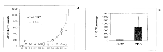
[0023] Figure 12. Effect of treatment with L2G7 mAb or PBS (control) on growth
of
U-118 tumors in groups of NM III Beige/Nude mice (n = 6). Arrow indicates when
injections began. (A) Tumor size vs day from tumor implantation. (B) Tumor
mass at end of
experiment.
DETAILED DESCRIPTION OF THE INVENTION
[0024] The invention provides neutralizing anti-HGF monoclonal antibodies,
pharmaceutical compositions comprising them, and methods of using them for the
treatment
of disease.
1. Antibodies
[0025] Antibodies are very large, complex molecules (molecular weight of -
450,000 or
about 1320 amino acids) with intricate internal structure. A natural antibody
molecule
contains two identical pairs of polyp eptide chains, each pair having one
light chain and one
heavy chain. Each light chain and heavy chain in turn consists of two regions:
a variable
("V") region involved in binding the target antigen, and a constant ("C")
region that interacts
with other components of the immune system. The light and heavy chain variable
regions
fold up together in 3-dimensional space to form a variable region that binds
the antigen (for
example, a receptor on the surface of a cell). Within each light or heavy
chain variable
region, there are three short segments (averaging 10 amino acids in length)
called the
complementarity determining regions ("CDRs"). The six CDRs in an antibody
variable
domain (three from the light chain and three from the heavy chain) fold up
together in 3-D
space to form the actual antibody binding site which locks onto the target
antigen. The
position and length of the CDRs have been precisely defined. Kabat, E. et al.,
Sequences of
Proteins of Immunological Interest, U.S. Department of Health and Human
Services, 1983,
6
CA 02563080 2011-07-26
1987: The part of a variable region not contained in the CDRs is called the
framework,
which forms the environment for the CDRs.
[0026] A monoclonal antibody (mAB) is a single molecular species of antibody
and
therefore does not encompass polyclonal antibodies produced by injecting an
animal (such as
a rodent, rabbit or goat) with an antigen, and extracting serum from the
animal. A humanized
antibody is a genetically engineered (monoclonal) antibody in which the CDRs
from a mouse
antibody ("donor antibody", which can also be rat, hamster or other similar
species) are
grafted onto a human antibody ("acceptor antibody"). Humanized antibodies can
also be
made with less than the complete CDRs from a mouse antibody (e.g., Pascalis et
al., J.
Inununol. 169:3076, 2002). Thus, a humanized antibody is an antibody having
CDRs from a
donor antibody and variable region framework and constant regions from a human
antibody.
Thus, typically a humanized antibody comprises (i) a light chain comprising
three CDRs
from a mouse antibody, e.g., L2G7, a variable region framework from a human
antibody,
and a human constant region, and (ii) a heavy chain comprising three CDRs from
a mouse
antibody, e.g., L2G7, a variable region framework from a human antibody and a
human
constant region. In addition, in order to retain high binding affinity, at
least one of two
additional structural elements can be employed. See, US Patent No. 5,530,101
and
5,585,089, which provide detailed
instructions for construction of humanized antibodies.
[00271 In the first structural element, the framework of the heavy chain
variable region of
the humanized antibody is chosen to have maximal sequence identity (between
65% and
95%) with the framework of the heavy chain variable region of the donor
antibody, by
suitably selecting the acceptor antibody from among the many known human
antibodies.
Sequence identity is determined when antibody sequences being compared are
aligned
according to the Kabat numbering convention. In the second structural element,
in
constructing the humanized antibody, selected amino acids in the framework of
the human
acceptor antibody (outside the CDRs) are replaced with corresponding amino
acids from the
donor antibody, in accordance with specified rules. Specifically, the amino
acids to be
replaced in the framework are chosen on the basis of their ability to interact
with the CDRs.
For example, the replaced amino acids can be adjacent to a CDR in the donor
antibody
sequence or within 4-6 angstroms of a CDR in the humanized antibody as
measured in 3-
dimensional space.
7
CA 02563080 2011-07-26
[0028] A chimeric antibody is an antibody in which the variable region of a
mouse (or
other rodent) antibody is combined with the constant region of a human
antibody; their
construction by means of genetic engineering is well-known. Such antibodies
retain the
binding specificity of the mouse antibody, while being about two-thirds human.
The
proportion of nonhuman sequence present in mouse, chimeric and humanized
antibodies
suggests that the immamogenicitys of chimeric antibodies is intermediate
between mouse and
humanized antibodies. Other types of genetically engineered antibodies that
may have
reduced immunogenicity relative to mouse antibodies include human antibodies
made using
phage display methods (Dower et al., W091/17271; McCafferty et al.,
W092/001047;
Winter, W092/20791; and Winter, FEBS Lett. 23:92, 1998)
or using transgenic animals (Lonberg et al., W093/12227; Kucherlapati
W091/10741).
[0029] As used herein, the term "human-like" antibody refers to a Mab in which
a
substantial portion of the amino acid sequence of one or both chains (e.g.,
about 50% or
imore) originates from human immunoglobulin genes. Hence, human-like
antibodies
encompass but are not limited to chimeric, humanized and human antibodies. As
used
herein, a "reduced-immunogenicity" antibody is one expected to have
significantly less
immunogenicity than a mouse antibody when administered to human patients. Such
antibodies encompass chimeric, humanized and human antibodies as well as
antibodies made
by replacing specific amino acids in mouse antibodies that may contibute to B-
or T-cell
epitopes, for example exposed residues (Padlan, Mol. Immunol. 28:489, 1991).
As used
. herein, a "genetically engineered" antibody is one for which the genes have
been constructed
or put in an unnatural environment (e.g., human genes in a mouse or on a
bacteriophage) with
the help of recombinant DNA techniques, and would therefore, e.g., not
encompass a mouse
mAb made with conventional hybridoma technology.
[0030] The epitope of a mAb is the region of its antigen to which the mAb
binds. Two
antibodies bind to the same or overlapping epitope if each competitively
inhibits (blocks)
binding of the other to the antigen. That is, a lx, 5x, 10x, 20x or 100x
excess of one antibody =
inhibits binding of the other by at least 50% but preferably 75%, 90% or even
99% as
measured in a competitive binding assay compared to a control lacking the
competing
antibody (see, e.g., Junghans et al., Cancer Res. 50:1495, 19901 .
Alternatively, two antibodies have the same epitope if essentially all amino
acid mutations in the antigen that reduce or eliminate binding of one antibody
reduce or
8
CA 02563080 2011-07-26
eliminate binding of the other. Two antibodies have overlapping epitopes if
some amino acid
mutations that reduce or eliminate binding of one antibody reduce or eliminate
binding of the
other.
2. Neutralizing anti-HGF Antibodies
5. [0031] A monoclonal antibody (mAb) that binds HGF (i.e., an anti-HGF
mAb) is said to
neutralize HGF, or be neutralizing, if the binding partially or completely
inhibits one or more
biological activities of HGF (i.e., when the mAb is used as a single agent).
Among the
biological properties of HGF that a neutralizing antibody may inhibit are the
ability of HGF
to bind to its cMet receptor, to cause the scattering of certain cell lines
such as Madin-Darby
canine kidney (MDCK) cells; to stimulate proliferation of (i.e., be mitogenic
for) certain cells
including hepatocytes, 4MBr-5 monkey epithelial cells, and various human tumor
cells; or to
stimulate angiogenesis, for example as measured by stimulation of human
vascular
endothelial cell (HUVEC) proliferation or tube formation or by induction of
blood vessels
when applied to the chick embryo chorioallantoic membrane (CAM). Antibodies of
the
invention preferably bind to human HGF, i.e., to the protein encoded by the
GenBank
sequence with Accession, number D90334.
[0032] A neutralizing mAb of the invention at a concentration of, e.g., 0.01,
0.1, 0.5, 1, 2,
5, 10, 20 or 50 g/m1 will inhibit a biological function of HGF (e.g.,
stimulation of =
proliferation or scattering) by about at least 50% but preferably 75%, more
preferably by
90% or 95% or even 99%, and most preferably approximately 100% (essentially
completely)
as assayed by methods described under ExamplOs or known in the art. Inhibition
is
considered complete if the level of activity is within the margin of error for
a negative control
lacking HGF. Typically, the extent of inhibition is measured when the amount
of HGF used
is just sufficient to fully stimulate the biological activity, or is 0.05,
0.1, 0.5, 1, 3 or 10 ilg/ml.
Preferably, at least 50%, 75%, 90%, or 95% or essentially complete inhibition
will be
achieved when the molar ratio of antibody to HGF is 0.5x, lx, 2x, 3x, 5x or
10x. Preferably,
the mAb will be neutralizing, i.e., inhibit the biological activity, when used
as a single agent,
but possible 2 mAbs will be needed together to give inhibition. Most
preferably, the mAb
will neutralize not just one but several of the biological activities listed
above; for purposes
herein, an anti-HGF mAb that used as a single agent neutralizes all the
biological activities of
HGF will be called "fully neutralizing", and such mAbs are most preferable.
MAbs of the
invention will preferably be specific for HOE, that is they will not bind, or
only bind to a
9
CA 02563080 2006-10-05
WO 2005/107800
PCT/US2004/026565
much lesser extent (e.g., Ka at least ten-fold less), proteins that are
related to HGF such as
fibroblast growth factor (FGF) and vascular endothelial growth factor (VEGF).
Preferred
antibodies lack agonistic activity toward HGF. That is, the antibodies block
interaction of
HGH with cMet without stimulating cells bearing HGF directly. MAbs of the
invention
typically have a binding affinity (Ka) for HGF of at least 107M-1 but
preferably 108 M-1 or
higher, and most preferably 109M-1 or higher or even 1010 M-1 or higher.
[0033] MAbs of the invention include anti-HGF antibodies in their natural
tetrameric form
(2 light chains and 2 heavy chains) and may be of any of the known isotypes
IgG, IgA, IgM,
IgD and IgE and their subtypes, i.e., human IgGl, IgG2, IgG3, IgG4 and mouse
IgGl, IgG2a,
IgG2b, and IgG3. The mAbs of the invention are also meant to include fragments
of
antibodies such as Fv, Fab and F(ab')2; bifunctional hybrid antibodies (e.g.,
Lanzavecchia et
al., Eur. J. Immunol. 17:105, 1987), single-chain antibodies (Huston et al.,
Proc. Natl. Acad.
Sci. USA 85:5879, 1988; Bird et al., Science 242:423, 1988); and antibodies
with altered
constant regions (e.g., U.S. Patent No. 5,624,821). The mAbs may be of animal
(e.g.,
mouse, rat, hamster or chicken) origin, or they may be genetically engineered.
Rodent mAbs
are made by standard methods well-known in the art, comprising multiple
immunization with
HGF in appropriate adjuvant i.p., i.v., or into the footpad, followed by
extraction of spleen or
lymph node cells and fusion with a suitable immortalized cell line, and then
selection for
hybridomas that produce antibody binding to HGF, e.g., see under Examples.
Chimeric and
humanized mAbs, made by art-known methods mentioned supra, are preferred
embodiments
of the invention. Human antibodies made, e.g., by phage display or transgenic
mice methods
are also preferred (see e.g., Dower et al., McCafferty et al., Winter, Lonberg
et al.,
Kucherlapati, supra). More generally, human-like, reduced immunogenicity and
genetically
engineered antibodies as defined herein are all preferred.
[0034] The neutralizing anti-HGF mAbs L1H4, L2C7 and L2G7 mAbs described infra
are
examples of the invention, with L2G7 a preferred example. Neutralizing mAbs
with the
same or overlapping epitope as any of these mAbs, e.g., as L2G7, provide other
examples. A
chimeric or humanized form of L2G7 or with LGF is an especially preferred
embodiment. A
mAb (including chimeric, humanized and human antibodies) that competes with
L2G7 for
binding to HGF and neutralizes HGF in at least one, and preferably all, in
vitro or in vivo
assays described herein is also preferred. MAbs that are 90%, 95%, 99% or 100%
identical
(determined by aligning antibody sequences according to the Kabat convention)
to L2G7 in
amino acid sequence, at least in the, CDRs are included in the invention.
Preferably such
CA 02563080 2006-10-05
WO 2005/107800
PCT/US2004/026565
antibodies differ from L2G7 by a small number of functionally inconsequential
amino acid
substitutions (e.g., conservative substitutions), deletions, or insertions.
Preferably such
antibodies retain the functional properties of L2G7, i.e., such antibodies
neutralize HGF in at
least one, and preferably all, in vitro or in vivo assays described herein.
For purposes of
classifying amino acids substitutions as conservative or nonconservative,
amino acids may be
grouped as follows: Group I (hydrophobic sidechains): norleucine, met, ala,
val, leu, ile;
Group II (neutral hydrophilic side chains): cys, ser, thr; Group III (acidic
side chains): asp,
glu; Group IV (basic side chains): asn, gin, his, lys, arg; Group V (residues
influencing chain
orientation): gly, pro; and Group VI (aromatic side chains): trp, tyr, phe.
Conservative
substitutions involve substitutions between amino acids in the same class. Non-
conservative
substitutions constitute exchanging a member of one of these classes for a
member of
another.
[0035] Native mAbs of the invention may be produced from their hybridomas.
Genetically
engineered mAbs, e.g., chimeric or humanized mAbs, may be expressed by a
variety of art-
known methods. For example, genes encoding their light and heavy chain V
regions may be
synthesized from overlapping oligonucleotides and inserted together with
available C regions =
into expression vectors (e.g., commercially available from Invitrogen) that
provide the
necessary regulatory regions, e.g., promoters, enhancers, poly A sites, etc.
Use of the CMV
promoter-enhancer is preferred. The expression vectors may then be transfected
using
various well-known methods such as lipofection or electroporation into a
variety of
mammalian cell lines such as CHO or non-producing myelomas including Sp2/0 and
NSO,
and cells expressing the antibodies selected by appropriate antibiotic
selection. See, e.g., US
Patent No. 5,530,101. Larger amounts of antibody may be produced by growing
the cells in
commercially available bioreactors.
[0036] Once expressed, the mAbs or other antibodies of the invention may be
purified
according to standard procedures of the art such as microfiltration,
ultrafiltration, protein A or
G affinity chromatography, size exclusion chromatography, anion exchange
chromatography,
cation exchange chromatography and/or other forms of affinity chromatography
based on
organic dyes or the like. Substantially pure antibodies of at least about 90
or 95%
homogeneity are preferred, and 98% or 99% or more homogeneity most preferred,
for
pharmaceutical uses.
11
CA 02563080 2006-10-05
WO 2005/107800
PCT/US2004/026565
3. Therapeutic Methods
[0037] In a preferred embodiment, the present invention provides a
pharmaceutical
formulation comprising the antibodies described herein. That is, the
antibodies can be used
in the manufacture of a medicament for treatment of disease. Pharmaceutical
formulations
(i.e., medicaments) of the antibodies contain the mAb in a physiologically
acceptable carrier,
optionally with excipients or stabilizers, in the form of lyophilized or
aqueous solutions.
Acceptable carriers, excipients or stabilizers are nontoxic to recipients at
the dosages and
concentrations employed, and include buffers such as phosphate, citrate, or
acetate at a pH
typically of 5.0 to 8.0, most often 6.0 to 7.0; salts such as sodium chloride,
potassium
chloride, etc. to make isotonic; antioxidants, preservatives, low molecular
weight
polypeptides, proteins, hydrophilic polymers such as polysorbate 80, amino
acids,
carbohydrates, chelating agents, sugars, and other standard ingredients known
to those skilled
in the art (Remington's Pharmaceutical Science 16th edition, Osol, A. Ed.
1980). The mAb is
typically present at a concentration of 1 - 100 mg/ml, e.g., 10 mg/mi.
[0038] Antibodies of the invention are typically substantially pure from
undesired
contaminant. This means that the antibody is typically at least about 50% w/w
(weight/weight) pure, as well as being substantially free from interfering
proteins and
contaminants. Preferably the antibodies are at least 90, 95% or 99% w/w pure.
Pharmaceutical compositions for parenteral administration are usually sterile,
substantially
isotonic and prepared in accordance with Good Manufacturing Practices of the
FDA or
similar body.
[0039] In another preferred embodiment, the invention provides a method of
treating a
patient with a disease using an anti-HGF mAb in a pharmaceutical formulation.
The mAb
prepared in a pharmaceutical formulation can be administered to a patient by
any suitable
route, especially parentally by intravenous infusion or bolus injection,
intramuscularly or
subcutaneously. Intravenous infusion can be given over as little as 15
minutes, but more
often for 30 minutes, or over 1, 2 or even 3 hours. The mAb can also be
injected directly into
the site of disease (e.g., a tumor), or encapsulated into carrying agents such
as liposomes.
The dose given will be sufficient to alleviate the condition being treated
("therapeutically
effective dose") and is likely to be 0.1 to 5 mg/kg body weight, for example
1, 2, 3 or 4
mg/kg, but may be as high as 10 mg/kg or even 15 or 20 mg/kg. A fixed unit
dose may also
be given, for example, 50, 100, 200, 500 or 1000 mg, or the dose may be based
on the
12
CA 02563080 2006-10-05
WO 2005/107800
PCT/US2004/026565
patient's surface area, e.g., 100 mg/m2. Usually between 1 and 8 doses, (e.g.,
1, 2, 3', 4, 5, 6,
7 or 8) are administered to treat cancer, but 10, 20 or more doses may be
given. The mAb
can be administered daily, biweekly, weekly, every other week, monthly or at
some other
interval, depending, e.g. on the half-life of the mAb, for 1 week, 2 weeks, 4
weeks, 8 weeks,
3-6 months or longer. Repeated courses of treatment are also possible, as is
chronic
administration. A regime of a dosage and intervals of administration that
alleviates or at least
partially arrests the symptoms of the disease (biochemical, histologic and/or
clinical),
including its complications and intermediate pathological phenotypes in
development of the
disease is referred to as a therapeutically effective regime.
[0040] The pharmaceutical compositions of the invention can also be used in
prophylaxis
of a patient at risk of cancer. Such patients include those having genetic
susceptibility to
cancer, patients who have undergone exposure to carcinogenic agents, such as
radiation or
toxins, and patients who have undergone previous treatment for cancer and are
at risk of
recurrence. A prophylactic dosage is an amount sufficient to eliminate or
reduce the risk,
lessen the severity, or delay the outset of the disease, including
biochemical, histologic and/or
clinical symptoms of the disease, its complications and intermediate
pathological phenotypes
presenting during development of the disease. Administration of a
pharmaceutical
composition in an amount and at intervals effective to effect one or more of
these objects is
referred to as a prophylactically effective regime.
[0041] Diseases especially susceptible to therapy with the anti-HGF mAbs of
this invention
include solid tumors known or suspected to require anOogenesis or to be
associated with
elevated levels of HGF, for example ovarian cancer, breast caliber, lung
cancer (small cell or
non-small cell), colon cancer, prostate cancer, pancreatic cancer, renal
cancer, gastric cancer,
liver cancer, head-and-neck tumors, melanoma, sarcomas, and brain tumors
(e.g.,
glioblastomas), of children or adults. Treatment can also be administered to
patients having
leukemias or lymphomas. In a preferred embodiment, the anti-HGF mAb can be
administered together with (i.e., before, during or after) other anti-cancer
therapy. For
example, the anti-HGF mAb may be administered together with any one or more of
the
chemotherapeutic drugs known to those of skill in the art of oncology, for
example Taxol
(paclitaxel) or its derivatives, platinum compounds such as carboplatin or
cisplatin,
anthrocyclines such as doxorubicin, alkylating agents such as
cyclophosphamide, anti-
metabolites such as 5-fluorouracil, or etoposide. The anti-HGF mAb can be
administered in
combination with two, three or more of these agents in a standard
chemotherapeutic regimen,
13
CA 02563080 2006-10-05
WO 2005/107800
PCT/US2004/026565
for example taxol and carboplatin, e.g. for breast and ovarian cancer. Other
agents with
which the anti-HGF mAb can be administered include biologics such as
monoclonal
antibodies, including HerceptinTM against the HER2 antigen, AvastinTM against
VEGF, or
antibodies to the EGF receptor, as well as small molecule anti-angiogenic or
EGF receptor
antagonist drugs. In addition, the anti-HGF mAb can be used together with
radiation therapy
or surgery.
[0042] Treatment (e.g., standard chemotherapy) including the anti-HGF mAb
antibody may
increase the median progression-free survival or overall survival time of
patients with these
tumors (e.g., ovarian, breast, lung, pancreas, brain and colon, especially
when relapsed or
refractory) by at least 30% or 40% but preferably 50%, 60% to 70% or even 100%
or longer,
compared to the same tretment (e.g., chemotherapy) but without anti-HGF mAb.
In addition
or alternatively, treatment (e.g., standard chemotherapy) including the anti-
HGF mAb may
increase the complete response rate, partial response rate, or objective
response rate
(complete + partial) of patients with these tumors (e.g., ovarian, breast,
lung, pancreas, brain
and colon, especially when relapsed or refractory) by at least 30% or 40% but
preferably
50%, 60% to 70% or even 100% compared to the same treatment (e.g.,
chemotherapy) but
without the anti-HGF mAb. Optionally, treatment can inhibit tumor invasion, or
metastasis.
[0043] Typically, in a clinical trial (e.g., a phase II, phase or phase III
trial), the
aforementioned increases in median progression-free survival and/or response
rate of the
patients treated with chemotherapy plus the anti-HGF mAb, relative to the
control group of
patients receiving chemotherapy alone (or plus placebo), will be statistically
significant, for
example at the p = 0.05 or 0.01 or even 0.001 level. It will also be
understood by one of skill
that the complete and partial response rates are determined by objective
criteria commonly
used in clinical trials for cancer, e.g., as listed or accepted by the
National Cancer Institute
and/or Food and Drug Administration.
4. Other Methods
[0044] The anti-HGF mAbs of the invention also find use in diagnostic,
prognostic and
laboratory methods. They may be used to measure the level of HGF in a tumor or
in the
circulation of a patient with a tumor, and therefore to follow and guide
treatment of the
tumor. For example, a tumor associated with high levels of HGF would be
especially
susceptible to treatment with an anti-HGF mAb. In particular embodiments, the
mAbs can be
used in an ELISA or radioimmunoassay to measure the level of HGF, e.g., in a
tumor biopsy
14
CA 02563080 2011-07-26
specimen or in serum or in media supernatant of HGF-secreting cells in cell
culture. The use
of two anti-HGF mAbs binding to different epitopes (i.e., not competing for
binding) will be
especially useful in developing a sensitive "sandwich" ELISA to detect HGF.
For various
assays, the mAb may be labeled with fluorescent molecules, spin-labeled
molecules, enzymes
or radioisotypes, and may be provided in the form of kit with all the
necessary reagents to
perform the assay for HGF. In other uses, the anti-HGF mAbs will be used to
purify HGF,
e.g., by affinity chromatography.
EXAMPLES
1. Generation of Anti-HGF mAbs
[0045] To generate mAbs which bind to and block the activities of human HGF,
recombinant human HGF (rHGF) was first produced in a mammalian expression
system.
cDNAs encoding the recombinant human HGF (rHGF) or rHGF-Flag peptide (8 amino
acid
residues of Flag attached to the c-terminus of HGF) were constructed in a pIND-
inducible
expression vector (No et al., Proc. Natl. Acad. Sci. USA. 93:3346, 1996).
These cDNAs
were then transfected into EcR-293 human kidney fibroblast cells (Invitrogen)
using Fugene
transfection reagent (Roche). Stable cell lines, 111-F11 and 24.1, secreting
HGF and HGF-
Flag respectively, were selected in the presence of 600 p,g/m1 of G418 and 400
pz/m1 of
Zeocin (Invitrogen). 111-F11 and 24.1 were induced to secrete HGF and HGF-Flag
by
treatment with 4 itM of Ponasterone A (Invitrogen) for 4-5 days in serum free
DMEM
containing glutamine and antibiotics. After aggregates were removed by
centrifugation at
15,000 rpm for 30 min at 4 C, HGF secreted into the culture supernatant was
concentrated
approximately 100-fold using a membrane ultrafiltration cartridge with an MW
50,000 cut-
off filter [amicon Centriprep YM-50 filter followed by microcon YM-50 filter
(Millipore)].
Such concentrated HIFI 1 culture supernatant contains ¨100 jig/m1 of HGF and
4204g/m1
of bovine serum albumin.
[0046] Balb/c mice were immunized in each hind foot pad >10 times at one week
intervals,
with 1-2 jig of purified rHGF (Pepro Tech) or 1-2 jig of rHGF plus 1-2 jig of
BSA
(concentrated H1 -F11 culture supernatant) resuspended in MPL-TDM ('Ribi
Immunochem.
Research). Three days after the final boost, popliteal lymph node cells were
fused with
murine myeloma cells, P3X63AgU.1 (ATCC CRL1597), using 35% polyethylene
glycol.
Hybridomas were selected in HAT medium as described (Chtmtharapai and Kim, J.
Immunol. 163:766, 1997) . Ten days after the
CA 02563080 2006-10-05
WO 2005/107800
PCT/US2004/026565
fusion, hybridoma culture supernatants were screened in a direct HGF binding
ELISA as well
as in an HGF-Flag capture ELISA. The latter assay was used to further confirm
the
specificity of anti-HGF mAbs selected using the direct HGF binding ELISA and
to select
mAbs that can bind to HGF in solution phase. Blocking activities of selected
mAbs were then
determined in the HGF-Flag/ cMet-Fc binding ELISA and in the MDCK scatter
assay as
described (Jeffers et al., Proc. Natl. Acad. Sci. USA 95:14417, 1998).
Selected hybridomas
were cloned twice using limiting dilution techniques. The isotype of mAbs were
determined
using an isotyping kit (Zymed). Ascites of selected mAbs were raised and
purified using
ImmunoPure (A/G) IgG Purification Kit (Pierce). Also biotinylated mAbs were
prepared
using EZ-sulfo-NHS-LC-Biotin according to the instructions provided by Pierce.
Each of the
assays referred to here is described in more detail below.For the direct HGF
binding ELISA,
microtiter plates (Maxisorb; Nunc) are coated with 50 td/well of Hl-F11
culture supernatants
containing rHGF, diluted in PBS at a 1:2 ratio of HGF/PBS, overnight at 4 C.
After washing
the plate, the nonspecific binding sites are blocked with PBS containing 2%
skim milk for 1
hr at room temperature (RT). After washing the plate, 50 id/well of purified
mAbs or
hybridoma culture supernatants are added to each well for 1 hr. After washing,
plates are
then incubated with 50 id/well of 1 ,g/m1 of HRP-goat anti-mouse IgG (HRP-
GaMIgG,
Cappel) for 1 hr. The bound HRP-GaMIgG is detected by the addition of the
tetramethylbenzidine substrate (Sigma). The reaction is stopped by the
addition of 1N H2SO4
and the plates are then read at 450 nm using an ELISA plate reader. Washes are
carried out 3
times in wash buffer (PBS containing 0.05% Tween 20).
[0047] For the HGF-Flag capture ELISA, microtiter plates are coated with 50
id/well of 2
iteml of goat antibodies specific to the Fe portion of mouse IgG (GaMIgG-Fc)
in PBS
overnight at 4 C and blocked with 2% skim milk for 1 hr at RT. After washing,
the plates are
incubated with 50 td/well of purified mAbs or hybridoma culture supernatants
for 1 hr. After
washing, plates are then incubated with 50 id/well of 24.1 cell culture
supernatant containing
rHGF-Flag. After washing, plates are then incubated with 50 Owell of BRP-M2
anti-Flag
inAb (Invitrogen) in the presence of 15 pg/m1 of murine IgG. The bound I-FRP-
anti-Flag M2
is detected by the addition of the substrate as described above. Washes are
carried out 3
times in wash buffer.
[0048] At least three mAbs, designated L1H4, L2C7 and L2G7, obtained from
hybridomas
generated by immunizing the Balb/c mice with rHGF in concentrated Hl-F11
culture
16
CA 02563080 2011-07-26
' supernatant as described above, showed binding in both the direct rHGF
binding ELISA and
the HGF-Flag capture ELISA and were selected for further study. These
hybridomas were
then cloned twice, ascites were raised in mice by standard methods, and mAbs
were purified
using a protein G/A column. Their isotypes were determined using an isotyping
kit (Zymed
Lab). The L2G7 hybridoma has been deposited on April 29, 2003 with the
American Type
Culture Collection, P.O. Box 1549 Manassas, VA 20108, as ATCC Number PTA-5162
under
the Budapest Treaty. These deposit will be maintained at an authorized
depository and
replaced in the event of mutation, nonviability or destruction for a period of
at least five years
after the most recent request for release of a sample was received by the
depository, for a
period of at least thirty years after the date of the deposit, or during the
enforceable life of the
related patent, whichever period is longest. All restrictions on the
availability to the public of
these cell lines will be irrevocably removed upon the issuance of a patent
from the
application.
[0049] Once a single, archtypal anti-human-HGF mAb, for example L2G7, has been
isolated that has the desired properties described herein of neutralizing HGF
in vitro and/or
inhibiting (e.g., completely) tumor growth in vivo, it is straightforward to
generate other
mAbs with similar properties, by using art-known methods. For example, mice
may be
immunized with HGF as described above, hybridomas produced, and the resulting
mAbs
screened for the ability to compete with the archtypal mAb for binding to HGF.
Alternatively, the method of Jespers et al., Biotechnology 12:899, 1994,
may be used to guide the selection of mAbs having the
same epitope and therefore similar properties to the archtypal mAb, e.g.,
L2G7. Using phage
display, first the heavy chain of the archtypal antibody is paired with a
repertoire of
(preferably human) light chains to select an HGF-binding mAb, and then the new
light chain
is paired with a repertoire of (preferably human) heavy chains to select a
(preferably human)
HGF-binding mAb having the same epitope as the archtypal mAb.
2. Characterization of Anti-HGF mAbs In Vitro
[0050] The binding epitopes of the antibodies were partially characterized by
a competitive
binding ELISA in which a 100x excess of unlabeled mAb was used to compete with
the
binding of the same or another biotinylated mAb in the HGF binding ELISA. Fig.
4 shows
that the binding of the anti-HGF mAbs, L1H4 and L2G7, was inhibited only by
themselves,
suggesting that they recognize unique epitopes. The binding of L2C7 was
inhibited by L2G7
17
CA 02563080 2006-10-05
WO 2005/107800
PCT/US2004/026565
but not by L1H4. This suggests that the L2C7 epitope overlaps with that of
L2G7 but not of
L1H4. However, L2C7 was not able to inhibit the binding of L2G7, suggesting
that the
L2C7 and L2G7 epitopes overlap but are distinct, and/or the affinity of L2C7
is much lower
than that of L2G7. The epitopes of L1H4, L2C7 and L2G7 are respectively
designated A, B
and C.
[0051] The relative binding abilities of the three anti-HGF mAbs were measured
using
purified antibodies in the direct HGF binding ELISA, in which rHGF is first
bound to the
plate. In this assay, L2C7 and L2G7 bound better than L1H4 (Fig. 5). The
ability of the
mAbs to bind rHGF-Flag in solution was also determined, using the HGF-Flag
capture
ELISA. All three mAbs were able to capture rHGF-Flag in solution phase but mAb
L2G7
was more effective than the others (Fig. 6). These results suggest that mAb
L2G7 has the
highest binding affinity to HGF among the three mAbs.
[0052] One of the biological activities of HGF is the ability to bind to its
receptor cMet, so
the ability of the anti-HGF mAbs to inhibit binding of HGF to cMET was
assayed. For this
assay, cMet-Fc was first produced by transfecting human fibroblast 293 cells
with cDNA
encoding residues 1-929 ECD of cMet linked with the Fc portion of human IgG1
(residues
216 to 446) as described by Mark et al., J. Biol. Chem. 267:26166, 1992 in the
pDisplay
expression vector (Invitrogen). Microtiter plates are coated with 50 ta/well
of 2 ptg/m1 of
goat antibodies specific to the Fc portion of human IgG (GocHIgG-Fc) in PBS
overnight at
4 C and blocked with 2% BSA for 1 hr at RT. After washing the plates, 50 Al of
culture
supernatant of 293 transfected with cMet-Fc cDNA is added to each well for 1
hr at RT.
After washing the plates, 50 0/well of 24.1 cell culture supernatant
containing rHGF-Flag,
preincubated with various concentrations of mAbs, is added to each well for 1
hr. After
washing, plates are then incubated with 50 1/well of HRP-M2 anti-Flag mAb
(Invitrogen).
The bound HRP-anti-Flag M2 is detected by the addition of the substrate as
described above.
Washes are carried out 3 times in wash buffer.
[0053] In this HGF-Flag/cMet-Fc binding inhibition assay, all three mAbs
demonstrated
some degrees of inhibition while an Ig control antibody did not (Fig. 7). MAb
L2G7 at >1
jig/m1 and mAb L1H4 at 50 pig/m1 completely abolished the binding of rHGF-Flag
to cMet-
Fc; mAb L2C7 even at 50 jig/m1 gave only 85% inhibition. Hence, mAb L2G7 was
much
more potent in inhibiting the interaction of rHGF-Flag with cMet-Fc (and
therefore
=
18
CA 02563080 2006-10-05
WO 2005/107800
PCT/US2004/026565
presumably HGF with its receptor cMet) than the other antibodies, consistent
with its
putatively greater affinity for HGF.
[0054] Since the receptor protein used in cMet-Fc/HGF-Flag binding ELISA is a
soluble
receptor protein, its conformation may be different from that of the natural
membrane bound
receptor. Furthermore, HGF binds to HSPG in addition to cMet and it is known
that the
HSPG-HGF interaction enhances various HGF activities. Thus, mAbs blocking the
interaction of HGF with soluble cMet may not necessarily have the capacity to
neutralize
HGF bioactivities on the cells. Thus, it is important to further confirm the
blocking activities
of mAbs in selected biological systems. HGF is known to be a potent scattering
factor.
Thus, the neutralizing activity of the anti-HGF mAbs was also determined using
the Madin-
Darby canine kidney (MDCK cells obtained from ATCC) scatter assay as described
(Jeffers
et al., Proc. Natl. Acad. Sci. USA 95:14417, 1998). MDCK cells grown in DMEM
supplemented with 5% FCS are plated at 103 cells/100 id/well in the presence
of
predetermined concentrations of rHGF with or without mAbs in DMEM with 5% FCS.
After
2 days incubation at 37 C in 5% CO2, cells are then washed in PBS, fixed in 2%
formaldehyde for 10 min at RT. After washing in PBS cells are stained with
0.5% crystal
violet in 50% ethanol (v/v) for 10 min at RT. Scattering activity is
determined by
microscopic examination.
[0055] Culture supernatant of the Hl-F11 clone secreting HGF, described above,
was used
as the source of HGF in the scatter assay. As little as 1:80 dilution of Hl-
F11 culture
supernatant induced the scattering and growth of MDCK cells. However, the
scattering
assays were carried out using a 1:20 dilution of Hl-F11 culture supernatant (-
3 gimp. MAb
L2G7 even at a 1:5 molar ratio of HGF/mAb inhibited the HGF induced scattering
of MDCK
by itself (Fig. 8), conclusively demonstrating that mAb L2G7 is indeed a
neutralizing mAb.
mAb L1H4 at >20 Rg/m1 could also neutralize scattering of MDCK induced by HGF,
while
mAb L2C7 even at 20 pg/m1 gave only a partial neutralizing activity (data not
shown).
[0056] The various characteristics of the three anti-HGF antibodies determined
in the
assays above are summarized in the Table 1.
19
CA 02563080 2006-10-05
WO 2005/107800
PCT/US2004/026565
Table 1. Characterization of mAbs to HGF
mAb lsotype Binding Block Block
Epitope HGF/ cMet-Fc binding MDCK scattering
L1 H4 G1, K A Weak Block
L2C7 G2b, K B Partial Block +/-
L2G7 G2a, K C Strong Block +++
[0057] HGF is a member of the heparin binding growth factor family including
fibroblast
growth factor (FGF) and vascular endothelial growth factor (VEGF). Also, HGF
has ¨40%
overall sequence similarity with plasminogen (Nakamura et al., Nature.
342:440, 1989) and
shares a similar domain structure with macrophage stimulating protein (MSF,
Wang et al.,
Scand. J. Immunol. 56:545, 2002). Thus, the binding specificity of the anti-
HGF antibodies
must be determined. The binding of anti-HGF mAbs to these HGF related proteins
(available
from R&D systems) is assayed using a direct binding ELISA similar to the one
for HGF
described above. MAb L2G7, mAb L2C7 and mAb L1H4 will not significantly bind
to these
proteins, demonstrating their specificity for HGF.
3. Ability of Anti-HGF MAbs to Inhibit Tumor-Promoting Biological Activities
of HGF
[0058] HGF has a number of biological activities that make it likely that it
plays a role in
the growth and invasiveness of certain human tumors. One such activity of HGF
is as a
powerful mitogen for hepatocytes and other epithelial cells (Rubin et al.,
Proc. Natl. Acad.
Sci. USA. 88:415, 1991). Thus, to further prove the neutralizing activity of
the anti-HGF
mAbs, the effects of the mAbs on the HGF-induced proliferation of 4MBr-5
monkey
epithelial cells (ATCC) or rat hepatocytes are determined. Hepatocytes are
isolated
according to a method described by Garrison and Haynes, J. Biol. Chem.
269:4264, 1985.
Cells are resuspended at 5 x 10⁴ cells/ml in DMEM containing 5% FCS and
stimulated with a
predetermined concentration of HGF with various concentration of mAbs. After
TA days
incubation at 37 C in 5% CO2, the level of cell proliferation is determined by
the addition of
3H-thymidine for 4 hrs. Cells are harvested using an automated cell harvester
and the level
of3H-thymidine incorporated is determined on a scintillation counter. At
sufficient
concentrations, mAb L2G7 may largely or completely inhibit HGF-induced
proliferation of
CA 02563080 2006-10-05
WO 2005/107800
PCT/US2004/026565
the cells, and mAbs L2C7 and L1H4 may at least partially inhibit
proliferation. These
antibodies may also inhibit the HGF-induced proliferation of other epithelial
cell lines.
[0059] For example, the inhibitory activity of L2G7 on the HGF-induced
proliferation of
mink lung Mv 1 Lu cells was determined (Borset et aL, J. hnmunol. Methods
189:59, 1996).
-- Cells grown in DMEM containing 10% FCS are harvested by treatment with
EDTA/trysin.
After washing, the cells are resuspended at 5 x 10⁴cells/m1 in serum free DMEM
with a
predetermined concentration (50 ng/ml) of HGF +/- various concentrations of
mAb. After 1
day incubation at 37 C in 5% CO2, the level of cell proliferation is
determined by the addition
of 1 Ci of3H-thymidine for an additional 24 hr. Cells are harvested onto
glass-fiber filters
-- using an automated cell harvester and the level of3H-thymidine incorporated
is determined
on a scintillation counter. Fig. 9 shows that the addition of 100-fold higher
molar
concentration of L2G7 mAb completely inhibited the proliferative response of
Mv 1 Lu cells.
Indeed, L2G7 even at a 3-fold molar ratio of mAb to HGF showed complete
inhibition,
while control IgG showed no inhibition even at 100-fold molar excess.
-- [0060] HGF is also reported to be a potent angiogenesis factor (Bussolino
et al., J. Cell
Biol. 119:629, 1992; Cherrington et aL, Adv. Cancer Res. 79:1, 2000), and
angiogenesis, the
formation of new blood vessels, is believed to be essential to the growth of
tumors.
Therefore, the ability of the anti-HGF mAbs to inhibit the angiogenic
properties of HGF is
shown in three assays: (i) proliferation of human vascular endothelial cells
(HUVEC), (ii)
-- tube formation of HUVEC, and (iii) development of new blood vessels on the
chick embryo
chorioallantoic membrane (CAM). Since HGF has been shown to synergize with
VEGF in
angiogenesis (Xin et al., Am. J. Pathol. 158:1111, 2001), these assays may be
performed both
in the presence and absence of VEGF.
[0061] The HUVEC proliferation assay is performed as described with a
modification
-- (Conn et al., Proc. Natl. Acad. Sci. USA 87:1323, 1990). HUVEC cells
obtained from
Clonetics are grown in Endothelial Growth Medium (EBM-2) containing 10% FCS
plus
endothelial cell growth supplements provided by Clonetics. Preferably cells
from passages 4
to 7 are used in this study. The cells are resuspended to be 105 cells/ml in
medium-199
containing antibiotics, 10 niM HEPES and 10% FCS (assay medium). HUVEC cells
(50
-- Al/well) are added to microtiter wells containing a suitable concentration
of HGF with various
concentrations of anti-HGF mAbs for 1 hr at 37 C. After cells are incubated
for 72 hr at
37 C in 5% CO2, the level of cell proliferation is determined by incorporation
of3H-
,
21
CA 02563080 2006-10-05
WO 2005/107800
PCT/US2004/026565
thymidine for 4 hrs. At sufficient concentrations, mAb L2G7 will largely or
completely
inhibit HGF-induced proliferation of the HUVEC, and mAbs L2C7 and L1H4 may at
least
partially inhibit proliferation.
[0062] Alternatively, the level of cell proliferation may be determined by the
well-known
colorimetric MTT assay. The HUVECs (104 cells/100 [d/well) are grown in serum
free
medium for 24 hr, and then incubated with 100 eul of of 50 ng/ml of HGF
(predetermined to
be a suboptimal amount) with various concentrations of mAb L2G7 for 72 hr. MTT
solution
(5 mg/ml) is added to each well (20 au1/200 Al medium) for 4 hr. Then 100 1
medium /well is
removed and mixed with 100 id/well of acidified isopropyl alcohol (0.04N HC1
in isopropyl).
The plates are read on an ELISA reader at 560 urn. The % maximum response is
calculated
as follows: [OD of HGF + mAB treated cells ¨ OD of untreated cells] / [OD of
HGF treated
cells ¨ OD of untreated cells] x 100. Fig 10 shows that even a 2-fold molar
excess of L2G7
mAB largely blocks the proliferation of HUVEC in response to HGF.
[0063] The endothelial tube assay is carried out essentially as described
(Matsumura, et al.,
J. Immunol. 158: 3408, 2001; Xin et al., Am. J. Pathol. 158:1111, 2001). HUVEC
(Clonetics) from passage 4-7 are grown in Clonetics EGM medium supplemented
with 10%
FBS and endothelial cell growth supplements. Plates are coated with Matrigel
(BD
Biosciences) according to the manufacture's instructions at 37 C for 30 mm,
and the cells are
seeded as 3 x 10⁶ cells/ml in 1 x basal medium with HGF and various
concentrations of anti-
HGF mAbs. Tube formation is evaluated under microscope at low-power (10x)
magnification. At sufficient concentrations, mAb L2G7 will largely or
completely inhibit
HGF-induced endothelial tube formation, and mAbs L2C7 and L1H4 may at least
partially
inhibit it.
[0064] The chick embryo chorioallantoic membrane (CAM) assay is performed
essentially
as described (Kim et al, Nature 362:841, 1992). Three-day old chicken embryos
are removed
from their shells and grown in petri dishes in 5% CO2 at 37 C. Seven days
later, dried
methylcellulose discs containing HGF with various concentrations of anti-HGF
mAbs are
layered onto the CAM. The methylcellulose discs are prepared by mixing 5 of
1.5%
methylcellulose in PBS with 5 aul of HGF preincubated with mAbs. Three days
later the
development of blood vessels around methylcellulose discs are examined. At
sufficient
concentrations, mAb L2G7 will largely or completely inhibit such blood vessel
formation,
and mAbs L2C7 and L1H4 may at least partially inhibit it.
22
CA 02563080 2006-10-05
WO 2005/107800
PCT/US2004/026565
[0065] HGF is also reported to promote tumor growth (Comoglio and Trusolino,
J. Clin.
Invest. 109:857, 2002). The ability of the anti-HGF antibodies to inhibit this
activity is
shown in two steps. First, a number of tumor cell lines are examined for their
ability to
secrete HGF and proliferate in response to HGF since HGF may be an autocrine
growth
factor for some of these cells. These cell lines include a panel of human
tumor cell lines
known to express HGF and cMet (Koochekpour et al., Cancer Res. 57:5391, 1997;
Wang et
al., J. Cell Biol. 153:1023, 2001). Specific cell lines to be tested include U-
118 glioma,
HCT116 colon carcinoma, A549 lung carcinoma and A431 epidermoid carcinoma
cells, all
available from the ATCC. Once such tumor cell lines are identified, the effect
of anti-HGF
mAbs on the proliferative response to HGF of these cells is determined, using
methods
similar to those described above. At sufficient concentrations, mAb L2G7 will
largely or
completely inhibit HGF-induced proliferation of many or all of these cell
lines, and mAbs
L2C7 and L1H4 may at least partially inhibit proliferation.
[0066] For example, human HCT116 tumor cells are seeded into 96-well
microtiter plates
at 5 x 10³cells/well in 200 Al of DMEM plus 5% FCS. After 24 hr incubation at
37 C in 5%
CO2, cells are washed with PBS and incubated in serum free DMEM for 48 hr.
Cells are then
incubated with 100 ng/ml of HGF +1-20 p.g/m1 of mAbs in DMEM for another 20
hr. As
controls, cells grown in DMEM alone or DMEM plus 10% FCS are included. At the
end of
the incubation, levels of cell proliferation are determined by incorporation
of311-thymidine
for 4 hr. The result of such an experiment was carried out in triplicates is
shown in Fig. 11.
HGF induced a moderate proliferation of the HCT116 cells, which was completely
abolished
by addition of L2G7 antibody (but not by the less potent L1114 antibody).
[0067] In all the assays described above, each anti-HGF antibody will
neutralize or inhibit
activity when used alone without other antagonists of HGF, i.e., as a single
agent, but
additive or synergistic effects may be achieved by administering the antibody
in conjunction
with other anti-HGF antibodies or other active agents.
4. Ability of anti-HGF mAbs to Inhibit Tumor Growth In Vivo
[0068] The ability of the anti-HGF antibodies to inhibit human tumor growth is
demonstrated in xenograft models in immunodeficient mice or other rodents such
as rat.
Ilustrative but not limiting examples of immunodeficient strains of mice that
can be used are
nude mice such as CD-1 nude, Nu/Nu, Balb/c nude, NM-III (NTH-bg-nu-xid BR);
scid mice
such as Fox Chase SOD (C.B-17 SCID), Fox Chase outbred SOD and SCID Beige;
mice
23
CA 02563080 2011-07-26
deficient in RAG enzyme; as well as nude rats. Experiments are carried out as
described
previously (Kim eta!,, Nature 362:841, 1992).
Human tumor cells grown in complete DMEM medium are harvested in HBSS. Female
immunodeficient, e.g., athymic nude mice (4-6 wks old) are injected s.c. with
typically 5x10⁶
cells in 0.2 ml of HBSS in the dorsal areas. When the tumor size reaches 50-
100 min3, the
mice are grouped randomly and appropriate amounts of the anti-HGF and control
inAbs
(typically between 0.1 and 1.0 mg, e.g. 0.5 mg) are administered i.p. once,
twice or three
times per week in a volume of, e.g., 0.1 ml, for e.g., 1, 2, 3, or 4 weeks or
the duration of the
experiment. Tumor sizes are determined typically twice a week by measuring in
two
dimensions [length (a) and width (b)]. Tumor volume is calculated according to
V = ab2/2
and expressed as mean tumor volume h SEM. The number of mice in each treatment
group
is at least 3, but more often between 5 and 10, e.g., 7. Statistical analysis
may be performed
using, e.g., Student's t test. In a variation of this experiment,
administration of the antibody
begins simultaneously or shortly after injection of the tumor cells. The
effect of the antibody
may also be measured by prolongation of the survival of the mice, or increase
in percent of
=
the mice surviving.
[00691 Various tumor cell lines known to secrete or respond to HGF are used in
separate
experiments, for example U118 human glioblastoma cells, and/or HCT116 human
colon
tumor cells. Preferred antibodies of the invention, such as human-like and
reduced-
imrnunogenicity antibodies and the L2G7 antibody and its chimeric and
humanized forms
and antibodies with the same epitope as L2G7, when used as a single agent,
will inhibit
growth of tumors by at least 25%, but possibly 40% or 50%, and as much as 75%
or 90% or
greater, or even completely inhibit tumor growth after some period of time or
cause tumor
regression or disappearance. This inhibition will take place for at least
tumor cell line such
as U118 in at least one mouse strain such as NIE1111 Beige/Nude, but
preferably will occur
for 2, 3, several, many, or even essentially all HGF-expressing tumor cell
lines of a particular
(e.g., glioma) or any type, when tested in one or more immunodeficient mouse
strains that do
not generate a neutralizing antibody response against the injected antibody.
Treatment with
some preferred antibodies in one or more of the xenograft models leads to the
indefinite
survival of 50%, 75%, 90% or even essentially all mice, who would otherwise
die or need to
be sacrificed because of growth of their tumor.
[0070] For example, such an experiment was performed with U-118 glioblastoma
cells,
grown in DMEM medium with FCS and harvested in HBSS. Female NM III Beige/Nude
24
CA 02563080 2011-07-26
mice (4-6 wks old) are injected S.C. with 106 cells in 0.2 ml of MSS in the
dorsal areas.
When the tumor size reaches ¨50 mm3, the mice are grouped randomly into 2
groups of 6
mice each, and 200 pg of the L2G7 mAb (treatment group) or of PBS (control
group) are
given i.p. twice a week in a volume of 0.1 ml. Tumor sizes are determined
twice a week as
described above. At the end of the experiment, the tumors are excised and
weighed. Fig. 12
shows that treatment with L2G7 completely inhibited tumor growth.
[0071] Similar tumor inhibition experiments are performed with the anti-HGF
antibody
administered in combination one or more chemotherapeutic agents such as 5-FU
(5-
fluorouracil) or CPT-11 (Camptosar) to which the tumor type is expected to be
responsive, as
described by Ashkenize et al., J. Clin.. Invest. 104:155, 1999. The
combination of the
antibody and chemotherapeutic drag may produce a greater inhibition of \tumor
growth than
either agent alone. The effect may be additive or synergistic, and strongly
inhibit growth, e.g.
by 80% or 90% or more, or even cause tumor regression or disappearance: The
anti-HGF
antibody may also be administered in combination with an antibody against
another growth
or angiogenic factor, for example anti-VEGF, and additive or synergistic
growth inhibition
and/or tumor regression or disappearance is expected.
[0072] Although the invention has been described with reference to the
presently preferred
embodiments, it should be understood that various modifications can be made
without
departing from the invention. Unless otherwise apparent from the context any
step, element,
embodiment, feature or aspect of the invention can be used with any other.
=