Note: Descriptions are shown in the official language in which they were submitted.
DEMANDES OU BREVETS VOLUMINEUX
LA PRESENTE PARTIE DE CETTE DEMANDE OU CE BREVETS
COMPREND PLUS D'UN TOME.
CECI EST LE TOME 1 _______________________ DE 2
NOTE: Pour les tomes additionels, veillez contacter le Bureau Canadien des
Brevets.
JUMBO APPLICATIONS / PATENTS
THIS SECTION OF THE APPLICATION / PATENT CONTAINS MORE
THAN ONE VOLUME.
THIS IS VOLUME 1 OF 2
NOTE: For additional volumes please contact the Canadian Patent Office.
õ
CA 02564114 2015-06-10
PDXI EXPRESSING ENDODERM
Related Applications
[0001] This application claims priority to: U.S. Patent Application Number
11/021,618
filed December 23, 2004; U.S. Patent Application Number 60/566,293 filed April
27, 2004; U.S.
Patent Application Number 60/587,942 filed July 14, 2004; and U.S. Patent
Application Number
60/586,566 filed July 9, 2004.
Field of the Invention
[0002] The
present invention relates to the fields of medicine and cell biology. In
particular, the present invention relates to mammalian
PDX1-positive
endoderm cells and methods of making, isolating and using such cells.
Background
[0003] Human
pluripotent stem cells, such as embryonic stem (ES) cells and embryonic
germ (EG) cells, were first isolated in culture without fibroblast feeders in
1994 (Bongso et al., 1994)
and with fibroblast feeders (Hogan, 1997). Later, Thomson, Reubinoff and
Shamblott established
continuous cultures of human ES and EG cells using mitotically inactivated
mouse feeder layers
(Reubinoff et al., 2000; Shamblott et al., 1998; Thomson et al., 1998).
[0004] Human ES
and EG cells (hESCs) offer unique opportunities for investigating
early stages of human development as well as for therapeutic intervention in
several disease states,
such as diabetes mellitus and Parkinson's disease. For example, the use of
insulin-producing 13-cells
derived from hESCs would offer a vast improvement over current cell therapy
procedures that utilize
cells from donor pancreases for the treatment of diabetes. However, presently
it is not known how to
generate an insulin-producing fl-cell from hESCs. As such, current cell
therapy treatments for
diabetes mellitus, which utilize islet cells from donor pancreases, are
limited by the scarcity of high
quality islet cells needed for transplant. Cell therapy for a single Type I
diabetic patient requires a
transplant of approximately 8 x le pancreatic islet cells. (Shapiro et al.,
2000; Shapiro et al., 2001a;
Shapiro et al., 2001b). As such, at least two healthy donor organs are
required to obtain sufficient
islet cells for a successful transplant. Human embryonic stem cells offer a
source of starting material
1
CA 02564114 2015-06-10
from which to develop substantial quantities of high quality differentiated
cells for human cell
therapies.
[0005] Two properties that make hESCs uniquely suited to cell therapy
applications are
pluripotence and the ability to maintain these cells in culture for prolonged
periods. Pluripotency is
defined by the ability of hES Cs to differentiate to derivatives of all 3
primary germ layers (endoderm,
mesoderm, ectoderm) which, in turn, form all somatic cell types of the mature
organism in addition
to extraembryonic tissues (e.g. placenta) and germ cells. Although
pluripotency imparts
extraordinary utility upon hESCs, this property also poses unique challenges
for the study and
manipulation of these cells and their derivatives. Owing to the large variety
of cell types that may
arise in differentiating hESC cultures, the vast majority of cell types are
produced at very low
efficiencies. Additionally, success in evaluating production of any given cell
type depends critically
on defining appropriate markers. Achieving efficient, directed differentiation
is of great importance
for therapeutic application of hESCs.
[0006] In order to use hESCs as a star _____________________ ting
material to generate cells that are useful in
cell therapy applications, it would be advantageous to overcome the foregoing
problems. For
example, in order to achieve the level of cellular material required for islet
cell transplantation
therapy, it would be advantageous to efficiently direct hESCs toward the
pancreatic islet/J3-cell
lineage at the very earliest stages of differentiation.
[0007] In addition to efficient direction of the differentiation
process, it would also be
beneficial to isolate and characterize intermediate cell types along the
differentiation pathway
towards the pancreatic islet/(3-cell lineage and to use such cells as
appropriate lineage precursors for
further steps in the differentiation.
Summary
[0008] Embodiments disclosed herein relate to compositions comprising PDX1-
expressing (PDX1-positive) endoderm cells as well as methods for producing the
same. Additional
embodiments relate to cell populations enriched in PDX1-positive endoderm and
methods for the
production of such cell populations. Other embodiments relate to methods of
increasing the
expression of PDX1 in endoderm cells as well as identifying factors useful for
finther differentiating
PDX1-negative and/or PDX1-positive endoderm. In some embodiments of the
compositions and
methods described throughout this application, the PDX1-positive endoderm
cells are PDX1-positive
foregut/midgut endoderm cells. In certain preferred embodiments of the
compositions and methods
described throughout this application, the PDX1-positive endoderm cells are
PDX1-positive foregut
endoderm cells. In other preferred embodiments, the PDX1-positive endoderm
cells are PDX1-
positive endoderm cells of the posterior portion of the foregut.
[0009] Some embodiments disclosed herein relate to cell cultures comprising
PDX1-positive foregut endoderm cells, wherein the PDX1-positive foregut
endoderm cells are
2
CA 02564114 2015-06-10
multipotent cells that can differentiate into cells, tissues or organs derived
from the anterior portion
of the gut tube. In some embodiments, the cell cultures comprise human cells.
In such human cell
cultures, PDX1-positive foregut endoderm can comprise at least about 2%, at
least about 5%, at least
about 10% or at least about 25% of the human cells in the culture. In some
embodiments, the at least
about 2%, the at least about 5%, the at least about 10% or the at least about
25% is calculated without
respect to any feeder cells present in said culture. The PDX1-positive foregut
endoderm cells in
certain embodiments of the cell cultures described herein can express a marker
selected from the
group consisting of the homeobox A13 (HOXA13) gene, the homeobox C6 (HOXC6)
gene and
SOX17. In other embodiments, cell cultures comprising PDX1-positive foregut
endoderm cells are
substantially free of visceral endoderm cells, parietal endoderm cells and/or
neural cells. In some
embodiments, the cell cultures further comprise one of more of the following:
a retinoid compound,
such as retinoic acid (RA), FGF-10 or B27.
= [0010] Additional embodiments disclosed herein relate to enriched,
isolated or
substantially purified PDXI -positive foregut endoderm cell populations,
wherein the PDXI-positive
foregut endoderm cells are multipotent cells that can differentiate into
cells, tissues or organs derived
= from the anterior portion of the gut tube. In some embodiments, the PDX1-
positive foregut
endoderm cells are derived from pluripotent cells, such as human embryonic
stem cells. Other
embodiments of the present invention, relate to a cell population which
comprises cells, wherein at
least about 90% of the cells are PDX1-positive foregut endoderm cells, and
wherein the PDX1-
positive foregut endoderm cells are multipotent cells that can differentiate
into cells, tissues or organs
derived from the anterior portion of the gut tube. In preferred embodiments,
the PDX-1 positive
foregut endoderm cells comprise at least about 95% of the cells in the cell
population. In even more
preferred embodiments, the PDX1-positive foregut endoderm cells comprise at
least about 98% of
the cells in the cell population.
[0011] Further
embodiments described herein relate to methods of producing PDX1-
positive foregut endoderm cells by providing a cell culture or cell population
comprising definitive
endoderm cells which do not substantially express PDX1 (PDX1-negative
definitive endoderm cells)
with a foregut differentiation factor, such as a retinoid. The retinoid, for
example RA, can be
supplied in a concentration ranging from about 0.01 i.tM to about 50 gM. In
some embodiments, the
differentiation of PDX1-negative definitive endoderm to PDX1 positive foregut
endoderm is
increased by providing the cell culture or cell population with FGF-10 and/or
B27. FGF-10 can be
supplied in a concentration ranging from about 5 ng/ml to about 1000 ng,/ml.
In some embodiments,
B27 is supplied to the cell culture or cell population at a concentration
ranging from about 0.1% to
about 20%. FGF-10 and/or B27 can be added to the cell culture or cell
population at about the same
time as the retinoid or each of the factors may be added separately with up to
several hours between
each addition. In certain embodiments, the retinoid is added to an
approximately 4-day-old PDX1-
3
CA 02564114 2015-06-10
negative definitive endoderm culture. In some embodiments, the retinoid is
added to an
approximately 5-day-old PDX1-negative definitive endoderm culture.
[0012] Still other
embodiments relate to methods of using a foregut differentiation
factor to further increase the production of PDX1-positive foregut endoderm
cells in a cell culture or
cell population that has been contacted with a retinoid, such as RA. In such
embodiments, the
differentiation of PDX1-negative definitive endoderm to PDX1 positive foregut
endoderm is
increased by providing the cell culture or cell population with activin A
and/or activin B. Activin A
and/or activin B can be supplied in a concentration ranging from about 5 ng/ml
to about 1000 ng/ml.
Other embodiments relate to methods of increasing the production of PDX1-
positive foregut
endoderm cells in a cell culture or cell population by differentiating PDX1-
negative cells in a
medium comprising a retinoid, wherein the medium has been previously
conditioned by the
maintenance or growth of certain cell types. Such cell types include, but are
not limited to,
embryonic stem cells or other pluripotent cells that have been differentiated
in. medium comprising
serum or members of the TGFP superfamily of growth factors, such as activin A,
activin B, Nodal
and/or bone morphogenic protein (BlVfP). In some embodiments, conditioned
medium is supplied to
the cell culture or cell population at a concentration ranging from about 1%
to about 100% of the
entire growth medium. TGEf3 superfamily growth factors and/or conditioned
medium can be added
to the cell culture or cell population at about the same time as the retinoid
or each of the factors may
be added separately with up to several hours between each addition.
10013] Embodiments disclosed herein also relate to methods of producing a cell
population enriched in PDX1-positive foregut endoderm cells. In certain
embodiments, these
methods comprise the step of obtaining a poptiation of pluripotent cells,
wherein at least one cell of
the pluripotent cell population comprises at least one copy of a nucleic acid
that is under the control
of the PDX1 promoter. In some embodiments, the nucleic acid comprises a
sequence encoding green
fluorescent protein (OPP) or a biologically active fragment thereof. In other
embodiments, additional
method steps include, differentiating the pluripotent cells so as to produce
PDX1-positive foregut
endoderm cells, wherein the PDX1-positive foregut endoderm cells are
multipotent cells that can
differentiate into cells, tissues or organs derived from the anterior portion
of the gut tube, and
separating PDX1-positive cells from PDX1-negative cells. In some embodiments
of the methods
described herein, the differentiation step further comprises providing the
pluripotent cell population
with at least one growth factor of the TGF13 superfamily in an amount
sufficient to promote
differentiation of the pluripotent cells to PDX1-negative definitive endoderm
cells, and providing the
PDX1-negative definitive endoderm cells with a foregut differentiation factor
in an amount sufficient
to promote differentiation of the PDX1-negative definitive endoderrn cells to
PDX1-positive
endoderm cells of the foregut.
4
CA 02564114 2015-06-10
[0014] Some embodiments disclosed herein relate to a method of increasing the
expression of the PDX1 gene product in SOX17-expressing (SOX17-positive)
definitive endoderm
cells by contacting such cells with a differentiation factor in an amount that
is sufficient to increase
the expression of the PDX1 gene product. In some embodiments, the
differentiation factor is
selected from the group consisting of RA, FGF-10 and B27.
[0015] Additional embodiments disclosed herein relate to a method of
identifying a differentiation factor capable of promoting the differentiation
of PDX1-negative
definitive endoderm cells to PDX1-positive foregut endoderm cells. In such
methods, PDX1-
negative definitive endoderm cells are contacted with a candidate
differentiation factor and it is
determined whether PDX1 expression in the cell population after contact with
the candidate
differentiation factor has increased as compared to PDX1 expression in the
cell population before
contact with the candidate differentiation factor. An increase in the PDX1
expression in the con
population indicates that the candidate differentiation factor is capable of
promoting the
differentiation of PDX1-negative definitive endoderm cells to PDXI -positive
foregut endoderm cells.
In some embodiments, PDX1 expression is determined by quantitative polymerase
chain reaction (Q-
PCR). Some embodiments of the foregoing method further comprise the step of
determining
expression of the HOXA.13 and/or the HOXC6 gene in the cell population before
and after contact
with the candidate differentiation factor. In some embodiments, the candidate
differentiation factor
is a small molecule, for example, a retinoid, such as RA. In others, the
candidate differentiation
factor is a polypeptide, for example, a growth factor, such as FGF-10.
[0016] Still other embodiments disclosed herein relate to a method of
identifying
a differentiation factor capable of promoting the differentiation of PDX1-
positive foregut endoderm
cells. In such methods, PDX1-positive foregut endoderm cells are contacted
with a candidate
differentiation factor and it is determined whether expression of a marker in
the population is
increased or decreased after contact with the candidate differentiation factor
as compared to the
expression of the same marker in the population before contact with the
candidate differentiation
factor. An increase or decrease in the expression of the marker indicates that
the candidate
differentiation factor is capable of promoting the differentiation of PDX1-
positive foregut endoderm
cells. In some embodiments, marker expression is determined by Q-PCR. In some
embodiments, the
candidate differentiation factor is a small molecule, for example, a retinoid,
such as RA. In others,
the candidate differentiation factor is a polyp eptide, for example, a growth
factor, such as FGF-10.
[0017] In certain
jurisdictions, there may not be any generally accepted definition of the
term "comprising." As used herein, the term "comprising" is intended to
represent "open" language .
which permits the inclusion of any additional elements. With this in mind,
additional embodiments
are described with reference to the numbered paragraphs below:
CA 02564114 2006-10-16
WO 2005/116073 PCMJS2005/014239
[0018] 1. A cell culture comprising human cells wherein at least about 2%
of said
human cells are pancreatic-duodenal homoebox factor-1 (PDX1) positive foregut
endoderm cells,
said PDX1-positive foregut endoderm cells being multipotent cells that can
differentiate into cells,
tissues or organs derived from the anterior portion of the gut tube.
[0019] 2. The cell culture of paragraph 1, wherein at least about 5% of
said human
cells are PDX1-positive foregut endoderm cells.
[0020] 3. The cell culture of paragraph 1, wherein at least about 10% of
said human
cells are PDX1-positive foregut endoderm cells.
[0021] 4. The cell culture of paragraph 1, wherein at least about 25% of
said human
cells are PDX1-positive foregut endoderm cells.
[0022] 5. The cell culture of paragraph 1, wherein human feeder cells are
present in
said culture, and wherein at least about 2% of human cells other than said
human feeder cells are
PDX1-positive foregut endoderm cells.
[0023] 6. The cell culture of paragraph 1, wherein said PDX1-positive
foregut
endoderm cells express the homeobox A13 (HOXA13) gene.
[0024] 7. The cell culture of paragraph 1, wherein said PDX1-positive
foregut
endoderm cells express the homeobox C6 (HOXC6) gene.
[0025] 8. The cell culture of paragraph 1, wherein said PDX1-positive
foregut
endoderm cells express SOX17.
[0026] 9. The cell culture of paragraph 1, wherein the expression of PDX1
is greater
than the expression of a marker selected from the group consisting of alpha-
fetoprotein (APP),
SOX7, SOX1, ZIC1 and NFM in said PDX1-positive foregut endoderm cells.
[0027] 10. The cell culture of paragraph 1, wherein said cell culture is
substantially free
of cells selected from the group consisting of visceral endodermal cells,
parietal endodermal cells and
neural cells.
[0028] 11. The cell culture of paragraph 1, wherein at least about 1 PDX1-
positive
foregut endoderm cell is present for about every 10 PDX1-negative definitive
endoderm cells in said
cell culture.
[0029] 12. The cell culture of paragraph 1, wherein at least about 1 PDX1-
positive
foregut endoderm cell is present for about every 5 PDX1-negative definitive
endoderm cells in said
cell culture.
[0030] 13. The cell culture of paragraph 1, wherein at least about 1 PDX1-
positive
foregut endoderm cell is present for about every 4 PDX1-negative definitive
endoderm cells in said
cell culture.
[0031] 14. The cell culture of paragraph 1 further comprising an embryonic
stem cell.
6
CA 02564114 2006-10-16
WO 2005/116073 PCMJS2005/014239
[0032] 15. The cell culture of paragraph 14, wherein said embryonic stem
cell is
derived from a tissue selected from the group consisting of the morula, the
inner cell mass (ICM) of
an embryo and the gonadal ridges of an embryo.
[0033] 16. The cell culture of paragraph 1 further comprising a retinoid.
[0034] 17. The cell culture of paragraph 16, wherein said retinoid is
retinoic acid (RA).
[0035] 18. The cell culture of paragraph 1 further comprising FGF-10.
[0036] 19. The cell culture of paragraph 1 further comprising 1327.
[0037] 20. The cell culture of paragraph 1 further comprising both RA and
FGF-10.
[0038] 21. The cell culture of paragraph 20 further comprising B27.
[0039] 22. A cell population comprising cells wherein at least about 90% of
said cells
are human PDX1-positive foregut endoderm cells, said PDX1-positive foregut
endoderm cells being
multipotent cells that can differentiate into cells, tissues or organs derived
from the anterior portion
of the gut tube.
[0040] 23. The cell population of paragraph 22, wherein at least about 95%
of said cells
are PDX1-positive foregut endoderm cells.
[0041] 24. The cell population of paragraph 22, wherein at least about 98%
of said cells
are PDX1-positive foregut endoderm cells.
[0042] 25. The cell population of paragraph 22, wherein said PDX1-positive
foregut
endoderm cells express the HOXA13 gene.
[0043] 26. The cell population of paragraph 22, wherein said PDX1-positive
foregut
endoderm cells express the HOXC6 gene.
[0044] 27. The cell population of paragraph 22, wherein said PDX1-positive
foregut
endoderm cells express SOX17.
[0045] 28. The cell population of paragraph 22, wherein the expression of
PDX1 is
greater than the expression of a marker selected from the group consisting of
AFP, SOX7, SOX1,
ZIC1 and NFM in said PDX1-positive foregut endoderm cells.
[0046] 29. A method of producing PDX1-positive foregut endoderm cells, said
method
comprising the steps of obtaining a cell population comprising PDX1-negative
definitive endoderm
cells and providing said cell population with a retinoid in an amount
sufficient to promote
differentiation of at least a portion of said PDX1-negative definitive
endoderm cells to PDX1-
positive foregut endoderm cells, wherein said PDX1-positive foregut endoderm
cells are multipotent
cells that can differentiate into cells, tissues or organs derived from the
anterior portion of the gut
tube.
[0049] 30. The method of paragraph 29 further comprising the step of
allowing
sufficient time for PDX1-positive foregut endoderm cells to form, wherein said
sufficient time for
7
CA 02564114 2006-10-16
WO 2005/116073 PCMJS2005/014239
PDX1-positive foregut endoderm cells to form has been determined by detecting
the presence of
PDX1-positive foregut endoderm cells in said cell population.
[0050] 31. The method of paragraph 29, wherein at least about 2% of said
PDX1-
negative definitive endoderm cells differentiate into PDX1-positive foregut
endoderm cells.
[0051] 32. The method of paragraph 29, wherein at least about 5% of said
PDX1-
negative definitive endoderm cells differentiate into PDX1-positive foregut
endoderm cells.
[0052] 33. The method of paragraph 29, wherein at least about 10% of said
PDX1-
negative definitive endoderm cells differentiate into PDX1-positive foregut
endoderm cells.
[0053] 34. The method of paragraph 29, wherein at least about 25% of said
PDX1-
negative definitive endoderm cells differentiate into PDX1-positive foregut
endoderm cells.
[0054] 35. The method of paragraph 29, wherein detecting the presence of
PDX1-
positive foregut endoderm cells in said cell population comprises detecting
the expression of PDX1.
[0055] 36. The method of paragraph 35, wherein the expression of PDX1 is
greater than
the expression of a marker selected from the group consisting of alpha-
fetoprotein (AFP), SOX7,
SOX1, ZIC1 and NFM in said PDX1-positive foregut endoderm cells.
[0056] 37. The method of paragraph 35, wherein the expression of said PDX1
is
determined by quantitative polymerase chain reaction (Q-PCR).
[0057] 38. The method of paragraph 35, wherein the expression of PDX1 is
determined
by immunocytochemistry.
[0058] 39. The method of paragraph 29, wherein said retinoid is RA.
[0059] 40. The method of paragraph 39, wherein RA is provided in a
concentration
ranging from about 0.011AM to about 50 M.
[0060] 41. The method of paragraph 39, wherein RA is provided in a
concentration
ranging from about 0.04 jAM to about 20 M.
[0061] 42. The method of paragraph 39, wherein RA is provided in a
concentration
ranging from about 0.1 M to about 10 M.
[0062] 43. The method of paragraph 39, wherein RA is provided in a
concentration
ranging from about 0.2 M to about 2.5 M.
[0063] 44. The method of paragraph 39, wherein RA is provided in a
concentration
ranging from about 0.5 M to about 1.5 M.
[0064] 45. The method of paragraph 39, wherein RA is provided in a
concentration of
about 1 M.
[0065] 46. The method of paragraph 39, wherein RA is provided when said
culture is
about 4-days-old.
[0066] 47. The method of paragraph 29 further comprising providing to said
culture a
factor selected from the group consisting of FGF-10, FGF-4, activin A, activin
B, B27, conditioned
8
CA 02564114 2006-10-16
WO 2005/116073 PCMJS2005/014239
medium, and combinations of said factors in an amount sufficient to enhance
the production of
PDX1-positive foregut endoderm cells.
[0067] 48. The method of paragraph 47, wherein said factor is selected from
the group
consisting of FGF-10, FGF-4, activin A and activin B.
[0068] 49. The method of paragraph 48, wherein said factor is provided in a
concentration ranging from about 10 ng/ml to about 500 ng/ml.
[0069] 50. The method of paragraph 48, wherein said factor is provided in a
concentration ranging from about 20 ng/ml to about 200 ng/ml.
[0070] 51. The method of paragraph 48, wherein said factor is provided in a
concentration ranging from about 25 ng/ml to about 75 ng/ml.
[0071] 52. The method of paragraph 48, wherein said factor is provided in a
concentration about 50 ng/ml.
[0072] 53. The method of paragraph 48, wherein said factor is provided at
approximately the same time as said retinoid.
[0073] 54. The method of paragraph 47 wherein said factor is B27.
[0074] 55. The method of paragraph 54, wherein B27 is provided in a
concentration
ranging from about 0.1% to about 20% of the total medium.
[0075] 56. The method of paragraph 54, wherein B27 is provided in a
concentration
ranging from about 0.2% to about 5% of the total medium.
[0076] 57. The method of paragraph 54, wherein B27 is provided in a
concentration
ranging from 0.5% to about 2% of the total medium.
[0077] 58. The method of paragraph 54, wherein B27 is provided in a
concentration of
about 1% of the total medium.
[0078] 59. The method of paragraph 54, wherein B27 is provided at
approximately the
same time as said retinoid.
[0079] 60. The method of paragraph 47 wherein said factor is conditioned
medium.
[0080] 61. The method of paragraph 60, wherein conditioned medium is
provided in a
concentration ranging from about 10% to about 100% of the total medium.
[0081] 62. The method of paragraph 60, wherein conditioned medium is
provided in a
concentration ranging from about 20% to about 80% of the total medium.
[0082] 63. The method of paragraph 60, wherein conditioned medium is
provided in a
concentration ranging from about 40% to about 60% of the total medium.
[0083] 64. The method of paragraph 60, wherein conditioned medium is
provided in a
concentration of about 50% of the total medium.
[0084] 65. The method of paragraph 60, wherein conditioned medium is
provided at
approximately the same time as said retinoid.
9
CA 02564114 2006-10-16
WO 2005/116073 PCMJS2005/014239
[0085] 66. The method of paragraph 60, wherein conditioned medium is
prepared by
contacting differentiated human embryonic stem cells (hESCs) with a cell
culture medium for about
24 hours.
[0086] 67. The method of paragraph 66, wherein said hESCs are
differentiated for
about 5 days in a cell culture medium selected from the group consisting of
RPMI supplemented
with 3% serum, low serum RPMI supplemented with activin A and low serum RPMI
supplemented
with BMP4.
[0087] 68. A PDX1-positive foregut endoderm cell produced by the
method of
paragraph 29.
[0088] 69. A method of producing a cell population enriched in PDX1-
positive foregut
endoderm cells, said method comprising the steps of obtaining a population of
pluripotent cells,
wherein at least one cell of said pluripotent cell population comprises at
least one copy of a nucleic
acid under the control of the PDX1 promoter, said nucleic acid comprising a
sequence encoding
green fluorescent protein (GFP) or a biologically active fragment thereof,
differentiating said
pluripotent cells so as to produce PDX1-positive foregut endoderm cells, said
PDX1-positive foregut
endoderm cells being multipotent cells that can differentiate into cells,
tissues or organs derived from
the anterior portion of the gut tube and separating said PDX1-positive foregut
endoderm cells from
PDX1-negative cells.
[0092] 70. The method of paragraph 69, wherein said enriched cell
population
comprises at least about 95% PDX1-positive foregut endoderm cells.
[0093] 71. The method of paragraph 69, wherein said enriched cell
population
comprises at least about 98% PDX1-positive foregut endoderm cells.
[0094] 72. The method of paragraph 69, wherein the differentiating
step further
comprises, providing said pluripotent cell population with at least one growth
factor of the TGFP
superfamily in an amount sufficient to promote differentiation of said
pluripotent cells to PDX1-
negative definitive endoderm cells, and providing said PDX1-negative
definitive endoderm cells with
a retinoid in an amount sufficient to promote differentiation of said PDX1-
negative definitive
endoderm cells to PDX1-positive foregut endoderm cells.
[0095] 73. The method of paragraph 72, wherein said retinoid is RA.
[0096] 74. An enriched population of PDX1-positive foregut endoderm
cells produced
by the method of paragraph 69.
[0097] 75. A method of increasing the expression of the PDX1 gene
product in a
SOX17 expressing definitive endoderm cell, said method comprising contacting
said definitive
endoderm cell with a differentiation factor in an amount sufficient to
increase expression of the
PDX1 gene product.
[0098] 76. The method of paragraph 75, wherein said differentiation
factor is a retinoid.
CA 02564114 2006-10-16
WO 2005/116073 PCMJS2005/014239
[0099] 77. The method of paragraph 76, wherein said differentiation
factor is RA.
[0100] 78. The method of paragraph 75, wherein said differentiation
factor is selected
from the group consisting of FGF-10, FGF-4, activin A, activin B, B27,
conditioned medium and
combinations of said factors.
[0101] 79. A method of identifying a differentiation factor capable of
promoting the
differentiation of PDX1-negative definitive endoderm cells to PDX1-positive
foregut endoderm cells,
said method comprising the steps of obtaining a population comprising PDX1-
negative defmitive
endoderm cells, contacting said population comprising PDX1-negative definitive
endoderm cells
with a candidate differentiation factor and determining if PDX1 expression in
said cell population
after contact with said candidate differentiation factor has increased as
compared to PDX1 expression
in said cell population before contact with said candidate differentiation
factor, wherein an increase
in said PDX1 expression in said cell population indicates that said candidate
differentiation factor is
capable of promoting the differentiation of PDX1-negative definitive endoderm
cells to PDX1-
positive foregut endoderm cells, said PDX1-positive foregut endoderm cells
being multipotent cells
that can differentiate into cells, tissues or organs derived from the anterior
portion of the gut tube.
[0105] 80. The method of paragraph 79, wherein said PDX1 expression is
determined
by Q-PCR.
[0106] 81. The method of paragraph 79 further comprising the step of
determining the
expression of the HOXA13 gene in said cell population before and after contact
with said candidate
differentiation factor.
[0107] 82. The method of paragraph 79 further comprising the step of
determining the
expression of the HOXC6 gene in said cell population before and after contact
with said candidate
differentiation factor.
[0108] 83. The method of paragraph 79, wherein said candidate
differentiation factor is
a small molecule.
[0109] 84. The method of paragraph 83, wherein said small molecule is a
retinoid.
[0110] 85. The method of paragraph 84, wherein said retinoid is RA.
[0111] 86. The method of paragraph 79, wherein said candidate
differentiation factor is
a polypeptide.
[0112] 87. The method of paragraph 79, wherein said candidate
differentiation factor is
a growth factor.
[0113] 88. The method of paragraph 79, wherein said candidate
differentiation factor is
FGF-10.
[0114] 89. A method of identifying a differentiation factor capable of
promoting the
differentiation of PDX1-positive foregut endoderm cells, said method
comprising the steps of
obtaining a population comprising PDX1-positive foregut endoderm cells,
contacting said population
11
CA 02564114 2006-10-16
WO 2005/116073 PCMJS2005/014239
comprising PDX1-positive foregut endoderm cells with a candidate
differentiation factor and
determining if expression of a marker in said population is increased or
decreased after contact with
said candidate differentiation factor, as compared to expression of the same
marker in said population
before contact with said candidate differentiation factor, wherein an increase
or decrease in
expression of said marker in said population indicates that said candidate
differentiation factor is
capable of promoting the differentiation of PDX1-positive foregut endoderm
cells.
[0118] 90. The method of paragraph 81, wherein said marker expression
is determined
by Q-PCR.
[0119] 91. The method of paragraph 81, wherein said candidate
differentiation factor is
a small molecule.
[0120] 92. The method of paragraph 81, wherein said candidate
differentiation factor is
a polypeptide.
[0121] 93. The method of paragraph 81, wherein said candidate
differentiation factor is
a growth factor.
[0122] 94. A vector comprising a reporter gene operably linked to a
PDX1 control
region.
[0123] 95. The vector of paragraph 94, wherein said reporter gene is
EGFP.
[0124] 96. A cell comprising the vector of paragraph 94.
[0125] 97. A cell comprising a reporter gene operably linked to a PDX1
control region.
[0126] 98. The cell of paragraph 97, wherein said reporter gene
operably linked to said
PDX1 control region is integrated into a chromosome.
[0127] 99. The cell of paragraph 97, wherein said reporter gene is
EGFP.
[0128] 100. The cell of paragraph 97, wherein said cell is
pluripotent.
[0129] 101. The cell of paragraph 100, wherein said cell is a hESC.
[0130] 102. The cell of paragraph 97, wherein said cell is a
definitive endoderm cell.
[0131] 103. The cell of paragraph 97, wherein said cell is a PDX1-
positive foregut
endoderm cell.
[0132] 104. A conditioned medium prepared by the steps of contacting
fresh cell culture
medium with a population of differentiated hESCs for about 24 hours, wherein
said hESCs have been
differentiated for about 5 days in a cell culture medium selected from the
group consisting of RPMI
supplemented with 3% serum, low serum RPMI supplemented with activin A and low
serum RPMI
supplemented with 13MP4 and removing said population of differentiated hESCs
from the medium.
[0135] 105. The conditioned medium of paragraph 104, wherein said
fresh cell culture
medium is RPMI.
[0136] 106. The conditioned medium of paragraph 105, wherein said RPMI
is low
serum RPMI.
12
CA 02564114 2016-01-04
CA 2564114
[0137] 107. A method for conditioning medium, said method comprising
the steps of
contacting fresh cell culture medium with a population of differentiated hESCs
for about 24 hours,
wherein said hESCs have been differentiated for about 5 days in a cell culture
medium selected from the
group consisting of RPMI supplemented with 3% serum, low serum RPM!
supplemented with activin A
and low serum RPM1 supplemented with BMP4 and removing said population of
differentiated hESCs
from the medium.
[0139] 108. The method of paragraph 107, wherein said fresh cell
culture medium is
RPM!.
[0140] 109. The method of paragraph 108, wherein said RPM' is low serum
RPM!.
[0141] It will be appreciated that the methods and compositions
described above relate to
cells cultured in vitro. However, the above-described in vitro differentiated
cell compositions may be
used for in vivo applications.
[0142] The claimed invention relates to producing PDX1-positive
endoderm cells by
culturing a cell population containing human definitive endoderm cells with a
retinoid. The method
may also include providing a conditioned medium to the cell population.
Brief Description of the Drawings
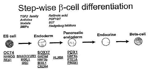
[0143] Figure 1 is a schematic of a proposed differentiation pathway
for the production of
beta-cells from hESCs. The first step in the pathway commits the ES cell to
the definitive endoderm
lineage and also represents the first step prior to further differentiation
events to pancreatic endoderm,
endocrine endoderm, or islet/beta-cells. The second step in the pathway shows
the conversion of
SOX17-positive/PDX1-negative definitive endoderm to PDXI-positive foregut
endoderm. Some factors
useful for mediating these transitions are italicized. Relevant markers for
defining the target cells are
underlined.
[0144] Figure 2 is a diagram of the human SOX17 eDNA which displays the
positions of
conserved motifs and highlights the region used for the immunization procedure
by GENOVAC.
[0145] Figure 3 is a relational dendrogram illustrating that SOX17 is
most closely related
to SOX7 and somewhat less to SOX18. The SOX17 proteins are more closely
related among species
homologs than to other members of the SOX group F subfamily within the same
species.
[0146] Figure 4 is a Western blot probed with the rat anti-S0X17
antibody. This blot
demonstrates the specificity of this antibody for human SOX17 protein over-
expressed in fibroblasts
13
CA 2564114
(lane 1) and a lack of immunoreactivity with EGFP (lane 2) or the most closely
related SOX family
member, SOX7 (lane 3).
[0147] Figures 5A-B are micrographs showing a cluster of SOXI 7- cells that
display a significant
number of AFP' co-labeled cells (A). This is in striking contrast to other
SOX17+ clusters (B) where
little or no AFP cells are observed.
[0148] Figures 6A-C are micrographs showing parietal endoderm and SOX17. Panel
A shows
immunocytochemistry for human Thrombomodulin (TM) protein located on the cell
surface of parietal
endoderm cells in randomly differentiated cultures of hES cells. Panel B is
the identical field shown in
A double-labeled for TM and SOX17. Panel C is the phase contrast image of the
same field with DAPI
labeled nuclei. Note the complete correlation of DAPI labeled nuclei and SOX17
labeling.
[0149] Figures 7A-B are bar charts showing SOX17 gene expression by
quantitative PCR (Q-PCR)
and anti-SOX17 positive cells by SOX17-specific antibody. Panel A shows that
activin A increases
SOX17 gene expression while retinoic acid (RA) strongly suppresses SOX17
expression relative to the
undifferentiated control media (SR20). Panel B shows the identical pattern as
well as a similar
magnitude of these changes is reflected in SOX17 + cell number, indicating
that Q-PCR measurement of
SOX17 gene expression is very reflective of changes at the single cell level.
[0150] Figure 8A is a bar chart which shows that a culture of differentiating
hESCs in the presence
of activin A maintains a low level of AFP gene expression while cells allowed
to randomly differentiate
in 10% fetal bovine serum (FBS) exhibit a strong upregulation of AFP. The
difference in expression
levels is approximately 7-fold.
[0151] Figures 8B-C are images of two micrographs showing that the suppression
of AFP
expression by activin A is also evident at the single cell level as indicated
by the very rare and small
clusters of AFP cells observed in activin A treatment conditions (bottom)
relative to 10% FBS alone
(top).
[0152] Figure 9 contains comparative images showing the quantitation of the
AFP' cell number
using flow cytometry. This figure demonstrates that the magnitude of change in
AFP gene expression
(Figure 8A) in the presence (panel B) and absence (panel A) of activin A
exactly corresponds to the
number of ATP' cells, further supporting the utility of Q-PCR analyses to
indicate changes occurring at
the individual cell level.
[0153] Figures 10A-F are micrographs which show that exposure of hESCs to
nodal, activin A and
activin B (NAA) yields a striking increase in the number of SOX17' cells over
the period of 5 days (A-
C). By comparing to the relative abundance of SOX1r cells to the total number
of cells present in each
field, as indicated by DAPI stained nuclei (D-F), it can be seen that
approximately 30-50% of all cells
are immunoreactive for SOX17 after five days treatment with NAA.
14
CA 2564114 2018-02-27
CA 02564114 2006-10-16
WO 2005/116073 PCMJS2005/014239
[0154] Figure 11 is a bar chart which demonstrates that activin A (0,
10, 30 or 100
ng/ml) dose-dependently increases SOX17 gene expression in differentiating
hESCs. Increased
expression is already robust after 3 days of treatment on adherent cultures
and continues through
subsequent 1, 3 and 5 days of suspension culture as well.
[0155] Figures 12A-C are bar charts which demonstrate the effect of
activin A on the
expression of MIXL1 (panel A), GATA4 (panel B) and HNF3b (panel C). Activin A
dose-dependent
increases are also observed for three other markers of definitive endoderm;
MDCL1, GATA4 and
HNF3b. The magnitudes of increased expression in response to activin dose are
strikingly similar to
those observed for SOX17, strongly indicating that activin A is specifying a
population of cells that
co-express all four genes (SOX17, MIXL1+, GATA4+ and HNF3b).
[0156] Figures 13A-C are bar charts which demonstrate the effect of
activin A on the
expression of AFP (panel A), SOX7 (panel B) and SPARC (panel C). There is an
activin A dose-
dependent decrease in expression of the visceral endoderm marker AFP. Markers
of primitive
endoderm (S0X7) and parietal endoderm (SPARC) remain either unchanged or
exhibit suppression
at some time points indicating that activin A does not act to specify these
extra-embryonic endoderm
cell types. This further supports the fact that the increased expression of
SOX17, MIXL1, GATA4,
and HNF3b are due to an increase in the number of definitive endoderm cells in
response to activin
A.
[0157] Figures 14A-B are bar charts showing the effect of activin A on
ZIC1 (panel A)
and Braehyury expression (panel B) Consistent expression of the neural marker
ZIC1 demonstrates
that there is not a dose-dependent effect of activin A on neural
differentiation. There is a notable
suppression of mesoderm differentiation mediated by 100 ng/ml of activin A
treatment as indicated
by the decreased expression of brachyury. This is likely the result of the
increased specification of
definitive endoderm from the mesendoderm precursors. Lower levels of activin A
treatment (10 and
30 ng/ml) maintain the expression of brachyury at later time points of
differentiation relative to
untreated control cultures.
[0158] Figures 15A-B are micrographs showing decreased parietal
endoderm
differentiation in response to treatment with activins. Regions of TMhi
parietal endoderm are found
through the culture (A) when differentiated in serum alone, while
differentiation to TM' cells is
scarce when activins are included (B) and overall intensity of TM
immunoreactivity is lower.
[0159] Figures 16A-D are micrographs which show marker expression in
response to
treatment with activin A and activin B. hESCs were treated for four
consecutive days with activin A
and activin B and triple labeled with SOX17, AFP and TM antibodies. Panel A -
SOX17; Panel B -
AFP; Panel C - TM; and Panel D - Phase/DAPI. Notice the numerous SOX17
positive cells (A)
associated with the complete absence of AFP (B) and TM (C) immunoreactivity.
CA 02564114 2006-10-16
WO 2005/116073 PCMJS2005/014239
[0160] Figure 17 is a micrograph showing the appearance of definitive
endoderm and
visceral endoderm in vitro from hESCs. The regions of visceral endoderm are
identified by
AFPhi/S0X171 /- while definitive endoderm displays the complete opposite
profile, SOX17"i/AFP101-.
This field was selectively chosen due to the proximity of these two regions to
each other. However,
there are numerous times when SOX17hi/AFPw- regions are observed in absolute
isolation from any
regions of APP" cells, suggesting the separate origination of the definitive
endoderm cells from
visceral endoderm cells.
[0161] Figure 18 is a diagram depicting the TGF13 family of ligands and
receptors.
Factors activating AR Smads and BR Smads are useful in the production of
definitive endoderm
from human embryonic stem cells (see, J Cell Physiol.187:265-76).
[0162] Figure 19 is a bar chart showing the induction of SOX17
expression over time as
a result of treatment with individual and combinations of TGFP factors.
[0163] Figure 20 is a bar chart showing the increase in SOX17+ cell
number with time=
as a result of treatment with combinations of TGFI3 factors.
[0164] Figure 21 is a bar chart showing induction of SOX17 expression
over time as a
result of treatment with combinations of TGF(El factors.
[0165] Figure 22 is a bar chart showing that activin A induces a dose-
dependent
increase in SOX17+ cell number.
[0166] Figure 23 is a bar chart showing that addition of Wnt3a to
activin A and activin
B treated cultures increases SOX17 expression above the levels induced by
activin A and activin B
alone.
[0167] Figures 24A-C are bar charts showing differentiation to
definitive endoderm is
enhanced in low FBS conditions. Treatment of hESCs with activins A and B in
media containing 2%
FBS (2AA) yields a 2-3 times greater level of SOX17 expression as compared to
the same treatment
in 10% FBS media (10AA) (panel A). Induction of the definitive endoderm marker
MlXL1 (panel
B) is also affected in the same way and the suppression of AFP (visceral
endoderm) (panel C) is
greater in 2% FBS than in 10% FBS conditions.
[0168] Figures 25A-D are micrographs which show SOX17+ cells are
dividing in
culture. SOX17 imrnunoreactive cells are present at the differentiating edge
of an hESC colony (C,
D) and are labeled with proliferating cell nuclear antigen (PCNA) (panel B)
yet are not co-labeled
with OCT4 (panel C). In addition, clear mitotic figures can be seen by DAPI
labeling of nuclei in
both SOX17+ cells (arrows) as well as OCT4+, undifferentiated hESCs
(arrowheads) (D).
[0169] Figure 26 is a bar chart showing the relative expression level
of CXCR4 in
differentiating hESCs under various media conditions.
16
CA 02564114 2006-10-16
WO 2005/116073 PCMJS2005/014239
[0170] Figures 27A-D are bar charts that show how a panel of
definitive endoderm
markers share a very similar pattern of expression to CXCR4 across the same
differentiation
treatments displayed in Figure 26.
[0171] Figures 28A-E are bar charts showing how markers for mesoderm
(BRACHYURY, MOX1), ectoderm (S0X1, ZIC1) and visceral endoderm (S0X7) exhibit
an inverse
relationship to CXCR4 expression across the same treatments displayed in
Figure 26.
[0172] Figures 29A-F are micrographs that show the relative difference
in SOX17
immunoreactive cells across three of the media conditions displayed in Figures
26-28.
[0173] Figures 30A-C are flow cytometry dot plots that demonstrate the
increase in
CXCR4 + cell number with increasing concentration of activin A added to the
differentiation media.
[0174] Figures 31A-D are bar charts that show the CXCR4 cells
isolated from the high
dose activin A treatment (A100-CX+) are even further enriched for definitive
endoderm markers than
the parent population (A100).
[0175] Figure 32 is a bar chart showing gene expression from CXCR4 +
and CXCR4
cells isolated using fluorescence-activated cell sorting (FACS) as well as
gene expression in the
parent populations. This demonstrates that the CXCR4 + cells contain
essentially all the CXCR4 gene
expression present in each parent population and the CXCR4- populations
contain very little or no
CXCR4 gene expression.
[0176] Figures 33A-D are bar charts that demonstrate the depletion of
mesoderm
(BRACHYURY, MOX1), ectoderm (ZIC1) and visceral endoderm (S0X7) gene
expression in the
CXCR4+ cells isolated from the high dose activin A treatment which is already
suppressed in
expression of these non-definitive endoderm markers.
[0177] Figures 34A-M are bar charts showing the expression patterns of
marker genes
that can be used to identify definitive endoderm cells. The expression
analysis of definitive
endoderm markers, FGF17, VWF, CALCR, FOXQ1, CMKOR1 and CRLP1 is shown in
panels G-L,
respectively. The expression analysis of previously described lineage marking
genes, SOX17,
SOX7, SOX17/S0X7, TM, ZIC1, and MOX1 is shown in panels A-F, respectively.
Panel M shows
the expression analysis of CXCR4. With respect to each of panels A-M, the
column labeled hESC
indicates gene expression from purified human embryonic stem cells; 2NF
indicates cells treated
with 2% FBS, no activin addition; 0.1A100 indicates cells treated with 0.1%
FBS, 100 ng/ml activin
A; 1A100 indicates cells treated with 1% FBS, 100 ng/ml activin A; and 2A100
indicates cells
treated with 2% FBS, 100 ng/ml activin A.
[0178] Figure 35 is a chart which shows the relative expression of the
PDX1 gene in a
culture of hESCs after 4 days and 6 days with and without activin in the
presence of retinoic acid
(RA) and fibroblast growth factor (FGF-10) added on day 4.
17
CA 02564114 2006-10-16
WO 2005/116073 PCMJS2005/014239
[0179] Figures 36A-F are charts which show the relative expression of
marker genes in
a culture of hESCs after 4 days and 6 days with and without activin in the
presence of retinoic acid
(RA) and fibroblast growth factor (FGF-10) added on day 4. The panels show the
relative levels of
expression of the following marker genes: (A) SOX17; (B) SOX7; (C) AFP; (D)
SOX1; (E) ZIC1;
and (F) NFM.
[0180] Figures 37A-C are charts which show the relative expression of
marker genes in
a culture of hESCs after 4 days and 8 days with and without activin in the
presence or absence of
combinations of retinoic acid (RA), fibroblast growth factor (FGF-10) and
fibroblast growth factor
(FGF-4) added on day 4. The panels show the relative levels of expression of
the following marker
genes: (A) PDX1; (B) SOX7; and (C) NFM.
[0181] Figures 38A-G are charts which show the relative expression of
marker genes in
a culture of definitive endoderm cells contacted with 50 ng/ml FGF-10 in
combination with either 1
M, 0.2 IAM or 0.04 jIM retinoic acid (RA) added on day 4. The panels show the
relative levels of
expression of the following marker genes: (A) PDX1; (B) HOXA3; (C) HOXC6; (D)
HOXA13; (E)
CDX1; (F) SOX I; and (G) NFM.
[0182] Figures 39A-E are charts which show the relative expression of
marker genes in
a culture of hESCs after 4 days and 8 days with and without activin in the
presence of combinations
of retinoic acid (RA), fibroblast growth factor (FGF-10) and one of the
following: serum replacement
(SR), fetal bovine serum (FBS) or B27. The panels show the relative levels of
expression of the
following marker genes: (A) PDX1; (B) SOX7; (C) AFP; (D) ZIC1; and (E) NFM.
[0183] Figures 40A-B are charts which show the relative expression of
marker genes for
pancreas (PDX1, HNF6) and liver (HNF6) in a culture of hESCs after 6 days
(just prior to addition of
RA) and at 9 days (three days after exposure to RA). Various conditions were
included to compare
the addition of activin B at doses of 10 ng/ml (a10), 25 ng/ml (a25) or 50
ng/ml (a50) in the presence
of either 25 ng/ml (A25) or 50 ng/ml (A50) activin A. The condition without
any activin A or activin
B (NF) serves as the negative control for definitive endoderm and PDX1-
positive endoderm
production. The panels show the relative levels of expression of the following
marker genes: (A)
PDX1and (B) HNF6.
[0184] Figures 41A-C are charts which show the relative expression of
marker genes in
a culture of hESCs with 100 ng/ml (A100), 50 ng/ml (A50) or without (NF)
activin A at 5 days (just
prior to retinoic acid addition) and at 2, 4, and 6 days after RA exposure
(day 7, 9, and 11,
respectively). The percentage label directly under each bar indicates the FBS
dose during days 3-5 of
differentiation. Starting at day 7, cells treated with RA (R) were grown in
RPMI medium comprising
0.5% FBS. The RA concentration was 2 1.1M on day 7, 11.IM on day 9 and 0.2
1.tM on day 11. The
panels show the relative levels of expression of the following marker genes:
(A) PDX1; (B) ZIC1;
(C) SOX7.
18
CA 02564114 2006-10-16
WO 2005/116073 PCMJS2005/014239
[0185] Figures 42A-B are charts which show the relative expression of
marker genes in
a culture of hESCs treated first with activin A in low FBS to induce
definitive endoderm (day 5) and
then with fresh (A25R) medium comprising 25 ng/ml activin A and RA or various
conditioned media
(MEFCM, CM#2, CM#3 and CM#4) and RA to induce PDX1-expressing endoderm. Marker
expression was determined on days 5, 6, 7, 8 and 9. The panels show the
relative levels of
expression of the following marker genes: (A) PDX1; (B) CDX1.
101861 Figure 43 is a chart which shows the relative expression of PDX1 in
a culture of
hESCs treated first with activin A in low FBS to induce definitive endodeim
and followed by fresh
media comprising activin A and retinoic acid (A25R) or varying amounts of RA
in conditioned
media diluted into fresh media. Total volume of media is 5 nil in all cases.
[0187] Figure 44 is a Western blot showing PDX1 immunoprecipitated from RA-
treated
definitive endoderm cells 3 days (d8) and 4 days (d9) after the addition of RA
and 50 neml
activin A.
[0188] Figure 45 is a summary chart displaying the results of a
fluorescence-activated
cell sort (FACs) of PDX1-positive foregut endoderm cells genetically tagged
with a EGFP reporter
under control of the PDX1 promoter.
[0189] Figure 46 is a chart showing relative PDX1 expression levels
normalized to
housekeeping genes for sorted populations of live cells (Live), EGFP-negative
cells (Meg) and
EGFP-positive cells (GFP+).
[0190] Figure 47 is a chart showing relative PDX1 expression levels
normalized to
housekeeping genes for sorted populations of live cells (Live), EGFP-negative
cells (Neg), the half of
the EGFP-positive cell population that has the lowest EGFP signal intensity
(Lo) and the half of the
EGFP-positive cell population that has the highest EGFP signal intensity (Hi).
[0191] Figures 48A-E are a charts showing the relative expression levels
normalized to
housekeeping genes of five pancreatic endoderm markers in sorted populations
of live cells (Live),
EGFP-negative cells (Meg) and EGFP-positive cells (GFP+). Panels: A ¨ NKX2.2;
B ¨ GLUT2; C ¨
HNF3 (3; D ¨ KRT19 and E ¨ HNF4oc.
[0192] Figure 49 are a charts showing the relative expression levels
normalized to
housekeeping genes of two non-pancreatic endoderm markers in sorted
populations of live cells
(Live), EGFP-negative cells (Meg) and EGFP-positive cells (GFP+). Panels: A ¨
ZIC1 and B ¨
GFAP.
Detailed Description
[0193] A crucial stage in early human development termed gastrulation
occurs 2-3
weeks after fertilization. Gastrulation is extremely significant because it is
at this time that the three
primary germ layers are first specified and organized (Lu et al., 2001;
Schoenwolf and Smith, 2000).
The ectoderm is responsible for the eventual formation of the outer coverings
of the body and the
19
CA 02564114 2006-10-16
WO 2005/116073 PCMJS2005/014239
entire nervous system whereas the heart, blood, bone, skeletal muscle and
other connective tissues
are derived from the mesoderm. Definitive endoderm is defined as the germ
layer that is responsible
for foimation of the entire gut tube which includes the esophagus, stomach and
small and large
intestines, and the organs which derive from the gut tube such as the lungs,
liver, thymus, parathyroid
and thyroid glands, gall bladder and pancreas (Grapin-Botton and Melton, 2000;
Kimelman and
Griffin, 2000; Tremblay et al., 2000; Wells and Melton, 1999; Wells and
Melton, 2000). A very
important distinction should be made between the definitive endoderm and the
completely separate
lineage of cells termed primitive endoderm. The primitive endoderm is
primarily responsible for
formation of extra-embryonic tissues, mainly the parietal and visceral
endoderm portions of the
placental yolk sac and the extracellular matrix material of Reichert's
membrane.
[0194] During gastrulation, the process of definitive endoderm formation
begins with a
cellular migration event in which mesendoderm cells (cells competent to form
mesoderm or
endoderm) migrate through a structure called the primitive streak. Definitive
endoderm is derived
from cells, which migrate through the anterior portion of the streak and
through the node (a
specialized structure at the anterior-most region of the streak). As migration
occurs, definitive
endoderm populates first the most anterior gut tube and culminates with the
formation of the
posterior end of the gut tube.
The PDX1 Gene Expression During Development
[0195] PDX1 (also called STF-1, ]DX-1 and LPF-1) is a transcription factor
that is
necessary for development of the pancreas and rostral duodenum. PDX1 is first
expressed in the
pancreatic endoderm, which arises from posterior foregut endoderm and will
produce both the
exocrine and endocrine cells, starting at E8.5 in the mouse. Later, PDX1
becomes restricted to beta-
cells and some delta-cells. This expression pattern is maintained in the
adult. PDX1 is also
expressed in duodenal endoderm early in development, which is adjacent to the
forming pancreas,
then in the duodenal enterocytes and enteroendocrine cells, antral stomach and
in the common bile,
cystic and biliary ducts. This region of expression also becomes limited, at
the time that pancreatic
expression becomes restricted, to predominantly the rostral duodenum.
PDX1-Positive Cells and Processes Related Thereto
[0196] Embodiments of the present invention relate to novel, defined
processes for the
production of PDX1-positive endoderm cells, wherein the PDX1-positive endoderm
cells are
multipotent cells that can differentiate into cells, tissues or organs derived
from the foregut/midgut
region of the gut tube (PDXI-positive foregut/midgut endoderm). As used
herein, "multipotent" or
"multipotent cell" refers to a cell type that can give rise to a limited
number of other particular cell
types. As used herein, "foregut/midgut" refers to cells of the anterior
portion of the gut tube as well
as cells of the middle portion of the gut tube, including cells of the
foregut/midgut junction.
CA 02564114 2006-10-16
WO 2005/116073 PCMJS2005/014239
[0197] Some preferred embodiments of the present invention relate to
processes for the
production of PDX1-positive foregut endoderm cells. In some embodiments, these
PDX1-positive
foregut endoderm cells are multipotent cells that can differentiate into
cells, tissues or organs derived
from the anterior portion of the gut tube (PDX1-positive foregut endoderm).
[0198] Additional preferred embodiments relate to processes for the
production of
PDX1-positive endoderm cells of the posterior portion of the foregut. hi some
embodiments, these
PDX1-positive endoderm cells are multipotent cells that can differentiate into
cells, tissues or organs
derived from the posterior portion of the foregut region of the gut tube.
[0199] The PDX1-positive foregut endoderm cells, such as those produced
according to
the methods described herein, can be used to produce fully differentiated
insulin-producing 13-cells.
In some embodiments of the present invention, PDX1-positive foregut endoderm
cells are produced
by differentiating definitive endoderm cells that do not substantially express
PDX1 (PDX1-negative
definitive endoderm cells; also referred to herein as definitive endoderm) so
as to form PDX1-
positive foregut endoderm cells. PDX1-negative definitive endoderm cells can
be prepared by
differentiating pluripotent cells, such as embryonic stem cells, as described
herein or by any other
known methods. A convenient and highly efficient method for producing PDX1-
negative definitive
endoderm from pluripotent cells is described in US Patent No. 11/021,618,
entitled DEFINITIVE
ENDODERM, filed December 23, 2004.
[0200] Processes of producing PDX1-positive foregut endoderm cells provide
a basis
for efficient production of pancreatic tissues such as acinar cells, ductal
cells and islet cells from
pluripotent cells. In certain preferred embodiments, human PDX1-positive
foregut endoderm cells
are derived from human PDX1-negative definitive endoderm cells, which in turn,
are derived from
hESCs. These human PDX1-positive foregut endoderm cells can then be used to
produce functional
insulin-producing 13-cells. To obtain useful quantities of insulin-producing p-
cells, high efficiency of
differentiation is desirable for each of the differentiation steps that occur
prior to reaching the
pancreatic islet/13-cell fate. Because differentiation of PDX1-negative
definitive endoderm cells to
PDX1-positive foregut endoderm cells represents an early step towards the
production of functional
pancreatic islet/f3-cells (as shown in Figure 1), high efficiency of
differentiation at this step is
particularly desirable.
[0201] In view of the desirability of efficient differentiation of PDX1-
negative
definitive endoderm cells to PDX1-positive foregut endoderm cells, some
aspects of the present
invention relate to in vitro methodology that results in approximately 2-25%
conversion of PDX1-
negative definitive endoderm cells to PDX1-positive foregut endoderm cells.
Typically, such
methods encompass the application of culture and growth factor conditions in a
defined and
temporally specified fashion. Further enrichment of the cell population for
PDX1-positive foregut
endoderm cells can be achieved by isolation and/or purification of the PDX1-
positive foregut
21
CA 02564114 2006-10-16
WO 2005/116073 PCMJS2005/014239
endoderm cells from other cells in the population by using a reagent that
specifically binds to the
PDX1-positive foregut endoderm cells. As an alternative, PDX1-positive foregut
endoderm cells can
be labeled with a reporter gene, such as green fluorescent protein (GFP), so
as to enable the detection
of PDX1 expression. Such fluorescently labeled cells can then be purified by
fluorescent activated
cell sorting (FACS). Further aspects of the present invention relate to cell
cultures and enriched cell
populations comprising PDX1-positive foregut endoderm cells as well as methods
for identifying
factors useful in the differentiation to and from PDX1-positive foregut
endoderm.
[0202] In order to
determine the amount of PDX1-positive foregut endoderm cells in a
cell culture or cell population, a method of distinguishing this cell type
from the other cells in the
culture or in the population is desirable. Accordingly, certain embodiments of
the present invention
relate to cell markers whose presence, absence and/or relative expression
levels are indicative of
PDX1-positive foregut endoderm cells as well as methods for detecting and
determining the
expression of such markers. As used herein, "expression" refers to the
production of a material or
substance as well as the level or amount of production of a material or
substance. Thus, determining
the expression of a specific marker refers to detecting either the relative or
absolute amount of the
marker that is expressed or simply detecting the presence or absence of the
marker. As used herein,
"marker" refers to any molecule that can be observed or detected. For example,
a marker can
include, but is not limited to, a nucleic acid, such as a transcript of a
specific gene, a polypeptide
product of a gene, a non-gene product polypeptide, a glycoprotein, a
carbohydrate, a glycolipd, a
lipid, a lipoprotein or a small molecule (for example, molecules having a
molecular weight of less
than 10,000 am).
[0203] In some
embodiments of the present invention, the presence, absence and/or
level of expression of a marker is determined by quantitative PCR (Q-PCR). For
example, the
amount of transcript produced by certain genetic markers, such as PDX1, SOX17,
SOX7, SOX1,
ZIC1, NFM, alpha-fetoprotein (AFP), homeobox A13 (HOXA13), homeobox C6
(HOXC6), and/or
other markers described
herein is determined by Q-PCR. In other embodiments,
immunohistoehemistry is used to detect the proteins expressed by the above-
mentioned genes. In
still other embodiments, Q-PCR and immunohistochemical techniques are both
used to identify and
determine the amount or relative proportions of such markers.
[0204] By using the
differentiation and detection methods described herein, it is
possible to identify PDX1-positive foregut endoderm cells, as well as
determine the proportion of
PDX1-positive foregut endoderm cells in a cell culture or cell population. For
example, in some
embodiments of the present invention, the PDX1-positive foregut endoderm cells
or cell populations
that are produced express the PDX1 gene at a level of at least about 2 orders
of magnitude greater
than PDX1-negative cells or cell populations. In other embodiments, the PDX1-
positive foregut
endoderm cells and cell populations that are produced express the PDX1 gene at
a level of more than
22
CA 02564114 2015-06-10
=
2 orders of magnitude greater than PDX1-negative cells or cell populations. In
still other
embodiments, the PDX1-positive foregut endoderm cells or cell populations that
are produced
express one or more of the markers selected from the group consisting of PDX1,
SOX17, HOXA13
and HOXC6 at a level of about 2 or more than 2 orders of magnitude greater
than PDXI-negative
definitive endoderm cells or cell populations.
10201 The
compositions and methods described herein have several useful features.
For example, the cell cultures and cell populations comprising PDX1-positive
endoderm, as well as
the methods for producing such cell cultures and cell populations, are useful
for modeling the early
stages of human development. Furthermore, the compositions and methods
described herein can also
serve for therapeutic intervention in disease states, such as diabetes
mellitus. For example, since
PDXI-positive foregut endoderm serves as the source for only a limited number
of tissues, it can be
used in the development of pure tissue or cell types.
PRODUCTION OF PDX1-NEGATIVE DEFINITIVE ENDODERM (DEFINITIVE ENDODERM)
FROM PLURIPOTENT CELLS
102061 Cell
cultures and/or cell populations comprising PDX1-positive foregut
.
endoderm cells are produced from pluripotent cells by first producing PDX1-
negative definitive
endoderm (also referred to as "definitive endoderm"). Processes for
differentiating pluripotent cells
to produce cell cultures and enriched cell populations comprising definitive
endoderm is described
briefly below and in detail in US 20050158853
entitled DEFINITIVE ENDODERM, filed
December 23, 2004. In some of these processes, the pluripotent cells used as
starting material are
stem cells. In certain processes, definitive endoderm cell cultures and
enriched cell populations
comprising definitive endoderm cells are produced from embryonic stem cells.
As used herein,
"embryonic" refers to a range of developmental stages of an organism beginning
with a single zygote
and ending with a multicellular structure that no longer comprises pluripotent
or totipotent cells other
than developed gametic cells. In addition to embryos derived by gamete fusion,
the term
"embryonic" refers to embryos derived by somatic cell nuclear transfer. A
preferred method for
deriving definitive endoderm cells utilizes human embryonic stem cells as the
starting material for
definitive endoderm production. Such pluripotent cells can be cells that
originate from the mortila,
embryonic inner cell mass or those obtained from embryonic gonadal ridges.
Human embryonic
stem cells can be maintained in culture in a pluripotent state without
substantial differentiation using
methods that are known in the art. Such methods are described, for example, in
US Patent Nos.
5,453,357, 5,670,372, 5,690,926 5,843,780, 6,200,806 and 6,251,671.
[0297) In
some processes for producing definitive endoderm cells, hESCs are
maintained on a feeder layer. In such processes, any feeder layer which allows
hESCs to be
maintained in a pluripotent state can be used. One commonly used feeder layer
for the cultivation of
human embryonic stem cells is a layer of mouse fibroblasts. More recently,
human fibroblast feeder
23
CA 02564114 2006-10-16
WO 2005/116073 PCMJS2005/014239
layers have been developed for use in the cultivation of hESCs (see US Patent
Application No.
2002/0072117). Alternative processes for producing definitive endoderm permit
the maintenance of
pluripotent hESC without the use of a feeder layer. Methods of maintaining
pluripotent hESCs under
feeder-free conditions have been described in US Patent Application No.
2003/0175956.
[0208] The human embryonic stem cells used herein can be maintained in
culture either
with or without serum. In some embryonic stem cell maintenance procedures,
serum replacement is
used. In others, serum free culture techniques, such as those described in US
Patent Application No.
2003/0190748, are used.
[0209] Stem cells are maintained in culture in a pluripotent state by
routine passage
until it is desired that they be differentiated into clefmitive endoderm. In
some processes,
differentiation to definitive endoderm is achieved by providing to the stem
cell culture a growth
factor of the TGFP superfamily in an amount sufficient to promote
differentiation to definitive
endoderm. Growth factors of the TGFP superfamily which are useful for the
production of definitive
endoderm are selected from the Nodal/Activin or BMP subgroups. In some
preferred differentiation
processes, the growth factor is selected from the group consisting of Nodal,
activin A, activin B and
BMP4. Additionally, the growth factor Wnt3a and other Wnt family members are
useful for the
production of definitive endoderm cells. In certain differentiation processes,
combinations of any of
the above-mentioned growth factors can be used.
[0210] With respect to some of the processes for the differentiation
of pluripotent stem
cells to definitive endoderm cells, the above-mentioned growth factors are
provided to the cells so
that the growth factors are present in the cultures at concentrations
sufficient to promote
differentiation of at least a portion of the stem cells to definitive endoderm
cells. In some processes,
the above-mentioned growth factors are present in the cell culture at a
concentration of at least about
ng/ml, at least about 10 ng/ml, at least about 25 ng/ml, at least about 50
ng/ml, at least about 75
ng/ml, at least about 100 ng/ml, at least about 200 ng/ml, at least about 300
ng/ml, at least about 400
ng/ml, at least about 500 ng/ml, at least about 1000 ng/ml, at least about
2000 ng/ml, at least about
3000 ng/ml, at least about 4000 ng/ml, at least about 5000 ng/ml or more than
about 5000 ng/ml.
[0211] In certain processes for the differentiation of pluripotent
stem cells to definitive
endoderm cells, the above-mentioned growth factors are removed from the cell
culture subsequent to
their addition. For example, the growth factors can be removed within about
one day, about two
days, about three days, about four days, about five days, about six days,
about seven days, about
eight days, about nine days or about ten days after their addition. In a
preferred processes, the
growth factors are removed about four days after their addition.
[0212] Cultures of definitive endoderm cells can be grown in medium
containing
reduced serum or no serum. Under certain culture conditions, serum
concentrations can range from
about 0.05% v/v to about 20% v/v. For example, in some differentiation
processes, the serum
24
CA 02564114 2006-10-16
WO 2005/116073 PCMJS2005/014239
concentration of the medium can be less than about 0.05% (v/v), less than
about 0.1% (v/v), less than
about 0.2% (v/v), less than about 0.3% (v/v), less than about 0.4% (v/v), less
than about 0.5% (v/v),
less than about 0.6% (v/v), less than about 0.7% (v/v), less than about 0.8%
(v/v), less than about
0.9% (v/v), less than about 1% (v/v), less than about 2% (v/v), less than
about 3% (v/v), less than
about 4% (v/v), less than about 5% (v/v), less than about 6% (v/v), less than
about 7% (v/v), less than
about 8% (v/v), less than about 9% (v/v), less than about 10% (v/v), less than
about 15% (v/v) or less
than about 20% (v/v). In some processes, definitive endoderm cells are grown
without serum or with
serum replacement. In still other processes, definitive endoderm cells are
grown in the presence of
B27. In such processes, the concentration of B27 supplement can range from
about 0.1% v/v to
about 20% v/v.
MONITORING THE DIFFERENTIATION OF PLURIPOTENT CELLS TO PDX1-NEGATIVE
DEFINITIVE ENDODERM (DEFINITIVE ENDODERM)
[0213] The progression of the hESC culture to definitive endoderm can
be monitored by
determining the expression of markers characteristic of definitive endoderm.
In some processes, the
expression of certain markers is determined by detecting the presence or
absence of the marker.
Alternatively, the expression of certain markers can be determined by
measuring the level at which
the marker is present in the cells of the cell culture or cell population. In
such processes, the
measurement of marker expression can be qualitative or quantitative. One
method of quantitating the
expression of markers that are produced by marker genes is through the use of
quantitative PCR (Q-
PCR). Methods of performing Q-PCR are well known in the art. Other methods
which are known in
the art can also be used to quantitate marker gene expression. For example,
the expression of a
marker gene product can be detected by using antibodies specific for the
marker gene product of
interest. In certain processes, the expression of marker genes characteristic
of definitive endoderm as
well as the lack of significant expression of marker genes characteristic of
hESCs and other cell types
is determined.
[0214] As described further in the Examples below, a reliable marker
of definitive
endoderm is the SOX17 gene. As such, the definitive endoderm cells produced by
the processes
described herein express the SOX17 marker gene, thereby producing the SOX17
gene product.
Other markers of definitive endoderm are MIXL1, GATA4, IINF3b, GSC, FGF17,
VWF, CALCR,
FOXQ1, CMKOR1 and CRIP 1 . Since definitive endoderm cells express the SOX17
marker gene at
a level higher than that of the SOX7 marker gene, which is characteristic of
primitive and visceral
endoderm (see Table 1), in some processes, the expression of both SOX17 and
SOX7 is monitored.
In other processes, expression of the both the SOX17 marker gene and the OCT4
marker gene, which
is characteristic of hESCs, is monitored. Additionally, because definitive
endoderm cells express the
SOX17 marker gene at a level higher than that of the APP, SPARC or
Thrombomodulin (TM)
marker genes, the expression of these genes can also be monitored.
CA 02564114 2006-10-16
WO 2005/116073 PCMJS2005/014239
[0215] Another marker of
definitive endoderm is the CXCR4 gene. The CXCR4 gene
encodes a cell surface chemokine receptor whose ligand is the ehemoattractant
SDF-1. The principal
roles of the CXCR4 receptor-bearing cells in the adult are believed to be the
migration of
hematopoetic cells to the bone marrow, lymphocyte trafficking and the
differentiation of various B
cell and macrophage blood cell lineages [Kim, C., and Broxmeyer, H. J.
Leukocyte Biol. 65, 6-15
(1999)]. The CXCR4 receptor also functions as a coreceptor for the entry of
HIV-1 into T-cells
[Feng, Y., et al. Science,
272, 872-877 (1996)]. In an extensive series of studies carried out by
[McGrath, K.E. et al. Dev. Biology 213, 442-456 (1999)], the expression of the
chemokine receptor
CXCR4 and its unique ligand, SDF-1 [Kim, C., and Broxmyer, H., J. Leukocyte
Biol. 65, 6-15
(1999)], were delineated during early development and adult life in the mouse.
The CXCR4/SDF1
interaction in development became apparent when it was demonstrated that if
either gene was
disrupted in transgenic mice [Nagasawa et al. Nature, 382, 635-638 (1996)],
Ma, Q., et al Immunity,
10, 463-471 (1999)] it resulted in late embryonic lethality. McGrath et al.
demonstrated that CXCR4
is the most abundant chemokine receptor messenger RNA detected during early
gastrulating embryos
(E7.5) using a combination of RNase protection and in situ hybridization
methodologies. In the
gastrulating embryo, CXCR4/SDF-1 signaling appears to be mainly involved in
inducing migration
of primitive-streak germlayer cells and is expressed on definitive endoderm,
mesoderm and
extraembryonic mesoderm present at this time. In E7.2-7.8 mouse embryos, CXCR4
and alpha-
fetoprotein are mutually exclusive indicating a lack of expression in visceral
endoderm [McGrath,
K.E. et al. Dev. Biology 213, 442-456 (1999)].
[0216] Since definitive
endoderm cells produced by differentiating pluripotent cells
express the CXCR4 marker gene, expression of CXCR4 can be monitored in order
to track the
production of definitive endoderm cells. Additionally, definitive endoderm
cells produced by the
methods described herein express other markers of definitive endoderm
including, but not limited to,
SOX17, MD(L 1, GA'TA4, HNF3b, GSC, FGF17, VWF, CALCR, FOXQ1, CMKOR1 and CRIP1.
Since definitive endoderm cells express the CXCR4 marker gene at a level
higher than that of the
SOX7 marker gene, the expression of both CXCR4 and SOX7 can be monitored. In
other processes,
expression of the both the CXCR4 marker gene and the OCT4 marker gene, is
monitored.
Additionally, because definitive endoderm cells express the CXCR4 marker gene
at a level higher
than that of the AFP, SPARC or Thrombomodulin (TM) marker genes, the
expression of these genes
can also be monitored.
[0217] It will be
appreciated that expression of CXCR4 in endodermal cells does not
preclude the expression of SOX17. As such, definitive endoderm cells produced
by the processes
described herein will substantially express SOX17 and CXCR4 but will not
substantially express
AFP, TM, SPARC or PDX1.
26
CA 02564114 2006-10-16
WO 2005/116073 PCMJS2005/014239
ENRICHMENT, ISOLATION AND/OR PURIFICATION OF DEFINITIVE ENDODERM
[0218] Definitive endoderm cells produced by any of the above-described
processes can
be enriched, isolated and/or purified by using an affinity tag that is
specific for such cells. Examples
of affinity tags specific for definitive endoderm cells are antibodies,
ligands or other binding agents
that are specific to a marker molecule, such as a polypeptide, that is present
on the cell surface of
definitive endoderm cells but which is not substantially present on other cell
types that would be
found in a cell-culture produced by the methods described herein. In some
processes, an antibody
which binds to CXCR4 is used as an affinity tag for the enrichment, isolation
or purification of
definitive endoderm cells. In other processes, the chemolcine SDF-1 or other
molecules based on
SDF-1 can also be used as affinity tags. Such molecules include, but not
limited to, SDF-1 fragments,
SDF-1 fusions or SDF-1 mimetics.
[0219] Methods for making antibodies and using them for cell isolation are
known in
the art and such methods can be implemented for use with the antibodies and
definitive endoderm
cells described herein. In one process, an antibody which binds to CXCR4 is
attached to a magnetic
bead and then allowed to bind to definitive endoderm cells in a cell culture
which has been
enzymatically treated to reduce intercellular and substrate adhesion. The
cell/antibody/bead
complexes are then exposed to a movable magnetic field which is used to
separate bead-bound
definitive endoderm cells from unbound cells. Once the definitive endoderm
cells are physically
separated from other cells in culture, the antibody binding is disrupted and
the cells are replated in
appropriate tissue culture medium.
[0220] Additional methods for obtaining enriched, isolated or purified
definitive
endoderm cell cultures or populations can also be used. For example, in some
embodiments, the
CXCR4 antibody is incubated with a definitive endoderm-containing cell culture
that has been
treated to reduce intercellular and substrate adhesion. The cells are then
washed, centrifuged and
resuspended. The cell suspension is then incubated with a secondary antibody,
such as an FITC-
conjugated antibody that is capable of binding to the primary antibody. The
cells are then washed,
centrifuged and resuspended in buffer. The cell suspension is then analyzed
and sorted using a
fluorescence activated cell sorter (FACS). CXCR4-positive cells are collected
separately from
CXCR4-negative cells, thereby resulting in the isolation of such cell types.
If desired, the isolated
cell compositions can be further purified by using an alternate affinity-based
method or by additional
rounds of sorting using the same or different markers that are specific for
definitive endoderm.
[0221] In still other processes, definitive endoderm cells are enriched,
isolated and/or
purified using a ligand or other molecule that binds to CXCR4. In some
processes, the molecule is
SDF-1 or a fragment, fusion or mimetic thereof.
[0222] In preferred processes, definitive endoderm cells are enriched,
isolated and/or
purified from other non-definitive endoderm cells after the stem cell cultures
are induced to
27
CA 02564114 2006-10-16
WO 2005/116073 PCMJS2005/014239
differentiate towards the definitive endoderm lineage. It will be appreciated
that the above-described
enrichment, isolation and purification procedures can be used with such
cultures at any stage of
differentiation.
[0223] In addition to the procedures just described, definitive
endoderm cells may also
be isolated by other techniques for cell isolation. Additionally, definitive
endoderm cells may also be
enriched or isolated by methods of serial subculture in growth conditions
which promote the selective
survival or selective expansion of the definitive endoderm cells.
[0224] Using the methods described herein, enriched, isolated and/or
purified
populations of definitive endoderm cells and or tissues can be produced in
vitro from pluripotent cell
cultures or cell populations, such as stem cell cultures or populations, which
have undergone at least
some differentiation. In some methods, the cells undergo random
differentiation. In a preferred
method, however, the cells are directed to differentiate primarily into
definitive endoderm. Some
preferred enrichment, isolation and/or purification methods relate to the in
vitro production of
definitive endoderm from human embryonic stem cells. Using the methods
described herein, cell
populations or cell cultures can be enriched in definitive endoderm content by
at least about 2- to
about 1000-fold as compared to untreated cell populations or cell cultures.
COMPOSITIONS COMPRISING PDX1-NEGATIVE DEFINITIVE ENDODERM (DEFINITIVE
ENDODERM)
[0225] Cell compositions produced by the above-described methods
include cell
cultures comprising definitive endoderm and cell populations enriched in
definitive endoderm. For
example, cell cultures which comprise definitive endoderm cells, wherein at
least about 50-80% of
the cells in culture are definitive endoderm cells, can be produced. Because
the efficiency of the
differentiation process can be adjusted by modifying certain parameters, which
include but are not
limited to, cell growth conditions, growth factor concentrations and the
timing of culture steps, the
differentiation procedures described herein can result in about 5%, about 10%,
about 15%, about
20%, about 25%, about 30%, about 35%, about 40%, about 45%, about 50%, about
55%, about 60%,
about 65%, about 70%, about 75%, about 80%, about 85%, about 90%, about 95%,
or greater than
about 95% conversion of pluripotent cells to definitive endoderm. In processes
in which isolation of
definitive endoderm cells is employed, for example, by using an affinity
reagent that binds to the
CXCR4 receptor, a substantially pure definitive endoderm cell population can
be recovered.
PRODUCTION OF PDX1-POSITVE FOREGUT ENDODERM FROM PDX1-NEGATIVE
DEFINITIVE ENDODERM
[0226] The PDX1-positive foregut endoderm cell cultures and
populations comprising
PDX1-positive foregut endoderm cells that are described herein are produced
from PDX1-negative
definitive endoderm, which is generated from pluripotent cells as described
above. A preferred
method utilizes human embryonic stem cells as the starting material. In one
embodiment, hESCs are
28
CA 02564114 2006-10-16
WO 2005/116073 PCMJS2005/014239
first converted to PDX1-negative definitive endoderm cells, which are then
converted to PDX1-
positive foregut endoderm cells. It will be appreciated, however, that the
starting materials for the
production of PDX1-positive foregut endoderm is not limited to definitive
endoderm cells produced
using pluripotent cell differentiation methods. Rather, any PDX1-negative
definitive endoderm cells
can be used in the methods described herein regardless of their origin.
[0227] In some embodiments of the present invention, cell cultures or
cell populations
comprising PDX1-negative definitive endoderm cells can be used for further
differentiation to cell
cultures and/or enriched cell populations comprising PDX1-positive foregut
endoderm cells. For
example, a cell culture or cell population comprising human PDX1-negative,
SOX17-positive
definitive endoderm cells can be used. In some embodiments, the cell culture
or cell population may
also comprise differentiation factors, such as activins, nodals and/or BMPs,
remaining from the
previous differentiation step (that is, the step of differentiating
pluripotent cells to definitive
endoderm cells). In other embodiments, factors utilized in the previous
differentiation step are
removed from the cell culture or cell population prior to the addition of
factors used for the
differentiation of the PDX1-negative, SOX17-positive definitive endoderm cells
to PDX1-positive
foregut endoderm cells. In other embodiments, cell populations enriched for
PDX1-negative,
SOX17-positive definitive endoderm cells are used as a source for the
production of PDX1-positive
foregut endoderm cells.
[0228] PDX1-negative definitive endoderm cells in culture are
differentiated to PDX1-
positive endoderm cells by providing to a cell culture comprising PDX1-
negative, SOX17-positive
definitive endoderm cells a differentiation factor that promotes
differentiation of the cells to PDX1-
positive foregut endoderm cells (foregut differentiation factor). In some
embodiments of the present
invention, the foregut differentiation factor is retinoid, such as retinoic
acid (RA). In some
embodiments, the retinoid is used in conjunction with a fibroblast growth
factor, such as FGF-4 or
FGF-10. In other embodiments, the retinoid is used in conjunction with a
member of the TGF13
superfamily of growth factors and/or a conditioned medium.
[0229] By "conditioned medium" is meant, a medium that is altered as
compared to a
base medium. For example, the conditioning of a medium may cause molecules,
such as nutrients
and/or growth factors, to be added to or depleted from the original levels
found in the base medium.
In some embodiments, a medium is conditioned by allowing cells of certain
types to be grown or
maintained in the medium under certain conditions for a certain period of
time. For example, a
medium can be conditioned by allowing hESCs to be expanded, differentiated or
maintained in a
medium of defined composition at a defined temperature for a defined number of
hours. As will be
appreciated by those of skill in the art, numerous combinations of cells,
media types, durations and
environmental conditions can be used to produce nearly an infinite array of
conditioned media. In
some embodiments of the present invention, a medium is conditioned by allowing
differentiated
29
CA 02564114 2006-10-16
WO 2005/116073 PCMJS2005/014239
pluripotent cells to be grown or maintained in a medium comprising about 1% to
about 20% serum
concentration. In other embodiments, a medium is conditioned by allowing
differentiated pluripotent
cells to be grown or maintained in a medium comprising about 1 ng/ml to about
1000 ng/ml activin
A. In still other embodiments, a medium is conditioned allowing differentiated
pluripotent cells to be
grown or maintained in a medium comprising about 1 ng/ml to about 1000 ng/ml
BMP4. In a
preferred embodiment, a conditioned medium is prepared by allowing
differentiated hESCs to be
grown or maintained for 24 hours in a medium, such as RPMI, comprising about
25 ng/ml activin A
and about 2 M RA.
[0230] In some embodiments of the present invention, the cells used to
condition the
medium, which is used to enhance the differentiation of PDX1-negative
definitive endoderm to
PDX1-positive foregut endoderm, are cells that are differentiated from
pluripotent cells, such as
hESCs, over about a 5 day time period in a medium such as RPMI comprising
about 0% to about
20% serum and/or one or more growth/differentiation factors of the TGFI3
superfamily.
Differentiation factors, such as activin A and BMP4 are supplied at
concentrations ranging from
about 1 ng/ml to about 1000 ng/ml. In certain embodiments of the present
invention, the cells used
to condition the medium are differentiated from hESCs over about a 5 day
period in low serum
RPMI. According to some embodiments, low serum RPMI refers to a low serum
containing
medium, wherein the serum concentration is gradually increased over a defined
time period. For
example, in one embodiment, low serum RPMI comprises a concentration of about
0.2% fetal bovine
serum (FBS) on the first day of cell growth, about 0.5% FBS on the second day
of cell growth and
about 2% FBS on the third through fifth day of cell growth. In another
embodiment, low serum
RPMI comprises a concentration of about 0% on day one, about 0.2% on day two
and about 2% on
days 3-6. In certain preferred embodiments, low serum RPMI is supplemented
with one or more
differentiation factors, such as activin A and BMP4. In addition to its use in
preparing cells used to
condition media, low serum RPMI can be used as a medium for the
differentiation of PDX1-positive
foregut endoderm cells from PDX1-negative definitive endoderm cells.
[0231] It will be appreciated by those of ordinary skill in the art that
conditioned media
can be prepared from media other than RPMI provided that such media do not
interfere with the
growth or maintenance of PDX1-positive foregut endoderm cells. It will also be
appreciated that the
cells used to condition the medium can be of various types. In embodiments
where freshly
differentiated cells are used to condition a medium, such cells can be
differentiated in a medium
other than RPMI provided that the medium does not inhibit the growth or
maintenance of such cells.
Furthermore, a skilled artisan will appreciate that neither the duration of
conditioning nor the
duration of preparation of cells used for conditioning is required to be 24
hours or 5 days,
respectively, as other time periods will be sufficient to achieve the effects
reported herein.
CA 02564114 2006-10-16
WO 2005/116073 PCMJS2005/014239
[0232] In
general, the use of a retinoid in combination with a fibroblast growth factor,
a
member of the TGFI3 superfamily of growth factors, a conditioned medium or a
combination of any
of these foregut differentiation factors causes greater differentiation of
PDX1-negative definitive
endoderm to PDX1-positive foregut endoderm than the use of a retinoid alone.
In a preferred
embodiment, RA and FGF-10 are both provided to the PDX1-negative definitive
endoderm cell
culture. In
another preferred embodiment, PDX1-negative definitive endoderm cells are
differentiated in a culture comprising a conditioned medium, activin A,
activin B and RA.
[0233] With
respect to some of the embodiments of differentiation processes described
herein, the above-mentioned foregut differentiation factors are provided to
the cells so that these
factors are present in the cell culture or cell population at concentrations
sufficient to promote
differentiation of at least a portion of the PDX1-negative defmitive endoderm
cell culture or cell
population to PDX1-positive foregut endoderm cells. When used in connection
with cell cultures
and/or cell populations, the term "portion" means any non-zero amount of the
cell culture or cell
population, which ranges from a single cell to the entirety of the cell
culture or cells population.
[0234] In some
embodiments of the present invention, a retinoid is provided to the cells
of a cell culture such that it is present at a concentration of at least about
0.01 M, at least about 0.02
IuM, at least about 0.04 M, at least about 0.08 uM, at least about 0.1 M, at
least about 0.2 M, at
least about 0.3 M, at least about 0.4 M, at least about 0.5 uM, at least
about 0.6 M, at least about
0.7 NI, at least about 0.8 M, at least about 0.9 M, at least about 1 uM, at
least about 1.1 uM, at
least about 1.2 M, at least about 1.3 ;AM, at least about 1.4 uM, at least
about 1.5 M, at least about
1.6 M, at least about 1.7 M, at least about 1.8 M, at least about 1.9 pM,
at least about 2 M, at
least about 2.1 pM, at least about 2.2 M, at least about 2.3 uM, at least
about 2.4 M, at least about
2.5 pM, at least about 2.6 M, at least about 2.7 M, at least about 2.8 M,
at least about 2.9 uM, at
least about 3 M, at least about 3.5 uM, at least about 4 uM, at least about
4.5 uM, at least about 5
AM, at least about 10 pM, at least about 20 M, at least about 30 M, at least
about 40 M or at least
about 50 M. As used herein, "retinoid" refers to retinol, retinal or retinoic
acid as well as
derivatives of any of these compounds. In a preferred embodiment, the retinoid
is retinoic acid.
[0235] In
other embodiments of the present invention, one or more differentiation
factors of the fibroblast growth factor family are present in the cell
culture. For example, in some
embodiments, FG1-4 can be present in the cell culture at a concentration of at
least about 10 ng/ml,
at least about 25 ng/ml, at least about 50 ng/ml, at least about 75 ng/ml, at
least about 100 ng/ml, at
least about 200 ng/ml, at least about 300 ng/ml, at least about 400 ng/ml, at
least about 500 ng/ml, or
at least about 1000 ng/ml. In further embodiments of the present invention,
FGF-10 is present in the
cell culture at a concentration of at least about 10 ng/ml, at least about 25
ng/ml, at least about 50
ng/ml, at least about 75 ng/ml, at least about 100 ng/ml, at least about 200
ng/ml, at least about 300
31
CA 02564114 2006-10-16
WO 2005/116073 PCMJS2005/014239
ng/ml, at least about 400 ng/ml, at least about 500 ng/ml, or at least about
1000 ng/ml. hi some
embodiments, either FGF-4 or FGF-10, but not both, is provided to the cell
culture along with RA.
In a preferred embodiment, RA is present in the cell culture at 1 IAM and FGF-
10 is present at a
concentration of 50 ng/ml.
[0236] In some embodiments of the present invention, growth factors of
the TGFI3
superfamily and/or a conditioned medium are present in the cell culture. These
differentiation factors
can be used in combination with RA and/or other mid-foregut differentiation
factors including, but
not limited to, FGF-4 and FGF-10. For example, in some embodiments, activin A
and/or activin B
can be present in the cell culture at a concentration of at least about 5
ng/ml, at least about 10 ng/ml,
at least about 25 ng/ml, at least about 50 ng/ml, at least about 75 ng/ml, at
least about 100 ng/ml, at
least about 200 ng/ml, at least about 300 ng/ml, at least about 400 ng/ml, at
least about 500 ng/ml, or
at least about 1000 ng/ml. In further embodiments of the present invention, a
conditioned medium is
present in the cell culture at a concentration of at least about 1%, at least
about 5%, at least about
10%, at least about 20%, at least about 30%, at least about 40%, at least
about 50%, at least about
60%, at least about 70%, at least about 80%, at least about 90%, or at least
about 100% of the total
medium. In some embodiments, activin A, activin B and a conditioned medium are
provided to the
cell culture along with RA. In a preferred embodiment, PDX1-negative
definitive endoderm cells are
differentiated to PDX1-positive foregut endoderm cells in cultures comprising
about 1 M RA, about
25 ng/ml activin A and low serum RPMI medium that has been conditioned for
about 24 hours by
differentiated hESCs, wherein the differentiated hESCs have been
differentiated for about 5 days in
low serum RPMI comprising about 100 ng/ml activin A. In another preferred
embodiment, activin B
and/or FGF-10 are also present in the culture at 25 ng/ml and 50 ng/ml,
respectively.
[0237] In certain embodiments of the present invention, the above-
mentioned foregut
differentiation factors are removed from the cell culture subsequent to their
addition. For example,
the foregut differentiation factors can be removed within about one day, about
two days, about three
days, about four days, about five days, about six days, about seven days,
about eight days, about nine
days or about ten days after their addition.
[0238] Cultures of PDX1-positive foregut endoderm cells can be grown in
a medium
containing reduced serum. Serum concentrations can range from about 0.05%
(v/v) to about 20%
(v/v). In some embodiments, PDX1-positive foregut endoderm cells are grown
with serum
replacement. For example, in certain embodiments, the serum concentration of
the medium can be
less than about 0.05% (v/v), less than about 0.1% (v/v), less than about 0.2%
(v/v), less than about
0.3% (v/v), less than about 0.4% (v/v), less than about 0.5% (v/v), less than
about 0.6% (v/v), less
than about 0.7% (v/v), less than about 0.8% (v/v), less than about 0.9% (v/v),
less than about 1%
(v/v), less than about 2% (v/v), less than about 3% (v/v), less than about 4%
(v/v), less than about 5%
(v/v), less than about 6% (v/v), less than about 7% (v/v), less than about 8%
(v/v), less than about 9%
32
CA 02564114 2006-10-16
WO 2005/116073 PCMJS2005/014239
(v/v), less than about 10% (v/v), less than about 15% (v/v) or less than about
20% (v/v). hi some
embodiments, PDX1-positive foregut endoderm cells are grown without serum. In
other
embodiments, PDX1-positive foregut endoderm cells are grown with serum
replacement.
[0239] hi still other embodiments, PDX1-positive foregut endoderm
cells are grown in
the presence of B27. In such embodiments, B27 can be provided to the culture
medium in
concentrations ranging from about 0.1% (v/v) to about 20% (v/v) or in
concentrations greater than
about 20% (v/v). In certain embodiments, the concentration of B27 in the
medium is about 0.1%
(v/v), about 0.2% (v/v), about 0.3% (v/v), about 0.4% (v/v), about 0.5% (v/v),
about 0.6% (v/v),
about 0.7% (v/v), about 0.8% (v/v), about 0.9% (v/v), about 1% (v/v), about 2%
(v/v), about 3%
(v/v), about 4% (v/v), about 5% (v/v), about 6% (v/v), about 7% (v/v), about
8% (v/v), about 9%
(v/v), about 10% (v/v), about 15% (v/v) or about 20% (v/v). Alternatively, the
concentration of the
added B27 supplement can be measured in terms of multiples of the strength of
a commercially
available B27 stock solution. For example, B27 is available from Invitrogen
(Carlsbad, CA) as a
50X stock solution. Addition of a sufficient amount of this stock solution to
a sufficient volume of
growth medium produces a medium supplemented with the desired amount of B27.
For example, the
addition of 10 ml of 50X B27 stock solution to 90 ml of growth medium would
produce a growth
medium supplemented with 5X B27. The concentration of B27 supplement in the
medium can be
about 0.1X, about 0.2X, about 0.3X, about 0.4X, about 0.5X, about 0.6X, about
0.7X, about 0.8X,
about 0.9X, about lx, about 1.1X, about 1.2X, about 1.3X, about 1.4X, about
1.5X, about 1.6X,
about 1.7X, about 1.8X, about 1.9X, about 2X, about 2.5X, about 3X, about
3.5X, about 4X, about
4.5X, about 5X, about 6X, about 7X, about 8X, about 9X, about 10X, about 11X,
about 12X, about
13X, about 14X, about 15X, about 16X, about 17X, about 18X, about 19X, about
20X and greater
than about 20X.
MONITORING THE DIFFERENTIATION OF PDX1-NEGATIVE DEFINITIVE ENDODERM TO
PDX1-POSITIVE ENDODERM
[0240] As with the differentiation of definitive endoderm cells from
pluripotent cells,
the progression of differentiation from PDX1-negative, SOX17-positive
definitive endoderm to
PDX1-positive foregut endoderm can be monitored by determining the expression
of markers
characteristic of these cell types. Such monitoring permits one to determine
the amount of time that
is sufficient for the production of a desired amount of PDX1-positive foregut
endoderm under
various conditions, for example, one or more differentiation factor
concentrations and environmental
conditions, hi preferred embodiments, the amount of time that is sufficient
for the production of a
desired amount of PDX1-positive foregut endoderm is determined by detecting
the expression of
PDX1. In some embodiments of the present invention, the expression of certain
markers is
determined by detecting the presence or absence of the marker. Alternatively,
the expression of
certain markers can be determined by measuring the level at which the marker
is present in the cells
33
CA 02564114 2006-10-16
WO 2005/116073 PCMJS2005/014239
of the cell culture or cell population. In such embodiments, the measurement
of marker expression
can be qualitative or quantitative. As described above, a preferred method of
quantitating the
expression markers that are produced by marker genes is through the use of Q-
PCR. In particular
embodiments, Q-PCR is used to monitor the progression of cells of the PDX1-
negative, SOX17-
positive definitive endoderm culture to PDX1-positive foregut endoderm cells
by quantitating
expression of marker genes characteristic of PDX1-positive foregut endoderm
and the lack of
expression of marker genes characteristic of other cell types. Other methods
which are known in the
art can also be used to quantitate marker gene expression. For example, the
expression of a marker
gene product can be detected by using antibodies specific for the marker gene
product of interest. In
some embodiments of the present invention, the expression of marker genes
characteristic of PDX1-
positive foregut endoderm as well as the lack of significant expression of
marker genes characteristic
of PDX1-negative definitive endoderm, hESCs and other cell types is
determined.
[0241] As described further in the Examples below, PDX1 is a marker
gene that is
associated with PDX1-positive foregut endoderm. As such, in some embodiments
of the present
invention, the expression of PDX1 is determined. In other embodiments, the
expression of other
markers, which are expressed in PDX1-positive foregut endoderm, including, but
not limited to,
SOX17, HOXA13 and/or HOXC6 is also determined. Since PDX1 can also be
expressed by certain
other cell types (that is, visceral endoderm and certain neural ectoderm),
some embodiments of the
present invention relate to demonstrating the absence or substantial absence
of marker gene
expression that is associated with visceral endoderm and/or neural ectoderm.
For example, in some
embodiments, the expression of markers, which are expressed in visceral
endoderm and/or neural
cells, including, but not limited to, SOX7, APP, SOX1, ZIC1 and/or NFM is
determined.
[0242] In some embodiments, PDX1-positive foregut endoderm cell
cultures produced
by the methods described herein are substantially free of cells expressing the
SOX7, AFP, SOX1,
ZIC1 or NFM marker genes. In certain embodiments, the PDX1-positive foregut
endoderm cell
cultures produced by the processes described herein are substantially free of
visceral endoderm,
parietal endoderm and/or neural cells.
ENRICHMENT, ISOLATION AND/OR PURIFICATION OF PDX1-POSITIVE FOREGUT
ENDODERM
[0243] With respect to additional aspects of the present invention,
PDX1-positive
foregut endoderm cells can be enriched, isolated and/or purified. In some
embodiments of the
present invention, cell populations enriched for PDX1-positive foregut
endoderm cells are produced
by isolating such cells from cell cultures.
[0244] In some embodiments of the present invention, PDX1-positive
foregut endoderm
cells are fluorescently labeled then isolated from non-labeled cells by using
a fluorescence activated
cell sorter (FACS). In such embodiments, a nucleic acid encoding green
fluorescent protein (GFP) or
34
CA 02564114 2006-10-16
WO 2005/116073 PCMJS2005/014239
another nucleic acid encoding an expressible fluorescent marker gene is used
to label PDX1-positive
cells. For example, in some embodiments, at least one copy of a nucleic acid
encoding GFP or a
biologically active fragment thereof is introduced into a pluripotent cell,
preferably a human
embryonic stem cell, downstream of the PDX1 promoter such that the expression
of the GFP gene
product or biologically active fragment thereof is under control of the PDX1
promoter. In some
embodiments, the entire coding region of the nucleic acid, which encodes PDX1,
is replaced by a
nucleic acid encoding GFP or a biologically active fragment thereof. In other
embodiments, the
nucleic acid encoding GFP or a biologically active fragment thereof is fused
in frame with at least a
portion of the nucleic acid encoding PDX1, thereby generating a fusion
protein. In such
embodiments, the fusion protein retains a fluorescent activity similar to GFP.
[0245] *Fluorescently marked cells, such as the above-described
pluripotent cells, are
differentiated to definitive endoderm and then to PDX1-positive foregut
endoderm as described
previously above. Because PDX1-positive foregut endoderm cells express the
fluorescent marker
gene, whereas PDX1-negative cells do not, these two cell types can be
separated. In some
embodiments, cell suspensions comprising a mixture of fluorescently-labeled
PDX1-positive cells
and unlabeled PDX1-negative cells are sorted using a FACS. PDX1-positive cells
are collected
separately from PDX1-negative cells, thereby resulting in the isolation of
such cell types. If desired,
the isolated cell compositions can be further purified by additional rounds of
sorting using the same
or different markers that are specific for PDX1-positve foregut endoderm.
[0246] In addition to the procedures just described, PDX1-positive
foregut endoderm
cells may also be isolated by other techniques for cell isolation.
Additionally, PDX1-positive foregut
endoderm cells may also be enriched or isolated by methods of serial
subculture in growth conditions
which promote the selective survival or selective expansion of said PDX1-
positive foregut endoderm
cells.
[0247] It will be appreciated that the above-described enrichment,
isolation and
purification procedures can be used with such cultures at any stage of
differentiation.
[0248] Using the methods described herein, enriched, isolated and/or
purified
populations of PDX1-positive foregut endoderm cells and/or tissues can be
produced in vitro from
PDX1-negative, SOX17-positive definitive endoderm cell cultures or cell
populations which have
undergone at least some differentiation. In some embodiments, the cells
undergo random
differentiation. In a preferred embodiment, however, the cells are directed to
differentiate primarily
into PDX1-positive foregut endoderm cells. Some preferred enrichment,
isolation and/or purification
methods relate to the in vitro production of PDX1-positive foregut endoderm
cells from human
embryonic stem cells.
[0249] Using the methods described herein, cell populations or cell
cultures can be
enriched in PDX1-positive foregut endoderm cell content by at least about 2-
to about 1000-fold as
CA 02564114 2006-10-16
WO 2005/116073 PCMJS2005/014239
compared to untreated cell populations or cell cultures. In some embodiments,
PDX1-positive
foregut endoderm cells can be enriched by at least about 5- to about 500-fold
as compared to
untreated cell populations or cell cultures. In other embodiments, PDX1-
positive foregut endoderm
cells can be enriched from at least about 10- to about 200-fold as compared to
untreated cell
populations or cell cultures. In still other embodiments, PDX1-positive
foregut endoderm cells can
be enriched from at least about 20- to about 100-fold as compared to untreated
cell populations or
cell cultures. In yet other embodiments, PDX1-positive foregut endoderm cells
can be enriched from
at least about 40- to about 80-fold as compared to untreated cell populations
or cell cultures. In
certain embodiments, PDX1-positive foregut endoderm cells can be enriched from
at least about 2- to
about 20-fold as compared to untreated cell populations or cell cultures.
COMPOSITIONS COMPRISING PDX1-POSITIVE FOREGUT ENDODERM
[0250] Some embodiments of the present invention relate to cell
compositions, such as
cell cultures or cell populations, comprising PDX1-positive endoderm cells,
wherein the PDX1-
positive endoderm cells are multipotent cells that can differentiate into
cells, tissues or organs
derived from the anterior portion of the gut tube (PDX1-positive foregut
endoderm). In accordance
with certain embodiments, the PDX1-positive foregut endoderm are mammalian
cells, and in a
preferred embodiment, the definitive endoderm cells are human cells.,
[0251] Other embodiments of the present invention relate to
compositions, such as cell
cultures or cell populations, comprising cells of one or more cell types
selected from the group
consisting of hESCs, PDX1-negative definitive endoderm cells, PDX1-positive
foregut endoderm
cells and mesoderm cells. In some embodiments, hESCs comprise less than about
5%, less than
about 4%, less than about 3%, less than about 2% or less than about 1% of the
total cells in the
culture. In other embodiments, PDX1-negative definitive endoderm cells
comprise less than about
90%, less than about 85%, less than about 80%, less than about 75%, less than
about 70%, less than
about 65%, less than about 60%, less than about 55%, less than about 50%, less
than about 45%, less
than about 40%, less than about 35%, less than about 30%, less than about 25%,
less than about 20%,
less than about 15%, less than about 12%, less than about 10%, less than about
8%, less than about
6%, less than about 5%, less than about 4%, less than about 3%, less than
about 2% or less than
about 1% of the total cells in the culture. In yet other embodiments, mesoderm
cells comprise less
than about 90%, less than about 85%, less than about 80%, less than about 75%,
less than about 70%,
less than about 65%, less than about 60%, less than about 55%, less than about
50%, less than about
45%, less than about 40%, less than about 35%, less than about 30%, less than
about 25%, less than
about 20%, less than about 15%, less than about 12%, less than about 10%, less
than about 8%, less
than about 6%, less than about 5%, less than about 4%, less than about 3%,
less than about 2% or
less than about 1% of the total cells in the culture.
36
CA 02564114 2006-10-16
WO 2005/116073 PCMJS2005/014239
[0252] Additional embodiments of the present invention relate to
compositions, such as
cell cultures or cell populations, produced by the processes described herein
comprise PDX1-positive
foregut endoderm as the majority cell type. In some embodiments, the processes
described herein
produce cell cultures and/or cell populations comprising at least about 99%,
at least about 98%, at
least about 97%, at least about 96%, at least about 95%, at least about 94%,
at least about 93%, at
least about 92%, at least about 91%, at least about 90%, at least about 85%,
at least about 80%, at
least about 75%, at least about 70%, at least about 65%, at least about 60%,
at least about 55%, at
least about 54%, at least about 53%, at least about 52% or at least about 51%
PDX1-positive foregut
endoderm cells. In preferred embodiments the cells of the cell cultures or
cell populations comprise
human cells. In other embodiments, the processes described herein produce cell
cultures or cell
populations comprising at least about 50%, at least about 45%, at least about
40%, at least about
35%, at least about 30%, at least about 25%, at least about 24%, at least
about 23%, at least about
22%, at least about 21%, at least about 20%, at least about 19%, at least
about 18%, at least about
17%, at least about 16%, at least about 15%, at least about 14%, at least
about 13%, at least about
12%, at least about 11%, at least about 10%, at least about 9%, at least about
8%, at least about 7%,
at least about 6%, at least about 5%, at least about 4%, at least about 3%, at
least about 2% or at least
about 1% PDX1-positive foregut endoderm cells. In preferred embodiments, the
cells of the cell
cultures or cell populations comprise human cells. In some embodiments, the
percentage of PDX1-
positive foregut endoderm cells in the cell cultures or populations is
calculated without regard to the
feeder cells remaining in the culture.
[0253] Still other embodiments of the present invention relate to
compositions, such as
cell cultures or cell populations, comprising mixtures of PDX1-positive
foregut endoderm cells and
PDX1-negative definitive endoderm cells. For example, cell cultures or cell
populations comprising
at least about 5 PDX1-positive foregut endoderm cells for about every 95 PDX1-
negative defmitive
endoderm cells can be produced. hi other embodiments, cell cultures or cell
populations comprising
at least about 95 PDX1-positive foregut endoderm cells for about every 5 PDX1-
negative definitive
endoderm cells can be produced. Additionally, cell cultures or cell
populations comprising other
ratios of PDX1-positive foregut endoderm cells to PDX1-negative definitive
endoderm cells are
contemplated. For example, compositions comprising at least about 1 PDX1-
positive foregut
endoderm cell for about every 1,000,000 PDX1-negative definitive endoderm
cells, at least about 1
PDX1-positive foregut endoderm cell for about every 100,000 PDX1-negative
definitive endoderm
cells, at least about 1 PDX1-positive foregut endoderm cell for about every
10,000 PDX1-negative
definitive endoderm cells, at least about 1 PDX1-positive foregut endoderm
cell for about every 1000
PDX1-negative definitive endoderm cells, at least about 1 PDX1-positive
foregut endoderm cell for
about every 500 PDX1-negative definitive endoderm cells, at least about 1 PDX1-
positive foregut
endoderm cell for about every 100 PDX1-negative definitive endoderm cells, at
least about 1 PDX1-
37
CA 02564114 2006-10-16
WO 2005/116073 PCMJS2005/014239
positive foregut endoderm cell for about every 10 PDX1-negative definitive
endoderm cells, at least
about 1 PDX1-positive foregut endoderm cell for about every 5 PDX1-negative
definitive endoderm
cells, at least about 1 PDX1-positive foregut endoderm cell for about every 4
PDX1-negative
definitive endoderm cells, at least about 1 PDX1-positive foregut endoderm
cell for about every 2
. PDX1-negative definitive endoderm cells, at least about 1 PDX-1 positive
foregut endoderm cell for
about every 1 PDX1-negative definitive endoderm cell, at least about 2 PDX1-
positive foregut
endoderm cells for about every 1 PDXI -negative definitive endoderm cell, at
least about 4 PDX1-
positive foregut endoderm cells for about every 1 PDX1-negative definitive
endoderm cell, at least
about 5 PDX1-positive foregut endoderm cells for about every 1 PDX1-negative
definitive endoderm
cell, at least about 10 PDX1-positive foregut endoderm cells for about every 1
PDX1-negative
definitive endoderm cell, at least about 20 PDX1-positive foregut endoderm
cells for about every 1
PDX1-negative definitive endoderm cell, at least about 50 PDX1-positive
foregut endoderm cells for
about every 1 PDX1-negative definitive endoderm cell, at least about 100 PDX1-
positive foregut
endoderm cells for about every 1 PDX1-negative definitive endoderm cell, at
least about 1000
PDX1-positive foregut endoderm cells for about every 1 PDX1-negative
definitive endoderm cell, at
least about 10,000 PDX1-positive foregut endoderm cells for about every 1 PDX1-
negative definitive
endoderm cell, at least about 100,000 PDX1-positive foregut endoderm cells for
about every 1
PDX1-negative definitive endoderm cell and at least about 1,000,000 PDX1-
positive foregut
endoderm cells for about every 1 PDX1-negative definitive endoderm cell are
contemplated.
[0254] In some embodiments of the present invention, the PDX1-negative
definitive
endoderm cells from which PDX1-positive foregut endoderm cells are produced
are derived from
human pluripotent cells, such as human pluripotent stem cells. In certain
embodiments, the human
pluripotent cells are derived from a morula, the inner cell mass of an embryo
or the gonadal ridges of
an embryo. In certain other embodiments, the human pluripotent cells are
derived from the gondal or
germ tissues of a multicellular structure that has developed past the
embryonic stage.
[0255] Further embodiments of the present invention relate to
compositions, such as cell
cultures or cell populations, comprising human cells, including human PDX1-
positive foregut
endoderm, wherein the expression of the PDX1 marker is greater than the
expression of the AFP,
SOX7, SOX1, ZIC1 and/or NFM marker in at least about 2% of the human cells. In
other
embodiments, the expression of the PDX1 marker is greater than the expression
of the AFP, SOX7,
SOX1, ZIC1 and/or NFM marker in at least about 5% of the human cells, in at
least about 10% of the
human cells, in at least about 15% of the human cells, in at least about 20%
of the human cells, in at
least about 25% of the human cells, in at least about 30% of the human cells,
in at least about 35% of
the human cells, in at least about 40% of the human cells, in at least about
45% of the human cells, in
at least about 50% of the human cells, in at least about 55% of the human
cells, in at least about 60%
of the human cells, in at least about 65% of the human cells, in at least
about 70% of the human cells,
38
CA 02564114 2006-10-16
WO 2005/116073 PCMJS2005/014239
in at least about 75% of the human cells, in at least about 80% of the human
cells, in at least about
85% of the human cells, in at least about 90% of the human cells, in at least
about 95% of the human
cells or in at least about 98% of the human cells. In some embodiments, the
percentage of human
cells in the cell cultures or populations, wherein the expression of PDX1 is
greater than the
expression of the AFP, SOX7, SOX1, ZIC1 and/or NFM marker, is calculated
without regard to
feeder cells.
[0256] It will be appreciated that some embodiments of the present
invention relate to
compositions, such as cell cultures or cell populations, comprising human PDX1-
positive foregut
endoderm cells, wherein the expression of one or more markers selected from
the group consisting of
SOX17, HOXA13 and HOXC6 is greater than the expression of the AFP, SOX7, SOX1,
ZIC1 and/or
NFM marker in from at least about 2% to greater than at least about 98% of the
human cells. In
some embodiments, the expression of one or more markers selected from the
group consisting of
SOX17, HOXA13 and HOXC6 is greater than the expression of the AFP, SOX7, SOX1,
ZIC1 and/or
NFM marker in at least about 5% of the human cells, in at least about 10% of
the human cells, in at
least about 15% of the human cells, in at least about 20% of the human cells,
in at least about 25% of
the human cells, in at least about 30% of the human cells, in at least about
35% of the human cells, in
at least about 40% of the human cells, in at least about 45% of the human
cells, in at least about 50%
of the human cells, in at least about 55% of the human cells, in at least
about 60% of the human cells,
in at least about 65% of the human cells, in at least about 70% of the human
cells, in at least about
75% of the human cells, in at least about 80% of the human cells, in at least
about 85% of the human
cells, in at least about 90% of the human cells, in at least about 95% of the
human cells or in at least
about 98% of the human cells. In some embodiments, the percentage of human
cells in the cell
cultures or populations, wherein the expression of one or more markers
selected from the group
consisting of SOX17, HOXA13 and HOXC6 is greater than the expression of the
AFP, SOX7,
SOX1, ZIC1 and/or NFM marker, is calculated without regard to feeder cells.
[0257] Additional embodiments of the present invention relate to
compositions, such as
cell cultures or cell populations, comprising mammalian endodermal cells, such
as human endodeim
cells, wherein the expression of the PDX1 marker is greater than the
expression of the AFP, SOX7,
SOX1, ZIC1 and/or NFM marker in at least about 2% of the endodermal cells. In
other
embodiments, the expression of the PDX1 marker is greater than the expression
of the AFP, SOX7,
SOX1, ZIC1 and/or NFM marker in at least about 5% of the endodemial cells, in
at least about 10%
of the endodermal cells, in at least about 15% of the endodermal cells, in at
least about 20% of the
endodermal cells, in at least about 25% of the endodermal cells, in at least
about 30% of the
endodeimal cells, in at least about 35% of the endodermal cells, in at least
about 40% of the
endodermal cells, in at least about 45% of the endodermal cells, in at least
about 50% of the
endodermal cells, in at least about 55% of the endodermal cells, in at least
about 60% of the
39
CA 02564114 2006-10-16
WO 2005/116073 PCMJS2005/014239
endodermal cells, in at least about 65% of the endodermal cells, in at least
about 70% of the
endodermal cells, in at least about 75% of the endodermal cells, in at least
about 80% of the
endodermal cells, in at least about 85% of the endodermal cells, in at least
about 90% of the
endodermal cells, in at least about 95% of the endodermal cells or in at least
about 98% of the
endodermal cells.
[0258] Still other embodiments of the present invention relate to
compositions, such as
cell cultures or cell populations, comprising mammalian endodermal cells, such
as human
endodermal cells, wherein the expression of one or more markers selected from
the group consisting
of SOX17, HOXA13 and HOXC6 is greater than the expression of the AFP, SOX7,
SOX1, ZIC1
and/or NFM marker in at least about 2% of the endodermal cells. In other
embodiments, the
expression of one or more markers selected from the group consisting of SOX17,
HOXA13 and
HOXC6 is greater than the expression of the AFP, SOX7, SOX1, ZIC1 and/or NFM
marker in at
least about 5% of the endodermal cells, in at least about 10% of the
endodermal cells, in at least
about 15% of the endodermal cells, in at least about 20% of the endodermal
cells, in at least about
25% of the endodermal cells, in at least about 30% of the endodermal cells, in
at least about 35% of
the endodermal cells, in at least about 40% of the endodermal cells, in at
least about 45% of the
endodermal cells, in at least about 50% of the endodeimal cells, in at least
about 55% of the
endodermal cells, in at least about 60% of the endodermal cells, in at least
about 65% of the
endodermal cells, in at least about 70% of the endodermal cells, in at least
about 75% of the
endodermal cells, in at least about 80% of the endodermal cells, in at least
about 85% of the
endodermal cells, in at least about 90% of the endodermal cells, in at least
about 95% of the
endodermal cells or at least about 98% of the endodermal cells.
[0259] Using the processes described herein, compositions comprising
PDX1-positive
foregut endoderm cells substantially free of other cell types can be produced.
With respect to cells in
cell cultures or in cell populations, the term "substantially free of' means
that the specified cell type
of which the cell culture or cell population is free, is present in an amount
of less than about 5% of
the total number of cells present in the cell culture or cell population. In
some embodiments of the
present invention, the PDX1-positive foregut endoderm cell populations or cell
cultures produced by
the methods described herein are substantially free of cells that
significantly express the AFP, SOX7,
SOX1, ZIC1 and/or NFM marker genes.
[0260] In one embodiment of the present invention, a description of a
PDX1-positive
foregut endoderm cell based on the expression of marker genes is, PDX1 high,
AFP low, SOX7 low,
SOX1 low, ZIC1 low and NFM low.
CA 02564114 2006-10-16
WO 2005/116073 PCMJS2005/014239
INCREASING EXPRESSION OF PDX1 IN A SOX17-POSITIVE DEFINITIVE ENDODERM
CELL
[0261] Some aspects of the present invention are related to methods of
increasing the
expression of the PDX1 gene product in cell cultures or cell populations
comprising SOX17-positive
defmitive endoderm cells. In such embodiments, the SOX17-positive definitive
endoderm cells are
contacted with a differentiation factor in an amount that is sufficient to
increase the expression of the
PDX1 gene product. The SOX17-positive definitive endoderm cells that are
contacted with the
differentiation factor can be either PDX1-negative or PDX1-positive. In some
embodiments, the
differentiation factor can be a retinoid. In certain embodiments, SOX17-
positive definitive endoderm
cells are contacted with a retinoid at a concentration ranging from about 0.01
1.1M to about 50 M. In
a preferred embodiment, the retinoid is RA.
[0262] In other embodiments of the present invention, the expression
of the PDX1 gene
product in cell cultures or cell populations comprising SOX17-positive
definitive endoderm cells is
increased by contacting the SOX17-positive cells with a differentiation factor
of the fibroblast
growth factor family. Such differentiation factors can either be used alone or
in conjunction with
RA. In some embodiments, the SOX17-positive definitive endoderm cells are
contacted with a
fibroblast growth factor at a concentration ranging from about 10 ng/ml to
about 1000 ng/ml. In a
preferred embodiment, the FGF growth factor is FGF-10.
[0263] In some embodiments of the presentinvention, the expression of
the PDX1 gene
product in cell cultures or cell populations comprising SOX17-positive
definitive endoderm cells is
increased by contacting the SOX17-positive cells with B27. This
differentiation factor can either be
used alone or in conjunction with one or both of retinoid and FGF family
differentiation factors. In
some embodiments, the SOX17-positive definitive endoderm cells are contacted
with B27 at a
concentration ranging from about 0.1% (v/v) to about 20% (v/v). In a preferred
embodiment, the
SOX17-positive definitive endoderm cells are contacted with RA, FGF-10 and
B27.
[0264] Methods for increasing the expression of the PDX1 gene product
in cell cultures
or cell populations comprising SOX17-positive defmitive endoderm cells can be
carried out in
growth medium containing reduced or no serum. In some embodiments, serum
concentrations range
from about 0.05% (v/v) to about 20% (v/v). In some embodiments, the SOX17-
positive cells are
grown with serum replacement.
IDENTIFICATION OF FACTORS CAPABLE OF PROMOTING THE DIFFERENTIATION OF
PDX1-NEGATIVE DEFINITIVE ENDODERM CELLS TO PDX1-POSITIVE FOREGUT
ENDODERM CELLS
[0265] Additional aspects of the present invention relate to methods
of identifying one
or more differentiation factors capable of promoting the differentiation of
PDX1-negative definitive
endoderm cells to PDX1-positive foregut endodemi cells. In such methods, a
cell culture or cell
41
CA 02564114 2006-10-16
WO 2005/116073 PCMJS2005/014239
population comprising PDX1-negative definitive endoderm cells is obtained and
the expression of
PDX1 in the cell culture or cell population is determined. After determining
the expression of PDX1,
the cells of the cell culture or cell population are contacted with a
candidate differentiation factor. In
some embodiments, the expression of PDX1 is determined at the time of
contacting or shortly after
contacting the cells with a candidate differentiation factor. PDX1 expression
is then determined at
one or more times after contacting the cells with the candidate
differentiation factor. If the
expression of PDX1 has increased after contact with the candidate
differentiation factor as compared
to PDX1 expression prior to contact with the candidate differentiation factor,
the candidate
differentiation factor is identified as capable of promoting the
differentiation of PDX1-negative
definitive endoderm cells to PDX1-positive foregut endoderm cells.
[0266] In some embodiments, the above-described methods of identifying
factors
capable of promoting the differentiation of PDX1-negative definitive endoderm
cells to PDX1-
positive foregut endoderm cells also include determining the expression of the
HOXA13 gene and/or
the HOXC6 gene in the cell culture or cell population. In such embodiments,
the expression of
HOXA13 and/or HOXC6 is determined both before and after the cells are
contacted with the
candidate differentiation factor. If the expression of PDX1 and HOXA13 has
increased after contact
with the candidate differentiation factor as compared to PDX1 and HOXA13
expression prior to
contact with the candidate differentiation factor, the candidate
differentiation factor is identified as
capable of promoting the differentiation of PDX1-negative definitive endoderm
cells to PDX1-
positive foregut endoderm cells. Similarly, if the expression of PDX1 and
HOXC6 has increased
after contact with the candidate differentiation factor as compared to PDX1
and HOXC6 expression
prior to contact with the candidate differentiation factor, the candidate
differentiation factor is
identified as capable of promoting the differentiation of PDX1-negative
definitive endoderm cells to
PDX1-positive foregut endoderm cells. In a preferred embodiment, a candidate
differentiation factor
is identified as being capable of promoting the differentiation of PDX1-
negative definitive endoderm
cells to PDX1-positive foregut endoderm cells by determining the expression of
PDX1, HOXA13
and HOXC6 both before and after contacting the cells of the cell culture or
cell population with the
candidate differentiation factor. In preferred embodiments, the expression of
PDX1, HOXA13
and/or HOXC6 is determined Q-PCR.
[0267] It will be appreciated that in some embodiments, the expression
of one or more
of PDX1, HOXA13 and HOXC6 can be determined at the time of contacting or
shortly after
contacting the cells of the cell cultures or cell populations with a candidate
differentiation factor
rather than prior to contacting the cells with a candidate differentiation
factor. In such embodiments,
the expression of one or more of PDX1, HOXA13 and HOXC6 at the time of
contacting or shortly
after contacting the cells with a candidate differentiation factor is compared
to the expression of one
42
CA 02564114 2006-10-16
WO 2005/116073 PCMJS2005/014239
or more of PDX1, HOXA13 and HOXC6 at one or more times after contacting the
cells with a
candidate differentiation factor.
[0268] hi some embodiments of the above-described methods, the one or
more times at
which PDX1 expression is determined after contacting the cells with the
candidate differentiation
factor can range from about 1 hour to about 10 days. For example, PDX1
expression can be
determined about 1 hour after contacting the cells with the candidate
differentiation factor, about 2
hours after contacting the cells with the candidate differentiation factor,
about 4 hours after
contacting the cells with the candidate differentiation factor, about 6 hours
after contacting the cells
with the candidate differentiation factor, about 8 hours after contacting the
cells with the candidate
differentiation factor, about 10 hours after contacting the cells with the
candidate differentiation
factor, about 12 hours after contacting the cells with the candidate
differentiation factor, about 16
hours after contacting the cells with the candidate differentiation factor,
about 24 hours after
contacting the cells with the candidate differentiation factor, about 2 days
after contacting the cells
with the candidate differentiation factor, about 3 days after contacting the
cells with the candidate
differentiation factor, about 4 days after contacting the cells with the
candidate differentiation factor,
about 5 days after contacting the cells with the candidate differentiation
factor, about 6 days after
contacting the cells with the candidate differentiation factor, about 7 days
after contacting the cells
with the candidate differentiation factor, about 8 days after contacting the
cells with the candidate
differentiation factor, about 9 days after contacting the cells with the
candidate differentiation factor,
about 10 days after contacting the cells with the candidate differentiation
factor or more than 10 days
after contacting the cells with the candidate differentiation factor.
[0269] Candidate differentiation factors for use in the methods
described herein can be
selected from compounds, such as polypeptides and small molecules. For
example, candidate
polypeptides can include, but are not limited to, growth factors, cytokines,
chemokines, extracellular
matrix proteins, and synthetic peptides. In a preferred embodiment, the growth
factor is from the
FGF family, for example FGF-10. Candidate small molecules include, but are not
limited to,
compounds generated from combinatorial chemical synthesis and natural
products, such as steroids,
isoprenoids, terpenoids, phenylpropanoids, alkaloids and flavinoids. It will
be appreciated by those
of ordinary skill in the art that thousands of classes of natural and
synthetic small molecules are
available and that the small molecules contemplated for use in the methods
described herein are not
limited to the classes exemplified above. Typically, small molecules will have
a molecular weight
less than 10,000 amu. In a preferred embodiment, the small molecule is a
retinoid, for example RA.
IDENTIFICATION OF FACTORS CAPABLE OF PROMOTING THE DIFFERENTIATION OF
PDX1-POSITIVE FOREGUT ENDODERM CELLS
[0270] Other aspects of the present invention relate to methods of
identifying one or
more differentiation factors capable of promoting the differentiation of PDX1-
positive foregut
43
CA 02564114 2006-10-16
WO 2005/116073 PCMJS2005/014239
endoderm cells. In such methods, a cell culture or cell population comprising
PDX1-positive foregut
endoderm cells is obtained and the expression of a marker in the cell culture
or cell population is
determined. After determining the expression of the marker, the cells of the
cell culture or cell
population are contacted with a candidate differentiation factor. In some
embodiments, the
expression of the marker is determined at the time of contacting or shortly
after contacting the cells
with a candidate differentiation factor. The expression of the same marker is
then determined at one
or more times after contacting the cells with the candidate differentiation
factor. If the expression of
the marker has increased or decreased after contact with the candidate
differentiation factor as
compared to the marker expression prior to contact with the candidate
differentiation factor, the
candidate differentiation factor is identified as capable of promoting the
differentiation of PDX1-
positive foregut endoderm cells. In preferred embodiments, expression of the
marker is determined
by Q-PCR.
[0271] k some embodiments of the above-described methods, the one or
more times at
which the marker expression is determined after contacting the cells with the
candidate
differentiation factor can range from about 1 hour to about 10 days. For
example, marker expression
can be determined about 1 hour after contacting the cells with the candidate
differentiation factor,
about 2 hours after contacting the cells with the candidate differentiation
factor, about 4 hours after
contacting the cells with the candidate differentiation factor, about 6 hours
after contacting the cells
with the candidate differentiation factor, about 8 hours after contacting the
cells with the candidate
differentiation factor, about 10 hours after contacting the cells with the
candidate differentiation
factor, about 12 hours after contacting the cells with the candidate
differentiation factor, about 16
hours after contacting the cells with the candidate differentiation factor,
about 24 hours after
contacting the cells with the candidate differentiation factor, about 2 days
after contacting the cells
with the candidate differentiation factor, about 3 days after contacting the
cells with the candidate
differentiation factor, about 4 days after contacting the cells with the
candidate differentiation factor,
about 5 days after contacting the cells with the candidate differentiation
factor, about 6 days after
contacting the cells with the candidate differentiation factor, about 7 days
after contacting the cells
with the candidate differentiation factor, about 8 days after contacting the
cells with the candidate
differentiation factor, about 9 days after contacting the cells with the
candidate differentiation factor,
about 10 days after contacting the cells with the candidate differentiation
factor or more than 10 days
after contacting the cells with the candidate differentiation factor.
[0272] As described previously, candidate differentiation factors for
use in the methods
described herein can be selected from compounds such as polypeptides and small
molecules.
[0273] Although each of the methods disclosed herein have been
described with respect
to PDX1-positive foregut endoderm cells, it will be appreciated that in
certain embodiments, these
methods can be used to produce compositions comprising the PDX1-positive
foregut/midgut
44
CA 02564114 2012-05-03
endoderm cells that are described herein and/or the PDX1-positive endoderm
cells of the posterior
portion of the foregut that are described herein. Furthermore, any of the PDX1-
positive endoderm
cell types disclosed in this specification can be utilized in the screening
methods described herein. =
[02741 Having generally described this invention, a further
understanding can be
obtained by reference to certain specific examples . which are provided herein
for purposes of
illustration only, and are not intended to be limiting.
EXAMPLES =
[0275] Many of the examples below describe the use of pluripotent human
cells.
Methods of producing pluripotent human cells are well known in the art and
have been described
numerous scientific publications, including U.S. Patent Nos. 5,453,357,
5,670,372, 5,690,926,
6,090,622, 6,200,806 and 6,251,671 as well as U.S. Patent Application
Publication No.
2004/0229350.
EXAMPLE 1
Human ES cells
[0276] For our studies of endoderm development we employed human
embryonic stem
cells, which are pluripotent and can divide seemingly indefinitely in culture
while maintaining a
normal kaiyotype. ES cells were derived from the 5-day-old embryo inner cell
mass using either
immunological or mechanical methods for isolation. In particular, the human
embryonic stem cell
line hESCyt-25 was derived from a supernumerary frozen embryo from an in vitro
fertilization cycle
following informed consent by the patient Upon thawing the hatched blastoc-yst
was plated on
mouse embryonic fibroblasts (MEF), in ES medium (DMEM, 20% PBS, non essential
amino acids,
beta-mercaptoethanol, ITS supplement). The embryo adhered to the culture dish
and after
approximately two weeks, regions of undifferentiated hESCs were transferred to
new dishes with
MEFs. Transfer was accomplished with mechanical cutting and a brief digestion
with dispaseTM,
followed by mechanical removal of the cell clusters, washing and re-plating.
Since derivation,
hESCyt-25 has been serially passaged over 100 times. We employed the hESCyt-25
human
embryonic stem cell line as our starting material for the production of
definitive endoderm.
[0277] It will be appreciated by those of skill in the art that stem
cells or other
pluripotent cells can also be used as starting material for the
differentiation procedures described
herein. For example, cells obtained from embryonic gonadal ridges, which can
be isolated by
methods known in the art, can be used as pluripotent cellular starting
material.
EXAMPLE 2
liESCyt-25 Characterization
[0278] The human embryonic stem cell line, hESCyt-25 has maintained a
normal
morphology, kaiyotype, growth and self-renewal properties over 18 months in
culture. This cell line
displays strong immunoreactivity for the OCT4, SSEA-4 and TRA-1-60 antigens,
all of which, are
CA 02564114 2006-10-16
WO 2005/116073 PCMJS2005/014239
characteristic of undifferentiated hESCs and displays alkaline phosphatase
activity as well as a
morphology identical to other established hESC lines. Furthermore, the human
stem cell line,
hESCyt-25, also readily forms embryoid bodies (EBs) when cultured in
suspension. As a
demonstration of its pluripotent nature, hESCyT-25 differentiates into various
cell types that
represent the three principal germ layers. Ectoderm production was
demonstrated by Q-PCR for
ZIC1 as well as immunocytochemistry (ICC) for nestin and more mature neuronal
markers.
Immunocytochemical staining for 13-III tubulin was observed in clusters of
elongated cells,
characteristic of early neurons. Previously, we treated EBs in suspension with
retinoic acid, to
induce differentiation of pluripotent stern cells to visceral endoderm (VE),
an extra-embryonic
lineage. Treated cells expressed high levels of a-fetoprotein (AFP) and SOX7,
two markers of VE,
by 54 hours of treatment. Cells differentiated in monolayer expressed AFP in
sporadic patches as
demonstrated by immunocytochemical staining. As will be described below, the
hESCyT-25 cell
line was also capable of fouling definitive endoderm, as validated by real-
time quantitative
polymerase chain reaction (Q-PCR) and immunocytochemistry for SOX17, in the
absence of AFP
expression. To demonstrate differentiation to mesoderm, differentiating EBs
were analyzed for
Brachyury gene expression at several time points. Brachyury expression
increased progressively
over the course of the experiment. In view of the foregoing, the hESCyT-25
line is pluripotent as
shown by the ability to form cells representing the three germ layers.
EXAMPLE 3
Production of SOX17 Antibody
[0279] A primary obstacle to the identification of definitive endoderm
in hESC cultures
is the lack of appropriate tools. We therefore undertook the production of an
antibody raised against
human SOX17 protein.
[0280] The marker SOX17 is expressed throughout the definitive endoderm
as it forms
during gastrulation and its expression is maintained in the gut tube (although
levels of expression
vary along the A-P axis) until around the onset of organogenesis. SOX17 is
also expressed in a
subset of extra-embryonic endoderm cells. No expression of this protein has
been observed in
mesoderm or ectoderm. It has now been discovered that SOX17 is an appropriate
marker for the
definitive endoderm lineage when used in conjunction with markers to exclude
extra-embryonic
lineages.
[0281] As described in detail herein, the SOX17 antibody was utilized
to specifically
examine effects of various treatments and differentiation procedures aimed at
the production of
SOX17 positive definitive endoderm cells. Other antibodies reactive to AFP,
SPARC and
Thrombomodulin were also employed to rule out the production of visceral and
parietal endoderm
(extra-embryonic endoderm).
46
CA 02564114 2006-10-16
WO 2005/116073 PCMJS2005/014239
[0282] In order to produce an antibody against SOX17, a portion of the
human SOX17
cDNA (SEQ ID NO: 1) corresponding to amino acids 172-414 (SEQ ID NO: 2) in the
carboxyteiininal end of the SOX17 protein (Figure 2) was used for genetic
immunization in rats at
the antibody production company, GENOVAC (Freiberg, Germany), according to
procedures
developed there. Procedures for genetic immunization can be found in US Patent
Nos. 5,830,876,
5,817,637, 6,165,993 and 6,261,281 as well as International Patent Application
Publication Nos.
W000/29442 and W099/13915.
[0283] Other suitable methods for genetic immunization are also
described in the non-
patent literature. For example, Barry et al. describe the production of
monoclonal antibodies by
genetic immunization in Biotechniques 16: 616-620, 1994. Specific examples of
genetic
immunization methods to produce antibodies against specific proteins can be
found, for example, in
Costaglia et al., (1998) Genetic immunization against the human thyrotropin
receptor causes
thyroiditis and allows production of monoclonal antibodies recognizing the
native receptor, J.
Innnunol. 160: 1458-1465; Kilpatrick et al (1998) Gene gun delivered DNA-based
immunizations
mediate rapid production of murine monoclonal antibodies to the Flt-3
receptor, Hybridotna 17: 569-
576; Schrnolke et al., (1998) Identification of hepatitis G virus particles in
human serum by E2-
specific monoclonal antibodies generated by DNA immunization, J. Virol. 72:
4541-4545;
Krasemann et al., (1999) Generation of monoclonal antibodies against proteins
with an
unconventional nucleic acid-based immunization strategy, J. Biotechnol. 73:
119-129; and Ulivieri et
al., (1996) Generation of a monoclonal antibody to a defined portion of the
Heliobacter pylori
vacuolating cytotoxin by DNA immunization, J. Biotechnol. 51: 191-194.
[0284] SOX7 and SOX18 are the closest Sox family relatives to SOX17 as
depicted in
the relational dendrogram shown in Figure 3. We employed the human SOX7
polypeptide as a
negative control to demonstrate that the SOX17 antibody produced by genetic
immunization is
specific for SOX17 and does not react with its closest family member. In
particular, SOX7 and other
proteins were expressed in human fibroblasts, and then, analyzed for cross
reactivity with the SOX17
antibody by Western blot and ICC. For example, the following methods were
utilized for the
production of the SOX17, SOX7 and EGFP expression vectors, their transfection
into human
fibroblasts and analysis by Western blot. Expression vectors employed for the
production of SOX17,
SOX7, and EGFP were pCMV6 (OriGene Technologies, Inc., Rockville, MD), pCMV-
SPORT6
(Invitrogen, Carlsbad, CA) and pEGFP-N1 (Clonetech, Palo Alto, CA),
respectively. For protein
production, telomerase immortalized MDX human fibroblasts were transiently
transfected with
supercoiled DNA in the presence of Lipofectamine 2000 (Invitrogen, Carlsbad,
CA). Total cellular
lysates were collected 36 hours post-transfecfion in 50 mM TRIS-HC1 (pH 8),
150 mM NaC1, 0.1%
SDS, 0.5% deoxycholate, containing a cocktail of protease inhibitors (Roche
Diagnostics
Corporation, Indianapolis, IN). Western blot analysis of 100 jig of cellular
proteins, separated by
47
CA 02564114 2012-05-03
SDS-PAGE on NuPAGETM (4-12 % gradient polyacrylarnide, Invitrogen, Carlsbad,
CA), and
transferred by electro-blotting onto PDVF membranes (Hercules, CA), were
probed with a 1/1000
dilution of the rat SOX17 anti-serum in 10 inIVITRIS-HCI (pH 8), 150 mM NaC1,
10% BSA, 0.05 %
Tween-20Tm (Sigma, St. Louis, MO), followed by Alkaline Phosphatase conjugated
anti-rat IgG
(Jackson ImmunoReseareh Laboratories, West Grove, PA), and revealed through
Vector Black
Alkaline Phosphatase staining (Vector Laboratories, Burlingame, CA). The
proteins size standard
used was wide range color markers (Sigma, St. Louis, MO).
[02851 In Figure 4, protein extracts made from human fibroblast cells
that were
transiently transfected with SOX17, SOX7 or EGFP eDNA's were probed on Western
blots with the
SOX17 antibody. Only the protein extract from hS0X17 iransfected cells
produced a band of
¨51Kda which closely matched the predicted 46 Kda molecular weight of the
human SOX17 protein.
There was no reactivity of the SOX17 antibody to extracts made from either
human SOX7 or EGFP
transfected cells. Furthermore, the SOX17 antibody clearly labeled the nuclei
of human fibroblast
cells transfected with the hS0X17 expression construct but did not label cells
transfected with EGFP
alone. As such, the SOX17 antibody exhibits specificity by ICC.
EXAMPLE 4
Validation of SOX17 Antibody as a Marker of Definitive Endoderm
[02861 Partially differentiated hESCs were co-labeled with SOX17 and AFP
antibodies
to demonstrate that the SOX17 antibody is specific for human SOX17 protein and
furthermore marks
definitive endoderm. It has been demonstrated that SOX17, SOX7 (which is a
closely related
member of the SOX gene family subgroup F (Figure 3)) and AFP are each
expressed in visceral
endoderm. However, AFP and SOX7 are not expressed in definitive endoderm cells
at levels
detectable by ICC, and thus, they can be employed as negative markers for
bonifide definitive
endoderm cells. It was shown that SOX17 antibody labels populations of cells
that exist as discrete
groupings of cells or are intermingled with AFP positive cells. In particular,
Figure 5A shows that
small numbers of SOX17 cells were co-labeled with AFP; however, regions were
also found where
there were little or no AFP+ cells in the field of SOX17 + cells (Figure 513).
Similarly, since parietal
endoderm has been reported to express SOX17, antibody co-labeling with SOX17
together with the
parietal markers SPARC and/or Thrombomodulin (TM) can be used to identify the
SOXI7+ cells that
are parietal endoderm. As shown in Figures 6A-C, Thrombomodulin and SOX17 co-
labeled parietal
endoderm cells were produced by random differentiation of ILES cells.
[0287] In view of the above cell labeling experiments, the identity of a
definitive
endoderm cell can be established by the marker profile SOXI7hi/AFPI /[The or
SPARCk]. In other
words, the expression of the SOX17 marker is greater than the expression of
the AFP marker, which
is characteristic of visceral endoderm, and the TM or SPARC markers, which are
characteristic of
48
CA 02564114 2006-10-16
WO 2005/116073 PCMJS2005/014239
parietal endoderm. Accordingly, those cells positive for SOX17 but negative
for AFP and negative
for TM or SPARC are definitive endoderm.
[0288] As a further evidence of the specificity of the
SOX17hi/AFp10rfmi0ispARci0
marker profile as predictive of definitive endoderm, SOX17 and AFP gene
expression was
quantitatively compared to the relative number of antibody labeled cells. As
shown in Figure 7A,
hESCs treated with retinoic acid (visceral endoderm inducer), or activin A
(definitive endoderm
inducer), resulted in a 10-fold difference in the level of SOX17 niRNA
expression. This result
mirrored the 10-fold difference in SOX17 antibody-labeled cell number (Figure
7B). Furthermore,
as shown in Figure 8A, activin A treatment of hESCs suppressed AFP gene
expression by 6.8-fold in
comparison to no treatment. This was visually reflected by a dramatic decrease
in the number of
AFP labeled cells in these cultures as shown in Figures 8B-C. To quantify this
further, it was
demonstrated that this approximately 7-fold decrease in AFP gene expression
was the result of a
similar 7-fold decrease in AFP antibody-labeled cell number as measured by
flow cytometry (Figures
9A-B). This result is extremely significant in that it indicates that
quantitative changes in gene
expression as seen by Q-PCR mirror changes in cell type specification as
observed by antibody
staining.
[0289] Incubation of hESCs in the presence of Nodal family members
(Nodal, activin A
and activin B - NAA) resulted in a significant increase in SOX17 antibody-
labeled cells over time.
By 5 days of continuous activin treatment greater than 50% of the cells were
labeled with SOX17
(Figures 10A-F). There were few or no cells labeled with AFP after 5 days of
activin treatment.
[0290] In summary, the antibody produced against the carboxy-terminal
242 amino
acids of the human SOX17 protein identified human SOX17 protein on Western
blots but did not
recognize SOX7, it's closest Sox family relative. The SOX17 antibody
recognized a subset of cells
in differentiating hESC cultures that were primarily SOX17+/AFPw" (greater
than 95% of labeled
cells) as well as a small percentage (< 5%) of cells that co-label for SOX17
and AFP (visceral
endoderm). Treatment of hESC cultures with activins resulted in a marked
elevation of SOX17 gene
expression as well as SOX17 labeled cells and dramatically suppressed the
expression of AFP
mRNA and the number of cells labeled with AFP antibody.
EXAMPLE 5
Q-PCR Gene Expression Assay
[0291] In the following experiments, real-time quantitative RT-PCR (Q-
PCR) was the
primary assay used for screening the effects of various treatments on liESC
differentiation. In
particular, real-time measurements of gene expression were analyzed for
multiple marker genes at
multiple time points by Q-PCR. Marker genes characteristic of the desired as
well as undesired cell
types were evaluated to gain a better understanding of the overall dynamics of
the cellular
populations. The strength of Q-PCR analysis includes its extreme sensitivity
and relative ease of
49
CA 02564114 2006-10-16
WO 2005/116073 PCMJS2005/014239
developing the necessary markers, as the genome sequence is readily available.
Furthermore, the
extremely high sensitivity of Q-PCR permits detection of gene expression from
a relatively small
number of cells within a much larger population. In addition, the ability to
detect very low levels of
gene expression provides indications for "differentiation bias" within the
population. The bias
towards a particular differentiation pathway, prior to the overt
differentiation of those cellular
phenotypes, is unrecognizable using immunocytochemical techniques. For this
reason, Q-PCR
provides a method of analysis that is at least complementary and potentially
much superior to
immunocytochemical techniques for screening the success of differentiation
treatments. Additionally,
Q-PCR provides a mechanism by which to evaluate the success of a
differentiation protocol in a
quantitative format at semi-high throughput scales of analysis.
[0292] The approach taken here was to perform relative quantitation
using SYBR Green
chemistry on a Rotor Gene 3000 instrument (Corbett Research) and a two-step RT-
PCR format.
Such an approach allowed for the banking of cDNA samples for analysis of
additional marker genes
in the future, thus avoiding variability in the reverse transcription
efficiency between samples.
[0293] Primers were designed to lie over exon-exon boundaries or span
introns of at
least 800 bp when possible, as this has been empirically determined to
eliminate amplification from
contaminating genomic DNA. When marker genes were employed that do not contain
introns or
they possess pseudogenes, DNase I treatment of RNA samples was performed.
[0294] We routinely used Q-PCR to measure the gene expression of
multiple markers of
target and non-target cell types in order to provide a broad profile
description of gene expression in
cell samples. The markers relevant for the early phases of hESC
differentiation (specifically
ectoderm, mesoderm, definitive endodeim and extra-embryonic endoderm) and for
which validated
primer sets are available are provided below in Table 1. The human specificity
of these primer sets
has also been demonstrated. This is an important fact since the hESCs were
often grown on mouse
feeder layers. Most typically, triplicate samples were taken for each
condition and independently
analyzed in duplicate to assess the biological variability associated with
each quantitative
determination.
[0295] To generate PCR template, total RNA was isolated using RNeasy
(Qiagen) and
quantitated using RiboGreen (Molecular Probes). Reverse transcription from 350-
500 ng of total
RNA was carried out using the iScript reverse transcriptase kit (BioRad),
which contains a mix of
oligo-dT and random primers. Each 20 I, reaction was subsequently diluted up
to 100 1.1L total
volume and 3 1_, was used in each 10 pi, Q-PCR reaction containing 400 nM
forward and reverse
primers and 5 1.11 2X SYBR Green master mix (Qiagen). Two step cycling
parameters were used
employing a 5 second denature at 85-94 C (specifically selected according to
the melting temp of the
amplicon for each primer set) followed by a 45 second anneal/extend at 60 C.
Fluorescence data was
collected during the last 15 seconds of each extension phase. A three point,
10-fold dilution series
CA 02564114 2006-10-16
WO 2005/116073 PCMJS2005/014239
was used to generate the standard curve for each run and cycle thresholds
(Ct's) were converted to
quantitative values based on this standard curve. The quantitated values for
each sample were
normalized to housekeeping gene performance and then average and standard
deviations were
calculated for triplicate samples. At the conclusion of PCR cycling, a melt
curve analysis was
performed to ascertain the specificity of the reaction. A single specific
product was indicated by a
single peak at the Tn, appropriate for that PCR amplicon. In addition,
reactions performed without
reverse transcriptase served as the negative control and do not amplify.
[0296] A first step in establishing the Q-PCR methodology was
validation of
appropriate housekeeping genes (HGs) in the experimental system. Since the HG
was used to
normalize across samples for the RNA input, RNA integrity and RT efficiency,
it was of value that
the HG exhibited a constant level of expression over time in all sample types
in order for the
normalization to be meaningful. We measured the expression levels of
Cyclophilin G, hypoxanthine
phosphoribosyltransferase 1 (HPRI), beta-2-microglobulin, hydroxynzethylbiane
synthase (HMBS),
TA TA-bindingprotein (TBP), and glucoronidase beta (GUS) in differentiating
hESCs. Our results
indicated that beta-2-nzicroglobulin expression levels increased over the
course of differentiation and
therefore we excluded the use of this gene for normalization. The other genes
exhibited consistent
expression levels over time as well as across treatments. We routinely used
both Cyclophilin G and
GUS to calculate a normalization factor for all samples. The use of multiple
HGs simultaneously
reduces the variability inherent to the normalization process and increases
the reliability of the
relative gene expression values.
[0297] After obtaining genes for use in normalization, Q-PCR was then
utilized to
determine the relative gene expression levels of many marker genes across
samples receiving
different experimental treatments. The marker genes employed have been chosen
because they
exhibit enrichment in specific populations representative of the early germ
layers and in particular
have focused on sets of genes that are differentially expressed in definitive
endoderm and extra-
embryonic endoderm. These genes as well as their relative enrichment profiles
are highlighted in
Table 1.
51
CA 02564114 2006-10-16
WO 2005/116073 PCMJS2005/014239
TABLE 1
Germ Layer Gene Expression Domains
Endoderm SOX17 definitive, visceral and parietal endoderm
MDCL1 endoderm and mesoderm
GATA4 definitive and primitive endoderm
HNF3b definitive endoderm and primitive endoderm, mesoderm, neural plate
GSC endoderm and mesoderm
Extra-embryonic SOX7 visceral endoderm
AFP visceral endoderm, liver
SPARC parietal endoderm
TM parietal endoderm/trophectoderm
Ectoderm ZIC1 neural tube, neural progenitors
Mesoderm BRACH nascent mesoderm
[0298] Since many genes are expressed in more than one germ layer it is
useful to
quantitatively compare expression levels of many genes within the same
experiment. SOX17 is
expressed in definitive endoderm and to a smaller extent in visceral and
parietal endoderm. SOX7
and AFP are expressed in visceral endoderm at this early developmental time
point. SPARC and TM
are expressed in parietal endoderm and Brachyury is expressed in early
mesoderm.
[0299] Definitive endoderm cells were predicted to express high levels
of 50X17
mRNA and low levels of AFP and SOX7 (visceral endoderm), SPARC (parietal
endoderm) and
Brachyury (mesoderm). In addition, ZIC1 was used here to further rule out
induction of early
ectoderm. Finally, GATA4 and HNF3b were expressed in both definitive and extra-
embryonic
endoderm, and thus, correlate with SOX17 expression in definitive endoderm
(Table 1). A
representative experiment is shown in Figures 11-14 which demonstrates how the
marker genes
described in Table 1 correlate with each other among the various samples, thus
highlighting specific
patterns of differentiation to defmitive endoderm and extra-embryonic endoderm
as well as to
mesodermal and neural cell types.
[0300] In view of the above data it is clear that increasing doses of
activin resulted in
increasing 50X17 gene expression. Further this SOX17 expression predominantly
represented
defmitive endoderm as opposed to extra-embryonic endoderm. This conclusion
stems from the
observation that SOX17 gene expression was inversely correlated with AFP,
SOX7, and SPARC
gene expression.
52
CA 02564114 2006-10-16
WO 2005/116073 PCMJS2005/014239
EXAMPLE 6
Directed Differentiation of Human ES Cells to Definitive Endoderm
[0301] Human ES cell cultures randomly differentiate if cultured under
conditions that
do not actively maintain their undifferentiated state. This heterogeneous
differentiation results in
production of extra-embryonic endoderm cells comprised of both parietal and
visceral endoderm
(AFP, SPARC and SOX7 expression) as well as early ectodermal and mesodermal
derivatives as
marked by ZIC1 and Nestin (ectoderm) and Brachyury (mesoderm) expression.
Definitive endoderm
cell appearance has not been examined or specified for lack of specific
antibody markers in ES cell
cultures. As such, and by default, early definitive endoderm production in ES
cell cultures has not
been well studied. Since satisfactory antibody reagents for definitive
endoderm cells have been
unavailable, most of the characterization has focused on ectoderm and extra-
embryonic endoderm.
Overall, there are significantly greater numbers of extra-embryonic and
neurectodermal cell types in
comparison to SOX17hi definitive endoderm cells in randomly differentiated ES
cell cultures.
[0302] As undifferentiated hESC colonies expand on a bed of fibroblast
feeders, the
cells at the edges of the colony take on alternative morphologies that are
distinct from those cells
residing within the interior of the colony. Many of these outer edge cells can
be distinguished by
their less uniform, larger cell body morphology and by the expression of
higher levels of OCT4. It
has been described that as ES cells begin to differentiate they alter the
levels of OCT4 expression up
or down relative to undifferentiated ES cells. Alteration of OCT4 levels above
or below the
undifferentiated threshold may signify the initial stages of differentiation
away from the pluripotent
state.
[0303] When undifferentiated colonies were examined by SOX17
immunocytochemistry, occasionally small 10-15-cell clusters of SOX17-positive
cells were detected
at random locations on the periphery and at the junctions between
undifferentiated hESC colonies.
As noted above, these scattered pockets of outer colony edges appeared to be
some of the first cells
to differentiate away from the classical ES cell morphology as the colony
expanded in size and
became more crowded. Younger, smaller fully undifferentiated colonies (< lmm;
4-5 days old)
showed no SOX17 positive cells within or at the edges of the colonies while
older, larger colonies (1-
mm diameter, > 5days old) had sporadic isolated patches of SOX17 positive, AFP
negative cells at
the periphery of some colonies or in regions interior to the edge that did not
display the classical
hESC morphology described previously. Given that this was the first
development of an effective
SOX17 antibody, definitive endoderm cells generated in such early
"undifferentiated" ES cell
cultures have never been previously demonstrated.
[0304] Based on negative correlations of SOX17 and SPARC gene
expression levels by
Q-PCR, the vast majority of these SOX17 positive, AFP negative cells will be
negative for parietal
endoderm markers by antibody co-labeling. This was specifically demonstrated
for TM-expressing
53
CA 02564114 2006-10-16
WO 2005/116073 PCMJS2005/014239
parietal endodelin cells as shown in Figures 15A-B. Exposure to Nodal factors
activin A and B
resulted in a dramatic decrease in the intensity of TM expression and the
number of TM positive
cells. By triple labeling using SOX17, AFP and TM antibodies on an activin
treated culture, clusters
of SOX17 positive cells that were also negative for AF'P and TM were observed
(Figures 16A-D).
These are the first cellular demonstrations of SOX17 positive definitive
endoderm cells in
differentiating hESC cultures (Figures 16A-D and 17).
[0305] With the SOX17 antibody and Q-PCR tools described above we have
explored a
number of procedures capable of efficiently programming hESCs to become
SOX17111/AFPI0 /
SPARC/TM1 definitive endoderm cells. We applied a variety of differentiation
protocols aimed at
increasing the number and proliferative capacity of these cells as measured at
the population level by
Q-PCR for SOX17 gene expression and at the level of individual cells by
antibody labeling of
SOX17 protein.
[0306] We were the first to analyze and describe the effect of TGF13
family growth
factors, such as Nodal/activin/BMP, for use in creating definitive endoderm
cells from embryonic
stem cells in in vitro cell cultures. In typical experiments, activin A,
activin B, BMP or combinations
of these growth factors were added to cultures of undifferentiated human stem
cell line hESCyt-25 to
begin the differentiation process.
[0307] As shown in Figure 19, addition of activin A at 100 ng/m1
resulted in a 19-fold
induction of SOX17 gene expression vs. undifferentiated hESCs by day 4 of
differentiation. Adding
activin B, a second member of the activin family, together with activin A,
resulted in a 37-fold
induction over undifferentiated hESCs by day 4 of combined activin treatment.
Finally, adding a
third member of the TGFP family from the Nodal/Activin and BMP subgroups,
BMP4, together with
activin A and activin B, increased the fold induction to 57 times that of
undifferentiated hESCs
(Figure 19). When SOX17 induction with activins and BMP was compared to no
factor medium
controls 5-, 10-, and 15-fold inductions resulted at the 4-day time point. By
five days of triple
treatment with activins A, B and BMP, SOX17 was induced more than 70 times
higher than hESCs.
These data indicate that higher doses and longer treatment times of the
Nodal/activin TGFE3 family
members results in increased expression of SOX17.
[0308] Nodal and related molecules activin A, B and BMP facilitate the
expression of
SOX17 and defmitive endoderm formation in vivo or in vitro. Furthermore,
addition of BMP results.
in an improved SOX17 induction possibly through the further induction of
Cripto, the Nodal co-
receptor.
[0309] We have demonstrated that the combination of activins A and B
together with
BMP4 result in additive increases in SOX17 induction and hence definitive
endoderm formation.
BMP4 addition for prolonged periods (>4 days), in combination with activin A
and B may induce
SOX17 in parietal and visceral endoderm as well as definitive endoderm. In
some embodiments of
54
CA 02564114 2006-10-16
WO 2005/116073 PCMJS2005/014239
the present invention, it is therefore valuable to remove BMP4 from the
treatment within 4 days of
addition.
[0310] To determine the effect of TGF13 factor treatment at the
individual cell level, a
time course of TGF13 factor addition was examined using SOX17 antibody
labeling. As previously
shown in Figures 10A-F, there was a dramatic increase in the relative number
of SOX17 labeled cells
over time. The relative quantification (Figure 20) shows more than a 20-fold
increase in SOX17-
labeled cells. This result indicates that both the numbers of cells as well
SOX17 gene expression
level are increasing with time of TGFf3 factor exposure. As shown in Figure
21, after four days of
exposure to Nodal, activin A, activin B and BlViP4, the level of SOX17
induction reached 168-fold
over undifferentiated hESCs. Figure 22 shows that the relative number of SOX17-
positive cells was
also dose responsive. activin A doses of 100 ng/rril or more were capable of
potently inducing
SOX17 gene expression and cell number.
[0311] In addition to the TGFI3 family members, the Wnt family of
molecules may play
a role in specification and/or maintenance of definitive endoderm. The use of
Wnt molecules was
also beneficial for the differentiation of hESCs to definitive endoderm as
indicated by the increased
SOX17 gene expression in samples that were treated with activins plus Wnt3a
over that of activins
alone (Figure 23).
[0312] All of the experiments described above were performed using a
tissue culture
medium containing 10% serum with added factors. Surprisingly, we discovered
that the
concentration of serum had an effect on the level of SOX17 expression in the
presence of added
activins as shown in Figures 24A-C. When serum levels were reduced from 10% to
2%, SOX17
expression tripled in the presence of activins A and B.
[0313] Finally, we demonstrated that activin induced SOX17+ cells
divide in culture as
depicted in Figures 25A-D. The arrows show cells labeled with SOX17/PCNA/DAPI
that are in
mitosis as evidenced by the PCNA/DAPI-labeled mitotic plate pattern and the
phase contrast mitotic
profile.
EXAMPLE 7
Chemokine receptor 4 (CXCR4) ex_pression correlates with markers for
definitive endoderm and not
markers for mesoderm, ectoderm or visceral endoderm
[0314] As described above, hESCs can be induced to differentiate to the
definitive
endoderm germ layer by the application of cytokines of the TGFP family and
more specifically of the
activin/nodal subfamily. Additionally, we have shown that the proportion of
fetal bovine serum
(FBS) in the differentiation culture medium effects the efficiency of
definitive endoderm
differentiation from hESCs. This effect is such that at a given concentration
of activin A in the
medium, higher levels of FBS will inhibit maximal differentiation to
definitive endoderm. In the
absence of exogenous activin A, differentiation of hESCs to the definitive
endoderm lineage is very
CA 02564114 2006-10-16
WO 2005/116073 PCMJS2005/014239
inefficient and the FBS concentration has much milder effects on the
differentiation process of
hESCs.
[0315] In these experiments, hESCs were differentiated by growing in
RPMI medium
(Invitrogen, Carlsbad, CA; cat# 61870-036) supplemented with 0.5%, 2.0% or 10%
FBS and either
with or without 100 ng/ml activin A for 6 days. In addition, a gradient of FBS
ranging from 0.5% to
2.0% over the first three days of differentiation was also used in conjunction
with 100 ng/ml of
activin A. After the 6 days, replicate samples were collected from each
culture condition and
analyzed for relative gene expression by real-time quantitative PCR. The
remaining cells were fixed
for immunofiuorescent detection of SOX17 protein.
[0316] The expression levels of CXCR4 varied dramatically across the 7
culture
conditions used (Figure 26). In general, CXCR4 expression was high in activin
A treated cultures
(A100) and low in those which did not receive exogenous activin A (NF). In
addition, among the
A100 treated cultures, CXCR4 expression was highest when FBS concentration was
lowest. There
was a remarkable decrease in CXCR4 level in the 10% FBS condition such that
the relative
expression was more in line with the conditions that did not receive activin A
(NF).
[0317] As described above, expression of the SOX17, GSC, MIXL1, and
HNF3f3 genes
is consistent with the characterization of a cell as definitive endoderm. The
relative expression of
these four genes across the 7 differentiation conditions mirrors that of CXCR4
(Figures 27A-D).
This demonstrates that CXCR4 is also a marker of definitive endoderm.
[0318] Ectodeim and mesodeini lineages can be distinguished from
definitive endoderm
by their expression of various markers. Early mesoderm expresses the genes
Brachymy and MOX1
while nascent neuro-ectoderm expresses SOX1 and ZIC1. Figures 28A-D
demonstrate that the
cultures which did not receive exogenous activin A were preferentially
enriched for mesoderm and
ectoderm gene expression and that among the activin A treated cultures, the
10% FBS condition also
had increased levels of mesoderm and ectoderm marker expression. These
patterns of expression
were inverse to that of CXCR4 and indicated that CXCR4 was not highly
expressed in mesoderm or
ectoderm derived from hESCs at this developmental time period.
[0319] Early during mammalian development, differentiation to extra-
embryonic
lineages also occurs. Of particular relevance here is the differentiation of
visceral endoderm that
shares the expression of many genes in common with definitive endoderm,
including SOX17. To
distinguish definitive endoderm from extra-embryonic visceral endoderm one
should examine a
marker that is distinct between these two. SOX7 represents a marker that is
expressed in the visceral
endoderm but not in the definitive endoderm lineage. Thus, culture conditions
that exhibit robust
SOX17 gene expression in the absence of SOX7 expression are likely to contain
definitive and not
visceral endoderm. It is shown in Figure 28E that SOX7 was highly expressed in
cultures that did
not receive activin A, SOX7 also exhibited increased expression even in the
presence of activin A
56
CA 02564114 2006-10-16
WO 2005/116073 PCMJS2005/014239
when FBS was included at 10%. This pattern is the inverse of the CXCR4
expression pattern and
suggests that CXCR4 is not highly expressed in visceral endoderm.
[0320] The relative number of SOX17 immunoreactive (S0X17+) cells
present in each
of the differentiation conditions mentioned above was also determined. When
hESCs were
differentiated in the presence of high dose activin A and low FBS
concentration (0.5% - 2.0%)
SOX17 cells were ubiquitously distributed throughout the culture. When high
dose activin A was
used but FBS was included at 10% (v/v), the SOX17+ cells appeared at much
lower frequency and
always appeared in isolated clusters rather than evenly distributed throughout
the culture (Figures
29A and C as well as B and E). A further decrease in SOX17+ cells was seen
when no exogenous
activin A was used. Under these conditions the SOX17+ cells also appeared in
clusters and these
clusters were smaller and much more rare than those found in the high activin
A, low FBS treatment
(Figure 29 C and F). These results demonstrate that the CXCR4 expression
patterns not only
correspond to definitive endoderm gene expression but also to the number of
definitive endoderm
cells in each condition.
EXAMPLE 8
Differentiation conditions that enrich for definitive endoderm increase the
proportion of CXCR4
positive cells
[0321] The dose of activin A also effects the efficiency at which
definitive endoderm
can be derived from hESCs. This example demonstrates that increasing the dose
of activin A
increases the proportion of CXCR4 + cells in the culture.
[0322] hESCs were differentiated in RPMI media supplemented with
0.5%-2% FBS
(increased from 0.5% to 1.0% to 2.0% over the first 3 days of differentiation)
and either 0, 10, or 100
ng/ml of activin A. After 7 days of differentiation the cells were dissociated
in PBS without
Ca2+/Mg2+ containing 2% FBS and 2 mM (EDTA) for 5 minutes at room temperature.
The cells were
filtered through 35 inn nylon filters, counted and pelleted. Pellets were
resuspended in a small
volume of 50% human serum/50% normal donkey serum and incubated for 2 minutes
on ice to block
non-specific antibody binding sites. To this, 1 il of mouse anti-CXCR4
antibody (Abeam, cat#
= ab10403-100) was added per 50 ill (containing approximately 105 cells)
and labeling proceeded for
45 minutes on ice. Cells were washed by adding 5 ml of PBS containing 2% human
serum (buffer)
and pelleted. A second wash with 5 ml of buffer was completed then cells were
resuspended in 50 [11
buffer per 105 cells. Secondary antibody (FITC conjugated donkey anti-mouse;
Jackson
ImmunoResearch, cat# 715-096-151) was added at 5 ig/m1 final concentration and
allowed to label
for 30 minutes followed by two washes in buffer as above. Cells were
resuspended at 5x10⁶ cells/ml
in buffer and analyzed and sorted using a FACS Vantage (Beckton Dickenson) by
the staff at the
flow cytometry core facility (The Scripps Research Institute). Cells were
collected directly into RLT
57
CA 02564114 2006-10-16
WO 2005/116073 PCMJS2005/014239
lysis buffer (Qiagen) for subsequent isolation of total RNA for gene
expression analysis by real-time
quantitative PCR.
[0323] The number of CXCR4 + cells as determined by flow cytometry were
observed to
increase dramatically as the dose of activin A was increased in the
differentiation culture media
(Figures 30A-C). The CXCR4 cells were those falling within the R4 gate and
this gate was set
using a secondary antibody-only control for which 0.2% of events were located
in the R4 gate. The
dramatically increased numbers of CXCR4 + cells correlates with a robust
increase in definitive
endoderm gene expression as activin A dose is increased (Figures 3 1A-D).
EXAMPLE 9
Isolation of CXCR4 positive cells enriches for definitive endoderm gene
expression and depletes
cells expressing markers of mesoderm, ectoderm and visceral endoderm
[0324] The CXCR4 + and CXCR4- cells identified in Example 8 above were
collected
and analyzed for relative gene expression and the gene expression of the
parent populations was
determined simultaneously.
[0325] The relative levels of CXCR4 gene expression was dramatically
increased with
increasing dose of activin A (Figure 32). This correlated very well with the
activin A dose-
dependent increase of CXCR4 + cells (Figures 30A-C). It is also clear that
isolation of the CXCR4+
cells from each population accounted for nearly all of the CXCR4 gene
expression in that population.
This demonstrates the efficiency of the FACS method for collecting these
cells.
[0326] Gene expression analysis revealed that the CXCR4 + cells contain
not only the
majority of the CXCR4 gene expression, but they also contained gene expression
for other markers
of definitive endoderm. As shown in Figures 3 1A-D, the CXCR4+ cells were
further enriched over
the parent A100 population for SOX17, GSC, HNF3B, and MIXL1. In addition, the
CXCR4
fraction contained very little gene expression for these definitive endoderm
markers. Moreover, the
CXCR4 + and CXCR4- populations displayed the inverse pattern of gene
expression for markers of
mesoderm, ectoderm and extra-embryonic endoderm. Figures 33A-D shows that the
CXCR4 4- cells
were depleted for gene expression of Brachyury, MOX1, ZIC1, and SOX7 relative
to the A100
parent population. This A100 parent population was already low in expression
of these markers
relative to the low dose or no activin A conditions. These results show that
the isolation of CXCR4+
cells from hESCs differentiated in the presence of high activin A yields a
population that is highly
enriched for and substantially pure definitive endoderm.
EXAMPLE 10
Quantitation of Definitive Endoderm Cells in a Cell Population Using CXCR4
[0327] To confirm the quantitation of the proportion of definitive
endoderm cells
present in a cell culture or cell population as determined previously herein
and as determined in
United States Provisional Patent Application No. 60/532,004, entitled
DEFINITIVE ENDODERM,
58
CA 02564114 2006-10-16
WO 2005/116073 PCMJS2005/014239
filed December 23, 2003, cells expressing CXCR4 and other markers of
definitive endoderm were
analyzed by FACS.
[0328] Using the methods such as those described in the above Examples,
hESCs were
differentiated to produce definitive endoderm. In particular, to increase the
yield and purity in
differentiating cell cultures, the serum concentration of the medium was
controlled as follows: 0.2%
FBS on dayl, 1.0% FBS on day 2 and 2.0% FBS on days 3-6. Differentiated
cultures were sorted by
FACS using three cell surface epitopes, E-Cadherin, CXCR4, and Thrombomodulin.
Sorted cell
populations were then analyzed by Q-PCR to determine relative expression
levels of markers for
definitive and extraembryonic-endoderm as well as other cell types. CXCR4
sorted cells taken from
optimally differentiated cultures resulted in the isolation of definitive
endoderm cells that were >98%
pure.
103291 Table 2 shows the results of a marker analysis for a definitive
endoderm culture
that was differentiated from hESCs using the methods described herein.
Table 2
Composition of Definitive Endoderm Cultures
Percent Percent Percent Percent
of Definitive Extraembryonic hES
Marker(s) culture Endoderm endoderm cells
50X17 70-80 100
Thrombomodulin <2 0 75
AFP <1 0 25
CXCR4 70-80 100 0
ECAD 10 0 100
other (ECAD
neg.) 10-20
Total 100 100 100 100
[0330] In particular, Table 2 indicates that CXCR4 and SOX17 positive
cells
(endoderm) comprised from 70%-80% of the cells in the cell culture. Of these
SOX17-expressing
cells, less than 2% expressed TM (parietal endoderm) and less than 1%
expressed AFP (visceral
endoderm). After subtracting the proportion of TM-positive and AFP-positive
cells (combined
parietal and visceral endoderm; 3% total) from the proportion of SOX17/CXCR4
positive cells, it can
be seen that about 67% to about 77% of the cell culture was definitive
endoderm. Approximately
59
CA 02564114 2006-10-16
WO 2005/116073 PCMJS2005/014239
10% of the cells were positive for E-Cadherin (ECAD), which is a marker for
hESCs, and about 10-
20% of the cells were of other cell types.
[0331] We have discovered that the purity of definitive endoderm in the
differentiating
cell cultures that are obtained prior to FACS separation can be improved as
compared to the above-
described low serum procedure by maintaining the FBS concentration at <0.5%
throughout the 5-6
day differentiation procedure. However, maintaining the cell culture at <0.5%
throughout the 5-6
day differentiation procedure also results in a reduced number of total
definitive endoderm cells that
are produced.
[0332] Definitive endoderm cells produced by methods described herein
have been
maintained and expanded in culture in the presence of activin for greater than
50 days without
appreciable differentiation. In such cases, SOX17, CXCR4, MIXL1, GATA4, HNF3I3
expression is
maintained over the culture period. Additionally, TM, SPARC, OCT4, AFP, SOX7,
ZTC1 and
BRACH were not detected in these cultures. It is likely that such cells can be
maintained and
expanded in culture for substantially longer than 50 days without appreciable
differentiation.
EXAMPLE 11
Additional Marker of Definitive Endoderm Cells
[0333] In the following experiment, RNA was isolated from purified
definitive
endoderm and human embryonic stem cell populations. Gene expression was then
analyzed by gene
chip analysis of the RNA from each purified population. Q-PCR was also
performed to further
investigate the potential of genes expressed in definitive endoderm, but not
in embryonic stem cells,
as a marker for definitive endoderm.
[0334] Human embryonic stem cells (hESCs) were maintained in DMEM/F12
media
supplemented with 20% KnockOut Serum Replacement, 4 ng/ml recombinant human
basic fibroblast
growth factor (bFGF), 0.1 mM 2-mercaptoethanol, L-glutamine, non-essential
amino acids and
penicillin/streptomycin. hESCs were differentiated to definitive endoderm by
culturing for 5 days in
RPMI media supplemented with 100 ng/ml of recombinant human activin A, fetal
bovine serum
(FBS), and penicillin/streptomycin. The concentration of FBS was varied each
day as follows: 0.1%
(first day), 0.2% (second day), 2% (days 3-5).
[0335] Cells were isolated by fluorescence activated cell sorting (FACS)
in order to
obtain purified populations of hESCs and definitive endoderm for gene
expression analysis.
Immuno-purification was achieved for hESCs using SSEA4 antigen (R&D Systems,
cat#
FAB1435P) and for definitive endoderm using CXCR4 (R&D Systems, cat# FAB170P).
Cells were
dissociated using trypsin/EDTA (Invitrogen, cat# 25300-054), washed in
phosphate buffered saline
(PBS) containing 2% human serum and resuspended in 100% human serum on ice for
10 minutes to
block non-specific binding. Staining was carried out for 30 minutes on ice by
adding 200 p.1 of
phycoerythrin-conjugated antibody to 5 x 10⁶ cells in 800 IA human serum.
Cells were washed twice
CA 02564114 2006-10-16
WO 2005/116073 PCMJS2005/014239
with 8 ml of PBS buffer and resuspended in 1 ml of the same. FACS isolation
was carried out by the
core facility of The Scripps Research Institute using a FACS Vantage (BD
Biosciences). Cells were
collected directly into RLT lysis buffer and RNA was isolated by RNeasy
according to the
manufacturers instructions (Qiagen).
[0336] Purified RNA was submitted in duplicate to Expression Analysis
(Durham, NC)
for generation of the expression profile data using the Affymetrix platform
and U133 Plus 2.0 high-
density oligonucleotide arrays. Data presented is a group comparison that
identifies genes
differentially expressed between the two populations, hESCs and definitive
endoderm. Genes that
exhibited a robust upward change in expression level over that found in hESCs
were selected as new
candidate markers that are highly characteristic of definitive endoderm.
Select genes were assayed
by Q-PCR, as described above, to verify the gene expression changes found on
the gene chip and
also to investigate the expression pattern of these genes during a time course
of hESC differentiation.
[0337] Figures 34A-M show the gene expression results for certain
markers. Results
are displayed for cell cultures analyzed 1, 3 and 5 days after the addition of
100 ng/ml activin A,
CXCR4-expressing definitive endoderm cells purified at the end of the five day
differentiation
procedure (CXDE), and in purified hESCs. A comparison of Figures 34C and G-M
demonstrates
that the six marker genes, FGF17, VWF, CALCR, FOXQ1, CMKOR1 and CRIP1, exhibit
an
expression pattern that is almost identical to each other and which is also
identical to the pattern of
expression of CXCR4 and the ratio of SOX17/S0X7. As described previously,
SOX17 is expressed
in both the definitive endoderm as well as in the SOX7-expressing extra-
embryonic endoderm. Since
SOX7 is not expressed in the definitive endoderm, the ratio of SOX17/S0X7
provides a reliable
estimate of definitive endoderm contribution to the SOX17 expression witnessed
in the population as
a whole. The similarity of panels G-L and M to panel C indicates that FGF17,
VWF, CALCR,
FOXQ1, CMKOR1 and CRIP1 are likely markets of definitive endoderm and that
they are not
significantly expressed in extra-embryonic endoderm cells.
[0338] It will be appreciated that the Q-PCR results described herein
can be further
confirmed by ICC.
EXAMPLE 12
Retinoic Acid and FGF-10 Induces PDX1 Specifically in Definitive Endoderm
Cultures
[0339] The following experiment demonstrates that RA and FGF-10 induces
the
expression of PDX1 in definitive endodelin cells.
[0340] Human embryonic stem cells were cultured with or without
activins for four
days. On day four, 1 1.1M RA and 50 ng/ml FGF-10 were added to the cell
culture. Forty-eight hours
after the RA/FGF-10 addition, the expression of the PDX1 marker gene and other
marker genes not
specific to foregut endoderm were quantitated by Q-PCR.
61
CA 02564114 2006-10-16
WO 2005/116073 PCMJS2005/014239
[0341] The application of RA to definitive endoderm cells caused a robust
increase in
PDX1 gene expression (see Figure 35) without increasing the expression of
visceral endoderm
(S0X7, AFP), neural (S0X1, ZIC1), or neuronal (NFM) gene expression markers
(see Figure 36A-
F). PDX1 gene expression was induced to levels approximately 500-fold higher
than observed in
definitive endoderm after 48 hours exposure to 1 M RA and 50 ng/ml FGF-10.
Furthermore, these
results show that substantial PDX1 induction occurred only in cell cultures
which had been
previously differentiated to definitive endoderm (S0X1 7) as indicated by the
160-fold higher PDX1
expression found in the activin treated cell cultures relative to those
cultures that received no activin
prior to RA application.
EXAMPLE 13
FGF-10 Provides Additional Increase in PDX1 Expression Over RA Alone
[0342] This Example shows that the combination of RA and FGF-10 induces
PDX1
expression to a greater extent than RA alone.
[0343] As in the previous Example, hESCs were cultured with or without
activins for
four days. On day four, the cells were treated with one of the following: 1 M
RA alone; 1 M RA
in combination with either FGF-4 or FGF-10; or 1 Jii,M RA in combination with
both FGF-4 and
FGF-10. The expression of PDX1, SOX7 and NFM were quantitated by Q-PCR ninety
six hours
after RA or RA/FGF.
[0344] The treatment of hESC cultures with activin followed by retinoic
acid induced a
60-fold increase in PDX1 gene expression. The addition of FGF-4 to the RA
treatment induced
slightly more PDX1 (approximately 3-fold over RA alone). However, by adding
FGF-10 and retinoic
acid together, the induction of PDX1 was further enhanced 60-fold over RA
alone (see Figure 37A).
This very robust PDX1 induction was greater than 1400-fold higher than with no
activin or RA/FGF
treatment. Interestingly, addition of FGF-4 and FGF-10 simultaneously
abolished the beneficial
effect of the FGF-10, producing only the modest PDX1 increase attributed to
FGF-4 addition.
[0345] Addition of RANGF-4 or RA/FGF-10 combinations did not increase the
expression of marker genes not associated with foregut endoderm when compared
to cells not
exposed to RA/FGF combinations (see Figure 37B-C).
EXAMPLE 14
Retinoic Acid Dose Affects Anterior-Posterior (A-P) Position In Vitro
[0346] To determine whether the dose of RA affects A-P position in in vitro
cell
cultures, the following experiment was performed.
[0347] Human embryonic stem cells were cultured with or without activins
for four
days. On day four, FGF-10 at 50 ng/ml was added to the culture in combination
with RA at 0.04
M, 0.2 M or 1.0 M. The expression of the PDX1 marker gene as well as other
markers not
specific for foregut endoderm were quantitated by Q-PCR.
62
CA 02564114 2006-10-16
WO 2005/116073 PCMJS2005/014239
[0348] The addition of retinoic acid at various doses, in combination
with FGF-10 at 50
ng/ml, induced differential gene expression patterns that correlate with
specific anterior-posterior
positional patterns. The highest dose of RA (1 uM) preferentially induced
expression of anterior
endoderm marker (HOXA3) and also produced the most robust increase in PDX1
(Figure 38A-B).
The middle dose of RA (0.2 11M) induced midgut endoderm markers (CDX1, HOXC6)
(see Figure
38C and 41E), while the lowest dose of RA (0.04 liM) preferentially induced a
marker of hindgut
endoderm (HOXA13) (see Figure 38D). The RA dose had essentially no effect on
the relative
expression of either neural (S0X1) or neuronal (NFM) markers (see Figure 38F-
G). This example
highlights the use of RA as a morphogen in vitro and in particular as a
morphogen of endoderm
derivatives of differentiating hESCs.
EXAMPLE 15
Use of B27 Supplement Enhances Expression of PDX1
[0349] PDX1 expression in definitive endoderm can be influenced by the
use of a
number of factors and cell growth/differentiation conditions. In the following
experiment, we show
that the use of B27 supplement enhances the expression of PDX1 in definitive
endoderm cells.
[0350] Human embryonic stem cells were induced to differentiate to
definitive
endoderm by treatment of undifferentiated hES cells grown on mouse embryonic
fibroblast feeders
with high dose activin A (100-200 ng/ml in 0.5-2 % FBS/DMEM/F12) for 4 days.
The no activin A
control received 0.5-2 % FBS/DMEM/F12 with no added activin A. At four days,
cultures received
either no activin A in 2% FBS (none), and in 2% serum replacement (SR), or 50
ng/ml activin A
together with 2 uM RA and 50 ng/ml FGF-10 in 2% FBS/DMEM/F12 (none, +FBS,
+B27) and
similarly in 2% Serum replacement (SR). B27 supplement, (Gibco/BRL), was added
as a 1/50
dilution directly into 2%FBS/DMEM/F12 (+B27). Duplicate cell samples where
taken for each point,
and total RNA was isolated and subjected to Q-PCR as previously described.
[0351] Figure 39A-E shows that serum-free supplement B27 provided an
additional
benefit for induction of PDX1 gene expression without inducing an increase in
the expression of
markers genes not specific for foregut endoderm as compared to such marker
gene expression in cells
grown without serum.
EXAMPLE 16
Use of Activin B to Enhance Induction of PDX1
[0352] This Example shows that the use of activin B enhances the
differentiation of
PDX1-negative cells to PDX1-positive cells in in vitro cell culture.
[0353] Human embryonic stem cells were induced to differentiate to
definitive
endoderm by treatment of undifferentiated hESCs grown on mouse embryonic
fibroblast feeders with
high dose activin A (50 ng/ml) in low serum/R_PMI for 6 days. The FBS dose was
0% on day one,
0.2% on day two and 2% on days 3-6. The negative control for definitive
endoderm production (NF)
63
CA 02564114 2006-10-16
WO 2005/116073 PCMJS2005/014239
received 2% FBS/RPMI with no added activin A. In order to induce PDX1
expression, each of the
cultures received retinoic acid at 2 uM in 2% FBS/RPMI on day 6. The cultures
treated with activin
A on days one through five were provided with different dosing combinations of
activin A and
activin B or remained in activin A alone at 5Ong/ml. The no activin A control
culture (NF) was
provided neither activin A nor activin B. This RA/activin treatment was
carried out for 3 days at
which time PDX1 gene expression was measured by Q-PCR from duplicate cell
samples.
[0354] Figure 40A shows that the addition of activin B at doses ranging
from 10-50
ng/ml (a10, a25 and a50) in the presence of 25 ng/ml (A25) or 50 ng/ml (A50)
of activin A increased
the PDX1 expression at least 2-fold over the culture that received only
activin A at 50 ng/ml. The
increase in PDX1 as a result of activin B addition was without increase in
HNF6 expression (see
Figure 40B), which is a marker for liver as well as pancreas at this time in
development. This result
suggests that the proportion of cells differentiating to pancreas had been
increased relative to liver.
EXAMPLE 17
Use of Serum Dose to Enhance Induction of PDX1
[0355] The expression of PDX1 in definitive endoderm cells is influenced by
the
amount of serum present in the cell culture throughout the differentiation
process. The following
experiment shows that the level of serum in a culture during the
differentiation of hESCs to PDX1-
negative definitive endoderm has an effect on the expression of PDX1 during
further differentiation
of these cells to PDX1-positive endoderm.
[0356] Human embryonic stem cells were induced to differentiate to
definitive
endoderm by treatment of undifferentiated hESCs grown on mouse embryonic
fibroblast feeders with
high dose activin A (100 ng/ml) in low serum/RPMI for 5 days. The FBS dose was
0.1% on day one,
0.5% on day two and either 0.5%, 2% or 10% on days 3-5. The no activin A
control (NF) received
the same daily FBS/RPMI dosing, but with no added activin A. PDX1 expression
was induced
beginning at day 6 by the addition of RA. During days 6-7, cultures received
retinoic acid at 2 [iM in
0.5% FBS/RPMI, 1 uM on day 8 and 0.2 11M on day 9-11. The activin A was
lowered to 50 ng/ml
during retinoic acid treatment and was left absent from the no activin A
control (NF).
[0357] Figure 41A shows that the FBS dosing during the 3 day period of
definitive
endoderm induction (days 3, 4 and 5) had a lasting ability to change the
induction of PDX1 gene
expression during the retinoic acid treatment. This was without significant
alteration in the
expression pattern of ZIC1 (Figure 41B) or SOX7 (Figure 41C) gene expression.
EXAMPLE 18
Use of Conditioned Medium to Enhance Induction of PDX1
[0358] Other factors and growth conditions which influence the expression
of PDX1 in
defmitive endoderm cells were also studied. The following experiment shows the
effect of
64
CA 02564114 2006-10-16
WO 2005/116073 PCMJS2005/014239
conditioned media on the differentiation of PDX1-negative definitive endoderm
cells to PDX1-
positive endoderm cells.
[0359] Human
embryonic stem cells were induced to differentiate to definitive
endoderm by treatment of undifferentiated hESCs grown on mouse embryonic
fibroblast feeders with
high dose activin A (100 ng/ml) in low serum/RPMI for 5 days. The FBS dose was
0.2% on day one,
0.5% on day two and 2% on days 3-5.
[0360] The
definitive endoderm cultures generated by 5 days of activin A treatment
were then induced to differentiate to PDX1 expressing endoderm by the addition
of RA in 2%
FBS/RPMI containing activin A at 25 ng/ml for four days. The RA was 2 pM for
the first two days
of addition, 1 M on the third day and 0.5 ,M on the fourth day. This base
medium for PDX1
induction was provided fresh (2A25R) or after conditioning for 24 hours by one
of four different cell
populations. Conditioned media (CM) were generated from either mouse embryonic
fibroblasts
(MEFCM) or from hESCs that were first differentiated for 5 days by one of
three conditions; i) 3%
FBS/RPMI (CM2), or activin
A (CM3) or ill) bone morphogenie protein 4 (BMP4) (CM4).
Activin A or BMP4 factors were provided at 100 ng/ml under the same FBS dosing
regimen
described above (0.2%, 0.5%, 2%). These three different differentiation
paradigms yield three very
different populations of human cells by which the PDX1 induction media can be
conditioned. The
3% FBS without added growth factor (NF) yields a heterogeneous population
composed in large part
of extraembryonic endoderm, ectoderm and mesoderm cells. The activin A treated
culture (A100)
yields a large proportion of definitive endoderm and the BMP4 treated culture
(B100) yields
primarily trophectoderm and some extraembryonic endoderm.
[0361] Figure
42A shows that PDX1 was induced equivalently in fresh and conditioned
media over the first two days of RA treatment. However, by the third day PDX1
expression had
started to decrease in fresh media and MEF conditioned media treatments. The
differentiated hESCs
produced conditioned media that resulted in maintenance or further increases
in the PDX1 gene
expression at levels 3 to 4-fold greater than fresh media. The effect of
maintaining high PDX1
expression in hESC-conditioned media was further amplified on day four of RA
treatment achieving
levels 6 to 7-fold higher than in fresh media. Figure 428 shows that the
conditioned media
treatments resulted in much lower levels of CDX1 gene expression, a gene not
expressed in the
region of PDX1 expressing endoderm. This indicates that the overall purity of
PDX1-expressing
endoderm was much enhanced by treating definitive endoderm with conditioned
media generated
from differentiated hESC cultures.
[0362] Figure
43 shows that PDX1 gene expression exhibited a positive dose response
to the amount of conditioned media applied to the definitive endoderm cells.
Total volume of media
added to each plate was 5 ml and the indicated volume (see Figure 43) of
conditioned media was
diluted into fresh media (A25R). It is of note that just 1 ml of conditioned
media added into 4 ml of
CA 02564114 2006-10-16
WO 2005/116073 PCMJS2005/014239
fresh media was still able to induce and maintain higher PDX1 expression
levels than 5 ml of fresh
media alone. This suggests that the beneficial effect of conditioned media for
induction of PDX1
expressing endoderm is dependent on the release of some substance or
substances from the cells into
the conditioned media and that this substance(s) dose dependently enhances
production of PDX1-
expressing endoderm.
EXAMPLE 19
Validation of Antibodies Which Bind to PDX1
[0363] Antibodies that bind to PDX1 are useful tools for monitoring the
induction of
PDX1 expression in a cell population. This Example shows that rabbit
polyclonal and IgY
antibodies to PDX1 can be used to detect the presence of this protein.
[0364] In a first experiment, IgY anti-PDX1 (IgY a-PDX1) antibody binding
to PDX1
in cell lysates was validated by Western blot analysis. hi this analysis, the
binding of IgY a-PDX1
antibody to 50 ps of total cell lysate from MDX12 human fibroblasts or MDX12
cells transfected 24
hrs previously with a PDX1 expression vector was compared. The cell lysates
separated by SDS-
PAGE, transferred to a membrane by electroblotting, and then probed with the
IgY a-PDX1 primary
antiserum followed by alkaline phosphatase conjugated rabbit anti-IgY (Rb a-
IgY) secondary
antibodies. Different dilutions of primary and secondary antibodies were
applied to separate strips of
the membrane in the following combinations: A (500x dilution of primary,
10,000x dilution of
secondary), B (2,000x, 10,000x), C (500x, 40,000x), D (2,000x, 40,000), E
(8,000x, 40,000x).
[0365] Binding was detected in cells transfected with the PDX1 expression
vector
(PDX1-positive) at all of the tested antibody combinations. Binding was only
observed in
untransfected (PDX1-negative) fibroblasts when using the highest
concentrations of both primary
and secondary antibody together (combination A). Such non-specific binding was
characterized by
the detection of an additional band at a molecular weight slightly higher than
PDX1 in both the
transfected and untransfected fibroblasts.
[0366] In a second experiment, the binding of polyclonal rabbit anti-PDX1
(Rb a-
PDX1) antibody to PDX1 was tested by immunocytochemistry. To produce a PDX1
expressing cell
for such experiments, MS1-V cells (ATCC # CRL-2460) were transiently
transfected with an
expression vector of PDX1-EGFP (constructed using pEGFP-N1, Clontech).
Transfected cells were
then labeled with Rb a-PDX1 and a-EGFP antisera. Transfected cells were
visualized by both
EGFP fluorescence as well as a-EGFP immunocytochemistry through the use of a
Cy5 conjugated
secondary antibody. PDX1 immunofluorescence was visualized through the use of
an a-Rb Cy3-
conjugated secondary antibody.
[0367] Binding of the Rb a-PDX1 and the a-EGPF antibodies co-localized with
GPF
expression.
66
CA 02564114 2006-10-16
WO 2005/116073 PCMJS2005/014239
EXAMPLE 20
Immunocytochemistry of Human Pancreatic Tissue
[0368] This Example shows that antibodies having specificity for PDX1 can
be used to
identify human PDX1-positive cells by immunocytochemistry.
[0369] In a first experiment, paraffin embedded sections of human pancreas
were
stained for insulin with guinea pig anti-insulin (Gp a-Ins) primary antibody
at a 1/200 dilution
followed by dog anti-guinea pig (D a-Gp) secondary antibody conjugated to Cy2
at a 1/100 dilution.
In a second experiment, the same paraffin embedded sections of human pancreas
were stained for
PDX1 with IgY a-PDX1 primary antibody at a 1/4000 dilution followed Rb a-IgY
secondary
antibody conjugated to AF555 at a 1/300 dilution. The images collected from
the first and second
experiments where then merged. In a third experiment, cells that were stained
with IgY a-PDX1
antibodies were also stained with DAPI.
[0370] Analysis of the human pancreatic sections revealed the presence of
strong
staining of islets of Langerhans. Although the strongest PDX1 signal appeared
in islets (insulin-
positive), weak staining was also seen in acinar tissue (insulin-negative).
DAPI and PDX1 co-
staining shows that PDX1 was mostly but not exclusively localized to the
nucleus.
EXAMPLE 21
Immunoprecipitation of PDX1 from Retinoic Acid Treated Cells
[0371] To further confirm PDX1 expression in definitive endoderm cells that
have been
differentiated in the presence of RA and the lack of PDX1 in definitive
endoderm cells that have not
been differentiated with RA, a rabbit anti-PDX1 (Rb a-PDX1) antibody was used
to
immunoprecipitate PDX1 from both RA differentiated and undifferentiated
definitive endoderm
cells. Immunoprecipitated RA was detected by Western blot analysis using IgY a-
PDX1 antibody.
[0372] To obtain undifferentiated and differentiated definitive endoderm
cell lysates for
immtmoprecipitation, hESCs were treated for 5 days with activin A at 100 ng/ml
in low serum
(definitive endoderm) followed by treatment with activin A at 50 ng/ml and 2
NI all-trans RA for
two days, 1 M for one day and 0.2 M for one day (PDX1-positive foregut
endoderm). As a
positive control cell lysates were also prepared from MS1-V cells (ATCC # CRL-
2460) transfected
with a PDX1 expression vector. PDX1 was immunoprecipitated by adding Rb a-PDX1
and rabbit-
specific secondary antibodies to each lysate. The precipitate was harvested by
centrifugation.
Immunoprecipitates were dissolved in SDS-containing buffer then loaded onto a
polyacrylamide gel.
After separation, the proteins were transferred to a membrane by
electroblotting, and then probed
with the IgY a-PDX1 primary antibody followed by labeled Rb a-IgY secondary
antibodies.
[0373] Immunoprecipitates collected from the MS1-V positive control cells
as well as
those from day 8 (lane d8, three days after the start of RA treatment) and day
9 (lane d9, four days
after the start of RA) cells were positive for PDX1 protein (Figure 44).
Precipitates obtained from
67
CA 02564114 2006-10-16
WO 2005/116073 PCMJS2005/014239
undifferentiated definitive endoderm cells (that is, day 5 cells treated with
activin A ¨ designated (A)
in Figure 44) and undifferentiated hESCs (that is, untreated day 5 cells ¨
designated as (NF) in Figure
44) were negative for PDX1.
EXAMPLE 22
Generation of PDX1 promoter-EGFP transgenic hESC lines
[0374] In order to use the PDX1 marker for cell isolation, we
genetically tagged PDX1-
positive foregut endoderm cells with an expressible reporter gene. This
Example describes the
construction of a vector comprising a reporter cassette which comprises a
reporter gene under the
control of the PDX1 regulatory region. This Example also describes the
preparation of a cell, such as
a human embryonic stem cell, transfected with this vector as well as a cell
having this reporter
cassette integrated into its genome.
[0375] PDX1-expressing definitive endoderm cell lines genetically
tagged with a
reporter gene were constructed by placing a GFP reporter gene under the
control of the regulatory
region (promoter) of the PDX1 gene. First, a plasmid construct in which EGFP
expression is driven
by the human PDX1 gene promoter was generated by replacing the CMV promoter of
vector pEGFP-
N1 (Clontech) with the human PDX1 control region (Genbank Accession No.
AF192496), which
comprises a nucleotide sequence ranging from about 4.4 kilobase pairs (kb)
upstream to about 85
base pairs (bp) downstream of the PDX1 transcription start site. This region
contains the
characterized regulatory elements of the PDX1 gene, and it is sufficient to
confer the normal PDX1
expression pattern in transgenic mice. In the resulting vector, expression of
EFGP is driven by the
PDX1 promoter. In some experiments, this vector can be transfected into hESCs.
[0376] The PDX1 promoter/EGFP cassette was excised from the above
vector, and then
subcloned into a selection vector containing the neomycin phosphotransferase
gene under control of
the phosphoglycerate ldnase-1 promoter. The selection cassette was flanked by
flp recombinase
recognition sites to allow removal of the cassette. This selection vector was
linearized, and then
introduced into hESCs using standard lipofection methods. Following 10-14 days
of selection in
G418, undifferentiated transgenic hESC clones were isolated and expanded.
EXAMPLE 23
Isolation of PDX1-Positive Foregut Endoderm
[0377] The following Example demonstrates that hESCs comprising the
PDX1
promoter/EGFP cassette can be differentiated into PDX1-positive endoderm cells
and then
subsequently isolated by fluorescence-activated cell sorting (FACS).
[0378] PDX1 promoter/EGFP transgenic hESCs were differentiated for 5
days in
activin A-containing media followed by two days in media comprising activin A
and RA. The
differentiated cells were then harvested by trypsin digestion and sorted on a
Becton Dickinson FACS
Diva directly into RNA lysis buffer or PBS. A sample of single live cells was
taken without gating
68
CA 02564114 2006-10-16
WO 2005/116073 PCMJS2005/014239
for EGFP (Live) and single live cells were gated into EGFP positive (GFP) and
GFP negative (Neg)
populations. In one experiment, the EGFP positive fraction was separated into
two equally sized
populations according to fluorescence intensity (Hi and Lo).
[0379] Following sorting, cell populations were analyzed by both Q-PCR and
immunocytochemistry. For Q-PCR analysis, RNA was prepared using Qiagen RNeasy
columns and
then converted to cDNA. Q-PCR was conducted as described previously. For
immunocytochemistry
analysis, cells were sorted into PBS, fixed for 10 minutes in 4%
paraformaldehyde, and adhered to
glass slides using a Cytospin centrifuge. Primary antibodies to Cytokeratin19
(KRT19) were from
Chemicon; to Hepatocyte nuclear factor 3 beta (HNF313) from Santa Cruz; to
Glucose Transporter 2
(GLUT2) from R&D systems. Appropriate secondary antibodies conjugated to FITC
(green) or
Rhodamine (Red) were used to detect binding of the primary antibodies.
[0380] A typical FACS sort of differentiated cells is shown in Figure 45.
The percent
isolated PDX1-positive cells in this example was approximately 7%, which
varied depending on the
differentiation efficiency from about 1% to about 20%.
[0381] Sorted cells were further subjected to Q-PCR analysis.
Differentiated cells
showed a correlation of EGFP fluorescence with endogenous PDX1 gene
expression. Compared to
non-fluorescing cells, the EGFP positive cells showed a greater than 20-fold
increase in PDX1
expression levels (Figure 46). The separation of high and low EGFP intensity
cells indicated that
EGFP expression level correlated with PDX1 expression level (Figure 47). In
addition to PDX1
marker analysis, sorted cells were subjected to Q-PCR analysis of several
genes that are expressed in
pancreatic endoderm. Products of each of these marker genes (NKX2.2, GLUT2,
KRT19, HNF4a
and HNF31:1) were all enriched in the EGFP positive fraction (Figures 48A-E).
In contrast, the neural
markers ZIC1 and GFAP were not enriched in sorted EGFP expressing cells
(Figures 49A and B).
[0382] By immunocytochemistry, virtually all the isolated PDX1-positive
cells were
seen to express KRT19 and GLUT2. This result is expected for cells of the
pancreatic endoderm
lineage. Many of these cells were also HNF313 positive by antibody staining.
[0236] The methods, compositions, and devices described herein are
presently
representative of preferred embodiments and are exemplary and are not intended
as limitations on the
scope of the invention. Changes therein and other uses will occur to those
skilled in the art which are
encompassed within the spirit of the invention and are defined by the scope of
the disclosure.
Accordingly, it will be apparent to one skilled in the art that varying
substitutions and modifications
may be made to the invention disclosed herein without departing from the scope
and spirit of the
invention.
[0383] As used in the claims below and throughout this disclosure, by the
phrase
"consisting essentially of' is meant including any elements listed after the
phrase, and limited to
other elements that do not interfere with or contribute to the activity or
acticm specified in the
69
CA 02564114 2006-10-16
WO 2005/116073 PCMJS2005/014239
disclosure for the listed elements. Thus, the phrase "consisting essentially
of' indicates that the listed
elements are required or mandatory, but that other elements are optional and
may or may not be
present depending upon whether or not they affect the activity or action of
the listed elements.
References
[0384] Numerous literature and patent references have been cited in the
present patent
application.
[0385] For some references, the complete citation is in the body of the
text. For other
references the citation in the body of the text is by author and year, the
complete citation being as
follows:
[0386] Alexander, J., Rothenberg, M., Henry, G. L., and Stainier, D. Y.
(1999).
Casanova plays an early and essential role in endoderm formation in zebrafish.
Dev Biol 215, 343-
357.
[0387] Alexander, J., and Stainier, D. Y. (1999). A molecular pathway
leading to
endoderm formation in zebrafish. Curr Biol 9, 1147-1157.
[0388] Aoki, T. 0., Mathieu, J., Saint-Etienne, L., Rebagliati, M. R.,
Peyrieras, N., and
Rosa, F. M. (2002). Regulation of nodal signalling and mesendoderrn formation
by TARAM-A, a
TGFbeta-related type I receptor. Dev Biol 241, 273-288.
[0389] Beck, S., Le Good, J. A., Guzman, M., Ben Haim, N., Roy, K.,
Beermann, F.,
and Constam, D. B. (2002). Extra-embryonic proteases regulate Nodal signalling
during gastrulation.
Nat Cell Biol 4, 981-985.
[0390] Beddington, R. S., Rashbass, P., and Wilson, V. (1992). Brachyury--a
gene
affecting mouse gastrulation and early organogenesis. Dev Suppl, 157-165.
[0391] Bongso, A., Fong, C. Y., Ng, S. C., and Ratnam, S. (1994). Isolation
and culture
of inner cell mass cells from human blastocysts. Hum Reprod 9, 2110-2117.
[0392] Chang, H., Brown, C. W., and Matzuk, M. M. (2002). Genetic analysis
of the
mammalian transforming growth factor-beta superfamily. Endocr Rev 23, 787-823.
[0393] Conlon, F. L., Lyons, K. M., Takaesu, N., Barth, K. S., Kispert, A.,
Herrmann,
B., and Robertson, E. J. (1994). A primary requirement for nodal in the
formation and maintenance
of the primitive streak in the mouse. Development 120, 1919-1928.
[0394] Dougan, S. T., Warga, R. M., Kane, D. A., Schier, A. F., and Talbot,
W. S.
(2003). The role of the zebrafish nodal-related genes squint and cyclops in
patterning of
mesendoderm. Development 130, 1837-1851.
[0395] Feldman, B., Gates, M. A., Egan, E. S., Dougan, S. T., Rennebeck,
G., Siroticin,
H. I., Schier, A. F., and Talbot, W. S. (1998). Zebrafish organizer
development and germ-layer
formation require nodal-related signals. Nature 395, 181-185.
CA 02564114 2006-10-16
WO 2005/116073 PCMJS2005/014239
[0396] Feng, Y., Broder, C. C., Kennedy, P. E., and Berger, E. A. (1996).
HIV-1 entry
cofactor: functional cDNA cloning of a seven-transmembrane, G protein-coupled
receptor. Science
272, 872-877.
[0397] Futaki, S., Hayashi, Y., Yamashita, M., Yagi, K., Bono, H.,
Hayashizaki, Y.,
Okazaki, Y., and Selciguchi, K. (2003). 'Molecular basis of constitutive
production of basement
membrane components: Gene expression profiles of engelbreth-holm-swarm tumor
and F9
embryonal carcinoma cells. J Biol Chem.
[0398] Grapin-Botton, A., and Melton, D. A. (2000). Endoderm development:
from
patterning to organogenesis. Trends Genet 16, 124-130.
[0399] Harris, T. M., and Childs, G. (2002). Global gene expression
patterns during
differentiation of F9 embryonal carcinoma cells into parietal endoderm. Funct
Integr Genomics 2,
105-119.
[0400] Hogan, B. L. (1996). Bone morphogenetic proteins in development.
Curr Opin
Genet Dev 6, 432-438.
[0401] Hogan, B. L. (1997). Pluripotent embryonic cells and methods of
making same
(U.S.A., Vanderbilt University).
[0402] Howe, C. C., Overton, G. C., Sawicki, J., Solter, D., Stein, P., and
Strickland, S.
(1988). Expression of SPARC/osteonectin transcript in murine embryos and
gonads. Differentiation
37, 20-25.
[0403] Hudson, C., Clements, D., Friday, R. V., Stott, D., and Woodland, H.
R. (1997).
Xsoxl7alpha and -beta mediate endoderm formation in Xenopus. Cell 91, 397-405.
[0404] Imada, M., Imada, S., Iwasaki, H., Kume, A., Yamaguchi, H., and
Moore, E. E.
(1987). Fetomodulin: marker surface protein of fetal development which is
modulatable by cyclic
AMP. Dev Biol 122,483-491.
[0405] Kanai-Azuma, M., Kanai, Y., Gad, J. M., Tajima, Y., Taya, C.,
Kurohmaru, M.,
Sanai, Y., Yonekawa, H., Yazaki, K., Tam, P. P., and Hayashi, Y. (2002).
Depletion of definitive gut
endoderm in Sox17-null mutant mice. Development 129, 2367-2379.
[0406] Katoh, M. (2002). Expression of human SOX7 in normal tissues and
tumors. Int
J Mol Med 9, 363-368.
[0407] Kikuchi, Y., Agathon, A., Alexander, J., Thisse, C., Waldron, S.,
Yelon, D.,
Thisse, B., and Stainier, D. Y. (2001). casanova encodes a novel Sox-related
protein necessary and
sufficient for early endoderm formation in zebrafish. Genes Dev 15, 1493-1505.
[0408] Kim, C. H., and Broxmeyer, H. E. (1999). Chemokines: signal lamps
for
trafficking of T and B cells for development and effector function. J Leukoc
Biol 65, 6-15.
[0409] Kimelman, D., and Griffin, K. J. (2000). Vertebrate mesendoderm
induction and
patterning. Curr Opin Genet Dev 10, 350-356.
71
CA 02564114 2006-10-16
WO 2005/116073 PCMJS2005/014239
[0410] Kubo A, Shinozaki K, Shannon JM, Kouskoff V, Kennedy M, Woo S,
Fehling
HJ, Keller G. (2004) Development of definitive endoderm from embryonic stem
cells in culture.
Development. /31,1651-62.
[0411] Kumar, A., Novoselov, V., Celeste, A. J., Wolfinan, N. M., ten
Dijke, P., and
Kuehn, M. R. (2001). Nodal signaling uses activin and transforming growth
factor-beta receptor-
regulated Smads. J Biol Chem 276, 656-661.
[0412] Laboslcy, P. A., Barlow, D. P., and Hogan, B. L. (1994a). Embryonic
germ cell
lines and their derivation from mouse primordial germ cells. Ciba Found Symp
182, 157-168;
discussion 168-178.
[0413] Labosky, P. A., Barlow, D. P., and Hogan, B. L. (1994b). Mouse
embryonic
germ (EG) cell lines: transmission through the germline and differences in the
methylation imprint of
insulin-like growth factor 2 receptor (Igf2r) gene compared with embryonic
stem (ES) cell lines.
Development 120, 3197-3204.
[0414] Lickert, H., Kutsch, S., Kanzler, B., Tamai, Y., Taketo, M. M., and
Kemler, R.
(2002). Formation of multiple hearts in mice following deletion of beta-
catenin in the embryonic
endoderm. Dev Cell 3, 171-181.
[0415] Lu, C. C., Brennan, J., and Robertson, E. J. (2001). From
fertilization to
gastrulation: axis formation in the mouse embryo. Curr Opin Genet Dev 11, 384-
392.
[0416] Ma, Q., Jones, D., and Springer, T. A. (1999). The chemokine
receptor CXCR4
is required for the retention of B lineage and granulocytic precursors within
the bone marrow
microenvironment. Immunity /0, 463-471.
[0417] McGrath KE, Koniski AD, Maltby KM, McGann JK, Palis J. (1999)
Embryonic
expression and function of the chemokine SDF-1 and its receptor, CXCR4. Dev
Biol. 213, 442-56.
[0418] Miyazono, K., Kusanagi, K., and Inoue, H. (2001). Divergence and
convergence
of TGF-beta/BMP signaling. J Cell Physiol 187, 265-276.
[0419] Nagasawa, T., Hirota, S., Tachibana, K., Takakura, N., Nishikawa,
S., Kitamura,
Y., Yoshida, N., Kikutani, H., and Kishimoto, T. (1996). Defects of B-cell
lymphopoiesis and bone-
marrow myelopoiesis in mice lacking the CXC chemokine PBSF/SDF-1. Nature 382,
635-638.
[0420] Niwa, H. (2001). Molecular mechanism to maintain stem cell renewal
of ES
cells. Cell Struct Funct 26, 137-148.
[0421] Ogura, H., Aruga, J., and Mikoshiba, K. (2001). Behavioral
abnormalities of
Zicl and Zic2 mutant mice: implications as models for human neurological
disorders. Behav Genet
31, 317-324.
[0422] Reubinoff, B. E., Pera, M. F., Fong, C. Y., Trounson, A., and Bongs
, A. (2000).
Embryonic stem cell lines from human blastocysts: somatic differentiation in
vitro. Nat Biotechnol
18, 399-404.
72
CA 02564114 2006-10-16
WO 2005/116073 PCMJS2005/014239
[0423] Rodaway, A., and Patient, R. (2001). Mesendoderm. an ancient germ
layer? Cell
105, 169-172.
[0424] Rodaway, A., Takeda, H., Koshida, S., Broadbent, J., Price, B.,
Smith, J. C.,
Patient, R., and Holder, N. (1999). Induction of the mesendoderm in the
zebrafish germ ring by yolk
cell-derived TGF-beta family signals and discrimination of mesoderm and
endoderm by FGF.
Development 126, 3067-3078.
[0425] Rohr, K. B., Schulte-Merker, S., and Tautz, D. (1999). Zebrafish
zicl expression
in brain and somites is affected by BMP and hedgehog signalling. Mech Dev 85,
147-159.
[04261 Schier, A. F. (2003). Nodal signaling in vertebrate development.
Annu Rev Cell
Dev Biol 19, 589-621.
[0427] Schoenwolf, G. C., and Smith, J. L. (2000). Gastrulation and early
mesodermal
patterning in vertebrates. Methods Mol Biol 135, 113-125.
[0428] Shamblott, M. J., Axelman, J., Wang, S., Bugg, E. M., Littlefield,
J. W.,
Donovan, P. J., Blumenthal, P. D., Huggins, G. R., and Gearhart, J. D. (1998).
Derivation of
pluripotent stem cells from cultured human primordial germ cells. Proc Natl
Acad Sci U S A 95,
13726-13731.
[0429] Shapiro, A. M., Lakey, J. R., Ryan, E. A., Korbutt, G. S., Toth, E.,
Warnock, G.
L., Kneteman, N. M., and Rajotte, R. V. (2000). Islet transplantation in seven
patients with type 1
diabetes mellitus using a glucocorticoid-free immunosuppressive regimen. N
Engl J Med 343, 230-
238.
[0430] Shapiro, A. M., Ryan, E. A., and Lakey, J. R. (2001a). Pancreatic
islet
transplantation in the treatment of diabetes mellitus. Best Pract Res Clin
Endocrinol Metab 15, 241-
264.
[0431] Shapiro, J., Ryan, E., Warnock, G. L., Kneteman, N. M., Lakey, J.,
Korbutt, G.
S., and Raj otte, R. V. (200 lb). Could fewer islet cells be transplanted in
type 1 diabetes? Insulin
independence should be dominant force in islet transplantation. Bmj 322, 861.
[0432] Shiozawa, M., Hiraoka, Y., Komatsu, N., Ogawa, M., Sakai, Y., and
Aiso, S.
(1996). Cloning and characterization of Xenopus laevis xSox7 cDNA. Biochim
Biophys Acta 1309,
73-76.
[0433] Smith, J. (1997). Brachyury and the T-box genes. Curr Opin Genet Dev
7, 474-
480.
[0434] Smith, J. C., Armes, N. A., Conlon, F. L., Tada, M., Umbhauer, M.,
and Weston,
K. M. (1997). Upstream and downstream from Brachyury, a gene required for
vertebrate mesoderm
formation. Cold Spring Harb Symp Quant Biol 62, 337-346.
73
CA 02564114 2006-10-16
WO 2005/116073 PCMJS2005/014239
[0435] Takash, W., Canizares, J., Bonneaud, N., Poulat, F., Mattei, M. G.,
Jay, P., and
Berta, P. (2001). SOX7 transcription factor: sequence, chromosomal
localisation, expression,
transactivation and interference with Wnt signalling. Nucleic Acids Res 29,
4274-4283.
[0436] Taniguchi, K., Hiraoka, Y., Ogawa, M., Sakai, Y., Kido, S., and
Aiso, S. (1999).
Isolation and characterization of a mouse SRY-related cDNA, mSox7. Biochim
Biophys Acta 1445,
225-231.
[0437] Technau, U. (2001). Brachyury, the blastopore and the evolution of
the
mesoderm. Bioessays 23, 788-794.
[0438] Thomson, J. A., Itskovitz-Eldor, J., Shapiro, S. S., Walcnitz, M.
A., Swiergiel, J.
J., Marshall, V. S., and Jones, J. M. (1998). Embryonic stem cell lines
derived from human
blastocysts. Science 282, 1145-1147.
[0439] Tremblay, K. D., Hoodless, P. A., Bikoff, E. K., and Robertson, E.
J. (2000).
Formation of the definitive endoderm in mouse is a Smad2-dependent process.
Development 127,
3079-3090.
[0440] Vandesompele, J., De Preter, K., Pattyn, F., Poppe, B., Van Roy, N.,
De Paepe,
A., and Speleman, F. (2002). Accurate normalization of real-time quantitative
RT-PCR data by
geometric averaging of multiple internal control genes. Genome Biol 3,
RESEARCH0034.
[0441] Varlet, I., Collignon, J., and Robertson, E. J. (1997). nodal
expression in the
primitive endodeim is required for specification of the anterior axis during
mouse gastrulation.
Development 124, 1033-1044.
[0442] . Vincent, S. D., Dunn, N. R., Hayashi, S., Norris, D. P., and
Robertson, E. J.
(2003). Cell fate decisions within the mouse organizer are governed by graded
Nodal signals. Genes
Dev 17, 1646-1662.
[0443] Weiler-Guettler, H., Aird, W. C., Rayburn, H., Husain, M., and
Rosenberg, R. D.
(1996). Developmentally regulated gene expression of thrombomodulin in
postimplantation mouse
embryos. Development 122, 2271-2281.
[0444] Weiler-Gueftler, H., Yu, K., Soff, G., Gudas, L. J., and Rosenberg,
R. D. (1992).
Thrombomodulin gene regulation by cAMP and retinoic acid in F9 embryonal
carcinoma cells.
Proceedings Of The National Academy Of Sciences Of The United States Of
America 89, 2155-
2159.
[0445] Wells, J. M., and Melton, D. A. (1999). Vertebrate endoderm
development.
Annu Rev Cell Dev Biol 15, 393-410.
[0446] Wells, J. M., and Melton, D. A. (2000). Early mouse endoderm is
patterned by
soluble factors from adjacent germ layers. Development 127, 1563-1572.
[0447] Willison, K. (1990). The mouse Brachyury gene and mesoderm
formation.
Trends Genet 6, 104-105.
74
CA 02564114 2006-10-16
WO 2005/116073 PCMJS2005/014239
[0448] Zhao, G. Q. (2003). Consequences of knocking out BMP signaling in
the mouse.
Genesis 35, 43-56.
[0449] Zhou, X., Sasaki, H., Lowe, L., Hogan, B. L., and Kuehn, M. R.
(1993). Nodal is
a novel TGF-beta-like gene expressed in the mouse node during gastrulation.
Nature 361, 543-547.
DEMANDES OU BREVETS VOLUMINEUX
LA PRESENTE PARTIE DE CETTE DEMANDE OU CE BREVETS
COMPREND PLUS D'UN TOME.
CECI EST LE TOME 1 DE 2
NOTE: Pour les tomes additionels, veillez contacter le Bureau Canadien des
Brevets.
JUMBO APPLICATIONS / PATENTS
THIS SECTION OF THE APPLICATION / PATENT CONTAINS MORE
THAN ONE VOLUME.
THIS IS VOLUME 1 OF 2
NOTE: For additional volumes please contact the Canadian Patent Office.