Note: Descriptions are shown in the official language in which they were submitted.
CA 02564122 2006-10-24
WO 2005/104779 PCT/US2005/014625
IMPLANTABLE MEDICAL DEVICES AND RELATED METHODS
CROSS REFERENCE TO RELATED APPLICATION
[001] The present application claims benefit under 35 U.S.C. 119(e) of U.S.
Provisional Application No. 60/566,222, filed April 28, 2004, which is hereby
incorporated by reference.
BACKGROUND OF THE INVENTION
[002] Some types of implantable devices provide for measurement of ECG and
other information which may be transmitted to an external recorder andlor
analysis
device. The information thus recorded can be used by a physician or other
medical care
provider to aid in diagnosis or treatment or for alerting emergency medical
services of a
life-threatening event. Current systems commercially available for the same or
similar
purpose include the Reveal implantable loop recorder (ILR) available from
Medtronic
(Minneapolis, Minnesota), animal monitoring devices available from Data
Sciences
International (St. Paul, Minnesota), mobile outpatient cardiac telemetry
systems and
services available from Cardionet (San Diego, California), and various
hardwired
systems.
[003] The Medtronic Reveal is an ECG monitor intended for diagnosis of
syncope or other rhythm disturbances. This device analyzes the ECG in real
time. The
device detects when a rhythm disturbance occurs and stores a segment of the
ECG strip
before and after the time of the rhythm disturbance. Issues with this include
limited
signal processing capability leading to poor detection accuracy. This device
is often
1
CA 02564122 2006-10-24
WO 2005/104779 PCT/US2005/014625
unable to, for example, detect atrial fibrillation accurately. In addition, it
often falsely
detects rhythm disturbances resulting in ECG's with no useful diagnostic
utility filling
the memory of the device. Memory in this device is limited to about 40
minutes, and the
patient must visit the clinic in order for the memory of the device to be
dumped and reset.
Once the memory fills, a syncopal event can no longer be recorded. Since these
events
can occur very infrequently, this can limit the diagnostic utility of the
device. The
Reveal includes ECG electrodes that are incorporated into the body of the
device. One
electrode is in the header and the 2nd electrodes is an uninsulated portion
located at the
opposite end of the metallic body of the device.
[004] The Data Sciences International (DSI) system for monitoring animals
involves an implanted ECG, temperature, and pressure transmitter that
telemeters a
continuous ECG. Information from this device is transmitted in real time to a
receiver.
The receiver forwards a signal to a computing device where the signals are
analyzed
(ECGs for arrhythmias, intervals; pressure for systolic, diastolic, and mean
pressure,
heart rate, dP/dt, etc.) The transmitter employs flexible leads for sensing
that extend
from the body of the device.
[005] The Cardionet system involves surface electrodes that are placed on the
patient for monitoring ECG. The ECG signal is telemetered to a computing
device that
analyzes the ECG and identifies rhythm abnormalities. This device can forward
a real
time ECG to a monitoring station, or can notify the monitoring station if an
abnormal
rhythm is identified. This system packetizes the telemetered signal,
incorporates time
synchronization, and the receiver identifies whether a particular packet was
received
properly. If a packet was not received properly, the computing device signals
to the
2
CA 02564122 2006-10-24
WO 2005/104779 PCT/US2005/014625
transmitter to resend a packet. This device requires that surface electrodes
be worn.
Wires from the surface electrodes are connected to the telemetry device worn
by the
patient. This can particularly be a problem while the patient is sleeping.
Also, since
surface electrodes must be worn, patient compliance is an issue. Most patients
are
unwilling to wear surface electrodes for more than about three to four weeks.
This system
provides the advantage of real time monitoring can be accomplished. If the
surface
electrodes come loose, this can be identified iunmediately by the monitoring
center and
the patient can be contacted to reposition the electrodes.
[006] Hardwired systems are available to serve this purpose. A computing
device connects directly to surface electrodes for recording and /or analyzing
ECG for the
purpose of providing diagnostic information to the physician. These devices
have no
telemetry link and have the disadvantage that the patient must wear surface
electrodes
and be coninected to the recorder. This can particularly be a problem while
the patient is
sleeping. Also, since surface electrodes must be worn, patient compliance is
an issue.
Most patients are unwilling to wear surface electrodes for more than about
three to four
weeks. Devices are often worn for two to four weeks. If problems have occurred
in the
recording, it will not be noticed for quite some time.
BRIEF SUMMARY OF THE INVENTION
[007] Implantable medical devices and associated methods are disclosed. In one
implementation, the implantable medical device comprises a conductive housing
and a
remote electrode that is mechanically coupled to the conductive housing by a
lead body.
An amplifier is electrically connected to the remote electrode and the
conductive housing
3
CA 02564122 2006-10-24
WO 2005/104779 PCT/US2005/014625
for providing a signal representative of a voltage difference between the
remote electrode
and the conductive housing. In some methods in accordance with the present
invention,
the implantable medical device is implanted in an implant site overlaying one
half of a rib
cage of a human body. The implantable medical device produces a signal
representative
of the voltage difference between the remote electrode and the conductive
housing and
the signal is transmitted to a receiver located outside the human body.
BRIEF DESCRIPTION OF THE DRAWING
[008] Figure 1 is a schematic illustration showing a system for monitoring one
or more physiological signals telemetered from an implantable medical device
implanted
in a human patient.
[009] Figure 2 is a plan view showing an implantable medical device that is
implanted in the body of a patient and a repeater that is supported by a
lanyard that
extends around the neck of the patient.
[0010] Figure 3 is a plan view sllowing an implantable medical device that is
implanted in a human body and a repeater that is supported by an elastic
gannent that
extends about the human body.
[0011] Figure 4 is an isometric view showing a portion of a human body with an
implantable medical device implanted therein.
[0012] Figure 5 is an isometric view showing a left implant site disposed in
the
left half of the human body shown in the previous figure.
[0013] Figure 6 is an isometric view showing a right implant site disposed'in
the
right half of the human body shown in the previous figure.
4
CA 02564122 2006-10-24
WO 2005/104779 PCT/US2005/014625
[0014] Figure 7 is a transverse cross-sectional view of a human body with an
implantable medical device implanted therein.
[0015] Figure 8 is a cross-sectional view showing an implantable medical
device
in accordance with an exemplary embodiment of the present invention.
[0016] Figure 9 is an additional cross sectional view of the implantable
medical
device shown in the previous figure.:
[0017] Figure 10 is an axial view of a lead assembly in accordance with an
exemplary embodiment of the present invention.
[0018] Figure 11 is a block diagram of an implantable medical device in
accordance with an exemplary embodiment of the present invention.
[0019] Figure 12 is a block diagram of an implantable medical device in
accordance with an additional exemplary embodiment of the present invention.
[0020] Figure 13 is a diagrarnmatic view of an implantable medical device in
accordance with an exemplary embodiment of the present invention.
[0021] Figure 14 is a schematic diagram showing an activity sensor and
associated circuitry.
[0022] Figure 15 is a diagrammatic view of an implantable medical device in
accordance with an exemplary embodiment of the present invention.
[0023] Figure 16 is a diagrammatic view of an implantable medical device in
accordance with an exemplary embodiment of the present invention.
[0024] Figures 17A and 17B are diagram views showing a threading tool and a
placement tool that may be employed to deploy an implantable medical device in
accordance with the present invention.
CA 02564122 2006-10-24
WO 2005/104779 PCT/US2005/014625
[0025] Figures 18A - 18C show electrodes incorporated into various portions of
a
housing of an implantable medical device.
[0026] Figure 19 is a block diagram of an implantable medical device that is
capable of producing a first signal that is representative of respiration and
a second signal
that is representative of ECG.
[0027] Figure 20A and figure 20B show the recharging of an implantable medical
device by transformer coupling energy from a recharging device located outside
the body
to a coil located inside the implantable medical device.
[0028] Figure 21 is a block diagram showing an implantable medical device and
a
recharging device.
[0029] Figure 22 is a diagrammatic view of an implantable medical device in
accordance with an additional exemplary embodiment of the present invention.
[0030] Figure 23 is a block diagram showing an implantable medical device and
a
recharging device that may be used to.recharge the implantable medical device.
[0031 ] Figure 24 is a block diagram showing an implantable medical device and
a
recharging device that may be used to recharge the implantable medical device.
[0032] Figure 25 is a flowchart illustrating an exemplary method in accordance
with the present invention.
[0033] Figure 26 is a diagram view showing a placement tool and an associated
method that inay be employed to deploy an implantable medical device in
accordance
with the present invention.
6
CA 02564122 2006-10-24
WO 2005/104779 PCT/US2005/014625
[0034] Figure 27 is an additional diagram view showing a placement tool and an
associated method that may be employed to deploy an implantable medical device
in
accordance with the present invention.
DETAILED DESCRIPTION
[0035] The following detailed description should be read with reference to the
drawings in which similar elements in different drawings are numbered the
same. The
drawings, which are not necessarily to scale, depict illustrative embodiments
and are not
intended to limit the scope of the invention.
[0036] Figure 1 is a schematic illustration showing a system for monitoring
one
or more physiological signals telemetered from implantable medical device 100
implanted in a human patient 20. In this illustrative embodiment, the system
measures
physiological signals such as ECG, pressure and/or temperature, and transmits
(e.g.,
wirelessly) the waveforms of these signals to repeater 140 worn by or kept
near patient
20. Repeater 140 receives the transmitted signals from implantable medical
device 100
and retransmits (e.g., wirelessly) the signals to receiver/analyzer/storage
buffer, RASB
142. Implantable medical device 100, repeater 140 and RASB 142 allow patient
20 to be
monitored when lying in bed sleeping or going about normal daily activities.
The RASB
142 may transmit the physiological data to a physician monitoring station S
via a network
144. Network 144 may comprise various networks without deviating from the
spirit and
scope of the present invention. Examples of networks that may be suitable in
some
applications include the Internet and modem communication via telephone lines.
Various
communication techniques are described in the following U.S. Patents:
5,113,869;
7
CA 02564122 2006-10-24
WO 2005/104779 PCT/US2005/014625
5,336,245; 6,409,674; 6,347,245; 6,577,901; 6,804,559; 6,820,057. The entire
disclosures of the above-mentioned U.S. Patents are hereby incorporated herein
by
reference. Various communication techniques are described in the following
U.S. Patent
Applications: US2002/0120200 and US2003/0074035. The entire disclosures of the
above-mentioned U.S. Patent Applications are also hereby incorporated herein
by
reference.
[0037] Implantable medical device 100 may be dedicated to patient monitoring,
or it may alternatively include a therapeutic function (e.g., pacing,
defibrillation, etc.) as
well. Repeater 140 may comprise a'barometric pressure sensor 146 that measures
barometric pressure and communicates the measurement to computing device 148.
Computing device 148 subtracts barometric pressure from pressure measured by
implantable medical device 100 to provide a gauge pressure measurement of
internal
body pressure. This gauge pressure signal is then retransmitted by repeater
140 to RASB
142, or it may be communicated back to a medical device implanted in patient
20 to aid
in controlling delivery of a therapy. The therapeutic function may be
contained within a
separate implantable device that is in communication with repeater 140 or/and
implantable medical device 100. This therapeutic function may be controlled in
part by
information derived separately or in combination from repeater 140 or/and
medical
device.
[0038] Implantable medical device 100 may transmit signals in real time or
pseudo real time (slightly delayed from real time). If the transmissions occur
in true real
time, and if the waveforms were to be transmitted either continuously or
frequently, in
order to achieve satisfactory battery life, the transmitter may employ a
modulation
8
CA 02564122 2006-10-24
WO 2005/104779 PCT/US2005/014625
scheme such as Pulse Interval Modulation (PIM) and use a relatively low
transmit carrier
frequency (for example, tens or hundreds of kHz). Another approach to
conserving
power might be to process the signals within the medical device to 'extract
the useful
information. If the volume of data comprising the useful information is much
less than
the signals from which it was derived, the useful information may then be
stored for later
transmission, or it may then be transmitted in real time or pseudo real time
to a receiver
located outside the body. One limitation that is apparent in the Medtronic
REVEAL
device (Minneapolis, MN) is that the device often fills memory with false
positive strips
of what it perceives to be aberrant rhythms. By transmitting the raw data to a
processor
located outside the body, the useful information contained in the signals can
be more
precisely extracted
[0039] A limitation of using PIM and a low carrier frequency is that the
transmit
range is relatively short and the signal transmission is subject to
interference. This
limitation can be overcome by locating repeater 140 in close proximity to
implantable
medical device 100. This can be accomplished by wearing repeater 140 in close
proximity to implantable medical device 100 by attaching it to lanyard or
clip, or by
securing it to a strap or elastic garment worn on patient 20.
[0040] Figure 2 is a plan view showing an implantable medical device 100 that
is
implanted in the body of a patient 20. A repeater 140 is supported by a
lanyard 150 that
extends around the neck of patient 20. Use of lanyard 150 allows repeater 140
to be
carried in close proximity to implantable medical device 100.
[0041] Figure 3 is a plan view showing an implantable medical device 100 that
is
implanted in a human body 22. A repeater 140 is supported by an elastic
garment 152
9
CA 02564122 2006-10-24
WO 2005/104779 PCT/US2005/014625
that extends about the human body 22. In the embodiment of figure 3,
implantable
medical device 100 comprises a housing 134, a lead body 154, and a remote
electrode
156. With reference to figure 3, it will be appreciated that housing 134 is
disposed in a
pocket 160 that has been formed in the tissue of human body 22. With
continuing
reference to figure 3, it will be appreciated that remote electrode 156 is
disposed in a
channel 158 that has been formed in the tissue of human body 22.
[0042] In some methods in accordance with the present invention, pocket 160
and
channel 158 are formed within a pre-selected implant site inside human body
22. Pocket
160 may be formed, for example, by making an incision with a cutting tool and
pushing a
blunt object through the incision to displace tissue and form pocket 160. For
example,
pocket 160 maybe formed by pushing gloved fingers through the incision.
Channel 158
may be formed, for example, by inserting a stylet into a lumen of lead body
154 and
advancing lead body 154 into the body so that tissue is displaced and channel
158 is
formed in the tissue. By way of a second example, channel 158 may be fonned by
inserting a groove director into pocket 160 and advancing the groove director
into the
body so that tissue is displaced and channel 158 is formed in the tissue. One
groove
director that may be suitable in some applications is commercially available
from
Universal Surgical Instruinents of Glen Cove, New York, USA which identifies
it by the
part number 88-42-2695.
[0043] Figure 4 is an isometric view showing a portion of a huinan body 22
with
an implantable medical device 100 implanted therein. In figure 4, a central
sagital plane
24 and a frontal plane 26 are shown intersecting human body 22. In the
embodiment of
figure 4, central sagital plane 24 and frontal plane 26 intersect one another
at a median
CA 02564122 2006-10-24
WO 2005/104779 PCT/US2005/014625
axis 42 of human body 22. With reference to figure 4, it will be appreciated
that central
sagital plane 24 bisects human body 22 into a right half 28 and a left half
30. Also with
reference to figure 4, it will be appreciated that frontal plane 26 divides
human body 22
into an anterior portion 32 and a posterior portion 34. In the embodiment of
figure 4,
central sagital plane 24 and a frontal plane 26 are generally perpendicular to
one another.
[0044] With reference to figure 4, it will be appreciated that implantable
medical
device 100 is implanted in tissue proximate a left arm 35 of human body 22. In
the
embodiment of figure 4, implantable medical device 100 comprises a housing
134, a
remote electrode 156 and a lead body 154 that mechanically couples remote
electrode
156 to housing 134.
[0045] Figure 5 is an isometric view showing a left implant site 44 disposed
in the
left half 30 of the human body 22 shown in the previous figure. With reference
to figure
5, it will be appreciated that an implantable medical device 100 is disposed
in the left
implant site 44. As shown in figure 5, left implant site 44 may be defined by
reference to
a plurality of planes. A first sagittal plane 50 is shown contacting a left-
most extent 62 of
a sternum 66 of human body 22. A second sagittal plane 52 is shown contacting
a left-
most extent 61 of a rib cage 40. Tn the embodiment of figure 5, left implant
site 44
extends laterally between first sagittal plane 50 and second sagittal plane
52. A superior
transverse plane 54 is shown contacting a lower surface 48 of a left clavicle
58 of human
body 22. An inferior. transverse plane 56 is shown contacting a lower extent
63 of
sternum 66. In the embodiment of figure 5, left implant site 44 extends
between superior
transverse plane 54 and inferior transverse plane 56. Some methods in
accordance with
the present invention, include the step of implanting implantable medical
device 100
11
CA 02564122 2006-10-24
WO 2005/104779 PCT/US2005/014625
within left implant site 44. In some methods in accordance with the present
invention,
implantable medical device 100 is implanted between the skin 60 of the human
body 22
and a front extent of rib cage 40.
[0046] Figure 6 is an isometric view showing a right implant site 46 disposed
in
the right half 28 of the human body 22 shown in the previous figure. With
reference to
figure 6, it will be appreciated that an implantable medical device 100 is
disposed in the
right implant site 46. As shown in figure 6, right implant site 46 may be
defined by
reference to a plurality of planes. A first sagittal plane 50' is shown
contacting a right-
most extent 64 of a sternum 66 of human body 22. A second sagittal plane 52'
is shown
contacting a right-most extent 65 of a rib cage 40. In the embodiment of
figure 6, right
implant site 46 extends laterally between first sagittal plane 50' and second
sagittal plane
52'. A superior transverse plane 54 is shown contacting a lower surface 67 of
a right
clavicle 68 of human body 22. An inferior transverse plane 56 is shown
contacting a
lower extent sternum 66. In the embodiment of figure 6, right implant site 46
extends
between superior transverse plane 54 and inferior transverse plane 56. Some
methods in
accordance with the present invention, include the step of implanting
implantable medical
device 100 within right implant site 46. In some methods in accordance with
the present
invention, implantable medical device 100 is implanted between the skin 60 of
the human
body 22 and a front extent of rib cage 40.
[0047] Figure 7 is a transverse cross-sectional view of a human body 22 with
an
implantable medical device 100 implanted therein. The skin 60 and rib cage 40
of human
body 22 are visible in this cross-sectional view. With reference to figure 7,
it will be
appreciated that implantable medical device 100 is disposed in a left implaiit
site 44 of
12
CA 02564122 2006-10-24
WO 2005/104779 PCT/US2005/014625
human body 22. Central sagital plane 24 is also shown in figure 7. With
reference to
figure 7, it will be appreciated that central sagital plane 24 bisects rib
cage 40 into a right
half 38 and a left half 36. With reference to figure 7, it will be appreciated
that left
implant site 44 generally overlays left half 36 of rib cage 40.
[0048] With reference to figure 7, it will be appreciated that implantable
medical
device 100 is disposed between skin 60 of human body 22 and a frontal extent
67 of the
rib cage 40 of human body 22. In the embodiment of figure 7, left implant site
44
extends between a first sagittal plane 50 and a second sagittal plane 52. In
figure 7, first
sagittal plane 50 is shown contacting a left-most extent 62 of a sternum 66 of
human
body 22. Also in figure 7, second sagittal plane 52 is shown contacting a left-
most extent
61 of rib cage 40.
[0049] In the embodiment of figure 7, implantable medical device 100 comprises
a housing 134, a lead body 154, and a remote electrode 156. In figure 7, lead
body 154 is
shown assuming a generally curved shape. In some useful embodiments of the
present
invention, lead body 154 has sufficient lateral flexibility to allow lead body
154 to
conform to the contour of left implant site 44. Also in some useful
embodiments of the
present invention, lead body 154 has sufficient lateral flexibility to allow
lead body 154
to flex in compliance with muscle movements of human body 22. With reference
to
figure 7, it will be appreciated that lead body 154 does not extend into a
chest cavity 68
of human body 20. Accordingly, it will be appreciated that lead 154 does not
extend into
a cavity of the heart of human body 20.
[0050] Figure 8 is a cross-sectional view showing an implantable medical
device
1.00 in accordance with an exemplary embodiment of the present invention.
Implantable
13
CA 02564122 2006-10-24
WO 2005/104779 PCT/US2005/014625
medical device 100 comprises a conductive housing 134, a header 162, and a
lead
assembly 200. Lead assembly 200 comprises a remote electrode 156 and a
connector pin
202. Remote electrode 156 and connector pin 202 are mechanically coupled to
one
another by a lead body 154 of lead assembly 200. Lead body 154 comprises a
coiled
conductor 206 and an outer sheath 204. In some useful embodiments, outer
sheath
comprises a flexible material. Examples of flexible materials that may be
suitable in
some applications include silicone rubber and polyurethane.
[0051] Remote electrode 156 and connector pin 202 are also electrically
connected to one another by coiled conductor 206. Coiled conductor 206 may
comprise
one or more filars wound in a generally helical shape. For example, coiled
conductor 206
may comprise four helically wound filars. Remote electrode 156 may comprise
various
materials without deviating from the spirit and scope of the present
invention. Examples
of materials that may be suitable in some applications include stainless
steel, Elgiloy,
MP-35N, titanium, gold and platinum. Remote electrode 156 may also comprise a
coating. Examples of coatings that may be suitable in some applications
include carbon
black, platinum black, and iridium oxide.
[0052] Header 162 defines a socket 208 that is dimensioned to receive a
connecting portion 220 of lead assembly 200. Remote electrode 156 may be
detachably
attached to conductive housing 134 by inserting connecting portion 220 of lead
assembly
200 into socket 208. In the embodiment of figure 8, a set screw 222 is
disposed in a
threaded hole defined by header 162. Set screw may be used to selectively lock
connecting portion 220 of lead assembly 200 in socket 208. An electrical
contact 224 is
14
CA 02564122 2006-10-24
WO 2005/104779 PCT/US2005/014625
also shown in figure 8. Electrical contact 224 may make contact with connector
pin 202
when connecting portion 220 of lead assembly 200 is disposed in socket 208.
[0053] Figure 9 is an additional cross sectional view of implantable medical
device 100 shown in the previous figure. In the embodiment of figure 9,
connecting
portion 220 of lead assembly 200 is disposed in socket 208 defined by header
162. In the
embodiment of figure 9, remote electrode 156 comprises a generally cylindrical
body
portion 226 having a generally circular lateral cross section. With reference
to figure 9 it
will be appreciated that remote electrode 156 also comprises a general rounded
tip
portion 228. In the embodiment of figure 9, tip portion 228 has a generally
hemispherical
shape.
[0054] With reference to figure 9, it will be appreciated that remote
electrode 156
and lead body 154 are both free of anchors. In some applications, providing a
remote
electrode that is free of anchors may facilitate removal of the remote
electrode from the
human body. Additionally, providing a lead body that is free of anchors may
facilitate
removal of the lead from the human body.
[0055] With reference to figure 9, it will be appreciated that lead body 154
separates remote electrode 156 and conductive housing 134 by a center-to-
center distance
D. In some useful embodiments, distance D is selected to be relatively large
so that a
voltage differential between. conductive housing 134 and remote electrode 156
is
relatively large. In some useful embodiments of the present invention,
distance D is
greater than about 4.0 centimeters and less than about 10.0 centimeters. In
some
particularly useful embodiments, distance D is greater than about 5.0
centimeters and less
than about 7.0 centimeters.
CA 02564122 2006-10-24
WO 2005/104779 PCT/US2005/014625
[0056] With continuing reference to figure 9, it will be appreciated that
implantable medical device 100 has an overall length L. In some useful
embodiments of
the present invention, overall length L is selected so that conductive housing
134, remote
electrode 156, and lead body 154 will all be received in an implant site
overlaying one
half of a rib cage of a human body. In som-e useful embodiments of the present
invention, overall length L is greater than abou.t 4.0 centimeters and less
than about 13.0
centimeters. In some particularly useful embodiments, overall length L is
greater than
about 5.0 centimeters and less than about 10.0 centimeters.
[0057] Conductive housing 134 may comprise various materials without
deviating from the spirit and scope of the present invention. Examples of
materials that
may be suitable in some applications include stainless steel, Elgiloy, MP-35N,
titanium,
gold and platinum. Conductive housing 134 may also comprise a conductive
coating.
Examples of conductive coatings that may be suitable in some applications
include
carbon black, platinum black, and iridium oxide. In the embodiment of figure
9,
conductive housing 134 is free of insulating coatings so that the entire outer
surface of
conductive housing 134 is available to make electrical connection with body
tissue.
Embodiments of the present invention are possible in which a portion of
conductive
housing 134 is covered with an insulating coating, for example, PARYLENE.
[0058] Figure 10 is an axial view of lead assembly 200 shown in the previous
figure. With reference to figure 10, it will be appreciated that remote
electrode 156, lead
body 154, and connecting portion 220 are all generally circular in cross
section. In some
applications, providing a remote electrode having a circular transverse cross-
section may
facilitate removal of the remote electrode from the human body. Additionally,
providing
16
CA 02564122 2006-10-24
WO 2005/104779 PCT/US2005/014625
a lead body having a circular transverse cross-section may facilitate removal
of the lead
from the human body.
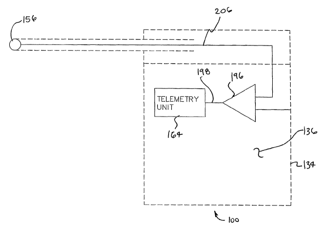
[0059] Figure 11 is a block diagram of an implantable medical device 100 in
accordance with an exemplary embodiment of the present invention. Implantable
medical device 100 of figure 11 comprises a conductive housing 134 defining a
cavity
136. In figure 11, an amplifier 196 is shown disposed in a cavity 136. A
remote
electrode 156 is electrically connected to amplifier 196 via a conductor 206.
Amplifier
196 is also electrically connected to conductive housing 134. In the
embodiment of
figure 11, amplifier 196 is capable of detecting a voltage difference between
conductive
housing 134 and remote electrode 156. Amplifier 196 is also capable of
producing a
signal 198 that is representative of the voltage difference between conductive
housing
134 and remote electrode 156. In figure 11, a telemetry unit 164 is shown
corinected to
amplifier 196. In some useful embodiinents of the present invention,
implantable
medical device 100 is disposed inside a human body and telemetry unit 164 is
capable of
transmitting signal 198 to a receiver located outside of the body.
[0060] Figure 12 is a block diagram of an implantable medical device 100 in
accordance with an additional exemplary embodiment of the present invention.
Implantable medical device 100 of figure 12 comprises a conductive housing 134
that is
electrically connected to an amplifier 196. In the embodiment of figure 12,
amplifier 196
is disposed within a cavity 136 defined by conductive housing 134. A remote
electrode
156 is electrically connected to amplifier 196 via a conductor 206. In the
embodiment of
figure 12, amplifier 196 is capable of detecting a voltage difference between
conductive
housing 134 and remote electrode 156. Amplifier 196 is also capable
of.producing a
17
CA 02564122 2006-10-24
WO 2005/104779 PCT/US2005/014625
signal 198 that is representative of the voltage difference between conductive
housing
134 and remote electrode 156.
[0061] In the embodiment of figure 12, a filter 232 is electrically connected
to
amplifier 196. Filter 232 may be capable of filtering signal 198. Filter 232
may
comprise, for example, a band-pass filter. When this is the case, filter 232
may pass a
portion of signal 198 having frequency's between about 0.5 Hz and about 80.0
Hz. Filter
232 is electrically connected to a telemetry unit 164. In some useful
embodiments of the
present invention, implantable medical device 100 is disposed inside a human
body and
telemetry unit 164 is capable of transmitting at least a portion of signal 198
to a receiver
located outside of the body.
[0062] Figure 13 is a diagrammatic view of an implantable inedical device 400
in
accordance with an exemplary embodiment of the present invention. Implantable
medical device 400 may be used to measure a number of signals. In the
embodiment of
figure 13, for example, implantable medical device 400 is capable of measuring
ECG,
pressure, patient activity, patient posture, impedance, respiratory rate,
respiratory effort,
glucose, and temperature. In the embodiment of figure 13, implantable medical
device
400 includes a telemetry unit 464 and remote sensing lead 466. Remote sensing
lead 466
is capable of sensing pressure from an artery or vein, and communicating such
signal to
telemetry unit 464 for transmission. Remote sensing lead 466 may also contain
one or
more electrodes for sensing ECG as well as a pressure sensor.
[0063] Remote sensing lead 466 may employ one of a variety of pressure sensing
means such as fiberoptic sensors, resonant sensor, piezoresistive sensors,
capacitive
sensors, and other sensors that can be fabricated in a diameter small enough
to be safely
18
CA 02564122 2006-10-24
WO 2005/104779 PCT/US2005/014625
introduced and reside within a vessel. In the preferred embodiment, the
pressure sensing
means may comprise a pressure transmission catheter (PTC 468), as described in
US
Patent No. 4,846,494 that can be introduced into an artery or vein. The entire
disclosure
of the above-mentioned U.S. patent is hereby incorporated by reference herein.
The PTC
approach as described in the '494 patent is advantageous in that it can be
fabricated in a
very small diameter. This is beneficial because the small size is less likely
to damage the
endothelial lining of the vessel and also because accidental pullout of the
sensing catheter
will result in far lesser complications.
[0064] PTC 468 refers the pressure signal to pressure sensor 484. Signal
processing electronics 486 converts the signal from pressure sensor 484 to a
signal that
can be communicated to telemetry unit 464 via flexible lead body 454 and
connector 488.
[0065] Remote sensing lead 466 may also incorporate a temperature sensor 490.
Temperature sensor 490 would preferably be located within conductive housing
434 and
the signal from temperature sensor 490 would be processed by signal processing
electronics 486. The temperature signal would preferably be multiplexed with
the
pressure signal for communication to telemetry unit 464 via flexible lead body
454-and
connector 488.
[0066] The housing of telemetry unit 464 may be constructed of three parts: a
metallic portion 480 fabricated of a metallic material (e.g., titanium), an RF
transparent
portion 478 fabricated of ceramic, and a header 442. In the embodiment of
figure 13,
metallic portion 480 and RF transparent portion 478 are joined together at a
seam 482. In
figure 13, a battery 408 can be seen disposed in metallic portion 480.
19
CA 02564122 2006-10-24
WO 2005/104779 PCT/US2005/014625
.[0067] Remote sensing lead 466 may also contain ECG sensing electrodes. In
some embodiments, for example, conductive housing 434 of implantable medical
device
400 may serve as one ECG sensing electrode while metallic portion 480 of the
housing of
telemetry unit 464 may serve as another ECG sensing electrode. Alternately,
the second
ECG sensing electrode could be incorporated into flexible lead body 454. This
arrangement provides for sufficient spacing between the two ECG sensing
electrodes to
obtain adequate ECG signal amplitude and sensing of important features of the
ECG such
as p-waves for detection of atrial fibrillation. Flexible lead body 454
includes a
conductor to connect the second ECG sensing electrode to signal processing
electronics
486. The ECG signal is preferably multiplexed with the pressure and
temperature signal
for communication to telemetry unit 464 via flexible lead body 454 and
connector 488.
[0068] Remote sensing lead 466 may further incorporate one or more conductors
in flexible lead body 454 to serve as a transmitting and/or receiving antenna.
Telemetry
unit 464 may contain an activity sensor. - The activity sensor may also
comprise, for
example, an accelerometer 494. As the patient moves about, g-forces placed on
the
accelerometer 494 by the patient may create an electrical signal that is
representative of
patient activity.
[0069] TU circuitry 470 contained in telemetry unit 464 is responsible for
controlling power to remote sensing lead 466 and for transmittin.g the signals
to repeater
440. In one exemplary embodiment, telemetry unit 464 has two operating states,
on and
off. When on, telemetry unit 464 transmits a PIM signal with a carrier
frequency of
about three hundred kHz. In another exemplary embodiment telemetry unit 464
compresses the signals to reduce the volume of data to be telemetered to
reduce the
CA 02564122 2006-10-24
WO 2005/104779 PCT/US2005/014625
power required by the transmitter. Power consumption can be further reduced by
storing
either the raw or compressed data in memory for a period of time, a few
seconds for
example, and then transmitting data at multiples of real time to repeater 440
or to RASB
442. In this approach, the transmitter is a high frequency transmitter
operating at about
nine hundred MHz, for example. Although such a high frequency transmitter
consumes
significantly more power when operating, it also provides for a much faster
data
transmission rate and therefore needs to operate for a much shorter period of
time. It
therefore allows several seconds of data stored in memory to be transmitted in
a fraction
of a second. Such an approach also allows the transmitter to employ more
reliable
communication means. For example, instead of using PIM, this approach allows
for the
use of frequency shift keying (FSK) modulation, a more robust modulation
scheme
compared to PIM. Further, transmitted data can be divided into packets and
error
correction codes (ECC) can be added to each packet. When a transmitted data
packet is
received at RASB 442, the ECC can be evaluated to determine if the packet was
received
correctly. RASB can either ignore such a corrupt packet, or it can be equipped
with bi-
directional communication such that it signals back to implantable medical
device 400
that the packet was not received correctly and request that it be
retransmitted by
implantable medical device 400.
[0070] Figure 14 is a schematic diagram showing an activity sensor 492 and
associated circuitry. Telemetry unit 464 (shown in the previous figure) may
contain
activity sensor 492 and it's associated circuitry. Activity sensor 492 may
comprise, for
example, an accelerometer 494. As the patient moves about, g-forces placed on
the
accelerometer 494 by the patient's movement create an electrical signal that
is amplified
21 *
CA 02564122 2006-10-24
WO 2005/104779 PCT/US2005/014625
by an amplifier 496. The output of amplifier 496 is a current source that
charges
capacitor 406 with a fixed amount of charge. Once that level of charge is
reached, a
pulse is triggered and the charge on capacitor 406 is dumped, indicating that
a quantum
of patient activity has occurred. Pulses are counted over a unit time, a few
minutes for
example, to indicate the degree of patient activity. In the embodiment of
figure 14, a
switch 495 and a controller 497 cooperate to dump the charge on capacitor 406.
[0071] Figure 15 is a diagramrnatic view of an implantable medical device 500
in
accordance with an additional exemplary embodiment of the present invention.
In the
embodiment of figure 15, implantable medical device 500 is used to monitor
ECG,
activity, and temperature. In this embodiment, since pressure is not
necessarily being
measured, the need for a remote sensing lead including a pressure sensor is
eliminated. A
temperature sensor 590 is contained within telemetry unit 564. Telemetry unit
564
includes TU circuitry 570. Data transmission approaches in this embodiment are
similar
in function to those previously described.
[0072] In the embodiment of figure 15, a first ECG electrode 572 and a second
ECG electrode 574 are integral to header 562. A remote electrode 556 is
contained at the
distal end of flexible lead 576. Flexible lead 576 allows for remote electrode
556 to be
directed to a site at the time of implantation that allows for a high quality
ECG. By
proper placement of telemetry unit 564 under the skin, it is possible to
obtain two ECG
channels using remote electrode 556 as a common electrode, allowing for
measurement
of two different ECG vectors. Further, if implantable medical device 500 were
only
capable of transmitting a single ECG channel, remote electrode 556 could be
selectively
paired by TU circuitry 570 to serve as a common electrode for either first ECG
electrode
22
CA 02564122 2006-10-24
WO 2005/104779 PCT/US2005/014625
572 or second ECG electrode 574. This would allow fine-tuning of the ECG
signal
following implantation via a programmable function incorporated into TU
circuitry 570.
Such fine-tuning would allow the physician to select that electrode pair that
provided, for
example, the highest amplitude p-wave, or the least amount of muscle noise.
Flexible
lead 576 could also incorporate additional conductive elements to accoinmodate
a
transmitting and/or receiving antenna for the transmitter contained in
telemetry unit 564.
[0073] Figure 16 is a diagrarnmatic view of an implantable medical device 600
in
accordance with an additional exemplary embodiment of the present invention.
In the
embodiment of Figure 16, implantable medical device 600 comprises a first
electrode 672
and a second electrode 674. By placing one electrode at the distal end of
flexible lead
676, sufficient spacing can be obtained between the two electrodes to detect a
good
quality ECG signal. In addition, placing an electrode on the end of flexible
lead 676
provides for a greater degree of flexibility in placement of the electrodes
relative to each
other. This has the potential to improve the diagnostic quality of the ECG
vector because
flexibility in positioning could allow the physician to adjust the relative
location of
electrodes to improve the amplitude of the p-wave, t-wave, or other clinically
significant
features of the ECG waveform. The housing 634 of implantable medical device
600 may
be constructed of three parts: a metallic portion 680 fabricated of a metallic
material (e.g.,
titanium:; an RF transparent portion 678 fabricated of ceramic, and a header
662. The
metallic portion 680 and RF transparent portion 678 are joined together at a
seam 682.
Metallic portion 680 is electrically insulated with parylene, except for the
portion
comprising first electrode 672. Flexible lead 676 may extend approximately
four to ten
centimeters distal to header 662.
23
CA 02564122 2006-10-24
WO 2005/104779 PCT/US2005/014625
[0074] Figures 17A and 17B are diagram views showing a threading too1300 and
a placement too1320 that may be employed to deploy an implantable medical
device 300
in accordance with the present invention. To implant implantable medical
device 300, an
incision 302 is made where the device is to be inserted under the skin 60 of
patient 20 and
a pocket is formed under the skin 60 distal to the incision to accommodate the
housing of
implantable medical device. Threading tool 300 has a hollow lumen and is
directed
through the incision 302 and under the skin 60 to the desired location for a
remote
electrode of the implantable medical device. Once in location, a guidewire 304
is
inserted into the lumen and threading too1300 is extracted. Guidewire 304 is
electrically
insulated with the exception of a distal portion thereof. To evaluate a
location for the
remote electrode of the implantable medical device, the proximal end of
guidewire 304
can be connected to an ECG monitoring instrument 306 while the other input to
the ECG
monitoring instrument 306 is connected to a temporary electrode 308 placed in
the
incision 302 at the approximate location where housing of the implantable
medical device
will be placed when the implantable medical device is implanted. If the result
is
satisfactory, the housing of the implantable medical device 300 and the
flexible lead 376
of the implantable medical device 300 are attached to a placement tool 320.
Placement
tool 320 contains a guide 322 through which guidewire 304 is inserted.
Placement tool
320 is then directed along guidewire 304 until guide 322 has reached the end
of
guidewire 304. Release 326 is then triggered, the housing of implantable
medical device
300 and flexible lead 376 from placement tool 320. Placement tool 320 may then
be
extracted, leaving the housing of implantable medical device 300 and flexible
lead 376 in
24
CA 02564122 2006-10-24
WO 2005/104779 PCT/US2005/014625
position. The housing of implantable medical device 300 is positioned within
the pocket
adjacent to the incision and the incision is closed.
[0075] Various alternative lead-less embodiments of implantable medical device
100 are contemplated. For example, as shown in Figures 18A - 18C, electrodes
may be
incorporated into various portions of the housing of the device 100. In each
of these
embodiments, the housing may include a case portion 1002 made of ceramic for
example,
and header portion 1004 made of a polymeric material. The electrodes 1006,
1008, 1010,
1012 may comprise a conductive material embedded in the header 1004 and/or
case
1002, and the orientation of the electrodes and the distance between the
electrodes may
be maintained by the non-conductive portions of the housing, such as the
ceramic case
1002 and/or the polymeric header 1004, in order to fix orientation for best
signal capture.
The housing holds and orientates one or more sensing electrodes 1006, 1008,
1010, 1012
for purposes of measuring ECG signals or other bio-potential signals such as
EEG, EMG,
.ECG etc., or respiratory effort and/or cardiac stroke volume via impedance.
These
signals may be transmitted and or recorded as described previously.
[0076] The electrodes 1006, 1008, 1010, 1012 may be made from any suitable
sensor electrode material (e.g., Stainless Steel, Elgiloy, MP-35N, Titanium,
etc.) and may
be coated to increase sensing capability (i.e.: carbon black, platinum black,
iridium oxide,
etc). The electrode surface may be smooth or porous coated, again to increase
sensing
capability. The electrodes may be located in-line or orthogonally opposed to
increase the
relative distance between them for improved capability. The macroscopic
surface area of
each electrode may vary depending on the application and the microscopic
surface fmish.
The electrodes may be disposed in or on (e.g., embedded or coated) the header
1004 or
CA 02564122 2006-10-24
WO 2005/104779 PCT/US2005/014625
the case 1002, and provided that they remain electrically isolated from each
other and the
rest of the structure. This may be accomplished by fabricating the case 1002
and/or
header 1004 of a non-conductive material, or if a conductive material is used
for the case
1002, by isolating the electrodes from the case with an insulating material.
[0077] A single header arrangement may be used as shown in figures 18A and
l OB, or a double header arrangement may be used as shown in Figure 18C. With
any one
of these arrangements, two, three, four or more electrodes may be used
depending on, the
number of electrical channels the device electronics allows for, the surface
area required
for each electrode and the signal to be measured in a given application.
Additional
electrodes may be provided via a flexible or semi-flexible wire lead
arrangement as
described previously herein, which would allow for further electrode spacing
for
increased signal resolution.
[0078] For measuring respiratory effort/respiratory rate, a constant current
carrier
signal may be injected between two electrodes. The carrier signal may be
amplitude
modulated by the changing impedance between the electrodes due to respiratory
effort.
The amplitude modulated signal may be demodulated and band-passed filtered for
respiratory signals producing a changing voltage proportional to respiratory
effort which
can then be transmitted and or recorded. Cardiac stroke volume can be attained
using
similar methods but with a band pass tailored to the cardiac signal. An intra-
cardiac
electrode as one of the electrodes in the configuration would provide an
improved
measurement of cardiac stroke volume. Each of these techniques could be
accomplished
using a four electrode method, as well, with one electrode pair providing the
constant
current, and another electrode pair to provide the measurement. This results
in a more
26
CA 02564122 2006-10-24
WO 2005/104779 PCT/US2005/014625
accurate measurement by eliminating the electrode impedance. All four
electrodes could
be configured in the header of the device, in the body of the device, via a
flexible or
semi-flexible wire arrangement, or in any combination of these electrode
types.
[0079] Figure 19 is a block diagram of an implantable medical device 700 that
is
capable of producing a first signal that is representative of respiration and
a second signal
that is representative of ECG. Implantable medical device 700 of figure 19
comprises a
conductive housing 734 that is electrically connected to a current source 234.
A remote
electrode 756 is also electrically connected to current source 234 via a
conductor 206. In
the embodiment of figure 19, current source 234 provides a substantially
constant current
traveling between conductive housing 734 and remote electrode 756.
[0080] In the embodiment of figure 19, an amplifier 796 is arranged to detect
a
voltage difference between conductive housing 734 and remote electrode 756.
Amplifier
796 is also capable of producing a signal 798 that is representative of the
voltage
difference between conductive housing 734 and remote electrode 756. In the
embodiment of figure 19, a first filter 230 and a second filter 232 are both
connected to
amplifier 796.
[0081] First filter 230 may comprise, for example, a band-pass filter that
passes a
portion of signal 798 that is related to the respiration of a human patient.
For example,
first filter 230 may pass a portion of signal 798 having frequency's between
about 0.2 Hz
and about 2.0 Hz. A de-modulator 233 is provided for demodulating the
respiration
related portion of signa1798.
[0082] Second filter 232 may comprise, for example, a band-pass filter that
passes a portion of signal 798 that is related to ECG. For example, second
filter 232 may
27
CA 02564122 2006-10-24
WO 2005/104779 PCT/US2005/014625
pass a portion of signal 798 having frequency's between about 0.2 Hz and about
80.0 Hz.
First filter 230 and second filter 232 are both electrically connected to a
telemetry unit
764. In some useful embodiments of the present invention, implantable medical
device
700 is disposed inside a human body and telemetry unit 764 is capable of
transmitting at
least a portion of signal 798 to a receiver located outside of the body.
[0083] To extend the useful life, an implantable medical device 800 in
accordance
with the present invention may contain a rechargeable battery. As shown in
Figure 20A
and figure 20B, recharging may be performed by transferring energy into
implantable
medical device 800 by transformer coupling energy from a recharging device
820,
located outside the body, to a coil located in implantable medical device 800.
The
secondary of the transformer coil, located in implantable medical device 800,
would drive
circuitry that would create a charging current for the rechargeable battery.
[0084] For convenience, the charging device may be battery powered and
portable and could be worn by patient 20 in an elastic garment 852 when
necessary for
recharging. The use of an elastic garment 852 would assure the device were
held stably
in proper position for charging. Alternately, recharging device 820 could
contain a
replaceable adhesive surface such that it could be located on the skin in
close proximity
to implantable medical device 800. In order to make it easy for the patient to
place the
recharging device properly, an indicator would tell the patient when the
device was
aligned properly, as measured by current being transferred into implantable
medical
device 800. A second indicator may tell the patient when the rechargeable
battery is
fully charged based on information transmitted from the implantable device to
the
recharging device.
28
CA 02564122 2006-10-24
WO 2005/104779 PCT/US2005/014625
[0085] Figure 21 is a block diagram showing an implantable medical device 800
and a recharging device 820. In the embodiment of figure 21, implantable
medical
device 800 is disposed inside a human body 22 and recharging device 820 is
disposed
outside of the human body 22. The skin 60 of the human body 22 is shown
extending
between implantable medical device 800 and recharging device 820 in figure 21.
[0086] In the embodiment of figure 21, recharging device comprises a first
coil
822 and a first battery 808 coupled to first coil 822 for exciting first coil
822. A control
circuit 826 is connected between first coil 822 and first battery 808. Control
circuit 826
is capable of generating the oscillating current necessary to inductively
couple first coil
822 of recharging device 820 with a second coil 824 of implantable medical
device 800.
[0087] Implantable medical device comprises a second battery 828 and a second
coil 824 coupled to second battery 828 for charging second battery 828. A
charging
circuit 899 is connected between second coil 824 and second battery. Charging
circuit
899 may comprise, for example, a voltage regulator that is capable of
controlling the
magnitude of the voltage that is applied to second battery 828 during
charging. Charging
circuit 899 may also comprise, for example, a current regulator that is
capable of
controlling the magnitude of the current that is applied to second battery 828
during
charging.
[0088] In the embodiment of figure 21, first coil 822 and second coil 824 are
inductively coupled to one another so that second battery 828 is charged while
first
battery 808 is depleted. With reference to figure 21, it will be appreciated
that recharging
device 820 comprises a housing 834 defining a cavity 836. In the embodiment of
figure
21, first battery 808 is disposed within cavity 836 defined by housing 834. In
some
29
CA 02564122 2006-10-24
WO 2005/104779 PCT/US2005/014625
useful embodiments of the present invention, first battery 808 is capable of
satisfying the
power requirements of recharging device 820. For example, first battery 808
may have
sufficient capacity to fully charge second battery 828 and -while, at the same
time,
compensating for energy lost during the charging of the second battery. In
such
embodiments, first battery 808 may be larger than second battery 828. Also in
such
embodiments, first battery 808 may be the sole source of power for recharging
device
820. This arrangement may allow the user of implantable medical device to
remain
ambulatory during the charging process.
[0089] Various charging techniques are described in the following U.S.
Patents:
3,454,012; 3,824,129; 3,867,950; 3,492,535; 4,014,346; 4,057,069; 4,082,097;
4,096,866; 4,172,459; 4,441,210; 4,562,840; 4,679,560; 4,741,339; 5,279,292;
5,350,413; 5,411,537; 5,690,693; 5,702,431; 5,991,665; 6,067,474; 6,154,677;
6,324,431; 6,505,077; 6,516,227; 6,549,807; and 6,850,803. The entire
disclosures of
the above-mentioned U.S. Patents are hereby incorporated herein by reference.
[0090] In another embodiment, battery 828 of implantable medical device 828
may be recharged by deriving power from an implanted power source. Such an
implanted power source may derive power from a human body by mechanical,
thermal
and/or chemical means. Examples of implantable power sources that derive power
from
a human body by thermal means include those described in U.S. Patent Numbers.
6,470,212 and No. 6,640,137. Examples of implantable power sources that derive
power
from a human body by mechanical means include those described in U.S. Patent
Numbers 3,943,936; 5,431,694; and 6,822,343 and U.K. Patent Application Number
GB
2350302. The entire disclosure of each of the above-mentioned patents and
patent
CA 02564122 2006-10-24
WO 2005/104779 PCT/US2005/014625
application is hereby incorporated by reference herein. The implantable power
source
may be connected to charging circuit 899 and/or second battery 828 by a first
wire and a
second wire.
[0091] In some useful embodiments of the present invention, implantable
medical
device 800 may include a charge counter to track the amount of charge that has
been
consumed from the battery. In addition, implantable medical device 800 also
incorporates a counter to track the amount of charge that has been depleted
from battery
828. By tracking charge added and charge depleted, remaining battery life can
be
determined and communicated to an external receiver. When battery 828 is fully
charged, both the charge added and charge depleted counters are reset to zero.
The
circuits used to count charge have some inherent error. If this error were
allowed to
accumulate through multiple charges and discharges of battery 828, the
remaining charge
in the battery as indicated by the charge added and charge depleted counters
battery life
indicator may have limited value. To address this problem, implantable medical
device
800 contains a circuit that measures charging current to battery 828. When the
charging
current present indicates that battery 828 has reached fitll charge, both the
charge
depleted and charge added counters are reset.
[0092] Figure 22 is a diagrammatic view of an implantable medical device 1100
in accordance with an additional exemplary embodiment of the present
invention.
Itnplantable medical device 1100 comprises a first energy storage element 1102
and a
second energy storage element 1104. In the embodiment of figure 22, first
energy
storage element 1102 comprises a capacitor 1106 -and second energy storage
element
1104 comprises a battery 1108. In the embodiment of Figure 22, implantable
medical
31
CA 02564122 2006-10-24
WO 2005/104779 PCT/US2005/014625
device 1100 employs a first energy storage element 1102, such as capacitor
1106, that
can store a smaller amount of charge than can be stored in battery 1108, but
can store
charge at a much faster rate than battery 1108. By placing a charging device
near
implantable medical device 1100 for a short period of time, first energy
storage element
1102 is fully charged. Once first energy storage element 1102 is fully
charged, additional
charge coupled into implantable medical device 1100 from the charging device
may be
directed toward charging battery 1108. Once the charging device is pulled away
and is
no longer coupling energy into implantable medical device 1100, the charge
stored in
first energy storage element 1102 is transferred into battery 1108.
[0093] This architecture, employing a fast charging element and a slower
charging element (e.g., a battery) may ha,ve advantages in certain situations.
For
example, suppose that battery 1108 had a charge capacity equal to about one
hundred and
fifty days of operation of implantable medical device 1100 and first energy
storage
element 1102 had a capacity of about seven days of operation. Normal charging
time for
battery 1108 may be about two hours, while charge time for first energy
storage element
1102 was only about thirty seconds. In this scenario, the patient could obtain
a charge
equal to about one full week of operation in about thirty seconds. Many
patients may
find this protocol more convenient than wearing a vest holding a recharging
device for
two hours every three months.
[0094] Figure 23 is a block diagram showing an implantable medical device 1100
and a recharging device 1120 that may be used to recharge implantable medical
device
1100. In the embodiment of figure 23, recharging device 1120 comprises a first
coil
1122 and a first battery 1108 coupled to first coil 1122 for exciting first
coil 1122. A
32
CA 02564122 2006-10-24
WO 2005/104779 PCT/US2005/014625
control-circuit 1126 is connected between first coil 1122 and first battery
1108. Control
circuit 1126 is capable of generating the oscillating current necessary to
inductively
couple first coil 1122 of recharging device 1120 with a second coil 1124 of
implantable
medical device 1100.
[0095] Implantable medical device 1100 comprises a first energy storage
element
1102 and a second energy storage element 1104. In the embodiment of figure 23,
first
energy storage element 1102 comprises a capacitor 1106 and second energy
storage
element 1104 comprises a second battery 1128. A second coil 1124 and a first
regulator
1130 are connected to first energy storage element 1102.
[0096] In the embodiment of figure 23, ~second coil 1124 and first regulator
1130
can cooperate to charge first energy storage element 1102. First regulator
1130 is
capable of controlling the flow of current and the magnitude of voltage
applied to first
energy storage element so that first energy storage element 1102 is charged at
a first
charging rate. First regulator 1130 may comprise, for example, a current
regulator and/or
a voltage regulator.
[0097] In the embodiment of figure 23, a second regulator 1132 is interposed
between the first energy storage element 1102 and second energy storage
element 1104.
Second regulator 1132 is capable of controlling the flow of current and the
magnitude of
voltage applied to second energy storage element so that second energy storage
element
1104 is charged at a second charging rate. Second regulator 1132 may comprise,
for
example, a current regulator and/or a voltage regulator.
[0098] In the embodiment of figure 23, capacitor 1106 is capable of being
charged at a faster rate than battery 1108. Accordingly, the second charging
rate is
33
CA 02564122 2006-10-24
WO 2005/104779 PCT/US2005/014625
slower than the first charging rate. Although one capacitor 1106 is
illustrated in figure
23, it will be appreciated that embodiments are possible in which capacitor
1106
comprises a plurality of capacitors.
[0099] As shown in figure 23, implantable medical device 1100 comprises a
housing 1134 defining a cavity 1136. Housing 1134 may comprise various
materials
without deviating from the spirit and scope of the present invention. Examples
of
materials that may be suitable in some applications include titanium and
stainless steel.
With reference to figure 23, it will be appreciated that second coil 1124,
first regulator
1130 and first battery 1108 are disposed in cavity 1136 defined by housing
1134.
[00100] Figure 24 is a block diagram showing an iinplantable medical
device 1200 and a recharging device 1220 that may be used to recharge
implantable
medical device 1200. In the embodiment of figure 24, recharging device 1220
comprises
a first coil 1222 and a first battery 1208 coupled to first coil 1222 for
exciting first coil
1222. A control circuit 1226 is connected between first coil 1222 and first
battery 1208.
Control circuit 1226 is capable of generating the oscillating current
necessary to
inductively couple first coil 1222 of recharging device 1220 with a second
coil 1224 of
implantable medical device 1200.
[00101] Second coil 1224 of implantable medical device 1200 is coupled to
a first energy storage element 1202 by a diode 1238. Implantable medical
device 1200
also includes a second energy storage element 1204. In the embodiment of
figure 24,
second coil 1224 and diode 1238 can cooperate to charge first energy storage
element
1202. In the embodiment of figure 24, a regulator 1232 is interposed between
the first
energy storage element 1202 and second energy storage element 1204. Regulator
1232 is
34
CA 02564122 2006-10-24
WO 2005/104779 PCT/US2005/014625
capable of controlling the flow of current and the magnitude of voltage
applied to second
energy storage element so that second energy storage element 1204 is charged
at a
controlled charging rate. Regulator 1232 may comprise, for example, a current
regulator
and/or a voltage regulator.
[00102] In the embodiment of figure 24, first energy storage element 1202
comprises a capacitor 1206 and second energy storage element 1204 comprises a
battery
1208. In this embodiment, capacitor 1206 is capable of being charged at a
faster rate than
battery 1208. Accordingly, regulator 1232 may be used to charge battery 1208
at a
-second charging, rate is slower than a first charging rate that capacitor
1206 is capable of.
Although one capacitor 1206 is illustrated in figure 24, it will be
appreciated that
embodiments are possible in which capacitor 1206 comprises a plurality of
capacitors.
[00103] As shown in figure 24, implantable medical device 1200 comprises
a housing 1234 defining a cavity 1236. Housing 1234 may comprise various
materials
without deviating from the spirit and scope of the present invention. Examples
of
materials that may be suitable in some applications include titanium and
stainless steel.
With reference to figure 24, it will be appreciated that second coil 1224,
first regulator
1230 and first battery 1208 are disposed in cavity 1236 defined by housing
1234.
[00104] Figure 25 shows a flowchart 1404 illustrating an exemplary
method in accordance with the present invention. Block 1402A of flowchart 1404
illustrates the step of forming a pocket 1460 in a left implant site 1444 in
the body of a
patient 20. In should be noted that pocket 1460 may be formed in a right
implant site
1446 of the body of patient 20 without deviating from the spirit and scope of
the present
invention. Pocket 1460 may be formed, for example, by making an incision 1403
with a
CA 02564122 2006-10-24
WO 2005/104779 PCT/US2005/014625
cutting tool and pushing a blunt object through the incision 1403 to displace
tissu:e and
form pocket 1460. Pocket 1460 may also be formed by pushing gloved fingers
through
incision 1403.
[00105] Block 1402B of flowchart 1404 illustrates the step of inserting an
implantable monitoring device 1400 in pocket 1460. Implantable monitoring
device may
comprise, for example, the implantable medical devices described herein.
Implantable
monitoring device 1400 may be inserted through incision 1403 so that the
housing of
implantable monitoring device 1400 is positioned within pocket 1460 adjacent
to incision
1403. Incision 1403 may then be closed and the patient may be allowed to go
about a
normal daily routine.
[00106] Block 1402C of flowchart 1404 illustrates the step of monitoring
the patient. Implantable monitoring device 1400 may detect various
physiological
parameters such as, for example, ECG, pressure and temperature. Implantable
monitoring device 1400 may transmit (e.g., wirelessly) signals related to
these parameters
to a repeater worn by or kept near patient 20. Patient 20 may be monitored
during
normal daily activity for a period of weeks, months and/or years.
[00107] A method in accordance with the present invention may include,
for example, the steps of placing an implantable monitoring device comprising
a
conductive -housing and a remote electrode in a left implant site 1444 and
detecting a
voltage difference between the remote electrode and the conductive housing.
This
method may further include the step of producing a signal representative of
the voltage
difference between the remote electrode and the conductive housing. The signal
may be
transmitted to a receiver 'located outside the human body. Information
obtained during
36
CA 02564122 2006-10-24
WO 2005/104779 PCT/US2005/014625
the monitoring step may be analyzed to determine what type of implantable
therapy
device may be appropriate for patient 20.
[00108] Block 1402D of flowchart 1404 illustrates the steps of removing
implantable monitoring device 1400 from pocket 1460 and inserting an
implantable
therapy device 1411 in pocket 1460. In some useful methods in accordance with
the
present invention, implantable monitoring device 1400 is removed from pocket
1460 and
iinplantable therapy device 1411 is inserted in pocket 1460 during a single
surgical
procedure. In the embodiment of figure 25, implantable monitoring device 1400
and
implantable therapy device 1411 have similar shapes and a similar in size.
[00109] Implantable therapy device 1411 may comprise various elements
without deviating from the spirit and scope of the present invention. Examples
of
implantable therapy devices that may be suitable in some applications include
pacemakers, defibrillators, and/or cardioverters. In some useful methods in
accordance
with the present invention, pocket 1460 is disposed in a location which will
allow leads
connected to implantable therapy device 1411 to travel through the vasculature
of patient
20 to the heart of patient 20.
[00110] Figures 26 and 27 are diagram views showing a placement tool
1320 that may be employed to deploy an implantable medical device 1300 in
accordance
with the present invention. Placement tool 1320 comprises a wall 1321 defining
a lumen
1325. A shaft 1327 has been inserted into the lumen 1325 of placement tool
1320. To
implant implantable medical device 1300, an incision 1302 is made where the
device is to
be inserted under the skin 60 of patient 20. Placement tool 1320 is directed
through the
incision 1302 and under the skin 60 until the distal end of placement tool
1320 is
37
CA 02564122 2006-10-24
WO 2005/104779 PCT/US2005/014625
proximate a desired location for implantable medical device 1300. Shaft 1327
is moved
distally so that implantable medical device 1300 exits the distal end of
placement tool
1320. Placement tool 1320 may then be extracted, leaving implantable medical
device
1300 in the desired position.
[00111] It should be recognized to those skilled in the art that the devices
described here can be applied for monitoring of other physiological signals
such as those
which can be measured on or within the heart, brain, bladder, transplanted
organs,
arteries, veins, and other body tissues.
[00112] Those skilled in the art will recognize that the present invention
may be manifested in a variety of forms other than the specific embodiments
described
herein. Accordingly, departures in form and detail may be made without
departing from
the spirit and scope of the present invention as described in the appended
claims.
38