Note: Descriptions are shown in the official language in which they were submitted.
CA 02564263 2006-10-17
CARDIAC REPERFUSION METHODS AND DEVICES
FIELD
This disclosure relates to methods and apparatus for reperfusing tissues.
BACKGROUND
When reperfusing tissue, such as when a vascular occlusion is cieared, there
is
a risk of reperfusion injury distal to the occlusion. Animal studies have
suggested the
possible efficacy of chloramphenicol in the prevention of such injuries but
systemic use
in humans has been generally seen as ineffective.
A number of catheter and guidewire designs are already known in the art:
US7,033,325 Sullivan discloses a guidewire comprising multiple radioopaque
marker
sections; US7,074,231 Jang, discloses a Convertible catheter system comprising
a
guidewire; and US4,946,466 Pinchuki & Martin discloses a transiuminal
angioplasty
apparatus comprising a hollow guidewire. US4,994,033 discloses an
intravascular
catheter, and its use to apply medicament to a stenotic lesion in a blood
vessel.
US4,946,466 discloses a hollow guidewire with a diameter of about 0.014
inches, a
balloon section and a hole to inflate the balloon and the use of:fluoroscopy
to view the
progress of the metallic guidewire during insertion.
SUMMARY
In an embodiment there is disclosed a hollow member for the intravascular
delivery of an agent, the member may have an end and a delivery hole proximate
said
end. In some embodiments there may be a marker positioned relative to the
delivery
hole.
In alternative embodiments, the member may be comprised in a catheter
assembly, may be a guidewire, may be comprised in an apparatus for performing
an
angioplasty or the member may be filled with the agent.
-1-
CA 02564263 2006-10-17
In alternative embodiments, the member may be selected from the group
consisting of: i) a cytochrome inhibitor; ii) a platelet activation factor
inhibitor; iii) a
Caspase inhibitor; and iv) a promoter of NO production.
In alternative embodiments, the intravascular delivery may be in a cardiac
blood
vessel; or may be in a blood vessel in the brain; or the member may be for use
in a
human.
In alternative embodiments, there is disclosed a method for reperfusing
tissue,
the method may comprise locally delivering an agent to the tissue prior to
reperfusing
the tissue.
In alternative embodiments, the method may further comprise controlling the
amount of the agent delivered to the tissue; or exposing the tissue to the
agent for a
predetermined time period prior to the reperfusing; or the agent may be
delivered
through an intravascular member; or the member may be comprised in a catheter
assembly.
In alternative embodiments, the member may include a marker and the method
may further comprise using the marker to determine the location of delivering
the
agent.
In alternative embodiments, the method may further comprise: i) delivering the
agent at a location relative to a vascular occlusion; and then ii) clearing
the vascular
occlusion.
In alternative embodiments, the occlusion may have first and second occlusion
ends and the intravascular member may be inserted through the occlusion from
the
first end and used to dispense the agent at the second end.
In alternative embodiments, the member may be a guidewire; the guidewire may
be comprised in a catheter assembly; or the guidewire may comprise a plurality
of
delivery holes; or may comprise a second marker; or may have an end, a
delivery hole
-2-
CA 02564263 2006-10-17
located relative to the end, a marker located relative to the delivery hole,
and methods
may further comprise using the marker to position the member relative to the
tissue.
In alternative embodiments, the agent may be a chemical; and the chemical may
be selected from the group consisting of: i) a cytochrome inhibitor; ii) a
platelet
activation factor inhibitor; iii) a Caspase inhibitor; and iv) a promoter of
NO production;
and v) a 2b3A receptor antagonist
In alternative embodiments, there is disclosed a kit for reperfusing tissue,
the kit
may comprise: i) a hollow member suitable for intravascular delivery of a
protective
agent; and ii) instructions to use the member to locally deliver an agent
prior to
reperfusing the tissue; the kit may further comprise instructions to fill the
member with
the agent prior the reperfusing; and the kit may further comprise instructions
to allow
the agent to contact the tissue for a predetermined time prior to the
reperfusion.
In alternative embodiments, there is disclosed use of a hollow member for the
local delivery of an agent for the prevention of reperfusion injury; and the
member may
be adapted to be insertable through a vascular lumen; and may have a delivery
hole
and a marker positioned at a defined location relative to the delivery hole.
In alternative embodiments, the use may comprise filling the member with the
agent; or may comprise dispensing a known quantity of the agent through the
member;
or may comprise allowing a determined time to pass between the delivery of the
agent
and reperfusing the tissue.
In alternative embodiments, the member may comprise a plurality of the markers
and a plurality of the delivery holes.
In alternative embodiments, the reperfusion may be reperfusion of a cardiac
blood vessel; or of a blood vessel in the brain.
-3-
CA 02564263 2006-10-17
In alternative embodiments, the agent may be selected from the group
consisting of: i) a cytochrome inhibitor; ii) a platelet activation factor
inhibitor; iii) a
Caspase inhibitor; iv) a promoter of NO production; and v) a 2b3A receptor
antagonist.
In alternative embodiments, there is disclosed use of a catheter assembly
having a hollow guidewire for the reperfusion of tissue associated with the
clearing of a
vascular occlusion, use may comprise locally delivering a chemical agent
through the
guidewire prior to the clearing of the vascular obstruction.
In alternative embodiments, there is disclosed use of a hollow member having
an end, a delivery hole positioned proximate the end, and a marker positioned
relative
to the delivery hole to manufacture an apparatus for the local intravascular
delivery of a
agent in the removal of a vascular occlusion.
In alternative embodiments, there is disclosed use of a locally administered
agent for prevention and treatment of reperfusion injury.
In alternative embodiments, there is disclosed use of a locally delivered
agent
for preventing and mitigating, and treatingreperfusion injury in a human
cardiac blood
vessel, the use may comprise sequentially: i) delivering a desired quantity of
the agent
at a desired intravascular location through a hollow intravascular member; and
ii)
waiting a desired time interval after the local delivery; and then iii)
carrying out the
reperfusion.
In alternative embodiments, there is disclosed a method for clearing a
vascular
occlusion in a cardiac blood vessel, the method may comprise sequentially: i)
exposing
tissue affected by the occlusion to a desired quantity of an agent for a
desired time;
and then ii) reperfusing the tissue.
In alternative embodiments, there is disclosed a method for clearing a
vascular
occlusion using a catheter assembly with a hollow guidewire, the method may
comprise
sequentially: i) passing the guidewire through the occlusion; ii) delivering a
desired
-4-
CA 02564263 2006-10-17
quantity of an agent through the guidewire; iii) waiting for a desired time;
and iv)
clearing the vascular occlusion.
In alternative embodiments, there is disclosed a method for intravascularly
deiivering a agent for use in reperfusion, the method may comprise: i) filling
a hollow
member with the agent; ii) feeding the member through a desired blood vessel;
iii)
expelling a desired quantity of the agent from the member at a desired
location.
In a further embodiment, there is disclosed an apparatus for the intravascular
delivery of an agent the apparatus comprising a fluid pressure source and a
member,
the member having an end and a delivery hole positioned relative to the end
and the
fluid pressure source being operatively connected to the member.
In further embodiments, the apparatus may be a catheter assembly.
In a further embodiment, there is disclosed a method for preparing tissues
prior
to reperfusion the method comprising delivering an agent to the tissue prior
to
reperfusing the tissue.
In further embodiments, the method may further comprise controlling the amount
of the agent delivered to the tissue; or may further comprise exposing the
tissue to the
agent for a predetermined time period prior to the reperfusing; or the agent
may be
delivered through an intravascular member; or the member may be comprised in a
catheter assembly.
In further embodiments, the member may include a marker and a delivery hole
and the method may further comprise using the marker to determine the location
of the
delivery hole.
In further embodiments, the method may further comprise: i) delivering the
agent
at a location relative to a vascular occlusion; and ii) clearing the vascular
occlusion.
-5-
CA 02564263 2006-10-17
In further embodiments, the method may include clearing a vascular occlusion
and the occlusion has first and second occlusion ends and the intravascular
member is
inserted through the occlusion from the first end and used to dispense the
agent at the
second end.
In further embodiments, the members disclosed may be used to prevent
reperfusion injury; and the uses may occur as part of an angioplasty
procedure; te
member may be filled with the agent; or may be a guidewire; or may have an
associated marker.
In further embodiments, the guidewire may have an end, and a delivery hole
located relative to the end, and the method may further comprise positioning
the
member relative to the tissue to be reperfused. Blood vessels may be blood
vessels in
the brain, or may be coronary blood vessels and tissue may be human tissue.
Features and advantages of the embodiments will become more apparent in light
of the
following detailed description of some embodiments thereof, as illustrated in
the
accompanying figures. As will be realized, the subject matter hereof is
capable of
modifications in various respects, all without departing from the scope of the
disclosure
and claims. Accordingly, the drawings and the description are to be regarded
as
illustrative in nature, and not as restrictive.
-6-
CA 02564263 2006-10-17
BRIEF DESCRIPTION OF THE DRAWINGS
Figure 1 is a perspective view of a first embodiment of a member.
Figure 2 is a side view of the embodiment of Figure 1.
Figure 3 is a cross sectional side view of the embodiment of Figure 1.
Figure 4 is a top view of the embodiment of Figure 1.
Figure 5 is an end view of the embodiment of Figure 1.
Figure 6 shows a second embodiment of a member.
Figure 7 shows a third embodiment of a member.
Figure 8 is a diagram showing the possible positioning of a guidewire
according
to an embodiment.
Figure 9 is a diagram of a catheter assembly according to an embodiment.
Figure 10 shows the arrangement of syringes of an embodiment.
Figure 11 shows a sketch of the spread of Evans Blue in a heart following
perfusion through a member of an embodiment.
DETAILED DESCRIPTION OF PREFERRED EMBODIMENTS
Definitions
In this application the following terms have the following meanings:
"Agent" (which may also be referred to as "protective agent", or "preventive
agent") means any species which is able to prevent, mitigate, reduce, or
control
reperfusion injury. Such active species may include chemical agents,
biologics, and
nanoparticies and may be pharmaceuticals or other bioactive chemicals, and may
include macromolecules, hormones, signalling molecules, organic and inorganic
compounds, microorganisms and other agents. By way of illustration and not of
limitation, possible agents for use in the embodiments disclosed herein may
include (a)
inhibitors of superoxide production which may be superoxide dismutases,
catalase, iron
chelating agents such as deferoxamine and may be cytochrome inhibitors and may
be
cytochrome p450 inhibitors, and may be chloramphenicol; (b) Platelet
Activation Factor
(PAF) inhibitors such as TCV309TM; (c) Caspase inhibitors; (d) promoters of NO
production such as nitroprusside and adenosine; and (e) 2b3A receptor
antagonists .
-7-
CA 02564263 2006-10-17
Suitable agents and suitable dosages for such agents will be readily apparent
to those
skilled in the art. In alternative embodiments the agent may be dissolved or
may be in
suspension or may be provided in an aqueous or other medium and may be
provided
at any suitable concentration in any suitable medium or carrier or form and
may be
provided in association with or may include any suitable carriers or
excipients. It will
further be understood that particular agents may be used in combinations with
other
agents or with other components. .
"Agent fluid pressure source" means any apparatus, device or combination of
elements suitable to provide a source of pressure for the delivery of agent
through a
member. Possible examples of suitable fluid pressure sources include syringes,
pumps, elevated reservoirs, or any other types of dispenser. A range of
alternatives
will be readily understood and adapted by those skilled in the art who will
readily be
able to understand and make suitable choices therebetween and adjustments
thereto.
"Angioplasty" means a surgical operation to repair a damaged blood vessel, or
to unblock a blood vessel, and may also be known by other names including
"coronary
artery balloon dilation", "balloon angioplasty" and "percutaneous coronary
intervention"
(PCI). The subject blood vessel may be an artery and in particular embodiments
it may
be a coronary artery or an artery in the brain. In particular embodiments an
angioplasty
may be carried out using a catheter apparatus, and the catheter apparatus may
,Gomprise a member that may be a guidewire.
"Catheter apparatus" has its normal meaning and includes an assembly
comprising one or more hollow, flexible tubes that can be inserted into a body
cavity,
duct, or vessel to allow the passage of fluids or distend a passageway or
carry out a
range of procedures as will be readily understood by those skilled in the art.
A wide
range of catheter designs will be well known to those skilled in the art and
those skilled
in the art will be readily able to select appropriate designs and to adapt
both
procedures and designs for the carrying out of various embodiments.
"Delivery hole" means any hole, orifice, port, slit, crack, aperture, opening,
space, gap, fissure, pore or micropore suitable for the delivery therethrough
of an agent
-8-
W
CA 02564263 2006-10-17
for use in an embodiment, it being understood that such holes may be of any
suitable
size and may be provided in a metal, or in a piastic or other suitable
material, and may
be created by any suitable methods including but not limited to laser or
mechanical
working of a substrate material or may be preformed in a material. It will be
understood
that in different embodiments such holes may be provided singly or in groups
and may
comprise any number of holes. It will be understood that in particular
embodiments
pluralities of holes may be organised in any regular or random fashion and may
form a
mesh or filter and may be positioned at a range of locations along a member.
The
choices between alternative designs will readily be made by those skilled in
the art.
"Guidewire" has its usual meaning and includes for example a wire used in the
treatment of a vascular occlusion, optionally as a part of a catheter
assembly.
"Marker" means a defined region, tag, indicator, material or other marker
suitable to permit an operator to localise a defined portion of a member. In
particular
embodiments the marker may be localised using fluoroscopy, or computer
tomography
in which cases the marker may generally be a radioopaque region having
suitable
properties. Suitable radioopaque materials may include by way of example gold,
platinum, lead and tungsten but a range of alternative materials, shapes,
constructions,
and detection methods will be readily apparent to those skilled in the art. In
alternative
embodiments it may be possible to locate the markers using alternative
techniques
such as MRI in which case the markers may be magnetopaque or may comprise
materials suitable to such alternative techniques, which materials may include
copper.
In particular embodiments the member may comprise multiple markers which have
different shapes, opacity, or properties so that their relative positions may
be
determined by an operator.
"Member" means a member suitable for conducting an agent, and may be
generally, or in part, tubular, and may include a conduit channel, hollow
portion,
reservoir, or other structure suitable for conducting a fluid. In some
embodiments the
member may be suitable for feeding along the lumen of a blood vessel, and may
be
coated, in whole or in part, with suitable materials to ensure its
compatibility with the
agent to be carried and to facilitate its movement through a blood vessel
lumen.
-9-
CA 02564263 2006-10-17
Typically the diameter of a member may be between 0.01 and 0.02 inches but in
particular embodiments the diameter may range from less than 0.010 inches,
0.010-
0.012 inches, 0.012-0.014 inches, 0.014-0.016 inches, 0.016-0.018 inches,
0.018-0.02
inches. In particular embodiments these diameters and diameters greater than
0.02
inches may all be possible and the choice of an appropriate dimension will be
readily
apparent to those skilled in the art. A member may be made from a range of
materials
and in a range of constructions all of which will be readily apparent to those
skilled in
the art. In particular embodiments the member may comprise flexible metals and
in
certain specific embodiments may comprise nitinol or stainless steel. In
certain
embodiments the member may be adapted to facilitate steerability, movement and
flexibility whilst maintaining the integrity of the member and any conduit
therein. In
some embodiments the member may be a guidewire. In particular embodiments the
member may be coated or treated with lubricious substances such as teflon,
hydrogels
or hydrophilic coatings. In some embodiments the member may have associated
structures such as holes, scrapers, or a range of suitable implements to
permit the
member to be advanced through a vascular occlusion.
"Reperfusion" means the restoration of blood flow to an ischemic organs,
tissues, or cells.
"Reperfusion injury" means injury resulting from the reperfusion of tissue and
may result from sudden exposure of ischemic organs, tissues or cells to
oxygenated
blood; it may include oxidative stress, apoptosis, inflammation, and ischemic
injuries.
Those skilled in the art will readily understand and implement a variety of
methods whereby suitable agents may be applied through the members disclosed,
and
will understand that a wide range of catheter designs are possible and will
readily
choose suitable designs.
Description of specific embodiments
In a first embodiment described with reference to Figures 1, 2, 3, 4 and 5,
there
is disclosed a hollow member generally designated 10, for the intravascular
delivery of
-10-
CA 02564263 2006-10-17
an agent. The member has an end 14, and at least one delivery hole 16. The at
least
one delivery hole 16 is positioned reiative to the end 14 of the member, and a
marker
18 is positioned relative to the delivery hole and proximate end 14. In the
illustrations
marker 18 is a radioopaque material making up material over length 19 the end
14 of
the member and may comprise platinum. It will be appreciated that a range of
alternative configurations and materials are possible. In the embodiment
illustrated the
marker 18 has a length 19 of from about 20mm to about 30mm, but in alternative
embodiments the length 19 may be adjusted in ways that will be readily
apparent to
those skilled in the art, so as to maintain a suitable balance of stiffness
and flexibility
for this portion of the member. Particular elements of the apparatuses
disclosed herein
may be within a region generally designated 70 at the end of the member 10.
In this embodiment a second marker 20 is presented on the opposite side of the
delivery holes 16 from first marker 14, it will be appreciated that in
alternative
embodiments the first maker, the second marker, or both markers may be
omitted, or
that additional markers or alternative marker placements may be adopted or
that the
markers may have the same or different shapes and properties. The location of
one or
both markers 18, 20 may be known relative to the delivery holes 16. Although
the
embodiments illustrated have markers 18 and 20 positioned on either side of
the
delivery holes 16, alternative arrangements, or the omission of one of the
markers, may
be possible in particular embodiments.
As will be seen, although closed at end 14 by marker 18, member 10 comprises
a lumen 22 defined by a wall 27 and communicating with delivery holes 16.
The member 10 will be compatible with the agent to be dispensed, and the
interior surface 26 of the member may optionally be coated with suitable
materials for
this purpose. In practice, prior to use, the member may be flushed with
heparin, or with
heparin saline or other anticoagulants. The exterior surface 28 of the member
may
optionally be coated with suitable lubricious substances to facilitate
movement of the
member 10 through the lumen of a blood vessel. Although the embodiment
illustrated
in Figures 1-5 comprises five delivery holes 16, and two markers, it will be
appreciated
that any number of holes, and any number of markers may be used to suit
particular
-11-
CA 02564263 2006-10-17
requirements. In particular alternative embodiments illustrated in Figures 6
and 7 the
delivery holes 16 may be arranged in alternative patterns around the member 10
and in
a range of sizes as set out herein. The only requirement may be that the agent
is
releasable through the delivery holes in a controlled manner. It will be
understood by
those skilled in the art that the number, size and distribution of the
delivery holes
should be adjusted to allow a suitable flow rate of agent out of the member,
so as to
permit rapid delivery of agent without damaging the interior of a blood vessel
or cavity
into which it is inserted.
Although in the first illustrated embodiment the length 19 of marker 18 may be
between about 20mm and about 30mm, a wide range of alternative sizes may be
possible, for example, lengths 19 of from about 5mm to about 10mm, about 10mm
to
about 15mm, about 15mm to about 20mm, about 20mm to about 25mm, about 25mm
to about 30mm, about 30mm to about 40mm or greater than about 40mm may all be
possible and suitable under particular circumstances. Likewise the size of
second
marker 20 may be changed and a range of suitable sizes may be possible. In the
first
embodiment marker 20 and marker 18 may be radioopaque and may be or may
comprise gold or platinum but other metallic or non-metallic substances may be
suitable and will be readily chosen by those skilled in the art to suit
particular purposes.
In the first embodiment distance 21 between markers 18 and 20 may be about
10mm,
but again a range of sizes may be possible, depending on operational
requirements,
and the number and disposition of delivery holes 16.
The member itself may be of conventional length, as determined by those
skilled
in the art, and may be sufficient to extend from a desired point of
introduction to the
vascular system to the identified target location whilst still leaving
sufficient length
outside the body for necessary manipulations.
In certain alternative embodiments there is also disclosed an apparatus
comprising a member and a fluid pressure source.
In alternative embodiments the member may be adapted for us in a mammal,
which may be a human. It will be appreciated that in certain embodiments the
markers
-12-
CA 02564263 2006-10-17
may be omitted entirely and the physical or radiographic properties of the
member as a
whole may be used to localise the end of the member. In some uses those
skilled in
the art will be able to localise and use the member without recourse to
radiography or
fluoroscopy and may rely on physically locating the end of the member
manually, by
feeling its location in the subject blood vessel.
In a second embodiment there is disclosed the use of a catheter assembly
having a hollow member 10, for the reperfusion of tissue associated with the
clearing of
a vascular occlusion. The vascular occlusion may be in a cardiac artery. An
example
of a catheter assembly is presented in Figure 9 and generally designated 40.
The
member is designated 10 and has an end 14. Catheter sheath 43 surrounds a
further
catheter element 44 having associated balloon member 45. The use of the
assembly
may comprise a series of steps, the details of which will be readily adapted
by those
skilled in the art to suit particular patients, particular objectives,
alternative catheter
designs and other variables.
a) Before use, the member 10 (which may be a guidewire), may be first
flushed and washed with heparin or heparin saline or other suitable
anticoagulants, and
then with agent. Before use the member may be filled with agent.
b) Necessary preparation for the procedure may vary with the condition and
location of the subject. A variety of routine procedures and precautions that
may be
implemented prior to, during and after the operation will be readily apparent
to those
skilled in the art who will be able to make appropriate choices therebetween.
These
include but are not limited to the following: The subject may be asked to stop
eating or
drinking for a suitable time before the operation, and in certain embodiments
such
suitable time may be up to 12 hours or more than 12 hours; Routine tests may
be
carried out before commencing the procedure, these may include: chest X-rays,
electrocardiograms and blood tests; Prior to the procedure it may also be
desirable that
the subject stop taking certain medications which may include those for
diabetes; The
subject's heart rate and rhythm may be monitored during the procedure using a
variety
of known techniques.
-13-
CA 02564263 2006-10-17
General anesthesia may or may not be used depending on the circumstances.
It may be desirable to administer anticoagulants to reduce blood clotting and
suitable
medications to relax the blood vessels that are to be treated. Subjects may
also be
given calcium blockers, nitrates or other suitable medications to reduce any
risk of
vascular spasm.
c) Briefly: A needle may first be inserted,then a wire therethrough. A sheath
may be guided into the bloodvessel over this wire, the sheath may contain a
one way
hemostatic valve to prevent blood flow out of the blood vessel. Then the
needle may be
removed and a sheath advanced along the wire into the blood vessel and then
the wire
removed A guiding catheter 43 with a wire may then be advanced along the blood
vessel up to the coronary artery. Then a third intravascular or coronary
guidewire may
be introduced and may be used to deliver an agent and to guide a coronary
catheter to
the site of treatment
An exemplary insertion point to access the coronary artery may be at the
femoral artery but in some embodiments the brachial or radial arteries, or
other blood
vessels, may also be suitable. Where the femoral artery is used, the insertion
site may
be in the groin. Before the procedure begins, the area for inserting the
catheter may be
prepared with antiseptic solution and a local anesthetic may be administered.
In some
embodiments a suitable needle is inserted into a chosen blood vessel and a
guidewire
introduced therethrough. The needle may then be removed and a catheter sheath
may
slipped over the wire and into the artery and then (where this is the target)
a guiding
catheter may be manipulated into a coronary artery. The catheter which is
advanced to
the coronary artery may be introduced through a vascular access sheath and may
be a
separate catheter from that initially introduced to the blood system.
Alternatively the
catheter sheath may be the first element introduced over a wire, following
puncture
with a needle. In alternative embodiments, insertion of the sheath may precede
insertion of the guide catheter which may precede insertion of the guidewire,
insertion
of the guidewire may precede the sheath, or alternative sequences may be
adopted. It
will be understood that different designs of guidewires may be used at
different stages
in the procedure: for introduction of a vascular access sheath; for advancing
a guiding
catheter towards the target artery which may be a coronary artery; and
finally,
-14-
CA 02564263 2006-10-17
intracoronary for guidance of coronary intervention catheters (balloons,
stents, etc). In
particular embodiments disclosed the member will be of the latter variety, an
intracoronary guidewire for guidance of an intervention catheter.
d) The member 10 may be advanced to a desired location, in some
embodiments this may be the entrance to the coronary artery. The catheter
operator
may use x-ray images to follow the location of the guide catheter 43 and or
member 42,
until it reaches the target which may be a blocked coronary artery. The
catheter
operator may inject a small amount of contrast agent, which may contain
iodine, or dye,
through the catheter to help in following the location of the catheter.
e) The member 10 may be advanced through the catheter to the location of
a target vascular occlusion and then advanced through the occlusion to a
position
where it may be used to dispense the desired agent downstream of the
occiusion.
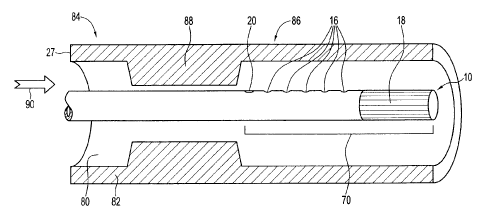
Figure 8 shows the placement of the end section 70 of a member 10 when
positioned
to deliver an agent in one embodiment. It will be observed that in this
embodiment the
member is disposed in the lumen 80 of a blood vessel 82, and extends from an
upstream side 84 to a downstream side 86 of an occlusion 88. Agent can thus be
delivered from delivery holes 16 downstream of the occlusion. It can be seen
that the
end 70 of the member 10 can be positioned relative to the occlusion 88 by
reference to
one or both of the markers 18, 20. Such positioning may occur prior to, during
or after
the end of the member is advanced through the occlusion. The direction of
blood flow
is shown by arrow 90. The location of an end portion 70 of the member may be
determined by x-ray techniques, fluoroscopy or any other suitable means. In
one
embodiment the agent may be chloramphenicol and about 10mg may be administered
in about 0.5-1.0ml volume. A wide range of suitable alternative agents (as
defined
herein) may be possible and will be readily selected, and dosages readily
determined,
by those skilled in the art.
Figure 10 illustrates a suitable arrangement of syringes for the controlled
delivery of an agent through a member. Two syringes 60 and 62 are
interconnected as
shown by a three way stop-cock which is adjusted as necessary to allow the
desired
flow of agent. Agent is transferred from a reservoir syringe 60 into a
dispensing syringe
- 15-
CA 02564263 2006-10-17
62 by compression of reservoir syringe 60. As desired dispensing syringe 62
may be
compressed to urge agent into the lumen of member 10 in a desired quantity and
rate.
It will be appreciated that a range of suitable one-way valves or other
control devices
may be provided between syringe 60 and 62 and between syringe 62 and member 10
as desired.
f) After delivering the agent the operator of the catheter may wait for a
predetermined time before clearing the occlusion, in some embodiments this may
be
about one minute but as disclosed herein alternative time periods are
possible. After a
suitable waiting period a balloon member 65 or other device may be used to
clear the
occlusion. In an embodiment a suitable balloon may be inflated for a period
that may
be up to a minute at the site of the blockage, in alternative embodiments this
inflation
step may be repeated and the procedure may be repeated at the sites of a
plurality of
biockages or the duration of the inflation step may be shortened or extended
in ways
readily apparent to those skilled in the art.
g) In some embodiments, once the occlusion has been cleared, a stent or
stents may be placed in the artery using a range of known procedures. Once a
stent or
stents are in place, the balloon catheter may be removed and angiograms may be
taken to see how well blood fiows through the cleared artery. In alternative
embodiments the blood vessel may be widened before, during, or after the stent
has
been opened up.
h) The various parts of the apparatus may be withdrawn from the patient
according to standard procedures. In particular alternative embodiments the
entire
procedure usually may take about 30 minutes to several hours. The sheath may
be left
in place for several hours after the procedure. The entry area may be kept
immobile
until the sheath has been out for an extended period which may be up to or
more than
about three hours. A range of standard procedures may be adopted to reduce or
prevent bleeding and infection. It may be desirable to monitor the patient's
heart and
vital signs for 12 to 24 hours after the procedure, and it may be desirable to
continue
treating with relaxants and anticoagulants. The patient may remain
hospitalized for
one or more days. After the procedure it may be desirable for the patient to
drink
- 16-
CA 02564263 2006-10-17
plenty of fluids to help rid their body of the contrast dye and to avoid
strenuous exercise
and lifting heavy objects for several days afterward.
It will be appreciated that in some embodiments the member 10 may be a
guidewire, or may take some other form and that in some embodiments the
apparatus
may include different combinations of sheath elements, guidewire elements,
guide
catheter elements, balloon catheter elements etc, and that in particular
elements one
or more of such elements may be omitted. It will also be appreciated that in
alternative
embodiments the sequence of placing and advancing the different elements may
be
changed, or as indicated some elements or their use may be omitted altogether.
The
choice between different catheter designs and different usage procedures will
be
readily made by those skilled in the art. As described above, in certain
embodiments
the member may not have associated markers.
Alternative Embodiments
A range of alternatives to the use of interconnected syringes will be readily
identified and implemented by those skilled in the art and may include
computer
controlled dispensing apparatuses, premeasured dispensers, or any other
mechanism
suitable to expel a controlled quantity of agent from the guidewire, at a
suitable rate.
In particular alternative embodiments the desired dosage of agent may vary
with
the subject, the circumstances and the particular agent selected. When the
agent is
chloramphenicol a dosage of between about 5mg and about 15mg may be suitable,
if
the agent is adenosine a dosage of between about 30ug and 50ug may be
suitable.
Generally the agent may be delivered in a volume of about 0.5ml to 1.0ml, but
larger or
smaller volumes may also be used and suitable parameters will be readily
determined
by a suitably skilled user. In alternative embodiments the method may further
comprise
controlling the amount of agent delivered to the tissue in relation to
parameters that will
be readily apparent to those skilled in the art. It will be understood that
the desired
dosage may change with the nature of the tissue to be reperfused, the time for
which
the tissue has been denied oxygen, the agent used, the temperature of the
tissue, the
composition of the reperfusion fluid and other parameters, all of which will
be readily
-17-
CA 02564263 2006-10-17
understood and evaluated by those skilled in the art. Suitable dosages for any
chosen
agents will be readily determined by those skilled in the art to suit
particular
circumstances.
In particular embodiments the tissue to be reperfused may be exposed to the
agent for any suitable period of time prior to the reperfusion step. In some
embodiments the tissue may be exposed to the agent for about 1 minute prior to
reperfusion, but in particular embodiments this time period may be up to 1
second, up
to 2 seconds, up to 5 seconds, up to 10 seconds, up to 20 seconds, up to 30
seconds,
up to 40 seconds, up to 50 seconds, up to 60 seconds, up to 80 seconds, up to
100
seconds, up to 120 seconds, up to 140 seconds, up to 160 seconds, up to 180
seconds or more than 180 seconds. In certain embodiments t the agent may be
delivered to the tissue only a minimal amount of time in advance of the
reperfusion
step.
In particular embodiments the methods and apparatuses and uses disclosed
herein may be directed at the treatment, or the prevention or the treatment
and
prevention of reperfusion injuries.
The method and apparatus of particular embodiments may each be directed at
the treatment of an occlusion or the reperfusion of tissue, or both, in a
cardiac blood
vessel, which may be a cardiac artery. Alternatively they may be directed at
other
blood vessels including blood vessels in the brain. In some embodiments an
occlusion
may be approached from an upstream direction but it will be appreciated that
in
alternative circumstances, such as during an operation, the agent may be
directly
introduced to the region downstream of an occlusion, may be approached from
the
downstream side of the occlusion through a blood vessel lumen, and may be
approached by injection directly through the blood vessel wall. In one
alternative
embodiment the agent may be introduced directly into the coronary sinus. In
further
alternative embodiments the method may comprise simply locally delivering an
agent to
the tissue prior to reperfusing the tissue. In further alternative embodiments
apparatuses and methods disclosed may be used in the reperfusion of tissue in
any
suitable blood vessels, including blood vessels in the brain and alternative
cardiac
-18-
CA 02564263 2006-10-17
blood vessels. It is also envisaged that in some embodiments the agent may be
deposited upstream of the occlusion, and a temporary hole introduced in the
occlusion
allowing the upstream blood pressure to force the agent through the hole to
the other
side of the occlusion.
The various embodiments may be provided in the form of kits for reperfusing
tissue. These may comprise a hollow member for intravascuiar delivery of an
agent
and exemplified in Figures 1-7, and in the specific embodiments disclosed
herein, as
well as instructions to use the member to locally deliver an agent prior to
reperfusing
tissue. In alternative embodiments a kit may further comprise instructions to
fill the
member with agent prior reperfusing tissue. In further alternative embodiments
a kit
may further comprise instructions to allow the agent to contact the tissue for
a
predetermined time prior to the reperfusion.
In further embodiments a hollow member may be used for the local delivery of
an agent for the prevention of reperfusion injury. In yet further alternative
embodiments
the member may be adapted to be insertable through a vascular lumen. Uses of
the
member may comprise filling the member with an agent, may comprise dispensing
a
known quantity of an agent through the member and may comprise allowing a
determined time to pass between the dispensing of an agent and reperfusing the
tissue
In alternative embodiments the member may be comprised in a catheter
assembly which may include one or more markers and the one or more markers may
be used to determine the location of delivering the agent. In further
alternative
embodiments the method may further comprise delivering the agent at a location
relative to a vascular occlusion and then clearing the vascular occlusion; the
occlusion
may have first and second occlusion ends and the intravascular member may be
inserted through the occlusion from the first end and used to dispense the
agent at the
second end. In alternative embodiments the member may be a guidewire and the
guidewire may be comprised catheter assembiy which may be an over the wire
catheter assembly or a rapid exchange catheter assembly or a mono-rail
catheter
assembly. In further refined embodiments the guidewire may comprise an end, a
delivery hole located relative to the end, a marker located relative to the
delivery hole,
-19-
CA 02564263 2006-10-17
and the method may further comprise using the marker to position the member
relative
to the tissue. In alternative embodiments of the method the guidewire may
comprise
more than one delivery holes, or may comprise more than one marker, or may
comprise more than one delivery hole and more than one marker. In particular
embodiments the agent may be a chemical, and may be an inhibitor of superoxide
production, may be a cytochrome inhibitor; may be a platelet activation factor
inhibitor;
may be a Caspase inhibitor; may be a 2b3A receptor antagonist and may be a
promoter of NO production. In some embodiments the reperfused tissue may be
associated with a cardiac blood vessel, or may be associated with a brain
blood vessel,
or may be any other suitable blood vessel. In particular embodiments the
method may
be carried out on a human subject in need thereof. In alternative embodiments
locally
administered chloramphenicol may be used for preventing reperfusion injury.
The
method of administration may include any of those methods set out in the
alternative
embodiments.
There is further disclosed the use of the member and variants of the various
embodiments to prevent reperfusion injury. In alternative embodiments the use
may be
carried out as part of an angioplasty procedure and locally delivered agents
may used
for preventing reperfusion injury in a human cardiac blood vessel. The uses
disclosed
may comprise sequentially delivering a desired quantity of an agent at a
desired
intravascular location through a hollow intravascular member; and waiting a
desired
time interval after the local delivery; and then carrying out the reperfusion.
In further
embodiments there is further disclosed a method for clearing a vascular
occlusion in a
cardiac blood vessel. The method may comprise sequentially exposing tissue
affected
by the occlusion to a desired quantity of a suitable agent for a desired time;
and then
reperfusing the tissue. There is further disclosed a method for clearing a
vascular
occlusion using a catheter assembly with a hollow member. The method may
comprise
sequentially passing the member through the occlusion; delivering a desired
quantity of
a suitable agent through the member; waiting for a desired time; and clearing
the
vascular occlusion.
There is further disclosed a method for intravascularly delivering a agent for
use
in reperfusion. The method may comprise filling a hollow member with an agent;
-20-
CA 02564263 2006-10-17
feeding the member through a desired blood vessel and expelling a desired
quantity of
the agent from the member at a desired location.
EXPERIMENTAL EXAMPLES:
The following examples are given by way of illustration and not limitation:
Background: Percutaneous coronary intervention (PCI) improves survival
from myocardial infarction (MI). Ischemia-reperfusion injury (IRI) is an
important factor
influencing the outcome following MI. Systemic strategies for IRI reduction
are
suboptimal due to the coronary occlusion.
Summary of Methods: Female juvenile pigs (n=14) were used. All animals
received 250mg of aspirin and 100 U/Kg of heparin IV. Tying the mid Left
Anterior
Descending Artery ("LAD") after distal TGT (Trans Guidewire Therapy) wire
placement
induced acute myocardial ischemia. The TGT system consisted of a 0.014 inch
nitinol
torquable guide wire and removable stopcock. The distal 30mm tip was flexible
and
radio-opaque. Immediately proximal to the tip were delivery holes to
facilitate TGT
delivery. TGT was delivered after 30 minutes of ischemia. The distal coronary
vascular bed was visualized by injection with radiographic contrast (n=2) and
Evans
blue (n=2). Drug effect was evaluated by comparison of TGT with 1 cc
heparinized
saline (n=5) or chloramphenicol 10mg (1cc) TGT with 40mg/Kg IV (n=5). Suture
removal allowed 2 hours of reperfusion prior to sacrifice. Hemodynamics
expressed as
the heart rate blood pressure product (RPP) was assessed continuously.
Echocardiographic LV ejection fraction (LVEF) was measured at baseline, end of
ischemia (pre-TGT) and pre-sacrifice. Device success was defined as successful
TGT
delivery.
Results: TGT was successfully performed in all animals. Luminal patency
was maintained allowing instantaneous TGT injections. X-ray dye and Evans blue
injection graphically demonstrated the TGT concept. The dye stained up to the
LAD
-21-
CA 02564263 2006-10-17
watershed demarcating the ischemic area at risk. TGT with chloramphenicol
showed
significantly better RPP (rate pressure product; pre-sacrifice;
chloramphenicol vs.
saline: 3720.0 (+/-510.2) vs. 2685.7 (+/-2227.6); p=0.02). Intravenous
inotrope support
was significantly less in the chloramphenicol group (chloramphenicol vs.
saline
(arbitrary units): mean 1.67 (+/-1.6) versus 7.0 (+/-5.1) vs. p=0.028). Left
ventricular
ejection fraction in TGT chloramphenicol-treated animals normalized during
reperfusion, returning to near baseline (27.3% +/-3.8 to 43.3% +/-11.4,
p<0.001).
Saline-TGT animals did not recover from ischemia with just minimal improvement
of EF
(35.1 %+/-12.2 to 38.9% +/-7.0, p=0.52).
Methods
Animal Model: All animals were maintained in accordance with the
principles outlined in the Guidelines of the Canadian Council of Animal Care
under the
supervision of the animal care committee of The University of British
Columbia.
Animal Pregaration: Female juvenile domestic swine, weight 30-50kg,
were studied. Swine have sparse collateral circulation and do not form
collaterals in
response to acute ischemia. Anesthesia was induced with an intramuscular
injection of
ketamine (20mg/kg) followed by inhaled isoflurane (0.5-2.0%). After general
anaesthesia the femoral artery was cannulated. A pulmonary artery cannula was
positioned via the right internal jugular vein. All animals received 250mg of
IV aspirin
and 100 U/Kg units of heparin intravenously to achieve an ACT above 250s.
TGT (Trans Guidewire Therapy) Wire Description: The TGT system
consisted of a 0.014 inch nitinol steerable guide wire and an attachable 3-way
stop
cock (Fig. 10). The wire tip was flexible and, if required, re-shapable during
a
procedure in order to meet specific anatomic circumstances. Furthermore, it
had a
torque device mounted on the proximal shaft. The distal 30mm of the wire was
relatively floppy and radio-opaque for visualization under fluoroscopy. A gold
marker
proximal to the radio-opaque part of the wire indicated the position of
multiple exit
ports. The body of the wire was treated with a hydrophilic coating.
-22-
CA 02564263 2006-10-17
Interventional Coronary and Surgical Procedures: A 6F Hockey stick guide
catheter cannuiated the left main coronary artery. The TGT wire, flushed with
heparin,
was positioned in the distal left anterior descending (LAD) artery. After a
midline
sternotomy acute regional myocardial ischemia was induced by occluding the mid
portion of the LAD with a surgical suture. Fluoroscopy ensured the suture was
above
the infusion ports of the TGT wire with distal TIMI 0 flow. Lidocaine was
given as a
bolus and infusion at the onset of ischemia and for the remainder of the
experiment.
TGT injection was performed at the end of a 30 minute ischemic period after
which the
suture was removed to allow reperfusion. Restoration of TIMI 3 flow was
documented
by cine-angiography. Animals were sacrificed after 2 hours of reperfusion and
the
hearts were harvested for immunohistochemistry.
The 30 minute duration of ischemia was chosen because this is known to cause
substantial damage (such as contraction band necrosis, coagulation necrosis
and
abundant infiltration of inflammatory cells (8)).
Device success was defined as successful placement of the TGT wire into the
LAD with the administration of TGT therapy.
Hemodynamic Asessment: There was continuous measurement of
hemodynamic parameters. Cardiac output was measured immediately before and
after
ischemia and pre-sacrifice. Hemodynamic stability and contractility were
supported
medically by IV dopamine, and epinephrine to maintain MAP above 50mmHg.
Boluses
of atropine (1 mg) and lidocaine (100mg) were given as appropriate for
treatment of
arrythmias. IV calcium and bicarbonate were given in appropriate doses during
cardiopulmonary resuscitation. Each administration of these drugs was recorded
as 1
arbitrary unit.
Trans Guidewire Theragv: TGT strategies included injection of 0.5cc
Evans blue or radiographic contrast to evaluate the distribution of injectate,
nitroglycerine, heparinized saline or chloramphenicol. The latter was prepared
as
-23-
CA 02564263 2006-10-17
10mg in 0.5cc of saline for TGT in addition to 40mg/Kg IV. Injections were
performed
just prior to reperfusion. Figure 10 (explained herein) shows an arrangement
of
syringes used for introduction of agent into the guidewire.
Echocardioarapy: Myocardial ejection fraction (EF) was quantitatively
determined using left ventricular end-diastolic and end-systolic volumes. The
volumes
were calculated using the modified Simpsons' formula by 2 independent
echocardiographers blinded to TGT therapy. Measurements were taken at
baseline,
end of ischemia and pre-sacrifice.
Western Blotting: After cardiac harvest the infarct border area was
immediately frozen in liquid nitrogen. The remainder was stored in 10%
formaldehyde
for histology. Samples were homogenized in a buffer containing 50 mM Tris/HCI
(pH 7.7), 100 mM sodium chloride, 1 % Triton X-100, 10% Glycerol, 2.5mM EDTA,
10mM NaF and a protease inhibitor cocktail (Sigma). Homogenates were
centrifuged
for 10 min at 11 500g at 4 C. Supernatant protein concentration was measured
using a
BCA assay kit (Pierce Laboratories). Proteins (50 pg) were separated using SDS-
polyacrylamide 7.5% gels for endothelial nitric oxide synthase (eNOS) and
inducible
nitric oxide synthase (iNOS) and 10 % gels for Caspase 3. Molecular weight
markers
(Santa Cruz Biotechnology) and positive controls (BD Transduction
laboratories) were
treated in a similar manner. Electrophoresis lasted 1.15 h (Bio-Rad, Hercules,
CA).
Proteins were blotted by electro diffusion for 1.5 h at 80mA on nitrocellulose
membranes. They were blocked with Tris-buffered saline containing 5%
(weight/volume) nonfat milk for 1 h and then biotted with primary iNOS, eNOS
and
Caspase-3 antibodies, 1/250 dilution, for 2h at room temperatures. The
membranes
were extensively washed with Tris-buffered saline and 0.2% Tween-20 (TBST) and
incubated for 1 h with goat anti rabbit antibodies conjugated with horseradish
peroxidase (HPO), 1/2000 dilution, (Santa Cruz Biotechnology). After 3 TBST
washings the immunocomplexes were developed using an enhanced HPO/luminol
chemiluminescence reaction, and recorded photographically (Hyper film ECL;
Amersham) by 10 s to 3 min exposure. Mouse macrophage +IFN/lysate for iNOS,
human endothelial cells for eNOS and Jurkat cell lysate for Caspase-3 (BD
-24-
CA 02564263 2006-10-17
Transduction laboratories) were used as positive controls, respectively.
Quantification
was performed using scanning densitometry with image J software.
TUNEL Staining: Terminal deoxy nucleotidyl transferase (TdT)-
mediated dUTP nick end labeling was used for detection of apoptosis (Chemicon
International, USA). Paraffin embedded tissue sections were fixed in 10%
formalin
then dewaxed in Xylene, taken through ethanol, incubated with Proteinase K,
washed
with distilled water, 3% hydrogen peroxide added. Finally, they were washed
with PBS
and incubated with TdT and stop/wash buffer. Digoxigenin dUTP was visualized
by an
antidigoxigenin peroxidase conjugate. A negative control using all reagents
except TdT
was performed in parallel. The brown apoptotic cell nuclei were detected by
high
power (x400) light microscopy. Ten optical fields consisting of approximately
500-1000
cells, were counted on each slide. The apoptosis index was defined as the
percentage
of apoptotic cells per 1000 cells.
Results:
Feasibility of Transauidewire Therapy: Two animals were studied with
radiographic contrast TGT, 2 with Evans Blue TGT, 5 with saline TGT, and 5
with
chloramphenicol TGT. All animals concurrently received TGT nitroglycerine. TGT
device success in these animals was 100%. The TGT wire could be safely and
easily
positioned into the apical LAD. TGT wire luminal patency was maintained for
the
duration of the ischemic period. TGT injection into the downstream occluded
LAD was
successfully achieved instantaneously at the end of the ischemic period in
all.
A 0.5cc Evans Blue TGT injection graphically demonstrated the feasibility
concept of Trans Guidewire Therapy (shown in sketch form in Figure 11). In a
heart
65, with ligature 68 with no staining proximal to the suture line Evans Blue
perfused up
to the lines of watershed with other coronaries demarcating the ischemic area
at risk
70. A similar pattern of distribution was seen radiographically following TGT
injection
or radiopaque contrast medium.
-25-
CA 02564263 2006-10-17
Early in our experience 2 additional animals were studied with systemic IV
chloramphenicol and nitroglycerine TGT injection. In the first of these
animais the LAD
surgical suture was initially non-occlusive. It was removed and a new suture
reapplied.
By the end of the experiment the distal wire tip had fractured. This led to a
design
advancement with extra crimping and soldering of the distal tip. No further
structural
problems were encountered in the other experiments. In the case of the second
animal
it was not possible to inject the TGT nitroglycerine at the end of the
ischemic period
because of wire luminal thrombosis. This led to protocol enhancement which
stipulated
that after successful placement the TGT wire lumen should be flushed with
500fU of
heparin (0.5cc of 1000IU/cc). No further thromboses were encountered. These 2
animals were not included in any further hemodynamic, echocardiographic or
immunohistochemical analysis.
Survival and Hemodynamics: Of the chloramphenicol TGT treated
animals all 5 survived the period of reperfusion to be sacrificed at 2h. Of
the Saline
TGT-treated animals, 2 died during the reperfusion period. Both suffered
terminal
cardiac arrest despite extensive resuscitation attempts.
The saline and chloramphenicol TGT-treated animals had an equivalent rate
pressure product (RPP) at baseline (Table 1).
Table 1.
Baseline End of ischemia Pre-sacrifice
Chloramphenicol 3980.8 +/- 594.5 2969.5 +/- 687.2 3720.0 +/- 510.2*
Saline 4808.4 +J- 731.5 3445 +/- 710.8 2685.7 +/- 2227.6**
p value 0.11 0.4 0.02
*p=0.21 when compared with baseline, **p=0.19 when compared with baseline.
With the onset of ischemia the overall mean RPP dropped from 4440.6,+/-768.4,
to 3173.3+/-684.9 (p=0.002). The RPP at the end of ischemia was not
significantly
-26-
CA 02564263 2006-10-17
different between the two groups. However, pre-sacrifice, the chloramphenicol
TGT
group RPP had returned almost to baseline. This was in stark contrast to the
saline
TGT group where the RPP continued to deteriorate leading to an overall 44%
drop
(chloramphenicol vs. saline: 3720.0 (+/-510.2) vs. 2685.7 (+/-2227.6);
p=0.02).
This difference in hemodynamic performance between the 2 groups was despite
significantly more supportive therapy in saline TGT-treated animals (Table 2).
Table 2.
Udocalne Dopamine Eplnephrine Atropine Bicarbonate Calcium Total
Chloramphenicol 4.5 1 1 2.5 1 0 10
Saline 9 2 16 6 3 6 42
Overall 42 arbitrary units of treatment were utilized in the saline animals
relative
to 10 arbitrary units in the chloramphenicol group (mean 7.0 (+/-5.1) vs. 1.67
(+/-1.6);
p=0.028). All drug groups were required more often for support of the saline
TGT
animals.
Echocardiographic Assessment: At baseline both the saline and
chloramphenicol TGT-treated animals had equivalent LV ejection fraction
(43.9%+/-
10.8 vs. 45.3%+/-12.0, p=0.82). After the 30 minute period of ischemia,
chloramphenicol-treated animals had a larger reduction in EF but made the
better
recovery during reperfusion returning to near baseline (27.3%+/-3.8 to 43.3
/a+/-11.4,
p<0.001). Saline-treated animals did not recover from ischemia with just
minimal
improvement of EF (35.1%+/-12.2 to 38.9%+/-7.0, p=0.52).
-27-
CA 02564263 2006-10-17
Immunohistochemistrv: The difference in Nos3/beta actin ratio for the
chloramphenicol and saline groups may be non significant (chloramphenicol vs.
saline
4.3 2.8 vs. 3.7 1.6, p=0.36). However, the concentration of caspase-3 may be
significantly lower in the chloramphenicol group (chloramphenicol vs. saline
346 110
vs. 578 104, p=0.007). The TUNEL staining was weakly positive in multiple
samples in
both groups and therefore non-discriminatory between TGT with chloramphenicol
or
saline.
The foregoing description of specific embodiments is illustrative of the
general nature of
the subject matter claimed so that others can readily modify and/or adapt such
embodiments for various applications without departing from the generic
concepts
presented herein. The claims hereof are to be understood to include without
limitation
all alternative embodiments and equivalents of the subject matter hereof.
Phraseology
and terminology employed herein are for the purpose of description and
illustration and
are not limiting. It will be appreciated that any aspects of the different
embodiments
disclosed herein may be combined with possible alternative embodiments, and
alternative combinations of features and accordingly that the limitations of
any one
claim may be combined with the limitations of any other claim or claims
without
departing from the spirit and intention of this disciosure.
-28-