Note: Descriptions are shown in the official language in which they were submitted.
CA 02564620 2006-10-23
WO 2005/106040 PCT/US2005/013897
METHOD AND DEVICE FOR SAMPLE PREPARATION CONTROL
BACKGROUND OF THE INVENTION
The present invention relates generally to nucleic acid assays and, more
particularly, to a device and method for preparing a sample for nucleic acid
amplification
and for verifying the integrity of the sample preparation process.
Methods for amplifying nucleic acids provide useful tools for the detection
of human pathogens, detection of human genetic polymorphisms, detection of RNA
and
DNA sequences, for molecular cloning, sequencing of nucleic acids, and the
like. In
particular, the polymerase chain reaction (PCR) has become an important tool
in the
cloning of DNA sequences, forensics, paternity testing, pathogen
identification, disease
diagnosis, and other useful methods where the amplification of a nucleic acid
sequence is
desired. See e.g., PCR Technology: Principles and Applications for DNA
Amplification
(Erlich, ed., 1992); PCR Protocols: A Guide to Methods and Applications (Innis
et al.,
eds, 1990).
The analysis of samples suspected of containing a nucleic acid sequence of
interest generally involves a series of sample preparation steps, which may
include
filtration, cell lysis, nucleic acid purification, and mixing with reagents.
To be confident
about the results of a nucleic acid assay, it would be useful to control for
the integrity of
the sample preparation process. The present invention addresses this and other
problems.
SUMMARY
According to one aspect, the invention provides a method for preparing a
sample
for a nucleic acid amplification reaction and for verifying the effectiveness
of the sample
preparation. The sample is suspected of containing target entities selected
from the group
consisting of cells, spores, microorganisms, and viruses, and the target
entities comprise
at least one target nucleic acid sequence. The method comprises the step of
introducing
the sample into a device having a mixing chamber for mixing the sample with
sample
preparation controls. The sample preparation controls are selected from the
group
consisting of cells, spores, microorganisms, and viruses, and the sample
preparation
controls comprise a marker nucleic acid sequence. The device further has a
lysing
chamber and a reaction chamber. The sample is mixed with the sample
preparation
controls in the mixing chamber. The method further comprises the steps of
subjecting the
sample preparation controls and the target entities, if present in the sample,
to a lysis
1'
CA 02564620 2006-10-23
WO 2005/106040 PCT/US2005/013897
treatment in the lysing chamber, subjecting nucleic acid released in the
lysing chamber to
nucleic acid amplification conditions in the reaction chamber, and detecting
the presence
or absence of the at least one target nucleic acid sequence and of the marker
nucleic acid
sequence. Positive detection of the marker nucleic acid sequence indicates
that the sample
preparation process was satisfactory, while the inability to detect the marker
nucleic acid
sequence indicates inadequate sample preparation.
In some embodiments, the lysing chamber contains solid phase material, and the
method further comprises the step of forcing the sample mixed with the sample
preparation controls to flow through the lysing chamber to capture the sample
preparation
controls and the target entities, if present in the sample, with the solid
phase material prior
to the lysis treatrrient. In some embodiments, the solid phase material
comprises at least
one filter having a pore size sufficient to capture the sample preparation
controls and the
target entities. The sample may be pre-filtered (e.g., to remove coarse
material) prior to
mixing the sample with the sample preparation controls. In some embodiments,
the lysis
treatment comprises subjecting the sample preparation controls and the target
entities to
ultrasonic energy using an ultrasonic transducer coupled to a wall of the
lysing chamber.
The lysis treatment may optionally comprise agitating beads in the lysing
chamber. In
some embodiments, the sample preparation controls are spores. In some
embodiments,
the mixing step comprises dissolving a dried bead containing the sample
preparation
controls. In some embodiments, the lysis treatment comprises contact with a
chemical
lysis agent. In some embodiments, the nucleic acid amplification conditions
comprise
polymerase chain reaction (PCR) conditions. In some embodiments, the presence
or
absence of the marker nucleic acid sequence is detected by determining if a
signal from a
probe capable of binding to the marker nucleic acid sequence exceeds a
threshold level.
According to another aspect, the invention provides a device for preparing a
sample for a nucleic acid amplification reaction and for verifying the
effectiveness of the
sample preparation. The sample is suspected of containing target entities
selected from
the group consisting of cells, spores, microorganisms, and viruses, and the
target entities
comprise at least one target nucleic acid sequence. The device comprises a
body having a
first chamber containing sample preparation controls to be mixed with the
sample. The
sample preparation controls are selected from the group consisting of cells,
spores,
microorganisms, and viruses, and the sample preparation controls comprise a
marker
nucleic acid sequence. The body also has a lysing chamber for subjecting the
sample
2
CA 02564620 2006-10-23
WO 2005/106040 PCT/US2005/013897
preparation controls and the target entities, if present in the sample, to a
lysis treatment to
release the nucleic acid therefrom. The body further has a reaction chamber
for holding
the nucleic acid for amplification and detection. The device further comprises
at least one
flow controller for directing the sample mixed with the sample preparation
controls to
flow from the first chamber into the lysing chamber and for directing the
nucleic acid
released in the lysing chamber to flow into the reaction chamber. The device
further
contains primers and probes for amplifying and detecting the marker nucleic
acid
sequence and the at least one target nucleic acid sequence.
In some embodiments, the lysing chamber contains solid phase material for
capturing the sample preparation controls and the target entities, if present
in the sample,
as the sample flows through the lysing chamber, the device further includes at
least one
waste chamber for receiving used sample fluid that has flowed through the
lysing
chamber, and the at least one flow controller is further capable of directing
used sample
fluid that has flowed through the lysing chamber to flow into the waste
chamber. In some
embodiments, the solid phase material comprises at least one filter having a
pore size
sufficient to capture the sample preparation controls and the target entities.
In some
embodiments, the device further comprises an ultrasonic transducer coupled to
a wall of
the lysing chamber to sonicate the lysing chamber. In some embodiments, the
device
further comprises beads in the lysing chamber for rupturing the sample
preparation
controls and the target entities. In some embodiments, the sample preparation
controls are
spores. In some embodiments, the sample preparation controls are in a dried
bead that is
dissolvable in liquid. In some embodiments, the primers and probes are in a
dried bead in
the reaction chamber, the bead being dissolvable in liquid. In some
embodiments, the
body includes a mixing chamber connected to the reaction chamber, and the
primers and
probes are in a dried bead in the mixing chamber, the bead being dissolvable
in liquid.
According to another aspect, the present invention provides a method for
determining the effectiveness of a lysis procedure. The method comprises the
steps of
mixing sample preparation controls with a sample suspected of containing
target entities
selected from the group consisting of cells, spores, microorganisms, and
viruses. The
target entities comprise at least one target nucleic acid sequence. The sample
preparation
controls are selected from the group consisting of cells, spores,
microorganisms, and
viruses, and the sample preparation controls comprise a marker nucleic acid
sequence.
The mixture of the sample preparation controls and the target entities, if
present in the
3
CA 02564620 2006-10-23
WO 2005/106040 PCT/US2005/013897
sample, are subjected to a lysis treatment. The method further comprises the
steps of
detecting the presence or absence of the marker nucleic acid sequence to
determine if
nucleic acid was released from the sample preparation controls during the
lysis treatment.
Positive detection of the marker nucleic acid sequence indicates satisfactory
lysis, while
the inability to detect the marker nucleic acid sequence indicates inadequate
lysis.
In some embodiments, the method further comprises the step of forcing the
sample mixed with the sample preparation controls to flow through a chamber
containing
solid phase material to capture the sample preparation controls and the target
entities, if
present in the sample, with the solid phase material prior to the lysis
treatment. In some
embodiments, the solid phase material comprises at least one filter having a
pore size
sufficient to capture the sample preparation controls and the target entities.
In some
embodiments, the sample is pre-filtered prior to mixing the sample with the
sample
preparation controls. In some embodiments, the lysis treatment comprises
subjecting the
sample preparation controls and the target entities to ultrasonic energy. The
lysis
treatment may also comprise agitating beads to rupture the sample preparation
controls
and the target entities. In some embodiments, the sample preparation controls
are spores.
In some embodiments, the mixing step comprises dissolving a dried bead
containing the
sample preparation controls. In some embodiments, the lysis treatment
comprises contact
with a chemical lysis agent. In some embodiments, the marker nucleic acid
sequence is
detected by amplifying the marker nucleic acid sequence (e.g., by PCR) and
detecting the
amplified marker nucleic acid sequence. In some embodiments, the amplified
marker
nucleic acid sequence is detected by determining if a signal from a probe
capable of
binding to the marker nucleic acid sequence exceeds a threshold level.
BRIEF DESCRIPTION OF THE DRAWINGS
Fig. I is a perspective view of the fluid control and processing device
according to an embodiment of the present invention;
Fig. 2 is another perspective view of the device of Fig. 1;
Fig. 3 is an exploded view of the device of Fig. 1;
= Fig. 4 is an exploded view of the device of Fig. 2;
Fig. 5 is an elevational view of a fluid control apparatus and gasket in the
device of Fig. 1;
Fig. 6 is a bottom plan view of the fluid control apparatus and gasket of
Fig. 5;
4
CA 02564620 2006-10-23
WO 2005/106040 PCT/US2005/013897
Fig. 7 is a top plan view of the fluid control apparatus and gasket of Fig. 5;
Fig. 8 is a cross-sectional view of the rotary fluid control apparatus of Fig.
7 along 8-8;
Figs. 9A-9LL are top plan views and cross-sectional views illustrating a
specific protocol for controlling and processing fluid using the fluid control
and
processing device of Fig. 1;
Fig. 10 is a cross-sectional view of a piston assembly; and
Fig. 11 is a cross-sectional view of a side-filtering chamber.
DESCRIPTION OF THE SPECIFIC EMBODIMENTS
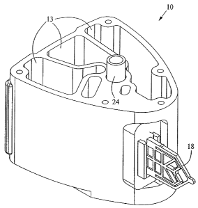
Figs. 1-4 show a fluid control and processing system 10 including a
housing 12 having a plurality of chambers 13. Fig. 1 shows the chambers 13
exposed for
illustrative purposes. A top cover will typically be provided to enclose the
chambers 13.
As best seen in Figs. 3 and 4, a fluid control device 16 and a reaction vessel
18 are
connected to different portions of the housing 12. The fluid control device in
the
embodiment shown is a rotary fluid control valve 16. The valve 16 includes a
valve body
having a disk portion 22 and a tubular portion 24. The disk portion 22 has a
generally
planar external port surface 23, as best seen in Fig. 3. The valve 16 is
rotatable relative to
the housing 12. The housing 12 includes a plurality of chamber ports 25 facing
the
20 external port surface 23 of the disk portion 22 of the valve 16 (Fig. 4) to
permit fluidic
communication between the chambers 13 and the valve 16. An optional seal or
gasket 26
is disposed between the disk portion 22 and the housing 12. The disk portion
22 further
includes a filter 27 and an outer wal128, and a toothed periphery 29.
As seen in Fig. 4, the disk portion 22 includes a lysing chamber 30. The
lysing chamber 30 may contain solid phase material for capturing cells,
spores, viruses, or
microorganisms to be lysed. Suitable solid phase materials include, without
limitation,
filters, beads, fibers, membranes, filter paper, glass wool, polymers, or
gels. In a specific
embodiment, the solid phase material is a filter having a pore size sufficient
to capture
target cells, spores, viruses, or microorganisms to be lysed.
As shown in Figs. 5-8, the outer wall 28 encloses the lysing chamber 30
and the bottom end of the disk portion 22 of the valve 16. In Fig. 8, the
lysing chamber
30 includes a first fluid processing port 32 coupled to a first fluid
processing channel 34,
and a second fluid processing port 36 coupled to a second fluid processing
channel 38.
The first fluid processing channe134 is coupled to a first outer conduit 40
ending at a first
5
CA 02564620 2006-10-23
WO 2005/106040 PCT/US2005/013897
external port 42 at the exterrial port surface 23, while the second fluid
processing channel
38 is coupled to a second outer conduit 44 ending at a second external port 46
at the
external port surface 23. A fluid displacement channel 48 is coupled to the
first fluid
processing channel 34 and first conduit 40 near one end, and to a fluid
displacement
chamber 50 at the other end. The first outer conduit 40 serves as a common
conduit for
allowing fluidic communication between the first external port 42 and either
or both of
the first fluid processing channel 34 and the fluid displacement channel 48.
The lysing
chamber 30 is in continuous fluidic communication with the fluid displacement
chamber
50.
As shown in Figs. 6-8, the external ports 42, 46 are angularly spaced from
one another relative to the axis 52 of the valve 16 by about 180 . The
external ports 42,
46 are spaced radially by the same distance from the axis 52. The axis 52 is
perpendicular to the external port surface 23. In another embodiment, the
angular spacing
between the external ports 42, 46 may be different. The configuration of the
channels in
the disk portion 22 may also be different in another embodiment. For example,
the first
fluid processing channel 34 and the first outer conduit 40 may be slanted and
coupled
directly with the fluid displacement chamber 50, thereby eliminating the fluid
displacement channel 48. The second fluid displacement channel 38 may also be
slanted
and extend between the second fluid processing port 36 and the second external
port 46
via a straight line, thereby eliminating the second outer conduit 44. In
addition, more
channels and external ports may be provided in the valve 16. As best seen in
Fig. 3, a
crossover channel or groove 56 is desirably provided on the external port
surface 23. The
groove 56 is curved and desirably is spaced from the axis 52 by a constant
radius. In one
embodiment, the groove 56 is a circular arc lying on a common radius from the
axis 52.
As discussed in more detail below, the groove 56 is used for filling the
vessel.
As shown in Fig. 8, the fluid displacement chamber 50 is disposed
substantially within the tubular portion 24 of the valve 16 and extends
partially into the
disk portion 22. A fluid displacement member in the form of a plunger or
piston 54 is
movably disposed in the chamber 50. When the piston 54 moves upward, it
expands the
volume of the chamber 50 to produce suction for drawing fluid into the chamber
50.
When the piston 54 moves downward, it decreases the volume of the chamber 50
to drive
fluid out of the chamber 50.
As the rotary valve 16 is rotated around its axis 52 relative to the housing
12 of Figs. 1-4, one of the external ports 42, 46 may be open and fluidicly
coupled with
6
CA 02564620 2006-10-23
WO 2005/106040 PCT/US2005/013897
one of the chambers 13 or reaction vessel 18, or both external ports 42, 46
may be
blocked or closed. In this embodiment, at most only one of the external ports
42, 46 is
fluidicly coupled with one of the chambers or reaction vessel 18. Other
embodiments
may be configured to permit both external ports 42, 46 to be fluidicly coupled
with
separate chambers or the reaction vessel 18. Thus, the valve 16 is rotatable
with respect
to the housing 12 to allow the external ports 42, 46 to be placed selectively
in fluidic
communication with a plurality of chambers which include the chambers 13 and
the
reaction vessel 18. Depending on which external port 42, 46 is opened or
closed and
whether the piston 54 is moved upward or downward, the fluid flow in the valve
16 can
change directions, the external ports 42, 46 can each switch from being an
inlet port to an
outlet port, and the fluid flow may pass through the processing region 30 or
bypass the
lysing chamber 30. In a specific embodiment, the first external port 42 is the
inlet port so
that the inlet side of the lysing chamber 30 is closer to the fluid
displacement chamber 54
than the outlet side of the lysing chamber 30.
Figs. 9A-9LL illustrate the operation of the valve 16 for conducting a
nucleic acid assay of a sample suspected of containing one or more target
entities (e.g.,
cells, spores, viruses, or microorganisms). The target entities comprise at
least one target
nucleic acid sequence for which the sample is being tested. A sample may be
introduced
into the housing 12 of the fluid control and processing device 10, which may
be
configured as a cartridge, by a variety of mechanisms, manual or automated.
For manual
addition, a measured volume of material may be placed into a receiving area of
the
housing 12 (e.g., one of the plurality of chambers) through an input port and
a cap is then
placed over the port. Alternatively, the receiving area may be covered by a
rubber or
similar barrier and the sample is injected into the receiving area by
puncturing the barrier
with a needle and injecting the sample through the needle. Alternatively, a
greater
amount of sample material than required for the analysis can be added to the
housing 12
and mechanisms within the housing 12 can effect the precise measuring and
aliquoting of
the sample needed for the specified protocol.
It may be desirable to place certain samples, such as tissue biopsy material,
soil, feces, exudates, and other complex material into another device or
accessory and
then place the secondary device or accessory into the housing causing a
mechanical
action which effects a function such as mixing, dividing, or extraction. For
example, a
piece of tissue may be placed into the lumen of a secondary device that serves
as the input
7
CA 02564620 2006-10-23
WO 2005/106040 PCT/US2005/013897
port cap. When the cap is pressed into the port, the tissue is forced through
a mesh that
slices or otherwise divides the tissue.
For automated sample introduction, additional housing or cartridge design
features are employed and, in many cases, impart sample collection
functionality directly
into the housing. With certain samples, such as those presenting a risk of
hazard to the
operator or the environment, such as human retrovirus pathogens, the transfer
of the
sample to the housing may pose a risk. Thus, in one embodiment, a syringe or
sipper may
be integrated into the device to provide a means for moving a sample directly
into the
housing. Alternatively, the device may include a venous puncture needle and a
tube
forming an assembly that can be used to acquire a sample. After collection,
the tube and
needle are removed and discarded, and the housing 12 is then placed in an
instrument to
effect processing. The advantage of such an approach is that the operator or
the
environment is not exposed to pathogens.
The input port can be designed with a consideration of appropriate human
factors as a function of the nature of the intended specimen. For example,
respiratory
specimens may be acquired from the lower respiratory tract as expectorants
from
coughing. Swab or brush samples may also be placed into the device. In the
former case,
the input port can be designed to allow the patient to cough directly into the
housing 12 or
to otherwise facilitate spitting of the expectorated sample into the housing.
For brush or
swab samples, the brush or swab is preferably placed in one of the chambers of
the device
10 and the sample is eluted off the brush or swab using, e.g., water or other
suitable
elution fluid. In addition, the housing 12 may include features that
facilitate the breaking
off and retaining of the end of the swab or brush in the sample-receiving
chamber.
In another embodiment, the housing 12 includes one or more input tubes
or sippers that may be positioned in a sample pool so that the sample material
flows into
the housing 12. Alternatively, a hydrophilic wicking material can function to
draw a
sample into the device. For example, the entire cartridge can be immersed
directly into
the sample, and a sufficient amount of sample is absorbed into the wicking
material and
wicks into the housing 12. The housing is then removed, and can be transported
to the
laboratory or analyzed directly using a portable instrument. In another
embodiment,
tubing can be utilized so that one end of the tube is in direct communication
with the
housing to provide a fluidic interface with at least one chamber and the other
end is
accessible to the external environment to serve as a receiver for sample. The
tube can
then be placed into a sample and serve as a sipper. Thus, the device may
include a variety
8
CA 02564620 2006-10-23
WO 2005/106040 PCT/US2005/013897
of features for collecting a sample from various different sources and for
moving the
sample into the housing 12, thereby reducing handling and inconvenience.
In Figs. 9A and 9AA, a sample is placed in a mixing chamber 60, e.g., by
pipetting, and then a lid is placed over the chamber 60. The sample will be
tested to
determine if it contains one or more target nucleic acid sequences. This
requires sample
preparation steps, e.g., lysing the target cells, spores, viruses, or
microorganisms
containing the target nucleic acid sequence. The chamber 60 contains sample
preparation
controls to be mixed with the sample. The sample preparation controls are also
cells,
spores, viruses, or microorganisms. The sample preparation controls contain a
marker
nucleic acid sequence different than the target nucleic acid sequence for
which the sample
is being assayed. The marker nucleic acid sequence will be detected in the
reaction
chamber 18 later in the assay, along with the target nucleic acid sequence if
the target
nucleic acid sequence is present in the sample. In order for the marker
nucleic acid
sequence to be detected, the sample preparation controls must be successfully
lysed to
release their nucleic acid and the nucleic acid must be successfully mixed
with
amplification reagents and amplified. The sample preparation controls thus
indicate that
sample preparation was adequate for the nucleic acid assay if they can be
detected and
inadequate if they cannot be detected. The sample preparation controls thus
verify that the
sample preparation was effective if they can be positively detected, so that
one can feel
confident in the assay results.
In one preferred embodiment, the sample preparation controls are spores
containing a specific marker nucleic acid sequence to be amplified and
detected. For
example, 2,000 to 10,000 spores containing a specific marker nucleic acid
sequence are
generally preferred, and more preferably about 6,000 spores are used as the
sample
preparation controls. The spores should be cleaned so that there is no
external nucleic
acid in order to prove that lysis step of the sample preparation is effective,
and not just
loosening external nucleic acid. In addition, the sample preparation controls
are
preferably stored in one of the chambers of the housing 12 in a lyophilized or
dried-down
bead that is quickly dissolvable in liquid. Methods for making such beads are
well known
in the art and are described in U.S. Patent 5,593,824 and in co-pending U.S.
patent
application Serial No. 10/672,266 filed Sept. 25, 2003, the disclosures of
which are
incorporated by reference herein.
The sample suspected of containing target cells, spores, viruses, or
microorganisms is mixed with the sample preparation controls in the chamber
60. The
9
CA 02564620 2006-10-23
WO 2005/106040 PCT/US2005/013897
mixing is preferably accomplished by dissolving a dried bead containing the
sample
preparation controls in the sample fluid. The first external port 42 is placed
in fluidic
communication with the chamber 60 by rotating the valve 16, and the piston 54
is pulled
upward to draw a fluid sample from the chamber 60 through the first outer
conduit 40 and
5' fluid displacement channel 48 to the fluid displacement chamber 50,
bypassing the lysing
chamber 30. For simplicity, the piston 54 is not shown in Figs. 9A-9LL. The
valve 16 is
then rotated to place the second external port 46 in fluidic communication
with a waste
chamber 64 as shown in Figs. 9B and 9BB. The piston 54 is pushed downward to
drive
the fluid sample mixed with the sample preparation controls through the lysing
chamber
30 to the waste chamber 64. In a specific embodiment, the lysing chamber 30
includes at
least one filter 27 having a pore size sufficient for capturing the target
cells, spores,
viruses, or microorganisms, if present in the sample, as well as capturing the
sample
preparation controls, as the sample fluid passes througli the lysing chamber
30. For this
reason, it is desirable that the sample preparation controls have the same
approximate size
or be slightly smaller than the target cells, spores, viruses, or
microorganisms in the
sample to prove that the filtration of the target entities, if they were
present in the sample,
was successful. In alternative embodiments, other solid phase materials may be
provided
in the lysing chamber 30.
In Figs. 9C and 9CC, the valve 16 is rotated to place the first external port
42 in fluidic communication with a wash chamber 66, and the piston 54 is
pulled upward
to draw a wash fluid from the wash chamber 66 into the fluid displacement
chamber 50,
bypassing the lysing chamber 30. The valve 16 is then rotated to place the
second
external port 46 in fluidic communication with the waste chamber 64 as shown
in Figs.
9D and 9DD. The piston 54 is pushed downward to drive the wash fluid through
the
lysing chamber 30 to the waste chamber 64. The above washing steps may be
repeated as
desired. The intermediate washing is used to remove unwanted residue within
the valve
16.
In Figs. 9E and 9EE, 'the valve 16 is rotated to place the first external port
42 in fluidic communication with a buffer chamber 70, and the piston 54 is
pulled upward
to draw a lysis buffer (e.g., water or water mixed with lysing agents) from
the buffer
chamber 70 into the fluid displacement chamber 50, bypassing the lysing
chamber 30.
The valve 16 is then rotated to place the second external port 46 in fluidic
communication
with the waste chamber 64 as shown in Figs. 9F and 9FF. The piston 54 is
pushed
CA 02564620 2006-10-23
WO 2005/106040 PCT/US2005/013897
downward to drive the buffer fluid into the lysing chamber 30. In Figs. 9G,
and 9GG, the
valve 16 is rotated to close the external ports 42, 46.
The sample preparation controls and the target cells, viruses, spores, or
microorganisms, if present, are subjected to a lysis treatment in the lysing
chamber 30.
The purpose of the lysis treatment is to break the outer walls of the sample
preparation
controls and of the target cells, viruses, spores, or microorganisms, if
present, to release
their nucleic acid. The sample preparation controls are preferably the same
level of
difficulty or more difficult to lyse than the target cells, viruses, spores,
or microorganisms
to prove that the lysis treatment was effective. Liberation of nucleic acids
from the cells,
viruses, spores, or microorganisms, and denaturation of DNA binding proteins
may
generally be performed by chemical, physical, or electrolytic lysis methods.
For example,
chemical methods generally employ lysing agents to disrupt the cells and
extract the
nucleic acids from the cells, followed by treatment of the extract with
chaotropic salts
such as guanidinium isothiocyanate or urea to denature any contaminating and
potentially
interfering proteins. Where chemical extraction and/or denaturation methods
are used, the
appropriate lysing agents are preferably in the lysis buffer stored in the
chamber 70 and
pumped into the lysing chamber 30.
Alternatively, physical methods may be used to extract the nucleic acids
and denature DNA binding proteins. U.S. Pat. No. 5,304,487, incorporated
herein by
reference in its entirety for all purposes, discusses the use of physical
protrusions within
microchannels or sharp edged particles within a chamber or channel to pierce
cell
membranes and extract their contents. Combinations of such structures with
piezoelectric
elements for agitation can provide suitable shear forces for lysis. More
traditional
methods of cell extraction may also be used, e.g., employing a channel with
restricted
cross-sectional dimension which causes cell lysis when the sample is passed
through the
channel with sufficient flow pressure. Alternatively, cell extraction and
denaturing of
contaminating proteins may be carried out by applying an alternating
electrical current. A
variety of other methods may be utilized within the device of the present
invention to
effect cell lysis/extraction, including, e.g., subjecting cells to ultrasonic
agitation, or
forcing cells through microgeometry apertures, thereby subjecting the cells to
high shear
stress resulting in rupture.
In one preferred embodiment, the lysis treatment comprises sonicating the
lysing chamber 30 using an ultrasonic transducer 76 coupled to the outer wall
28 of the
lysing chamber 30. The ultrasonic transducer 76, preferably an ultrasonic
horn, is placed
11
CA 02564620 2006-10-23
WO 2005/106040 PCT/US2005/013897
in contact with the wall 28 to transmit ultrasonic energy into the lysing
chamber 30 to
facilitate lysing of the cells, spores, viruses, or microorganisms. Suitable
ultrasonic horns
are commercially available from Sonics & Materials, Inc. having an office at
53 Church
Hill, Newton, Connecticut 06470-1614, U.S.A. Alternatively, the ultrasonic
transducer
may comprise a piezoelectric disk or any other type of ultrasonic transducer
that may be
coupled to the wall 28. In addition, beads (e.g., glass or polystyrene beads)
are preferably
agitated in the lysing chamber 30 to rupture the cells, spores, viruses, or
microorganisms.
The pressure waves or pressure pulses created by the transducer 76 vibrating
against the
wall 28 causes the beads to move in ballistic motion in the lysis buffer and
cause the
rupturing. In these embodiments employing an ultrasonic transducer, the lysis
buffer
should be an ultrasonic transmission medium, e.g., deionized water. The lysis
buffer may
also include one or more lysing agents to aid in the lysis. In the presently
preferred
embodiment, the wall 28 is a deflectable plastic wall as described in co-
pending U.S.
patent application Ser. No. 09/972,221 filed October 4, 2001 the disclosure of
which is
incorporated by reference herein.
In Figs. 9H and 9HH, the valve 16 is rotated to place the second external
port 46 in fluidic communication with a reagent chamber 78, and the piston 54
is pushed
downward to elute the lysate in the lysing chamber 30 to the reagent chamber
78. The
reagent chamber 78 preferably contains all of the necessary nucleic acid
amplification
reagents and probes (e.g., enzyme, primers, and fluorescent probes) to amplify
and detect
the marker nucleic acid sequence of the sample preparation controls and the
one or more
target nucleic acid sequences for which the sample is being tested. Any excess
lysate is
dispensed into the waste chamber 64 via the second external port 46 after
rotating the
valve 16 to place the port 46 in fluidic communication with the waste chamber
64, as
shown in Figs. 91 and 911. The lysate containing nucleic acid released in the
lysing
chamber 30 is then mixed in the reagent chamber 78. This is carried out by
placing the
fluid displacement chamber 50 in fluidic communication with the reagent
chamber 78 as
shown in Figs. 9J and 9JJ, and moving the piston 54 up and down. Toggling of
the
mixture through the filter in the processing region 30, for instance, allows
larger particles
trapped in the filter to temporarily move out of the way to permit smaller
particles to pass
through.
The reagents and probes for amplifying and detecting the marker nucleic
acid sequence of the sample preparation controls and the one or more target
nucleic acid
sequences for which the sample is being tested are preferably stored in
chamber 78 in a
12
CA 02564620 2006-10-23
WO 2005/106040 PCT/US2005/013897
lyophilized or dried-down bead that= is quickly dissolvable in liquid. Methods
for making
such beads are well known in the art and are described in U.S. Patent
5,593,824 and in
co-pending U.S. patent application Serial No. 10/672,266 filed Sept. 25, 2003,
the
disclosures of which are incorporated by reference herein. In an alternative
embodiment,
the reagents and probes are stored in the reaction chamber of the reaction
vessel 18.
In Figs. 9K, 9KK, and 9K'K', the valve 16 is rotated to place the first
external port 42 in fluidic communication with a first branch 84 coupled to
the reaction
vessel 18, while the second branch 86 which is coupled to the reaction vessel
18 is placed
in fluidic communication with the crossover groove 56. The first branch 84 and
second
branch 86 are disposed at different radii from the axis 52 of the valve 16,
with the first
branch 84 having a common radius with the first external port 42 and the
second branch
86 having a common radius with the crossover groove 56. The crossover groove
56 is
also in fluidic communication with the reagent chamber 78 (Fig. 9K), and
serves to bridge
the gap between the reagent chamber 78 and the second branch 86 to provide
crossover
flow therebetween. The external ports are disposed within a range of external
port radii
from the axis and the crossover groove is disposed within a range of crossover
groove
radii from the axis, where the range of external port radii and the range of
crossover
groove radii are non-overlapping. Placing the crossover groove 56 at a
different radius
from the radius of the external ports 42, 46 is advantageous because it avoids
cross-
contamination of the crossover groove 56 by contaminants that may be present
in the area
near the surfaces between the valve 16 and the housing 12 at the radius of the
external
ports 42, 46 as a result of rotational movement of the valve 16. Thus, while
other
configurations of the crossover groove may be used including those that
overlap with the
radius of the external ports 42, 46, the embodiment as shown is a preferred
arrangement
that isolates the crossover groove 56 from contamination from the area near
the surfaces
between the valve 16 and the housing 12 at the radius of the external ports
42, 46.
To fill the reaction vessel 18, the piston 54 is pulled upward to draw the
reaction mixture in the reagent chamber 78 through the crossover groove 56 and
the
second branch 86 into the reaction vessel 18. The valve 16 is then rotated to
place the
second extetnal port 46 in fluidic communication with the first branch 84 and
to close the
first external port 42, as shown in Figs. 9L and 9LL. The piston 54 is pushed
downward
to pressurize the reaction mixture inside the reaction vessel 18. The reaction
vessel 18
has a reaction chamber for holding the reaction mixture for nucleic acid
amplification and
detection. The reaction chamber may be inserted into a thermal reaction sleeve
for
13
CA 02564620 2006-10-23
WO 2005/106040 PCT/US2005/013897
performing nucleic acid amplification and detection. The two branches 84, 86
allow
filling and evacuation of the reaction chamber of the reaction vessel 18. The
vessel
maybe connected to the housing 12 by ultrasonic welding, mechanical coupling,
or the
like, or be integrally formed with the housing 12 such as by molding.
The reaction mixture in the reaction chamber of the vessel 18 is subjected
to nucleic acid amplification conditions. Amplification of an RNA or DNA
template
using reactions is well known (see U.S. Patents 4,683,195 and 4,683,202; PCR
Protocols:
A Guide to Methods and Applications (Innis et al., eds, 1990)). Methods for
amplifying
and detecting nucleic acids by PCR using a thermostable enzyme are disclosed
in U.S.
Pat. No. 4,965,188, which is incorporated herein by reference. PCR
amplification of
DNA involves repeated cycles of heat-denaturing the DNA, annealing two
oligonucleotide primers to sequences that flank the DNA segment to be
amplified, and
extending the annealed primers with DNA polymerase. The primers hybridize to
opposite strands of the target sequence and are oriented so that DNA synthesis
by the
polymerase proceeds across the region between the primers, effectively
doubling the
amount of the DNA segment. Moreover, because the extension products are also
complementary to and capable of binding primers, each successive cycle
essentially
doubles the amount of DNA synthesized in the previous cycle. This results in
the
exponential accumulation of the specific target fragment, at a rate of
approximately 2n
per cycle, where n is the number of cycles. Methods such as polymerase chain
reaction
(PCR) and ligase chain reaction (LCR) can be used to amplify nucleic acid
sequences of
target DNA sequences directly from mRNA, from cDNA, from genomic libraries or
cDNA libraries.
Isothermic amplification reactions are also known and can be used
according to the methods of the invention. Examples of isothermic
amplification
reactions include strand displacement amplification (SDA) (Walker, et al.
Nucleic Acids
Res. 20(7):1691-6 (1992); Walker PCR Methods Appl 3(1):1-6 (1993)),
transcription-
mediated amplification (Phyffer, et al., J. Clin. Microbiol. 34:834-841
(1996); Vuorinen,
et al. , J. Clin. Microbiol. 33:1856-1859 (1995)), nucleic acid sequence-based
amplification (NASBA) (Compton, Nature 350(6313):91-2 (1991), rolling circle
amplification (RCA) (Lisby, Mol. Biotechnol. 12(1):75-99 (1999)); Hatch et
al., Genet.
Anal. 15(2):35-40 (1999)) and branched DNA signal amplification (bDNA) (see,
e.g.,
Iqbal et al., Mol. Cell Probes 13(4):315-320 (1999)). Other amplification
methods
14
CA 02564620 2006-10-23
WO 2005/106040 PCT/US2005/013897
known to those of skill in the art include CPR (Cycling Probe Reaction), SSR
(Self-
Sustained Sequence Replication), SDA (Strand Displacement Amplification), QBR
(Q-
Beta Replicase), Re-AMP (formerly RAMP), RCR (Repair Chain Reaction), TAS
(Transcription Based Amplification System), and HCS.
The nucleic acid amplification reaction is preferably carried out using a
thermal processing instrument that heats and/or cools the reaction mixture in
the vessel 18
to the temperatures needed for the amplification reaction. The thermal
processing
instrument can also optionally comprise one or more detection mechanisms for
detecting
the marker nucleic acid sequence of the sample preparation controls and the
one or more
target nucleic acid sequences for which the sample is being tested. A
preferred thermal
processing instrument with built in optical detectors for amplifying and
detecting nucleic
acid sequences in the vessel 18 is described in commonly assigned U.S. Patent
Nos.
6,369,893 and 6,391,541, the disclosures of which are incorporated by
reference herein.
There are also many other known ways to control the temperature of a reaction
mixture
and detect nucleic acid sequences in the reaction mixture that are suitable
for the present
invention. For example, other instruments for nucleic acid amplification and
detection are
described, e.g., in U.S. Patent Nos. 5,958,349; 5,656,493; 5,333,675;
5,455,175;
5,589,136 and 5,935,522.
The detection of the marker nucleic acid sequence of the sample
preparation controls and of the one or more target nucleic acid sequences for
which the
sample is being tested is preferably carried out using probes. The reaction
vesse118
preferably has one or more transparent or light-transmissive walls through
which signals
from the probe may be optically detected. Preferably hybridization probes are
used to
detect and quantify the nucleic acid sequences. There are many different types
of assays
that employ nucleic acid hybridization probes. Some of these probes generate
signals
with a change in the fluorescence of a fluorophore due to a change in its
interaction with
another molecule or moiety. Typically, the interaction is brought about by
changing the
distance between the fluorophore and the interacting molecule or moiety. These
assays
rely for signal generation on fluorescence resonance energy transfer, or
"FRET." FRET
utilizes a change in fluorescence caused by a change in the distance
separating a first
fluorophore from an interacting resonance energy acceptor, either another
fluorophore or
a quencher. Combinations of a fluorophore and an interacting molecule or
moiety,
including quenching molecules or moieties, are known as "FRET pairs." The
mechanism
of FRET-pair interaction requires that the absorption spectrum of one member
of the pair
CA 02564620 2006-10-23
WO 2005/106040 PCT/US2005/013897
overlaps the emission spectrum of the other member, the first fluorophore. If
the
interacting molecule or moiety is a quencher, its absorption spectrum must
overlap the
emission spectrum of the fluorophore. Stryer, L., Ann. Rev. Biochem. 1978, 47:
819-846;
BIOPHYSICAL CHEMISTRY part II, Techniques for the Study of Biological
Structure
and Function, (C. R. Cantor and P. R. Schimmel, eds., 1980), pages 448-455,
and Selvin,
P. R., Methods in Enzymology 246: 300-335 (1995). Efficient, or a substantial
degree of,
FRET interaction requires that the absorption and emission spectra of the pair
have a
large degree of overlap. The efficiency of FRET interaction is linearly
proportional to that
overlap. Haugland, R. P., Yguerabide, Jr., and Stryer, L., Proc. Natl. Acad.
Sci. USA 63:
24-30 (1969). Non-FRET probes have also been described. See, e.g., U.S. Patent
No.
6,150,097.
Another preferred method for detection of amplification products is the 5'
nuclease PCR assay (also referred to as the TaqMan assay) (Holland et al.,
Proc. Natl.
Acad. Sci. USA 88: 7276-7280 (1991); Lee et al., Nucleic Acids Res. 21: 3761-
3766
(1993)). This assay detects the accumulation of a specific PCR product by
hybridization
and cleavage of a doubly labeled fluorogenic probe (the "TaqMan(W probe)
during the
amplification reaction. The fluorogenic probe consists of an oligonucleotide
labeled with
both a fluorescent reporter dye and a quencher dye. During PCR, this probe is
cleaved by
the 5'-exonuclease activity of DNA polymerase if, and only if, it hybridizes
to the
segment being amplified. Cleavage of the probe generates an increase in the
fluorescence
intensity of the reporter dye.
Another method of detecting amplification products that relies on the use
of energy transfer is the "beacon probe" method described by Tyagi and Kramer
(Nature
Biotecli. 14:303-309 (1996)), which is also the subject of U.S. Pat. Nos.
5,119,801 and
5,312,728. This method employs oligonucleotide hybridization probes that can
form
hairpin structures. On one end of the hybridization probe (either the 5' or 3'
end), there is
a donor fluorophore, and on the other end, an acceptor moiety. In the case of
the Tyagi
and Kramer method, this acceptor moiety is a quencher, that is, the acceptor
absorbs
energy released by the donor, but then does not itself fluoresce. Thus when
the beacon is
in the open conformation, the fluorescence of the donor fluorophore is
detectable,
whereas when the beacon is in hairpin (closed) conformation, the fluorescence
of the
donor fluorophore is quenched. When employed in PCR, the molecular beacon
probe,
which hybridizes to one of the strands of the PCR product, is in "open
conformation," and
fluorescence is detected, while those that remain unhybridized will not
fluoresce (Tyagi
16
CA 02564620 2006-10-23
WO 2005/106040 PCT/US2005/013897
and Kramer, Nature Biotechnol. 14: 303-306 (1996). As a result, the amount of
fluorescence will increase as the amount of PCR product increases, and thus
may be used
as a measure of the progress of the PCR.
To be confident about the detection, or lack thereof, of a target nucleic
acid sequence in a sample, one should control for the integrity of the sample
preparation.
This is why the sample preparation controls are subjected to the same
treatment as the
target entities (e.g., target cells, spores, viruses, or microorganisms
containing a target
nucleic acid sequence) in the sample. If the marker nucleic acid sequence of
the sample
preparation controls is detected, then the sample preparation is deemed
satisfactory. If the
presence of the marker nucleic acid sequence cannot be detected, then the
sample
preparation is deemed inadequate and the outcome of the test for the target
nucleic acid
sequence is deemed "unresolved". Preferably, the presence or absence of the
marker
nucleic acid sequence is detected by determining if a signal from a
hybridization probe
capable of binding to the marker nucleic acid sequence exceeds a threshold
level, e.g., a
predetermined fluorescent threshold level that must be met or exceeded for the
assay to be
deemed valid.
To operate the valve 16 of Figs. 3-8, a motor such as a stepper motor is
typically coupled to the toothed periphery 29 of the disk portion 22 to rotate
the valve 16
relative to the housing 12 for distributing fluid with high precision. The
motor can be
computer-controlled according to the desired protocol. A linear motor or the
like is
typically used to drive the piston 54 up and down with precision to provide
accurate
metering, and may also be computer-controlled according to the desired
protocol.
The use of a single valve produces high manufacturing yields due to the
presence of only one failure element. The concentration of the fluid control
and
processing components results in a compact apparatus (e.g., in the form of a
small
cartridge) and facilitates automated molding and assembly. As discussed above,
the
system advantageously includes dilution and mixing capability, intermediate
wash
capability, and positive pressurization capability. The fluid paths inside the
system are
normally closed to minimize contamination and facilitate containment and
control of
fluids within the system. The reaction vessel is conveniently detachable and
replaceable,
and may be disposable in some embodiments.
The components of the fluid control and processing system may be made
of a variety of materials that are compatible with the fluids being used.
Examples of
suitable materials include polymeric materials such as polypropylene,
polyethylene,
17
CA 02564620 2006-10-23
WO 2005/106040 PCT/US2005/013897
polycarbonate, acrylic, or nylon. The various chambers, channels, ports, and
the like in
the system may have various shapes and sizes.
Fig. 10 shows another embodiment in which a piston assembly 210
including a piston rod 212 connected to a piston shaft 214 having a smaller
cross-section
than the rod 212 for driving small amounts of fluids. The thin piston shaft
214 may bend
under an applied force if it is too long. The piston rod 212 moves along the
upper portion
of the barrel or housing 216, while the piston shaft 214 moves along the lower
portion of
the barrel 216. The movement of the piston rod 212 guides the movement of the
piston
shaft 214, and absorbs much of the applied force so that very little bending
force is
transmitted to the thin piston shaft 214.
Fig. 11 shows another embodiment in which the sample is pre-filtered
before being mixed with the sample preparation controls. The sample is
preferably pre-
filtered in a side chamber 220 that is incorporated into the device. The side
chamber 220
includes an inlet port 222 and an outlet port 224. In this example, the side
chamber 220
includes a filter 226 disposed at the inlet port 222. Sample fluid is directed
to flow via
the inlet port 222 into the side chamber 220 and out via the outlet port 224
for side
filtering. This allows filtering of a fluid sample or the like using the fluid
control device
of the invention. The fluid may be recirculated to achieve better filtering by
the filter
226. This prefiltering is useful to remove coarse material, that might
otherwise clog up
the other parts of the device, before mixing the sample with the sample
preparation
controls. After the sample is pre-filtered, it is mixed with the sample
preparation controls,
e.g., in the chamber 66 of Fig. 9C or another chamber of the housing 12. The
use of a side
chamber is advantageous, for instance, to avoid contaminating the valve and
the other
chambers in the device.
The above-described arrangements of devices and methods are merely
illustrative of applications of the principles of this invention and many
other embodiments
and modifications may be made without departing from the spirit and scope of
the
invention as defined in the claims.
For example, although a rotary-valve cartridge has been described as a
preferred embodiment, the sample preparation control of the present invention
is suitable
for many other cartridge designs. Alternative cartridge designs are described
in U.S.
Patent Nos. 6,391,541, 6,440,725, and 6,168,948 the disclosures of which are
incorporated by reference herein. Moreover, when a rotary valve cartridge is
used, the
18
CA 02564620 2006-10-23
WO 2005/106040 PCT/US2005/013897
cartridge may have more or fewer chamber than shown in the preferred
embodiments and
many different sample preparation protocols may be executed.
The scope of the invention should, therefore, be determined not with
reference to the above description, but instead should be determined with
reference to the
appended claims along with their full scope of equivalents.
19